Liver Flukes Ppt
63
TREMATODES
Transcript of Liver Flukes Ppt
TREMATODES

EUKARYOTES
UNICELLULAR MULTICELLULAR
Helminths
Nematodes(roundworms)
Cestodes(Tapeworms)
Trematodes(leaf-like)
Intestinal Lungs BloodLiver
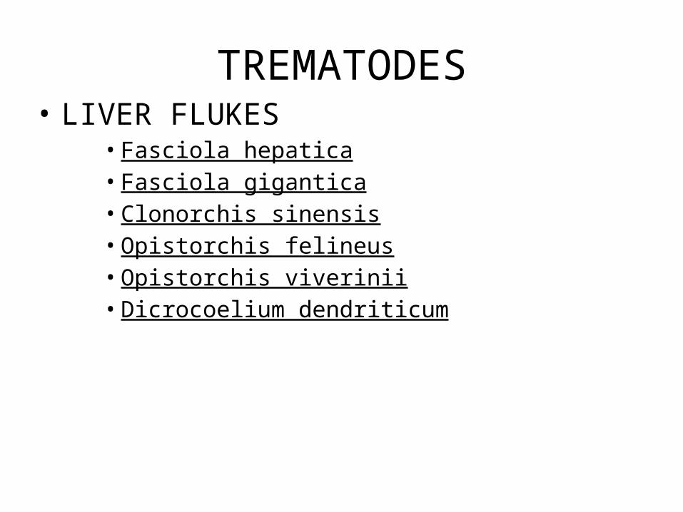
TREMATODES• LIVER FLUKES
• Fasciola hepatica• Fasciola gigantica• Clonorchis sinensis• Opistorchis felineus• Opistorchis viverinii• Dicrocoelium dendriticum
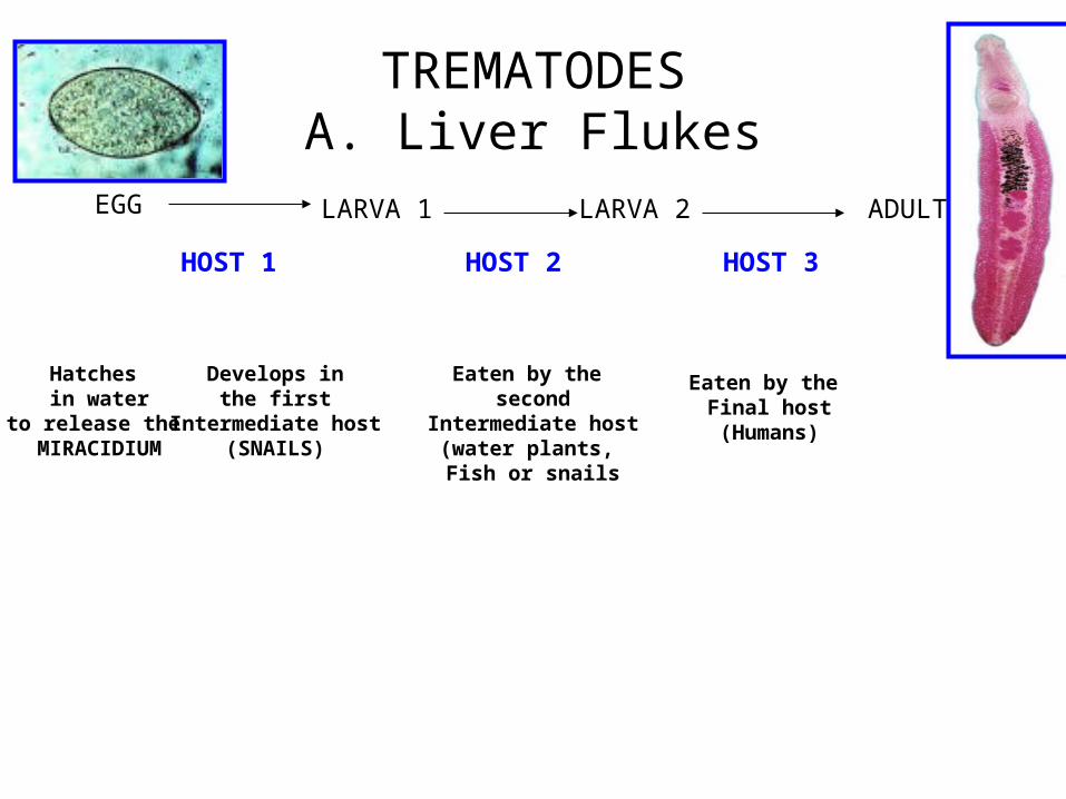
TREMATODESA. Liver Flukes
EGG LARVA 1 LARVA 2 ADULT
Hatches in water
to release the MIRACIDIUM
Develops inthe first
Intermediate host(SNAILS)
Eaten by the second
Intermediate host(water plants, Fish or snails
Eaten by the Final host(Humans)
HOST 1 HOST 2 HOST 3

Adult Trematodes
• Flat, elongated, leaf-shaped
• Ovoid, conical or cylindrical depending on state of contraction
• Integument may be covered with spines, tubercles or ridges (partially or completely)
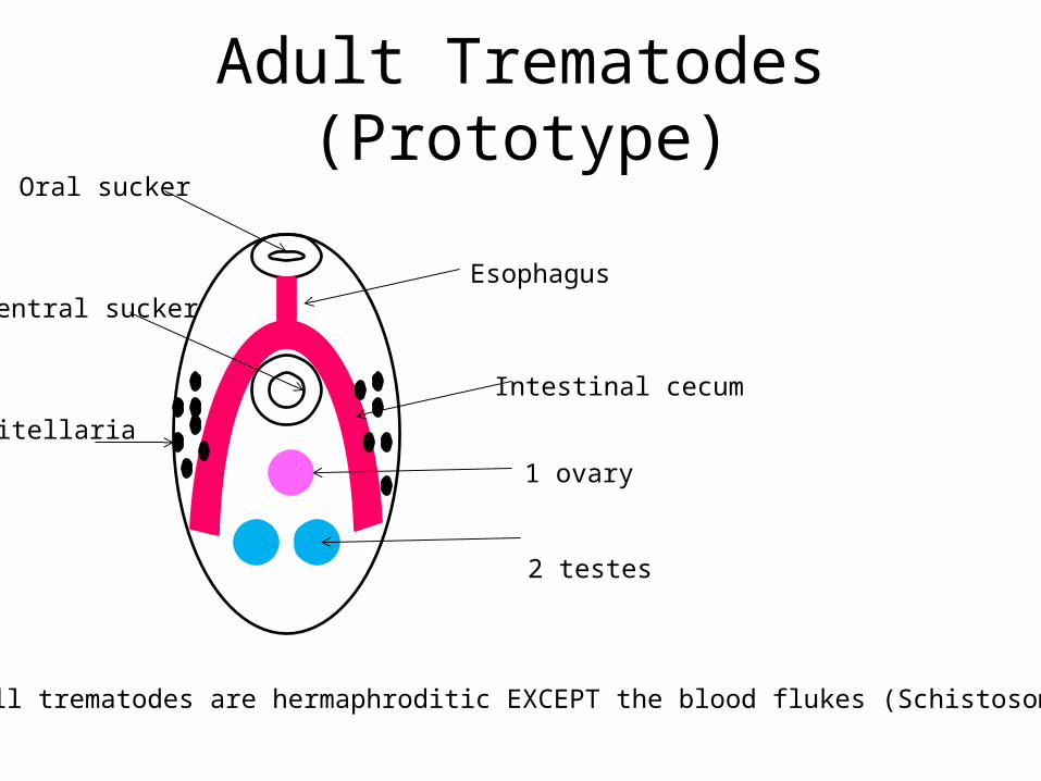
Adult Trematodes (Prototype)Oral sucker
Ventral suckerEsophagus
Intestinal cecum
2 testes
1 ovary
Vitellaria
All trematodes are hermaphroditic EXCEPT the blood flukes (Schistosoma sp.)

Trematode Egg (Prototype)
• Contains a fertilized ovum
• Shape, appearance and size – depending on the species
• Contains a cap-like operculum, EXCEPT Schistosoma sp.

TREMATODES
EGG LARVA 1 LARVA 2 ADULT
Hatches in water
to release the
SNAIL
SECOND INTERMEDIATE
HOST FINAL HOSTFish ,
crustaceansSnails
Aquatic plants
FIRST INTERMEDIATE
HOST
All follow the above cycle, EXCEPT Schistosoma sp.
Sporocyst
Redia I
Redia II
Cercaria
MIRACIDIUM
Metacercaria(encysted cercaria)
Adult
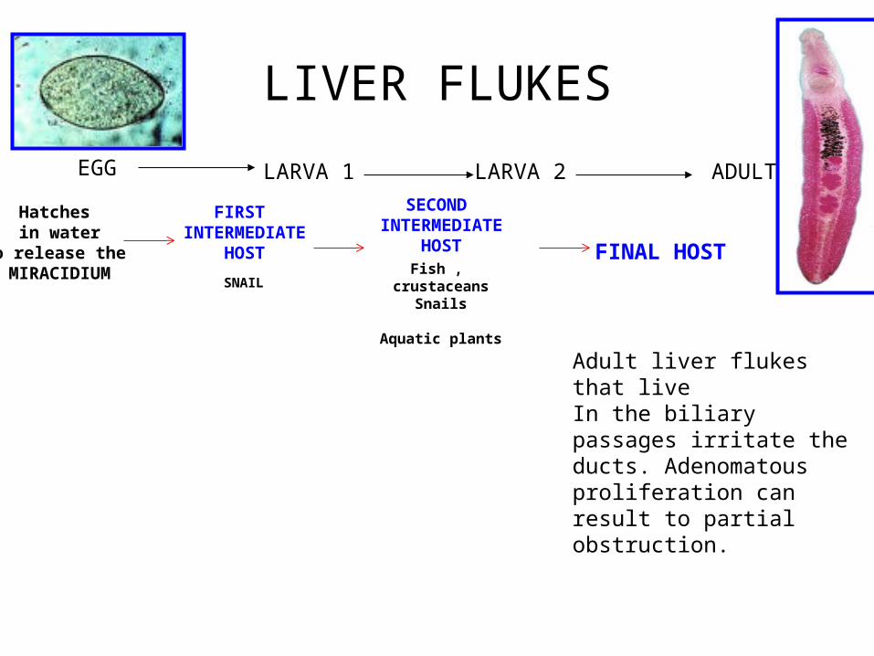
LIVER FLUKES
EGG LARVA 1 LARVA 2 ADULT
Hatches in water
to release the MIRACIDIUM SNAIL
SECOND INTERMEDIATE
HOST FINAL HOSTFish ,
crustaceansSnails
Aquatic plants
FIRST INTERMEDIATE
HOST
Adult liver flukes that live In the biliary passages irritate the ducts. Adenomatous proliferation can result to partial obstruction.
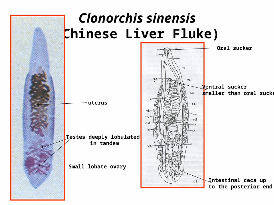
Clonorchis sinensis(Chinese Liver Fluke)
Ventral sucker smaller than oral sucker
Oral sucker
Intestinal ceca up to the posterior end
Testes deeply lobulated in tandem
Small lobate ovary
uterus

Clonorchis sinensis(Chinese Liver Fluke)
operculum
rimmed
Small protuberance
Thicker posterior end

EGG LARVA 1 LARVA 2 ADULT
FIRST INTERMEDIATE
HOST
SECOND INTERMEDIATE
HOST
FINALHOST
operculate snails of severalgenera, including Alocinma
and Parafossarulus, Bithynia ( Bulimus),
Semisulcospira, Melanoides tuberculatus
fresh water fish of the family Cyprinidae ,
Ctenopharyngodon idellus
Humans get infected by: >eating uncooked fish containing the infective metacercaria > ingestion of the cysts in drinking water
Clonorchis sinensis(Chinese Liver Fluke)

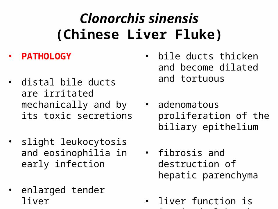
• PATHOLOGY
• distal bile ducts are irritated mechanically and by its toxic secretions
• slight leukocytosis and eosinophilia in early infection
• enlarged tender liver
• bile ducts thicken and become dilated and tortuous
• adenomatous proliferation of the biliary epithelium
• fibrosis and destruction of hepatic parenchyma
• liver function is impaired although SGPT and SGOT are normal
Clonorchis sinensis(Chinese Liver Fluke)

• SYMPTOMATOLOGY
• Light Infections – produce only mild symptoms or
go unnoticed
• Moderate Infections – indigestion– Epigastric discomfort unrelated
to meals– weakness – loss of weight
• Heavy infections– complicated by cholelethiasis
and bouts of pyogenic cholangitis
Clonorchis sinensis(Chinese Liver Fluke)
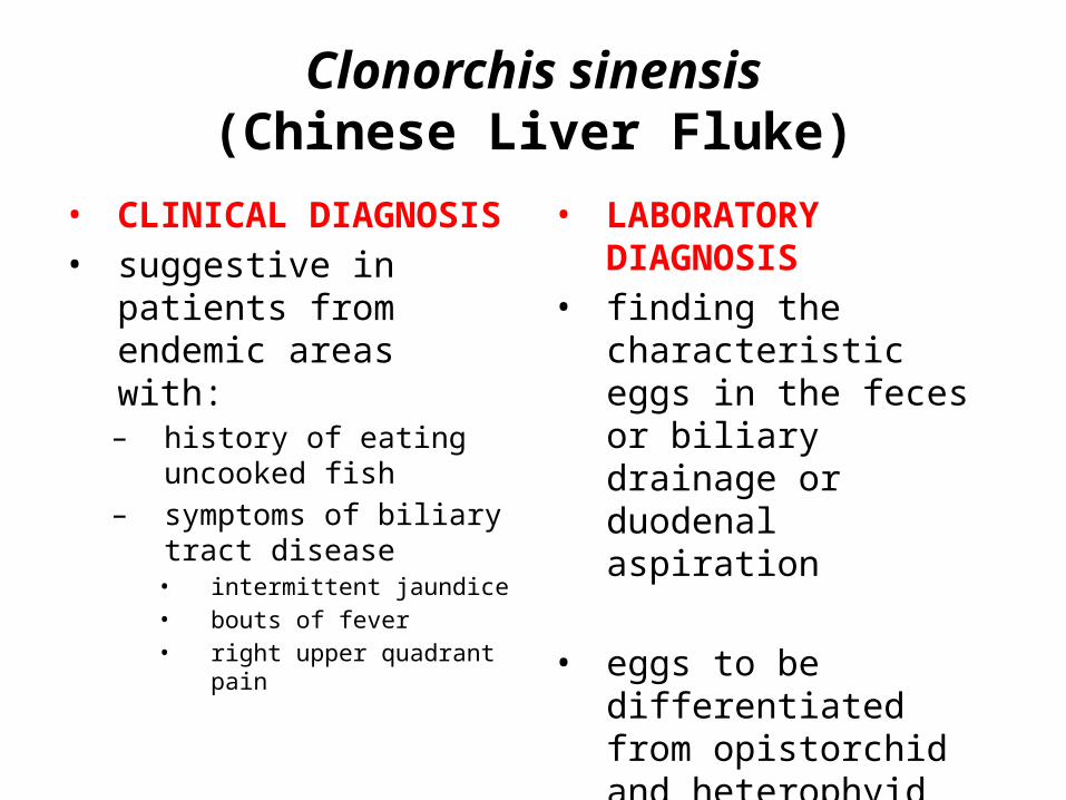
• CLINICAL DIAGNOSIS• suggestive in patients from
endemic areas with: – history of eating uncooked
fish– symptoms of biliary tract
disease • intermittent jaundice • bouts of fever • right upper quadrant pain
• LABORATORY DIAGNOSIS• finding the characteristic
eggs in the feces or biliary drainage or duodenal aspiration
• eggs to be differentiated from opistorchid and heterophyid flukes
Clonorchis sinensis(Chinese Liver Fluke)

• TREATMENT• chloroquine diphosphate • praziquantel
Heavy infections complicatedby obstructive jaundice:
1. cholecystectomy with 2. choledocholithotomy3. exploration of the common duct4. drainage procedure such as
sphincteroplasty or choledochoduodenostomy
• PREVENTION AND CONTROL
• thorough cooking of all freshwater fish
• sterilization of human feces by storage or by the addition of ammonium sulfate
• Human and animal feces should not be disposed in bodies of water.
Clonorchis sinensis(Chinese Liver Fluke)

Opistorchis felineus(Cat Liver Fluke)
Oral sucker subterminal
Ventral sucker Same size as oral sucker
2 testes lobed Oblique to each other
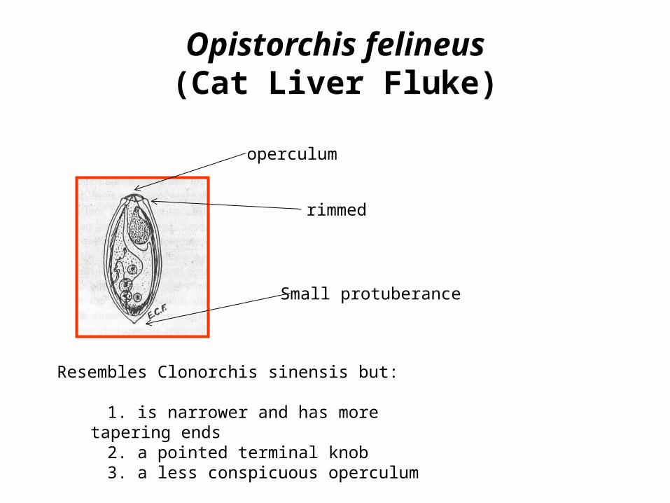
Opistorchis felineus(Cat Liver Fluke)
operculum
rimmed
Small protuberance
Resembles Clonorchis sinensis but:
1. is narrower and has more tapering ends 2. a pointed terminal knob 3. a less conspicuous operculum
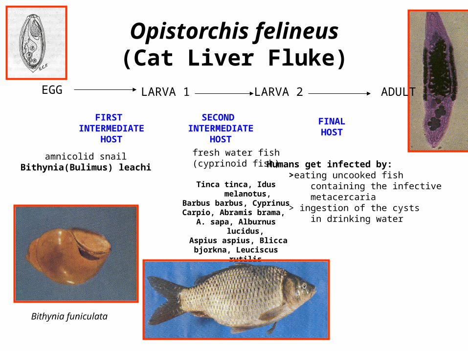
EGG LARVA 1 LARVA 2
FIRST INTERMEDIATE
HOST
SECOND INTERMEDIATE
HOST
FINALHOST
amnicolid snailBithynia(Bulimus) leachi
fresh water fish(cyprinoid fish)
Tinca tinca, Idus melanotus,Barbus barbus, CyprinusCarpio, Abramis brama,
A. sapa, Alburnus lucidus, Aspius aspius, Blicca
bjorkna, Leuciscus rutilis Scardiinius erythopthalmus
Humans get infected by: >eating uncooked fish containing the infective metacercaria > ingestion of the cysts in drinking water
Opistorchis felineus(Cat Liver Fluke)
ADULT
Bithynia funiculata


• PATHOLOGY
• inflammatory and proliferative changes of the biliary epithelium
• fibrosis of the distal biliary vessels
• pathologic changes may extend to the proximal
bile ducts and gallbladder periportal fibrosis
Opistorchis felineus(Cat Liver Fluke)

• SYMPTOMATOLOGY• Light Infections
– Asymptomatic
• Moderate Infections– moderate, painful
enlargement of the liver– passive congestion of the
spleen– Icterus– local eosinophilia in the wall
of the bile ducts
• Heavy Infections• invade the pancreas with digestive
disturbances• bile stones may form around eggs as
nuclei and cause cholecystitis with colic
• loss of appetite as patient becomes toxic
• scar tissue around the bile ducts encroaches on liver cells and portal vessels ---- collateral venous circulation, edema of the face and limbs and at times ascitis
Opistorchis felineus(Cat Liver Fluke)
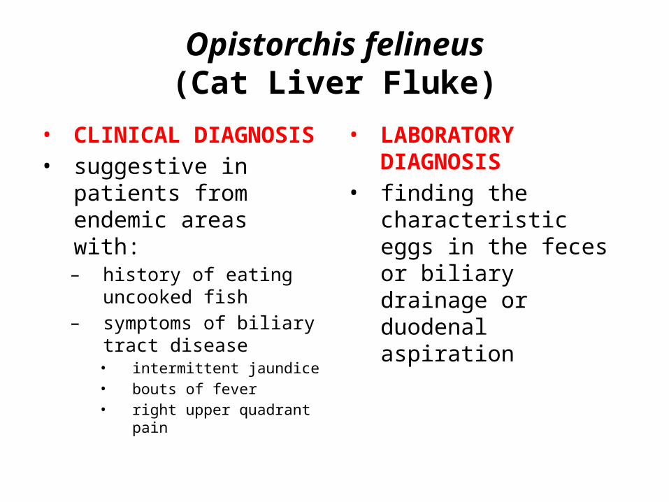
• CLINICAL DIAGNOSIS• suggestive in patients from
endemic areas with: – history of eating uncooked
fish– symptoms of biliary tract
disease • intermittent jaundice • bouts of fever • right upper quadrant pain
• LABORATORY DIAGNOSIS• finding the characteristic
eggs in the feces or biliary drainage or duodenal aspiration
Opistorchis felineus(Cat Liver Fluke)

• TREATMENT• praziquantel
• PREVENTION AND CONTROL
• thorough cooking of all freshwater fish
• sterilization of human feces by storage or by the addition of ammonium sulfate
• Human and animal feces should not be disposed in bodies of water.
Opistorchis felineus(Cat Liver Fluke)
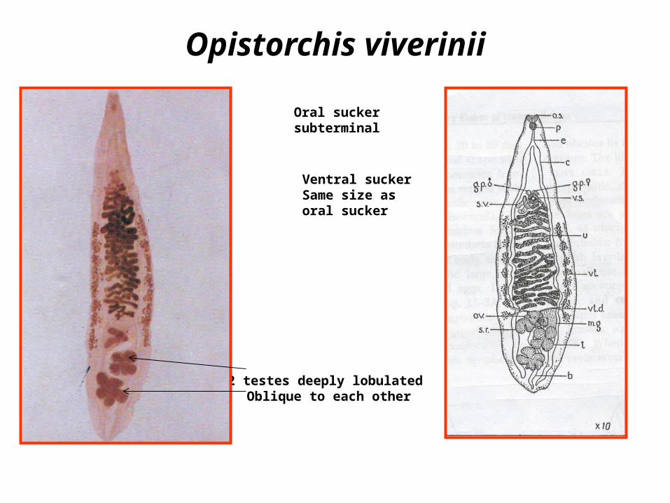
Opistorchis viverinii
Oral sucker subterminal
Ventral sucker Same size as oral sucker
2 testes deeply lobulated Oblique to each other
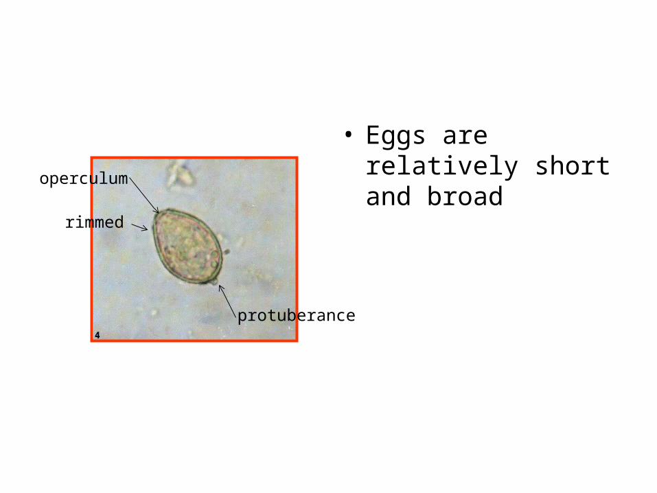
• Eggs are relatively short and broad
operculum
rimmed
protuberance

EGG LARVA 1 LARVA 2
FIRST INTERMEDIATE
HOST
SECOND INTERMEDIATE
HOST
FINALHOST
Snails :Bithynia goniomphalus
Bithynia funiculateBithynia laevis
fresh water fish
Punteus orphoidesHampala dispar
Cyclocheilichthys siaja
Humans get infected by: >eating uncooked fish containing the infective metacercaria > ingestion of the cysts in drinking water
ADULT
Opistorchis viverinii
Natural definitive hosts:civet cat, cat, dog and other fish- eating mammalsMAN IS AN ACCIDENTAL HOST.


• PATHOLOGY • dilatation and thickening of
bile duct walls
• presence of stones and sludge in the gallbladder
• hyperplastic biliary epithelium from presence of worms
• further stimulated by nitrosamines in local fermented foods or by nitrosocompounds produced by activated
• macrophages in chronically affected tissues
• striking association with cholangiocarcinoma
Opistorchis viverinii
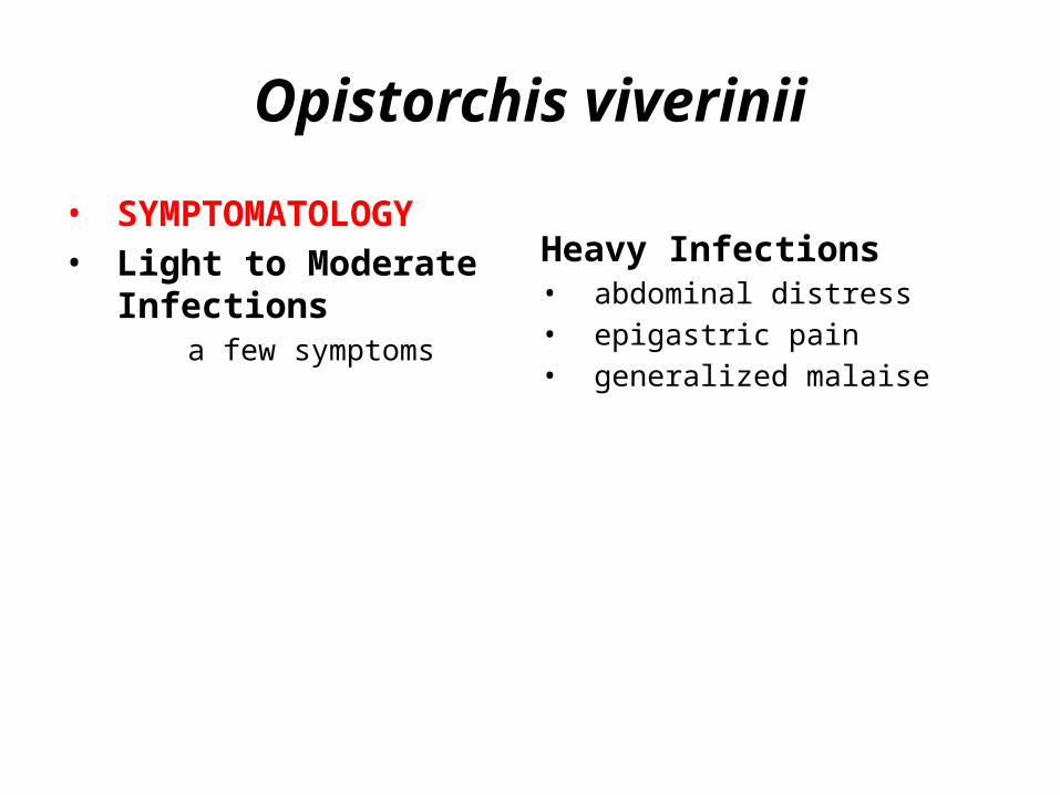
• SYMPTOMATOLOGY• Light to Moderate
Infections a few symptoms
Heavy Infections• abdominal distress• epigastric pain• generalized malaise
Opistorchis viverinii

• CLINICAL DIAGNOSIS• suggestive in patients from
endemic areas with: – history of eating uncooked
fish– symptoms of biliary tract
disease • intermittent jaundice • bouts of fever • right upper quadrant pain
• LABORATORY DIAGNOSIS• finding the characteristic
eggs in the feces or biliary drainage or duodenal aspiration
• Ultrasonography to screen the presence of cholangiocarcinoma
Opistorchis viverinii
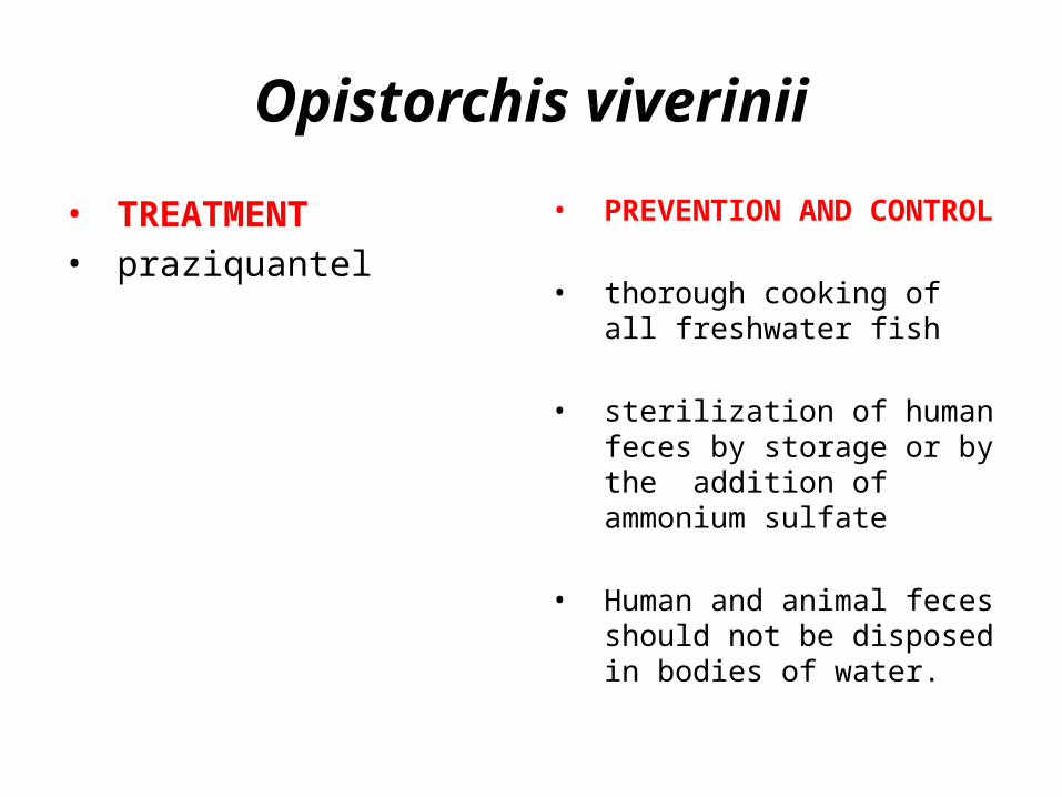
• TREATMENT• praziquantel
• PREVENTION AND CONTROL
• thorough cooking of all freshwater fish
• sterilization of human feces by storage or by the addition of ammonium sulfate
• Human and animal feces should not be disposed in bodies of water.
Opistorchis viverinii

• TREATMENT• praziquantel
• PREVENTION AND CONTROL
• thorough cooking of all freshwater fish
• sterilization of human feces by storage or by the addition of ammonium sulfate
• Human and animal feces should not be disposed in bodies of water.
Opistorchis viverinii
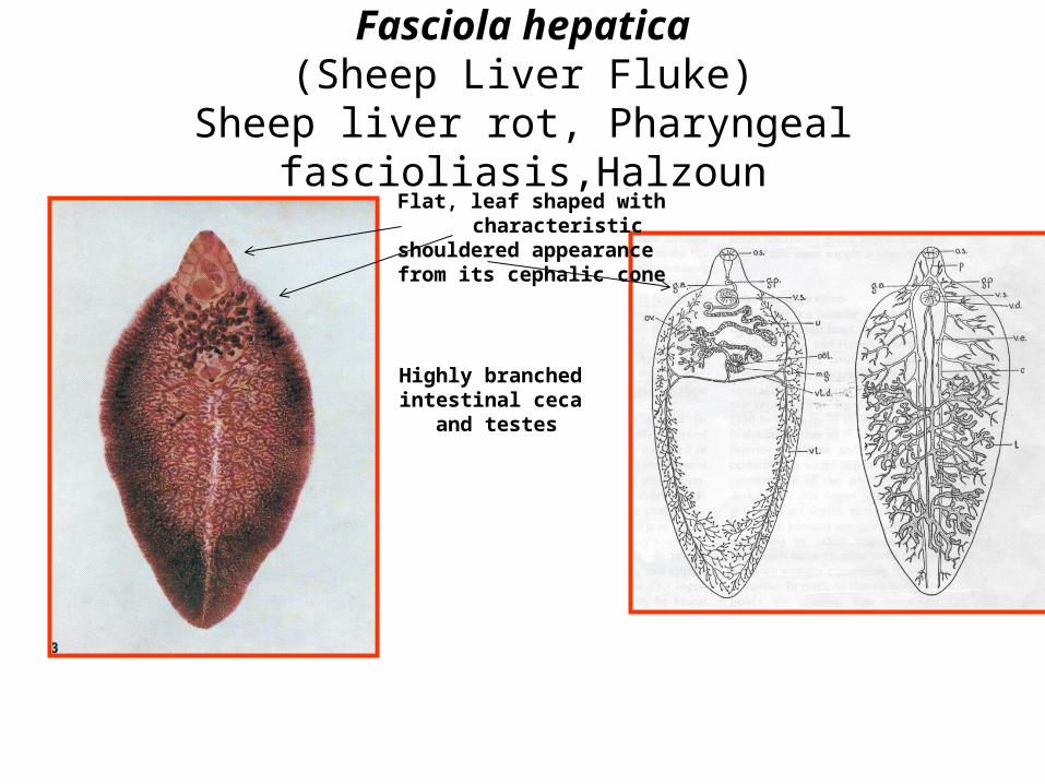
Fasciola hepatica(Sheep Liver Fluke)
Sheep liver rot, Pharyngeal fascioliasis,HalzounFlat, leaf shaped with characteristic
shouldered appearance from its cephalic cone
Highly branched intestinal ceca
and testes

ovaloperculated
Fasciola hepatica(sheep liver fluke)

EGG LARVA 1 LARVA 2
FIRST INTERMEDIATE
HOST
SECOND INTERMEDIATE
HOST
FINALHOST
Lymneid snails a. Lymnaea philippinensis b. Lymnea swinhoe
aquatic vegetationwatercress FASCIOLIASIS
Humans get infected by: >eating uncooked watercress containing the infective metacercaria > ingestion of the cysts in drinking water
PHARYNGEAL FASCIOLIASIS (HALZOUN)Humans get infected by: >eating raw sheep or goat liver
Fasciola hepatica(sheep liver fluke)
Natural definitive hosts:herbivorous animals like sheep.
MAN IS AN ACCIDENTAL HOST.
ADULT

PATHOLOGY
Acute Or Invasive Phase period during which the fluke migrates from the intestine to the liver and its burrowing through the liver parenchyma
no significant change from the intestine to the liver Parasite may wander or be carried by blood after penetrating a blood vessel to ectopic sites such as lungs, subcutaneous Tissues, brain and the orbit ---- abscesses or fibrotic lesions
Migration to the liver parenchyma traumatic and necrotic lesions
Chronic Or Latent Phase period when the parasite has already reached the bile ducts
a. obstruction in the vessel
b. inflammatory and adenomatous changes of the biliary epithelium
c. fibrosis of the ducts
d. pressure atrophy of the liver parenchyma
e. intensive periductal fibrosis
f. Heavy infections 1. erosion of the epithelium 2. young worms wander back into the liver to produce abscess pockets and to seed the vital liver tissue with their eggs

Symptomatology
1. colic and obstructive jaundice2. coughing and vomiting3. generalized abdominal rigidity4. acute epigastric pain and tenderness5. urticaria6. early leukocytosis and eosinophilia7. irregular fever8. more or less persistent diarrhea9. marked anemia10.hemoglobinuria11.cholelithiasis – common complication
PHARYNGEAL FASCIOLIASIS OR HALZOUN
due to ingestion of infected raw sheep and goat livers Adult worm lodges temporarily in the pharyngeal mucosa causing:
>edematous congestion of the soft palate, pharynx, larynx, nasal fossae, Eustachian tube ------ suffocation
>dyspnea
>deafness
>asphyxiation

DIAGNOSIS
Clinical – based on1. biliary symptoms2. moderate to high eosinophilia3. eating watercress as a green salad
(metacercaria in vegetation)
Laboratory 1. recovery of the eggs in the patient’s stool or from duodenal or biliary tract drainage 2. serodiagnosis – helpful but not adapted for routine diagnosis
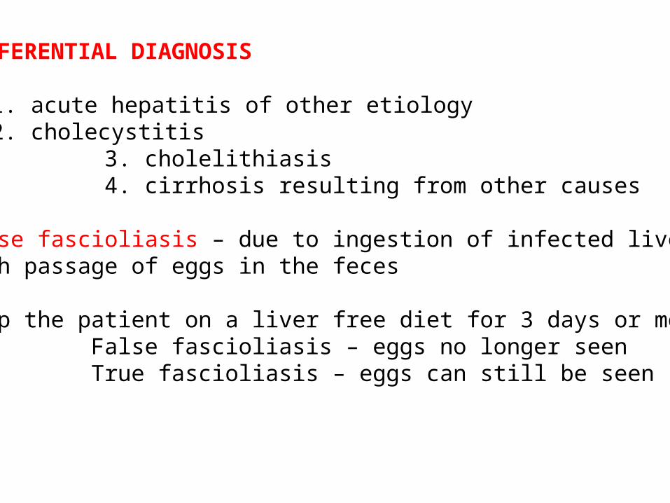
DIFFERENTIAL DIAGNOSIS
1. acute hepatitis of other etiology2. cholecystitis
3. cholelithiasis 4. cirrhosis resulting from other causes
False fascioliasis – due to ingestion of infected liverswith passage of eggs in the feces Keep the patient on a liver free diet for 3 days or more False fascioliasis – eggs no longer seen True fascioliasis – eggs can still be seen

Treatment
1. bithionol (dichlorophenol) 2. dehydroemetine hydrochloride3. emetine hydrochloride 4. hexachloroparaxylene 5. praziquantel
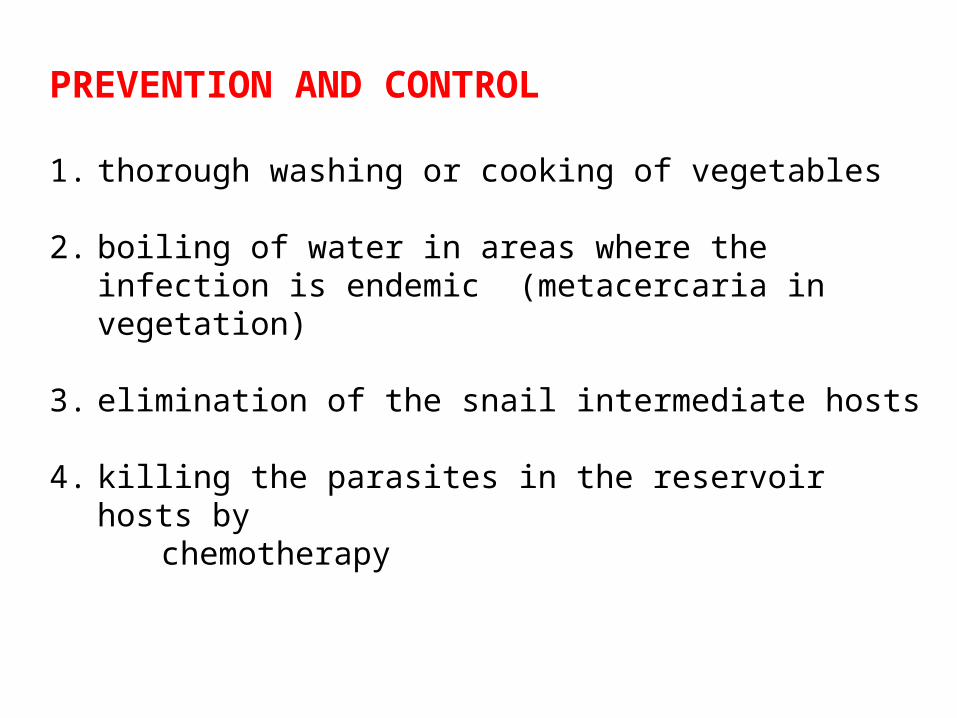
PREVENTION AND CONTROL
1. thorough washing or cooking of vegetables
2. boiling of water in areas where the infection is endemic (metacercaria in vegetation)
3. elimination of the snail intermediate hosts
4. killing the parasites in the reservoir hosts by chemotherapy

Fasciola gigantica(Giant Liver Fluke)
Flat, leaf shaped more elongated
cephalic cone is shorter and less prominent
shoulders are practically lacking
ventral sucker is larger
Highly branched intestinal ceca
and testes (more anterior)

ovaloperculated
Fasciola gigantica(Giant liver fluke)
Eggs are larger.
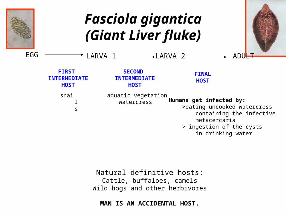
EGG LARVA 1 LARVA 2
FIRST INTERMEDIATE
HOST
SECOND INTERMEDIATE
HOST
FINALHOST
snails
aquatic vegetationwatercress Humans get infected by:
>eating uncooked watercress containing the infective metacercaria > ingestion of the cysts in drinking water
Fasciola gigantica(Giant Liver fluke)
Natural definitive hosts:Cattle, buffaloes, camels
Wild hogs and other herbivores
MAN IS AN ACCIDENTAL HOST.
ADULT
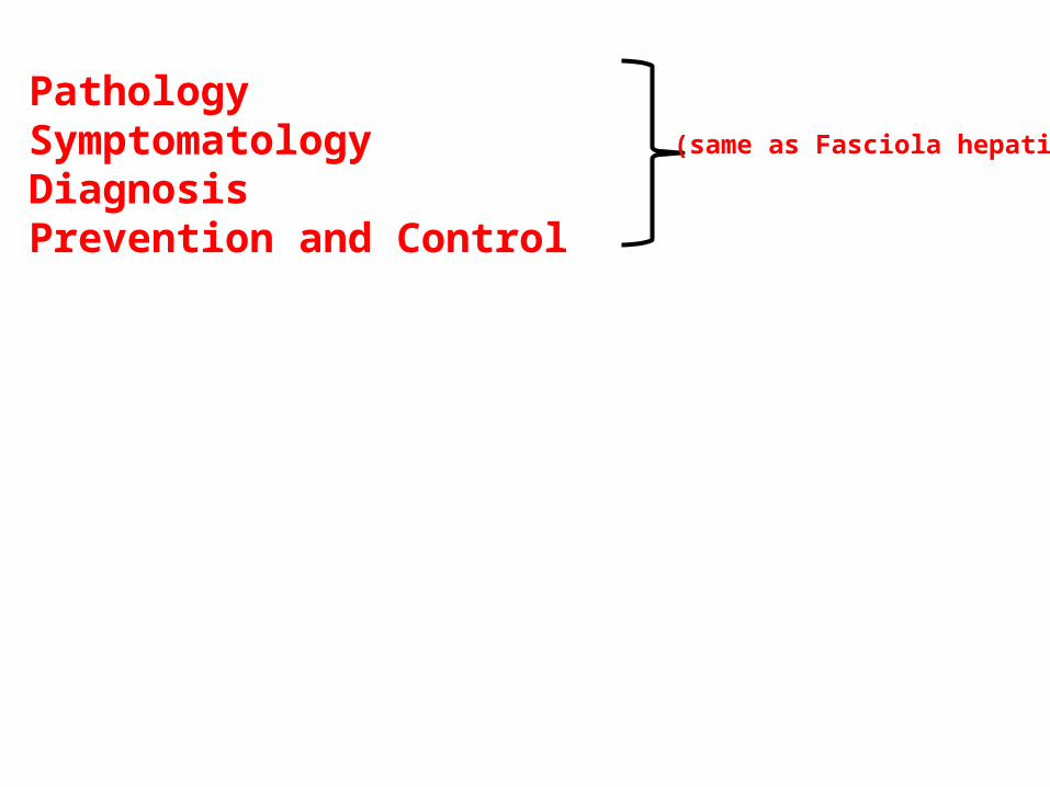
PathologySymptomatology DiagnosisPrevention and Control
(same as Fasciola hepatica)

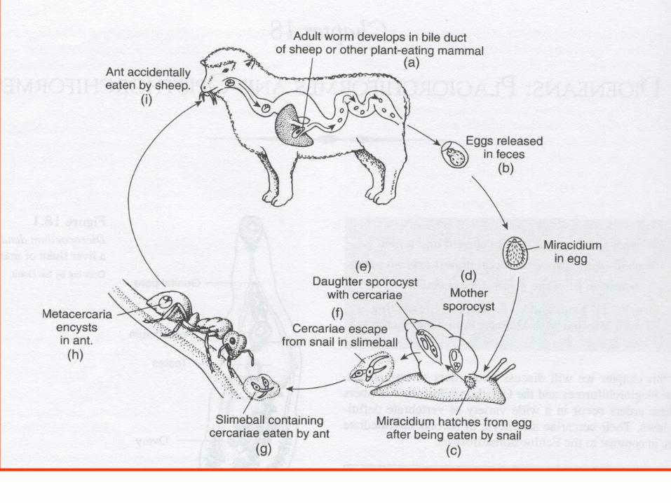
Dicrocoelium dendriticum(Lancet Fluke)
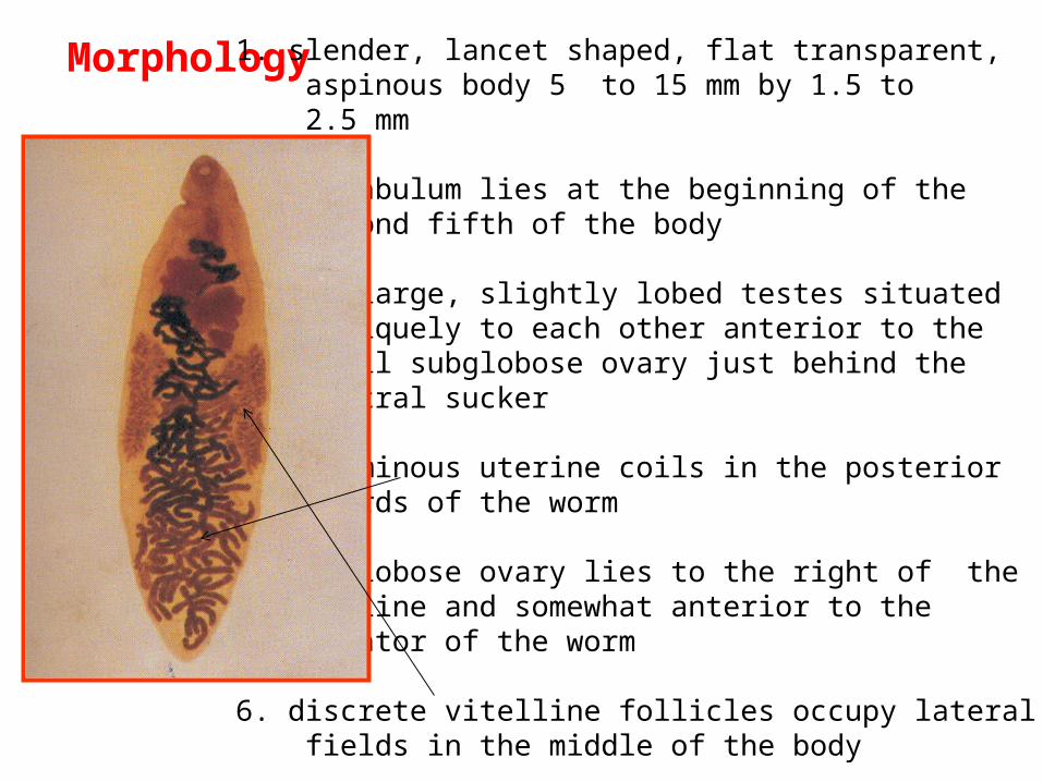
Morphology 1. slender, lancet shaped, flat transparent, aspinous body 5 to 15 mm by 1.5 to 2.5 mm
2. acetabulum lies at the beginning of the second fifth of the body
3. two large, slightly lobed testes situated obliquely to each other anterior to the small subglobose ovary just behind the ventral sucker
4. voluminous uterine coils in the posterior thirds of the worm
5. subglobose ovary lies to the right of the midline and somewhat anterior to the equator of the worm
6. discrete vitelline follicles occupy lateral fields in the middle of the body
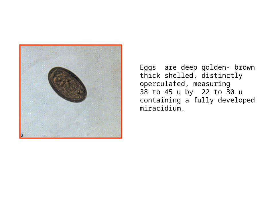
Eggs are deep golden- brownthick shelled, distinctly operculated, measuring 38 to 45 u by 22 to 30 u containing a fully developed miracidium.

EGG LARVA 1 LARVA 2
FIRST INTERMEDIATE
HOST
SECOND INTERMEDIATE
HOST
FINALHOST
land snails of the genera Abida, Cochlicopa, Helicella and Zebrina
Ants(Formica fusca) Humans get infected by:
> ants harboring the metacercaria
Dicrocoelium dendriticum(Lancet fluke)
ADULT
Eggs are deep golden- brownthick shelled, distinctly operculated, measuring 38 to 45 u by 22 to 30 u containing a fully developed miracidium.
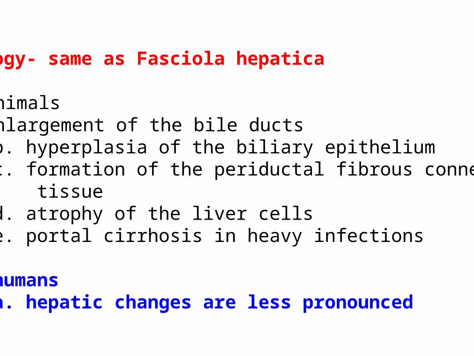
Pathology- same as Fasciola hepatica
1. in animalsa. enlargement of the bile ducts
b. hyperplasia of the biliary epithelium c. formation of the periductal fibrous connective tissue d. atrophy of the liver cells e. portal cirrhosis in heavy infections
2. in humans a. hepatic changes are less pronounced

Symptomatology
In Humansa. digestive disturbancesb. flatulence
c. vomiting d. biliary colic e. chronic constipation or diarrhea f. enlarged liver g. systemic toxemia less pronounced that in fascioliasis
LABORATORY DIAGNOSIS
1. finding the eggs consistently in the feces and duodenal drainage
2. eliminate spurious infections from eating livers containing the eggs

TREATMENT – same as Clonorchis sinensis
1. praziquantel – 25 mg per kg TID for 2 consecutive days
PREVENTION AND CONTROL
2. no effective measures of control3. Fresh herbs collected from grazing areas for use as food for humans should be washed to remove the ants.

___________________________________________________________________________Scientific name Common name Infective Diagnostic
stage stage___________________________________________________________________________Fasciola hepatica sheep liver fluke metacercaria unembryonated ova___________________________________________________________________________Fasciola gigantica giant liver fluke metacercaria unembryonated ova___________________________________________________________________________Clonorchis sinensis Chinese liver fluke metacercaria embryonated ova___________________________________________________________________________Opistorchis felineus cat liver fluke metacercaria embryonated ova___________________________________________________________________________Opistorchis metacercaria embryonated ovaviverrini___________________________________________________________________________Dicrocoelium lancet fluke metacercaria embryonated ovadendriticum
______________________________________________________

___________________________________________________________________________Scientific name 1ST IH 2ND IH FH AFH ___________________________________________________________________________Fasciola hepatica snail aquatic vegetation herbivorous animals man like sheep___________________________________________________________________________Fasciola gigantica snail aquatic vegetation camels, wild hogs, man cattle and water buffalo___________________________________________________________________________Clonorchis sinensis snail fresh water fish man ___________________________________________________________________________Opistorchis felineus snail fresh water fish cats, dogs,fox,wolves seals man___________________________________________________________________________Opistorchis snail fresh water fish civet cat, cat,dog manviverrini and other fish eating mammals___________________________________________________________________________Dicrocoelium snail ants sheep man dendriticum___________________________________________________________________________

TREMATODESA. Liver Flukes
EGG LARVA 1 LARVA 2 ADULTHatches in waterto release the Larva.
Develops inThe firstIntermediate host(SNAILS)
Develops inThe secondIntermediate host(water plants, Fish or snails
Eaten by the Final host(Humans)
LIVER FLUKESFasciola hepatica Goes to the liver, bile duct
Causing destruction
Opistorchis felineus(cat liver fluke)
Clonorchis sinensis(Chinese liver fluke) Can cause liver cancerEating raw
Fresh waterfish
Can cause bile stones.destruction of the bileducts and liver
Eating rawFresh waterfish
Eating watercress


![Microproteinuria during Opisthorchis viverriniInfection: A ......liver flukes Opisthorchis viverrini and Clonorchis sinensis [1]. In Southeast Asia alone, up to 67 million people are](https://static.fdocuments.net/doc/165x107/604a246ab262a95d9267572c/microproteinuria-during-opisthorchis-viverriniinfection-a-liver-flukes.jpg)