Light-sheet microscopy for everyone? Experience of ...
16
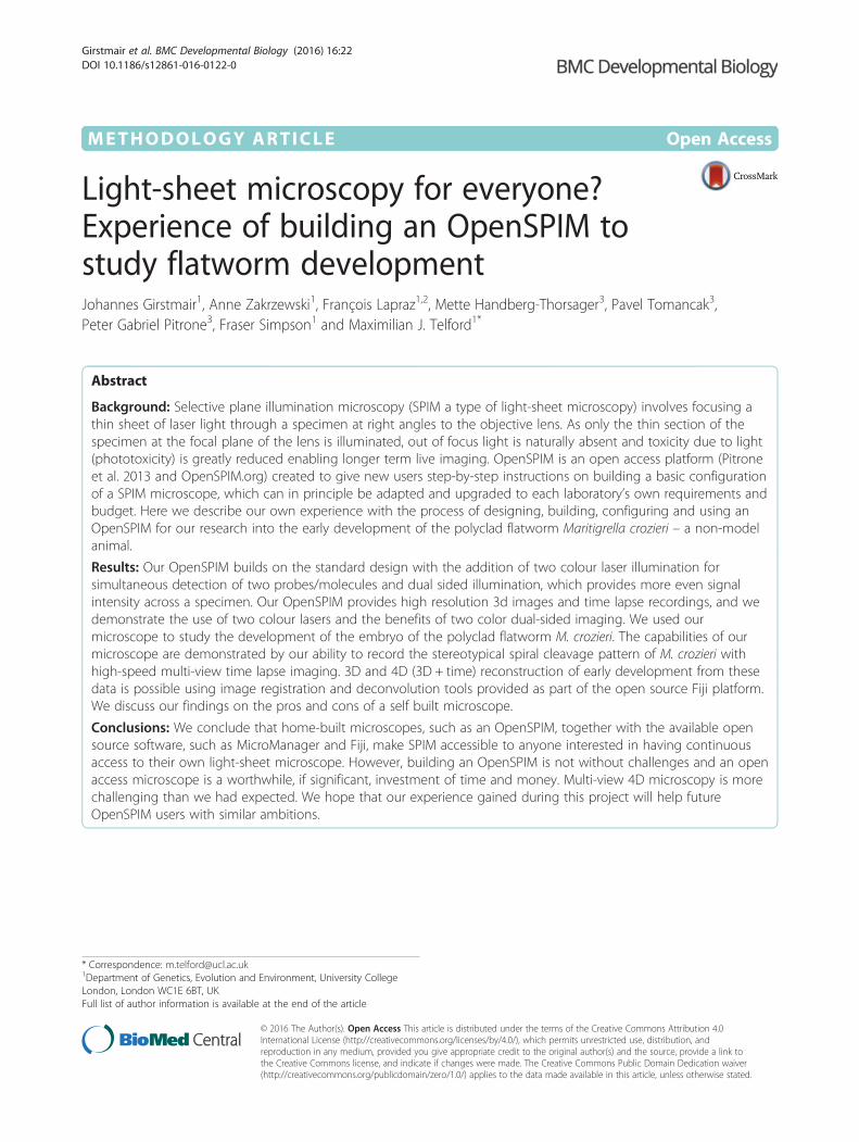
METHODOLOGY ARTICLE Open Access Light-sheet microscopy for everyone? Experience of building an OpenSPIM to study flatworm development Johannes Girstmair 1 , Anne Zakrzewski 1 , François Lapraz 1,2 , Mette Handberg-Thorsager 3 , Pavel Tomancak 3 , Peter Gabriel Pitrone 3 , Fraser Simpson 1 and Maximilian J. Telford 1* Abstract Background: Selective plane illumination microscopy (SPIM a type of light-sheet microscopy) involves focusing a thin sheet of laser light through a specimen at right angles to the objective lens. As only the thin section of the specimen at the focal plane of the lens is illuminated, out of focus light is naturally absent and toxicity due to light (phototoxicity) is greatly reduced enabling longer term live imaging. OpenSPIM is an open access platform (Pitrone et al. 2013 and OpenSPIM.org) created to give new users step-by-step instructions on building a basic configuration of a SPIM microscope, which can in principle be adapted and upgraded to each laboratory’s own requirements and budget. Here we describe our own experience with the process of designing, building, configuring and using an OpenSPIM for our research into the early development of the polyclad flatworm Maritigrella crozieri – a non-model animal. Results: Our OpenSPIM builds on the standard design with the addition of two colour laser illumination for simultaneous detection of two probes/molecules and dual sided illumination, which provides more even signal intensity across a specimen. Our OpenSPIM provides high resolution 3d images and time lapse recordings, and we demonstrate the use of two colour lasers and the benefits of two color dual-sided imaging. We used our microscope to study the development of the embryo of the polyclad flatworm M. crozieri. The capabilities of our microscope are demonstrated by our ability to record the stereotypical spiral cleavage pattern of M. crozieri with high-speed multi-view time lapse imaging. 3D and 4D (3D + time) reconstruction of early development from these data is possible using image registration and deconvolution tools provided as part of the open source Fiji platform. We discuss our findings on the pros and cons of a self built microscope. Conclusions: We conclude that home-built microscopes, such as an OpenSPIM, together with the available open source software, such as MicroManager and Fiji, make SPIM accessible to anyone interested in having continuous access to their own light-sheet microscope. However, building an OpenSPIM is not without challenges and an open access microscope is a worthwhile, if significant, investment of time and money. Multi-view 4D microscopy is more challenging than we had expected. We hope that our experience gained during this project will help future OpenSPIM users with similar ambitions. * Correspondence: [email protected] 1 Department of Genetics, Evolution and Environment, University College London, London WC1E 6BT, UK Full list of author information is available at the end of the article © 2016 The Author(s). Open Access This article is distributed under the terms of the Creative Commons Attribution 4.0 International License (http://creativecommons.org/licenses/by/4.0/), which permits unrestricted use, distribution, and reproduction in any medium, provided you give appropriate credit to the original author(s) and the source, provide a link to the Creative Commons license, and indicate if changes were made. The Creative Commons Public Domain Dedication waiver (http://creativecommons.org/publicdomain/zero/1.0/) applies to the data made available in this article, unless otherwise stated. Girstmair et al. BMC Developmental Biology (2016) 16:22 DOI 10.1186/s12861-016-0122-0
Transcript of Light-sheet microscopy for everyone? Experience of ...
METHODOLOGY ARTICLE Open Access
Light-sheet microscopy for everyone?Experience of building an OpenSPIM tostudy flatworm developmentJohannes Girstmair1, Anne Zakrzewski1, François Lapraz1,2, Mette Handberg-Thorsager3, Pavel Tomancak3,Peter Gabriel Pitrone3, Fraser Simpson1 and Maximilian J. Telford1*
Abstract
Background: Selective plane illumination microscopy (SPIM a type of light-sheet microscopy) involves focusing athin sheet of laser light through a specimen at right angles to the objective lens. As only the thin section of thespecimen at the focal plane of the lens is illuminated, out of focus light is naturally absent and toxicity due to light(phototoxicity) is greatly reduced enabling longer term live imaging. OpenSPIM is an open access platform (Pitroneet al. 2013 and OpenSPIM.org) created to give new users step-by-step instructions on building a basic configurationof a SPIM microscope, which can in principle be adapted and upgraded to each laboratory’s own requirements andbudget. Here we describe our own experience with the process of designing, building, configuring and using anOpenSPIM for our research into the early development of the polyclad flatworm Maritigrella crozieri – a non-modelanimal.
Results: Our OpenSPIM builds on the standard design with the addition of two colour laser illumination forsimultaneous detection of two probes/molecules and dual sided illumination, which provides more even signalintensity across a specimen. Our OpenSPIM provides high resolution 3d images and time lapse recordings, and wedemonstrate the use of two colour lasers and the benefits of two color dual-sided imaging. We used ourmicroscope to study the development of the embryo of the polyclad flatworm M. crozieri. The capabilities of ourmicroscope are demonstrated by our ability to record the stereotypical spiral cleavage pattern of M. crozieri withhigh-speed multi-view time lapse imaging. 3D and 4D (3D + time) reconstruction of early development from thesedata is possible using image registration and deconvolution tools provided as part of the open source Fiji platform.We discuss our findings on the pros and cons of a self built microscope.
Conclusions: We conclude that home-built microscopes, such as an OpenSPIM, together with the available opensource software, such as MicroManager and Fiji, make SPIM accessible to anyone interested in having continuousaccess to their own light-sheet microscope. However, building an OpenSPIM is not without challenges and an openaccess microscope is a worthwhile, if significant, investment of time and money. Multi-view 4D microscopy is morechallenging than we had expected. We hope that our experience gained during this project will help futureOpenSPIM users with similar ambitions.
* Correspondence: [email protected] of Genetics, Evolution and Environment, University CollegeLondon, London WC1E 6BT, UKFull list of author information is available at the end of the article
© 2016 The Author(s). Open Access This article is distributed under the terms of the Creative Commons Attribution 4.0International License (http://creativecommons.org/licenses/by/4.0/), which permits unrestricted use, distribution, andreproduction in any medium, provided you give appropriate credit to the original author(s) and the source, provide a link tothe Creative Commons license, and indicate if changes were made. The Creative Commons Public Domain Dedication waiver(http://creativecommons.org/publicdomain/zero/1.0/) applies to the data made available in this article, unless otherwise stated.
Girstmair et al. BMC Developmental Biology (2016) 16:22 DOI 10.1186/s12861-016-0122-0

BackgroundLight-sheet illumination for microscopy is an old technol-ogy enjoying a dramatic recent renaissance due to introduc-tion of selective plane illumination microscopy (SPIM) [1].The principle of SPIM is to use optics to form a thin sheetof light that passes through the specimen. Unlike a stand-ard microscope in SPIM the objective lens is placed per-pendicular to the direction of the light such that the sheetof light illuminates the specimen only at the focal plane ofthe lens. This has two important benefits; it eliminates scat-tered light from out of focus areas of the specimen provid-ing a natural means of optical sectioning and, because onlythe imaged area is illuminated, the total amount of lighthitting the specimen is orders of magnitude less than inconventional fluorescence microscopy meaning thatphotodamage/phototoxicity is enormously reduced andimaging over long periods is possible [1]. This latter bene-fit is of great significance for live imaging. OpenSPIM isan open access light-sheet microscopy design [2]; http://openspim.org; see also [3]. The OpenSPIM resource givesusers step-by-step guidance for building a basic configur-ation of a SPIM microscope and includes appropriate
open source software for image acquisition and processingsuch as Fiji (http://fiji.sc/Fiji), micromanager (https://www.micro-manager.org/), multiview reconstruction plu-gins [4, 5] deconvolution [6] and big data viewer (http://fiji.sc/BigDataViewer). The design can be adapted andupgraded according to the users specific requirementsand budget. We have designed an OpenSPIM microscopecapable of dual-sided illumination (the so called T-configuration proposed on the OpenSPIM wiki). Themicroscope was built following instructions from the web-site http://openspim.org with modifications required toextend the capabilities of the basic single sided illumin-ation described there (Fig. 1).To test our system we have imaged the early embryo-
genesis and the larval stage of the polyclad flatwormMaritigrella crozieri, a promising new evo-devo modelwithin Plathyhelminthes [7]. Eggs of this polyclad flat-worm undergo stereotypical spiral cleavage to produce aciliated planktotrophic larval stage known as Müllerlarva that shows morphological similarities to plankto-trophic larval stages found in marine annelids and mol-luscs. The embryonic and post-embryonic development
Fig. 1 OpenSPIM with dual-sided illumination, hardware-controlled laser triggering and all hardware components
Girstmair et al. BMC Developmental Biology (2016) 16:22 Page 2 of 16

of M. crozieri has been previously described [8]. Recentflatworm phylogenies confirm the basal position of poly-clad flatworms within the rhabditophoran Platyhelmin-thes [9, 10] making M. crozieri and other polycladflatworms an interesting system for evo-devo studieswithin Platyhelminthes and amongst other Lophotrocho-zoa. Here we demonstrate that, on both live and fixedmaterial, we were able to visualize the stereotypicalspiral cleavage pattern of M. crozieri with high-speedtime-lapse sequences and were able to 3D-reconstruct anumber of individual time points of the early embryonicdevelopment using Fiji’s bead based registration softwareand multi-view deconvolution plugins [4, 6].In this report we describe a real life experience of build-
ing an OpenSPIM microscope. We discuss the difficultieswe encountered, the real costs involved including the timespent and difficulties encountered as well as describingthe limitations and significant benefits of the system.
ResultsOur OpenSPIM produces high quality images which wecompare to scanning electron micrographs (SEM)To test whether our OpenSPIM microscope can producehigh quality images, we stained fixed 1 day old Müller’slarvae with a monoclonal Mouse anti-Acetylated Tubulin
antibody (Sigma) and used a secondary antibody conju-gated to Alexa Fluor® 568 Goat anti-Mouse (Invitrogen™).Our OpenSPIM images (shown as maximum projections)show cilia covering the whole epidermis of the polycladlarva (Fig. 2a-e). This dense film of short cilia can easily bedistinguished from longer cilia comprising the ciliary bandalong the eight lobes (Fig. 2e). Our OpenSPIM imagesshow a clear resemblance to scanning electron microscopyimages [7] of similar stage larvae (Fig. 2d, e and f) con-firming reliable image acquisition with OpenSPIM at thelevel of embryo morphology. For a more detailed compari-son of several views see also Additional file 1.
The advantage of OpenSPIM multi-view reconstructionsover confocal microscopy and single image in M. crozieriStandard confocal microscopes lack the possibility ofmulti-view imaging and reconstruction. This is import-ant for the study of M. crozieri larvae and embryos dueto the attenuation of light intensity caused by theopaque yolk meaning we can only visualize one side ofthe embryo. We have found that the opacity causes sig-nificant signal loss, which becomes especially obvious inMaritigrella when the confocal z-stack of an imagedspecimen is rotated. We demonstrate this here in a fixed
Fig. 2 Maximum projections of fixed Müller’s larvae stained with Acetylated tubulin and imaged with OpenSPIM (a-e). a Anterior view b Lateralview c Ventral view d Posterior view e magnified view of area boxed in d (f) Posterior view of a M. crozieri larval stage obtained with scanningelectron microscopy for comparison with imaging acquired by our OpenSPIM. Ap, apical plate; oh, oral hood; vll, ventro-lateral lobe; ll, lateral lobe;dll, dorso-lateral lobe; sop, sub-oral plate; cb, ciliary bands (long cilia). All scalebars are 50 μm
Girstmair et al. BMC Developmental Biology (2016) 16:22 Page 3 of 16

larva labeled with the nucleic-stain SytoxGreen (Fig. 3,right).The second drawback of a confocal is the necessity of
using a slide with coverslip, which tends to cause de-formation of our topologically complex larvae. The rota-tion of a confocal imaged larva reveals the slightlysqueezed body shape of the larva. Confocal z-stacks arenot suitable for further image processing (e.g. image pat-tern registration as described by [11, 12].
In contrast to confocal imaging, OpenSPIM offers amulti-view reconstruction method, whereby different an-gles of the same specimen can be fused into a single z-stack as shown in Fig. 3 (left side). The reconstructedlarva not only keeps its natural shape, but also includesthe information obtained from each individual angle,resulting in a whole-mount containing high-resolutionsignal from all sides. For Maritigrella larvae andembryos multi-view reconstructions achieved with
Fig. 3 A comparison of a multi-view reconstructed larva (multi-view deconvolution of several angles) stained with the nucleic marker SytoxGreen(left side) with a larva with the same staining captured with a Leica TCS SP8 confocal laser microscopy (right side)
Girstmair et al. BMC Developmental Biology (2016) 16:22 Page 4 of 16

OpenSPIM create a crucial advantage over confocalmicroscopy.We further visualized to what extent average fusion
and deconvolution [6] can improve results over a singleview in an M. crozieri embryo when imaged with ourOpenSPIM. This is relevant e.g. for early cleavage obser-vations, when nuclei of the macromeres shift from theanimal pole towards the vegetal pole of the embryo andare thus difficult to see.In terms of imaging the entire embryo, it is not sur-
prising that we found a clear benefit gained by applyingmulti-view imaging (average fusion or multi-view decon-volution of 5 angles) over a single one angle view. Thedifferences are shown in Fig. 4a-c; in the single angleview the small macromeres (cells A-D) at the vegetal ex-treme of the embryo are not visible (Fig. 4a). The acqui-sition of several angles (Fig. 4b and c) reveals themissing cells and makes clear that embryonic 3D recon-structions, which should include all nuclei information,depends on multi-view imaging. A slight improvementof multi-view deconvolution with 12 iterations over aver-age fusion could be achieved (Fig. 4b and c), but appearsto be less critical for nuclei staining in early staged M.crozieri embryos.
Dual-sided illumination efficiently compensates axialintensity attenuation in semi-transparent specimensDual-sided illumination for OpenSPIM microscopycan be achieved by building a so-called T-configuration, whereby the laser beam gets split intotwo beams and a second optical path is installed onthe optical breadboard. This allows the illuminationof specimens from two sides, instead of one, as dem-onstrated originally by [13], and was also suggestedas a potential extension in the original OpenSPIM
publication [2]. The benefit of having dual-sided illu-mination in our OpenSPIM was tested on fixed M.crozieri embryos stained with the nucleic acidmarker SytoxGreen. In single-sided illumination im-ages (where one of the two illumination paths hasbeen completely obscured - left or right respect-ively), a significant loss of signal during acquisitionon the side of the missing illumination path becomesobvious due to axial intensity attenuation caused byour opaque and yolky specimens (Fig. 5a and c). Thelight attenuation is especially apparent when singlenuclei from opposed illumination sites (left andright) are directly compared to each other (Fig. 5aand c, insets). In contrast, a more complete pictureof the stained nuclei is achieved by using both illu-mination paths simultaneously (Fig. 5c, insets). Thissimple test clearly demonstrates the benefit of usingdual-sided illumination for our slightly opaque endo-lecithal polyclad embryos.
Two laser lines allow the visualization of two detectionchannelsOur OpenSPIM is equipped with two individual lasers(λ = 488 nm and 561 nm) that allow visualization of twodetection channels. The twin laser system was tested onfixed 1 day old M. crozieri larvae stained with the nu-cleic marker SytoxGreen (488). The 561 laser was usedin the same specimens to visualize auto-fluorescence ofgland cells (rhabdites). The larvae have gland cell scat-tered mostly around the apical plate and on the ventralside of the animals, which is shown in Fig. 6 in a singlespecimen, in which both channels (green and red) havebeen combined. Here we simply demonstrate the useand precise alignment of the twin laser beams (488 nmand 561 nm). Precise alignment is required during multi
Fig. 4 The benefit gained by applying the multi-view deconvolution method (5-angles used) vs. simple maximum projections of an acquiredstack (1-angle) on an early staged M. crozieri embryo. a Depth color coding (Fire) of all nuclei after maximum projection of a raw z-stack (1-angle)b Depth color coding (Fire) of all nuclei after a maximum projection of a raw z-stack (1-angle) c Maximum projection of all nuclei after multi-viewdeconvolution. Additionally all nuclei missing in the single angle maximum projection (from b) have been coloured in red
Girstmair et al. BMC Developmental Biology (2016) 16:22 Page 5 of 16

color imaging to obtain good quality images from bothchannels. The initial laser beam alignments in a multiplelaser system (in our case VersaLase) is done by themanufacturer and an OpenSPIM user can later onlyalign one wavelength, e.g. 488 nm, while the other wave-length(s) (in our case the 561 nm) is presumed to coin-cide. It is worth noting that we have transported ourOpenSPIM by train and car and that the default
alignment of our laser system alignment has proven ro-bust during travelling.
OpenSPIM image acquisition with hardware controlledlaser triggering is more than twice as fastWith the aim of reducing image acquisition time, we in-corporated ESio’s TTL controller box (http://www.esi-magingsolutions.com/) into our OpenSPIM microscope;
Fig. 5 A single and dual-sided illumination, imaging test showing maximum projections of nuclei stained with the nucleic acid marker SytoxGreen. aMaximum projections of embryonic nuclei using left illumination path of the OpenSPIM microscope, b Both illumination paths and c Right illuminationpaths respectively. Image stacks were acquired in the following order: left illumination, right illumination, dual-sided illumination (a following c following b).All stacks and related insets have been processed identically
Fig. 6 Several angles of a 3D-rendered Müller’s larva showing nuclei in green (captured with the 488 laser detection channel) and gland cells inred (captured with the 561 laser detection channel)
Girstmair et al. BMC Developmental Biology (2016) 16:22 Page 6 of 16

this enables hardware-controlled synchronization of thetiming of camera exposure and laser triggering. To testour ESio TTL controller, a 100 μm thick single colorstack was imaged (1280 × 1080 resolution in 16-bit andan exposure time of 32 ms) with a constant step size of1.5 μm. In this test, the software-controlled image
acquisition (by MicroManager without the TTL control-ler box) completed the acquisition in 43.5 s. In compari-son, when hardware-controlled imaging is used, wherelasers are triggered with the TTL controller box fromESio, image acquisition took 17.5 s demonstrating a sig-nificant reduction of image acquisition time.
Fig. 7 Summary figure of the early embryonic development of the polyclad flatworm M. crozieri (1–128-cell stages) (A-J); SEM pictures (A-J) havebeen captured for comparison, stills from time-lapse sequences (A-J’), multi-view 3D reconstructions (A-J”); all embryos are shown from animalside. Note that time-lapse images (A-J’) are presented as captured by the OpenSPIM (mirror images) and therefore cleavage direction is oppositeto 3D and SEM images. All scale bars are 50 μm
Girstmair et al. BMC Developmental Biology (2016) 16:22 Page 7 of 16

Fiji’s bead based registration algorithm and multi-viewdeconvolution is essential to visualize all nuclei in M. cro-zieri embryos along the animal-vegetal axisHaving the possibility to 3D rotate the specimen and usinga faster image acquisition set-up opens up the possibilityof carrying out whole-embryo time-lapse videos of the de-velopment of an embryo. Fiji’s bead based registration al-gorithm and multi-view deconvolution plugins [4, 6] makeit possible to fuse and deconvolve z-stacks imaged at mul-tiple angles, acquired sequentially at any given time-point.We imaged the early development of Maritigrella cover-ing the spiral cleavage and the formation of the four quad-rants (Fig. 7A-J’). To ensure that the development isnormal in our live-imaging experiment, we fixed speci-mens from series of cleavage stages with fixed specimens,for which both 3D reconstructions of immunostainedOpenSPIM imaged embryos and SEM imaged embryoswere performed (Fig. 7A-J & A-J”). The 3D-reconstructedseries of fixed embryos of several stages are also availableas 3D models (see Additional file 2).
Rapid in vivo time-lapse sequences captured with Open-SPIM show the dynamic early embryonic development ofM. crozieriWith the OpenSPIM equipped with two illuminationpaths and capable of rapidly producing, high-qualityimage stacks, we aimed to visualize the embryonic devel-opment in M. crozieri up to the 128-cell stage to furthertest the potential of OpenSPIM for live-imaging and, ul-timately, for lineage tracing. We created a time-lapse se-quence of a developing embryo injected at the one cellstage with a nuclear marker (H2B:GFP) and a mem-brane marker (CAAX:GFP). Our time lapse covers 18 hand shows the stereotypical spiral cleavage and forma-tion of four quartets and further development in M. cro-zieri. The sequence visualizes the embryo from theanimal pole (Fig. 7A-J’ and Additional file 3) and con-sists of 273 individual time-points. During early cleav-age, the live specimens had similar morphology atspecific time points when compared with fixed speci-mens (SEM and 3D reconstructions) at the same devel-opmental stages.
DiscussionSummary of the capacity of our new OpenSPIMNew modifications tested for OpenSPIMWhen designing microscopes for in vivo imaging withthe purpose of tracing cells, one of the goals is to have ahigh imaging speed in order to have the best time reso-lution. One of our own modifications included hard-ware-controlled imaging that appears to be an elegantway of removing unwanted delays during image acquisi-tion. Another relatively new implementation, at least inthe context of OpenSPIM, is the use of dual-sided
illumination, which we tested on our specimens. Wedemonstrated that bringing the light-sheet simultan-eously from two sides to our opaque and yolky sam-ples results in a significant increase in signal acrossthe sample as shown for the nucleic markers ofstained embryos (Fig. 5).
Limitations of our self-built OpenSPIMIt is worth mentioning that simultaneous dual-sided illu-mination can lead to the light-sheet widening and a re-duction in image quality due to additional lightscattering effects and shadowing [13]. In our sample,these issues do not outweigh the benefits gained bydual-sided illumination in comparison to the otherwisemuch more severe attenuation effect observed, but it isassumed to have an impact on the final image quality.Another limitation of conventional light-sheet micros-
copy concerns the thickness of the light-sheet. Thinnerlight-sheets are governed by the illumination objective andthe thickness depends in particular on their numerical aper-ture. Ideally the thickness of the light-sheet is uniformacross the field of view. In reality, it widens on each side ofview and its narrowest point resides in the middle. Fortu-nately, that is where the sample is normally located. Whena static light-sheet is created by passing a pencil beamthrough a cylindrical lens, the ratio of the light-sheet thick-ness between the center and the side of the field of view de-pends on the numerical aperture of the illuminationobjective and the size of the field of view, i.e. the magnifica-tion of the detection objective. OpenSPIM as described pre-sents a good compromise, however the sample should bepositioned as centrally as possible for optimal sectioning.To address these problems in light-sheet microscopy,
and of particular importance for specimens larger thanour embryos, more advanced light-sheet microscopes il-luminate the sample from left and right sides sequen-tially (rather than simultaneously as presented here (seeFig. 5) and also take advantage of pivoting (scanning)the light-sheet as described for the mSPIM [13]. Thissignificantly reduces scattering and attenuation acrossthe field of view. Alternatively, a light-sheet can be gen-erated by scanning a Gaussian beam up and down acrossthe field of view [14]. Thus, the light-sheet that is cre-ated can be further modified in various ways [15, 16].However, such a light-sheet formation paradigm goes be-yond the original OpenSPIM design. Nevertheless it canbe implemented on the OpenSPIM platform and it is ex-pected that the community of users forming aroundOpenSPIM will do so.
Points for consideration before purchasing and building anOpenSPIMThere is little doubt that a self-built light-sheet micro-scope is significantly more affordable than existing
Girstmair et al. BMC Developmental Biology (2016) 16:22 Page 8 of 16

commercially available alternatives. A laboratory consid-ering whether to embark on building one ought to con-sider two factors. First is the question of whether thefinished microscope will be an adequate alternative interms of image quality and ease of use for the specifictask. Second, it is essential when considering an Open-SPIM to factor in the hidden costs involved, most obvi-ously the costs implied by the time spent building themicroscope, learning to use it and learning to use theopen source software required to run the microscopeand to process the data acquired.For our purposes the quality and speed of acquisi-
tion that we were able to achieve with our home-built OpenSPIM device provides valuable, high qualitydata that suit our requirements. However, despite thefact that the assembly of an OpenSPIM is indeedquite straightforward, this step remains only oneof the many challenges to overcome. Here wesummarize various steps we feel are worth consider-ing before building an OpenSPIM in order to avoidassembling an expensive toy that will be forgottenshortly after (Fig. 8).
Before you beginBefore beginning we would recommend prospectiveOpenSPIM users to image your own specimens onan established OpenSPIM system. This will providevaluable information on whether the system will besuitable for your purposes as well as show what isrequired in terms of hardware for capturing high-quality images of your particular specimens. This isalso an opportunity to gain skills such as correctlyaligning the light-sheets, getting familiar with the ac-quisition software, finding the optimal mountingstrategy for the specimens and will inform decisionsfor the OpenSPIM design selected, as discussed inthe next section. There are many OpenSPIM systemsaround the world; the current estimate is 70. Thesystem from Tomancak lab also regularly travels topractical courses and was extensively used duringthe EMBO course on Light-sheet Microscopy inDresden (August 2014 http://openspim.org/EMBO_practical_course_Light_sheet_microscopy and upcom-ing August 2016).
Designing your OpenSPIMThe basic design of an OpenSPIM can be taken fromthe open access platform (http://openspim.org). How-ever, modifications, which might meet more specificneeds of individual users, require well thought-throughdecisions especially considering that most divergencesfrom the basic plan will involve higher costs and, verylikely, additional trouble shooting.We have discussed the most significant amendments
we have made in building our own OpenSPIM. Dualsided illumination allows us to image our relativelyopaque, yolky embryos optimally and we have shown thebenefits of this. Twin lasers allow us to observe morethan one labelled molecule per sample. As the most ex-pensive component, the laser system is of particular im-portance, especially when it comes to multi-channelacquisition. Our OpenSPIM can be easily upgraded upto as many as 4 different laser wavelengths whose beamsare aligned within the laser system itself. This alignmentof two lasers has proven robust even when travelling,meaning the microscope is fairly portable.As our embryos are extracted from animals living in trop-
ical waters they can be left at room temperature (23° C)during development. Therefore our OpenSPIM chambertemperature is currently not temperature regulated, butdepending on the experiment or the specimens used forimaging a more sophisticated control of the chambertemperature might often be advantageous or even a re-quirement. While we have no experience of controllingvariables in the acquisition chamber, we think thattemperature control could be achieved easily by simplyplacing the chamber on top of a heating/cooling plate.Additional regulations such as pH or CO2 controls, al-though feasible, would require more elaborate chambermodifications to avoid, for example, bubbling or flow dis-turbance of the chamber water created by the connectedgas supply and reliable measuring systems.
Time taken for purchasingWhile this might sound trivial, we found that purchasingelements occupied a significant time. There were threereasons for this, first that there is a multitude of sup-pliers to be negotiated with. Second, as some of the partsare expensive we (like many institutions) were required
Fig. 8 Flow chart illustrating steps necessary for establishing a home-built OpenSPIM
Girstmair et al. BMC Developmental Biology (2016) 16:22 Page 9 of 16

to obtain multiple quotes for each item. Third, thechoices made regarding some parts had knock-on effectsregarding the choice or specification of other parts. Fi-nally it should not be forgotten that some of the parts ofthe OpenSPIM are bespoke and require a workshop ormanufacturer for their production.
AssemblyThe assembly of all parts can be considered fairly easy,also thanks to the information provided from the http://openspim.org website. Usually this should be the leasttime consuming (and the most fun) task.
Software and hardware integrationThe MicroManager software and information providedon the OpenSPIM website makes correct hardware con-figuration relatively easy. However - at least in our ex-perience - establishing the correct links betweenhardware components and the acquisition computercauses time consuming problems. Additional time forhardware testing and configuration should be allowed.As an example, we experienced a major issue installing asimple FTDI chip driver, which is necessary for theESio’s TTL controller box to communicate with the ac-quisition computer. Solving this problem required add-itional testing of the hardware and interaction with theoriginal suppliers. Moreover, we strongly recommendinteracting with the growing OpenSPIM online commu-nity via the mailing list, since many users are experien-cing the same problems and it is the power of thiscommunity that will help you overcome them. Besides,hardware software integration is not something that atypical biologist can master with ease. Involving com-puter scientists or engineers on the undergraduate level,which should be relatively easy at any large University, islikely to smooth many integration problems. It will alsogive the students valuable experience with open accesshardware and electronics and connect them with the ac-tive online communities in these areas.
A fast way to correctly align the OpenSPIMLearning how to correctly align the light-sheet is askill that might require some help, but it is not par-ticularly difficult to learn. Within the materials andmethods we describe what we learned and how wecurrently align our two excitation light-sheets by sim-ply adjusting the 25 mm and 50 mm telescope lensesand the two adjuster knobs (Horizontal & Vertical) ofthe Gimbal mounts of each corner mirror. It is arelatively fast approach (and certainly not the onlyone), but in our experience the results achieved interms of image quality are more than satisfying.
Time needed to complete building the OpenSPIMAltogether it took us about 7 months from ordering theOpenSPIM parts to acquiring a first image. This timespan is surely highly variable and some delays we experi-enced and mentioned above (see time taken for purchas-ing) can probably be improved or avoided at all.
Image processingImaging and processing and the challenge of multi-view4D microscopy of acquired data can be straightforwardor become a major issue depending on the operator’sambitions. Acquisition of z-stacks of fixed specimensand subsequent processing with Fiji can be easily learnedand more sophisticated processing such as multi-viewdeconvolution can be learned using online Fiji tutorialson how to use the necessary plugins.Considerably more challenging, in our experience, is
long-term multi-view 4D microscopy of live embryos.This live-imaging setup (keeping the embryo alive anddeveloping normally during acquisition for example) isclearly important. It is also essential to consider thechallenge of post-processing the huge amount of datathat are generated. Home-built OpenSPIMs are inprinciple capable of creating elaborate multi-view 4Dmicroscopy videos on difficult specimens (e.g. opaqueembryos with scattered emission-light). This was suc-cessfully demonstrated on Drosophila embryos, wheredata from six angles per time point have been acquiredand the OpenSPIM data generated were subsequentlysuccessfully reconstructed using Fiji’s bead based regis-trations algorithm and fused via multi-view deconvolu-tion. However, multi-view 4D microscopy requires anefficient work flow saving the produced data onto harddrives. The second major requirement is a precise 4Dmotor system to keep the positional information of thespecimen over time as exact as possible in concert withacquisition software that allows the correction of minordrifting of the specimen. Finally major computational re-sources are needed for processing the data generated, es-pecially if multi-view deconvolution of hundreds oftime-points is intended. In our opinion multi-view 4Dmicroscopy is one of the most demanding and challen-ging experiments one can undertake with a home-builtOpenSPIM and will thus be discussed further in the fol-lowing section.
Inefficient data saving can prolong time-point intervals dur-ing imagingMulti-view 4D microscopy requires software that reliablysaves large amounts of data on hard-drives without run-ning into the problem of a data bottleneck. In single-view time-lapse videos, we observed that the creation ofclosely spaced time-points (intervals from about 90 s/time-point) with the MicroManager SPIMacquisition
Girstmair et al. BMC Developmental Biology (2016) 16:22 Page 10 of 16

plugin (available at OpenSPIM.org) can cause delaysafter a certain amount of time has passed. The interesthere is, perhaps, less in the specifics of this issue andmore in the observation that running an OpenSPIM (asopposed to a commercial system) will require the oper-ator to get involved in many such technical challenges.Alternatively, as this is clearly a solvable issue, one couldinvest in collaboration with software engineers to iden-tify the problem and adjust the open source software ac-cordingly. Expert help from microManager, Fiji andOpenSPIM communities is expected and required. Toensure the problem is solved one has to invest in the so-lution. These communities are not compensated for de-veloping the resources and their ability to fix specificproblems is limited.
Combining multi view acquisition with long term in vivoexperimentsIn our experience, long term in vivo multi view experi-ments with the aim of acquiring many time-points withseveral angles is not an easy task. Our living embryos oc-casionally undergo dynamic developmental processes,which can cause minor drifts during imaging. Addition-ally the automated correct positioning for each angle re-lies on the smooth and precise running of the USB-4Dstage motors system (x, y, z, and twister motors) and onadvanced acquisition software. Recently anti-drift plu-gins have been developed and implemented into Micro-Manager and are currently being further improved. Weanticipate that these developments will bring major ben-efits for multi-view multi time point acquisition with anOpenSPIM.
Processing of acquired multi-view data is challengingThe creation of multi-view 4D videos with an Open-SPIM has many interesting challenges. One importantquestion that remains is how to deal with the hugeamount of data generated (Table 1 provides examples ofour acquired data size). The processing of single time-points is feasible on a decent desktop computer (our sys-tem information can be found in the Table 4), keeping inmind that SPIM registration processes such as multi-view deconvolution can require up to 128 GB of memoryto successfully deconvolve a single time-point withoutcompromising image quality and depending on
parameters such as z-stack size, resolution, bit-rate, etc.To handle hundreds of time-points, even when imagingquality standards are lowered, a cluster computer with asophisticated pipeline to organise the processing becomesa necessity. Cluster processing will certainly get more ac-cessible in the future and an automated workflow for mul-tiview SPIM recordings, see [17], but the need to set upsuch a pipeline and to have access to a cluster computershould also be borne in mind if OpenSPIM multi-view 4Dmicroscopy is required. Different laboratories have cur-rently already developed a range of increasingly user-friendly tools to visualize, handle and automatically extractinformation from large-scale light-sheet data [18–21].
ConclusionWe have described the design and assembly of a T-configuration OpenSPIM with twin lasers. We haveshown that a home-built SPIM microscope can be usedas a scientific instrument to study the embryonic devel-opment of the polyclad flatworm M. crozieri on fixedspecimens and in vivo. With our microscope we haveproduced high-quality 3D images of fixed larvae andhave captured in detail the early embryonic developmentup to the 128 cell stage in a series of 3D reconstructedtime-points.One of our major goals is to use OpenSPIM for 4D
microscopy (3D time lapse). Our OpenSPIM time-lapsevideos, presented in Fig. 6 and Additional file 3, demon-strate our ability to image the embryogenesis of M. cro-zieri in unprecedented detail over time by single-viewstack acquisition.We have highlighted the problems encountered at all
stages of building our OpenSPIM in the hope that thiswill help future users with similar ambitions. Buildingour microscope has been a fascinating challenge, and weconclude that OpenSPIM is eminently possible for any-one interested having continuous access to their ownlight-sheet microscope.
MethodsOpenSPIM - summaryAn OpenSPIM microscope capable of dual-sided illumin-ation (T-configuration) was built on a 600 x 900 x 12.7 mmaluminium breadboard following instructions from thewebsite http://openspim.org/. The principal components of
Table 1 Examples of acquired data size. (TP = time-points)
Figures/Supplementary Video TP Resolution Bitrate Z-step Z-stack Acquired data size
Per image Per z-stack In total
Fig. 3 - AcTub frontal view 1 1280 × 1080 16-bit 3 μm 101 slices 2.6 MB 266 MB 266 MB
Fig. 6 - dual-sided illumination 1 2560 × 2160 16-bit 0.5 μm 301 slices 11 MB 3.1 GB 3.1 GB
Fig. 8J (3D) - 5 angles multi-view fusion 1 758 × 758 32-bit Isotropic 635 slices 2.2 MB 1.4 GB 1.4 GB
Additional file 3 - 18 h time-lapse 273 1280 × 1080 16-bit 3 μm 94 slices 2.6 MB 244.4 MB 66.72 GB
Girstmair et al. BMC Developmental Biology (2016) 16:22 Page 11 of 16

our microscope comprise a multiple wavelength laser sys-tem (Stradus VersaLase™ from Laser2000 http://www.la-ser2000.co.uk/versalase.php) producing two individualwavelengths (λ = 488 and 561 nm); a Zyla 5.5 3 TapsCMOS camera from Andor (http://www.andor.com/); anda USB 4D-stage from Picard Industries (http://www.picard-industries.com/). The acquisition chamber (designed byPGP and manufactured by Pieter Fourie Design and Engin-eering CC; http://www.pfde.co.uk) includes openings fortwo 10x illumination objectives (Olympus UMPLFLN10xWleft and right, N.A. 0.30) and one aperture for a 40x acquisi-tion objective (Olympus; LUMPLFLN40xW, N.A. 0.80).The optical breadboard, rails and rail carriers, optical ele-ments and mirrors were purchased from Thorlabs (http://www.thorlabs.com/), fluorescence clean up and emissionfilters from AHF (http://www.ahf.de/). We included acomplete list of all purchased parts (Table 2) and a sum-mary of the costs (Table 3).
OpenSPIM - assemblyMirror components, optical elements and the acquisitionchamber of the OpenSPIM were assembled andmounted on rail carriers as described in the video guideon the OpenSPIM website and is summarized within theAdditional file 4 in 14 simplified steps and schematicallyrepresented in Additional file 5.
OpenSPIM - alignment of illumination paths along therailsThe two illumination paths were aligned along the op-tical rails using alignment disks (DG05-1500-H1-MD,Thorlabs) and ring-activated iris apertures (SM1D12D,Thorlabs). Fine-tuning of the light paths was applied byadjusting the Kinematic Mounts (KM05/M, Thorlabs) ofthe laser reflecting mirrors. Note that it is important tothink of appropriate laser safety measures during laser ad-justments. For example we use special laser safety eyewear(from laservision) and avoid wearing reflective objects. Amore detailed step-by-step description of how we alignedthe light-sheet before image acquisition can be found inthe Additional file 4 and Additional file 6: Figure S3.
OpenSPIM - configuration of the acquisition computerAll necessary hardware component drivers were installedon a HPZ820 workstation computer (see Table 4 forcomputer specifications) and the OpenSPIM hardwareconfigured with the open source microscopy softwareMicroManager (version 1.4.19; November 7, 2014 re-lease; https://www.micro-manager.org/).
OpenSPIM - processing of acquired dataPost-processing of acquired data was performed with thelatest version of the freely available imaging software Fiji[22]. For the 3D reconstructions, we took advantage of
the bead based registration algorithm and the multi-viewdeconvolution plugin [4–6].
Animal cultureAdult specimens of M. crozieri were collected in coastalmangrove areas in the Lower Florida Keys, USA in No-vember 2014. Eggs without egg-shells (to produce‘naked’ embryos) were obtained from adults by pokingwith a needle (BD Microlance 3) and raised in Petridishes coated with 2 % agarose (diluted in filtered artifi-cial seawater) or gelatin coated Petri dishes at roomtemperature in penicillin-streptomycin (100 μg/ml peni-cillin; 200 μg/ml streptomycin) treated Millipore filteredartificial seawater (35–36 ‰).
In vitro synthesis of mRNAThe plasmids carrying the nuclear marker pCS2-H2B-GFP(GFP-Histone) and the surface marker pDestTol2pA2-CAAX-EGFP [23] were linearized with the restriction en-zymes NotI and BglII respectively. Ambion’s SP6 mMES-SAGE mMACHINE kit was used to produce cappedmRNA.
MicroinjectionsFine-tipped microinjection needles were pulled on a Sut-ter P-97 micropipette puller (parameters: P = 300; H =560; Pu = 140; V = 80; T = 200.) and microinjections ofsynthesized mRNA (~300–400 ng/μl per mRNA innuclease-free water) were carried out under a LeicaDMI3000 B inverted scope with a Leica micromanipula-tor and a Picospitzer® III at room temperature.
Imaging of embryos for 3D-reconstructions & time-lapseimage acquisitionFor 3D reconstructions of fixed embryos, glass capillar-ies were mounted into the OpenSPIM chamber via a1 ml BD Plastikpak (REF 300013) syringe and embryosembedded in 1 % agarose containing 0.5 μm sized fluo-sphere beads (1:2500, F8813 from Life Technologies) toenable registration of images taken from different angles.For imaging agarose was pushed out of the glass capil-lary once mounted onto the OpenSPIM chamber filledwith water to a point that the embryo is visible outsidethe capillary. This is a regular procedure in SPIM mi-croscopy that usually requires a minimum density of0.6 % agarose to become stable in holding the specimenin place during imaging. This is a necessary step to avoidsevere optical aberration that would otherwise be causedby the lasers passing through scattering materials suchas glass capillaries.In contrast to fixed embryos, live embryos were briefly
incubated in 0.2 % low melting agarose and immediatelysucked into fluorinated ethylene propylene (FEP) tubes(Bola S1815-04), which were mounted into the
Girstmair et al. BMC Developmental Biology (2016) 16:22 Page 12 of 16

Table 2 List of quantity and materials used for building the OpenSPIM
Laser2000
Stradus VersaLase™ VersaLase 488/561
Heat sink (special modification)
Pieter Fourie Design and Engineering CC
2x RC1 vertical slit stilt
11x RC1 Ø1/2" lens stilt
3x Metal objective holder ring
1x Detection axis holder, base
1x Detection axis holder, top
1x Infinity space tube
2x Ø1"/Ø25.4 mm microscopy fluorescence emission filter holder, base
2x Ø1"/Ø25.4 mm microscopy fluorescence emission filter holder, top
8x RAIL CARRIER 15.4 mm, MOD ONLY
1x Acrylic sample chamber T, OLYMPUS
1x Metal chamber holder T, OLYMPUS
8x INSERT FOR RAIL CARRIER 15.4 mm (RC1 MODIFIED)
2x RC1 MOD, Ø1/2" lens stilt
2x RC1 Iris stilt
5x RC1 Ø1/2" mirror stilt
AHF Fluorescent filters
1x F72-866; 446/523/600/677 HC Quadband Filter (Emission Filter)
1x F59-486; Dual Line Laser Clean-up ZET 488/561
Picard Industries
1x USB-4D-STAGE
Thorlabs parts
2x DG05-1500-H1-MD; Ø1/2" SM05-Mounted Frosted Glass Alignment Disk w/Ø1 mm Hole
2x NE20A-A; Ø25 mm AR-Coated Absorptive Neutral Density Filter, SM1-Threaded Mount, 350-700 nm, OD: 2.0
5x TRF90/M; 90° Flip Mount for Ø1" Filters and Optics, Metric
2x VA100/M; Adjustable Mechanical Slit, Metric
10x LMR05/M; Lens Mount for Ø1/2" Optics, One Retaining Ring Included, M4 Tap
5x KM05/M; Kinematic Mount for Ø12.7 mm Optics, Metric
2x GM100/M; Ø25.4 mm Gimbal Mirror Mount, Metric, One Retaining Ring Included
2x RSP1X15/M; Metric Rotation Mount, 360° Continuous or 15° Indexed Rotation
2x BB1-E02; Ø1" Broadband Dielectric Mirror, 400-750 nm
5x BB05-E02; Ø1/2" Broadband Dielectric Mirror, 400-750 nm
2x AC127-050-A-ML; f = 50 mm, Ø1/2" Achromatic Doublet, SM05-Threaded Mount, ARC: 400-700 nm
2x AC127-025-A-ML; f = 25 mm, Ø1/2" Achromatic Doublet, SM05-Threaded Mount, ARC: 400-700 nm
2x AC127-019-A-ML; f = 19 mm, Ø1/2" Achromatic Doublet, SM05-Threaded Mount, ARC: 400-700 nm
2x AC127-075-A-ML; f = 75 mm, Ø1/2" Achromatic Doublet, SM05-Threaded Mount, ARC: 400-700 nm
2x ACY254-050-A; f = 50 mm, Ø1" Cylindrical Achromat, AR Coating: 350 - 700 nm
24x RC1; Rail Carrier, 1" x 1", 1/4" (M6) Counterbored Mounting Hole
2x LMR1/M; Lens Mount for Ø1" Optics, One Retaining Ring Included, M4 Tap
2x SM1D12D; Ring-Activated SM1 Iris Diaphragm
1x MB6090/M; Aluminum Breadboard, 600 mm x 900 mm x 12.7 mm, M6 Taps
3x AV2/M; Sorbothane Feet, M6 Thread, 20 - 32 kg (44 - 70.4 lb) Load, 4 Pieces
Girstmair et al. BMC Developmental Biology (2016) 16:22 Page 13 of 16

OpenSPIM chamber filled with filtered artificial seawaterand antibiotics via a 1 ml BD Plastikpak (REF 300013)syringe. The use of FEP tubes has been previously de-scribed [24] and allows the specimen to remain insidethe tube during image acquisition without causing anyblurring to the acquired images, as would be the casewith other mounting materials such as glass capillaries.Using FEP tubes enables us to take advantage of mount-ing specimens in lower percentage agarose, thus per-turbing embryo growth and development less. Tocapture high-speed time-lapse videos of early quartetformation at the start of development (3–4 cell stage),time-points were captured every 90 s. The interval be-tween images at later stages was gradually increasedfrom 2 to 3 min (4–8-cell stage), 4 min (8–128-cellstage) and finally every 7 min.Finding the samples with high magnification objectives
(40x and higher) can be a time consuming process. Wetherefore adjust the color-coded markings of our glasscapillaries to the same calibration number of the mount-ing syringe and use FEP tubes of similar lengths tostandardize the mounting procedure. Additionally we
bring the USB 4D stage to its home position beforemounting. When a conventional LED lamp beam is di-rected against the chamber, FEP tubes and glass capillar-ies, as well as specimens become clearly visible as soon asthe agarose is in focus. In our experience the easiest wayof finding the sample is by going initially to the tip of theFEP tube or capillary, then by focusing on the agarose andscreening for the specimen from bottom upwards.
Fixation and imaging of embryos used for scanningelectron microscopy (SEM)Batches of embryos were raised until developmentreached the desired stage (1-cell, 2-cell, 4-cell, 8-cell, 16-cell, 32-cell, 64-cell, 128-cell and intermediate phases).Fixation was done at 4 °C for 1 h in 2.5 % glutaralde-hyde, buffered with phosphate buffered saline (PBS;0.05 M PB/0.3 M NaCl, pH 7.2) and post-fixed at 4 °Cfor 20 min in 1 % osmium tetroxide buffered with PBS.Fixed specimens were dehydrated in an ethanol series,dried via critical point drying, and subsequently sput-tered coated with carbon or gold/palladium in a Gatan681 High Resolution Ion Beam Coater and examined
Table 2 List of quantity and materials used for building the OpenSPIM (Continued)
3x RLA300/M; Dovetail Optical Rail, 300 mm, Metric
5x RLA150/M; Dovetail Optical Rail, 150 mm, Metric
2x HW-KIT1/M; M4 Cap Screw and Hardware Kit
1x HW-KIT2/M; M6 Cap Screw and Hardware Kit
1x SPW602; SM1 Spanner Wrench, Graduated, Length = 3.88"
1x BS004; 50:50 Non-Polarizing Beamsplitter Cube, 400 - 700 nm, 1/2"
1x BS127CAM; 12.7 mm (0.50") Beamsplitter Cube Adapter for Compact 30 mm Cage Cube
1x CM1-4ER/M; Compact Clamping 4-Port Prism/Mirror 30 mm Cage Cube, M4 Tap
3x CL3/M; Compact Variable Height Clamp, M6 Tapped
1x PH30/M; Post Holder with Spring-Loaded Hex-Locking Thumbscrew, L = 30 mm
1x TR40/M; Ø12.7 mm x 40 mm Stainless Steel Optical Post, M4 Stud, M6-Tapped Hole
Olympus
2x N2667500; UMPLFLN10XW objective (N.A. 0.30)
1x N2667700; LUMPLFLN40XW objective (N.A. 0.80)
Video camera mounts & adapters
1x U-TLU single port tube with lens
1x U-TV1x video camera adapter (projection lens)
1x U-CMAD3 video camera mount adapter
Andor
1x Camera Zyla 5.5 3 Tap ex-demo model
ESImaging
1x ESio TTL Controller
Misco.co.uk
1x LN47340; Drobo 5D 5 Bays DAS Thunderbold x2 (10Gbs x2)
5x LN46168; Red WD30EFRX 3 TB HDD
Girstmair et al. BMC Developmental Biology (2016) 16:22 Page 14 of 16

with a Jeol 7401 high resolution Field Emission ScanningElectron Microscope (SEM).
Immunohistochemistry1 day old larvae were relaxed for 10 to 15 min in 7.14 %MgCl2 * 6H2O and fixed for 60 min in 4 % formaldehyde(from 16 % paraformaldehyde: 43368 EM Grade, AlfaAesar)in 0.1 M phosphate buffer saline (PBS) at room temperatureor at 4 °C overnight, followed by a 5 times washing step inPBS. The larvae were subsequently stepwise transferred into100 % methanol (25 %, 50 %, 75 %, 2 × 100 %) and storedat -20 °C. Embryos were fixed in the same way but withoutthe MgCl2 relaxation step.Larvae and embryos were rehydrated from methanol
to 0.1 % Triton X-100 in 0.1 M phosphate-buffered sa-line (PBST) by four PBST washing steps, each reducingthe concentration of methanol in PBST by 25 %. Larvae(not embryos) were subsequently treated with proteinaseK (0.1 mg/ml in PBST) for 8 min and quickly rinsed sev-eral times in PBST. Two drops of Image-iT™FX SignalEnhancer (Molecular Probes) were added to specimens,followed by four PBST washes (5 min each) and a 2-hblocking step in 1 % bovine serum albumin diluted inPBST (BSA solution). Primary antibody (1:250 monoclo-nal Mouse anti-Acetylated Tubulin antibody fromSigma, which labels stabilized microtubules and ciliatedcells) and a secondary antibody (1:500 Alexa Fluor® 568Goat anti-Mouse from Invitrogen™) were diluted in BSAsolution. Primary antibody incubation took place at 4 °Covernight in the dark, followed by several washes ofPBST. Then secondary antibody incubation took place at
4 °C overnight in the dark, followed by several washes ofPBST. Additionally 0.1 uM of the nuclear stain Sytox-Green (Invitrogen) was added during the final wash tospecimens for 30 min and rinsed with PBST for 1 h.
Additional files
Additional file 1: Figure S1. Images (maximum projections) of fixedMüller’s larvae stained with Acetylated tubulin and captured with ourOpenSPIM images show a clear resemblance to scanning electronmicroscopy images of similar stage larvae. (TIF 7028 kb)
Additional file 2: Video 1. (A-J) 3D-models of a series of fixed embryosof several stages reconstructed using Fiji’s bead based registrationsalgorithm and multi-view deconvolution. (MP4 5355 kb)
Additional file 3: Video 2. Left: 18 h of continuous live imaging of theembryogenesis of M. crozieri by single-view stack acquisition (live stainingachieved by mRNA injections: CAAX-GFP marking the membranes andH2B-GFP marking the nuclei). Cell stages (defined by manually countingthe nuclei) are shown at the top left corner in red. Time at the top rightcorner (cyan) indicate hours post oviposition (hpo). Scale bar = 50 um.Right: Selected scanning microscopy pictures, which correspond tolive-imaging stages. (MP4 10684 kb)
Additional file 4: Supplementary Methods. (DOCX 17 kb)
Additional file 5: Figure S2. Schematic assembly of the OpenSPIM; (A)Step 1 - Installation of breadboard feet; Step 2 - Installation of laserheatsink and fixation of laser system (VersaLase) on top (B) Step 3 -Cutting and installation of rail system onto the optical breadboard (C)Step 4 - Installation of pre-assembled acquisition chamber (D) Step 5 -Installation of the beam splitter (E) Step 6 - Installation of all corner andlaser reflecting mirrors (F) Step 7 - Installation of detection axis holder,infinity space tube, camera and its corresponding connection adapterunits to the infinity space tube (U-CMAD3, U-TV1x-2 and U-TLU) (G) Step8 - Installation of optical elements (beam expanders, telescope); Step9 - Installation of clean-up and emission filters (H) Step 10 - Installation ofPicard 4D stage on its correct position (I) Step 11 - Plugging in thecontroller boxes (Esio TTL controller box & VersaLase control box),VersaLase, Camera, USB 4D-stage and connecting them up with theacquisition computer. (TIF 2639 kb)
Additional file 6: Figure S3. A and B Schematic drawing of laser beamvisualized on agarose hanging from above into the water filledacquisition chamber, Also seen in A and B are three alignment steps ofthe laser beam (1-3); C and D Actual misaligned and aligned laser beamsvisualized on agarose by removing emission filters and cylindrical lenses;E and F SPIM images (maximum projections) acquired with misalignedand aligned laser beams. The initially visible fuzzy beam is indicated by abright blue horizontal stripe in between orange arrows. This coarse beamis then brought into focus with the detection objective (step1) andtherefore appears as a much thinner laser beam indicated by a bluehorizontal stripe in an hourglass-like shape. Note that in this example thefocal point of the beam is at this point still shifted to the left (verticalgrey line) and need further adjustments (step2). B The laser beam isshifted from the top to a central position within the field of view (step3).(TIF 8563 kb)
AcknowledgementsWe would like to thank Peter Brunt (from Laser 2000 (UK) Ltd) for hissupport with the multiple laser system (VersaLase) and the whole Tomancaklab at the MBI-CBG in Dresden and in particular Christopher Schmied for thevaluable SPIM processing tips and help during our visits. We also want tothank Anna Czarkwiani for her input during writing the paper and PaolaOliveri for helping us setting up the microinjections at UCL.
FundingThe OpenSPIM parts were principally purchased using the Biotechnologyand Biological Sciences Research Council grant (BB/H006966/1). JG wasfunded by the Marie Curie ITN ‘NEPTUNE’ grant (no. 317172), under the FP7of the European Commission. A.Z. was supported by the European Research
Table 4 Acquisition and processing computer information
Product: HP Z820 Workstation
Processor: 2x Xeon E5-2630 v2 2.60Ghz
Drives: 1x 256GB SSD; 3x 3 TB Hard drives
Graphics: 2x Nvidia Quadro K4000 graphic cards
Memory: 128GB RAM
Table 3 openSPIM Telford lab expenses (rounded; inc. VAT)
Laser 488/561 £18,520.00
Self-made parts £2,000.00
Fluorescence filters £880.00
USB 4D-stage £3,240.00
Breadboard and optical elements £5,400.00
Objectives and camera mount £4,500.00
Camera £6,500.00
TTL control box £270.00
Processing computer £4,400.00
Data storage (15 TB) £1,100.00
Total £46,810.00
Girstmair et al. BMC Developmental Biology (2016) 16:22 Page 15 of 16

Council (ERC-2012-AdG 322790) and F.L. by the Biotechnology and BiologicalSciences Research Council grant (BB/H006966/1). P.T., M.H.-T. and P.P. weresupported by The European Research Council Community’s SeventhFramework Program (FP7/2007–2013), grant agreement 260746. M.J.T. issupported by a Royal Society Wolfson Research Merit Award.
Availability of data and materialThe data supporting the results of this manuscript are included in the bodyof the manuscript and as supplemental data.
Authors’ contributionsMJT and JG designed the experiments. PGP, PT, JG and FL designed theOpenSPIM. MHT helped JG establishing live-imaging and a microinjection setupfor M. crozieri animals. JG and PGP built the OpenSPIM. JG and FSsampled and cultured experimental animals. JG obtained and injectedembryos from gravid animals. JG performed OpenSPIM live-imaging and 3Dreconstructions. AZ and JG made SEM images. JG prepared the figures. JG andMJT analysed the data and wrote the manuscript draft. PT and MHTcontributed to the writing. All authors read and approved the final manuscript.
Competing interestsThe authors declare that they have no competing interests.
Consent for publicationNot applicable.
Ethics approval and consent to participateFlatworms are not regulated in directive 2010/63/EU of the EuropeanParliament or the UK Animals (Scientific Procedures) Act 1986, but care hasbeen taken to minimize potential suffering of animals.
Author details1Department of Genetics, Evolution and Environment, University CollegeLondon, London WC1E 6BT, UK. 2CNRS, CBD UMR5547, Université deToulouse, UPS, Centre de Biologie du Développement, Bâtiment 4R3, 118Route de Narbonne, 31062 Toulouse, France. 3Max Planck Institute ofMolecular Cell Biology and Genetics, Pfotenhauerstr. 108, 01307 Dresden,Germany.
Received: 8 January 2016 Accepted: 7 June 2016
References1. Huisken J, Swoger J, Del Bene F, et al. Optical sectioning deep inside live
embryos by selective plane illumination microscopy. Science. 2004;305:1007–9. doi:10.1126/science.1100035.
2. Pitrone P, Schindelin J, Stuyvenberg L, et al. OpenSPIM - an open accessplatform for light sheet microscopy. Nat Methods. 2013;10(7):598-9. doi:10.1038/nmeth.2507.
3. Gualda EJ, Vale T, Almada P, et al. OpenSpinMicroscopy: an open-sourceintegrated microscopy platform. Nat Methods. 2013;10:599–600. doi:10.1038/nmeth.2508.
4. Preibisch S, Saalfeld S, Schindelin J, Tomancak P. Software for bead-basedregistration of selective plane illumination microscopy data. Nat Methods.2010;7:418–9. doi:10.1038/nmeth0610-418.
5. Schmied C, Stamataki E, Tomancak P. Open-source solutions for SPIMageprocessing. Methods Cell Biol. 2014;123:505–29. doi:10.1016/B978-0-12-420138-5.00027-6.
6. Preibisch S, Amat F, Stamataki E, et al. Efficient Bayesian-based multiviewdeconvolution. Nat Methods. 2014;11:645–8. doi:10.1038/nmeth.2929.
7. Lapraz F, Rawlinson KA, Girstmair J, et al. Put a tiger in your tank: thepolyclad flatworm Maritigrella crozieri as a proposed model for evo-devo.Evodevo. 2013;4:29. doi:10.1186/2041-9139-4-29.
8. Rawlinson KA. Embryonic and post-embryonic development of the polycladflatworm Maritigrella crozieri; implications for the evolution of spiralian lifehistory traits. Front Zool. 2010;7:12. doi:10.1186/1742-9994-7-12.
9. Laumer CE, Hejnol A, Giribet G. Nuclear genomic signals of the“microturbellarian” roots of platyhelminth evolutionary innovation. Elife.2015;4:1–31. doi:10.7554/eLife.05503.
10. Egger B, Lapraz F, Tomiczek B, et al. A transcriptomic-phylogenomic analysisof the evolutionary relationships of flatworms. Curr Biol. 2015;1–7. doi:10.1016/j.cub.2015.03.034.
11. Tomer R, Denes AS, Tessmar-Raible K, Arendt D. Profiling by imageregistration reveals common origin of annelid mushroom bodies andvertebrate pallium. Cell. 2010;142:800–9. doi:10.1016/j.cell.2010.07.043.
12. Asadulina A, Panzera A, Verasztó C, et al. Whole-body gene expressionpattern registration in Platynereis larvae. Evodevo. 2012;3:27. doi:10.1186/2041-9139-3-27.
13. Huisken J, Stainier DYR. Even fluorescence excitation by multidirectionalselective plane illumination microscopy (mSPIM). Opt Lett. 2007;32:2608–10.doi:10.1364/OL.32.002608.
14. Keller PJ, Schmidt AD, Wittbrodt J, Stelzer EHK. Reconstruction of zebrafishearly embryonic development by scanned light sheet microscopy. Science.2008;322:1065–9. doi:10.1126/science.1162493.
15. Huisken J. Slicing embryos gently with laser light sheets. Bioessays. 2012;34:406–11. doi:10.1002/bies.201100120.
16. Fahrbach F, Rohrbach A. A line scanned light-sheet microscope with phaseshaped self-reconstructing beams. Opt Express. 2010;18:2608–10. doi:10.1364/OE.18.024229.
17. Schmied C, Steinbach P, Pietzsch T, et al. An automated workflow forparallel processing of large multiview SPIM recordings. Bioinformatics. 2015;32(7):1112-4. doi:10.1093/bioinformatics/btv706.
18. Peng H, Bria A, Zhou Z, et al. Extensible visualization and analysis formultidimensional images using Vaa3D. Nat Protoc. 2014;9:193–208. doi:10.1038/nprot.2014.011.
19. Pietzsch T, Saalfeld S, Preibisch S, Tomancak P. BigDataViewer: visualizationand processing for large image data sets. Nat Methods. 2015;12:481–3. doi:10.1038/nmeth.3392.
20. Amat F, Höckendorf B, Wan Y, et al. Efficient processing and analysis oflarge-scale light-sheet microscopy data. Nat Protoc. 2015;10:1679–96. doi:10.1038/nprot.2015.111.
21. Stegmaier J, Amat F, Lemon WC, et al. Real-time three-dimensional cellsegmentation in large-scale microscopy data of developing technologyreal-time three-dimensional cell segmentation in large-scale microscopydata of developing embryos. Dev Cell. 2016;36:225–40. doi:10.1016/j.devcel.2015.12.028.
22. Schindelin J, Arganda-Carreras I, Frise E, et al. Fiji: an open-source platformfor biological-image analysis. Nat Methods. 2012;9:676–82. doi:10.1038/nmeth.2019.
23. Kwan KM, Fujimoto E, Grabher C, et al. The Tol2kit: A multisite gateway-based construction kit forTol2 transposon transgenesis constructs. Dev Dyn.2007;236:3088–99. doi:10.1002/dvdy.21343.
24. Kaufmann a, Mickoleit M, Weber M, Huisken J. Multilayer mounting enableslong-term imaging of zebrafish development in a light sheet microscope.Development. 2012;139:3242–7. doi:10.1242/dev.082586.
• We accept pre-submission inquiries
• Our selector tool helps you to find the most relevant journal
• We provide round the clock customer support
• Convenient online submission
• Thorough peer review
• Inclusion in PubMed and all major indexing services
• Maximum visibility for your research
Submit your manuscript atwww.biomedcentral.com/submit
Submit your next manuscript to BioMed Central and we will help you at every step:
Girstmair et al. BMC Developmental Biology (2016) 16:22 Page 16 of 16