Light and Electron Microscopic Localization of Substance...
9
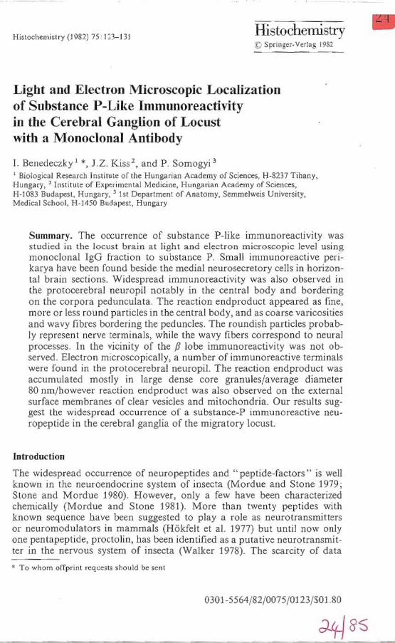
Histochemistry (J982) 75'123-131 Histochemistry !C Spr lll ger-Vcrlag 1982 Light and Electron Microscopic Localization of Substance P-Like Immunoreactivity in the Cerebral Ganglion of Locust with a MOl1oclonal Antibody 1. Benedeczk y I "', J. Z. Kiss 2, and P. Somogyi 3 , Biologicnl Research I ns titu te of the Hungari an Acade my of Sciences, H-8 23 7 Tihany, Hungary, 2 Institute of Experimental Medicine, Hungarian Academy of Sciences, H-IOS) BudapeSl , Hunga ry , 3 1st Department of Anatomy. SemmelW(is University, Medical School, H-1450 Budapest, Hun&ary Summar y. The occurrence of substance P-like immunoreactivity was s tudied in the locust brain a t li ght and electron micr oscopic level us ing monoc !onal T gG fraction lO substance P. Sma ll immunoreactive pe ri - karya ha ve been fou nd beside the medial neurosecretory cells in horizon- tal brain sections. Wides pread immuno reactivity was also observed in the protocerebral neu ropil notably in the central body and bo rdering on the corpora peduncu!ata, Th e reaction end product appeared as fine, more or less round part icles in the central body, and as coarse varicosities and wavy fi bres bordering the peduncles. The roundish particles probab- ly represent nerve terminals, while the wavy fibers correspond to neural processes. T n the vicini ty of the P lobe immunoreactivity was not ob- served. El ectron mic roscopica lly , a num ber of immunoreactive terminals were found in the protocerebral neuropil. The reac ti on endp roducl was accumulated most ly in large dense core granules/average diameter 80 nm/however reacti on endproduct was al so observed on the external surface membranes of clear ves icl es and mitochondria. Our results sug- gest t he widespread occurrence of a substance-P immunoreactive neu- ropeptide in the cerebral ganglia of the migr atory locust. Introd uc ti on The widesprea d occurrence of neuropeptides and" peptide-factors" is well known in the neuroendocrine system of insecta (Mordue and Stone 1979; Stone and Mordue 1980). However, only a few have been charac terized chemically (Mo rdue and Stone 1981). M o re than twe nt y peptides with known sequen ce bave been suggested to play a role as neurot ran smitters or neuromodu la tors in mammals (H6kfelt et al. 1977) but until now only one pentapeptide. proctolin, has been identified as a putative neurotransmit- ter in the nervous system of insecta (Walker 1978). Th e scarcity of data • To whom offp rint shou ld be sent 0301 -5564/ 82/0075/0123/ 501 .80 •
-
Upload
phamnguyet -
Category
Documents
-
view
218 -
download
2
Transcript of Light and Electron Microscopic Localization of Substance...
Histochemistry (J982) 75'123-131 Histochemistry !C Sprlllger-Vcrlag 1982
Light and Electron Microscopic Localization of Substance P-Like Immunoreactivity in the Cerebral Ganglion of Locust with a MOl1oclonal Antibody
1. Benedeczk y I "', J . Z. Kiss 2, and P. Somogyi 3
, Biologicnl Research Ins titu te of the Hungarian Academy of Sciences, H-8237 Tihany, Hungary, 2 Institute of Experimental Medicine, Hungarian Academy of Sciences, H-IOS) BudapeSl , Hungary, 3 1st Department of Anatomy. SemmelW(is University, Medical School, H-1450 Budapest, Hun&ary
Summary. The occurrence of substance P-like immunoreactivi ty was studied in the locust brain a t light and electron microscopic level using monoc!onal TgG fraction lO substance P. Sma ll immunoreactive peri karya have been fou nd beside the medial neurosecretory cells in horizontal brain sections. Widespread immuno reactivity was also observed in the protocerebral neuropil notably in the central body and bordering on the corpora peduncu!ata , The reaction end product appeared as fine, more or less round particles in the centra l body, and as coarse varicosities and wavy fibres bordering the peduncles. T he roundish particles probably represent nerve terminals, while the wavy fibe rs co rrespond to neural processes . T n the vicini ty of the P lobe immunoreactivity was not observed. Elec tron microscopica lly, a num ber of immunoreactive terminals were found in the protocerebral neuropil. T he reacti on endproducl was accumula ted mostly in large dense core granules/ave rage diameter 80 nm/however reaction endproduct was also observed on the external surface membranes of clear vesicles and mitochondria. Our results suggest the widespread occurrence of a substance-P immuno reactive neuropeptide in the cerebral ganglia of the migratory locust.
Introduction
The widespread occurrence of neuropeptides and" peptide-factors" is well known in the neuroendocrine system of insecta (Mordue and Stone 1979; Stone and Mordue 1980). However, only a few have been characterized chemically (Mo rdue and Stone 1981). More than twenty peptides with known sequence bave been suggested to play a role as neurotransmitters or neuromodula tors in mam mals (H6kfelt et al. 1977) but until now only one pentapeptide. proctolin, has been ide ntified as a putative neurotransmitter in the nervous system of insecta (Walker 1978). The sca rcity of data
• To whom offprint reque s t~ should be sent
0301 -5564/82/0075/0123/501 .80
•
". T. Benedecz.ky et al.
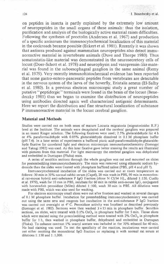
on peptides in insecta is partly explained by the extremely low amount of neuropeptides in the small organs of these animals: thus the isola tion, purification and analysis of the biologically active material raises dLfficuihes. Following the synthesis o f proctolin (Andersen et al. 1967) and production of a specific antiser um the immunocytochemical localization of this material in the cockroach became possible (Eckert et al. 1981 ). Recently it was shown tha t antisera prod uced against mammalian neuropep tides also detect immunoreactive material in invertebra te animals (Duve a nd Thorpe 1980). Thus somatostatin-like materi al was dem onstrated in the neurosecretory cells of locust (Doer-Schou et a!. 1978) and neurophysi n and vasopressin-like material was found in the suboesophageal ganglion in the same animal (Remy et al. 1979). Very recently immunohistochemical evidence has been reported that some gastro-entero-pancreatic peptides from vertebrates are detecta ble in the nervous system of the larva of the hoverfly, Eristalis aeneus (EI-Salhy et a!. 1980). Tn a previous electron microscopic study a great number of puta tive" peptidergic" terminals were found in the brain of the locust (Benedeczky 1981) thus we began to examine the d istribution of neuropeptides using a ntibod ies directed again well characterized anti genic determinants. Here we report the distribution and fi ne structural localization of substance p immunoreactive mate~al in the locust cerebral ganglion.
Material and Methods
Studies were carried out on both sexes of maLUrt Locusta migratoda (migratorioides R.F .) bred at the Institute. The animals were decapitated and the cerebllll ganglion was prepared in an insect Ringer solution. The following fixatives were used; 2.5% glutaraldehyde for 4 h or 4% paraforma:dehyde with 0.050/. glutaraldehyde dissolved in 0.1 M phosphate buffer (pH 7 4). In a later series of experiments a buffered picric acid 'paraformaldehyde-glutaraldehyde fixative for correlated light and electron microscopic immullohystochemistry (Somogy; and Takagi1 982) was used. As this later fixative gave better staining the results are illustrated with pictures from this material. For light microscopy the cerebra l ganglion was dehydrated and embedded In Durcupan (Fluka) resin.
A series of semitbin se<:tions through the whole ganglion was cut and mounted on slides for postembedding immunocytochemistry. The resin was removed using ethanol ic sodium hydroxide Ihen the slides were treated with phosphate buffered saline (PBS. pH 4 and pH 7).
Immunocytochemical incubation of the slides was carried out at room temperature as follows : 30 min in 20% normal rabbit serum (Capel), 20 min wath in pes, 90 min in monoclon_ al ral·mouse: hybrid anli ·substance P IgG fracti on (clone N CI/34 HL. diluted 1: 150. Cuello el aJ. 1979). wash fllr 10 min in PBS. incubale for 60 min in rabbit anti·mouse IgO, conjugated with horserad1sh peroxidase (Miles) diluted 1: lOO, wash 30 min in PBS. All ditutions were made with PBS, which was also used for washing.
For electron microscopy small sl ices were cut arter fixa tion and washed in several changes of 0.1 M phosphate buffer. Incubation for preembedding immullocytochemiwy was carried out usmg the same sera snd reagents bUI incubation in the anti·substance P IgG fract ion was carried out o~ernighl at 4° C. Peroxidase activity was localised as described previously (Somogyi et al. 1982). Sections were then washed 3 x 15 min in phosphate buffer. Semithin sections, on slides, were treated with 0.1% 050. in phosphate buffer for 5 min. Tissue slices which were stained using the precmbedding method were treated with 20/. 050. in phosphate buffer for I h. then washcd in phosphate buffer, dehy<lrated and embedded in Durcupan (Fluka) . To enhanee contrast 1"10 uranyl acelate was included in the 70% ethanol for 40 min. No lead itaining was used To test the specificity of the reaction, incubalio ns were carried out either omiu ing the monoclonal IgO fraction or replacing it wi th normal ra t serum at dihllions 1 : J 50 and 1 : 1 ,000.

Fig. I a. HOrIZontally sectioned cerebral gan&lion of the migratory locust. Substance P immunoreactive eells ( .... ) can be seen in the vicinity of media] neurosecretory !;ells (MNC). x 200. b High magnification light mia-ograph of a subslan!;e P neuron containing fine granulated immunoreactive material. x ],000. C Signet-ring shape immunoreactive cell rrom the same area of the locust brain. )( 800

126 J. Bene<leezky et a1.
t"ig. 2. Horizontal sectiou of the cerebral gangl ion at the level of the centra l body (CB). Abundant immunoreactive material is found In the central body as well as around the corpora pedunculata (Pl. 0, ocellus. Insert: grains of reaction endproduct in the central body. x 400
Results
The monodonal IgG fraction used in this ~Ludy has been shown to recognise the C terminal portion of substance P (Cuello et aL 1979). As the same antigenic sile may be present in several :nolecules we refer to the specific staini ng observed as substance P immunoreactivity. No endogenous peroxidase activity was observed in the controls.
Substance P Immunoreactivity was nol detectable in the nerve cells of the surrace of the horizon tally sectioned brain. The substance P immunoreactive cells occur mainly in the pars intercerebralis. alongside the large neurosecretory cells (Fig. 1 a). These relative small cells have oval or pearshaped form, their diameter ranging between 15-20 j.lm. At high magnification (Figs. 1 b and c) a fine granulated reaction product was observed in the perikarya. The most abundant reaction product was accumulated in the protocerebral neuropil at the level of the cen tral body (Fig. 2). A fioe granular evenly distributed immunoreactivity was observed in the whole cross5ection of the cemral body (Fig. 2, and Fig. 2 insert). Strong immuno-

Fig. )1. Coarse va ricosities and fibres can be seen in the neuropi l Irea between the centra l body (CB) IDd corpora peduDCulata (P). )I 500. b Ul tranructural localization or substa~ P immunoreactivity in the protoccfebral neuropil or the locust. Densely labelled vesieles ( -) are present in the axon terminal (A). x 40.000

'" I. Btnedeezky et al.
Fig .... Dia,mm of the horizontally se<:uoned (crebral ganglion of the locust. Dense grains and lines represent the substl'lnce P-!ike immunoreactivity in the plane of central body (CB). P, peduncles ; P. beta lobe. Groups of roundish black cells at the top and bonom of the diagram are nOI Immunon:.cti ve
reactivity was also fo und in the neu ropil lying between the cen tral body and corpo ra pedunculata (Fig. 2). Coarse varicosities and fibers could be observed in this area (Fig. 3a) and occasionally long characteristic wavy segments of Immunoreactive axons were also conspicuous (Fig. 3a). Knoblike immunoreactive particles (probably bo utons) were frequently observed in this area (Fig. 3 a). The peduncles themselves rarely contained immuno reactive elements. The orientation of the immunoreactive axons is mainly posterio-anterior, and some axons reach the cor tex of the frontal ocellus (Figs. 2 and 4). T he immunoreactivi ty usually disappears in the depth of the P lobe and only a small amount o f reaction product was detectable in this area of the protocerebral neuropile. At the electron microscopic level electron dense reaction product was observable in nerve terminals of the protoce rebra l neuropil (Fig. 3 b). The round granulated vesicles (80-100 nm in diameter) contained reaction endproduct over their core. T he clear vesicles had electron dense reactio n product associated wi th the externai surface of their membrane. Other organelles (as for example mitochondria) in the terminals were also la belled with a small amount of reacti on product (Fig. 3 b) on thei r external membrane surfaces.
Discllssion
Substance P is widely dIstributed in the central nervo us system of vertebrates (Leeman and Mroz 1974; Takaha shi and Otsuka 1975) and it may be a neurotransmitter in primary sensory systems (Stern 1963; Krnjevic 1974 ;

Sub$IAno: P·M:e lmmunocadivilY in the Brain '" Leeman and MrOl 1974; Talc.ahashi and Otsuka 1975). Little IS known about the ocx:urrence of substance P in invertebrates. Substance P-Jike mate· rlal was demonstrated in the brain of the hoverfly larva (El Salhy et al. 1980). In addition substance P like Immunoreactivity was found lfI the gut of the snail by Van Noorden et al. (1980). Companng our study with pre· ~Iousdata (El Salhy et al. 1980) the main difference is Ihat we could demon· strate substance P·like immunoreactivity in the protooerebral neuropil whIle El Salhy et at. (1980) detected it only in the somata of a few nerve cells. This diffuence is all the more surprisina since in both expenments u!;l':d a similar Immunocytochemical method (unlabelled an tlbody·enzyme and only the anlisera, fixation procedure aDd embedding media were ditIeren!. Using only glutaraldehyde fixation we observed a relatively weak immunore· actiVIty. Introducing the picric·acid containing fixati~e (Somogyi and Takagi 1982) the immunoreactivi ty was observed in the same regions in the brain but the reaction producl was more abundant and ils density, was higher. As regards Ihe occurrence of immunoreactive nerve cells, we could find substance P·like immunoreactivity only in a few somata, result in agreement with those of El Salhy et al . (1980).
There may be more substance P C(lntaining nerve cells in t~ locust brain , but the ooncentrallon of the neurotransmitter may no t be high enough for the immunocytochemical detectIon. It is also pOSSIble, that substance P is present in a masked form (bound 10 other molecules) in the perikarya thus the detection wi th an antibody recognising the C terminal part is no t possible. The widespread occurrence of the substance P· like immunoreactivi· ty in the locust brain, as for e;<ample in the central body, suggests that It may play a role as a mediator or modUlator. However, in contrast to mammals, where atleaSI two substances have been found to show substance P activity (Ben Ari et al. 1979) nothing is known abou t the substance respon· sible for the immunoreaclion ill invertebrates. Thus the fUllction of a sub· &lance P immunoreactive system remains uncertain. The centra l body, whIch IS the main neuropIl area where substance P immunOre8Cllvl1y was found is well known as a higher motor center (BullO(:k and Horridge 1965) On the other hand the close morphological contact of substance P·like immuno· reactive fibers with the ocellus suggests that subslllncc P, as in the case in mammals, may occur also in sensory syslems. [n light nllcTOgraphs It
is dear, that the appearance of substance P·like Immunoreactivi ty IS usua lly finely granulate<! and bulbous varicosilies are present along the fibe.., . It is likely that these structures repIeKDI Derve terminals iD the central body and also in other l)lrts of the protocerebral neuropil Electron microscopic examinations have confirmed Ihis assumption since t~ ele(:tron dense reaction product was observed in nerve terminals. In these terminals, as in mammals (Pickel et a!. 1977, 1979; Somogyi et al. 1982), the electron dense reaction produet was localised mainly in granules having a d,ameter of 60-80 nm. Reaction product was also observed in the axoplasm and on the surface ofmilocbondria and clear vesIcles. SiDce large granulated vesicles (average diameter 80 nm) arc very common in the locust brain they may be responsible for the storage or ehemical mediators (BenedecUy 1981)
130 J. Benedcczky et al.
and we suppose, that substance P immunoreactive material is also stored in these granules. The immunoreactive terminals a lso contained small clear vesicles conta ining no reaction end product. This raises the possibility tha t the immunoreactive material occurs together with some other substance(s) in the terminals.
Acknowledgement . The authors arc grateful 10 Dr. A.C. Cuello for [he gi ft of monoclonal anll-substance P antibody.
References
Andenon OW, Zimmerman lE, Callahan FM (1961) A reinvcSligaLion of the mi~ed carbonic anhydride method of peptide synthesiS. J Am Chem Soc 89: 5012-S017
Ben-An V, Pradelles P, G ros C, Dray F (1979) IdentifICation of authentic substance P in Slrialoni,ra! and amyedaloid nuclei using combined high performance liquid chromatogra. phy and radiOlmmunoasuy Brain Res 173:360-363
Scnedec:2.ky I (198]) The ultras tructure and cytochemiStry of aminergic and peptidcrgie termin· als in Locusta migratoria migratorioides R Fin : R6:zsa KS (cd) Adv physiol sa, vol 22 . Perpmon Press, O~ford, Akademiai Kiild6, Budapest, pp 509-523
Bullock TH, Horridge GA (1965) Structure and functi on in the nervous system of invertebrates. WH Freeman, San Francisco and London, pp 1138-1139
Cuello AC, Galrre G, Milslein C (1979) Detection of substance P in the central nervous system by a monoclonal antibody. Pro<: Nail Acad Se; USA 76 : 3532-3536
Doerr·Sehott J, Joly L, Dobeis MP (1978) Sur l'u;islence dans \a pars intercerebralis d'un inSCCIC (Locus/a migraloria R Cl F) de ccllulcs neurostcretriccs liunl un antiserum an ti · somatostalinc. eR Acad Sei Pans 286:93-95
Duve H, Thorpc: A (1980) Localisation of pancreatic polypeptide (PP)-hke immunoreactive material in neurones of the brain of the blowny, Ca lliphora erythrocephala (Diplera). Cell Tissue Res 210:101-109
Eckert M. Agricola H, Penzlin H (1981) Immunocytochemical identification of proctohnhke immunoreactivity in the terminal ganglion and hmdgut of the cockroach Periplaneta ameri· cana (L). Cell Tissue Res 217 ' 633-645
EI·Salhy M, Abeu·EI·Ela R, Falkmer S, Grimehus L, Wilander E (1980) Immunohistochemical evidence of gastro-cntero·panereatic neurohormonal peptides of ver tebrate type in the ner· vous system of the larva of a dipteran insect, the hoverfly, Efulalls uentus. Regulatory Peptides J : 187-204
H6kfelt T, lohansson 0 , Kellerth JO, LjungdahJ A, Nilsson G, Nygards A. Pernow B (1977) Immunohistochemical dlstribU1;on of substance: P. In: EuJer US von, Pemow B (eds) Sub$Iance P (Nobel Symposium 37) Raven Press, New York, pp 117- 145
Krnjevic K ( 1 97~) Chemical nature of synaptic transmission in vertebrales. Physiol Rev $4 418-$40
Leeman SE, Mro:.e EA ( 1974) Substance: P. LIfe Sci 15 : 2033-2044 Mordue W, StOne JV (1979) Insect hormones. In Barrington EJW (cd) Hormones and evolu
tion, Vol I : Chap 5. AcademiC Press, London Mordue W, Stone JV (1981) Structure and function of Insect peptide hormones. Insect Biochem
11 ]5]-360 Pickel VM . Reis DJ, Leeman SE (1977) Ultra5tfl.IClurallocalization of substlnce P in neurons
of rat spinal cord. Brain Res 122: 534-$40 Pickd VM , Joh TH, Reis DJ, Lec:man SE, Miller RJ (1979) Electron mlCroseopie localization
of substancc P and enkephalin in axon terminals related to dendrites of eatecholaminergic neurons. Brain Res 160: 387-400
Remy Ch, Girardie J, Dubois M (1979) Vertebrate neuropepllde.bke substance In the suboc:so· phageal ganglion of two insects: Locusta migtlloria RF (Orthoplera) and Bomby:c mor; L. (Lepidoplti!ro). immuncytological investigation . Gcn Comp EndocrinoI37 :93-JOO

,
Substance P-lilce Immunoreactivity in the Brain III
SomogYI P, Prist!ey JV, Cuel!o AC, Smith AD, Bolam JP (1982) Synaptic conne<:lions of substance P immunoreactive nerve terminals in the substance nigra of the nil: a correlated liaht and eJeclron mIcroscopic study. Cell Tissue Res 223:469-486
Somogyi P, T.kagi H (1982) A nOle on the use of picric acid-paraformaldehyde-glutaraldehyde fixative for correlated light a nd ele<;tron microscopic immunocytochemistry Neuroscience in press
Stern P (1963) Substance P as a sensory transmitter and its other central effects. Ann NY Acad Se; 104: 403-414
Stone JV , Mordue W (1980) Isolation of insect neuropcptides. Insect Bioc:hem 10:229-2]9 Takahashi R, Otsuka M (1975) Regional dIstribution of substance P in the spinal cord and
nerve roots of the Cllt IInd the effect of dorsal root section. Brain Res 87 : 1-11 Van Noordcn S, Fritsch HAR, Ori110 TA l, Polak JM, Pearse AGE (1980) Immunocytochemical
slIinina for vertebrate peptides in the nervous S)'$tem of a gastropod moltuse. Gcn Comp Endocr 40 :375-376
Walker RJ (1978) Polypeptides as central transmitters. Gen Pharmacol 9 : 129-138
Received May 3, 1982