Histology, Histochemistry and Scanning Electron Microscopy...
6
Veterinary Research International | January-March, 2015 | Vol 3 | Issue 1 | Pages 01-06 © 2015 Jakraya Publications (P) Ltd VETERINARY RESEARCH INTERNATIONAL Journal homepage: www.jakraya.com/journal/vri ORIGINAL ARTICLE Histology, Histochemistry and Scanning Electron Microscopy of Tubal Tonsil of the Young Pigs Ranjit, Pawan Kumar*, Tej Parkash, Pawan Kumar, Ruchita Pal and Gurdial Singh Department of Veterinary Anatomy, College of Veterinary Sciences, Lala Lajpat Rai University of Veterinary and Animal Sciences, Hisar-125 004 (Haryana), India. *Corresponding Author: Pawan Kumar Email: [email protected] Received: 27/12/2014 Revised: 28/02/2015 Accepted: 03/03/2015 Abstract The tubal tonsil of the pig was lined by pseudostratified columnar ciliated epithelium with goblet cells which showed modification into follicle associated epithelium being characterized by lack of cilia, goblet cells, reduced height of epithelium and heavy infiltration of lymphoid tissue. Some surface cells showed protrusions which were comparable to M-cells. In addition, small patches of simple and stratified cuboidal epithelium were also observed. Propria submucosa was comprised of loose irregular connective tissue especially lymphoid and glandular tissue. The lymphoid tissue had small, medium, and large sized lymphocytes, plasma cell and macrophages. The interfollicular areas were having more number of blood capillaries and few high endothelial venules. A dense layer of elastic fibres was also present which separated the lymphoid follicles and tissue from glandular tissue. The goblet cells and mucous glandular acini showed a strong positive reaction for different moieties of carbohydrates. The scanning electron microscopy revealed surface of tubal tonsil a dense mat of cilia with bulbous free endings which masked the presence of other types of cells. Different types of microvillus cells, bursh cells, M-cells and few goblet cells were observed at places where density of cilia was less. Key words: Follicle associated epithelium, M cells, Young Pigs, Tubal tonsil, High Endothelial Venules. 1. Introduction Tonsils, major components of the pharyngeal mucosa-associated lymphoid tissue, represented first line of defense against ingested and inhaled foreign antigens (Gebert et al., 1996; Caramelli et al., 2003). The ‘‘Waldeyer ring’’ consists of nasopharyngeal tonsil and the tubal tonsil were located in the nasopharynx region, the lingual tonsil and tonsils of the soft palate were located in the oropharynx region, and the paraepiglottic tonsil was located in the laryngopharynx region (Cocquyt et al., 2005; Casteleyn et al., 2007; 2008). Respiratory tract associated lymphoid tissue was considered an integral part of the local immune system because of its probable involvement in uptake, transport and presentation of antigen to cells of the immune system (McDermott et al., 1982; Gebert and Pabst, 1999). Morphologically, lymphoid tissue of the pharyngeal tonsil shared characteristics of gut associated lymphoid tissue (GALT), bronchus associated lymphoid tissue (BALT) and other mucosa associated lymphoid tissue (MALT) in that it contained follicle-associated epithelium (FAE) and secondary lymphoid follicles but no afferent lymphatics (Korsrud and Brandtzaeg, 1981; Mair et al., 1987; Chen et al., 1989). The FAE consisted of a mesh of epithelial cells of altered shape and contained mobile infiltrating lymphocytes, macrophages, dendritic cells, and a network of capillaries (Perry and Whyte, 1998). Some of its epithelial cells with shortened microvilli were known as M cells which were specialized cells involved in active transport of soluble and particulate matter across the epithelium (Gebert et al., 1996) by fluid phase or receptor mediated endocytosis at the apical membrane (Neutra et al., 1996). The cells of immune system and tonsillar epithelial cells recognize pathogens via toll like receptors (TLRs), a group of pattern recognition receptors (PRRs), which were capable of recognizing conserved microbial structures and thus able to discriminate between self and non-self- antigens (Lange et al., 2009). 2. Material and Methods
Transcript of Histology, Histochemistry and Scanning Electron Microscopy...

Veterinary Research International | January-March, 2015 | Vol 3 | Issue 1 | Pages 01-06 © 2015 Jakraya Publications (P) Ltd
VETERINARY RESEARCH INTERNATIONAL Journal homepage: www.jakraya.com/journal/vri
ORIGINAL ARTICLE
Histology, Histochemistry and Scanning Electron Microscopy of Tubal Tonsil of the Young Pigs Ranjit, Pawan Kumar*, Tej Parkash, Pawan Kumar, Ruchita Pal and Gurdial Singh Department of Veterinary Anatomy, College of Veterinary Sciences, Lala Lajpat Rai University of Veterinary and Animal Sciences, Hisar-125 004 (Haryana), India. *Corresponding Author: Pawan Kumar Email: [email protected] Received: 27/12/2014 Revised: 28/02/2015 Accepted: 03/03/2015
Abstract The tubal tonsil of the pig was lined by pseudostratified columnar
ciliated epithelium with goblet cells which showed modification into follicle associated epithelium being characterized by lack of cilia, goblet cells, reduced height of epithelium and heavy infiltration of lymphoid tissue. Some surface cells showed protrusions which were comparable to M-cells. In addition, small patches of simple and stratified cuboidal epithelium were also observed. Propria submucosa was comprised of loose irregular connective tissue especially lymphoid and glandular tissue. The lymphoid tissue had small, medium, and large sized lymphocytes, plasma cell and macrophages. The interfollicular areas were having more number of blood capillaries and few high endothelial venules. A dense layer of elastic fibres was also present which separated the lymphoid follicles and tissue from glandular tissue. The goblet cells and mucous glandular acini showed a strong positive reaction for different moieties of carbohydrates. The scanning electron microscopy revealed surface of tubal tonsil a dense mat of cilia with bulbous free endings which masked the presence of other types of cells. Different types of microvillus cells, bursh cells, M-cells and few goblet cells were observed at places where density of cilia was less. Key words: Follicle associated epithelium, M cells, Young Pigs, Tubal tonsil, High Endothelial Venules.
1. Introduction Tonsils, major components of the pharyngeal
mucosa-associated lymphoid tissue, represented first line of defense against ingested and inhaled foreign antigens (Gebert et al., 1996; Caramelli et al., 2003). The ‘‘Waldeyer ring’’ consists of nasopharyngeal tonsil and the tubal tonsil were located in the nasopharynx region, the lingual tonsil and tonsils of the soft palate were located in the oropharynx region, and the paraepiglottic tonsil was located in the laryngopharynx region (Cocquyt et al., 2005; Casteleyn et al., 2007; 2008). Respiratory tract associated lymphoid tissue was considered an integral part of the local immune system because of its probable involvement in uptake, transport and presentation of antigen to cells of the immune system (McDermott et al., 1982; Gebert and Pabst, 1999). Morphologically, lymphoid tissue of the pharyngeal tonsil shared characteristics of gut associated lymphoid tissue (GALT), bronchus associated lymphoid tissue (BALT) and other mucosa associated lymphoid tissue (MALT)
in that it contained follicle-associated epithelium (FAE) and secondary lymphoid follicles but no afferent lymphatics (Korsrud and Brandtzaeg, 1981; Mair et al., 1987; Chen et al., 1989). The FAE consisted of a mesh of epithelial cells of altered shape and contained mobile infiltrating lymphocytes, macrophages, dendritic cells, and a network of capillaries (Perry and Whyte, 1998). Some of its epithelial cells with shortened microvilli were known as M cells which were specialized cells involved in active transport of soluble and particulate matter across the epithelium (Gebert et al., 1996) by fluid phase or receptor mediated endocytosis at the apical membrane (Neutra et al., 1996). The cells of immune system and tonsillar epithelial cells recognize pathogens via toll like receptors (TLRs), a group of pattern recognition receptors (PRRs), which were capable of recognizing conserved microbial structures and thus able to discriminate between self and non-self-antigens (Lange et al., 2009). 2. Material and Methods

Ranjit et al…Histology, Histochemistry and Scanning Electron Microscopy of Tubal Tonsil of the Young Pigs
Veterinary Research International | January-March, 2015 | Vol 3 | Issue 1 | Pages 01-06 © 2015 Jakraya Publications (P) Ltd
2
2.1 Light Microscopy The present study was conducted on 10 young
male pigs of 8-10 months age, of local or mixed breed. The heads were procured from local slaughter house immediately after decapitation. The mid sagittal sections of heads were made to collect tissues. The tissues for histomorphological and histochemical studies were collected from 5 heads and fixed in 10 per cent neutral buffered formalin solution for 48 hours. The fixed tissues were processed for routine paraffin technique for light microscopy. The paraffin sections of 5-6 µ were cut and stained with routine Harris’ hematoxylin and eosin stain, Gomori’s method for reticulum, Weigert’s method for elastic fibres, Crossman’s trichrome stain for collagen fibres (Crossman, 1937), McManus’ method for glycogen (PAS), Alcian blue method for muco-substances (pH 2.5), PAS-Alcian blue method for acidic and neutral mucosubstances (pH 2.5), Meyer’s mucicarmine method for mucin and colloidal iron method for acid mucopolysaccharides (Luna, 1968). 2.2 Scanning Electron Microscopy
Fresh tissues from 5 pig heads were collected for scanning electron microscopy. The tissues were fixed in 2 per cent glutaraldehyde solution for 6 hours after thorough washing in chilled 0.1 M phosphate buffer (pH 7.4). The tissues were again washed twice with 0.1 M phosphate buffer and rest of the procedure was carried out at EM-Laboratory, AIRF, JNU, New Delhi. The processed tissues were viewed in scanning electron microscope (Zeiss EVO-40). Energy-dispersive X-ray spectroscopy (EDX) was carried out at 15 Kv using Bruker X- flash detector 3010 (Germany) for elemental composition and quantificational analysis.
3. Results and Discussion
The surface of tubal tonsil lined by pseudostratified columnar ciliated epithelium with goblet cells (Figs 1-12) showed folds and invaginations which increased the surface area as reported in horse (Mair et al., 1987; Kumar and Timoney, 2005) and sheep (Kumar and Kumar, 2012) and provided better opportunities for entrapment and attachment of foreign particles including the pathogens. The epithelium was composed of basal cells, supporting/ sustentacular cells and goblet cells. The oval shaped nuclei of basal cells were vertically arranged as reported in sheep (Kumar and Kumar, 2012) and horse (Kumar and Timoney, 2005). Cytoplasm of all the cell types was finely granular and eosinophilic. The epithelium showed infiltration of isolated lymphocytes. The number of goblet cells was reduced in the epithelium when the
latter was associated with underlying lymphoid follicle. A similar type of epithelium was called lymphoepithelium in the horse (Kumar and Timoney, 2005) and sheep (Kumar and Kumar, 2012) because of association of large amount of lymphoid tissue. At places, respiratory epithelium was modified into follicle associated epithelium (FAE) characterized by absence of goblet cells, reduced number of cell layers and a large amount of infiltration of lymphoid cells due to interrupted basement membrane (Figs 1,2,7, 9,12). The cells of FAE lacked cilia and contained microvillous cells. Some of these cells with very small sized microvilli and close association with lymphocytes were identified as M cells as reported in different species (Kumar and Timoney, 2005; Kumar and Kumar, 2012). In some places an excessive lymphoid infiltration into the FAE obscured the occurrence of epithelial cells. Lack of goblet cells and mucus may facilitate direct contact of microorganisms with the FAE including M cells. The outermost layer was comprised of microvillus (MV) cells some of which had depressed surfaces with shortened microvilli, round to oval nuclei and a close association with lymphocytes (Fig 2). These were designated as M cells as reported in the horse (Kumar and Timoney, 2005) and sheep (Kumar and Kumar, 2012). The FAE was involved in transcytosis of antigens, transportation of immunocytes, and mucosal protection (Brandtzaeg and Halstensen, 1992). A few MV cells of this region were also associated with intra-epithelial lymphocytes in the absence of underlying lymphoid tissue as reported in the horse (Kumar and Timoney, 2005) and sheep (Kumar and Kumar, 2012) similar to intestinal-villous M-cells which were not associated with Peyer’s patches (Jang et al., 2004). Isolated patches of stratified squamous non-keratinized epithelium were interspersed in between the respiratory epithelium as reported in sheep (Kumar and Kumar, 2012). Lymphoid cells and loose irregular connective tissue formed by sparsely distributed reticular and collagen fibres were scattered throughout the propria submucosa (Figs 4-6) as reported in horse (Kumar and Timoney, 2005).
Lymphoid follicles were constituted by small, medium and large sized lymphocytes, macrophages and plasma cells as reported in horse (Kumar and Timoney, 2005 and sheep (Kumar and Kumar, 2012). High endothelial venules (Fig 13) were located towards the parafollicular area. The epithelium of endothelial venules was low cuboidal to high cuboidal and their strongly basophilic nuclei protruded into the lumen.
The goblet cells, glandular acini and their ducts were strongly PAS positive by McManus’method which indicated the presence of glycogen (Figs 7-12) as reported in horse (Kumar and Timoney, 2005) and in sheep (Kumar and Kumar, 2012). The positivity was
Ranjit et al…Histology, Histochemistry and Scanning Electron Microscopy of Tubal Tonsil of the Young Pigs
Veterinary Research International | January-March, 2015 | Vol 3 | Issue 1 | Pages 01-06 © 2015 Jakraya Publications (P) Ltd
3
Fig 1 Fig 2 Fig 3
Fig 4 Fig 5 Fig 6
Fig 7 Fig 8 Fig 9
Fig 10 Fig 11 Fig 12

Ranjit et al…Histology, Histochemistry and Scanning Electron Microscopy of Tubal Tonsil of the
Veterinary Research International | January© 2015 Jakraya Publications (P) Ltd
Fig 13
Fig 14
Fig 16Figs 14-17: Scanning electron micrograph showing; 14. A dense arrangement of ciliated cells in the form of a mat masking the appearance of other cells. x 1510. 15. Presence of ciliated cells, different type of microvillus cells, and cells having microvilli and cilia. x 5950. 16. Higher magnification showing presence of ciliated cells, different type of microvillus cells, and cells having both microvilli and cilia. x 7330. 17. Distribution of small size microvilli of microvillus cells at higher magnification. x 3290.
reduced after diastase treatment indicating the presence of other mucopolysaccharides in addition to glycogen. These cells also showed strong positive reaction for PAS-Alcian blue method with a predominance of neutral mucopolysaccharides than the mucopolysaccharides (Fig 8). These cells also showed strong Alcianophilic positive reaction indicating the presence of weakly sulphated mucopolysaccharides,
Ranjit et al…Histology, Histochemistry and Scanning Electron Microscopy of Tubal Tonsil of the
January-March, 2015 | Vol 3 | Issue 1 | Pages 01-06 15 Jakraya Publications (P) Ltd
4
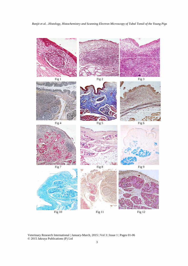
Figs 1-13. Photomicrograph of tubal tonsil showing 1. Pseudostratified columnarepithelium with goblet cells and scattered lymphoid tissue in the superficial propria submucosa. H & E. × 200. 2. A small modified patch of respiratory epithelium showing microvillus cells with absence of goblet and ciliated cells and the underlying lymphoid tissue. H. & E. x 200. 3. Showing presence of lymphoid follicles and interfollicular areas. PAS. x 100. 4. Presence of reticular fibres at the base of the respiratory epithelium, surrounding the lymphoid follicles and interfollicular areas. Gomori’s method x 100. 5. Distribution of collagen fibres in the propria submucosa. Crossman’s trichrome x 100. 6. Presence of dense layer of elastic fibres in deeper part of propria submucosa. Note presence of isolated elastic fibres in subepithelial portion. Weigert’s method x 100. 7. Presence of glycogen in isolated patches of respiratory epithelium glandular acini aabsence of reaction in FAE (arrow). McManus’ PAS method x 100. 8. Presence of acidic and neutral mucopolysaccharides in the goblet cells of the respiratory epithelium and glandular acini. PAS AB x 100. 9. Presence and absence of Alcianophiliin respiratory and FAE, respectively. Note a strong positive reaction in mucus glandular acini. Alcian blue x 100. 10. Absence and presence of acidic mucopolysaccharides in surface epithelium and glandular acini, respectively. Colloidal iron method x 100. 11. Presence of mucin in goblet cells of epithelium and glandular acini. Note positive reaction in glandular duct opening towards surface of the epithelium. Mayer’s mucicarmine method x 100. 12. Absence of mucin in modified simple cuboidal epithelium of FAE. Note a strong positive reaction in mucus glandular acini. Mayer’s mucicarmine method x 100. 13. Presence of high endothelial venules in the lymphoid tissue. H. & E. x 400.
Fig 14 Fig 15
Fig 16 Fig 17
: Scanning electron micrograph showing; 14. A dense arrangement of ciliated cells in the form of a mat masking the appearance of other cells. x 1510. 15. Presence of ciliated cells, different type of microvillus cells, and cells having
and cilia. x 5950. 16. Higher magnification showing presence of ciliated cells, different type of microvillus cells, and cells having both microvilli and cilia. x 7330. 17. Distribution of small size microvilli of microvillus cells at higher
reduced after diastase treatment indicating the presence of other mucopolysaccharides in addition to glycogen. These cells also showed strong positive reaction for
Alcian blue method with a predominance of saccharides than the acidic
8). These cells also showed strong Alcianophilic positive reaction indicating the presence of weakly sulphated mucopolysaccharides,
sialomuccins, and hyaluronic acid (secretions of mucus acini were also strongly positive by colloidal iron method and Mayer’s mucicarmine methods indicating presence of acidic mucopolysaccharides and mucin, respectively (12). The glandular ducts showed varying affinity formucopolysaccharides. The interglaintraglandular ducts showed few PAS positive cells as
Ranjit et al…Histology, Histochemistry and Scanning Electron Microscopy of Tubal Tonsil of the Young Pigs
. Photomicrograph of tubal tonsil showing 1. Pseudostratified columnar-ciliated epithelium with goblet cells and scattered lymphoid tissue in the superficial propria
200. 2. A small modified patch of respiratory epithelium showing microvillus cells with absence of goblet and ciliated cells and the underlying lymphoid tissue. H. & E. x 200. 3. Showing presence of lymphoid follicles and interfollicular areas.
4. Presence of reticular fibres at the base of the respiratory epithelium, surrounding the lymphoid follicles and interfollicular areas. Gomori’s method x 100. 5. Distribution of collagen fibres in the propria submucosa. Crossman’s trichrome x 100. 6.
esence of dense layer of elastic fibres in deeper part of propria submucosa. Note presence of isolated elastic fibres in subepithelial portion. Weigert’s method x 100. 7. Presence of glycogen in isolated patches of respiratory epithelium glandular acini and ducts. Note absence of reaction in FAE (arrow). McManus’ PAS method x 100. 8. Presence of acidic and neutral mucopolysaccharides in the goblet cells of the respiratory epithelium and glandular acini. PAS AB x 100. 9. Presence and absence of Alcianophilic positive reaction in respiratory and FAE, respectively. Note a strong positive reaction in mucus glandular acini. Alcian blue x 100. 10. Absence and presence of acidic mucopolysaccharides in
iron method x 100. 11. Presence of mucin in goblet cells of epithelium and glandular acini. Note positive reaction in glandular duct opening towards surface of the epithelium. Mayer’s mucicarmine method
dal epithelium of FAE. Note a strong positive reaction in mucus glandular acini. Mayer’s mucicarmine method x 100. 13. Presence of high endothelial venules in the lymphoid tissue. H. & E. x 400.
: Scanning electron micrograph showing; 14. A dense arrangement of ciliated cells in the form of a mat masking the appearance of other cells. x 1510. 15. Presence of ciliated cells, different type of microvillus cells, and cells having both
and cilia. x 5950. 16. Higher magnification showing presence of ciliated cells, different type of microvillus cells, and cells having both microvilli and cilia. x 7330. 17. Distribution of small size microvilli of microvillus cells at higher
sialomuccins, and hyaluronic acid (Fig 9). The i were also strongly positive
by colloidal iron method and Mayer’s mucicarmine methods indicating presence of acidic mucopolysaccharides and mucin, respectively (Figs 10-
). The glandular ducts showed varying affinity for mucopolysaccharides. The interglandular and intraglandular ducts showed few PAS positive cells as

Ranjit et al…Histology, Histochemistry and Scanning Electron Microscopy of Tubal Tonsil of the Young Pigs
Veterinary Research International | January-March, 2015 | Vol 3 | Issue 1 | Pages 01-06 © 2015 Jakraya Publications (P) Ltd
5
Fig 18: EDS spectrum of tubal tonsil
demonstrated by McManus’ method and Mayer’s mucicarmine. The cells of the glandular ducts showed negative reaction for Alcian blue. However, secretions present in the lumen of these ducts had a strong Alcianophilic positive reaction. The regions of FAE towards the surface epithelum, crypts and the modified cuboidal type FAE did not exhibit positive reaction by any of the method employed for the demonstration of mucopolysaccharides during present study. The proteins could not be demonstrated by bromphenol blue method in epithelial cells or glandular acini. However, a very weak reaction was observed in the blood capillaries.
Scanning electron microscopy of the tubal tonsil revealed a dense mat of ciliated cells (Fig 14) with interspersed microvillus and goblet cells. Majority of the ciliated cells possessed tufts of uniform sized cilia with bulbous endings and these masked the appearance of other cells as reported in horse (Kumar and Timoney, 2005) and sheep (Casteleyn et al., 2010; Kumar and Kumar, 2012). The microvillus cells of different types along with ciliated and brush cells were visible in areas where density of cilia was less (Figs 15-16). The region of FAE possessed microvillus cells which were categorized into different types depending on the size of microvilli (Fig 17) as reported earlier in the horse (Kumar and Timoney, 2005) and in sheep (Casteleyn et al., 2010; Kumar and Kumar, 2012). Type I cells possessed microvilli of uniform size and shape with distinct outlines Type II cells had larger microvilli. Occasional cells with very short microvilli
or depressed surfaces were identified as M cells as reported earlier in the horse (Kumar and Timoney, 2005) and sheep (Kumar and Kumar, 2012). EDX presented different elements in the tubal tonsil (Table 1; Graph 1). Table 1: EDS elemental analysis of tubal tonsil of pig Element Series unn. C norm. C Atom. C Error [wt.-%] [wt.-%] [at.-%] [%]
Carbon K-series 29.36 29.36 35.39 9.0
Sodium K-series 0. 55 0.55 0.34 0.1
Zinc K-series 2.98 2.98 0.66 0.1
Selenium L-series 0.00 0.00 0.00 0.0
Silicon K-series 0.00 0.00 0.00 0.0
Nitrogen K-series 22.32 22.32 23.08 6.9
Oxygen K-series 44.79 44.79 40.53 13.6
Total: 100.00 100.00 100.00
4. Conclusion
The tubal tonsil having pseudostratified columnar ciliated epithelium showed modification into follicle associated epithelium containing a few M cells. The histoarchitecture and cellular components led the tubal tonsil as part of mucosa associated lymphoid tissue and it appeared as to be an inductive site for processing of antigens entering the respiratory system.
2 4 6 8 10 12 14keV
0
2
4
6
8
10
12
14
cps/eV
C O
Na Zn Zn
Se
Se
Si
N

Ranjit et al…Histology, Histochemistry and Scanning Electron Microscopy of Tubal Tonsil of the Young Pigs
Veterinary Research International | January-March, 2015 | Vol 3 | Issue 1 | Pages 01-06 © 2015 Jakraya Publications (P) Ltd
6
References Brandtzaeg P and Halstensen TS (1992). Immunology and
immunopathology of tonsils. Immunology and Immunopathology, 47: 64-75.
Caramelli M, Acutis P, Bozzetta E, Casalone C, Gagna C and Ru G (2003). Bovine spongiform encaphalopathy in Italian herds. Veterinary Record, 153: 711-712.
Casteleyn C, Simoens P and Van den Broeck W (2008). Larynx-associated lymphoid tissue (LALT) in young cattle. Veterinary Immunology and Immunopathology, 124: 394-397.
Casteleyn C, Van Den Broeck W and Simoens P (2007). Histological characteristics and stereological volume assessment of the ovine tonsils. Veterinary Immunology and Immunopathology, 120: 124-135.
Chen W, Alley MR and Manktelow BW (1989). Respiratory tract-associated lymphoid tissue in conventionally raised sheep. Journal of Comparative Pathology, 101: 327-340.
Cocquyt G, Baten T, Simoens P and Van Den Broeck W (2005). Anatomical localization and histology of the ovine tonsils. Veterinary Immunology and Immunopathology, 107: 79-86.
Crossman GA (1937). A modification of Mallory's connective tissue stain with a discussion of principles involved. Anatomical Record, 69: 33-38.
Gebert A, and R Pabst (1999). M cells at locations outside the gut. Semin. Immunology, 11: 165-170.
Gebert A, HJ Rothkotter and R Pabst (1996). M cells in Peyer’s patches of the intestine. International Review of Cytology, 167: 91-159.
Jang MH, Kweon MN, Iwatani K, Ya,mamoto M, Terahara K, Sasakawa C, Suzuki T, Nochi Tyokota Y, Rennert PD, Hiroi T, Tamagawa H, Iijima H, Kunisawa J, Yuki Y and Kiyono H (2004). Intestinal villous M cells: an antigen entry site in the mucosal epithelium.
Proceedings of National Academy of Sciences, 101: 6110-6115.
Korsrud FR, and P Brandtzaeg (1981).Immunohistochemical evaluation of J-chain expression by intra- and extra-follicular immunoglobulin-producing human tonsillar cells. Scandinavian Journal of Immunology, 13: 271-280.
Kumar Pawan and Kumar P (2012). Histology, histochemistry and scanning electron microscopic studies on the tubal tonsil of sheep (Ovis aries). Indian Journal of Animal Sciences, 82: 61-63.
Kumar Pawan and Timoney J F, 2005. Histology, immunohistochemistry and ultrastructure of the equine tubal tonsil. Anatomia Histologia and Embryologia, 34:141-148.
Lange MJ, Lasiter JC and Misfeldt ML (2009). Toll-like receptors in tonsillar epithelial cells. International Journal of Pediatrics andd Otorhinolaryngology, 73: 613-621.
Luna LG (1968). Manual of Histologic Staining Methods of the Armed Forces Institute of Pathology. 3rd edn., McGraw-Hill Book Co., New York.
Mair TS, EH Batten, C R Stokes and FJ Bourne (1987). The histological features of the immune system of the equine respiratory tract. Journal of Comparative Pathology, 97: 575-586.
McDermott MR, Befus AD and Bienenstock J (1982). The structural basis for immunity in the respiratory tract. International Review of Experimental Pathology, 23: 47-112.
Neutra MR, A Frey and J P Kraehenbuhl (1996). Epithelial M cells: gateways for mucosal infection and immunization. Cell, 86: 345-348.
Perry M and Whyte A (1998). Immunology of the tonsils. Immunology Today, 19: 414-421.