Laura D. Kramer HHS Public Access 1,4,* -...
25
Rearing of Culex spp. and Aedes spp. Mosquitoes Elizabeth Kauffman 1 , Anne Payne 1 , Mary A. Franke 1 , Michael A. Schmid 2 , Eva Harris 3 , and Laura D. Kramer 1,4,* 1 Wadsworth Center, New York State Department of Health, Albany, New York, USA 2 Rega Institute for Medical Research, Virology and Chemotherapy, Department of Immunology and Microbiology, University of Leuven, Leuven, Belgium 3 Division of Infectious Diseases and Vaccinology, School of Public Health, University of California, Berkeley, California, USA 4 School of Public Health, State University of New York at Albany, Albany, New York, USA Abstract Mosquito-transmitted pathogens cause major public health problems and contribute substantially to the global burden of disease. Aedes mosquitoes transmit dengue, Zika, yellow fever, and Chikungunya viruses; Culex mosquitoes transmit West Nile, Japanese encephalitis, and Saint Louis encephalitis viruses, among others. Experiments utilizing laboratory-reared colonized mosquitoes can address many issues such as vector biology, vector competence, vector-pathogen interaction, and vector control. The establishment of healthy and standardized mosquito colonies requires generation and implementation of protocols, attention to detail, and an understanding of the factors that affect mosquito fitness, such as temperature and humidity, nutrient quality and availability, population density, blood feeding and mating behavior, and egg-laying requirements. Here, we present a standard protocol for the rearing of Culex spp. and Aedes spp. mosquitoes and maintenance of the mosquito colony. Keywords Mosquito; Egg; Larva; Pupa; Adult; Colony; Blood-feeding; Aedes; Culex Background Mosquitoes undergo complete metamorphosis with four life stages: egg, larva, pupa, and adult. The immature stages are always aquatic. Successful maintenance of mosquitoes in the laboratory depends on providing conditions that are optimal for each developmental stage. These requirements will vary with species, and in fact, many mosquito species, such as Cx. restuans, have not been colonized successfully in the lab. Colony maintenance is a labor- intensive process requiring time and attention to detail in handling and record keeping. When standard protocols are successful, the fitness of the mosquitoes is maintained and their suitability for repeatable experimentation is optimal. This protocol is designed for * For correspondence: [email protected]. HHS Public Access Author manuscript Bio Protoc. Author manuscript; available in PMC 2017 October 24. Published in final edited form as: Bio Protoc. 2017 September 5; 7(17): . doi:10.21769/BioProtoc.2542. Author Manuscript Author Manuscript Author Manuscript Author Manuscript
Transcript of Laura D. Kramer HHS Public Access 1,4,* -...

Rearing of Culex spp. and Aedes spp. Mosquitoes
Elizabeth Kauffman1, Anne Payne1, Mary A. Franke1, Michael A. Schmid2, Eva Harris3, and Laura D. Kramer1,4,*
1Wadsworth Center, New York State Department of Health, Albany, New York, USA
2Rega Institute for Medical Research, Virology and Chemotherapy, Department of Immunology and Microbiology, University of Leuven, Leuven, Belgium
3Division of Infectious Diseases and Vaccinology, School of Public Health, University of California, Berkeley, California, USA
4School of Public Health, State University of New York at Albany, Albany, New York, USA
Abstract
Mosquito-transmitted pathogens cause major public health problems and contribute substantially
to the global burden of disease. Aedes mosquitoes transmit dengue, Zika, yellow fever, and
Chikungunya viruses; Culex mosquitoes transmit West Nile, Japanese encephalitis, and Saint
Louis encephalitis viruses, among others. Experiments utilizing laboratory-reared colonized
mosquitoes can address many issues such as vector biology, vector competence, vector-pathogen
interaction, and vector control. The establishment of healthy and standardized mosquito colonies
requires generation and implementation of protocols, attention to detail, and an understanding of
the factors that affect mosquito fitness, such as temperature and humidity, nutrient quality and
availability, population density, blood feeding and mating behavior, and egg-laying requirements.
Here, we present a standard protocol for the rearing of Culex spp. and Aedes spp. mosquitoes and
maintenance of the mosquito colony.
Keywords
Mosquito; Egg; Larva; Pupa; Adult; Colony; Blood-feeding; Aedes; Culex
Background
Mosquitoes undergo complete metamorphosis with four life stages: egg, larva, pupa, and
adult. The immature stages are always aquatic. Successful maintenance of mosquitoes in the
laboratory depends on providing conditions that are optimal for each developmental stage.
These requirements will vary with species, and in fact, many mosquito species, such as Cx. restuans, have not been colonized successfully in the lab. Colony maintenance is a labor-
intensive process requiring time and attention to detail in handling and record keeping.
When standard protocols are successful, the fitness of the mosquitoes is maintained and their
suitability for repeatable experimentation is optimal. This protocol is designed for
*For correspondence: [email protected].
HHS Public AccessAuthor manuscriptBio Protoc. Author manuscript; available in PMC 2017 October 24.
Published in final edited form as:Bio Protoc. 2017 September 5; 7(17): . doi:10.21769/BioProtoc.2542.
Author M
anuscriptA
uthor Manuscript
Author M
anuscriptA
uthor Manuscript

colonization and maintenance of fresh water Culex species and container-breeding Aedes species. It has been used successfully with Cx. pipiens, Cx, quinquifasciatus, Cx. tarsalis, Ae. aegypti, Ae. albopictus, Ae. triseriatus, and Ae. japonicus, among others.
Since most diseases transmitted by mosquitoes are BSL-2 and BSL-3 biological agents,
experimental research with these vectors requires containment based on established
guidelines (Benedict et al., 2004) as well as cooperation with institutional biosafety
committees. Work with non-indigenous species also requires containment that assures no
escape will occur. The Arbovirus Insectary Facility, Wadsworth Center, New York State
Department of Health in Albany, NY consists of connecting arthropod BSL-2 (ABSL-2) and
ABSL-3 labs. Mosquito rearing is carried out in the ABSL-2 facility under ABSL-2
guidelines. These containment guidelines are important to prevent escape of mosquitoes into
the surrounding environment, preventing introduction of new species. Mosquitoes that will
be infected with virus for experimental purposes are transferred into the ABSL-3 facility and
handled following arthropod ABSL-3 guidelines. The mosquitoes are transferred into the
ABSL-3 lab via a pass-through chamber that can be accessed from only one side at a time.
See references (Gerberg et al., 1994; Higgs and Beaty, 1996; Higgs, 2005; Imam et al., 2014) for additional information on mosquito rearing and containment.
Materials and Reagents
1. Clear polystyrene cups, disposable, capacity 250 ml, height 7.5 cm, bottom
diameter 5 cm, top diameter 8.5 cm (e.g., Solo, clear plastic cup, 9 oz, https://
www.solocup.com/products/clear-plastic-cup/)
2. Wooden applicator sticks
3. Oviposition dish for Aedes spp.: black plastic dish (ramekin), approximately 10
cm width × 5 cm height (e.g., Portion cup, black plastic soufflé,
WebstaurantStore Food Service and Supply, catalog number: 127P400B)
Note: The dish is half-filled with distilled water, and brown seed germination
paper (Anchor Paper Company, 38-lb regular weight creped seed germination
paper) or a fluted coffee filter with bottom removed is placed partially
submerged around the edges.
4. Plastic bag
5. Damp sponge
6. Transfer pipets, 1 ml, polyethylene, disposable (Biologix, catalog number: 30–
0135)
7. Paper towel
8. Mosquito emergence jars (Mosquito Breeder) (BioQuip, catalog number: 1425)
Note: The apparatus consists of two plastic 1-liter (L) jars that can be screwed
together. Water and mosquito larvae are placed in the bottom jar, and emerging
adults fly into the upper portion. A mesh hole is provided on top for respiration
Kauffman et al. Page 2
Bio Protoc. Author manuscript; available in PMC 2017 October 24.
Author M
anuscriptA
uthor Manuscript
Author M
anuscriptA
uthor Manuscript

and food. The BioQuip breeder is equipped with a funnel between the upper and
lower units, but in our insectary, better viability of the adults has been achieved
by removing the funnel.
9. Mosquito-holding cartons, created from 0.5- or 1-L ice cream cartons or white
food containers with lids (Solo, catalog number: KH16A-J8000, https://
www.solocup.com/; available also from Amazon.com). See Note 1 for details on
how to modify for mosquito containers
10. Fish net, 10 cm, with fine-mesh netting
11. T-175 flasks (e.g., Nunc™ Non-treated Flasks, Thermo Fisher Scientific, Thermo
Scientific™, catalog number: 178883)
12. Cotton coil (e.g., Fantasea 100 % Cotton Coil/12 M per Bag (FSC501), available
at Amazon.com)
13. Petri dish cover
14. 15-ml conical centrifuge tube
15. Natural hog sausage casing, available in coils of approximately 32–35 mm
diameter, in brine, available at local butcher shops or on-line
Note: Store in air tight container at 4 °C for up 6 months. Do not freeze.
16. Gloves
17. 0.22 µm Nalgene vacuum filter unit
18. Tulle fabric
19. Duct tape
20. Gorilla glue (Gorilla Glue Company, Cincinnati, Ohio), or other polyurethane
adhesive that expands slightly as it dries
21. Culex spp. and Aedes spp. Mosquitoes
22. Larval food: Kaytee Koi’s Choice Premium Fish Food (available at pet supply
stores such as PetSmart or Petco, or at Amazon.com)
Note: Prepare by grinding in coffee grinder or blender, store in aliquots at −20 °C
for up to 6 months. Another brand of koi premium fish food could be substituted.
23. Sugar cubes (e.g., Domino Dots sugar cubes)
24. Defibrinated chicken blood for Culex rearing (100 ml, Colorado Serum, catalog
number: 31143); defibrinated sheep blood for Aedes rearing (100 ml, Colorado
Serum, catalog number: 31123)
Note: Store at 4 °C and use within 2 weeks.
25. Bleach
26. Ethyl alcohol 190 proof (PHARMCO-AAPER, UN1170; http://
www.pharmcoaaper.com)
Kauffman et al. Page 3
Bio Protoc. Author manuscript; available in PMC 2017 October 24.
Author M
anuscriptA
uthor Manuscript
Author M
anuscriptA
uthor Manuscript

27. Sucrose (Avantor Performance Materials, J.T. Baker®, catalog number: 4072-07)
28. 70% ethanol (see Recipes)
29. 10% sucrose solution (see Recipes)
30. 50% sucrose solution (see Recipes)
Equipment
1. Grinder for preparation of larval food (coffee grinder or blender)
2. Flask, side-arm filtering, 2 L capacity
3. Mosquito cages, collapsible, aluminum frame, aluminum 20 × 20 mesh, nylon
feeding hammock, access through knitted polyester stockinette sleeve; sizes 30
cm3, 46 cm3, or 60 cm3 (BioQuip, catalog number: 1450)
4. Larval flats, 35.6 cm length × 27.9 cm width × 8.3 cm height (Sterilite, catalog
number: 1963)
Note: Maximum liquid capacity 1 L to prevent spillage during transport.
5. Glove box, side entry, acrylic, size 30 × 24 × 24 inches (SP Scienceware - Bel-
Art Products - H-B Instrument, catalog number: H50026-0000)
6. Spray bottle
7. Water bath
8. Freezer
9. 1-L glass bottle
10. Magnetic stir plate
11. Dissecting microscope (e.g., Carl Zeiss, model: Stemi 2000)
12. Battery-powered aspirator (Clarke, catalog number: 13500)
Note: It consists of a handheld ‘flashlight’ aspirator body operating off two D-
cell batteries, a 16-cm length × 1.25 cm diameter inlet tube, which connects via a
stopper into a collecting tube (5 cm height × 2.5 cm diameter) screened on one
end.
Procedure
1. Collection and hatching of eggs
a. Culex spp. lay their eggs in rafts directly on the water surface, ideally
250–300 eggs per raft. Females settle carefully on still water and lay
eggs one by one, arranging them into a head-down array that sticks
together to form the raft, which must remain on the water surface to
hatch. In the laboratory mosquito colony, collect Culex spp. egg rafts 3–
7 days (d) after blood feeding, by placing a clear polystyrene 250 ml
Kauffman et al. Page 4
Bio Protoc. Author manuscript; available in PMC 2017 October 24.
Author M
anuscriptA
uthor Manuscript
Author M
anuscriptA
uthor Manuscript

cup (oviposition dish) partially filled with distilled water in the
mosquito cage overnight (Figure 1A). The egg rafts (Figure 2) are
transferred to a larval flat filled with 1 L of distilled water and a pinch
(approximately 0.2 g) of ground larval food. Transfer the intact egg
rafts by gently picking them up with a wooden applicator stick and
placing them on the water surface of the larval flat in the same
orientation as in the oviposition cup. If the eggs have already hatched,
pour the contents of the oviposition dish directly into the larval flat.
Most eggs hatch into larvae within 48 h.
b. Aedes spp. lay their eggs singly on damp substrates just above the water
line. Their eggs can withstand long periods of desiccation and remain
viable. In the laboratory colony, collect Aedes spp. eggs 3–5 d after
blood feeding by placing an oviposition dish containing distilled water
and a partially submerged fluted coffee filter, or seed paper in the
mosquito cage (Figure 1B). After eggs are deposited, remove the paper
with attached eggs from the oviposition dish, allow it to partially dry,
place it into a plastic bag, and place the bag in a sealed air-tight plastic
container for at least three days to allow the eggs to embryonate. Place a
damp sponge in the container to maintain humidity. Under these
conditions, the Aedes eggs will remain viable for 3–6 months (Ae. aegypti) or 1–2 months (Ae. albopictus). Ae. albopictus, unlike Ae. aegypti, will not tolerate extreme desiccation. Thus, regularly test
viability by hatching a small section of the paper containing eggs. Once
a month, check eggs for desiccation by observing under a microscope
(Figure 3). If they are shrunken and deflated, they may have desiccated,
or may not be embryonated.
To hatch Ae. albopictus eggs, place egg papers into a larval flat with 1
L of distilled water and a pinch (0.2 g) of ground larval food. After the
papers are thoroughly wet, they will sink into the water.
To hatch Ae. aegypti eggs, place egg papers in a 2-L side-arm flask
containing 1 L of distilled water and place under laboratory house
vacuum for 1 h to deoxygenate the water. After the deoxygenation step,
pour the flask contents into an empty larval flat and add a pinch (0.2 g)
of ground larval food.
Note: For both species, examine the flats daily for hatched larvae, which are 1–1.5 mm long and tend to accumulate in corners of the flat, but will ‘wiggle’ and disperse if the flat is disturbed.
2. Larval rearing
a. During this aquatic stage, the Culex and Aedes species larvae will feed
voraciously. They often suspend head down, from the surface of the
water breathing through siphon tubes. Larvae shed (molt) their
exoskeleton four times (4 instar stages), growing larger after each molt.
Kauffman et al. Page 5
Bio Protoc. Author manuscript; available in PMC 2017 October 24.
Author M
anuscriptA
uthor Manuscript
Author M
anuscriptA
uthor Manuscript

After the fourth instar, larvae metamorphose into pupae. The entire
larval stage lasts from 6 to 8 days, depending on temperature, crowding,
and nutrition. Larval body lengths range from approximately 1–1.5 mm
for first instar, 1.5–3 mm for second instar, 3–5 mm for third instar, and
3.5–7 mm for fourth instar (Video 1 shows fourth instar larvae
swimming and suspended at the water surface).
b. After eggs have hatched, use 1-ml disposable transfer pipets to transfer
the first instar larvae into flats containing 1 L of deionized water. Aim
for a density of 250–300 larvae per flat (approximately 2–3 pipet squirts
if removing larvae from a concentrated area of the hatching flat). The
larval density is important because crowding leads to stress,
competition for food, longer pupation times and smaller adults.
Approximately six larval flats will be needed to fill a 30 cm3 mosquito
colony cage and one flat with approximately 200 larvae for
experimental use (Figure 4).
c. Feed the larvae daily with ground larval food. Always make sure that
there is a small amount of food at the bottom of the pan. Food density
should be assessed daily. The following approximations can be used as
a guideline: first and second instar, 0.2 g per flat; third and fourth instar,
0.4 g per flat. A scum on the surface of the flat indicates overfeeding,
which will affect larval fitness. The scum should be removed by
skimming the surface with a paper towel. Do not change the water in
the flats because larvae feed best in water conditioned during
breakdown of food. If the larvae become too dense, they can be divided
into new flats (resulting in slower development) or added to less dense
flats from the same batch of eggs. The rearing process proceeds much
more efficiently, if egg hatching, larval rearing, pupae, and adult
development are as closely synchronized as possible. Keep flats covered
with plastic lids to prevent emerging mosquitoes from escaping from
the flats.
3. Pupae
After the fourth instar, larvae begin to pupate, approximately 6–8 days after
hatching and rearing at 26–28 °C. Pupae have short curved bodies with a large
head and flippers for swimming at the other end. They are lighter than water and
rest at the surface, breathing through a pair of respiratory trumpets. When
disturbed, they swim in jerky movements and then float back to the surface (see
Video 1 that shows both fourth instar larvae and pupae). Pupation continues for
3–4 days, and the pupal stage lasts for 1–2 days, depending on species and
temperature. During daily larval feeding, check the flats for pupae, which float at
the surface and appear darker in color than the larvae. Since the pupal stage lasts
only days, the pupae should be moved from the larval flats (Figure 5) to
mosquito emergence jars as soon as detected to avoid emergence of adults into
the larval flats. If mosquitoes are allowed to emerge in the flats, the covered flats
Kauffman et al. Page 6
Bio Protoc. Author manuscript; available in PMC 2017 October 24.
Author M
anuscriptA
uthor Manuscript
Author M
anuscriptA
uthor Manuscript

must be transferred to a glove box, and the adult mosquitoes collected with an
aspirator, as described in step 4b and Video 2. Separate pupae from larvae using
one of the following methods and place them into an emergence jar with the
bottom chamber half-full of deionized water (Figure 6). Label emergence jars
with hatch date and mosquito species/strain. Place a sugar cube or other sugar
source on the mesh top of the emergence jar to be available to adults after they
emerge, but do not add food to the water, as pupae do not feed.
a. Picking pupae manually.
Cut off the tip of a 1 ml transfer pipet and draw in pupae one by one
from the water surface and transfer to an emergence jar (Figure 7). This
is the quickest method when there are < 20 pupae in the flat
b. Separating pupae from larvae by the ice-water method.
Pour the contents of the larval flat through a fish net with the drained
water going into a clean flat. Using a small amount of distilled water,
wash contents of fish net into a clear plastic cup. Fill the cup with icy
cold water, swirl gently, which will cause larvae to sink to the bottom
and pupae to rise to the top. Use a 1 ml transfer pipette to transfer pupae
to the emergence jar. Pour the remaining larvae into the flat that
contains strained rearing water. This is a good method to use when
there are many pupae but still a substantial number of larvae in the flat.
This method also allows one to easily combine larval flats to maintain
an optimal density. Pupal separators also can be purchased or made, but
because male and female pupae vary in size, the separators may be
difficult to adjust so they work well.
4. Adults
a. Adults will emerge from pupae in approximately 2 days. After
emergence, the adult will sit on the water surface until its body dries
and hardens. Most mosquitoes mate shortly after emergence from the
pupal stage. The spermatozoa are passed by the male into the
spermatheca of the female and usually serve to fertilize all eggs laid
throughout the female’s lifetime. Males live for only 3–5 days after
emergence, while females can live up to one or two months under
laboratory conditions, typically laying as many as three sets of eggs
before dying.
b. Transfer the adults from the emergence jars into a mosquito colony cage
(Figure 8) or into mosquito holding cartons for experimental use. Open
the emergence jar within a glove box and use the battery-powered
aspirator to collect adults (Figure 10 and Video 2). After aspirating the
adults into the collecting tube, stopper the inlet tube, detach the
collecting tube from the aspirator, and remove collection tube with
attached inlet tube through the sleeve of the glove box. Expel
mosquitoes into a colony cage or mosquito holding carton by inserting
Kauffman et al. Page 7
Bio Protoc. Author manuscript; available in PMC 2017 October 24.
Author M
anuscriptA
uthor Manuscript
Author M
anuscriptA
uthor Manuscript

the inlet tube through the sleeve of the cage (Figure 8A) or the port of
the holding carton (Figure 9) and gently blowing through the collection
tube screen (Video 2). For the mosquito colony, all adults, both female
and male, are transferred. For experimental use, one male is included
for every ten females in the holding carton. Collecting the adults within
the glove box provides an opportunity to sort female and males. Males
are easily identified because they are smaller than females and have
branched and feathery antennae (Figure 11). Before beginning transfer
of adults within the glove box, clean the box with 70% ethanol in a
spray bottle, wipe down, and allow the fumes to clear. After transfer is
complete, check the glove box for loose mosquitoes and clean the box
again. The 70% ethanol will also kill mosquitoes that may have hidden
in corners and crevices.
c. Healthy mosquito colonies are maintained by regulating the population
size and feeding larvae appropriately. For a 30-cm3 cage, a population
of 2,000–3,000 is ideal; low populations result in bottlenecks, and
overcrowding can decrease fitness. The population can be estimated by
the number of newly emerged adults that are introduced. Place a sugar
cube or other source of carbohydrate, such as cotton wad soaked in 10%
sucrose, in the nylon feeding hammock on top of the cage and provide
water by placing water-soaked white paper towels on the screened top
of the cage, keeping them moist with a cotton coil stuffed into a T175
flask filled with deionized water (Figure 8B). Check the water daily
(paper towels should be moist), and replace paper towels and cotton
coils once every 2 weeks, or sooner if mold develops. Cover the cage
with clear plastic to maintain moisture. Clean cages twice per week by
removing the paper cage liner, aspirating dead mosquitoes, and
replacing with a new liner.
d. Mosquitoes placed in holding cartons are maintained by placing cotton
pads soaked in 10% sucrose on the mesh top and covering with a Petri
dish cover. The entire mesh top is then covered with plastic wrap and
secured with a rubber band. Check the sucrose pads and moisten them
daily.
5. Blood feeding
a. Only female mosquitoes feed on blood, and all female mosquitoes,
except those that are autogenous (such as Cx. pipiens form molestus),
must take a blood meal in order to obtain nutrients necessary to develop
eggs.
b. For routine maintenance, mosquito colonies should be fed with blood
every other week to select for longer-lived mosquitoes. Before feeding,
remove the sugar cube and pre-warm a water bath to 45 °C. Prepare the
blood meal by mixing 9.5 ml of defibrinated chicken blood (Culex spp.)
or sheep blood (Aedes spp.) with 0.5 ml of 50% sucrose in a 15-ml
Kauffman et al. Page 8
Bio Protoc. Author manuscript; available in PMC 2017 October 24.
Author M
anuscriptA
uthor Manuscript
Author M
anuscriptA
uthor Manuscript

conical centrifuge tube. Cut a 5–7 inch piece of sausage casing. Open
the casing and rinse well inside and out. Knot one end and fill with
water 3 times to check for leaks. Pour 5–10 ml of prepared blood meal
into the casing and knot or tie off the other end (Figure 12). Warm the
blood meal by placing the filled casing in a beaker with warm deionized
water, and place the beaker in the 45 °C water bath. After 5 min,
remove the blood meal, dab excess water on a paper towel, and place it
in the feeding hammock of the cage for 1–2 h. After removing the
blood meal, clean the hammock and replace the sugar cube. Collect
eggs 2–7 days after blood feeding (see step 1).
6. Insectary maintenance
a. The temperature should be maintained at 24–28 °C and humidity should
be 70–80%, or as high as can be achieved if that is not possible. Floor
humidifiers can assist in maintaining humidity; they should be filled
daily using deionized water.
b. Room lights should be set to a 16-h light, 8-h dark cycle with 0.5-h
crepuscular periods at dawn and dusk. Dawn: 6:00–6:30, Light: 6:30–
21:30, Dusk: 21:30–22:00, or set to whatever environment you want to
replicate.
c. Clean all flats & lids, emergence jars, aspirator collection tubes, etc. by
soaking for at least 15 min in 10% Bleach. Rinse well. Do not use soap.
Make fresh 10% Bleach once per month.
d. Wear gloves when working with mosquito colonies; wash hands.
e. Mosquito cages should be emptied, cleaned with 70% ethanol and
autoclaved 1–2 times/year.
f. Complete weekly logs recording egg laying, blood feeding, flats,
temperature range, and mosquito fitness.
g. Do not discard live mosquitoes! Freeze adults overnight in mosquito
cartons (kill cups: see Note 1) before disposal. Pour contents of
containers with larvae or pupae through a fish net into a bucket so that
larvae/pupae are trapped on the mesh. Rinse the net with very hot water,
then wipe the mesh with paper towel and discard in stock pot. Check
the bucket for larvae before discarding in the sink. Run hot water in the
sink for several minutes.
Data analysis
This protocol does not generate information that needs extensive data analysis. However,
experimental use of mosquitoes reared using this protocol for vector competence studies, life
table studies, and experimental protocols, such as generation of mosquito salivary gland
extract for intradermal inoculation of mice, will produce data requiring analysis. See Bio-
Kauffman et al. Page 9
Bio Protoc. Author manuscript; available in PMC 2017 October 24.
Author M
anuscriptA
uthor Manuscript
Author M
anuscriptA
uthor Manuscript

protocol Schmid et al. (2017), which also includes information on example data and data
analysis.
Notes
Modify ice cream cartons for use as mosquito holding cartons. Cut out the inside top of the
lid. Stretch tulle screening over the top of the cup and secure the edges with Gorilla wood
glue. Replace the lid. The lid then will be sealed and cannot be opened to introduce or
remove mosquitoes. Cut a 2-cm diameter hole (port) in the lower part of the carton and
cover it with duct tape. This tape will be used to open and close the container to insert
mosquitoes from the aspirator collection tube, or remove mosquitoes for experimental use or
disposal. To keep mosquitoes from sticking to the tape at the port, place a port-size piece of
tape with sticky side facing the tape. A rubber band around the tape is often used to keep the
port closed. At least one of these cartons is used as a ‘kill cup’, which is used to kill excess
mosquitoes by inserting mosquitoes through the side port, then closing and placing the
carton in the freezer overnight (see Figure 9).
Recipes
1. 70% ethanol
a. Combine 74 ml of ethyl alcohol with 26 ml of distilled water
b. Label as flammable and store at room temperature for up to 3 months
2. 10% sucrose solution
a. Add 100 g sucrose to a 1-L glass bottle and fill to 1-L mark with
distilled water
b. Stir until dissolved using a magnetic stir plate
c. Filter-sterilize with a 0.22 µm Nalgene vacuum filter unit
d. Store at 4 °C for up to 6 months
3. 50% sucrose solution
a. Add 500 g sucrose to a 1-L glass bottle and fill to 1-L mark with
distilled water
b. Stir until dissolved using a magnetic stir plate and gentle heating
c. Filter-sterilize with a 0.22 µm Nalgene vacuum filter unit
d. Store at 4 °C for up to 6 months
Acknowledgments
Establishment of the high-containment insectary facilities at the Wadsworth Center, New York State Department of Health, was made possible through funding grant from NIH (1 C06 RR 17715-01) and matching funds from Wadsworth Center. We thank all the insectary staff who over the years have assisted in rearing mosquitoes to establish colonies and supply mosquitoes for use in laboratory studies, and for their help in developing protocols.
Kauffman et al. Page 10
Bio Protoc. Author manuscript; available in PMC 2017 October 24.
Author M
anuscriptA
uthor Manuscript
Author M
anuscriptA
uthor Manuscript

References
1. Benedict MQ, Tabachnick W, Higgs S, Wesson ED. Arthropod containment guidelines (Version 3.1). A project of the American Committee of Medical Entomology and American Society of Tropical Medicine and Hygiene. Vector Borne Zoonotic Dis. 2004; 3(2):61–98.
2. Gerberg EJ, Barnard DR, Ward RA. Manual for mosquito rearing and experimental techniques. American Mosquito Control Association. 1994
3. Higgs, S., Beaty, BJ. Rearing and containment of mosquito vectors. In: Beaty, BJ., Marquardt, WC., editors. Biology of Disease Vectors. 1. University of Colorado PRess; 1996. p. 595-605.
4. Higgs, S. Care, maintenance, and experimental infection of mosquitoes. In: Marquardt, WC., editor. Biology of Disease Vectors. 2. Elsevier; 2005. p. 727-39.
5. Imam H, Zarnigar, Sofi G, Seikh A. The basic rules and methods of mosquito rearing (Aedes aegypti). Trop Parasitol. 2014; 4(1):53–55. [PubMed: 24754030]
6. Schmid MA, Kauffman E, Payne A, Harris E, Kramer LD. Preparation of mosquito salivary gland extract and intradermal inoculation of mice. Bio Protoc. 2017; 7(14):e2407.
Kauffman et al. Page 11
Bio Protoc. Author manuscript; available in PMC 2017 October 24.
Author M
anuscriptA
uthor Manuscript
Author M
anuscriptA
uthor Manuscript

Figure 1. Oviposition containers for Culex (A) and Aedes (B)
Kauffman et al. Page 12
Bio Protoc. Author manuscript; available in PMC 2017 October 24.
Author M
anuscriptA
uthor Manuscript
Author M
anuscriptA
uthor Manuscript

Figure 2. Culex egg raft and enlarged view of eggs
Kauffman et al. Page 13
Bio Protoc. Author manuscript; available in PMC 2017 October 24.
Author M
anuscriptA
uthor Manuscript
Author M
anuscriptA
uthor Manuscript

Figure 3. Aedes eggs on paper (A) and enlarged view of eggs (B)
Kauffman et al. Page 14
Bio Protoc. Author manuscript; available in PMC 2017 October 24.
Author M
anuscriptA
uthor Manuscript
Author M
anuscriptA
uthor Manuscript
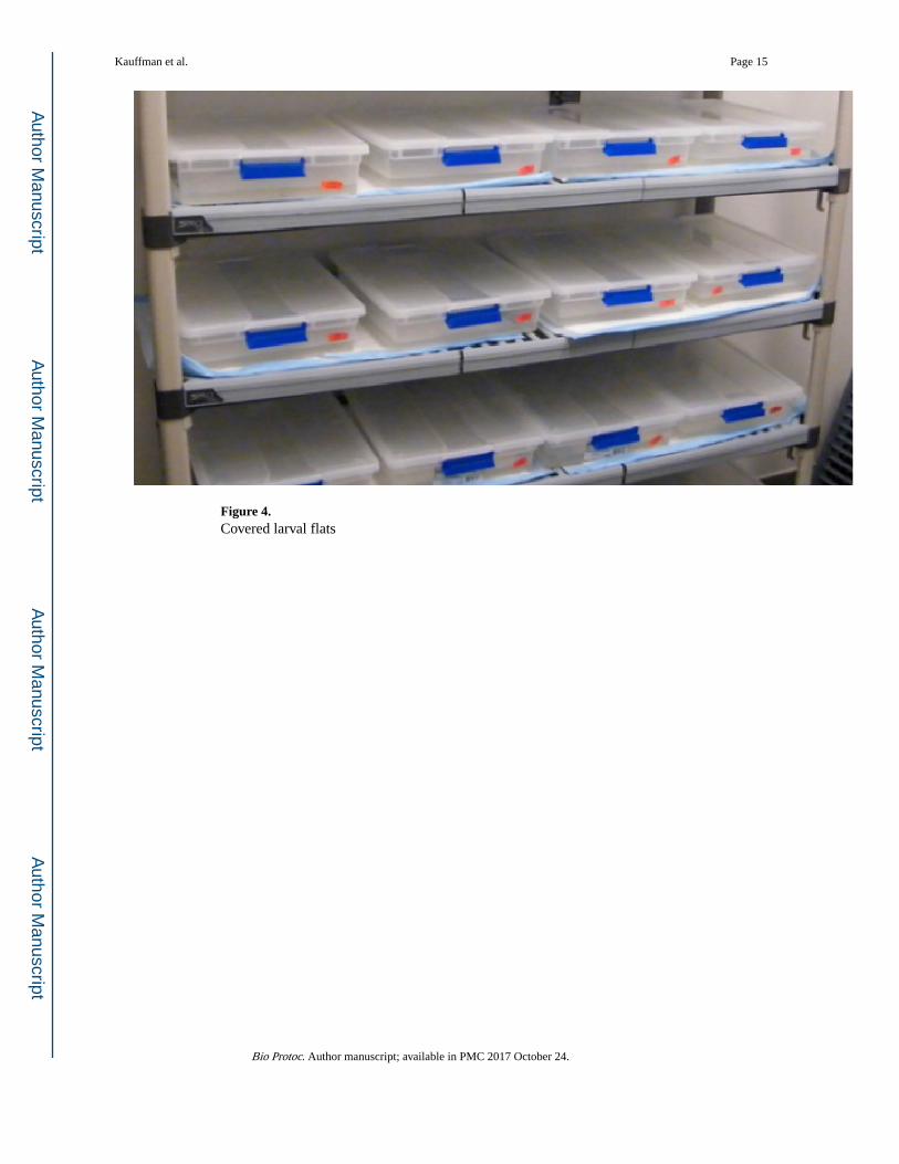
Figure 4. Covered larval flats
Kauffman et al. Page 15
Bio Protoc. Author manuscript; available in PMC 2017 October 24.
Author M
anuscriptA
uthor Manuscript
Author M
anuscriptA
uthor Manuscript
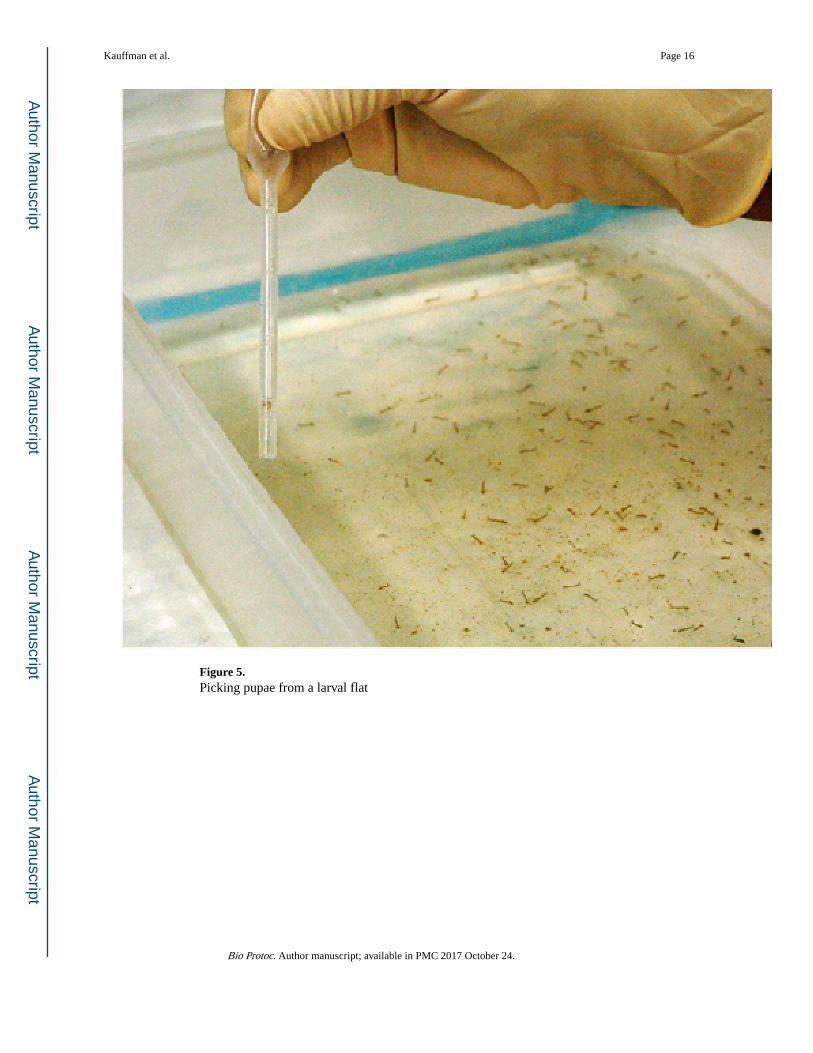
Figure 5. Picking pupae from a larval flat
Kauffman et al. Page 16
Bio Protoc. Author manuscript; available in PMC 2017 October 24.
Author M
anuscriptA
uthor Manuscript
Author M
anuscriptA
uthor Manuscript

Figure 6. Mosquito emergence jar containing pupae and adults
Kauffman et al. Page 17
Bio Protoc. Author manuscript; available in PMC 2017 October 24.
Author M
anuscriptA
uthor Manuscript
Author M
anuscriptA
uthor Manuscript
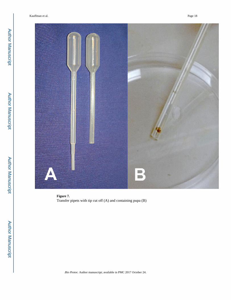
Figure 7. Transfer pipets with tip cut off (A) and containing pupa (B)
Kauffman et al. Page 18
Bio Protoc. Author manuscript; available in PMC 2017 October 24.
Author M
anuscriptA
uthor Manuscript
Author M
anuscriptA
uthor Manuscript

Figure 8. Mosquito cage front (A) and top (B) views
Kauffman et al. Page 19
Bio Protoc. Author manuscript; available in PMC 2017 October 24.
Author M
anuscriptA
uthor Manuscript
Author M
anuscriptA
uthor Manuscript

Figure 9. Mosquito holding cartons
Kauffman et al. Page 20
Bio Protoc. Author manuscript; available in PMC 2017 October 24.
Author M
anuscriptA
uthor Manuscript
Author M
anuscriptA
uthor Manuscript

Figure 10. Hand-held mosquito aspirator
Kauffman et al. Page 21
Bio Protoc. Author manuscript; available in PMC 2017 October 24.
Author M
anuscriptA
uthor Manuscript
Author M
anuscriptA
uthor Manuscript

Figure 11. Male and female mosquitoes
Kauffman et al. Page 22
Bio Protoc. Author manuscript; available in PMC 2017 October 24.
Author M
anuscriptA
uthor Manuscript
Author M
anuscriptA
uthor Manuscript

Figure 12. Sausage casing before and after filling with blood meal
Kauffman et al. Page 23
Bio Protoc. Author manuscript; available in PMC 2017 October 24.
Author M
anuscriptA
uthor Manuscript
Author M
anuscriptA
uthor Manuscript

Video 1. Culex larvae and pupae
Kauffman et al. Page 24
Bio Protoc. Author manuscript; available in PMC 2017 October 24.
Author M
anuscriptA
uthor Manuscript
Author M
anuscriptA
uthor Manuscript

Video 2. Transferring adult mosquitoes from emergence jar
Kauffman et al. Page 25
Bio Protoc. Author manuscript; available in PMC 2017 October 24.
Author M
anuscriptA
uthor Manuscript
Author M
anuscriptA
uthor Manuscript