Landscape of somatic mutations and clonal evolution in ... · Landscape of somatic mutations and...
6
Landscape of somatic mutations and clonal evolution in mantle cell lymphoma Sílvia Beà a,1 , Rafael Valdés-Mas b , Alba Navarro a , Itziar Salaverria a , David Martín-Garcia a , Pedro Jares a , Eva Giné a , Magda Pinyol a , Cristina Royo a , Ferran Nadeu a , Laura Conde a , Manel Juan a , Guillem Clot a , Pedro Vizán c , Luciano Di Croce c , Diana A. Puente b , Mónica López-Guerra a , Alexandra Moros a , Gael Roue a , Marta Aymerich a , Neus Villamor a , Lluís Colomo a , Antonio Martínez a , Alexandra Valera a , José I. Martín-Subero a , Virginia Amador a , Luis Hernández a , Maria Rozman a , Anna Enjuanes a , Pilar Forcada d , Ana Muntañola d , Elena M. Hartmann e , María J. Calasanz f , Andreas Rosenwald e , German Ott g , Jesús M. Hernández-Rivas h , Wolfram Klapper i , Reiner Siebert j , Adrian Wiestner k , Wyndham H. Wilson l , Dolors Colomer a , Armando López-Guillermo a , Carlos López-Otín b,2 , Xose S. Puente b,1,2 , and Elías Campo a,1,2 a Institut d’Investigacions Biomèdiques August Pi i Sunyer, Hospital Clínic, Universitat de Barcelona, 08036 Barcelona, Spain; b Instituto Universitario de Oncología, Universidad de Oviedo, 33006 Oviedo, Spain; c Center for Genomic Regulation and Universitat Pompeu Fabra, 08003 Barcelona, Spain; d Mutua de Terrassa, 08221 Terrassa, Spain; e Institute of Pathology, University of Würzburg, 97080 Würzburg, Germany; f Departamento de Genética, Universidad de Navarra, 31080 Pamplona, Spain; g Robert-Bosch-Krankenhaus and Dr. Margarete Fischer-Bosch Institute of Clinical Pharmacology, 70376 Stuttgart, Germany; h Centro de Investigación del Cáncer, Universidad de Salamanca, 37007 Salamanca, Spain; i Hematopathology Section and Lymph Node Registry, University of Kiel, D-24105 Kiel, Germany; j Institute of Human Genetics, University of Kiel, D-24105 Kiel, Germany; k National Heart, Lung, and Blood Institute, Bethesda, MD 20892; and l National Cancer Institute, Bethesda, MD 20892 Edited* by Louis M. Staudt, National Institutes of Health, Bethesda, MD, and approved September 19, 2013 (received for review August 7, 2013) Mantle cell lymphoma (MCL) is an aggressive tumor, but a subset of patients may follow an indolent clinical course. To understand the mechanisms underlying this biological heterogeneity, we performed whole-genome and/or whole-exome sequencing on 29 MCL cases and their respective matched normal DNA, as well as 6 MCL cell lines. Recurrently mutated genes were investigated by targeted sequencing in an independent cohort of 172 MCL patients. We identified 25 significantly mutated genes, including known drivers such as ataxia-telangectasia mutated (ATM), cyclin D1 (CCND1), and the tumor suppressor TP53; mutated genes encoding the anti-apoptotic protein BIRC3 and Toll-like receptor 2(TLR2); and the chromatin modifiers WHSC1, MLL2, and MEF2B. We also found NOTCH2 mutations as an alternative phenomenon to NOTCH1 mutations in aggressive tumors with a dismal progno- sis. Analysis of two simultaneous or subsequent MCL samples by whole-genome/whole-exome (n = 8) or targeted (n = 19) sequenc- ing revealed subclonal heterogeneity at diagnosis in samples from different topographic sites and modulation of the initial muta- tional profile at the progression of the disease. Some mutations were predominantly clonal or subclonal, indicating an early or late event in tumor evolution, respectively. Our study identifies molec- ular mechanisms contributing to MCL pathogenesis and offers po- tential targets for therapeutic intervention. next-generation sequencing | cancer genetics | cancer heterogeneity M antle cell lymphoma (MCL) is a mature B-cell neoplasm characterized by the t(11;14)(q13;q32) translocation leading to the overexpression of cyclin D1 (1). CCND1 is a weak oncogene that requires the cooperation of other oncogenic events to transform lymphoid cells (2). Molecular studies have identified alterations in components of the cell-cycle regulation, DNA damage response, and cell survival pathways (3, 4), but the profile of mutated genes contributing to the pathogenesis of MCL and cooperating with CCND1 is not well defined (1). Most MCL cases have a rapid evolution and an aggressive behavior with an un- favorable outcome with current therapies (5). Paradoxically, a subset of patients follows an indolent clinical evolution with stable disease even in the absence of chemotherapy (6, 7). This favorable behavior has been associated with IGHV-mutated (8, 9) and lack of expression of SOX11 (10, 11), a transcription factor highly specific of MCL that contributes to the aggressive behavior of this tumor (12). However, the molecular mechanisms re- sponsible for this clinical heterogeneity are not well understood. To gain insight into the molecular pathogenesis of MCL we performed whole-genome sequencing (WGS) and whole-exome sequencing (WES) of 29 MCL and further investigated mutated genes in an expanded series of patients. We also analyzed the subclonal heterogeneity of the tumors and their modulation during the evolution of the disease. Results Landscape of Mutations in MCL. We performed WGS and WES of 4 and 29 MCL, respectively. These patients were representa- tive of the broad clinical and biological spectrum of the disease, including five patients with an indolent clinical evolution. In Significance This is a comprehensive whole-genome/whole-exome analysis of mantle cell lymphoma (MCL). We sequenced 29 MCL cases and validated the findings by target sequencing of 172 addi- tional tumors. We identified recurrent mutations in genes regulating chromatin modification and genes such as NOTCH2 that have a major impact on clinical outcome. Additionally, we demonstrated the subclonal heterogeneity of the tumors al- ready at diagnosis and the modulation of the mutational ar- chitecture in the progression of the disease. The identification of new molecular mechanisms may open perspectives for the management of MCL patients. Author contributions: S.B., C.L.-O., X.S.P., and E.C. designed research; S.B., R.V.-M., A.N., I.S., D.M.-G., P.J., M.P., C.R., F.N., L. Conde, M.J., P.V., L.D.C., D.A.P., M.L.-G., A. Moros, G.R., L. Colomo, A. Martínez, A.V., J.I.M.-S., V.A., L.H., A.E., R.S., and E.C. performed research; P.V., L.D.C., M.A., P.F., A. Muntañola, E.M.H., A.R., G.O., J.M.H.-R., W.K., R.S., A.W., W.H.W., and D.C. contributed new reagents/analytic tools; S.B., R.V.-M., A.N., I.S., D.M.-G., P.J., E.G., M.P., C.R., M.J., G.C., G.R., N.V., J.I.M.-S., M.R., M.J.C., R.S., D.C., A.L.-G., C.L.-O., X.S.P., and E.C. analyzed data; and S.B., C.L.-O., X.S.P., and E.C. wrote the paper. The authors declare no conflict of interest. *This Direct Submission article had a prearranged editor. Freely available online through the PNAS open access option. Data deposition: Next-generation sequencing data have been deposited at the European Genome-Phenome Archive under accession no. EGAS00001000510. Affymetrix SNP6. 0 array and HU133+2.0 gene expression data have been deposited at Gene Expression Omnibus (GEO) under accession nos. GSE46969 and GSE36000, respectively. 1 To whom correspondence may be addressed. E-mail: [email protected], sbea@clinic. ub.es, or [email protected]. 2 C.L.-O., X.S.P., and E.C. contributed equally to this work. This article contains supporting information online at www.pnas.org/lookup/suppl/doi:10. 1073/pnas.1314608110/-/DCSupplemental. 18250–18255 | PNAS | November 5, 2013 | vol. 110 | no. 45 www.pnas.org/cgi/doi/10.1073/pnas.1314608110
Transcript of Landscape of somatic mutations and clonal evolution in ... · Landscape of somatic mutations and...

Landscape of somatic mutations and clonal evolutionin mantle cell lymphomaSílvia Beàa,1, Rafael Valdés-Masb, Alba Navarroa, Itziar Salaverriaa, David Martín-Garciaa, Pedro Jaresa, Eva Ginéa,Magda Pinyola, Cristina Royoa, Ferran Nadeua, Laura Condea, Manel Juana, Guillem Clota, Pedro Vizánc,Luciano Di Crocec, Diana A. Puenteb, Mónica López-Guerraa, Alexandra Morosa, Gael Rouea, Marta Aymericha,Neus Villamora, Lluís Colomoa, Antonio Martíneza, Alexandra Valeraa, José I. Martín-Suberoa, Virginia Amadora,Luis Hernándeza, Maria Rozmana, Anna Enjuanesa, Pilar Forcadad, Ana Muntañolad, Elena M. Hartmanne,María J. Calasanzf, Andreas Rosenwalde, German Ottg, Jesús M. Hernández-Rivash, Wolfram Klapperi, Reiner Siebertj,Adrian Wiestnerk, Wyndham H. Wilsonl, Dolors Colomera, Armando López-Guillermoa, Carlos López-Otínb,2,Xose S. Puenteb,1,2, and Elías Campoa,1,2
aInstitut d’Investigacions Biomèdiques August Pi i Sunyer, Hospital Clínic, Universitat de Barcelona, 08036 Barcelona, Spain; bInstituto Universitario deOncología, Universidad de Oviedo, 33006 Oviedo, Spain; cCenter for Genomic Regulation and Universitat Pompeu Fabra, 08003 Barcelona, Spain; dMutua deTerrassa, 08221 Terrassa, Spain; eInstitute of Pathology, University of Würzburg, 97080 Würzburg, Germany; fDepartamento de Genética, Universidad deNavarra, 31080 Pamplona, Spain; gRobert-Bosch-Krankenhaus and Dr. Margarete Fischer-Bosch Institute of Clinical Pharmacology, 70376 Stuttgart, Germany;hCentro de Investigación del Cáncer, Universidad de Salamanca, 37007 Salamanca, Spain; iHematopathology Section and Lymph Node Registry, University ofKiel, D-24105 Kiel, Germany; jInstitute of Human Genetics, University of Kiel, D-24105 Kiel, Germany; kNational Heart, Lung, and Blood Institute, Bethesda,MD 20892; and lNational Cancer Institute, Bethesda, MD 20892
Edited* by Louis M. Staudt, National Institutes of Health, Bethesda, MD, and approved September 19, 2013 (received for review August 7, 2013)
Mantle cell lymphoma (MCL) is an aggressive tumor, but a subsetof patients may follow an indolent clinical course. To understandthe mechanisms underlying this biological heterogeneity, weperformed whole-genome and/or whole-exome sequencing on29 MCL cases and their respective matched normal DNA, as wellas 6 MCL cell lines. Recurrently mutated genes were investigatedby targeted sequencing in an independent cohort of 172 MCLpatients. We identified 25 significantly mutated genes, includingknown drivers such as ataxia-telangectasia mutated (ATM), cyclinD1 (CCND1), and the tumor suppressor TP53; mutated genesencoding the anti-apoptotic protein BIRC3 and Toll-like receptor2 (TLR2); and the chromatin modifiers WHSC1, MLL2, and MEF2B.We also found NOTCH2 mutations as an alternative phenomenonto NOTCH1 mutations in aggressive tumors with a dismal progno-sis. Analysis of two simultaneous or subsequent MCL samples bywhole-genome/whole-exome (n = 8) or targeted (n = 19) sequenc-ing revealed subclonal heterogeneity at diagnosis in samples fromdifferent topographic sites and modulation of the initial muta-tional profile at the progression of the disease. Some mutationswere predominantly clonal or subclonal, indicating an early or lateevent in tumor evolution, respectively. Our study identifies molec-ular mechanisms contributing to MCL pathogenesis and offers po-tential targets for therapeutic intervention.
next-generation sequencing | cancer genetics | cancer heterogeneity
Mantle cell lymphoma (MCL) is a mature B-cell neoplasmcharacterized by the t(11;14)(q13;q32) translocation
leading to the overexpression of cyclin D1 (1). CCND1 is a weakoncogene that requires the cooperation of other oncogenic eventsto transform lymphoid cells (2). Molecular studies have identifiedalterations in components of the cell-cycle regulation, DNAdamage response, and cell survival pathways (3, 4), but the profileof mutated genes contributing to the pathogenesis of MCL andcooperating with CCND1 is not well defined (1). Most MCL caseshave a rapid evolution and an aggressive behavior with an un-favorable outcome with current therapies (5). Paradoxically,a subset of patients follows an indolent clinical evolution withstable disease even in the absence of chemotherapy (6, 7). Thisfavorable behavior has been associated with IGHV-mutated (8, 9)and lack of expression of SOX11 (10, 11), a transcription factorhighly specific of MCL that contributes to the aggressive behaviorof this tumor (12). However, the molecular mechanisms re-sponsible for this clinical heterogeneity are not well understood.
To gain insight into the molecular pathogenesis of MCL weperformed whole-genome sequencing (WGS) and whole-exomesequencing (WES) of 29 MCL and further investigated mutatedgenes in an expanded series of patients. We also analyzed thesubclonal heterogeneity of the tumors and their modulationduring the evolution of the disease.
ResultsLandscape of Mutations in MCL. We performed WGS and WES of4 and 29 MCL, respectively. These patients were representa-tive of the broad clinical and biological spectrum of the disease,including five patients with an indolent clinical evolution. In
Significance
This is a comprehensive whole-genome/whole-exome analysisof mantle cell lymphoma (MCL). We sequenced 29 MCL casesand validated the findings by target sequencing of 172 addi-tional tumors. We identified recurrent mutations in genesregulating chromatin modification and genes such as NOTCH2that have a major impact on clinical outcome. Additionally, wedemonstrated the subclonal heterogeneity of the tumors al-ready at diagnosis and the modulation of the mutational ar-chitecture in the progression of the disease. The identificationof new molecular mechanisms may open perspectives for themanagement of MCL patients.
Author contributions: S.B., C.L.-O., X.S.P., and E.C. designed research; S.B., R.V.-M., A.N.,I.S., D.M.-G., P.J., M.P., C.R., F.N., L. Conde, M.J., P.V., L.D.C., D.A.P., M.L.-G., A. Moros,G.R., L. Colomo, A. Martínez, A.V., J.I.M.-S., V.A., L.H., A.E., R.S., and E.C. performedresearch; P.V., L.D.C., M.A., P.F., A. Muntañola, E.M.H., A.R., G.O., J.M.H.-R., W.K., R.S., A.W.,W.H.W., and D.C. contributed new reagents/analytic tools; S.B., R.V.-M., A.N., I.S., D.M.-G.,P.J., E.G., M.P., C.R., M.J., G.C., G.R., N.V., J.I.M.-S., M.R., M.J.C., R.S., D.C., A.L.-G., C.L.-O.,X.S.P., and E.C. analyzed data; and S.B., C.L.-O., X.S.P., and E.C. wrote the paper.
The authors declare no conflict of interest.
*This Direct Submission article had a prearranged editor.
Freely available online through the PNAS open access option.
Data deposition: Next-generation sequencing data have been deposited at the EuropeanGenome-Phenome Archive under accession no. EGAS00001000510. Affymetrix SNP6.0 array and HU133+2.0 gene expression data have been deposited at Gene ExpressionOmnibus (GEO) under accession nos. GSE46969 and GSE36000, respectively.1To whom correspondence may be addressed. E-mail: [email protected], [email protected], or [email protected].
2C.L.-O., X.S.P., and E.C. contributed equally to this work.
This article contains supporting information online at www.pnas.org/lookup/suppl/doi:10.1073/pnas.1314608110/-/DCSupplemental.
18250–18255 | PNAS | November 5, 2013 | vol. 110 | no. 45 www.pnas.org/cgi/doi/10.1073/pnas.1314608110

addition, we performed WES of six MCL cell lines. Selectedmutated genes were investigated in a validation series of 172MCL patients (SI Appendix, Tables S1–S6). We detected about3,700 somatic mutations per tumor (1.2 per Mb) by WGS (SIAppendix, Figs. S1A and S2A and Dataset S1). The most com-mon substitution was the transition C > T/G > A, usually oc-curring in a CpG context. Two IGHV-mutated MCL showeda higher proportion of A > C/T > G mutations than the twoIGHV-unmutated cases (SI Appendix, Fig. S1B). The break-points of the t(11;14) translocation differed in the four cases (13)(SI Appendix, Table S7). We next investigated the presence ofregional clustering of somatic mutations by constructing “rainfallplots” (SI Appendix, Fig. S2B). Foci of hypermutation or kataegis,a phenomenon recently described in breast cancer (14), wereobserved in three cases. They were more frequent in the twoIGHV-mutated tumors. These clusters occurred around the11q13 breakpoint of the t(11;14); the Ig genes at 2p11, 14q32.33,and 22q11.22; and the deleted 9p21.3 region, but we also ob-served this phenomenon in regions without apparent structuralalterations and lacking coding genes (SI Appendix, Table S8).The same clusters of hypermutation were observed in the se-quential sample of case M003, suggesting that kataegis in MCLmay occur as an early phenomenon that remains stable duringthe evolution of the disease.We further characterized the spectrum of mutations in 29
MCL by WES (SI Appendix, Tables S1, S3, and S9 and DatasetS2). All these cases were also analyzed by SNP array for copynumber alterations (CNA) and copy number neutral loss ofheterozygosity (SI Appendix, Fig. S3 and Table S10). We iden-tified 652 protein-coding genes with somatic mutations affectingthe structure of the encoded protein (nonsynonymous changes,frameshifts in the coding sequence, and mutations affecting ca-nonical splicing sites) with a median of 20 mutations per case(range 8–47). Twenty-five of the 33 mutated genes in at least twosamples were mutated at a rate significantly higher than expectedby chance, and all tumors harbored mutations in at least 1 ofthese 25 genes (Fig. 1 and SI Appendix, Table S11). Similarly, fiveof the six MCL cell lines also had mutations in at least one of therecurrently mutated genes identified in primary tumors (Fig. 1,SI Appendix, and Dataset S3). Chromosomal CNAs were presentin 26/29 (90%) cases (mean 11.1 ± 9.2 per altered case) (SIAppendix, Fig. S3 and Table S10). Tumors expressing SOX11showed a significantly higher number of CNAs than SOX11-negative MCLs (mean 13 ± 9 versus 2 ± 2; P = 2.1 × 10⁻⁵),despite the similar number of somatic mutations (mean 24 ± 12versus 17 ± 9; P = 0.141) (Fig. 1). Interestingly, five patients whodid not need treatment (median 55 mo, range 4–147) had sig-nificantly fewer somatic protein-coding mutations (mean 11 ± 4versus 25 ± 11, P = 3.4 × 10⁻⁵) and CNAs (mean 2 ± 3 versus12 ± 9; P = 0.001) than patients who required treatment at di-agnosis (n = 24).
Recurrent Somatic Mutations in MCL. ATM, CCND1, and TP53,previously described as drivers in MCL, were among the mostfrequently mutated genes in this study. ATM mutations werefound in 12 of the 22 (55%) tumors expressing SOX11, but innone of the SOX11-negative MCL (P = 0.023) (Table 1, Fig. 1,and SI Appendix, Table S9). Six of the mutations were associatedwith deletions of the wild-type allele, whereas five cases with no11q loss had two different ATM mutations per case and only onewas a single mutation with no 11q deletion (SI Appendix, Fig.S4A). These mutations mainly truncated or affected functionaldomains (SI Appendix, Fig. S5). CCND1 mutations were foundpredominantly in exon 1 (9 of 10 CCND1-mutated cases) (SIAppendix, Fig. S6) and were more frequent in SOX11-negativeMCL [6/7 (86%) versus 4/22 (18%); P = 0.03] and with IGHV-mutated [7/12 (58%) versus 3/16 (19%), P = 0.05], suggestingtheir acquisition in the germinal center microenvironment(Fig. 1, Table 1, and SI Appendix, Table S9). TP53 mutations [8/29 (28%)] were associated with 17p alterations in six cases, andonly one case without 17p alteration had two mutations. TP53
mutations were equally distributed in tumors regardless ofSOX11 expression or IGHV mutations (Fig. 1).We also identified recurrent mutations in WHSC1, MLL2,
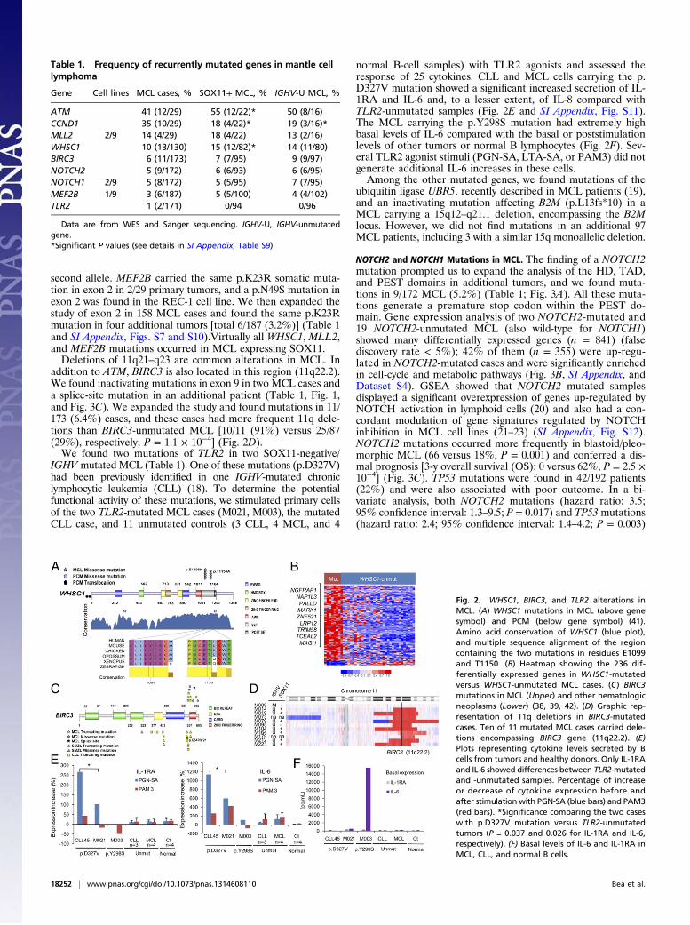
BIRC3, MEF2B, and TLR2 (Table 1; Fig. 1), as well as NOTCH2in one case. WHSC1 encodes a histone 3 methyltransferase oflysine-36 (H3K36) that has not been found previously mutated inlymphomas. Two missense mutations (p.E1099K and p.T1150A)were recurrently found in two cases each. Both residues are inclose proximity and affect two of the most conserved domains inexons 18 and 19. We further analyzed these exons in 101 addi-tional tumors and confirmed the presence of the same mutationsin nine more cases [total 13/130 (10%)] (Table 1, Fig. 2A, and SIAppendix, Table S9 and Fig. S7). Interestingly, WHSC1 (alsonamed MMSET or NSD2) is the target of the IGH-translocationt(4;14) in plasma cell myeloma (PCM), where it is overexpressedand associated with an increase in H3K36 and a decrease inH3K27 methylation across the genome (15). Gene expressionanalysis of 8 WHSC1-mutated and 31 WHSC1-unmutated MCLidentified 236 genes differentially expressed (false discovery rate<5%) with the majority of these genes [192/236 (81%)] up-reg-ulated in the WHSC1-mutated cases (Fig. 3B; SI Appendix, TableS12). A gene set enrichment analysis (GSEA) using previouslypublished lymphoid gene expression signatures (15) demonstratedthat WHSC1-mutated cases displayed significant overexpressionof several signatures related to proliferation and cell-cycle regula-tion (SI Appendix, Fig. S8) (16). Interestingly, WHSC1-mutatedMCL showed a highly significant overexpression of the gene signa-ture up-regulated in PCM with the t(4;14) translocation over-expressing WHSC1 (17). In addition, WHSC1-mutated MCLshowed overexpression of genes up-regulated by wild-typeWHSC1or the gain-of-function exon 19 mutant WHSC1 in the KMS11PCM cell line (15) (SI Appendix, Fig. S8).In addition, the histone methyltransferase MLL2 was mutated
in 4/29 primary tumors and 2/6 MCL cell lines. Four of the sixmutations were truncating and two were missense changes andaffected the conserved FYRN and FYRC domains (Table 1 andSI Appendix, Fig. S9). None of these cases had a deletion of the
Fig. 1. WES in 29 cases and 6 MCL cell lines. Heatmap with the mutationpattern of the 25 significantly recurrent mutated genes. Each rowrepresents a gene and each column represents a primary tumor/cell line.Vertical bar graphs show the total number of somatic mutations by WES(blue) and somatic CNAs by SNP array (green) for primary tumors, and thetotal number of nonpolymorphic variants and CNAs for cell lines. The plotbelow the case label indicates sample characteristics (SOX11 expression andIGHV gene status).
Beà et al. PNAS | November 5, 2013 | vol. 110 | no. 45 | 18251
MED
ICALSC
IENCE
S
second allele. MEF2B carried the same p.K23R somatic muta-tion in exon 2 in 2/29 primary tumors, and a p.N49S mutation inexon 2 was found in the REC-1 cell line. We then expanded thestudy of exon 2 in 158 MCL cases and found the same p.K23Rmutation in four additional tumors [total 6/187 (3.2%)] (Table 1and SI Appendix, Figs. S7 and S10).Virtually all WHSC1, MLL2,and MEF2B mutations occurred in MCL expressing SOX11.Deletions of 11q21–q23 are common alterations in MCL. In
addition to ATM, BIRC3 is also located in this region (11q22.2).We found inactivating mutations in exon 9 in two MCL cases anda splice-site mutation in an additional patient (Table 1, Fig. 1,and Fig. 3C). We expanded the study and found mutations in 11/173 (6.4%) cases, and these cases had more frequent 11q dele-tions than BIRC3-unmutated MCL [10/11 (91%) versus 25/87(29%), respectively; P = 1.1 × 10⁻⁴] (Fig. 2D).We found two mutations of TLR2 in two SOX11-negative/
IGHV-mutated MCL (Table 1). One of these mutations (p.D327V)had been previously identified in one IGHV-mutated chroniclymphocytic leukemia (CLL) (18). To determine the potentialfunctional activity of these mutations, we stimulated primary cellsof the two TLR2-mutated MCL cases (M021, M003), the mutatedCLL case, and 11 unmutated controls (3 CLL, 4 MCL, and 4
normal B-cell samples) with TLR2 agonists and assessed theresponse of 25 cytokines. CLL and MCL cells carrying the p.D327V mutation showed a significant increased secretion of IL-1RA and IL-6 and, to a lesser extent, of IL-8 compared withTLR2-unmutated samples (Fig. 2E and SI Appendix, Fig. S11).The MCL carrying the p.Y298S mutation had extremely highbasal levels of IL-6 compared with the basal or poststimulationlevels of other tumors or normal B lymphocytes (Fig. 2F). Sev-eral TLR2 agonist stimuli (PGN-SA, LTA-SA, or PAM3) did notgenerate additional IL-6 increases in these cells.Among the other mutated genes, we found mutations of the
ubiquitin ligase UBR5, recently described in MCL patients (19),and an inactivating mutation affecting B2M (p.L13fs*10) in aMCL carrying a 15q12–q21.1 deletion, encompassing the B2Mlocus. However, we did not find mutations in an additional 97MCL patients, including 3 with a similar 15q monoallelic deletion.
NOTCH2 and NOTCH1 Mutations in MCL. The finding of a NOTCH2mutation prompted us to expand the analysis of the HD, TAD,and PEST domains in additional tumors, and we found muta-tions in 9/172 MCL (5.2%) (Table 1; Fig. 3A). All these muta-tions generate a premature stop codon within the PEST do-main. Gene expression analysis of two NOTCH2-mutated and19 NOTCH2-unmutated MCL (also wild-type for NOTCH1)showed many differentially expressed genes (n = 841) (falsediscovery rate < 5%); 42% of them (n = 355) were up-regu-lated in NOTCH2-mutated cases and were significantly enrichedin cell-cycle and metabolic pathways (Fig. 3B, SI Appendix, andDataset S4). GSEA showed that NOTCH2 mutated samplesdisplayed a significant overexpression of genes up-regulated byNOTCH activation in lymphoid cells (20) and also had a con-cordant modulation of gene signatures regulated by NOTCHinhibition in MCL cell lines (21–23) (SI Appendix, Fig. S12).NOTCH2 mutations occurred more frequently in blastoid/pleo-morphic MCL (66 versus 18%, P = 0.001) and conferred a dis-mal prognosis [3-y overall survival (OS): 0 versus 62%, P = 2.5 ×10⁻⁴] (Fig. 3C). TP53 mutations were found in 42/192 patients(22%) and were also associated with poor outcome. In a bi-variate analysis, both NOTCH2 mutations (hazard ratio: 3.5;95% confidence interval: 1.3–9.5; P = 0.017) and TP53mutations(hazard ratio: 2.4; 95% confidence interval: 1.4–4.2; P = 0.003)
Table 1. Frequency of recurrently mutated genes in mantle celllymphoma
Gene Cell lines MCL cases, % SOX11+ MCL, % IGHV-U MCL, %
ATM 41 (12/29) 55 (12/22)* 50 (8/16)CCND1 35 (10/29) 18 (4/22)* 19 (3/16)*MLL2 2/9 14 (4/29) 18 (4/22) 13 (2/16)WHSC1 10 (13/130) 15 (12/82)* 14 (11/80)BIRC3 6 (11/173) 7 (7/95) 9 (9/97)NOTCH2 5 (9/172) 6 (6/93) 6 (6/95)NOTCH1 2/9 5 (8/172) 5 (5/95) 7 (7/95)MEF2B 1/9 3 (6/187) 5 (5/100) 4 (4/102)TLR2 1 (2/171) 0/94 0/96
Data are from WES and Sanger sequencing. IGHV-U, IGHV-unmutatedgene.*Significant P values (see details in SI Appendix, Table S9).
Fig. 2. WHSC1, BIRC3, and TLR2 alterations inMCL. (A) WHSC1 mutations in MCL (above genesymbol) and PCM (below gene symbol) (41).Amino acid conservation of WHSC1 (blue plot),and multiple sequence alignment of the regioncontaining the two mutations in residues E1099and T1150. (B) Heatmap showing the 236 dif-ferentially expressed genes in WHSC1-mutatedversus WHSC1-unmutated MCL cases. (C) BIRC3mutations in MCL (Upper) and other hematologicneoplasms (Lower) (38, 39, 42). (D) Graphic rep-resentation of 11q deletions in BIRC3-mutatedcases. Ten of 11 mutated MCL cases carried dele-tions encompassing BIRC3 gene (11q22.2). (E)Plots representing cytokine levels secreted by Bcells from tumors and healthy donors. Only IL-1RAand IL-6 showed differences between TLR2-mutatedand -unmutated samples. Percentage of increaseor decrease of cytokine expression before andafter stimulation with PGN-SA (blue bars) and PAM3(red bars). *Significance comparing the two caseswith p.D327V mutation versus TLR2-unmutatedtumors (P = 0.037 and 0.026 for IL-1RA and IL-6,respectively). (F) Basal levels of IL-6 and IL-1RA inMCL, CLL, and normal B cells.
18252 | www.pnas.org/cgi/doi/10.1073/pnas.1314608110 Beà et al.

were identified as independent risk factors for OS (SI Appendix,Fig. S13).NOTCH1 mutations have been recently described in MCL
(20). We investigated this gene and found truncating mutationsin 8/172 (4.6%) MCL cases, as well as in MINO and REC-1 cells(Table 1 and SI Appendix, Fig. S14). NOTCH1-mutated tumorswere predominantly blastoid/pleomorphic (67 versus 19%, P =0.03) and showed shorter survival than NOTCH1-unmutatedMCL (3-y OS: 33 versus 60%, P = 0.026) (SI Appendix, Fig. S15).NOTCH1 and NOTCH2 mutations occurred in different subsetsof tumors because only 1 of the 16 patients with mutations inthese genes had mutations in both. Taken together, NOTCH1/2mutations were present in 9.5% of MCL and identified a subsetof tumors with more adverse biological and clinical featuresincluding blastoid/pleomorphic morphology (67 versus 13%,P = 1.3 × 10⁻⁵) and a significant shorter survival (3-y OS: 24versus 63%, P = 3.4 × 10⁻⁴) (Fig. 3C).
Sequencing Simultaneous and Subsequent MCL Samples RevealsClonal Heterogeneity. To explore the subclonal architecture ofMCL, we sequenced a second tumor sample obtained simulta-neously from two different topographic sites (n = 6) or at twotime points (diagnosis and disease progression, n = 2). Analysisof the frequency of reads supporting somatic substitution allowedthe estimation of major subclonal populations (24). Four of thesix patients with simultaneous samples showed the same muta-tions and CNAs in the peripheral blood (PB) sample and cor-responding lymphoid tissue, suggesting the presence of a singlemajor clone at both sites. In contrast, two cases (M023 andM026) had a subset of common mutations in both topographicsites, but they also carried a subset of mutated genes that wereexclusive of the PB or tissue tumor sample (Fig. 4 A and B). Thispattern of alterations is consistent with two major subpopu-lations derived from an initial founder clone differentially rep-resented in the two topographic sites. In addition, we also observeda different pattern of genomic alterations at diagnosis and pro-gression in the two cases with sequential samples (Fig. 4C and Dand SI Appendix, Table S10). Patient M003 had stable diseasewithout treatment for more than 3 y and then rapidly progressed.The major clone identified at diagnosis gained new mutationsand genomic complexity at the time of clinical progression. Thisnew clone carried the same 17p deletion and somatic mutationsobserved in the initial sample, but had a 16% increase in thenumber of whole-genome mutations (from 3,928 to 4,665) (SIAppendix, Fig. S1A). These included nonsynonymous mutationsin additional genes, a complex DNA copy number profile with 20acquired CNAs, and chromothripsis involving chromosomes 4and 12 (Fig. 4C and SI Appendix, Fig. S2A). Patient M002 wastreated with chemotherapy after diagnosis and relapsed 3 y later.The major clone at relapse maintained a subset of CNAs andsomatic mutations present at diagnosis, but 11 of the initialmutations had disappeared and other mutated genes had
emerged (Fig. 4D). This pattern of evolution is consistent withthe eradication of the major initial subclone by chemotherapyand the relapse of a new subclone after treatment.Comparison of the allele frequency of mutations in the dif-
ferent simultaneous or evolving subclones may help to infer thedynamic architecture of somatic events in the evolution oftumors (24, 25). Eight of the 25 recurrently mutated genes inMCL (ATM, CCND1, MLL2, KCNC2, KIAA1671, PCSK2,TNRC6B, and TRPM6) were present at similar allelic frequencyin the two subclones of different cases, suggesting that theyrepresent early events. In contrast, four recurrently mutatedgenes (ABCA3, TLR2, TP53, andWHSC1) were seen in only oneof the two simultaneous or emerging subclones at progression,supporting the notion that they might constitute later events. Wefurther analyzed by Sanger sequencing 11 additional serial and 8simultaneous tumor samples from different topographic sites(PB and lymphoid tissues) (SI Appendix, Fig. S16). Interestingly,we observed that BIRC3 mutations were absent at diagnosis intwo cases that acquired the mutation in a posttreatment sampleassociated with the acquisition of an 11q22.1–q24.2 deletion inone of them. Another case showed the BIRC3 mutation in the PBbut not in the simultaneous lymph node. Additionally, NOTCH1was mutated in only one of the synchronic samples in two cases.
DiscussionWe have conducted a comprehensive genomic study of MCLthat has revealed the heterogeneous spectrum of somatic mu-tations of this tumor, with several molecular mechanisms con-tributing to the pathogenesis and the clinical progression of thedisease. The genome sequencing of simultaneous and sequentialtumor samples has highlighted the subclonal heterogeneity of themutations already present at diagnosis and their dynamic evo-lution in the progression of the disease.The whole-genome analysis showed a relative low number of
global somatic mutations in MCL (1.2 per Mb), slightly higher toCLL (26) or acute myeloid leukemia (27), but lower than in otherlymphoid or nonhematopoietic tumors (28, 29). Interestingly, weidentified a distinct mutational signature characterized by A > C/T > G substitutions in a TpA context in the two MCL withIGHV-mutated. This signature was initially identified in CLLwith IGHV-mutated and more recently confirmed as a uniquefeature of lymphoid neoplasms originating in germinal centercells (26, 29) and has been attributed to the action of the error-prone DNA polymerase η during the IGHV somatic hyper-mutation process (26).The most commonly mutated genes were the previously
identified MCL drivers ATM, CCND1, and TP53. These muta-tions were differentially distributed in subtypes of the diseaseaccording to the IGHV mutational status and SOX11 expression.Thus, ATM mutations were seen only in SOX11-positive tumors,whereas CCND1 mutations were preferentially detected inMCL with IGHV-mutated and TP53 mutations were equally
Fig. 3. NOTCH2 alterations in mantle cell lymphoma. (A) NOTCH2 mutations in MCL (Upper) and other hematologic neoplasms (Lower) (37, 38, 43). (B)Heatmap showing the 841 differentially expressed genes in NOTCH2-mutated versus NOTCH2-unmutated MCL cases. (C) Actuarial probability of overallsurvival of MCL patients according to NOTCH2 mutation and NOTCH2 or NOTCH1 mutation.
Beà et al. PNAS | November 5, 2013 | vol. 110 | no. 45 | 18253
MED
ICALSC
IENCE
S

distributed among the different groups. The integration of theCNA and somatic mutations showed that only TP53, ATM, andBIRC3 recurrent mutations were associated with allelic losses(17p and 11q, respectively), whereas CDKN2A, the only genetargeted by recurrent homozygous deletions, did not carry so-matic mutations in other cases. Interestingly, three mutatedgenes (WHSC1, MLL2, and MEF2B) function as chromatinmodifiers, and altogether these mutations occurred in 10 of the29 (14%) cases examined by WES. The analysis of WHSC1 andMEF2B in an expanded series confirmed the relative high fre-quency of these mutations in MCL (10% and 3.2%, respec-tively). WHSC1 mutations have not been described previouslyin lymphomas, but this gene is the target of the t(4;14) trans-location in PCM, and the same mutation observed in exon 18 hasbeen recently detected in an acute lymphoblastic leukemia(ALL) patient (30). The WHSC1 overexpression in PCM and themutation in ALL seem to have an activating function becausethey increase the H3K36 methylation associated with a methylationdecrease in H3K27 across the genome (15). OurWHSC1-mutatedprimary MCL had the same overexpressed gene signaturesmodulated by WHSC1 activation in PCM, suggesting that thesemutations may also have a similar activating function in MCL.The mutations observed in MLL2 and MEF2B are similar tothose detected in diffuse large B-cell lymphoma (DLBCL) andfollicular lymphomas, but their functional consequences are notyet well understood (31–33). Notably, virtually all MLL2, WHSC1,andMEF2Bmutations were found in MCL with IGHV-unmutatedor expressing SOX11.Recent studies have suggested a role of microenvironment
stimuli in sustaining MCL (1). TLRs mediate cell responses tospecific pathogen-associated molecular patterns (34). In thatsense, we found mutations of TLR2 in two SOX11-negative/IGHV-mutated MCL, and these mutations were associated withincreased production of IL-1RA and IL-6 by the tumor cells.Notably, high levels of IL-1RA have been associated with ag-gressive behavior in some lymphomas (35), and IL-6 sustainsgrowth and survival of MCL cells (36). These findings suggestthat TLR2 mutations may contribute to the pathogenesis ofa subset of SOX11-negative/IGHV-mutated MCL by modulatingtumor microenvironment responses.
In addition to the statistically significant mutations describedabove, we found a mutation in NOTCH2 in one MCL. Similaractivating mutations have been recently described in splenicmarginal zone lymphomas and in NOTCH1 in aggressive CLLand MCL (37, 38). These findings prompted us to expand thestudy of NOTCH2 and NOTCH1 mutations in MCL and foundtheir presence in 5.2% and 4.7% of the tumors, respectively. Allthese mutations generated truncating and likely more activeproteins and occurred in a subset of tumors with very aggressiveclinical behavior. Only 1 of 16 tumors had simultaneous muta-tions in both genes, suggesting that they may give a similar se-lective advantage to the cells. All these findings indicate thatNOTCH1/2 mutations in MCL activate this pathway and area frequent mechanism involved in the aggressive behavior ofthe tumors.The spectrum of mutations described in this work highlights
the existence of common molecular features as well as relevantdifferences in the genetic alterations present in different sub-types of lymphoid neoplasms. Thus, NOTCH1 mutations havebeen found in CLL (18, 26) whereas NOTCH2 is mutated insplenic marginal zone lymphomas (37, 38); however, none ofthese tumors carry mutations in both NOTCH genes as observedin MCL. In addition, MLL2 and MEF2B mutations are sharedwith DLBCL (31–33) but are uncommon in CLL, whereas MCLhas frequent mutations of ATM and BIRC3 that are also fre-quent in CLL (39) but uncommon in DLBCL. In contrast,WHSC1 mutations appear to be specific for MCL while lackingmutations in genes frequently mutated in other hematologicalneoplasias (e.g., MYD88, CARD11, EZH2, SF3B1).Recent genomic studies have revealed the complex subclonal
heterogeneity of different tumors and its dynamic evolution inthe course of the disease (40). The sequence of two simultaneousor longitudinal samples in different topographic sites in our studyhas revealed that some tumors may have at least two majorsubclones already at diagnosis with different representation intwo topographic sites, lymph nodes, and peripheral blood. Inaddition, the evolution of the tumors is also heterogeneous withthe eradication of some clones by the chemotherapy treatmentand the emergency of other new clones at the progression of thedisease or at relapse after chemotherapy. The analysis of the
Fig. 4. Subclonal architecture in MCL. Representation of four informative MCL patients with two tumor samples analyzed. The mutation clusters are rep-resented in the upper panel of each case, the shared and stable mutations are in green color, in blue the mutations specific of the first sample and in red themutations identified only in the second sample. In the lower panel of each case, a detailed representation of the percentage of mutated reads in each of thesamples (Left and Right) with the same color code, and with the significantly recurrent mutated genes highlighted in the same color. (A–B) Cases M023 andM026 have two major subclones derived from an initial founder clone differentially represented in simultaneous samples of lymph node (LN) and peripheralblood (PB). (C) Longitudinal analysis in patient M003 at diagnosis and at disease progression previous to treatment. (D) Longitudinal analysis in patient M002at diagnosis and at first relapse.
18254 | www.pnas.org/cgi/doi/10.1073/pnas.1314608110 Beà et al.

allelic frequency of the mutations allows the recognition of initialand acquired mutations that are probably relevant in the pro-gression of the disease (24, 25). The recognition of this molec-ular heterogeneity at diagnosis and progression may have futureclinical relevance for the management of patients with targetedtherapies to specific mutations.In summary, this whole-genome/whole-exome study of MCL
has revealed genes and pathways recurrently mutated that maycontribute to lymphomagenesis in cooperation with the t(11;14)translocation. The differential distribution of these mutations inclinical and molecular subtypes of the disease illustrates the re-lationship between genomic alterations and tumor heterogeneity.We have also shown the intratumoral heterogeneity and sub-clonal architecture of MCL that may have relevance in theclinical evolution of the tumors. These alterations highlightmechanisms in the pathogenesis of this lymphoma and offerpotential targets for therapeutic intervention.
Materials and MethodsPatients and Specimens. We sequenced the genomes of 29 patients withMCL and six MCL cell lines. Additional samples from 172 patients wereobtained for clinical validation. All patients gave informed consent forsample collection and analysis. DNA and RNA were purified from tumorsand normal cells. Additional details are provided in SI Appendix, SI Materialsand Methods.
Whole-Genome and Whole-Exome Sequencing and Analysis. Whole-genomeand whole-exome sequencing (Agilent SureSelect Human All Exon 50 MB)were performed as previously described (18, 26) using a HiSeq 2000 in-strument. Sequence data analysis was performed using the Sidrón mutationcaller as described (18, 26). Additional details are provided in SI Appendix, SIMaterials and Methods.
Microarray Experiments. Genotyping and copy number and gene expressionanalysis were performed using Affymetrix SNP6.0 arrays and HU133+ 2.0GeneChip (Affymetrix), respectively. Additional details are provided inSI Appendix, SI Materials and Methods.
ACKNOWLEDGMENTS. We thank M. Prieto, S. Guijarro, M. Sánchez, L. Pla,S. Martín, C. Capdevila, N. de Moner, N. Villahoz, and C. Muro for their assis-tance; and the Spanish Centro Nacional de Análisis Genómico, Centro Nacionalde Genotipado, and Institut d’Investigacions Biomèdiques August Pi i SunyerGenomic Unit for their services. This work was developed at the Centro EstherKoplowitz, Barcelona, Spain, and supported by the Fondo de InvestigacionesSanitarias (PI11/01177, PI10/01404); Association for International CancerResearch (12-0142); Lymphoma Research Foundation; Red Temática deInvestigación Cooperativa en Cáncer (RD06/0020/0039; RD12/0036/0036); PlanNacional (SAF08/03630, SAF10/21165, SAF12/38432); Generalitat de Catalu-nya 2009-SGR-992; and the European Regional Development Fund. C.L-O.is a Botín Foundation investigator and E.C. is an Institució Catalana deRecerca i Estudis Avançats-Academia investigator. A.W. and W.H.W. aresupported by the intramural research program of National Heart, Lung,and Blood Institute and National Cancer Institute, respectively.
1. Jares P, Colomer D, Campo E (2012) Molecular pathogenesis of mantle cell lymphoma.J Clin Invest 122(10):3416–3423.
2. Gladden AB, Woolery R, Aggarwal P, Wasik MA, Diehl JA (2006) Expression of con-stitutively nuclear cyclin D1 in murine lymphocytes induces B-cell lymphoma. Oncogene25(7):998–1007.
3. Beà S, et al. (2009) Uniparental disomies, homozygous deletions, amplifications, andtarget genes in mantle cell lymphoma revealed by integrative high-resolution whole-genome profiling. Blood 113(13):3059–3069.
4. Rosenwald A, et al. (2003) The proliferation gene expression signature is a quantita-tive integrator of oncogenic events that predicts survival in mantle cell lymphoma.Cancer Cell 3(2):185–197.
5. Vose JM (2012) Mantle cell lymphoma: 2012 update on diagnosis, risk-stratification,and clinical management. Am J Hematol 87(6):604–609.
6. Martin P, et al. (2009) Outcome of deferred initial therapy in mantle-cell lymphoma.J Clin Oncol 27(8):1209–1213.
7. Fernàndez V, et al. (2010) Genomic and gene expression profiling defines indolentforms of mantle cell lymphoma. Cancer Res 70(4):1408–1418.
8. Orchard J, et al. (2003) A subset of t(11;14) lymphoma with mantle cell features dis-plays mutated IgVH genes and includes patients with good prognosis, nonnodaldisease. Blood 101(12):4975–4981.
9. Navarro A, et al. (2012) Molecular subsets of mantle cell lymphoma defined by theIGHV mutational status and SOX11 expression have distinct biologic and clinicalfeatures. Cancer Res 72(20):5307–5316.
10. Ondrejka SL, Lai R, Smith SD, Hsi ED (2011) Indolent mantle cell leukemia: A clinico-pathological variant characterized by isolated lymphocytosis, interstitial bone mar-row involvement, kappa light chain restriction, and good prognosis. Haematologica96(8):1121–1127.
11. Royo C, et al. (2012) Non-nodal type of mantle cell lymphoma is a specific biologicaland clinical subgroup of the disease. Leukemia 26(8):1895–1898.
12. Vegliante MC, et al. (2013) SOX11 regulates PAX5 expression and blocks terminalB-cell differentiation in aggressive mantle cell lymphoma. Blood 121(12):2175–2185.
13. Greisman HA, et al. (2012) IgH partner breakpoint sequences provide evidence thatAID initiates t(11;14) and t(8;14) chromosomal breaks in mantle cell and Burkittlymphomas. Blood 120(14):2864–2867.
14. Nik-Zainal S, et al.; Breast Cancer Working Group of the International Cancer GenomeConsortium (2012) Mutational processes molding the genomes of 21 breast cancers.Cell 149(5):979–993.
15. Martinez-Garcia E, et al. (2011) The MMSET histone methyl transferase switchesglobal histone methylation and alters gene expression in t(4;14) multiple myelomacells. Blood 117(1):211–220.
16. Shaffer AL, et al. (2006) A library of gene expression signatures to illuminate normaland pathological lymphoid biology. Immunol Rev 210(1):67–85.
17. Zhan F, et al. (2006) The molecular classification of multiple myeloma. Blood 108(6):2020–2028.
18. Quesada V, et al. (2012) Exome sequencing identifies recurrent mutations of thesplicing factor SF3B1 gene in chronic lymphocytic leukemia. Nat Genet 44(1):47–52.
19. Meissner B, et al. (2013) The E3 ubiquitin ligase UBR5 is recurrently mutated in mantlecell lymphoma. Blood 121(16):3161–3164.
20. Kridel R, et al. (2012) Whole transcriptome sequencing reveals recurrent NOTCH1mutations in mantle cell lymphoma. Blood 119(9):1963–1971.
21. Palomero T, et al. (2006) NOTCH1 directly regulates c-MYC and activates a feed-for-ward-loop transcriptional network promoting leukemic cell growth. Proc Natl AcadSci USA 103(48):18261–18266.
22. Sharma VM, et al. (2006) Notch1 contributes to mouse T-cell leukemia by directlyinducing the expression of c-myc. Mol Cell Biol 26(21):8022–8031.
23. Weng AP, et al. (2004) Activating mutations of NOTCH1 in human T cell acute lym-phoblastic leukemia. Science 306(5694):269–271.
24. Landau DA, et al. (2013) Evolution and impact of subclonal mutations in chroniclymphocytic leukemia. Cell 152(4):714–726.
25. Puente XS, López-Otín C (2013) The evolutionary biography of chronic lymphocyticleukemia. Nat Genet 45(3):229–231.
26. Puente XS, et al. (2011) Whole-genome sequencing identifies recurrent mutations inchronic lymphocytic leukaemia. Nature 475(7354):101–105.
27. Ley TJ, et al. (2010) DNMT3A mutations in acute myeloid leukemia. N Engl J Med363(25):2424–2433.
28. Lawrence MS, et al. (2013) Mutational heterogeneity in cancer and the search for newcancer-associated genes. Nature 499(7457):214–218.
29. Alexandrov LB, et al. (2013) Signatures of mutational processes in human cancer.Nature 500(7463):415–421.
30. Oyer JA, et al. (2013) Point mutation E1099K in MMSET/NSD2 enhances its methyl-tranferase activity and leads to altered global chromatin methylation in lymphoidmalignancies. Leukemia, 10.1038/leu.2013.204.
31. Morin RD, et al. (2011) Frequent mutation of histone-modifying genes in non-Hodgkin lymphoma. Nature 476(7360):298–303.
32. Pasqualucci L, et al. (2011) Analysis of the coding genome of diffuse large B-celllymphoma. Nat Genet 43(9):830–837.
33. Lohr JG, et al. (2012) Discovery and prioritization of somatic mutations in diffuse largeB-cell lymphoma (DLBCL) by whole-exome sequencing. Proc Natl Acad Sci USA109(10):3879–3884.
34. Chiron D, Bekeredjian-Ding I, Pellat-Deceunynck C, Bataille R, Jego G (2008) Toll-likereceptors: Lessons to learn from normal and malignant human B cells. Blood 112(6):2205–2213.
35. Charbonneau B, et al. (2012) Pretreatment circulating serum cytokines associatedwith follicular and diffuse large B-cell lymphoma: A clinic-based case-control study.Cytokine 60(3):882–889.
36. Zhang L, et al. (2012) Role of the microenvironment in mantle cell lymphoma: IL-6 isan important survival factor for the tumor cells. Blood 120(18):3783–3792.
37. Kiel MJ, et al. (2012) Whole-genome sequencing identifies recurrent somatic NOTCH2mutations in splenic marginal zone lymphoma. J Exp Med 209(9):1553–1565.
38. Rossi D, et al. (2012) The coding genome of splenic marginal zone lymphoma: Acti-vation of NOTCH2 and other pathways regulating marginal zone development. J ExpMed 209(9):1537–1551.
39. Rossi D, et al. (2012) Disruption of BIRC3 associates with fludarabine chemorefrac-toriness in TP53 wild-type chronic lymphocytic leukemia. Blood 119(12):2854–2862.
40. Gerlinger M, et al. (2012) Intratumor heterogeneity and branched evolution revealedby multiregion sequencing. N Engl J Med 366(10):883–892.
41. Chapman MA, et al. (2011) Initial genome sequencing and analysis of multiple my-eloma. Nature 471(7339):467–472.
42. Rossi D, et al. (2011) Alteration of BIRC3 and multiple other NF-κB pathway genes insplenic marginal zone lymphoma. Blood 118(18):4930–4934.
43. Zhang J, et al. (2013) Genetic heterogeneity of diffuse large B-cell lymphoma. ProcNatl Acad Sci USA 110(4):1398–1403.
Beà et al. PNAS | November 5, 2013 | vol. 110 | no. 45 | 18255
MED
ICALSC
IENCE
S