1 SOMATIC MUTATIONS OF ERBB4: SELECTIVE LOSS-OF ...
23
1 SOMATIC MUTATIONS OF ERBB4: SELECTIVE LOSS-OF-FUNCTION PHENOTYPE AFFECTING SIGNAL TRANSDUCTION PATHWAYS IN CANCER Denis Tvorogov 1 , Maria Sundvall 1 , Kari Kurppa 1 , Maija Hollmén 1,2 , Susanna Repo 3,4 , Mark S. Johnson 3 , and Klaus Elenius 1,5 1 MediCity Research Laboratory, and Department of Medical Biochemistry and Genetics, University of Turku, 2 Turku Graduate School of Biomedical Sciences, 3 Structural Bioinformatics Laboratory, Department of Biochemistry and Pharmacy, Åbo Akademi University, FIN-20520 Turku, Finland, 4 Present address: Department of Plant and Microbial Biology, University of California, Berkeley, CA 94720, USA, and 5 Department of Oncology, Turku University Hospital, FIN-20520 Turku, Finland Address correspondence to: Dr. Klaus Elenius, Department of Medical Biochemistry and Genetics, University of Turku, Kiinanmyllynkatu 10, FIN-20520 Turku, Finland, Tel:+358-2- 3337569, Fax:+358-2-3337229, E-mail:[email protected] Cancer drugs targeting ErbB receptors, such as EGFR and ErbB2, are currently in clinical use. However, the role of ErbB4 as a potential cancer drug target has remained controversial. Recently, somatic mutations altering the coding region of ErbB4 were described in patients with breast, gastric, colorectal or non-small cell lung cancer, but the functional significance of these mutations is unknown. Here we demonstrate that 2 out of 10 of the cancer-associated mutations of ErbB4 lead to loss of ErbB4 kinase activity due to disruption of functionally important structural features. Interestingly, the kinase- dead ErbB4 mutants were as efficient as wild- type ErbB4 in forming a heterodimeric neuregulin receptor with ErbB2 and promoting phosphorylation of Erk1/2 and Akt in an ErbB2 kinase-dependent manner. However, the mutant ErbB4 receptors failed to phosphorylate STAT5 and suppressed differentiation of MDA-MB-468 mammary carcinoma cells. These findings suggest that the somatic ErbB4 mutations have functional consequences and lead to selective changes in ErbB4 signaling. The ErbB/EGFR/HER receptor tyrosine kinase subfamily includes EGFR, ErbB2, ErbB3 and ErbB4. ErbB receptors bind several epidermal growth factor (EGF)-like growth factors including the neuregulins (NRG). Ligand- induced extracellular homo- or heterodimerization of ErbB receptors is followed by autophosphorylation at intracellular tyrosine residues by juxtaposed tyrosine kinase domains. The phosphorylated tyrosines in the cytoplasmic receptor tail serve as binding sites for various intracellular signal transduction molecules that mediate the cellular responses to ErbB stimulation (1, 2). Recent crystallographic and biochemical analyses have indicated that intracellular tyrosine kinases of EGFR and ErbB4 are activated allosterically in an asymmetrical fashion (3, 4). In the activated dimer, the C- terminal lobe of one kinase domain contacts with the N-terminal lobe of another kinase domain, thereby breaking its intrinsic autoinhibited conformation and facilitating catalysis (3, 4). Activation mechanisms of protein kinases have shared features and the relative spatial orientation of certain residues that are highly conserved within the eukaryotic protein kinome is essential for successful catalysis (5). These include residues that participate in nucleotide binding and transfer of the -phosphate group of adenosine triphosphate (ATP) to the hydroxyl oxygen atom of a substrate. Conserved regulatory elements of protein kinases include the activation (A)-loop, C-helix, phosphate binding (P)-loop, and catalytic (C)-loop. The A- loop is involved in stabilizing the inactive conformation, while the C-helix, located in the N-terminal lobe, mediates conformational changes within the catalytic center that activate the kinase. The aspartate-phenylalanine-glycine (DFG) motif at the base of the A-loop and the P- loop participate in binding and coordination of ATP, whereas the C-loop contains the catalytic aspartate residue (D843 in ErbB4), which processes the substrate tyrosine for catalysis (5). http://www.jbc.org/cgi/doi/10.1074/jbc.M805438200 The latest version is at JBC Papers in Press. Published on December 19, 2008 as Manuscript M805438200 Copyright 2008 by The American Society for Biochemistry and Molecular Biology, Inc. by guest on April 9, 2018 http://www.jbc.org/ Downloaded from
Transcript of 1 SOMATIC MUTATIONS OF ERBB4: SELECTIVE LOSS-OF ...

1
SOMATIC MUTATIONS OF ERBB4: SELECTIVE LOSS-OF-FUNCTION PHENOTYPE AFFECTING SIGNAL TRANSDUCTION PATHWAYS IN
CANCER Denis Tvorogov
1, Maria Sundvall
1, Kari Kurppa
1, Maija Hollmén
1,2, Susanna Repo
3,4,
Mark S. Johnson3, and Klaus Elenius1,5 1MediCity Research Laboratory, and Department of Medical Biochemistry and Genetics, University of Turku, 2Turku Graduate School of Biomedical Sciences, 3Structural Bioinformatics Laboratory, Department of Biochemistry and Pharmacy, Åbo Akademi University, FIN-20520 Turku, Finland, 4Present address: Department of Plant and Microbial Biology, University of California, Berkeley, CA 94720, USA, and 5Department of Oncology, Turku University Hospital, FIN-20520 Turku, Finland Address correspondence to: Dr. Klaus Elenius, Department of Medical Biochemistry and
Genetics, University of Turku, Kiinanmyllynkatu 10, FIN-20520 Turku, Finland, Tel:+358-2-
3337569, Fax:+358-2-3337229, E-mail:[email protected]
Cancer drugs targeting ErbB receptors, such as EGFR and ErbB2, are currently in clinical use. However, the role of ErbB4 as a potential cancer drug target has remained controversial. Recently, somatic mutations altering the coding region of ErbB4 were described in patients with breast, gastric, colorectal or non-small cell lung cancer, but the functional significance of these mutations is unknown. Here we demonstrate that 2 out of 10 of the cancer-associated mutations of ErbB4 lead to loss of ErbB4 kinase activity due to disruption of functionally important structural features. Interestingly, the kinase-dead ErbB4 mutants were as efficient as wild-type ErbB4 in forming a heterodimeric neuregulin receptor with ErbB2 and promoting phosphorylation of Erk1/2 and Akt in an ErbB2 kinase-dependent manner. However, the mutant ErbB4 receptors failed to phosphorylate STAT5 and suppressed differentiation of MDA-MB-468 mammary carcinoma cells. These findings suggest that the somatic ErbB4 mutations have functional consequences and lead to selective changes in ErbB4 signaling. The ErbB/EGFR/HER receptor tyrosine kinase subfamily includes EGFR, ErbB2, ErbB3 and ErbB4. ErbB receptors bind several epidermal growth factor (EGF)-like growth factors including the neuregulins (NRG). Ligand-induced extracellular homo- or heterodimerization of ErbB receptors is followed by autophosphorylation at intracellular tyrosine residues by juxtaposed tyrosine kinase domains.
The phosphorylated tyrosines in the cytoplasmic receptor tail serve as binding sites for various intracellular signal transduction molecules that mediate the cellular responses to ErbB stimulation (1, 2). Recent crystallographic and biochemical analyses have indicated that intracellular tyrosine kinases of EGFR and ErbB4 are activated allosterically in an asymmetrical fashion (3, 4). In the activated dimer, the C-terminal lobe of one kinase domain contacts with the N-terminal lobe of another kinase domain, thereby breaking its intrinsic autoinhibited conformation and facilitating catalysis (3, 4). Activation mechanisms of protein kinases have shared features and the relative spatial orientation of certain residues that are highly conserved within the eukaryotic protein kinome is essential for successful catalysis (5). These include residues that participate in nucleotide binding and transfer of the -phosphate group of adenosine triphosphate (ATP) to the hydroxyl oxygen atom of a substrate. Conserved regulatory elements of protein kinases include the activation (A)-loop, C-helix, phosphate binding (P)-loop, and catalytic (C)-loop. The A-loop is involved in stabilizing the inactive conformation, while the C-helix, located in the N-terminal lobe, mediates conformational changes within the catalytic center that activate the kinase. The aspartate-phenylalanine-glycine (DFG) motif at the base of the A-loop and the P-loop participate in binding and coordination of ATP, whereas the C-loop contains the catalytic aspartate residue (D843 in ErbB4), which processes the substrate tyrosine for catalysis (5).
http://www.jbc.org/cgi/doi/10.1074/jbc.M805438200The latest version is at JBC Papers in Press. Published on December 19, 2008 as Manuscript M805438200
Copyright 2008 by The American Society for Biochemistry and Molecular Biology, Inc.
by guest on April 9, 2018
http://ww
w.jbc.org/
Dow
nloaded from

2
While EGFR and ErbB2 are well-characterized oncogenes and targets for cancer therapeutics (2), the relevance of ErbB4 as a cancer drug target is poorly understood. Indeed, both roles as a human oncogene as well as a tumor suppressor gene have been proposed (6, 7). Lack of knowledge of tumor-associated genetic changes in ErbB4 has precluded addressing their potential loss-of-function or gain-of-function phenotypes. Recently, 9 different somatic mutations targeting the ErbB4 kinase domain were reported in patients with either breast, gastric, colorectal or non-small cell lung cancer (8). In EGFR, similar mutations targeting the kinase domain sensitize patients to treatment with tyrosine kinase inhibitors (9, 10). In addition, one somatic mutation targeting the cytoplasmic tail of ErbB4 has been reported in a colorectal cancer patient (11). However, the functional effect of the cancer-associated mutations on ErbB4 activity has not been addressed. Here, we exploited the recently reported crystal structure of ErbB4 kinase domain (4) to predict the functionality of 9 somatic ErbB4 kinase domain mutants (8). In addition, we analyzed the basal and ligand-induced phosphotyrosine content of the 9 kinase domain and 1 cytoplasmic domain mutations (8, 11) in different cell backgrounds. Our data demonstrate that 2 out of the 10 mutations disrupt the catalytic activity of the ErbB4 tyrosine kinase and there are clear structural reasons for the observed effects. Intriguingly, despite loss of kinase activity, the 2 mutant receptors were able to form a functional heterodimer with ErbB2 and activate mitogen-activated protein kinase (MAPK) Erk and phosphoinositide 3-kinase (PI3-K)/Akt pathways. However, kinase activity of ErbB4 was required for NRG-induced activation of signal transducer and activator of transcription 5 (STAT5), resulting in a failure of ErbB4 mutants to activate STAT5. Interestigly, when overexpressed in a breast cancer cell line, the 2 kinase-dead mutants gained an ability to suppress the formation of differentiated acinar structures, unlike the wild-type ErbB4 that promoted differentiation. Thus, while most of the ErbB4 mutations are surface mutations and are not located either near the binding/active site or near the dimerization surface, and hence appear to be innocuous, for 2 of the mutants that occur within the binding/active site, the structural and experimental data suggest that the
mutations affect kinase activity but not dimerization. These alterations also associate with a selective loss-of-function phenotype affecting specific signal transduction pathways and cellular responses in cancer cells.
EXPERIMENTAL PROCEDURES Structural analysis and molecular modeling of ErbB4- The crystal structure of the ErbB4 kinase domain reported by Qiu et al. (Protein Data Bank (PDB) (12) ID: 3BCE (4)) was used as a template for the structural models of ErbB4 with the mutations G802dup and D861Y. The model of ErbB4 with the point mutation D861Y was created with the Bodil modeling environment (13). In order to investigate the structural consequences of the D861Y mutation on the function and ATP-binding properties of ErbB4, the crystal structure of the EGFR kinase domain in complex with the non-hydrolyzable ATP analog AMP-PNP (PDB ID: 2ITX) (14) was used to position the ligand in the structure of ErbB4. The two kinase structures were first superimposed with Vertaa (15) implemented in Bodil and then AMP-PNP and a conserved water molecule important for ATP-binding were copied into the “mutated” model structure of ErbB4. Where necessary, new rotamers for side chains within the ATP-binding site were selected from the rotamer library (16) implemented in Bodil. The structural model of ErbB4 with the insertion G802dup was produced with Modeller version 9.4 (17). The model was energy minimized with Gromacs version 3.3.3 (18) using the steepest descent algorithm and OPLS_AA/L all-atom force field until the potential energy change between two steps was less than 2000 kJ mol-1 nm-1. Thereafter, the model was visually analyzed and, if necessary for docking purposes, new rotamers for side chains within the ATP-binding site were introduced using Bodil. The ATP analog AMP-PNP was docked into the G802dup model with GOLD version 3.2 (19) by imposing a hydrogen bonding constraint between the main-chain nitrogen atom of M799 and the N1 atom of AMP-PNP. The active site radius was set to 10 Å centered on the H atom of V732. The most appropriate docking pose of AMP-PNP to the ErbB4 model was chosen based on visual analysis of the docked conformations. Although ATP binds to ErbB4 and EGFR complexed with a metal cation (20), we decided to construct the
by guest on April 9, 2018
http://ww
w.jbc.org/
Dow
nloaded from

3
models without the addition of the cations, because there were no Mg2+ atoms in the EGFR structure that was used as a template in the docking of AMP-PNP. Figures of the models were produced with Pymol version 1.1 (21). Cell culture, expression plasmids, and transfection- COS-7, MCF-7 and 32D cells were cultured as described (22, 23). MDA-MB-468 cells were maintained in DMEM (Gibco) supplemented with 10% fetal calf serum (FCS) (Promocell), 50 μg/ml streptomycin (Sigma), and 100 IU/ml penicillin (Sigma). Mutations targeting the tyrosine kinase or cytoplasmic domains of ErbB4 reported by Soung et al. (8) and Parsons et al. (11) were introduced into pcDNA3.1ErbB4JM-aCYT-1, pcDNA3.1ErbB4JM-aCYT-2 or pBABE-puroErbB4JM-aCYT-2 using the QuikChange site-directed mutagenesis kit (Stratagene). Similarly, a conserved lysine residue (K751) within the ATP-binding site of ErbB4 was mutated to arginine in pcDNA3.1ErbB4JM-aCYT-2-HA to generate kinase-dead pcDNA3.1ErbB4JM-aCYT-2K721R-HA. All mutations were confirmed by sequencing.
pcDNA3.1ErbB2 was generated by cloning a 4.2
kb insert of human ErbB2 (24) into the HindIII
site of pcDNA3.1(-) vector (Invitrogen).
pME18S-STAT5a has been described (25). COS-7 and MCF-7 cells were transfected with pcDNA3.1ErbB4JM-aCYT-2 wild-type and mutants as indicated in the figures by using the FuGENE 6 Transfection Reagent (Roche) according to the manufacturer’s recommendations. In order to generate stable 32D or MDA-MB-468 cell lines expressing wild-type ErbB4 JM-a CYT-2 or its G802dup or D861Y mutants, Phoenix Ampho HEK293 cells were transfected with the corresponding pBABE-puroErbB4JM-aCYT-2 mutant constructs and used for retrovirus production. Subsequently, 32D and MDA-MB-468 cells were infected with retroviral supernatants and stable pools were selected with puromycin (Sigma). Immunoprecipitation and Western blotting- In order to analyze ErbB4 tyrosine phosphorylation in MCF-7 transfectants, cells were starved overnight, stimulated for 10 min with 25 ng/ml neuregulin-1 (NRG-1; R&D), and lysed. ErbB4 was immunoprecipitated from lysates with anti-ErbB4 antibodies (sc-283; Santa Cruz) and
analyzed for phosphotyrosine content by Western blotting with anti-phosphotyrosine antibodies (4G10; Upstate Biotech Inc.). ErbB4 loading was controlled by Western blotting with the sc-283 antibody. To analyze ErbB4 tyrosine phosphorylation in the MDA-MB-468 background, cells were starved overnight and stimulated for 15 min with 50 ng/ml NRG-1. Cell lysates were analyzed by Western blotting using a phospho-specific antibody against Tyr1284 of ErbB4 (Cell Signaling). Loading was controlled by Western blotting with anti-ErbB4 (E-200; Abcam) and anti-actin (sc-1616; Santa Cruz) antibodies. COS-7 transfectants transiently expressing the indicated ErbB4 constructs with or without ErbB2 or STAT5a were starved overnight in the absence of serum and stimulated for 10 min with 25 ng/ml NRG-1. Cell lysates were analyzed for ErbB4, Erk1/2, Akt, and STAT5 phosphorylation by Western blotting using phospho-specific antibodies (Cell Signaling). Loading was controlled by Western blotting with antibodies recognizing total ErbB4 (sc-283), Erk1/2 (Cell Signaling), Akt (sc-1618; Santa Cruz) or STAT5 (sc-835; Santa Cruz). ErbB2 was immunoprecipitated with anti-ErbB2 antibodies trastuzumab (Roche) or sc-284 (Santa Cruz) and analyzed for phosphorylation by Western blotting with an anti-phosphotyrosine antibody (4G10). ErbB2 loading was controlled by Western blotting with an anti-ErbB2 antibody (sc-284). In order to inhibit tyrosine kinase activity, cells were incubated for 1 h in the presence of 1 μM gefitinib (AstraZeneca) or M578440 (AZ10398863; AstraZeneca) prior to stimulation with or without NRG-1. In vitro kinase assay- 32D cells stably expressing wild-type ErbB4 JM-a CYT-2 or its engineered G802dup or D861Y mutants were analyzed for ErbB4 in vitro kinase activity in the presence and absence of 10 μM ATP (Roche) as described (23). ErbB4 was immunoprecipitated with anti-ErbB4 antibodies (sc-283) and analyzed for phosphotyrosine content by Western blotting with anti-phosphotyrosine antibodies (4G10). ErbB4 loading was controlled by Western blotting with the sc-283 antibody.
by guest on April 9, 2018
http://ww
w.jbc.org/
Dow
nloaded from

4
Three-dimensional cultures- 2x10⁴ MDA-MB-468 transfectants in 20 μl of DMEM + 10% FCS containing 0 or 50 ng/ml NRG-1 were suspended into 180 μl of cold Matrigel (BD Biosciences) supplemented with or without 50 ng/ml NRG-1. The cell-Matrigel suspension was divided to duplicate 96-well plate wells, and the Matrigel was allowed to polymerize at 37 °C for 30 min. One hundred μl of DMEM + 10% FCS containing 0 or 50 ng/ml NRG-1 was added on top of the polymerized Matrigel. The cells were maintained in Matrigel at 37 °C for 6 days, after which formed colonies were counted using Olympus CK40 light microscope. From each well, the colonies were counted from 4 individual views through the whole thickness of the Matrigel using 200x magnification. MTT proliferation assay- To analyze the proliferation of MDA-MB-468 transfectants, 3x10³ cells were plated onto 96-well plate wells in triplicates in 100 μl of DMEM + 10% FCS containing 0 or 50 ng/ml NRG-1. At indicated time points, the number of viable cells were estimated using CellTiter 96 Non-Radioactive Cell Proliferation Assay (MTT; Promega) following manufacturer’s recommendations.
RESULTS Nine of the recently reported somatic mutations of ErbB4 target the tyrosine kinase domain (8), whereas a missense mutation, I1030M, targets the cytoplasmic tail of ErbB4 (11) (Fig. 1A). In order to systematically analyze the location and model significance of the nine kinase domain mutations, we exploited the recently reported structure of the ErbB4 kinase domain (4). In case of four of the mutations in ErbB4, R782Q, G802dup, P854Q and D861Y, the targeted amino acids are conserved within the human ErbB receptor family (26); three other mutated positions, A773, E810 and E872, are conserved in all family members except for the weak kinase ErbB3, whereas V721 varies as leucine and isoleucine and T926 varies as alanine and leucine. In Figure 1B the mutated amino acids are mapped to the structure of a monomer of the wild-type ErbB4 kinase domain, showing their positioning relative to important structural and functional features of the ErbB4 structure. The mutation V721I is located in close proximity to the asymmetrical dimer interface, but the
equivalent position in the wild-type EGFR kinase domain is isoleucine and in the structure of the asymmetric dimer this position is not involved in forming the dimer interface (3). The mutations A773S and R782Q are located close to each other on the surface of the kinase domain at the C-terminal end of the C-helix (A773S) and within the loop connecting the C-helix to the -sheet of the N-terminal lobe of the kinase domain (R782Q). G802dup is located on the surface of the kinase domain, in a loop connecting the N-terminal and C-terminal lobes, also called the hinge region, whereas D861Y is located at the base of the A-loop. E810K, P854Q, E872K and T926M are located on the surface of the kinase domain. E810 is at the C-terminal end of helix 3, P854 is within a loop connecting the two strands within the small -sheet of the C-terminal lobe, E872 is within the A-loop, and T926 is located at the beginning of helix 9. In order to address their function in vivo, the somatic mutations were engineered by site-directed mutagenesis to one of the ErbB4 isoforms expressed in cancer, ErbB4 JM-a CYT-2 (27). Unexpectedly, none of the 10 mutations demonstrated enhanced basal or ligand-induced tyrosine phosphorylation in MCF-7 breast cancer or COS-7 cells compared to wild-type ErbB4 (Fig. 2A; data not shown). Moreover, sensitivity of the ErbB4 mutants to inhibition by the tyrosine kinase inhibitor gefitinib was not different from wild-type ErbB4 in COS-7 cells (IC50 for ErbB4 0.5 μM). In contrast, 2 mutants, G802dup and D861Y, showed clearly reduced tyrosine phosphorylation in vivo (Fig. 2A). Moreover, results of an in vitro kinase assay demonstrate that in 32D cells devoid of endogenous ErbB expression both G802dup and D861Y mutants had markedly reduced tyrosine kinase activity (Fig. 2B). Even after a maximal exposure time only a weak phosphotyrosine signal was detected in the G802dup mutant, whereas the D861Y mutant was completely kinase-dead (data not shown). The findings were reproduced by analyzing the mutants in the context of the other cancer-associated isoform, ErbB4 JM-a CYT-1 (27) (Supplemental Fig. 1), as well as in the context of stable transfections into NIH 3T3 cells (data not shown). These data indicate that 2 out of 10 ErbB4 mutants, G802dup and D861Y, have lost their kinase activity.
by guest on April 9, 2018
http://ww
w.jbc.org/
Dow
nloaded from

5
The D861Y mutation is located in the DFG motif, a highly conserved triplet at the base of the activation loop of eukaryotic protein kinases that is crucial for successful phosphotransfer (28). In ErbB4, the DFG motif consists of D861, F862 and G863. In order to investigate the structural consequences of introducing a tyrosine residue at amino acid position 861, we generated a model as described in the Experimental procedures, replacing the side chain of aspartate by tyrosine in the ErbB4 crystal structure. Based on our model of wild-type ErbB4 bound to the non-hydrolyzable ATP analog, AMP-PNP, it is likely that upon ATP-binding, D861 is hydrogen-bonded to a conserved water molecule (w1 in Fig. 3A). This water molecule would additionally be coordinated by two key residues conserved throughout the family — the side chains of D843 and N848 from the C-loop (Fig. 3A). Furthermore, D843 is thought to act as a catalytic base, removing the hydrogen atom from the hydroxyl group of the substrate tyrosine residue (29). Thus, introduction of a bulky tyrosine residue into the DFG motif by mutation of D861 not only disrupts the hydrogen bonding network that functions to properly position the ATP terminal phosphate and the side chain of the catalytic D843 (Fig. 3A), but the mutation may also directly interfere with the binding of the substrate. Moreover, a compensatory hydrogen bond cannot be formed between the hydroxyl group of tyrosine at position 861 and the side chain of D843 due to steric hindrance (replacing the D861 – w1 – D843 interactions by direct hydrogen bonding of D861Y to D843). Furthermore, the size difference, tyrosine versus aspartate, is sufficient to expect some alterations of the local structure, too. These observations indicate that D861Y mutation disrupts the functionally important DFG-motif, resulting in catalytic incompetence. In order to investigate the structural consequences of the insertion of an additional glycine residue adjacent to G802 in the hinge region of the kinase domain, we generated a model of ErbB4 G802dup. Since G802 forms part of the ATP binding site and is located in close proximity to the adenine ring, the ATP-analog AMP-PNP was docked into the model in order to assess how the insertion of a glycine adjacent to G802 could affect ATP-binding. The structural model suggests that the insertion of a glycine would lead to increased flexibility of the
main chain (a typical property of glycine) as well as to the positional displacement of G802 and C803 relative to each other (Fig. 3B vs. C). As a consequence, these changes could affect binding to ATP as well as other residues lining the ATP-binding pocket, altering both the size and shape of the pocket. In our docking studies with the model structure of the ErbB4 G802dup (Fig. 3C), not only are residues within the pocket shifted from their wild-type positions seen in the X-ray structure (Fig. 3B), but we also observed that the ribose ring of AMP-PNP was shifted approximately 1.5 Å towards the N-terminal lobe of the kinase. Consequently, the phosphate groups of AMP-PNP were also shifted (Fig. 3C) in comparison to the complex proposed for wild-type ErbB4 (Fig. 3B). This suggests that for ErbB4 G802dup, interactions of ATP with the important functional residues may no longer be optimally positioned for binding and catalysis. These docking results were obtained by implementing a hydrogen-bonding constraint in the docking run, which forces AMP-PNP to the ATP-binding pocket and does not explore the possibility that AMP-PNP would not bind to ErbB4 G802dup at all. In an early computational study on EGFR, Liu et al. (30) suggested that the size of the hydrophobic slot formed by L718 (L699 in ErbB4) and G796 (G802 in ErbB4) is important for proper binding of an inhibitor, gefitinib. The insertion of glycine in G802dup ErbB4 would affect the size and shape of the hydrophobic slot (Fig. 3B and C), and therefore, the G802dup insertion might exert its effect entirely through its effects on ATP binding. Taken together, our docking studies suggest two possibilities for the experimentally observed reduced kinase activity of ErbB4 G802dup: 1) ATP cannot bind to G802dup mutant, or 2) ATP can still bind to the mutant, but in a conformation where the interactions important for the phosphotransfer reaction are no longer optimal and hence kinase activity is severely reduced. In order to analyze the ability of G802dup and D861Y mutants to activate different signal transduction pathways, mutant receptors were overexpressed in COS-7 cells and phosphorylation of different signaling proteins was analyzed by Western blotting. Intriguingly, the kinase-dead mutants were as efficient as wild-type ErbB4 in their ability to activate Erk or Akt kinases, both in the context of JM-a
by guest on April 9, 2018
http://ww
w.jbc.org/
Dow
nloaded from

6
CYT-2 (Fig. 4A and B) and JM-a CYT-1 (Supplemental Fig. 1) isoforms. While there was a tendency for slightly attenuated promotion of Erk activity, in particular with the completely kinase-dead D861Y mutant (Fig. 4B), the difference when compared to wild-type ErbB4 did not reach statistical significance in three independent experiments (P = 0.17). The ErbB4 mutants also efficiently mediated ligand-activated tyrosine phosphorylation of endogenously expressed ErbB2 (Fig. 4C). Moreover, overexpression of ErbB4 mutants together with ErbB2 was more efficient than overexpression of ErbB2 alone in stimulating Erk and Akt phosphorylation (Fig. 5). Neither mutants nor the wild-type ErbB4 significantly promoted tyrosine phosphorylation of either EGFR or ErbB3 that were expressed in low levels in the COS-7 cells (data not shown). These data indicate that despite the lack of kinase activity, both G802dup and D861Y mutants were able to form a functional heterodimer with ErbB2 and activate both Erk and PI3-K/Akt signaling pathways. Besides activating Erk and PI3-K/Akt signal transduction pathways, ErbB4 has previously been shown to bind to and activate STAT5 signaling (31, 32). Interestingly, in contrast to activation of Erk, Akt or ErbB2, the kinase-dead ErbB4 mutants had lost their ability to activate STAT5 (Fig. 6A), suggesting that the G802dup and D861Y mutations render ErbB4 selectively unable to activate downstream signaling pathways. Similar to the naturally occurring somatic kinase-dead mutants, also a previously characterized engineered ErbB4 mutant K751R (33) with a disrupted ATP binding site was unable to mediate STAT5 phosphorylation (Fig. 6B). Taken together, these findings indicate that the loss of STAT5 activation by the 2 somatic ErbB4 mutants was due to the loss of ErbB4 kinase activity, rather than secondary to a concomitant structural change that altered the substrate specificity or the pattern of ErbB4 tyrosine phosphorylation.
Stimulation of neuregulin-dependent ErbB2 phosphorylation by the mutant ErbB4 receptors (Fig. 4C), as well as stimulation of ErbB4 mutant phosphorylation by ErbB2 overexpression (Fig. 5 vs. Fig. 4A), suggested that the kinase-dead ErbB4 mutants can activate and heterodimerize with ErbB2. This mechanism resembles that previously reported for the naturally kinase-impaired ErbB3 (34, 35). To
test whether the activation of downstream signaling pathways was dependent on the activation of ErbB2 kinase by heterodimerizing with ErbB4 mutants, tyrosine kinase inhibitors (TKI) with different selectivities for ErbB2 and ErbB4 were analyzed for their effects on signaling cascades downstream of ErbBs in COS-7 transfectants. Both of the kinase inhibitors that were used, gefitinib (TKI “2/4”) and the M578440 compound (TKI “2”), efficiently blocked phosphorylation of endogenously expressed ErbB2 (Fig. 7A), while only gefitinib was active in blocking phosphorylation of transfected ErbB4, as well as the kinase activity-dependent cleavage of the full-length 180 kD ErbB4 into the 80 kD carboxyterminal fragment (Fig. 7B) at the inhibitor concentrations that were used. Both TKIs that blocked ErbB2 activity, also suppressed both Erk and Akt activation (Fig. 7C). In contrast, phosphorylation of STAT5 was only down-regulated when gefitinib with selectivity for ErbB4 was tested (Fig. 7B). These findings suggest that while Erk and Akt pathways can be stimulated in the absence of ErbB4 kinase activity via indirect activation of ErbB2, stimulation of STAT5 requires the kinase activity of ErbB4 itself. ErbB4 has previously been shown to induce differentiation of mammary carcinoma cells in three-dimensional cultures, involving STAT5a activation induced by ligand-activated ErbB4 (36). In order to address the functional consequences of the differences in cellular signaling induced by the ErbB4 kinase domain mutants, stable transfectants of human MDA-MB-468 breast cancer cells were generated (Fig. 8A), and analyzed for growth in two (Fig. 8B) and three dimensions (Fig. 8C and D). The 2 kinase-dead ErbB4 mutants did not significantly differ from wild-type ErbB4 in their ability to promote two-dimensional growth when assessed by MTT proliferation assays measuring the number of viable cells (Fig. 8B). Under three-dimensional anchorage-independent conditions in Matrigel, most MDA-MB-468 cells grew as disorganized colonies (Fig. 8C, right), with less than 20% of colonies organizing into acinar structures (Fig. 8C, left). Upon ligand-activation of an overexpressed wild-type ErbB4, the percentage of differentiated acinar structures exceeded 30% (Fig. 8D). Interestingly, ligand-stimulation of both kinase-dead ErbB4 mutants induced a significant reduction, rather than
by guest on April 9, 2018
http://ww
w.jbc.org/
Dow
nloaded from
7
induction, in the percentage of differentiated acinar colonies (Fig. 8D). These data suggests, that the lack of kinase activity and STAT5 activation by ErbB4 G802dup and D861Y mutants associates with a reduced potency to promote breast cancer cell differentation. Taken together, our results show that somatic kinase domain mutations of ErbB4 may result in a loss of kinase activity but only selective inability to activate specific signal transduction pathways (Fig. 9).
DISCUSSION
The protein kinase domain is the most frequently observed domain structure among reported cancer genes (37). Gain-of-function mutations in the kinase domains typically confer sensitivity of tumors to targeted drugs, suggesting that tumors are dependent on the signaling by the mutant kinases. For example, lung cancer patients with somatic kinase domain mutations of EGFR are sensitive to the treatment with EGFR tyrosine kinase inhibitors, such as gefitinib and erlotinib (9, 10). In the absence of inhibitors, several of the mutated EGFRs demonstrate enhanced phosphorylation and kinase activity. Moreover, activating mutations of the ErbB2 tyrosine kinase domain have been reported in lung cancer (38, 39). The positioning of the ErbB4 mutations within the tyrosine kinase domain is similar to the localization of the described EGFR and ErbB2 mutations, implying that they conferred a gain-of-function oncogenic phenotype for ErbB4. However, none of the 10 somatic cancer-associated mutations of ErbB4 demonstrated increased catalytic activity or sensitivity to the TKI gefitinib. Instead, two of the ErbB4 mutants had lost their kinase activity. Of all the 10 ErbB4 mutations, only 2 (G802dup and D861Y) resulted in aberrant kinase activity. D861Y disrupts the catalytically important DFG-motif. Mutation at the corresponding site of EGFR also yields a functionally inactivated kinase domain (40). According to our structural modeling, the G802dup insertional mutation occurs where it would alter the ATP binding pocket, leading to hindered ATP binding and/or forcing ATP into a conformation unsuitable for catalysis. However, other mutations also targeted structurally interesting locations, including R782Q, P854Q and E872K. In the active conformation of ErbB4, the side chain of R782 is fully extended
and hydrogen bonded to the main-chain carbonyl group of A773 in the C-helix, but in the inactive structures of ErbB4, the side chain of R782 is positioned so that a hydrogen bond is not possible. Thus, it seems likely that this hydrogen bond assists in positioning the C helix into the active conformation, and therefore the R782Q mutation would affect this key interaction since the side chain of glutamine is considerably shorter. P854, in turn, is located in a tight loop where the main chain of the residue adopts a cis-conformation. A glutamine at this position, as in the case with the P854Q mutation, would be expected to have a trans-conformation and likely disrupt the wild-type conformation of the loop. As for E872K, it is located on the surface of the receptor in the A-loop. In EGFR, the mutation of the corresponding residue E842 to serine decreases the in vitro kinetics of phosphate transfer (41). Despite the possibility that the E872K, R782Q and P854K mutants would lead to structural alterations, the level of autophosphorylation was at the level of wild-type ErbB4. The remaining four mutations, V721I, A773S, E810K, and T926M, are located at positions on the structure where they are exposed to solvent, or are involved in main-chain interactions (i.e. A773S), and thus these sites can more readily accept changes than elsewhere. Tyrosine kinase activity is thought to be indispensable for the function of several receptor tyrosine kinases, and drugs inhibiting the kinase activity have been developed to block their signaling (42). However, ErbB receptors devoid of intrinsic kinase activity can form a functional signaling unit with another ErbB receptor with intact tyrosine kinase activity. For example, catalytically incompetent ErbB3 is a functional NRG receptor in a heterodimeric complex with any other catalytically active ErbB receptor (9, 10). In addition, EGFR may also signal via mechanisms that are not dependent on intact tyrosine kinase activity (43). Furthermore, intact kinase activity of ErbB4 ICD is not required for its ability to bind to Eto2 and regulate Eto2-mediated transcriptional repression (44). In support of only a partial loss-of-function, the 2 kinase-dead ErbB4 mutants demonstrated a selective inability to trigger downstream signaling pathways. The mutant receptors were able to activate Erk and PI3-K/Akt signaling pathways to a similar extent as wild-type ErbB4. However, unlike wild-type
by guest on April 9, 2018
http://ww
w.jbc.org/
Dow
nloaded from

8
ErbB4, the mutant receptors failed to activate STAT5 signaling. Whereas Erk and PI3-K/Akt signaling pathways have been implicated in survival and proliferation of cancer cells, the role for STAT5 signaling in cancer is not yet fully understood, although increased STAT5 signaling has been associated with transformation (45). However, a recent study has demonstrated that STAT5a expression is epigenetically silenced by overexpression of an oncogenic tyrosine kinase and suggests a tumor-suppressive function for STAT5a (46). In the context of ErbB4, STAT5 has been suggested to mediate differentiation downstream of ErbB4 (32, 47). These latter observations are also consistent with our findings that the kinase-dead ErbB4 mutants that failed to activate STAT5 also had lost their ability to promote breast cancer cell differentiation in a three-dimensional in vitro model. Significantly, in our analyses the mutants had not only lost their ability to promote differentiation but had gained an ability to actively suppress differentiation. These data may indicate that mutations leading to loss of kinase activity in ErbB4 may lead to a qualitative shift in the balance between tumorigenic and suppressive pathways, favoring cancer cell proliferation and survival over differentiation. Taken together, our observations suggest that somatic mutations of the ErbB4 kinase domain may functionally affect ErbB4 signaling. Unlike similar kinase domain mutations of EGFR and ErbB2 that typically enhance intrinsic kinase activity, some of the ErbB4 mutants demonstrated suppressed kinase activity. However, the lack of kinase activity did not lead to full loss of all ErbB4 signaling activities in cells, as the mutant receptors were still capable of activating major growth-promoting signaling pathways, such as Erk and PI3-K/Akt, via hetrodimeric activation of ErbB2. In contrast, the loss of signaling activity resulted in the selective inability to activate signaling pathways, such as STAT5 pathway, which requires the
activity of ErbB4’s intrinsic kinase domain. Consistent with these experimental results, the 2 mutants that affect kinase activity are positioned where they could exert their effects on the internal binding/active site at the adenine (G802dup) and the -phosphate (D861Y) ends of the bound ATP analog. They are not surface mutations (although G802 is surface exposed, it has no side chain and the addition of another glycine would effect the relative positioning of nearby residues within the binding cavity). Thus, the location of these mutants would not be expected to interfere with either ErbB4 homodimerization or ErbB4/ErbB2 heterodimerization, while dramatically affecting ErbB4 intrinsic kinase function.
While this manuscript was under review,
The Sequencing Project reported an
identification of 9 novel somatic ErbB4
mutations that targeted either the extracellular (7
mutations) or the kinase (2 mutations) domain,
in lung adenocarcinoma samples (48).
ACKNOWLEDGEMENTS We thank Minna Santanen, Mika Savisalo, and Maria Tuominen for excellent technical assistance. We also thank Dr. Garry Nolan for providing Phoenix Ampho HEK293 cells, Drs. Johanna Ivaska and Johanna Nevo for help with the Matrigel differentiation assay, and the CSC–Scientific Computing Ltd., Espoo, Finland for providing us with access to the program GOLD. This work has been supported by the Academy of Finland, Finnish Cancer Organizations, Finnish Cultural Foundation, Foundation for the Finnish Cancer Institute, Foundation of Åbo Akademi University (Center of Excellence Program in Cell Stress), Oscar Öflund's Foundation, and Tor, Joe and Pentti Borg's Memorial Fund, Sigrid Jusélius Foundation, TEKES National Agency for Technology, Turku University Central Hospital.
REFERENCES
1. Schlessinger J. Cell signaling by receptor tyrosine kinases. Cell
2000;103(2):211-25.
2. Hynes NE, Lane HA. ERBB receptors and cancer: the complexity of targeted
inhibitors. Nat Rev Cancer 2005;5(5):341-54.
3. Zhang X, Gureasko J, Shen K, Cole PA, Kuriyan J. An allosteric mechanism
for activation of the kinase domain of epidermal growth factor receptor. Cell
2006;125(6):1137-49.
by guest on April 9, 2018
http://ww
w.jbc.org/
Dow
nloaded from
9
4. Qiu C, Tarrant MK, Choi SH, Sathyamurthy A, Bose R, Banjade S, et al.
Mechanism of activation and inhibition of the HER4/ErbB4 kinase. Structure
2008;16(3):460-7.
5. Huse M, Kuriyan J. The conformational plasticity of protein kinases. Cell
2002;109(3):275-82.
6. Gullick WJ. c-erbB-4/HER4: friend or foe? J Pathol 2003;200(3):279-81.
7. Sundvall M, Iljin K, Kilpinen S, Sara H, Kallioniemi OP, Elenius K. Role of
ErbB4 in breast cancer. J Mammary Gland Biol Neoplasia 2008;13(2):259-68.
8. Soung YH, Lee JW, Kim SY, Wang YP, Jo KH, Moon SW, et al. Somatic
mutations of the ERBB4 kinase domain in human cancers. Int J Cancer
2006;118(6):1426-9.
9. Lynch TJ, Bell DW, Sordella R, Gurubhagavatula S, Okimoto RA, Brannigan
BW, et al. Activating mutations in the epidermal growth factor receptor underlying
responsiveness of non-small-cell lung cancer to gefitinib. N Engl J Med
2004;350(21):2129-39.
10. Paez JG, Janne PA, Lee JC, Tracy S, Greulich H, Gabriel S, et al. EGFR
mutations in lung cancer: correlation with clinical response to gefitinib therapy.
Science 2004;304(5676):1497-500.
11. Parsons DW, Wang TL, Samuels Y, Bardelli A, Cummins JM, DeLong L, et
al. Colorectal cancer: mutations in a signalling pathway. Nature 2005;436(7052):792.
12. Berman HM, Westbrook J, Feng Z, Gilliland G, Bhat TN, Weissig H, et al.
The Protein Data Bank. Nucleic Acids Res 2000;28(1):235-42.
13. Lehtonen JV, Still DJ, Rantanen VV, Ekholm J, Bjorklund D, Iftikhar Z, et al.
BODIL: a molecular modeling environment for structure-function analysis and drug
design. J Comput Aided Mol Des 2004;18(6):401-19.
14. Yun CH, Boggon TJ, Li Y, Woo MS, Greulich H, Meyerson M, et al.
Structures of lung cancer-derived EGFR mutants and inhibitor complexes: mechanism
of activation and insights into differential inhibitor sensitivity. Cancer Cell
2007;11(3):217-27.
15. Johnson M, Lehtonen, J. Bioinformatics. Oxford University Press 2000:15-50.
16. Lovell SC, Word JM, Richardson JS, Richardson DC. The penultimate
rotamer library. Proteins 2000;40(3):389-408.
17. Sali A, Blundell TL. Comparative protein modelling by satisfaction of spatial
restraints. J Mol Biol 1993;234(3):779-815.
18. Van Der Spoel D, Lindahl E, Hess B, Groenhof G, Mark AE, Berendsen HJ.
GROMACS: fast, flexible, and free. J Comput Chem 2005;26(16):1701-18.
19. Jones G, Willett P, Glen RC, Leach AR, Taylor R. Development and
validation of a genetic algorithm for flexible docking. J Mol Biol 1997;267(3):727-48.
20. Brignola PS, Lackey K, Kadwell SH, Hoffman C, Horne E, Carter HL, et al.
Comparison of the biochemical and kinetic properties of the type 1 receptor tyrosine
kinase intracellular domains. Demonstration of differential sensitivity to kinase
inhibitors. J Biol Chem 2002;277(2):1576-85.
21. DeLano WL. The PyMOL molecular graphics system. 2007:available at
http:pymol.sourceforge.net/.
22. Sundvall M, Korhonen A, Paatero I, Gaudio E, Melino G, Croce CM, et al.
Isoform-specific monoubiquitination, endocytosis, and degradation of alternatively
spliced ErbB4 isoforms. Proc Natl Acad Sci U S A 2008;105(11):4162-7.
23. Maatta JA, Sundvall M, Junttila TT, Peri L, Laine VJ, Isola J, et al. Proteolytic
cleavage and phosphorylation of a tumor-associated ErbB4 isoform promote ligand-
independent survival and cancer cell growth. Mol Biol Cell 2006;17(1):67-79.
by guest on April 9, 2018
http://ww
w.jbc.org/
Dow
nloaded from

10
24. Coussens L, Yang-Feng TL, Liao YC, Chen E, Gray A, McGrath J, et al.
Tyrosine kinase receptor with extensive homology to EGF receptor shares
chromosomal location with neu oncogene. Science 1985;230(4730):1132-9.
25. Pircher TJ, Flores-Morales A, Mui AL, Saltiel AR, Norstedt G, Gustafsson
JA, et al. Mitogen-activated protein kinase kinase inhibition decreases growth
hormone stimulated transcription mediated by STAT5. Mol Cell Endocrinol
1997;133(2):169-76.
26. Plowman GD, Culouscou JM, Whitney GS, Green JM, Carlton GW, Foy L, et
al. Ligand-specific activation of HER4/p180erbB4, a fourth member of the epidermal
growth factor receptor family. Proc Natl Acad Sci U S A 1993;90(5):1746-50.
27. Junttila TT, Sundvall M, Lundin M, Lundin J, Tanner M, Harkonen P, et al.
Cleavable ErbB4 isoform in estrogen receptor-regulated growth of breast cancer cells.
Cancer Res 2005;65(4):1384-93.
28. Hanks SK, Quinn AM, Hunter T. The protein kinase family: conserved
features and deduced phylogeny of the catalytic domains. Science
1988;241(4861):42-52.
29. Johnson LN, Noble ME, Owen DJ. Active and inactive protein kinases:
structural basis for regulation. Cell 1996;85(2):149-58.
30. Liu B, Bernard B, Wu JH. Impact of EGFR point mutations on the sensitivity
to gefitinib: insights from comparative structural analyses and molecular dynamics
simulations. Proteins 2006;65(2):331-46.
31. Schulze WX, Deng L, Mann M. Phosphotyrosine interactome of the ErbB-
receptor kinase family. Mol Syst Biol 2005;1:2005 0008.
32. Jones FE, Welte T, Fu XY, Stern DF. ErbB4 signaling in the mammary gland
is required for lobuloalveolar development and Stat5 activation during lactation. J
Cell Biol 1999;147(1):77-88.
33. Sundvall M, Peri L, Maatta JA, Tvorogov D, Paatero I, Savisalo M, et al.
Differential nuclear localization and kinase activity of alternative ErbB4 intracellular
domains. Oncogene 2007;26(48):6905-14.
34. Citri A, Skaria KB, Yarden Y. The deaf and the dumb: the biology of ErbB-2
and ErbB-3. Exp Cell Res 2003;284(1):54-65.
35. Sergina NV, Rausch M, Wang D, Blair J, Hann B, Shokat KM, et al. Escape
from HER-family tyrosine kinase inhibitor therapy by the kinase-inactive HER3.
Nature 2007;445(7126):437-41.
36. Muraoka-Cook RS, Sandahl M, Husted C, Hunter D, Miraglia L, Feng SM, et
al. The intracellular domain of ErbB4 induces differentiation of mammary epithelial
cells. Mol Biol Cell 2006;17(9):4118-29.
37. Futreal PA, Coin L, Marshall M, Down T, Hubbard T, Wooster R, et al. A
census of human cancer genes. Nat Rev Cancer 2004;4(3):177-83.
38. Stephens P, Hunter C, Bignell G, Edkins S, Davies H, Teague J, et al. Lung
cancer: intragenic ERBB2 kinase mutations in tumours. Nature 2004;431(7008):525-
6.
39. Wang SE, Narasanna A, Perez-Torres M, Xiang B, Wu FY, Yang S, et al.
HER2 kinase domain mutation results in constitutive phosphorylation and activation
of HER2 and EGFR and resistance to EGFR tyrosine kinase inhibitors. Cancer Cell
2006;10(1):25-38.
40. Du X, Tabeta K, Hoebe K, Liu H, Mann N, Mudd S, et al. Velvet, a dominant
Egfr mutation that causes wavy hair and defective eyelid development in mice.
Genetics 2004;166(1):331-40.
by guest on April 9, 2018
http://ww
w.jbc.org/
Dow
nloaded from

11
41. Timms JF, Noble ME, Gregoriou M. An investigation of the role of Glu-842,
Glu-844 and His-846 in the function of the cytoplasmic domain of the epidermal
growth factor receptor. Biochem J 1995;308 (Pt 1):219-29.
42. Blume-Jensen P, Hunter T. Oncogenic kinase signalling. Nature
2001;411(6835):355-65.
43. Weihua Z, Tsan R, Huang WC, Wu Q, Chiu CH, Fidler IJ, et al. Survival of
cancer cells is maintained by EGFR independent of its kinase activity. Cancer Cell
2008;13(5):385-93.
44. Linggi B, Carpenter G. ErbB-4 s80 intracellular domain abrogates ETO2-
dependent transcriptional repression. J Biol Chem 2006;281(35):25373-80.
45. Yu H, Jove R. The STATs of cancer--new molecular targets come of age. Nat
Rev Cancer 2004;4(2):97-105.
46. Zhang Q, Wang HY, Liu X, Wasik MA. STAT5A is epigenetically silenced
by the tyrosine kinase NPM1-ALK and acts as a tumor suppressor by reciprocally
inhibiting NPM1-ALK expression. Nat Med 2007;13(11):1341-8.
47. Williams CC, Allison JG, Vidal GA, Burow ME, Beckman BS, Marrero L, et
al. The ERBB4/HER4 receptor tyrosine kinase regulates gene expression by
functioning as a STAT5A nuclear chaperone. J Cell Biol 2004;167(3):469-78.
48. Ding L, Getz G, Wheeler DA, Mardis ER, McLellan MD, Cibulskis K, et al.
Somatic mutations affect key pathways in lung adenocarcinoma. Nature
2008;455(7216):1069-75.
49. Stamos J, Sliwkowski MX, Eigenbrot C. Structure of the epidermal growth
factor receptor kinase domain alone and in complex with a 4-anilinoquinazoline
inhibitor. J Biol Chem 2002;277(48):46265-72.
FIGURE LEGENDS
FIG. 1. Schematic and structural presentation of cancer-associated somatic mutations of the ErbB4 tyrosine kinase domain. A) ErbB4 consists of a ligand binding extracellular domain with two cysteine rich regions (Cys), a hydrophobic transmembrane domain (black rectangle) and an intracellular domain that contains the tyrosine kinase. The regions in ErbB4 kinase domain equivalent to the P-loop, C-helix, C-loop and A-loop (shaded boxes) were located after aligning the ErbB4 sequence with the EGFR sequence and identifying the corresponding regions (49). Arrows indicate the individual mutations within the ErbB4 kinase domain (8) and cytoplasmic tail (11). B) Sites of somatic kinase domain mutations are shown for the crystal structure of the monomer of the wild-type ErbB4 kinase domain (PDB ID: 3BCE) (4). The general secondary structure of the backbone is shown in green (helices, coil ribbons; strands, elongated ribbons; loops, thin ropes) and the location and side chains of the wild-type residues where somatic mutations take place are shown as a stick representation with orange carbon, red oxygen and blue nitrogen atoms. FIG 2. Tyrosine phosphorylation and kinase activity of ErbB4 mutants. A) MCF-7 transfectants expressing vector, wild-type ErbB4 JM-a CYT-2 or the indicated ErbB4 mutants were analyzed for ErbB4 tyrosine phosphorylation after stimulating cells with or without NRG-1. B) 32D cells stably expressing wild-type ErbB4 JM-a CYT-2 or its engineered G802dup or D861Y mutants were analyzed for ErbB4 in vitro kinase activity in the presence and absence of 10 μM ATP. In both A) and B) ErbB4 was immunoprecipitated with anti-ErbB4 antibodies and analyzed for phosphotyrosine content by Western blotting with anti-phosphotyrosine antibodies. ErbB4 loading was controlled by Western blotting.
by guest on April 9, 2018
http://ww
w.jbc.org/
Dow
nloaded from

12
FIG. 3. Structural models of ErbB4 D861Y and G802dup mutations. The structural models of the mutated proteins were generated as described in the Experimental procedures. The docked ATP analog, AMP-PNP, is shown as stick representations with green carbon, blue nitrogen, red oxygen and orange phosphate atoms. In A), the active site of the ErbB4 kinase domain is shown in detail with the C-helix highlighted in a light brown color (right side, center). The amino acids important for the function of ErbB4 are labeled and their side chains shown as stick representations, putative hydrogen bonds with dashed yellow lines and the conserved water molecule with a red sphere (w1). At position 861, the side chains of both the aspartate of wild-type ErbB4 (centrally located and hydrogen-bonded to w1) and tyrosine of the mutant (oriented to the left; carbon atoms in magenta) are shown. AMP-PNP docked B) to the wild-type ErbB4 crystal structure (cyan) and C) to the G802dup model structure (pink); the Connolly surface, generated with the program Pymol, is shown. The location of important side chains is labeled and indicated with yellow surface color. FIG. 4. Signaling of ErbB4 mutants. COS-7 transfectants transiently expressing the indicated vector and ErbB4 JM-a CYT-2 constructs were starved overnight in the absence of serum and stimulated for 10 min with NRG-1. A) Cell lysates were analyzed for ErbB4, Erk1/2 and Akt phosphorylation by Western blotting using phospho-specific antibodies. Loading was controlled by Western blotting with antibodies recognizing total ErbB4, Erk1/2 and Akt. B) Erk and Akt phosphorylation was analyzed as in (A), and signals from Western films were quantified by densitometry from three independent experiments. Quantities of phospho-specific signals were normalized by quantities of corresponding total proteins in the same sample. Mean and standard deviations are shown. C) ErbB2 was immunoprecipitated with anti-ErbB2 antibody and phosphorylated ErbB2 was detected by Western blotting with an anti-phosphotyrosine antibody. Total ErbB2 was analyzed by Western blotting using an anti-ErbB2 antibody. ErbB4 E810K was analyzed in A) and C) as a control representing a somatic mutation that did not decrease ErbB4 kinase activity. FIG. 5. Phosphorylation of ErbB4 mutants by ErbB2. COS-7 cells were transiently transfected with the indicated ErbB4 JM-a CYT-2 or vector control constructs together with a plasmid encoding ErbB2. Cells were starved overnight in the absence of serum and stimulated for 10 min with NRG-1. Transfectants were analyzed for ErbB4, Erk1/2, and Akt phosphorylation by Western blotting using phospho-specific antibodies. Loading was controlled by Western blotting with antibodies recognizing total ErbB4, Erk1/2, Akt or ErbB2. ErbB4 E810K was analyzed as a control representing a somatic mutation that did not decrease ErbB4 kinase activity. FIG. 6. Inability of ErbB4 mutants to activate STAT5. COS-7 transfectants transiently expressing the indicated vector, ErbB4 JM-a CYT-2 and STAT5a constructs were starved overnight in the absence of serum and stimulated for 10 min with NRG-1. Cell lysates were analyzed for ErbB4 and STAT5 phosphorylation by Western blotting using phospho-specific antibodies. Loading was controlled by Western blotting with antibodies recognizing total ErbB4 or STAT5. ErbB4 E810K was analyzed as a control representing a somatic mutation that did not decrease ErbB4 kinase activity. Kinase-dead ErbB4 = engineered ErbB4 K751R with a mutated ATP-binding site. FIG. 7. The role of ErbB2 and ErbB4 kinase activity on ErbB4 mutant signaling. COS-7 cells were transfected either with an empty vector or a construct encoding the D861Y ErbB4 JM-a CYT-2 mutant in the presence (B) or absence (A and C) of STAT5a. Cells were starved overnight in the absence of serum, incubated for 1 hour in the presence of 1 μM of tyrosine kinase inhibitors (TKI), and treated for 10 min with NRG-1. TKIs “2/4” and “2” refer to gefitinib and M578440 (AZ10398863) blocking activity of both ErbB2 and ErbB4, or ErbB2 only, respectively, at the 1 μM concentration used. A) ErbB2 was immunoprecipitated with anti-ErbB2 antibodies and analyzed for phosphorylation by Western blotting with anti-
by guest on April 9, 2018
http://ww
w.jbc.org/
Dow
nloaded from
13
phosphotyrosine antibody. Loading was controlled by Western blotting with an anti-ErbB2 antibody. B) and C) Phosphorylation of STAT5, ErbB4, Erk1/2, and Akt was analyzed by Western blotting using phospho-specific antibodies. Loading was controlled by antibodies recognizing total Erk1/2, Akt, STAT5 and ErbB4. FIG. 8. The ability of ErbB4 mutants to induce proliferation and differentiation of breast cancer cells. A) Analysis of ErbB4 tyrosine phosphorylation of MDA-MB-468 cell transfectants expressing wild-type ErbB4 JM-a CYT-2 or the indicated ErbB4 mutants and the vector control cells. Cells were starved overnight in the absence of serum and stimulated for 15 min with 50 ng/ml NRG-1. ErbB4 tyrosine phosphorylation was analyzed from the cell lysates by Western blotting with a phospho-specific anti-ErbB4 antibody. Loading was controlled by antibodies recognizing ErbB4 and actin. B) To analyze proliferation of MDA-MB-468 transfetants, 3x10³ cells/96-well plate well were grown in triplicates in the presence or absence of 50 ng/ml NRG-1. At day 1 and day 5, the number of viable cells was determined by MTT assays. The number of cells on day 5 was divided by the number of cells on day 1 in order to normalize cell proliferation to the original cell number. Mean and standard deviations are shown. C) and D) To analyze differentiation of MDA-MB-468 transfectants, cells were suspended into Matrigel and grown for 6 days in the presence or absence of 50 ng/ml NRG-1. The colonies were counted, and the percentage of differentiated colonies (acini) of all formed colonies (differentiated and undifferentiated) was calculated. Representative images of a differentiated acinus and an undifferentiated colony are shown in (C). (D) shows the mean and standard deviations of the data from three experimens. The P-values were calculated using student’s t-test. FIG. 9. Schematic model of the signaling by ErbB2/ErbB4 heterodimer including either a wild-type (left) or a kinase-dead mutant (right) ErbB4.
by guest on April 9, 2018
http://ww
w.jbc.org/
Dow
nloaded from

Fig. 1
978
715
P-loop αC-helix C-loop A-loop
V72
1I
R78
2Q
G80
2dup
E81
0K
P85
4QD
861Y
E87
2K
T92
6M
A77
3S
Kinase domainCys CysN – – C
ErbB4
I103
0MA
B
by guest on April 9, 2018
http://ww
w.jbc.org/
Dow
nloaded from

Fig. 2
A ErbB4: wt G802dup D861Y+NRG-1: _ +_ +_
E810K+_ +_
–+_ +_ +_ +_ +_ +_ +_
I1030MR782Q P854QV721I A773S E872K T926M
1 2 3 4 5 6 7 8 9 10 11 12 13 14 15 16 17 18 19 20 21 22 23 24
IP: αErbB4blot: αpTyr
blot: αErbB4
– pErbB4(180 kD)
– pErbB4(80 kD)
– ErbB4(180 kD)
– ErbB4(80 kD)
B
kinase assayIP: αErbB4blot: αpTyr
ErbB4: wt G802dup D861Y+ATP: _ +_ +_
– pErbB4(180 kD)
blot: αErbB4
1 2 3 4 5 6
– ErbB4(180 kD)
by guest on April 9, 2018
http://ww
w.jbc.org/
Dow
nloaded from
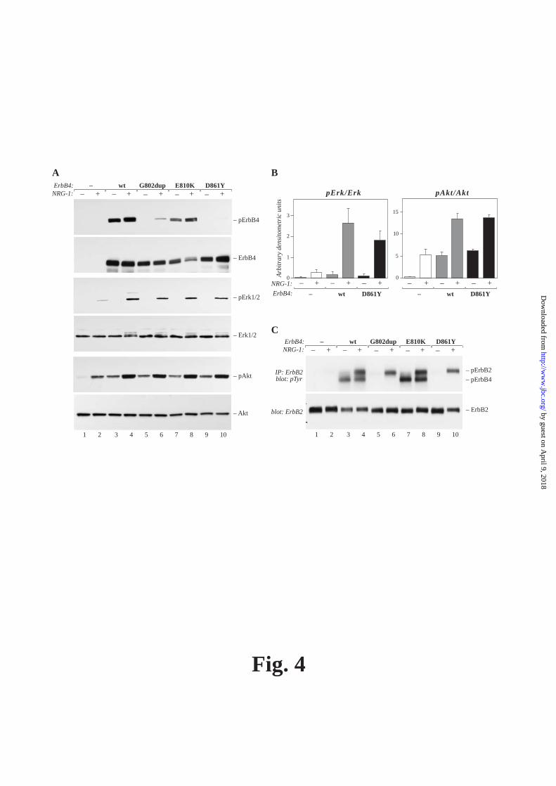
Fig. 4
A
– pErk1/2
– Erk1/2
– pAkt
– Akt
– pErbB4
– ErbB4
1 2 3 4 5 6 7 8 9 10
ErbB4: wt G802dup D861Y+NRG-1: _ +_ +_
E810K+_ +_
–
C
– pErbB2
– ErbB2
ErbB4: wt G802dup D861Y+NRG-1: _ +_ +_
E810K+_ +_
–
1 2 3 4 5 6 7 8 9 10
– pErbB4IP: ErbB2blot: pTyr
blot: ErbB2
0
1
2
3
0
5
10
15
NRG-1: +_ +_ +_ +_ +_ +_
ErbB4: wt D861Y – wt D861Y –
Arb
itra
ry d
ensi
tom
etri
c un
its
pErk/Erk pAkt/Akt
B
by guest on April 9, 2018
http://ww
w.jbc.org/
Dow
nloaded from
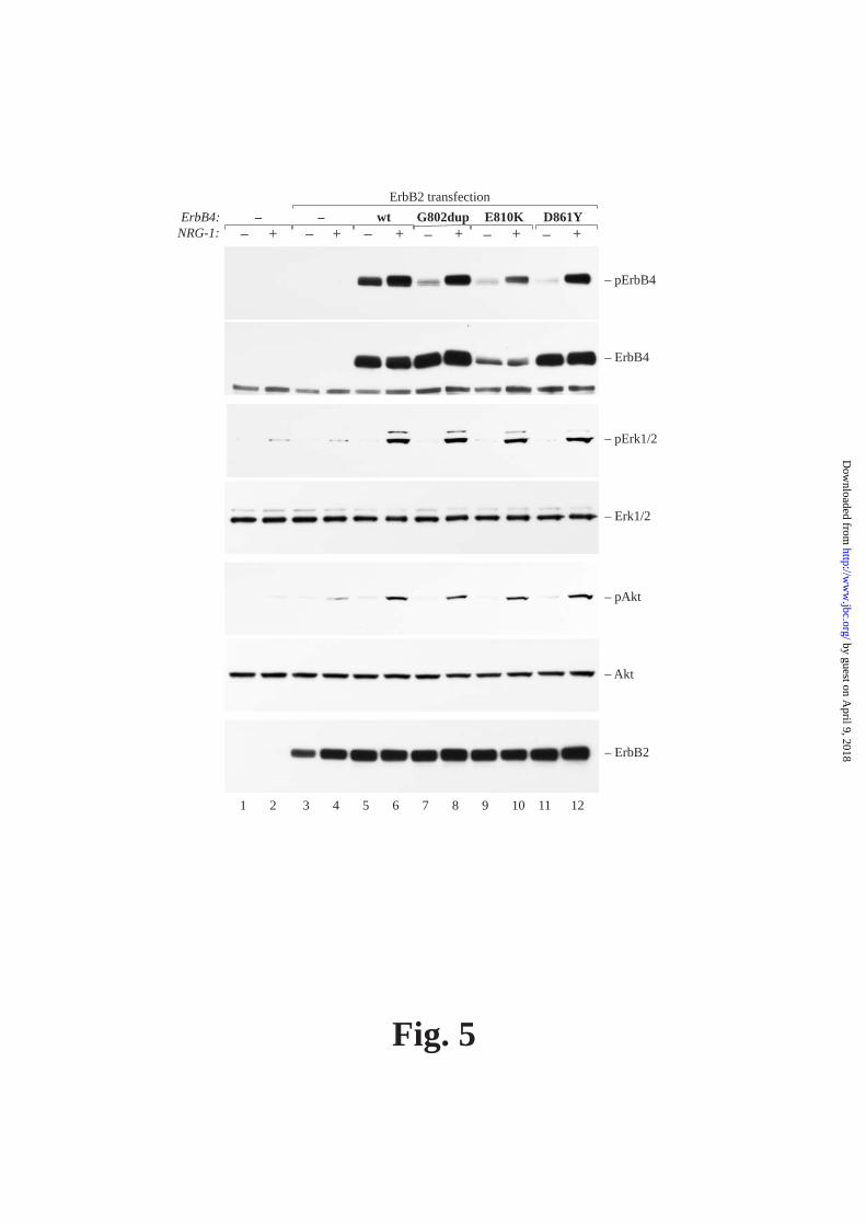
Fig. 5
– pErk1/2
– Erk1/2
– pAkt
– Akt
– pErbB4
– ErbB4
1 2 3 4 5 6 7 8 9 10 11 12
ErbB4: wt G802dup D861Y+NRG-1: _ +_ +_
E810K+_ +_
–+_
–
– ErbB2
ErbB2 transfection
by guest on April 9, 2018
http://ww
w.jbc.org/
Dow
nloaded from

Fig. 6
– pSTAT5
– STAT5
– ErbB4
ErbB4: wt G802dup D861Y+NRG-1: _ +_ +_
E810K+_ +_
–
1 2 3 4 5 6 7 8 9 10
A BErbB4: wt kinase-dead
NRG-1: +_ +_
– pSTAT5
– STAT5
– pErbB4
– ErbB4
1 2 3 4
by guest on April 9, 2018
http://ww
w.jbc.org/
Dow
nloaded from
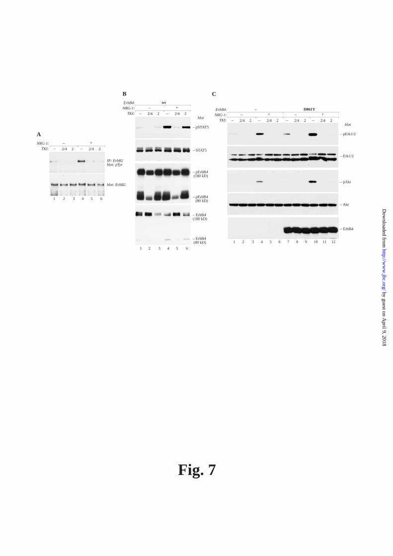
Fig. 7
IP: ErbB2blot: pTyr
blot: ErbB2
1 2 3 4 5 6
TKI:NRG-1: +_
2/4_ 2 2/4_ 2
A
B
– pErbB4(180 kD)
TKI:
ErbB4:NRG-1: +_
wt
2/4_ 2 2/4_ 2
– pSTAT5
– STAT5
blot
– pErbB4(80 kD)
– ErbB4(180 kD)
– ErbB4(80 kD)
1 2 3 4 5 6
C
– pErk1/2
– Erk1/2
– pAkt
– Akt
– ErbB4
1 2 3 4 5 6 7 8 9 10 11 12
TKI:
ErbB4: D861YNRG-1: +_ +_
–
2/4_ 2 2/4_ 2 2/4_ 2 2/4_ 2blot
by guest on April 9, 2018
http://ww
w.jbc.org/
Dow
nloaded from
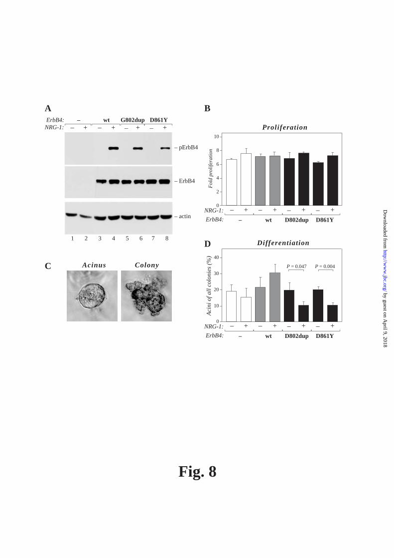
Fig. 8
A
– pErbB4
– ErbB4
ErbB4: wt G802dup D861Y+NRG-1: _ +_ +_ +_
–
1 2 3 4 5 6 7 8
– actin
Acinus ColonyC
B
D
0
2
4
6
8
10
Fol
d pr
olif
erat
ion
NRG-1: +_ +_ +_
ErbB4: wt D802dup –
+_
D861Y
Proliferation
0
10
20
30
40
Aci
ni o
f all
col
onie
s (%
)
P = 0.047 P = 0.004
NRG-1: +_ +_ +_
ErbB4: wt D802dup –
+_
D861Y
Differentiation
by guest on April 9, 2018
http://ww
w.jbc.org/
Dow
nloaded from

Fig. 9
ErkAkt
STAT5
ErbB2 ErbB4 wt
ErkAkt
ErbB2 ErbB4 mutant
NRG NRG
by guest on April 9, 2018
http://ww
w.jbc.org/
Dow
nloaded from

Johnson and Klaus EleniusDenis Tvorogov, Maria Sundvall, Kari Kurppa, Maija Hollmen, Susanna Repo, Mark S.
transduction pathways in cancerSomatic mutations of ERBB4: Selective loss-of-function phenotype affecting signal
published online December 19, 2008J. Biol. Chem.
10.1074/jbc.M805438200Access the most updated version of this article at doi:
Alerts:
When a correction for this article is posted•
When this article is cited•
to choose from all of JBC's e-mail alertsClick here
Supplemental material:
http://www.jbc.org/content/suppl/2008/12/23/M805438200.DC1
by guest on April 9, 2018
http://ww
w.jbc.org/
Dow
nloaded from