Labs 9 – 13 Bone Physiology and Anatomy and Skeletal System
76
Labs 9 – 12 Bone Physiology and Anatomy and Skeletal System Please refer to the bone list for markings to learn
Transcript of Labs 9 – 13 Bone Physiology and Anatomy and Skeletal System
Labs 9 – 12Bone Physiology and Anatomy
and Skeletal SystemPlease refer to the bone list for
markings to learn
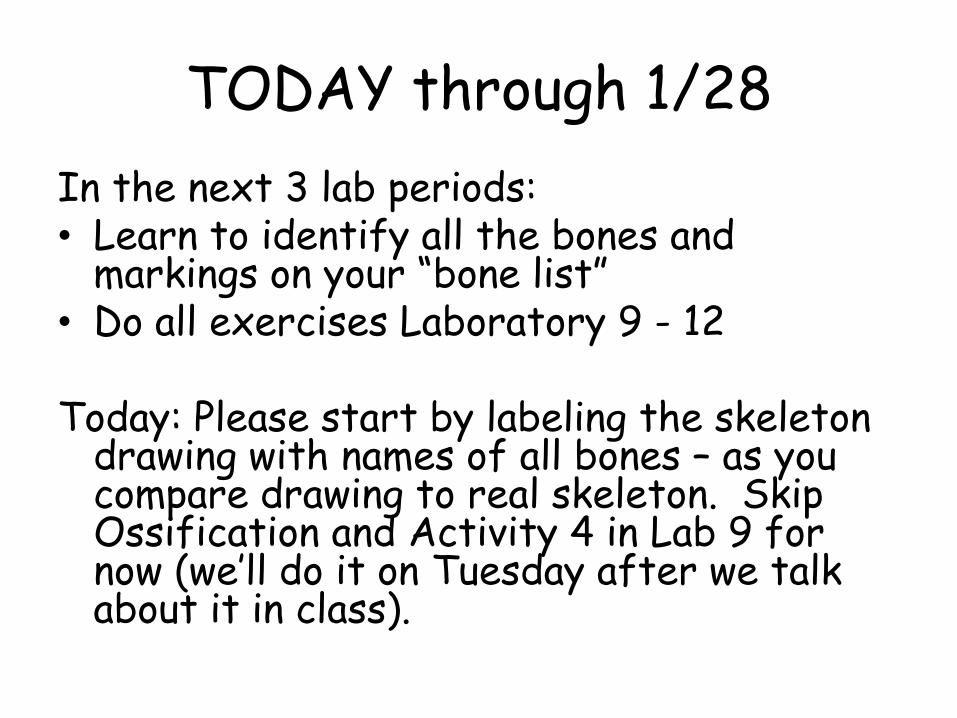
TODAY through 1/28
In the next 3 lab periods: • Learn to identify all the bones and
markings on your “bone list” • Do all exercises Laboratory 9 - 12
Today: Please start by labeling the skeleton drawing with names of all bones – as you compare drawing to real skeleton. Skip Ossification and Activity 4 in Lab 9 for now (we’ll do it on Tuesday after we talk about it in class).
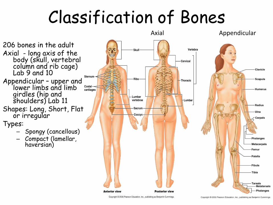
Classification of Bones
206 bones in the adultAxial - long axis of the
body (skull, vertebral column and rib cage) Lab 9 and 10
Appendicular – upper and lower limbs and limb girdles (hip and shoulders) Lab 11
Shapes: Long, Short, Flat or irregular
Types: – Spongy (cancellous)– Compact (lamellar,
haversian)
Axial Appendicular
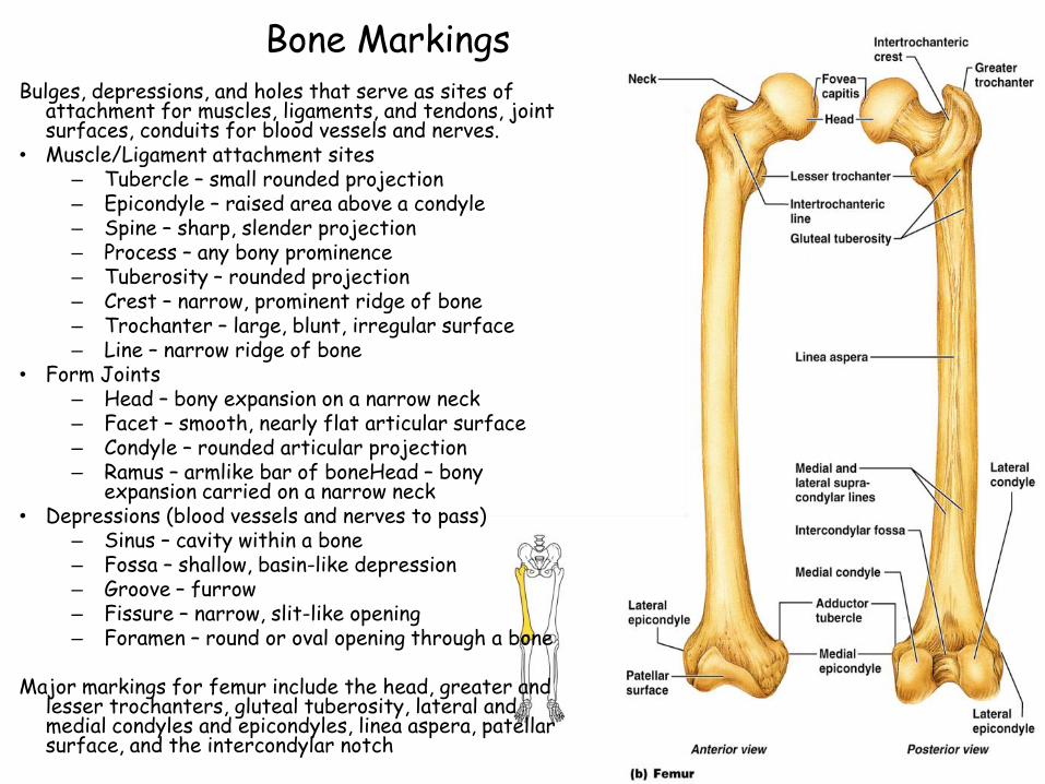
Bone MarkingsBulges, depressions, and holes that serve as sites of
attachment for muscles, ligaments, and tendons, joint surfaces, conduits for blood vessels and nerves.
• Muscle/Ligament attachment sites– Tubercle – small rounded projection– Epicondyle – raised area above a condyle– Spine – sharp, slender projection– Process – any bony prominence– Tuberosity – rounded projection– Crest – narrow, prominent ridge of bone– Trochanter – large, blunt, irregular surface– Line – narrow ridge of bone
• Form Joints– Head – bony expansion on a narrow neck– Facet – smooth, nearly flat articular surface– Condyle – rounded articular projection– Ramus – armlike bar of boneHead – bony
expansion carried on a narrow neck• Depressions (blood vessels and nerves to pass)
– Sinus – cavity within a bone– Fossa – shallow, basin-like depression– Groove – furrow– Fissure – narrow, slit-like opening– Foramen – round or oval opening through a bone
Major markings for femur include the head, greater and lesser trochanters, gluteal tuberosity, lateral and medial condyles and epicondyles, linea aspera, patellar surface, and the intercondylar notch

Bone Markings
Table 6–1 (2 of 2)
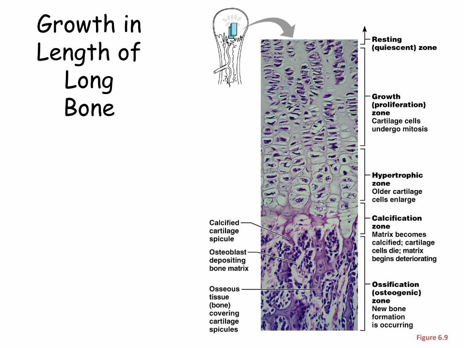
Growth in Length of
Long Bone
Figure 6.9
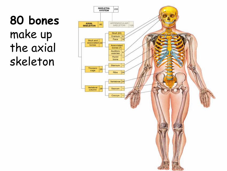
80 bonesmake up the axialskeleton
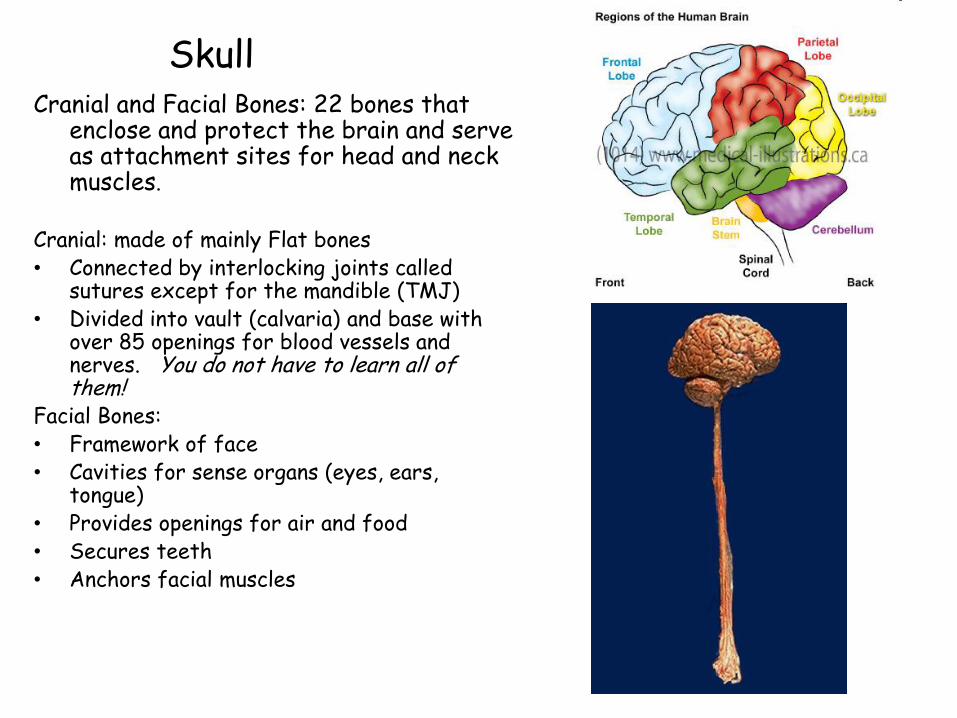
SkullCranial and Facial Bones: 22 bones that
enclose and protect the brain and serve as attachment sites for head and neck muscles.
Cranial: made of mainly Flat bones• Connected by interlocking joints called
sutures except for the mandible (TMJ)• Divided into vault (calvaria) and base with
over 85 openings for blood vessels and nerves. You do not have to learn all of them!
Facial Bones:• Framework of face• Cavities for sense organs (eyes, ears,
tongue) • Provides openings for air and food• Secures teeth• Anchors facial muscles
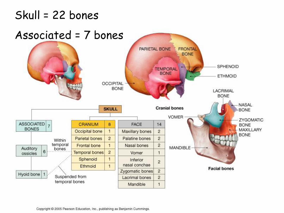
Skull = 22 bones
Associated = 7 bones
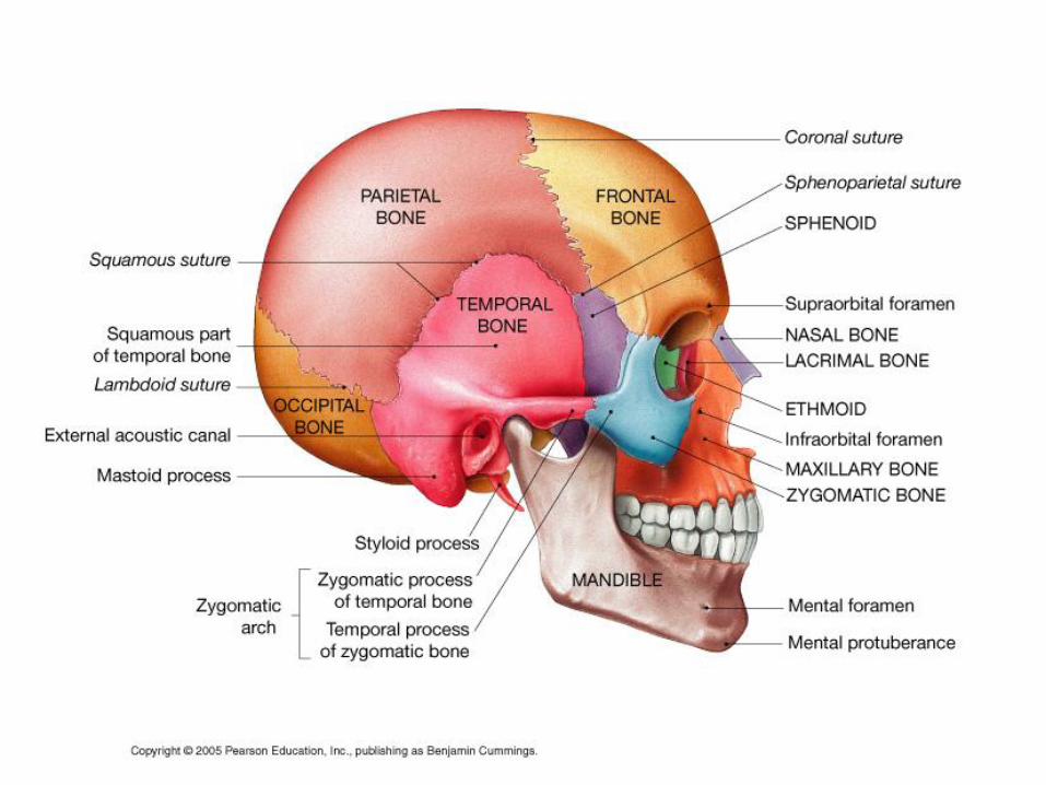
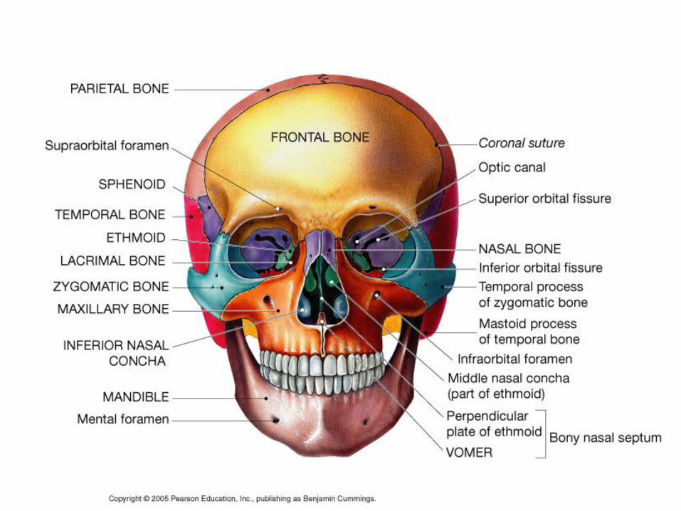
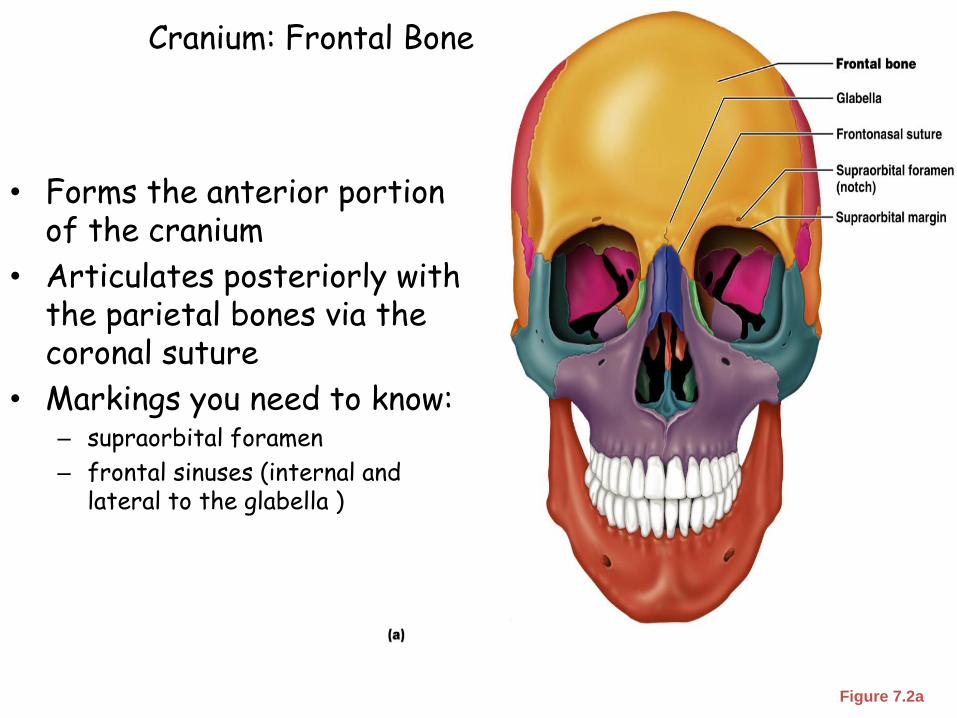
Cranium: Frontal Bone
Figure 7.2a
• Forms the anterior portion of the cranium
• Articulates posteriorly with the parietal bones via the coronal suture
• Markings you need to know:– supraorbital foramen
– frontal sinuses (internal and lateral to the glabella )
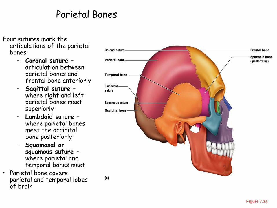
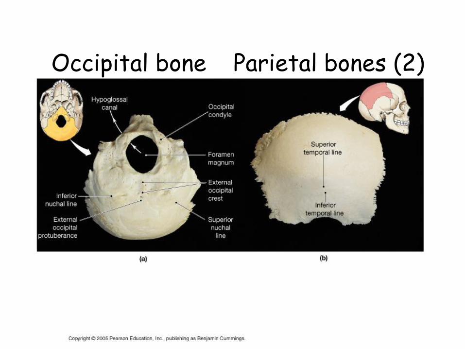
Parietal Bones
Four sutures mark the articulations of the parietal bones
– Coronal suture –articulation between parietal bones and frontal bone anteriorly
– Sagittal suture –where right and left parietal bones meet superiorly
– Lambdoid suture –where parietal bones meet the occipital bone posteriorly
– Squamosal or squamous suture –where parietal and temporal bones meet
• Parietal bone covers parietal and temporal lobes of brain
Figure 7.3a
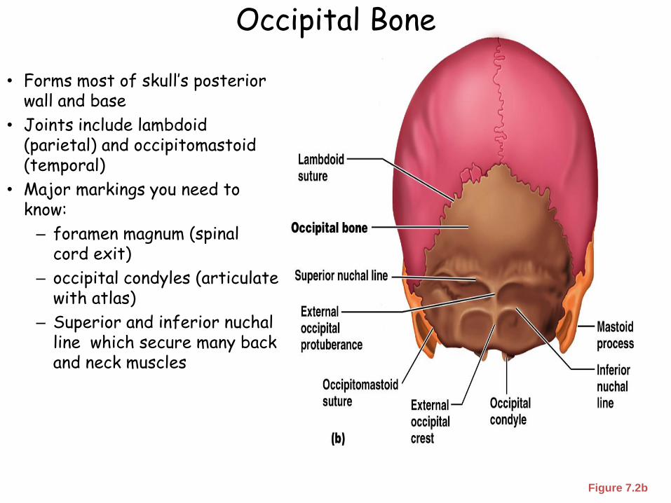
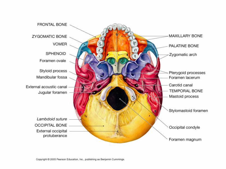
Occipital Bone
• Forms most of skull’s posterior wall and base
• Joints include lambdoid(parietal) and occipitomastoid(temporal)
• Major markings you need to know:
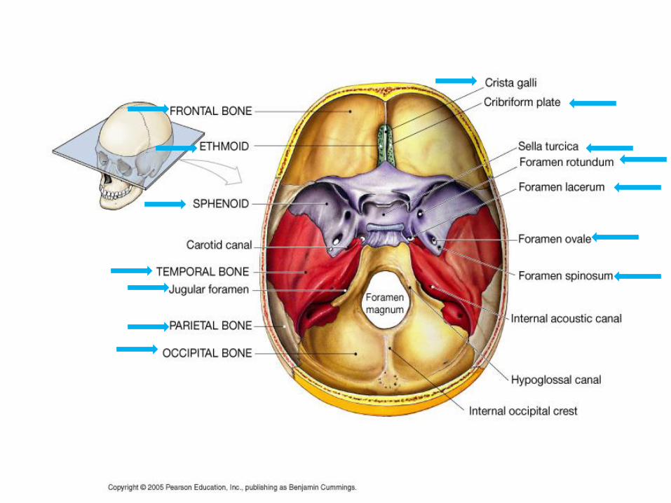
– foramen magnum (spinal cord exit)
– occipital condyles (articulate with atlas)
– Superior and inferior nuchalline which secure many back and neck muscles
Figure 7.2b
Occipital bone Parietal bones (2)
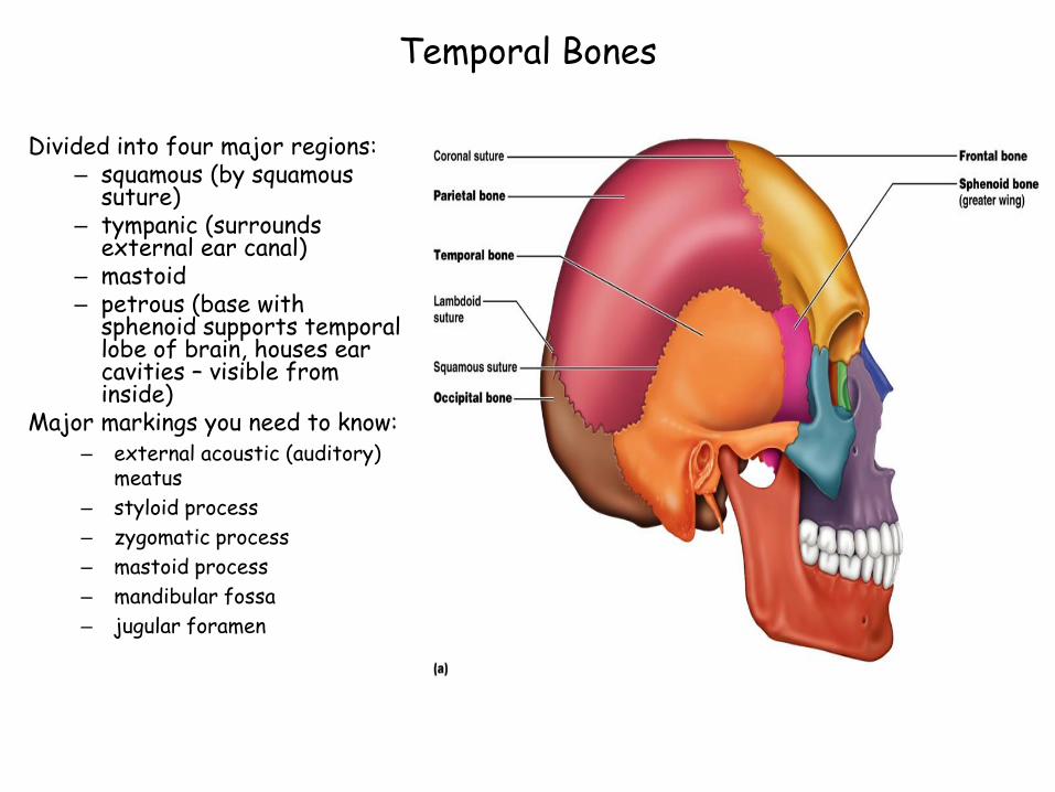
Temporal Bones
Divided into four major regions:– squamous (by squamous
suture)– tympanic (surrounds
external ear canal)– mastoid– petrous (base with
sphenoid supports temporal lobe of brain, houses ear cavities – visible from inside)
Major markings you need to know:– external acoustic (auditory)
meatus
– styloid process
– zygomatic process
– mastoid process
– mandibular fossa
– jugular foramen
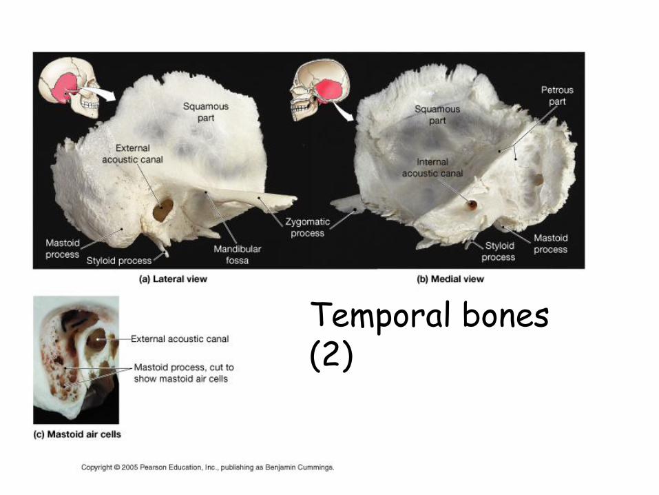
Temporal bones (2)
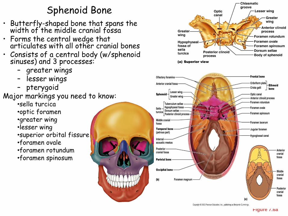
Sphenoid Bone
Figure 7.6a
• Butterfly-shaped bone that spans the width of the middle cranial fossa
• Forms the central wedge that articulates with all other cranial bones
• Consists of a central body (w/sphenoid sinuses) and 3 processes:
– greater wings – lesser wings– pterygoid
Major markings you need to know:•sella turcica•optic foramen•greater wing•lesser wing•superior orbital fissure•foramen ovale•foramen rotundum•foramen spinosum
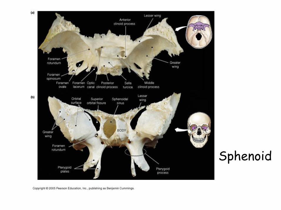
Sphenoid
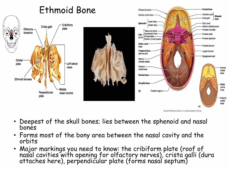
Ethmoid Bone
• Deepest of the skull bones; lies between the sphenoid and nasal bones
• Forms most of the bony area between the nasal cavity and the orbits
• Major markings you need to know: the cribiform plate (roof of nasal cavities with opening for olfactory nerves), crista galli (dura attaches here), perpendicular plate (forms nasal septum)
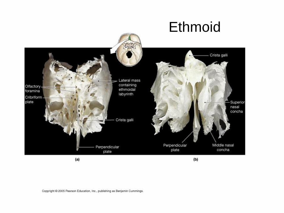
Ethmoid

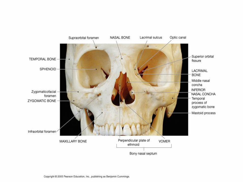
Skull: Other Bones
• Zygomatic: Irregularly shaped bones (cheekbones) that form the cheeks and the inferior-lateral margins of the orbits
• Nasal bones – thin medially fused bones that form the bridge of the nose
• Lacrimal bones – contribute to the medial walls of the orbit and contain a deep groove called the lacrimal fossa that houses the lacrimal glands
• Palatine bones – two bony plates that form the posterior portion of the hard palate, the postero-lateral walls of the nasal cavity, and a small part of the orbits
• Vomer – plow-shaped bone that forms part of the nasal septum
• Inferior nasal conchae – paired, curved bones in the nasal cavity that form part of the lateral walls of the nasal cavity
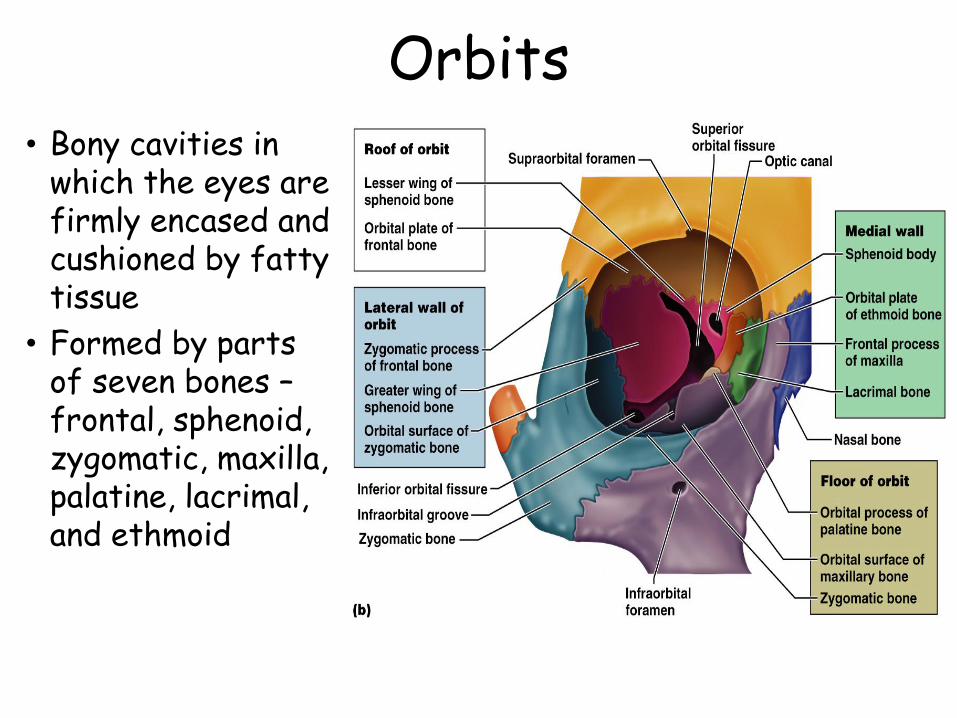
Orbits• Bony cavities in
which the eyes are firmly encased and cushioned by fatty tissue
• Formed by parts of seven bones –frontal, sphenoid, zygomatic, maxilla, palatine, lacrimal, and ethmoid
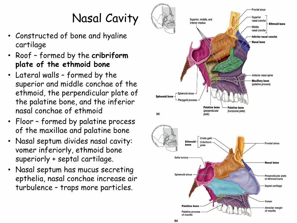
Nasal Cavity
• Constructed of bone and hyaline cartilage
• Roof – formed by the cribriform plate of the ethmoid bone
• Lateral walls – formed by the superior and middle conchae of the ethmoid, the perpendicular plate of the palatine bone, and the inferior nasal conchae of ethmoid
• Floor – formed by palatine process of the maxillae and palatine bone
• Nasal septum divides nasal cavity: vomer inferiorly, ethmoid bone superiorly + septal cartilage.
• Nasal septum has mucus secreting epthelia, nasal conchae increase air turbulence – traps more particles.
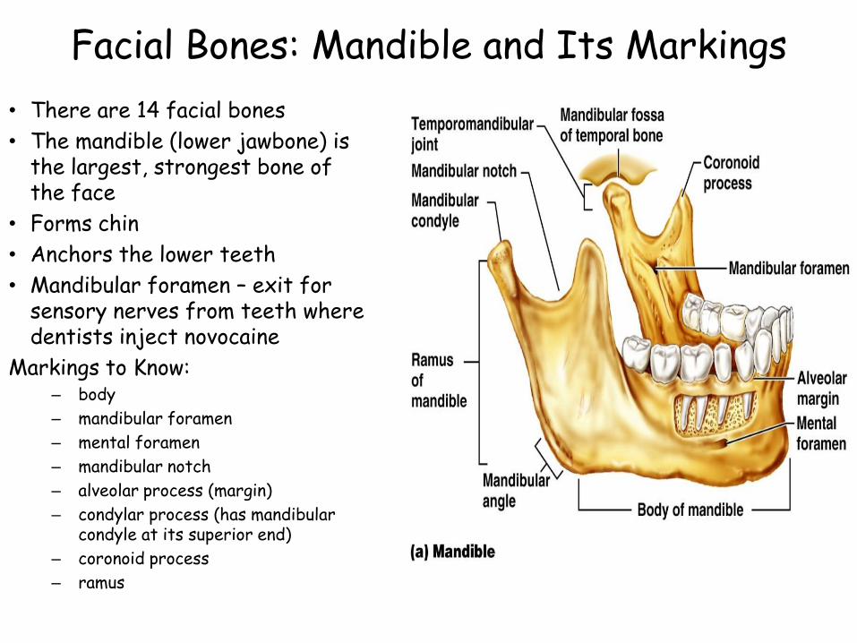
Facial Bones: Mandible and Its Markings
• There are 14 facial bones
• The mandible (lower jawbone) is the largest, strongest bone of the face
• Forms chin
• Anchors the lower teeth
• Mandibular foramen – exit for sensory nerves from teeth where dentists inject novocaine
Markings to Know:– body
– mandibular foramen
– mental foramen
– mandibular notch
– alveolar process (margin)
– condylar process (has mandibular condyle at its superior end)
– coronoid process
– ramus
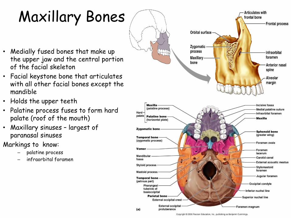
Maxillary Bones
• Medially fused bones that make up the upper jaw and the central portion of the facial skeleton
• Facial keystone bone that articulates with all other facial bones except the mandible
• Holds the upper teeth
• Palatine process fuses to form hard palate (roof of the mouth)
• Maxillary sinuses – largest of paranasal sinuses
Markings to know:– palatine process
– infraorbital foramen

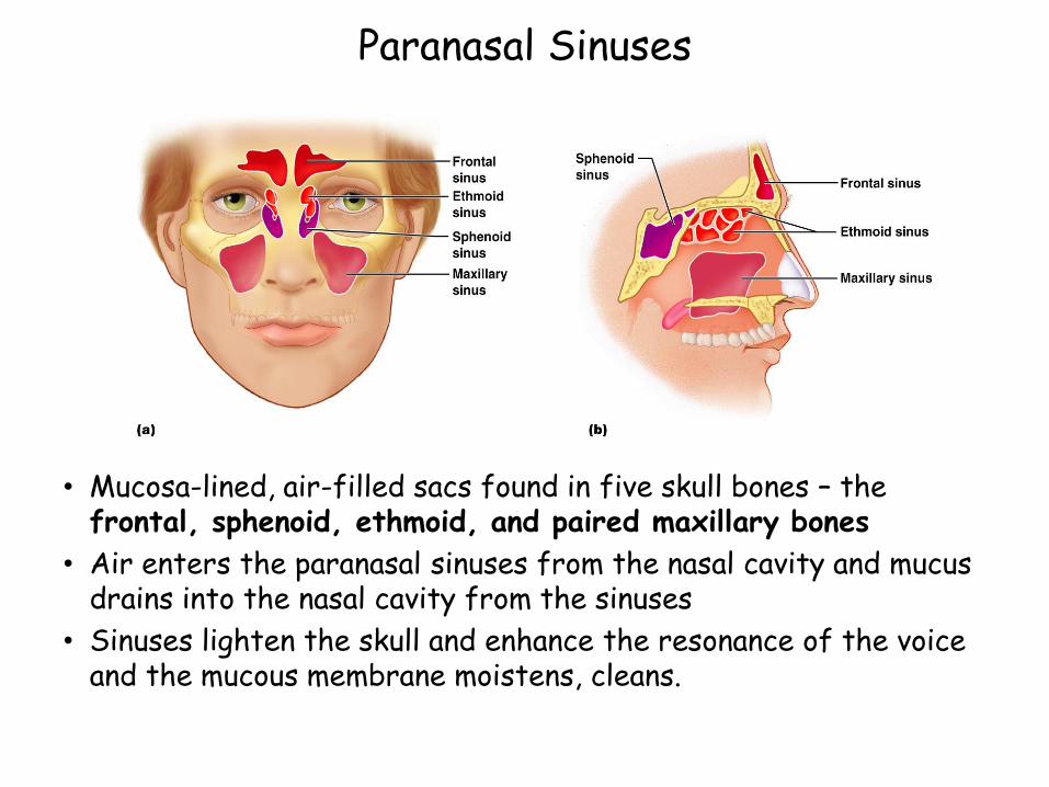
Paranasal Sinuses
• Mucosa-lined, air-filled sacs found in five skull bones – the frontal, sphenoid, ethmoid, and paired maxillary bones
• Air enters the paranasal sinuses from the nasal cavity and mucus drains into the nasal cavity from the sinuses
• Sinuses lighten the skull and enhance the resonance of the voice and the mucous membrane moistens, cleans.
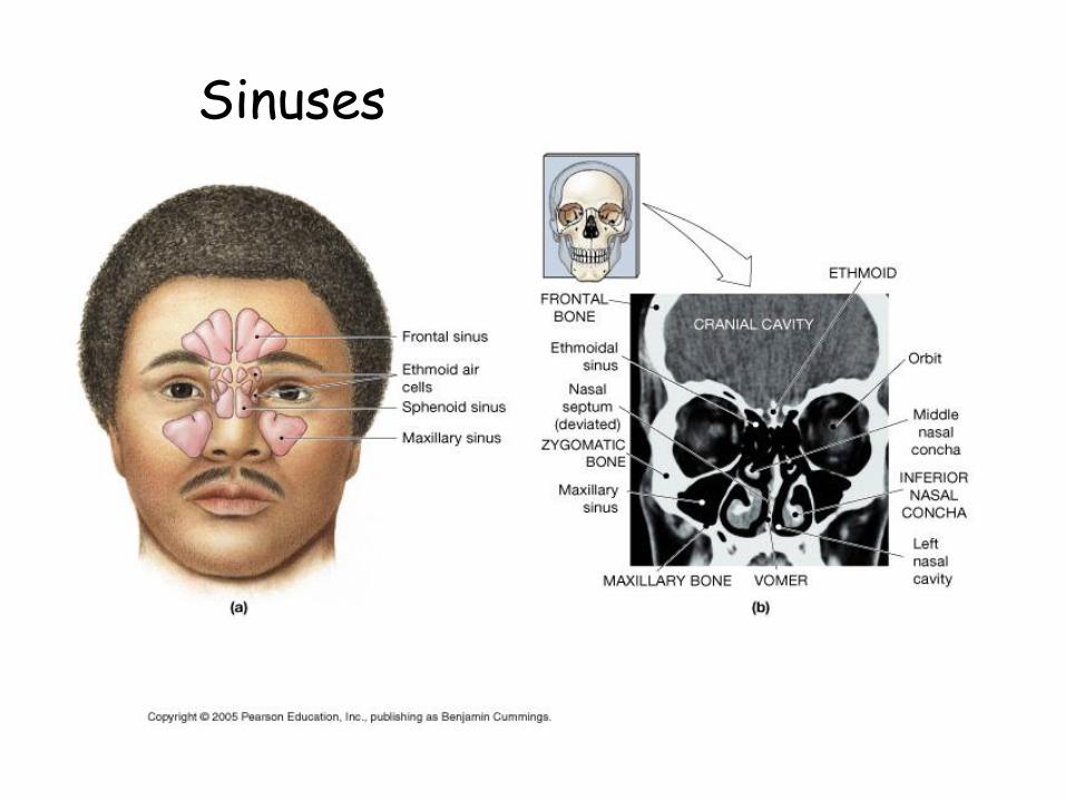
Sinuses
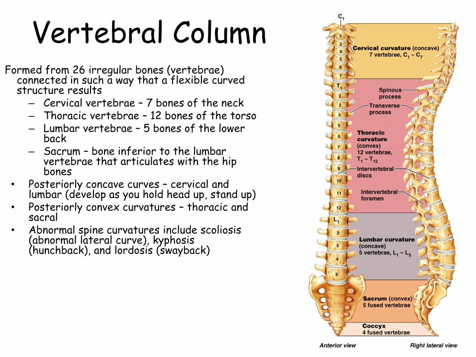
Vertebral ColumnFormed from 26 irregular bones (vertebrae)
connected in such a way that a flexible curved structure results
– Cervical vertebrae – 7 bones of the neck– Thoracic vertebrae – 12 bones of the torso– Lumbar vertebrae – 5 bones of the lower
back– Sacrum – bone inferior to the lumbar
vertebrae that articulates with the hip bones
• Posteriorly concave curves – cervical and lumbar (develop as you hold head up, stand up)
• Posteriorly convex curvatures – thoracic and sacral
• Abnormal spine curvatures include scoliosis (abnormal lateral curve), kyphosis(hunchback), and lordosis (swayback)
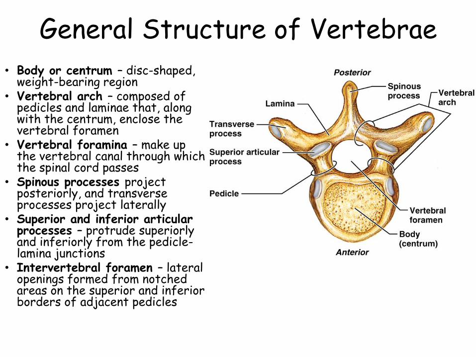
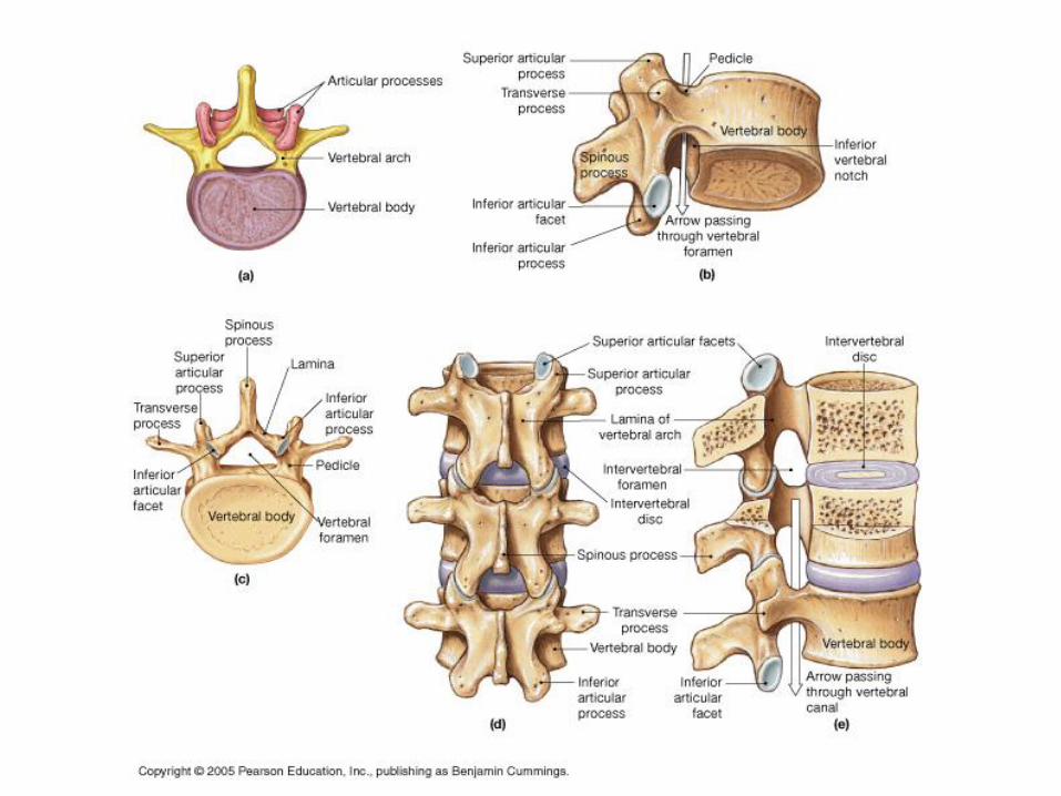
General Structure of Vertebrae
• Body or centrum – disc-shaped, weight-bearing region
• Vertebral arch – composed of pedicles and laminae that, along with the centrum, enclose the vertebral foramen
• Vertebral foramina – make up the vertebral canal through which the spinal cord passes
• Spinous processes project posteriorly, and transverse processes project laterally
• Superior and inferior articular processes – protrude superiorly and inferiorly from the pedicle-lamina junctions
• Intervertebral foramen – lateral openings formed from notched areas on the superior and inferior borders of adjacent pedicles
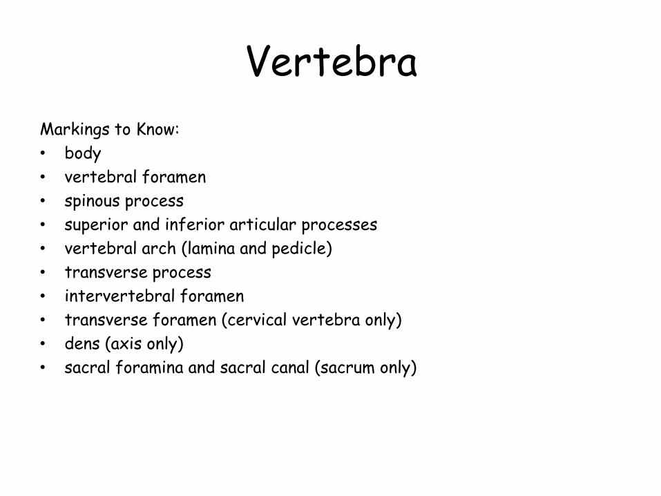
Vertebra
Markings to Know:
• body
• vertebral foramen
• spinous process
• superior and inferior articular processes
• vertebral arch (lamina and pedicle)
• transverse process
• intervertebral foramen
• transverse foramen (cervical vertebra only)
• dens (axis only)
• sacral foramina and sacral canal (sacrum only)
Vertebrae
• Bodies get larger as you descend (more weight)
• Foramen get smaller as you descend (less information in spinal cord)
• Shape of spinous process helps to identify vertebrae from each region:
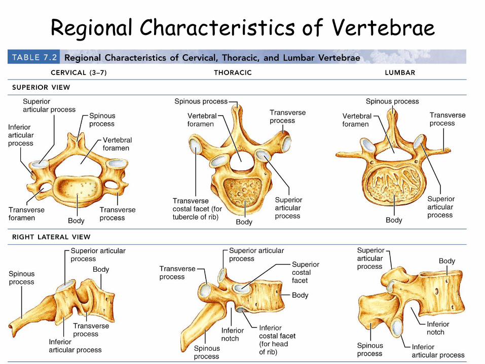
Regional Characteristics of Vertebrae
Table 7.2.2
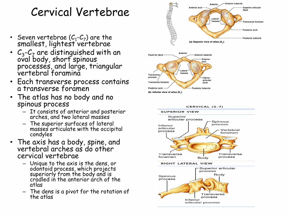
Cervical Vertebrae
• Seven vertebrae (C1-C7) are the smallest, lightest vertebrae
• C3-C7 are distinguished with an oval body, short spinous processes, and large, triangular vertebral foramina
• Each transverse process contains a transverse foramen
• The atlas has no body and no spinous process– It consists of anterior and posterior
arches, and two lateral masses– The superior surfaces of lateral
masses articulate with the occipital condyles
• The axis has a body, spine, and vertebral arches as do other cervical vertebrae– Unique to the axis is the dens, or
odontoid process, which projects superiorly from the body and is cradled in the anterior arch of the atlas
– The dens is a pivot for the rotation of the atlas
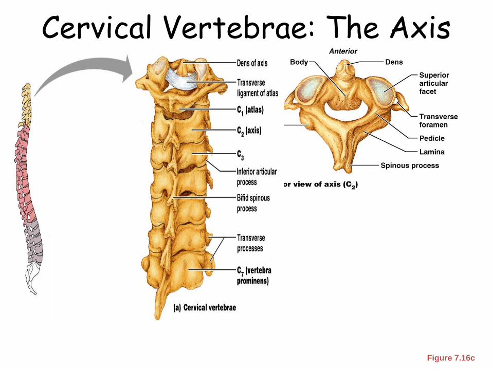
Cervical Vertebrae: The Axis (C2)
Figure 7.16c
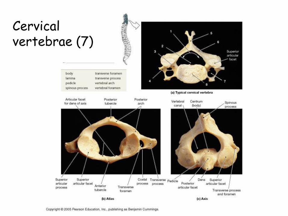
Cervical vertebrae (7)
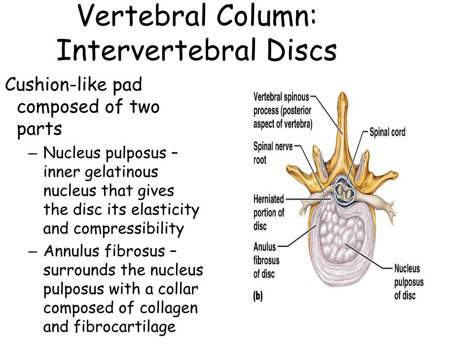
Vertebral Column: Intervertebral Discs
Cushion-like pad composed of two parts– Nucleus pulposus –
inner gelatinous nucleus that gives the disc its elasticity and compressibility
– Annulus fibrosus –surrounds the nucleus pulposus with a collar composed of collagen and fibrocartilage
Regional Characteristics of Vertebrae
Table 7.2.1
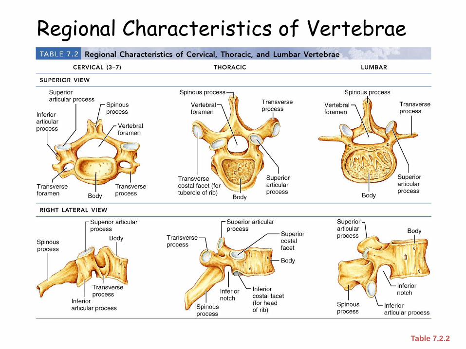
Regional Characteristics of Vertebrae
Table 7.2.2
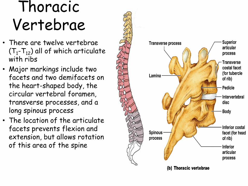
Thoracic Vertebrae
• There are twelve vertebrae (T1-T12) all of which articulate with ribs
• Major markings include two facets and two demifacets on the heart-shaped body, the circular vertebral foramen, transverse processes, and a long spinous process
• The location of the articulate facets prevents flexion and extension, but allows rotation of this area of the spine
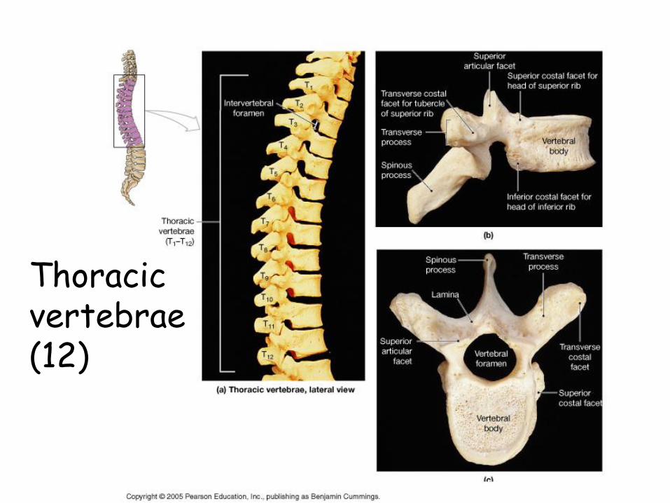
Thoracic vertebrae (12)
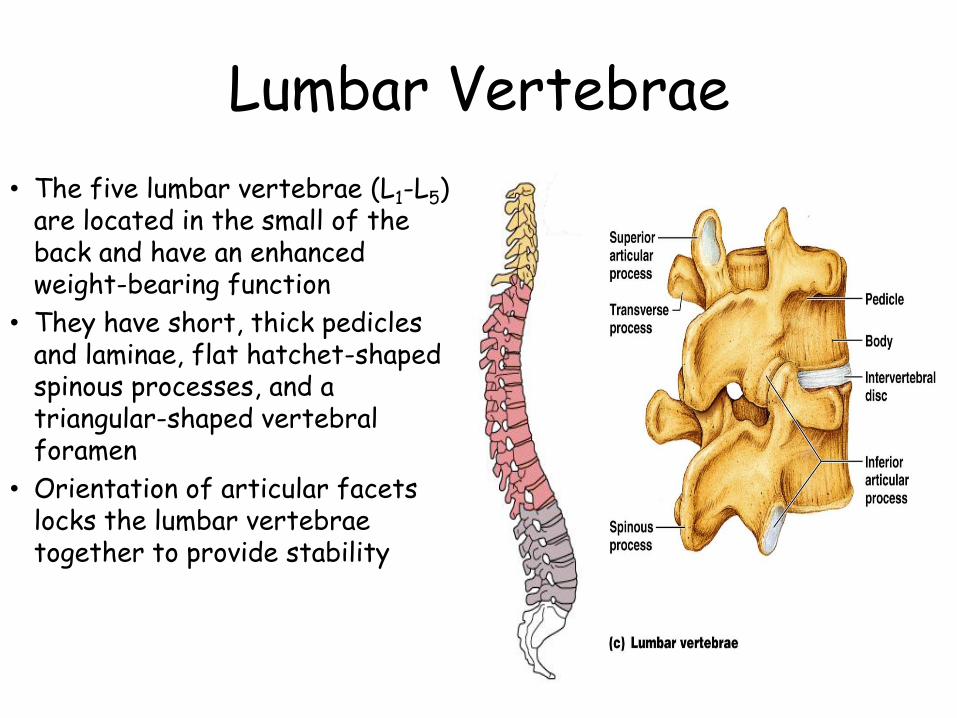
Lumbar Vertebrae
• The five lumbar vertebrae (L1-L5) are located in the small of the back and have an enhanced weight-bearing function
• They have short, thick pedicles and laminae, flat hatchet-shaped spinous processes, and a triangular-shaped vertebral foramen
• Orientation of articular facets locks the lumbar vertebrae together to provide stability
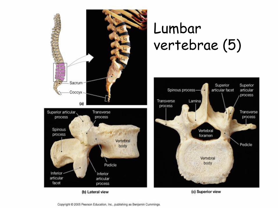
Lumbar vertebrae (5)
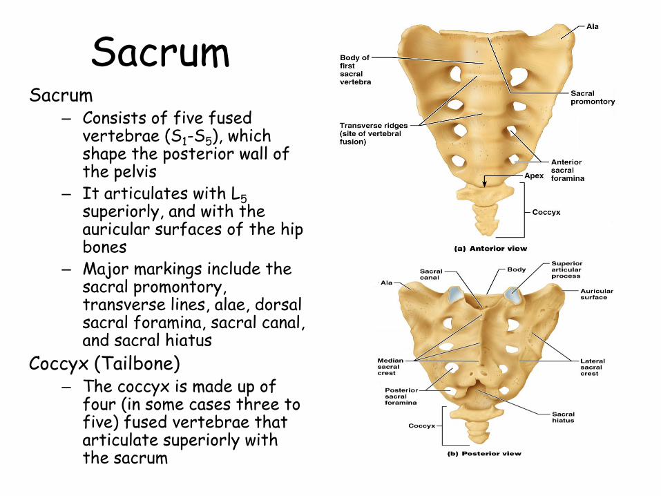
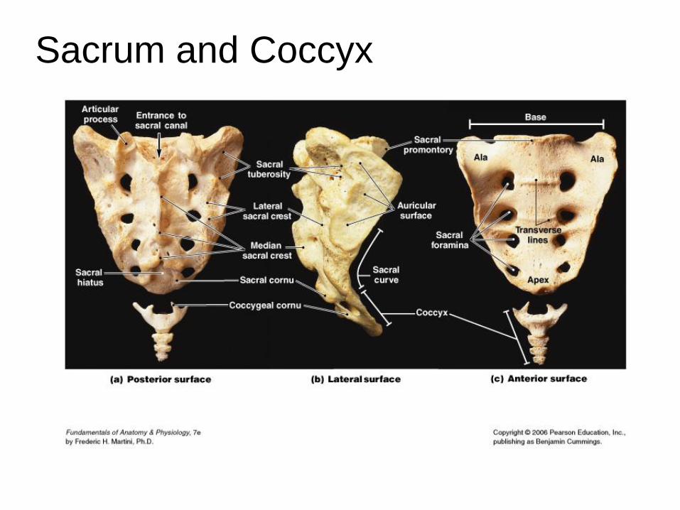
SacrumSacrum
– Consists of five fused vertebrae (S1-S5), which shape the posterior wall of the pelvis
– It articulates with L5superiorly, and with the auricular surfaces of the hip bones
– Major markings include the sacral promontory, transverse lines, alae, dorsal sacral foramina, sacral canal, and sacral hiatus
Coccyx (Tailbone)– The coccyx is made up of
four (in some cases three to five) fused vertebrae that articulate superiorly with the sacrum
Sacrum and Coccyx
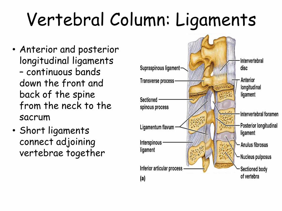
Vertebral Column: Ligaments
• Anterior and posterior longitudinal ligaments – continuous bands down the front and back of the spine from the neck to the sacrum
• Short ligaments connect adjoining vertebrae together
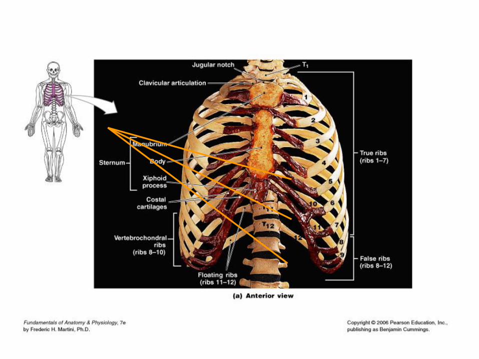
Bony Thorax (Thoracic Cage)
The thoracic cage is composed of the thoracic vertebrae dorsally, the ribs laterally, and the sternum and costal cartilages anteriorly
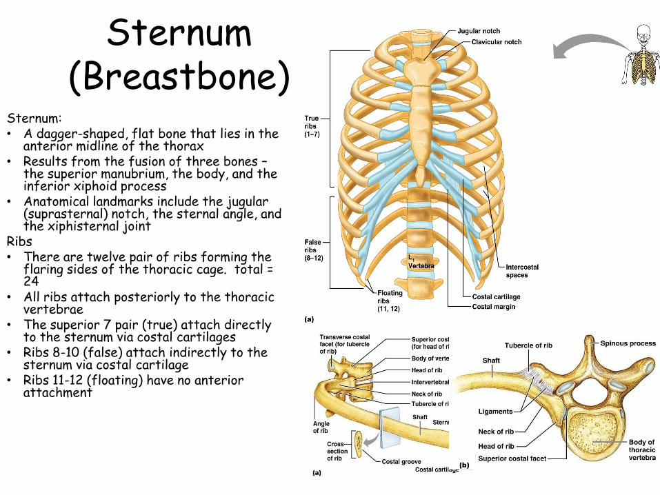
Sternum (Breastbone)
Sternum:• A dagger-shaped, flat bone that lies in the
anterior midline of the thorax• Results from the fusion of three bones –
the superior manubrium, the body, and the inferior xiphoid process
• Anatomical landmarks include the jugular (suprasternal) notch, the sternal angle, and the xiphisternal joint
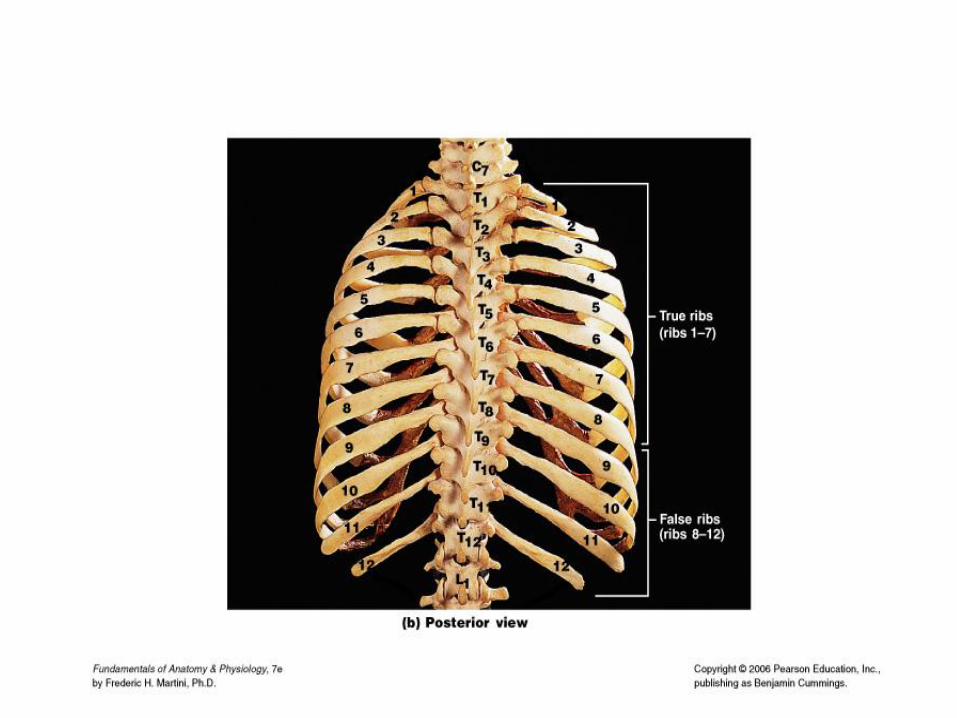
Ribs• There are twelve pair of ribs forming the
flaring sides of the thoracic cage. total = 24
• All ribs attach posteriorly to the thoracic vertebrae
• The superior 7 pair (true) attach directly to the sternum via costal cartilages
• Ribs 8-10 (false) attach indirectly to the sternum via costal cartilage
• Ribs 11-12 (floating) have no anterior attachment
Ribs and Sternum
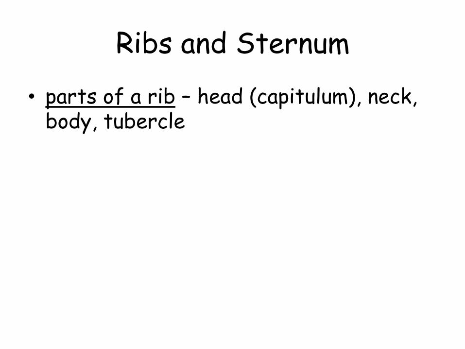
• parts of a rib – head (capitulum), neck, body, tubercle
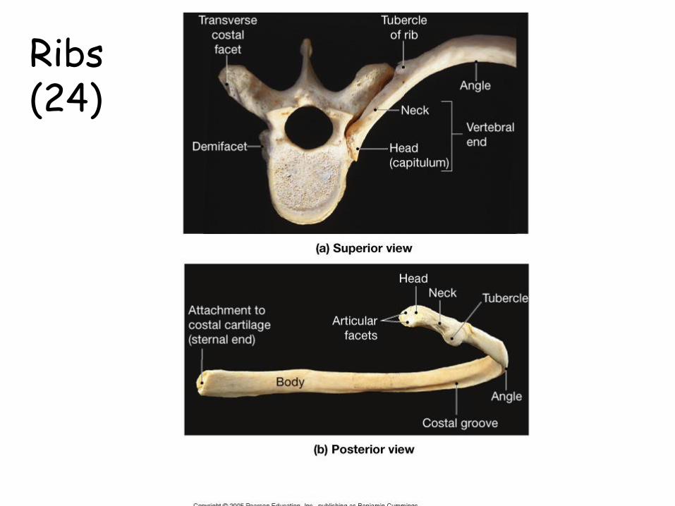
Ribs (24)
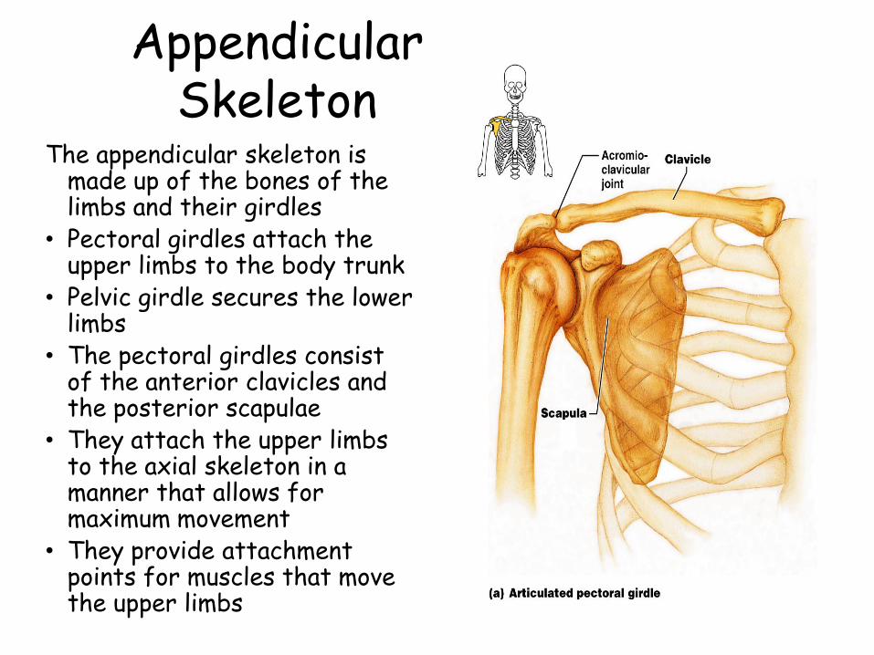
Appendicular Skeleton
The appendicular skeleton is made up of the bones of the limbs and their girdles
• Pectoral girdles attach the upper limbs to the body trunk
• Pelvic girdle secures the lower limbs
• The pectoral girdles consist of the anterior clavicles and the posterior scapulae
• They attach the upper limbs to the axial skeleton in a manner that allows for maximum movement
• They provide attachment points for muscles that move the upper limbs
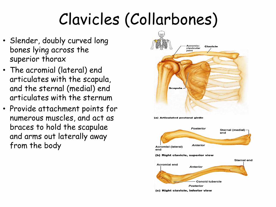
Clavicles (Collarbones)• Slender, doubly curved long
bones lying across the superior thorax
• The acromial (lateral) end articulates with the scapula, and the sternal (medial) end articulates with the sternum
• Provide attachment points for numerous muscles, and act as braces to hold the scapulae and arms out laterally away from the body
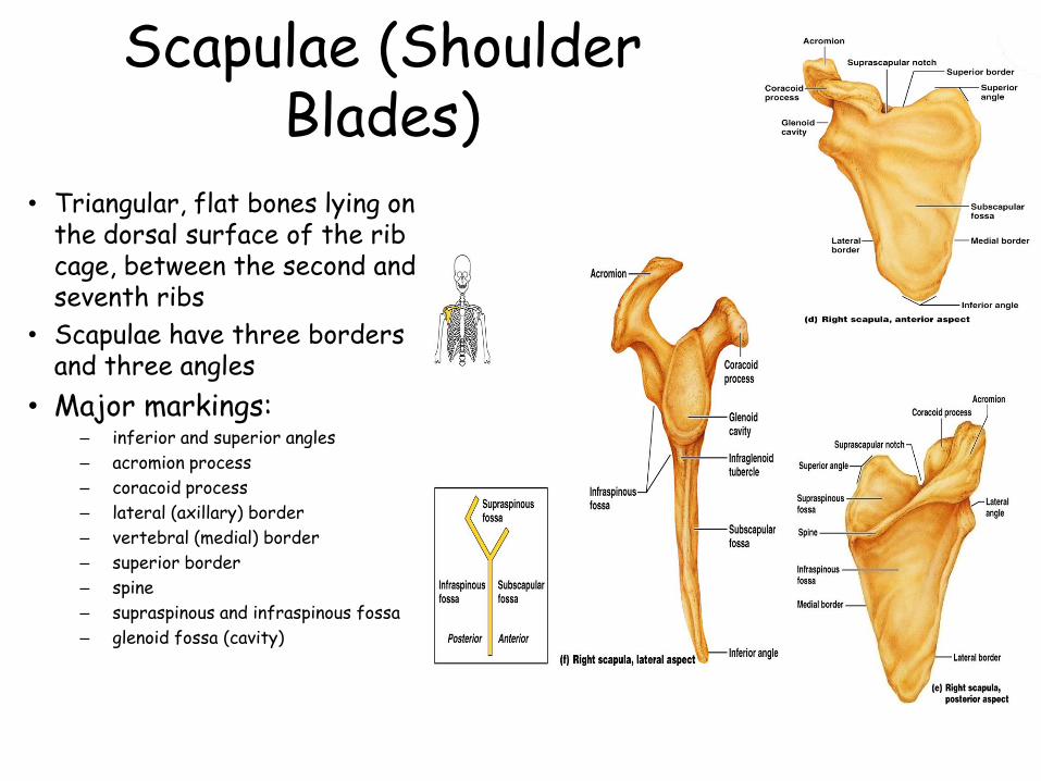
Scapulae (Shoulder Blades)
• Triangular, flat bones lying on the dorsal surface of the rib cage, between the second and seventh ribs
• Scapulae have three borders and three angles
• Major markings:– inferior and superior angles
– acromion process
– coracoid process
– lateral (axillary) border
– vertebral (medial) border
– superior border
– spine
– supraspinous and infraspinous fossa
– glenoid fossa (cavity)
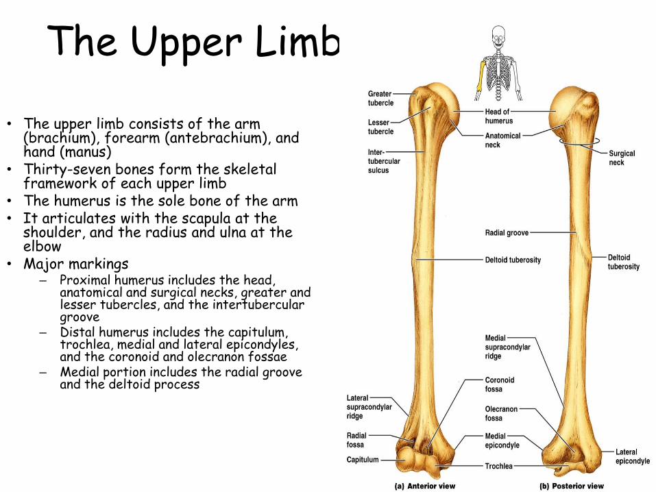
The Upper Limb
• The upper limb consists of the arm (brachium), forearm (antebrachium), and hand (manus)
• Thirty-seven bones form the skeletal framework of each upper limb
• The humerus is the sole bone of the arm• It articulates with the scapula at the
shoulder, and the radius and ulna at the elbow
• Major markings– Proximal humerus includes the head,
anatomical and surgical necks, greater and lesser tubercles, and the intertubercular groove
– Distal humerus includes the capitulum, trochlea, medial and lateral epicondyles, and the coronoid and olecranon fossae
– Medial portion includes the radial groove and the deltoid process
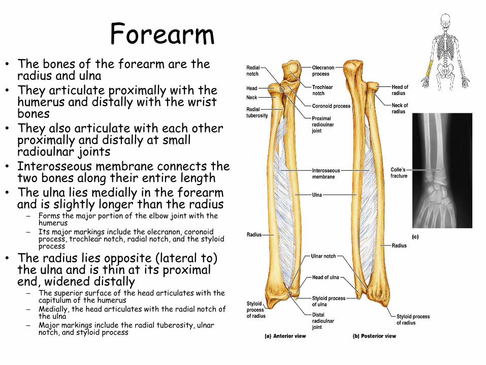
Forearm• The bones of the forearm are the
radius and ulna • They articulate proximally with the
humerus and distally with the wrist bones
• They also articulate with each other proximally and distally at small radioulnar joints
• Interosseous membrane connects the two bones along their entire length
• The ulna lies medially in the forearm and is slightly longer than the radius
– Forms the major portion of the elbow joint with the humerus
– Its major markings include the olecranon, coronoid process, trochlear notch, radial notch, and the styloid process
• The radius lies opposite (lateral to) the ulna and is thin at its proximal end, widened distally
– The superior surface of the head articulates with the capitulum of the humerus
– Medially, the head articulates with the radial notch of the ulna
– Major markings include the radial tuberosity, ulnar notch, and styloid process
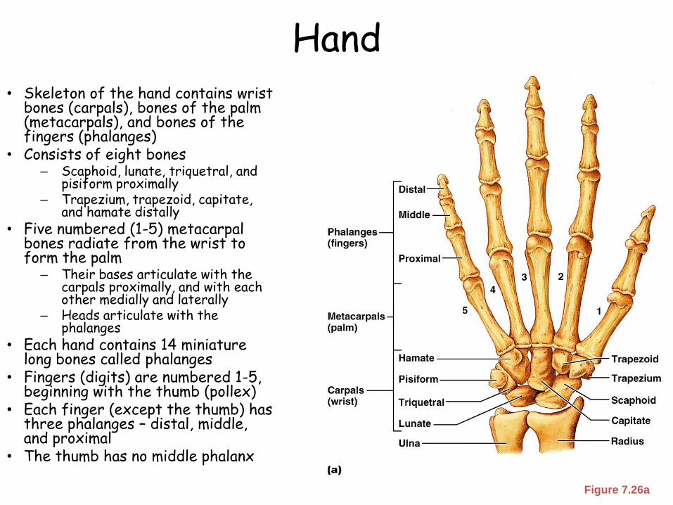
Hand• Skeleton of the hand contains wrist
bones (carpals), bones of the palm (metacarpals), and bones of the fingers (phalanges)
• Consists of eight bones– Scaphoid, lunate, triquetral, and
pisiform proximally– Trapezium, trapezoid, capitate,
and hamate distally• Five numbered (1-5) metacarpal
bones radiate from the wrist to form the palm
– Their bases articulate with the carpals proximally, and with each other medially and laterally
– Heads articulate with the phalanges
• Each hand contains 14 miniature long bones called phalanges
• Fingers (digits) are numbered 1-5, beginning with the thumb (pollex)
• Each finger (except the thumb) has three phalanges – distal, middle, and proximal
• The thumb has no middle phalanx
Figure 7.26a
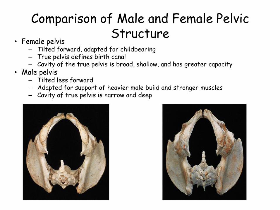
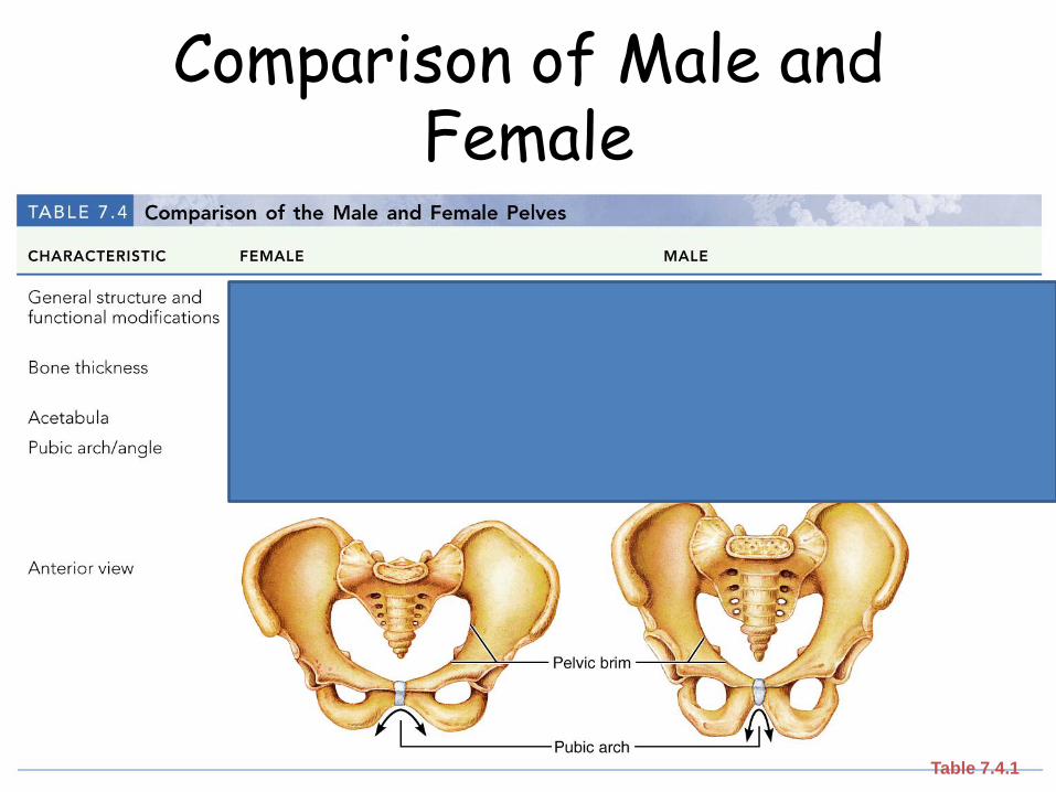
Comparison of Male and Female Pelvic Structure
• Female pelvis– Tilted forward, adapted for childbearing– True pelvis defines birth canal– Cavity of the true pelvis is broad, shallow, and has greater capacity
• Male pelvis– Tilted less forward– Adapted for support of heavier male build and stronger muscles– Cavity of true pelvis is narrow and deep
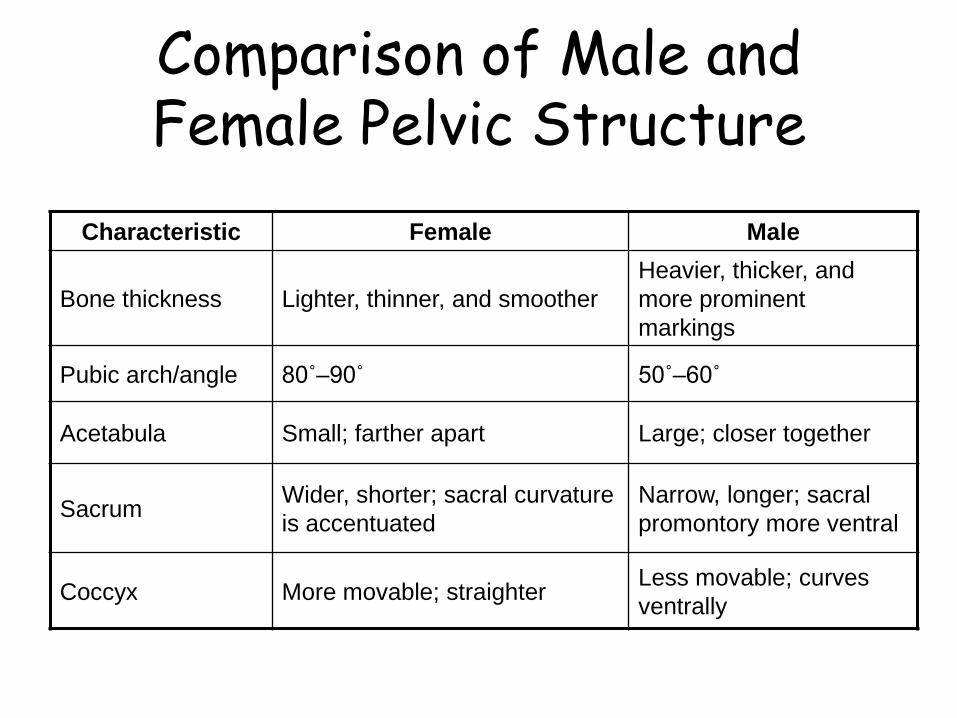
Characteristic Female Male
Bone thickness Lighter, thinner, and smoother
Heavier, thicker, and
more prominent
markings
Pubic arch/angle 80˚–90˚ 50˚–60˚
Acetabula Small; farther apart Large; closer together
SacrumWider, shorter; sacral curvature
is accentuated
Narrow, longer; sacral
promontory more ventral
Coccyx More movable; straighterLess movable; curves
ventrally
Comparison of Male and Female Pelvic Structure
Comparison of Male and Female
Table 7.4.1
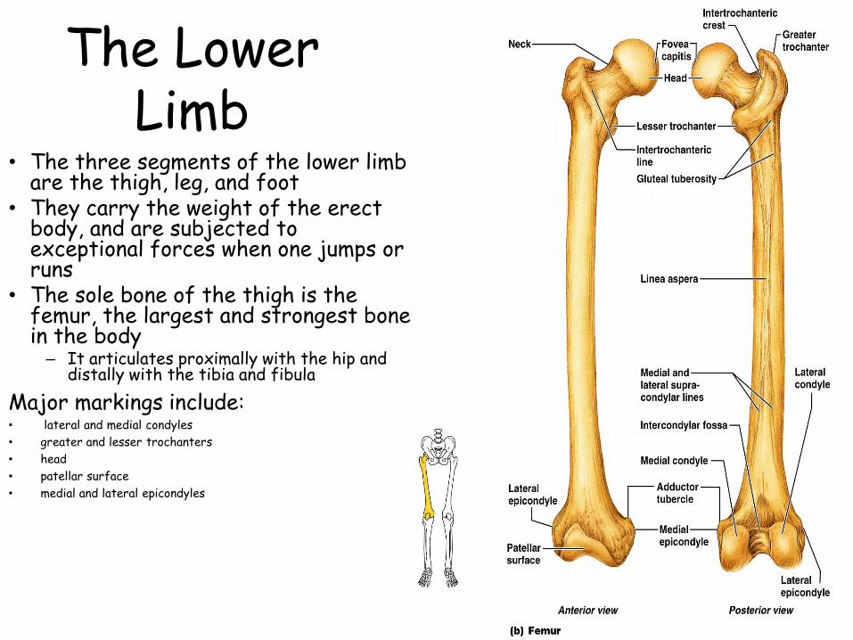
The Lower Limb
• The three segments of the lower limb are the thigh, leg, and foot
• They carry the weight of the erect body, and are subjected to exceptional forces when one jumps or runs
• The sole bone of the thigh is the femur, the largest and strongest bone in the body– It articulates proximally with the hip and
distally with the tibia and fibula
Major markings include:• lateral and medial condyles
• greater and lesser trochanters
• head
• patellar surface
• medial and lateral epicondyles
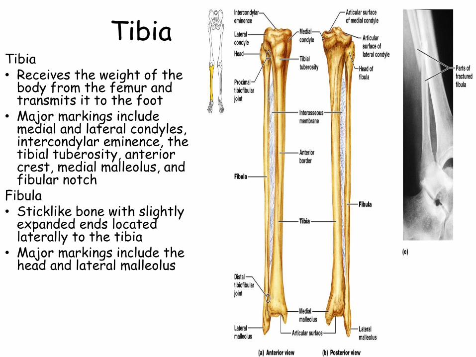
TibiaTibia• Receives the weight of the
body from the femur and transmits it to the foot
• Major markings include medial and lateral condyles, intercondylar eminence, the tibial tuberosity, anterior crest, medial malleolus, and fibular notch
Fibula• Sticklike bone with slightly
expanded ends located laterally to the tibia
• Major markings include the head and lateral malleolus
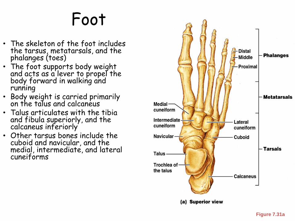
Foot• The skeleton of the foot includes
the tarsus, metatarsals, and the phalanges (toes)
• The foot supports body weight and acts as a lever to propel the body forward in walking and running
• Body weight is carried primarily on the talus and calcaneus
• Talus articulates with the tibia and fibula superiorly, and the calcaneus inferiorly
• Other tarsus bones include the cuboid and navicular, and the medial, intermediate, and lateral cuneiforms
Figure 7.31a
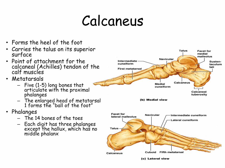
Calcaneus
• Forms the heel of the foot• Carries the talus on its superior
surface• Point of attachment for the
calcaneal (Achilles) tendon of the calf muscles
• Metatarsals– Five (1-5) long bones that
articulate with the proximal phalanges
– The enlarged head of metatarsal 1 forms the “ball of the foot”
• Phalanges– The 14 bones of the toes– Each digit has three phalanges
except the hallux, which has no middle phalanx
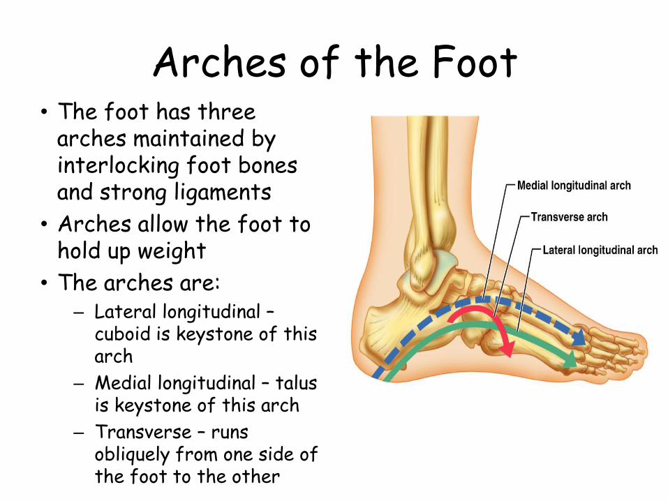
Arches of the Foot• The foot has three
arches maintained by interlocking foot bones and strong ligaments
• Arches allow the foot to hold up weight
• The arches are:– Lateral longitudinal –
cuboid is keystone of this arch
– Medial longitudinal – talus is keystone of this arch
– Transverse – runs obliquely from one side of the foot to the other
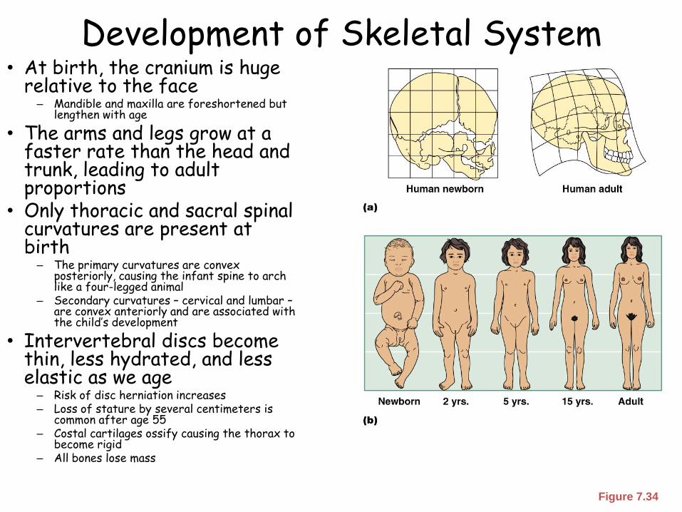
Development of Skeletal System• At birth, the cranium is huge
relative to the face– Mandible and maxilla are foreshortened but
lengthen with age
• The arms and legs grow at a faster rate than the head and trunk, leading to adult proportions
• Only thoracic and sacral spinal curvatures are present at birth
– The primary curvatures are convex posteriorly, causing the infant spine to arch like a four-legged animal
– Secondary curvatures – cervical and lumbar –are convex anteriorly and are associated with the child’s development
• Intervertebral discs become thin, less hydrated, and less elastic as we age
– Risk of disc herniation increases– Loss of stature by several centimeters is
common after age 55 – Costal cartilages ossify causing the thorax to
become rigid– All bones lose mass
Figure 7.34
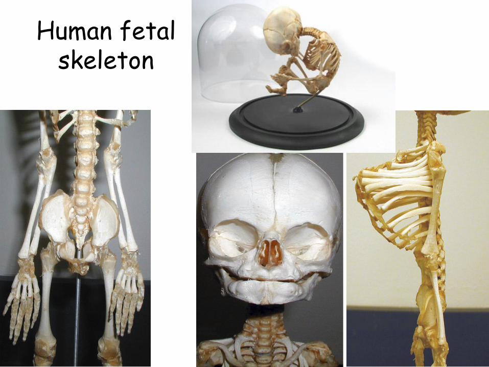
Human fetal skeleton
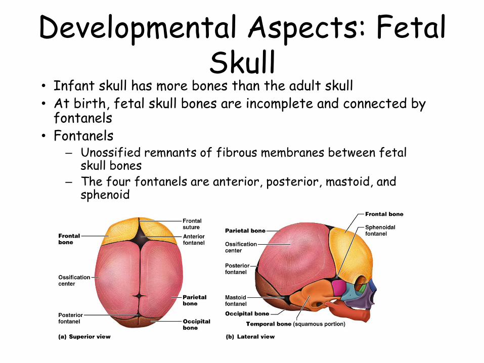
Developmental Aspects: Fetal Skull
• Infant skull has more bones than the adult skull• At birth, fetal skull bones are incomplete and connected by
fontanels• Fontanels
– Unossified remnants of fibrous membranes between fetal skull bones
– The four fontanels are anterior, posterior, mastoid, and sphenoid
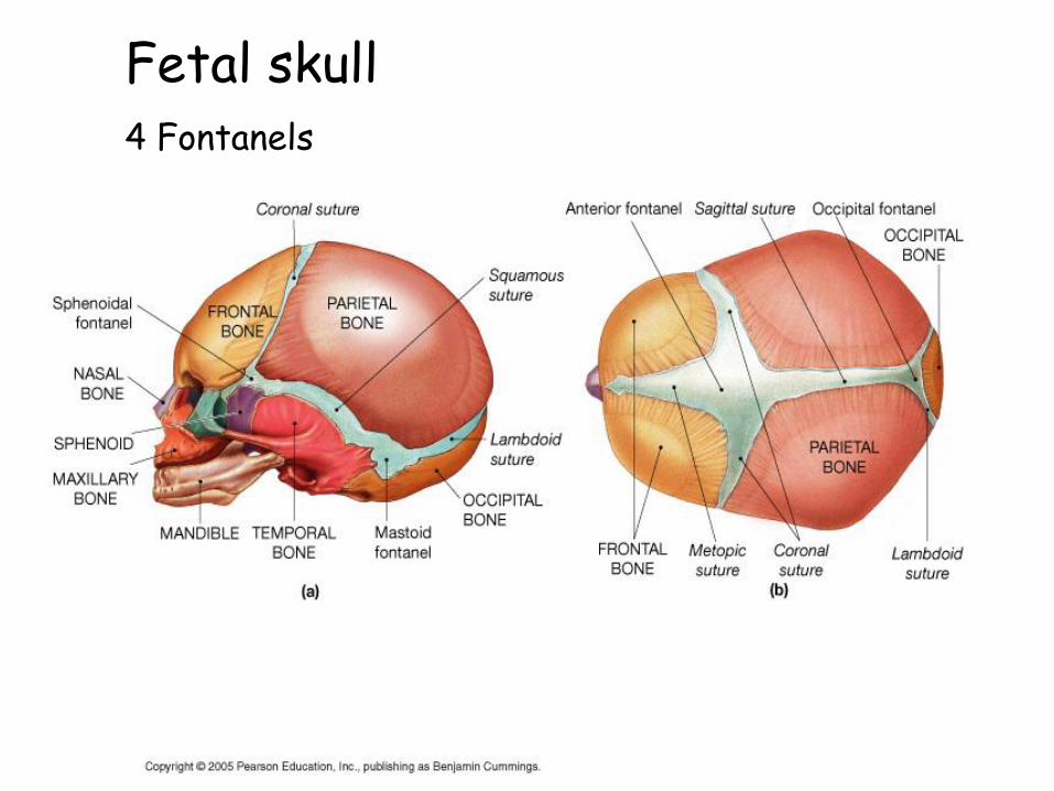
Fetal skull4 Fontanels