KOB OBT Program
24
Kyudai Oral Bioscience & OBT Research Center Joint International Symposium 2019 PROGRAM & ABSTRACTS March 2 – 3, 2019 Lecture Room A/B, Faculty of Dental Science, Kyushu University K OB 2019
Transcript of KOB OBT Program
Kyudai Oral Bioscience
&
OBT Research Center
Joint International Symposium 2019
PROGRAM & ABSTRACTS
March 2 – 3, 2019
Lecture Room A/B, Faculty of Dental Science, Kyushu University
K OB 2019
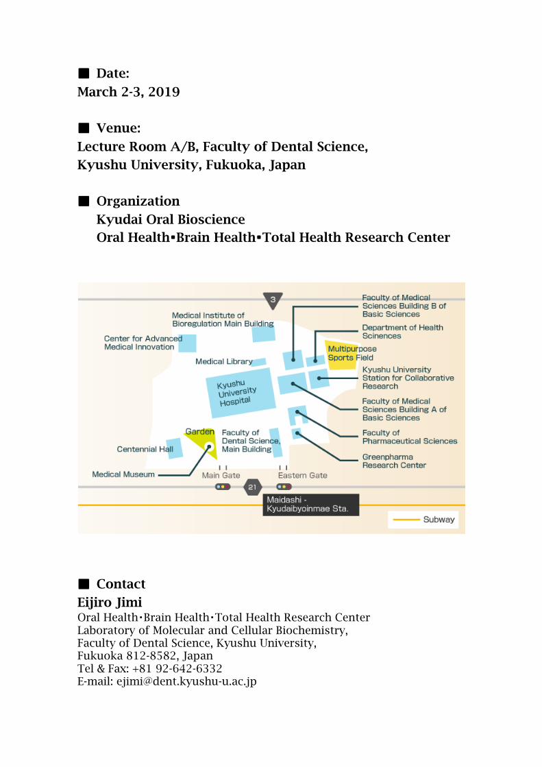
■ Date:
March 2-3, 2019
■ Venue:
Lecture Room A/B, Faculty of Dental Science,
Kyushu University, Fukuoka, Japan
■ Organization
Kyudai Oral Bioscience Oral Health・Brain Health・Total Health Research Center
■ Contact
Eijiro Jimi Oral Health・Brain Health・Total Health Research Center Laboratory of Molecular and Cellular Biochemistry, Faculty of Dental Science, Kyushu University, Fukuoka 812-8582, Japan Tel & Fax: +81 92-642-6332 E-mail: [email protected]
KOB & OBT Joint International Symposium 2019
PROGRAM
KOB & OBT Joint International Symposium 2019
Chaired by Dr. Zhou Wu
Session 1
Oral Health and Brain Health
Critical roles of Cathepsin B in linkage between periodontitis and neuroinflammation
Junjun Ni 1, Zhou Wu 1,2
1Department of Aging Science and Pharmacology, 2OBT Research Center Faculty of Dental Science, Kyushu University.
Neuroinflammation, inflammation of the brain, is strongly implicated in Alzheimer’s
disease (AD), which can be enhanced by systemic inflammation. The concept suggests a possible link between periodontitis and AD because periodontitis is an oral chronic infection that elicits systemic inflammatory responses. There is now growing clinical evidence that chronic periodontitis is closely linked to the initiation and progression of AD. Chronic periodontitis can be a significant source of covert systemic inflammation within the general population, because a high percentage of adults are suffering from periodontitis, and the prevalence of periodontitis increases with age. In the present talk, we will introduce our recently works on clarifying the mechanisms linking between periodontitis and AD, thereby helping to understand how does oral infection impact on brain.
We have recently found that Cathepsin (Cat) B plays a critical role in inducing AD-like phenotypes, including microglia-mediated neuroinflammation, intracellular Aβ accumulation in neurons and impairment of the learning and memory functions in the middle-aged mice following chronic systemic exposure to LPS from P.gingivalis (PgLPS). The microglia CatB- dependent in PgLPS-mediated intracellular Aβ accumulation in neuron. We have clarified the novel mechanisms of CatB involving in microglia-mediated neuroinflammation. Firstly, CatB involves in chronically NF-kB activation in microglia, result in prolonging microglia-mediated neuroinflammation, because CA-074Me, the specific CatB inhibitor, inhibits the autophagic IkBa degradation. Secondly, CatB involves in microglia aging, because leakage of CatB in microglia during aging are responsible for mitochondria-derived reactive oxygen species (ROS) generation result in proinflammatory mediator production. The CatB-dependent microglia aging promotion are important for periodontitis inducing AD-like phenotypes, because the senescent-type microglia respond in an exaggerated manner to systemic inflammation.
In conclusion, the novel mechanisms of CatB in involving microglia-mediated neuroinflammation are dependent on mitochondria-derived ROS generation and NF-kB activation. We propose that CatB may as a therapeutic target for preventing periodontitis-associated cognitive decline in AD.
The mechanism of periodontitis link to Alzheimer’s disease: gingipains involvement
Yicong Liu
Zhe Jiang University, China
Alzheimer’s disease (AD) is the world’s most common neurodegenerative disease. Although clinical evidence has already showed the positive link between chronic periodontitis and cognitive decline in AD, the precise mechanism underlying the relationship remains unclear.
Porphyromonas gingivalis (P.g), the keystone pathogen in chronic periodontitis, has been identified in the brain of AD patients, and the level of the toxic cysteine proteinases from P.g called gingipains has been found to correlate with tau and ubiquitin pathology. We hypothesized that Gingipains may activate microglia, brain-resident immune cells, directly resulting in neuroinflammation. Gingipains comprises Rgp and Kgp, and both Rgp and Kgp are mediated host cell responses and the subsequent intracellular signaling in the infected cells. In the present talk, I will introduce our recently works for clarifying the potential effects of Rgp and Kgp on microglia activation. We provide the first evidence that Rgp and Kgp cooperatively contribute to the P. g-induced cell migration and expression of proinflammatory mediators through the activation of PAR2. The subsequent activation of PI3K/Akt and mitogen-activated protein kinase/ERK kinase/ERK pathways contributes to cell migration and inflammatory response of microglia.
It is known that female not only have a higher risk of developing AD, but also showed significantly age-related faster decline than male. Finally, I will introduce our ongoing works on exploring gender difference of microglia-mediated neuroinflammation during P.g infection. We found that the anti-inflammatory mediators in the P.g infected microglia, including TGFb1, Arginase-1 and IL-10 were significantly lower in the P.g infected microglia from female aged mice brain than that of from male aged ones, and the estrogen receptor (ER)-a agonist increased the Arginase-1 expression in the P.g infected primary microglia from the aged female mice brain.
In conclusion, gingipains may be key mediators of periodontitis link to AD, gingipains inhibitors could be valuable for treating periodontitis-related neurodegeneration in AD. And ER-a agonist could be beneficial for reducing the risk of AD in old female individuals by suppressing the microglia-mediated neuroinflammation.

Spontaneous Activity-Dependent Discrete Circuit Formation in the Developing Olfactory Bulb
Satoshi Fujimoto
Graduate School of Medical Sciences, Kyushu University, Fukuoka, Japan
In the mouse olfactory system, odor information detected by ~1000 types of olfactory receptors is represented by the glomerular map on the olfactory bulb. Mitral cells, which are second-order neurons, extend multiple dendrites to multiple glomeruli in the early stages of development, but dendrites are pruned as development progresses and finally connect one primary dendrite to one glomerulus. However, it remains unknown how this discrete connectivity is established. Here we found that dendrite pruning was perturbed by silencing neuronal activity in mitral cells, while the pruning occurred normally under the blockade of activity derived from olfactory sensory neurons. Spontaneous activity of the mitral cell was directly observed by two photon calcium imaging of awake neonates in vivo and even isolated olfactory bulbs in vitro. We also found that the pattern of activity becomes decorrelated during development. Pharmacological experiments revealed that spontaneous activity is mediated by glutamatergic synaptic transmission and gap junction. Dendrite pruning was suppressed in the mitral cells lacking NMDA receptors. Thus, the spontaneous activity generated in the olfactory bulb establishes the discrete olfactory circuit.
Exploring Potential Therapies for Alzheimer’s Disease
Zhenzhen Quan1, Hong Qing1
Beijing Institute of Technology, China
Alzheimer’s disease (AD) is a progressive neurodegenerative disease which affects at least 47 million people worldwide as per World Alzheimer Report of 2016. Currently, there are no specific drugs or therapies that could cure AD effectively. Revealing the mechanism and exploring effective therapy for ND are the hotspots for current research. My topic is mainly based on two parts: 1) NSC transplantation on AD study. We initially approved that NSC transplantation significantly improves the ability of short-term memory and certainly ameliorates the ability of long-term memory in tau/tta mice; we also found that NSC transplantation could reduce the number of neurofibrillary tangles in hippocampal CA1 region; the transplanted NSC could differentiate into neurons and microglia. These indicated NSC transplantation has the potential to improve the ability of learning and memory in AD model mice which is possibly due to its ability of integrating into the impaired neural circuits and eliminating the neurofibrillary tangles; 2) Application of optogenetics on AD study. Previous studies have indicated the important role of CA3 on learning and memory, our recent studies found that, manipulation of hippocampal CA3 firing via luminopsins significantly modulates spatial and episodic short-term memory, especially working memory. Regarding this, we further found that manipulation of CA3 by optogenetics could ameliorate the cognitive dysfunctions of AD model mice through improvement of the synapse plasticity of CA3 circuits.
KOB & OBT Joint International Symposium 2019
Chaired by Dr. Masafumi Moriyama
Session 2
Oral Health
The effect of systemically-administered Mesenchymal stem cells for
peri-implant Mucosa
Ikiru Atsuta1*, Yasunori Ayukawa1, Nobuyuki Ueda1, Takayoshi Yamaza2, Yuri Matsuura1, Ryosuke Kondo1, Kiyoshi Koyano1
1Section of Implant and Rehabilitative Dentistry, Division of Oral Rehabilitation,
Faculty of Dental Science, Kyushu University, Fukuoka, Japan 2Department of Molecular Cell and Oral Anatomy, Faculty of Dental Science, Kyushu
University, Fukuoka, Japan Dental implant therapy is one of the most important prosthodontic options for edentulous patients. Dental implants based on the concept of “osseointegration”, a term explaining the fixation of a titanium implant in the bone, have shown dramatic continual improvement. However, the peri-implant tissue is always exposed to the possibility of inflammation because the titanium body penetrates into the oral mucosa. Actually, the peri-implant tissue has many close similarities to normal gingiva, but many reports have noted that the ability of the peri-implant epithelium to seal against the oral environment is poor. Therefore, the improvement of local defense in mucosa is strongly expected to enable successful outcomes in oral implant patients. In this study, we have attracted attention to mesenchymal stem cells (MSCs), because these cells generate a great deal of interest in many clinical settings, including immuno-modulation and tissue engineering, where studies have demonstrated the feasibility of transplanted MSCs becoming a new mode of cellular therapy. However, the relationship between MSC-based therapy and PIE-implant interface sealing is not well understood. The hypothesis of the present study was that systemic MSCs accumulate around the implant in the early stage and promote PIE formation and soft tissue attachment to the implant surface.
A Novel disease discovered and established in 21th Century: IgG4-related disease
-Mechanistic insights from both clinical and immunologic understanding of this condition -
Takashi Maehara1, Hamid Mattoo2, Vinay Mahajan2, Emanuel Della-Torre3, Cory Perugino4, Masafumi Moriyama1,5, Miho Ohta1, Naoki Kaneko2, Noriko Ishiguro1, Akira Chinju1, Ryusuke Munemura1, John Stone4, Shiv Pillai2, Seiji Nakamura1. 1Section of Oral and Maxillofacial Oncology, Division of Maxillofacial Diagnostic and
Surgical Sciences, Faculty of Dental Science, Kyushu University, Fukuoka, Japan 2Ragon Institute of MGH, MIT, and Harvard, Massachusetts General Hospital,
Harvard Medical School, Boston, MA, USA 3Universita Vita-Saluta San Raffaele, IRCCS San Raffaele Scientific Institute, Milan, Italy 4Division of Rheumatology, Allergy and Immunology, Massachusetts General Hospital,
Harvard Medical School, Boston, Massachusetts, USA 5Faculty of Dental Science, OBT Research Center, Kyushu University, Fukuoka, Japan
Objective: IgG4-related disease (IgG4-RD) is a chronic, systemic inflammatory condition with T cells and polarized class-switching to IgG4 secreting B cells within severe tissue fibrosis. Important mechanistic insights in this disease have been derived from studies of B-cell depletion therapy. Actually CD4+T cells are the most abundant cells within tissue lesions, so disease-specific CD4+T cells could provide help to specific B cells during T-dependent immune responses. We thus investigated disease-related CD4+T cells in this disease on International IgG4-RD collaboration research. Methods: We used flow cytometry to identify CD4+ effector/memory T cells in 101 patients with IgG4-RD. Next-generation sequencing of the T cell receptor β chain gene was used to identify clonal expansions of effector CD4+T cells. We use transcriptomes of isolating CD4+ cytotoxic T lymphocytes (CD4+CTLs). Furthermore, by using IL-4 capture assay, we use transcriptomes of isolating IL-4 secreting Tfh cells based on their function. Tissue infiltration by these T cells was examined using quantitative multi-color imaging in IgG4-RD subjects compared with healthy controls and other diseases. Results: CD4+ effector/memory T cells were expanded in IgG4-RD. Next-generation sequencing revealed prominent clonal expansions of CD4+CTLs and Tfh cells in subjects with IgG4-RD. Clonally-expanded CD4+CTLs that expressed pro-fibrotic cytokine. IL-4 expressing Tfh cells are surprisingly sparse in human secondary lymphoid organs (SLOs). Notably, most Tfh cells in IgG4-RD expressed IL-4. IL-4+Tfh cells can be detected in cell-cell contact with IgG4+B cells, some of these B cells express AID in SLOs and TLOs. The proportion of IL-4+Tfh cells in IgG4-RD tissues correlates tightly with tissue IgG4 plasma cell numbers and plasma IgG4 levels in patients but not with the total plasma levels of other isotypes. The numbers of CD4+CTLs declined during clinical responses to B cells depletion therapy, suggesting a role of these cells in the pathogenesis of this disease. Conclusions: IgG4-RD is prominently linked to the tissue infiltration of clonally expanded CD4+CTLs and IL-4+Tfh cells. Especially 1) reactivation of CD4+ CTLs might require antigen presentation by activated B cells in tissue sites, 2) disease-related Tfh subpopulation in SLOs and TLOs that is linked to IgG4 class switching.
PTHrP-PPR autocrine regulation of mesenchymal progenitor cell fate orchestrates tooth eruption
Akira Takahashi1,2, Yoichiro Ogino1, Kiyoshi Koyano1, Noriaki Ono2, Wanida Ono2
1Division of Oral Rehabilitation, Faculty of Dental Science, Kyushu University 2Department of Orthodontics and Pediatric Dentistry, University of Michigan,
School of Dentistry
Formation of functional skeletal tissues requires highly organized steps of mesenchymal progenitor cell differentiation. The dental follicle (DF), a sac-like membranous tissue surrounding the developing tooth bud, harbors mesenchymal progenitor cells for various differentiated cells constituting the tooth root-bone interface, and coordinates tooth eruption in a manner dependent on signaling by parathyroid hormone-related peptide (PTHrP) and its receptor, PTH/PTHrP receptor (PPR). In humans, primary failure of tooth eruption, a rare autosomal dominant disorder that exclusively affects tooth eruption, is characterized by a cessation of tooth eruption before emergence despite an unobstructed eruption path. PFE is caused by loss-of-function mutations in PPR. However, the identity of mesenchymal progenitor cells in the DF and how they are regulated by PTHrP-PPR signaling remain unknown. Here we show that the PTHrP-PPR autocrine signal maintains physiological cell fates of DF mesenchymal progenitor cells to establish the functional periodontal attachment apparatus and orchestrates tooth eruption. A single cell RNA-seq analysis revealed cellular heterogeneity of PTHrP+ cells, wherein PTHrP+ DF subpopulations abundantly expressed PPR. Cell lineage analysis using tamoxifen-inducible PTHrP-creER mice revealed that PTHrP+ DF cells differentiated into cementoblasts on the acellular cementum, periodontal ligament (PDL) cells and alveolar cryptal bone osteoblasts during tooth root formation. PPR-deficiency induced a cell fate shift of PTHrP+ DF mesenchymal progenitor cells to non-physiological cementoblast-like cells precociously forming the cellular cementum on the root surface associated with upregulation of Mef2c and matrix proteins, resulting in loss of the proper periodontal attachment apparatus and primary failure of tooth eruption closely resembling human genetic conditions caused by PPR mutations. These findings reveal a unique mechanism whereby proper cell fates of mesenchymal progenitor cells are tightly maintained by an autocrine system mediated by PTHrP-PPR signaling to achieve functional formation of skeletal tissues.
KOB & OBT Joint International Symposium 2019
Chaired by Prof. Eijiro Jimi
Special Lecture
From Kyushu to Okayama: My research history
Ryusuke Yoshida
Department of Oral Physiology, Graduate School of Medicine, Dentistry and Pharmaceutical Sciences, Okayama University
After I obtained a Ph.D. in Science from Kobe University in 2002, my research career in the taste field started as a post-doctoral fellow at the professor Ninomiya’s laboratory in Kyushu University. At that time, my main research theme was to analyze molecular and physiological properties of taste receptor cells. To do this, a predecessor and I developed the experimental setup for recording taste cell responses, which is able to apply taste stimuli restricted to the apical side with keeping the structure of taste buds. After recording taste responses, the taste cell was collected from a taste bud and subjected with RT-PCR to analyze molecular expression. I had a hard time to establish this system; I spent about 3 years for the first publication of a paper in this field. This technique was greatly improved by using transgenic mice expressing fluorescent protein in a subset of taste bud cells and laser scanning microscopy. This technique has further advantages; we are able to measure transmitter release from taste cells and also able to analyze the effect of endocrine factors on taste cell responses since apical and basolateral side of a taste bud is completely segregated in this recording system. Now, famous researchers in taste field of USA comment on this technique as “Technical tour de force”. In 2016, I was selected as a member of OBT research center. This would be a very important step to get current position in Okayama University. In addition, many members in Kyushu University kindly supported for this personnel affair. From October in 2018, I moved to Okayama University as a professor in Department of Oral Physiology. Although I still have not get deeply into my research works because of many other works, problems in man power, etc., now I ready to take a new step in Okayama University. I hope that this talk will be as reference for and encourage young researchers.
KOB & OBT Joint International Symposium 2019
Chaired by Prof. Fusanori Nishimura
Session 3
UGM Summer Course Report
KOB & OBT Joint International Symposium 2019
Chaired by
Session 4
Graduate Student’s Session
TLR8+CD68+monocyte/macrophage contribute to the pathogenesis of Sjögren’s syndrome via TNF-α secretion.
Mizuki Sakamoto, Masafumi Moriyama, and Seiji Nakamura
1OBT Research Center, 2Section of Oral and Maxillofacial Oncology, Division of
Maxillofacial Diagnostic and Surgical Sciences, Faculty of Dental Science, Kyushu University, Fukuoka, Japan
Objectives Sjögren’s syndrome (SS) is an autoimmune disease characterized by lymphocytic infiltration into the salivary and lacrimal gland with the concomitant destruction of glandular tissue. Recent studies indicate that Toll like receptors (TLRs) play an important role in the initiation of autoimmune diseases including rheumatoid arthritis and systemic lupus erythematosus. In this study, we thus addressed to identify the disease-associated TLR related molecules in SS, and then examined both expression and function of those molecules. Methods Gene expression was analyzed by using DNA microarray in labial salivary glands from 3 patients with typical SS and 3 healthy controls (HC). DNA microarray analysis was performed in the two groups to screen for TLR family (TLR1-TLR10). The candidate TLRs were validated by real-time PCR and immunohistochemical staining in SS (n=19), and HC (n=10). Moreover, the productivity of TNF-α in human monocyte cell line (U937) was examined by ELISA. Finally, we established human Tlr8 transgenic/mouse Tlr8 knock-out (huTlr8 Tg/mTlr8 KO) mice and confirmed their phenotypes in each organ (submandibular gland, lung, kidney, pancreas, and liver). Results DNA microarray and PCR validated the up-regulation of TLR8 in SS compared with HC. Immunofluorescence double staining confirmed that the expression of TLR8 almost merged with that of CD68, known as a monocyte/macrophage marker. Moreover, TNF-α productivity of U937 was significantly increased by over-expression of TLR8 in comparison with those of untreated U937 and TLR8 KO U937. In vivo, huTlr8 Tg/mTlr8 KO mice showed strong infiltration of inflammatory cells in submandibular glands and lungs compared with Wild-type mice. Conclusion TLR8 is considered to recognize self-RNA as well as viral RNA and then accelerates autoimmune responses via production of inflammatory cytokines, such as TNF-α. Therefore, our current data suggest that TLR8+CD68+ monocyte/macrophages might promote the initiation of SS via TNF-α secretion.
Characterization of disrupted microbiota on the tongue dorsum
observed in frail elderly adults
Mikari Asakawa1, Toru Takeshita1,2, Shinya Kageyama1, Kenji Takeuchi1,2, Michiko Furuta1, Shino Suma1, Yoshihisa Yamashita1
1Section of Preventive and Public Health Dentistry, Division of Oral Health, Growth
and Development and 2OBT Research Center, Faculty of Dental Science, Kyushu University, Fukuoka, Japan.
Tongue microbiota is a primary source of numerous microorganisms constantly ingested with saliva, and the overgrowth of non-commensal bacteria due to the disruption of the tongue microbiota would threaten the health of elderly adults with swallowing impairment. This study investigated tongue microbiota composition of 325 institutionalized frail elderly adults aged ≥70 years by 16S rRNA gene amplicon sequencing and explored disrupted tongue microbiota in the elderly adults. The tongue microbiota of most individuals was constituted by well-known predominant oral bacterial taxa such as Streptococcus salivarius, Rothia mucilaginosa, and Veillonella atypica, whereas Streptococcus agalactiae, Corynebacterium simulans, and Actinomyces radicidentis deposited as non-oral bacterial taxa in the expanded Human Oral Microbiome Database (eHOMD 16S rRNA RefSeq version 15.1) occupied ≥10% in the microbiota of the 24 individuals. These individuals demonstrated significantly poorer physical function and nutritional condition than the individuals with the normal microbiota. These results suggest that tongue coating could be a reservoir of unexpected bacteria in the frail elderly adults, especially those with declined physical function or nutritional condition.
SPOCK-1 is a novel epithelial mesenchymal transition (EMT) inducer in calcium channel blocker-induced gingival overgrowth.
Rehab Alshargabi
Department of Periodontology, Division of Oral Rehabilitation, Faculty of Dental
Science, Kyushu University, Fukuoka, Japan. 【Background】 Drug-induced gingival overgrowth (DIGO) is a side effect associated with Ca+2 channel blockers such as Nifedipine. The exact mechanism by which those drugs causes overgrowth is not fully understood. However, previous studies suggested that both EMT and reduced catheptin-L activity could contribute to the development of DIGO. Moreover, elongated rete pegs are typical feature of DIGO. SPOCK-1, a multi-domain secreted proteoglycan, has been reported to induce EMT in several cancer cells as well as to inhibit cathepsin-L activity. In addition, SPOCK-1 belongs to SPARC family, some of which are highly expressed in basement membrane zone. Therefore, we hypothesized that SPOCK-1 may associate with the pathology of DIGO. 【Methods】 Calcium channel blocker-induced gingival overgrowth (CCBGO) and non-overgrowth samples were collected from patients undergoing periodontal surgery in the Periodontology Section at Kyushu University Hospital. SPOCK-1 protein level was checked by western blotting and IHC staining. Spock-1 transgenic (Tg) mice were generated. Spock-1 Tg mice and wild-type (WT) littermates were sacrificed at 8 weeks after birth. The expression of TGF-β1, E-Cadherin, Vimentin, Mmp2, Mmp9, Slug were examined by western blotting and IHC staining following morphological observation. 【Results】 SPOCK-1 protein level was significantly up-regulated in CCBGO samples compared with control samples. Morphological observation of Tg mice revealed more thickened gingival tissues at the molar region compared with WT mice. Histologically, more elongated rete pegs and accelerated collagen accumulation were observed. E-cadherin protein expression was significantly down-regulated while TGF-β1, Vimentin, Mmp2, Mmp9, Slug expressions were up-regulated. 【Conclusion】 The results strongly indicate the importance of SPOCK-1 as an inducer of the EMT process in the pathophysiology of DIGO.
Effects of TNF-a on Senescent human dental pulp cells
Aoi Nozu¹, Sayuri Hamano¹,², Atsushi Tomokiyo³, Daigaku Hasegawa³, Shinichiro Yoshida³, Hideki Sugii³,
Hiromi Mitarai4, Keita Ipposhi¹,³, Naohisa Wada4, Hidefumi Maeda¹,³ 1
Department of Endodontology and Operative Dentistry, Division of Oral Rehabilitation, Faculty of Dental Science, Kyushu University,
2OBT Research Center,
Faculty of Dental Science, Kyushu University, 3Division of Endodontology, Kyushu
University Hospital, 4Division of General Dentistry, Kyushu University Hospital
Objectives: Aging is characterized by chronic, low-grade inflammation, and this phenomenon has been termed as inflammaging. It has been reported that the concentration of tumor necrosis factor-a (TNF-a) is higher in the blood of aged people compared with that of young people. Therefore, the aim of this study was to investigate how TNF-a affected pulp cells during aging. Methods: Human dental pulp cells (HDPCs) were cultured until reaching the plateau of their growth, and the cells were isolated at actively (yHDPCs) or inactively (sHDPCs) proliferating stages, whose gene expression of senescence-related factors, TNF-a, and its receptor, TNFR1 was examined by quantitative PCR. In addition, on odontoblastic differentiation of yHDPCs and sHDPCs cultured in control culture medium (CM) or CM containing 2mM CaCl2 as differentiation medium (DM) with or without TNF-a was examined by quantitative PCR and alizarin red staining. Furthermore, the contribution of the signaling via TNFR1 to mineralization of sHDPCs was examined by transfection of TNFR1 siRNA. Results: sHDPCs expressed senescence-related molecules while yHDPCs did not. The expression level of TNF-a in sHDPCs was significantly higher than that in yHDPCs. When these HDPCs were cultured in DM, the mineralization activity of sHDPCs was lower compared with that of yHDPCs. However, the administration of TNF-a to this culture medium altered these responses: yHDPCs showed downregulated mineralization per a cell, while sHDPCs exhibited a significant increase. Furthermore, the expression of TNFR1 in sHDPCs was significantly upregulated compared with that in yHDPCs. Downregulation of TNFR1 expression led to decreased mineralization of TNF-a-treated sHDPCs. Conclusions: These results suggested that sHDPCs preserved the odontoblastic differentiation capacity, and TNF-a promoted odontoblastic differentiation of sHDPCs through increased expression of TNFR1.
KOB & OBT Joint International Symposium 2019
Chaired by Dr. Shinsuke Mizutani
Session 5
Oral Health & Total Health

Oral function and decline in cognitive function
Masahiro Yamaguchi1), Shinsuke Mizutani2,3), Rui Egashira1), Toru Naito1)
1) Section of Geriatric Dentistry, Department of General Dentistry, Fukuoka Dental College, Fukuoka, Japan 2) OBT Research Center, Faculty of Dental Science, Kyushu University, Fukuoka, Japan
3) Section of Geriatric Dentistry and Perioperative Medicine in Dentistry, Division of Maxillofacial Diagnostic and Surgical Sciences, Faculty of Dental Science, Kyushu University, Fukuoka, Japan
In Japan, about 4.62 million elderly people aged 65 years or older are thought to be developing dementia. A condition called mild cognitive impairment (MCI) occurs in the preliminary stage of dementia, and if left untreated, about 40% of those with MCI will develop dementia within 5 years; however, dementia onset may be delayed by appropriate treatment. Therefore, it is important to notice MCI at an early stage and take effective countermeasures. For patients who do not have a family doctor, dentists who have frequent contact with the elderly who visit for regular dental checkups could easily become involved in the early detection of MCI. Recent findings have suggested that oral conditions such as periodontal disease or oral function are related to a decline in cognitive function. For example, epidemiological studies have reported that severe periodontitis and the impaired ability to pronounce /pa/ are related to MCI in Japanese people. However, few studies have examined oral condition or function in detail. Therefore, we examined present teeth, periodontal inflammation, degree of periodontal disease, masticatory function, swallowing function, and tongue pressure in dental outpatients. We used the Japanese version of the Montreal Cognitive Assessment and the Mini-Mental State Examination as cognitive function tests. In addition, we measured serum apolipoprotein A1, complement protein C3, and transthyretin as biomarkers for MCI. The results showed that higher age and low tongue pressure were related to a decline in cognitive function. In this presentation, we introduce studies on the relationship between oral condition/function and MCI. We also discuss our study results, which suggest an association between low tongue pressure and a decline in cognitive function.
Necroptosis of Adipocytes Induced by Osteocalcin
Takahito Otani1, Miho Matsuda2, Akiko Mizokami3, Norio Kitagawa1, Hiroshi Takeuchi4, Eijiro Jimi2,3, Tetsuichiro Inai1 and Masato Hirata5
1Division of Functional Structure, Department of Morphological Biology, Fukuoka
Dental College 2Laboratory of Molecular and Cellular Biochemistry, Faculty of Dental Science,
Kyushu University 3OBT Research Center, Faculty of Dental Science, Kyushu University
4Division of Applied Pharmacology, Kyushu Dental University 5School of Dental Medicine, Fukuoka Dental College
Osteocalcin, one of non-collagenous bone matrix proteins, is known to regulate the metabolism of glucose and energy in the uncarboxylated form designated as GluOC. We examined the effects of GluOC on adipocytes and adipose tissue of mice, and then revealed the signaling pathway on which GluOC increased the expressions of adiponectin, an adipokine regulating the metabolism of glucose and lipids, and peroxisome proliferator-activated receptor-γ (PPARγ), a master regulator of adipogenesis, via its putative receptor GPRC6A. Furthermore, we also found that GluOC at the concentration of over 20 ng/ml inversely abolished the expression of adiponectin, and that the mechanism was induced by the programmed necrosis (necroptosis) in 3T3-L1 adipocytes. The adipocyte bound by GluOC via GPRC6A increased the expression of FoxO1, a transcription factor regulating cell death, via cAMP-PKA-CREB signaling axis together with activation of p300, a transcriptional coactivator of CREB followed by the expression of FasL, a member of tumor necrosis factor superfamily. FasL expressed in the plasma membrane triggered Fas activation followed by the phosphorylation and trimerization of mixed lineage kinase domain-like protein (MLKL), a major regulator of necroptosis. Finally, calcium influx, reactive oxygen species (ROS) production, lipid peroxidation and mitochondrial fragmentation occurred in the downstream of MLKL activation leading to necroptosis. Although high-dose GluOC decreased the cell number of adipocytes, it was maximally about by 30% even after 96 h, probably because direct cell-cell contact was required for necroptosis via Fas/FasL. We have here revealed the signaling pathway on which high-dose GluOC induced necroptosis in 3T3-L1 adipocytes.