KERNICTERUS AND PREMATURITYadc.bmj.com/content/archdischild/30/154/501.full.pdf · KERNICTERUS AND...
8
KERNICTERUS AND PREMATURITY BY V. M. CROSSE, T. C. MEYER and J. W. GERRARD From Sorrento Maternity Hospital, Birmingham (RECEIVED FOR PUBLICATION JUNE 30, 1955) At one time kernicterus was most commonly associated with haemolytic disease of the newborn. With the introduction of adequate exchange trans- fusions this complication, whether due to Rh or to ABO incompatibility, has been virtually eliminated. In 1950 two groups of workers, Aidin, Corner and Tovey in this country and Zuelzer and Mudgett in America, independently drew attention to the fact that kernicterus was sometimes associated with pre- maturity only. Other workers (Gerrard, 1952; Govan and Scott, 1953) have also given details of cases and suggested possible aetiological factors. The present paper draws attention to the early signs of the disease, its mortality and the sequelae in survivors. An attempt has also been made to assess its incidence, its relationship to such factors as sex, birth weight, maturity and birth order, and ante-natal and post-natal factors. Among the post-natal factors specifically studied has been the level of the serum bilirubin in those developing kernicterus; these levels have been compared with those in normals. Material and Methods Three groups of babies have been studied. First, 60 premature babies in three Birmingham units who developed kernicterus in the absence of iso-immunization during the years 1951-54. Kemicterus was demon- strated either at necropsy in those who died in the neonatal period or by follow-up studies in the survivors. These cases have been compared first with 60 controls (a control was the first baby in the same 500 g. weight group admitted to the same premature baby unit after the case developing kernicterus), secondly, when possible, with all the premature babies (2,608) admitted to the units during the same period. A second group of 91 children has been studied to assess the importance, if any, of blood group incom- patibility between the mother and child. This group was composed of 56 dying in the neonatal period, in which the diagnosis was confirmed at necropsy, and of 35 who survived with varying degrees of- choreoathetosis and perceptive deafness. The latter had all been deeply jaundiced in infancy, and their history was characteristic of kernicterus as seen in prematures. This group included the 60 cases already mentioned. Iso-immuniza- tion was excluded by a negative direct Coombs test on the baby's blood in 59 instances; by the absence of any anomalous agglutinins and of immune anti-A and anti-B in the maternal serum in 83; and by a negative indirect Coombs test, using maternal serum and either the infant's or the father's red cells, in 50. A third group of cases has been studied to determine whether there is any relationship between the degree of jaundice and the development of kemicterus. This group included 46 normal full-term and 47 premature babies. The serum bilirubin was estimated on the second, fourth and sixth days. The mean serum bilirubin levels in babies of different birth weight groups were then calculated. Twelve premature babies who developed kernicterus in the absence of iso-immunization were also studied; these 12 were all included in the first group of 60. The total, direct and indirect, serum bili- rubin levels were estimated on venous blood by the method of Malloy and Evelyn (1937). Results The following results are based on our findings in the first group of prematures. The Signs of Kernicterus. The first 24 or 48 hours of life in the premature baby are the most critical, but once these are passed the chances of ultimate survival are good. The signs of kernicterus (Table 1) develop in a baby who is jaundiced after this initial adjustment to an extra-uterine environment. The signs include head retraction, an expressionless facies, usually with oculogyric movements, changes in muscle tone, cyanotic attacks, a refusal to suck in those bottle-fed, vomiting and, terminally, haemor- rhage, usually from the mouth. In severe cases these signs are self evident, but in those less affected they are easily missed. Jaundice may be overlooked, especially in artificial light, because the plethoric colour of prematures tends to conceal the underlying icterus. If the skin, how- ever, is stretched between two fingers, jaundice becomes immediately apparent. Oculogyric move- iO1 copyright. on 30 May 2018 by guest. Protected by http://adc.bmj.com/ Arch Dis Child: first published as 10.1136/adc.30.154.501 on 1 December 1955. Downloaded from
Transcript of KERNICTERUS AND PREMATURITYadc.bmj.com/content/archdischild/30/154/501.full.pdf · KERNICTERUS AND...

KERNICTERUS AND PREMATURITYBY
V. M. CROSSE, T. C. MEYER and J. W. GERRARDFrom Sorrento Maternity Hospital, Birmingham
(RECEIVED FOR PUBLICATION JUNE 30, 1955)
At one time kernicterus was most commonlyassociated with haemolytic disease of the newborn.With the introduction of adequate exchange trans-fusions this complication, whether due to Rh or toABO incompatibility, has been virtually eliminated.In 1950 two groups of workers, Aidin, Corner andTovey in this country and Zuelzer and Mudgett inAmerica, independently drew attention to the factthat kernicterus was sometimes associated with pre-maturity only. Other workers (Gerrard, 1952;Govan and Scott, 1953) have also given details ofcases and suggested possible aetiological factors.The present paper draws attention to the early
signs of the disease, its mortality and the sequelae insurvivors. An attempt has also been made toassess its incidence, its relationship to such factorsas sex, birth weight, maturity and birth order, andante-natal and post-natal factors. Among thepost-natal factors specifically studied has been thelevel of the serum bilirubin in those developingkernicterus; these levels have been compared withthose in normals.
Material and MethodsThree groups of babies have been studied. First,
60 premature babies in three Birmingham units whodeveloped kernicterus in the absence of iso-immunizationduring the years 1951-54. Kemicterus was demon-strated either at necropsy in those who died in theneonatal period or by follow-up studies in the survivors.These cases have been compared first with 60 controls(a control was the first baby in the same 500 g. weightgroup admitted to the same premature baby unit after thecase developing kernicterus), secondly, when possible,with all the premature babies (2,608) admitted to theunits during the same period.A second group of 91 children has been studied to
assess the importance, if any, of blood group incom-patibility between the mother and child. This group wascomposed of 56 dying in the neonatal period, in which thediagnosis was confirmed at necropsy, and of 35 whosurvived with varying degrees of- choreoathetosis andperceptive deafness. The latter had all been deeplyjaundiced in infancy, and their history was characteristic
of kernicterus as seen in prematures. This groupincluded the 60 cases already mentioned. Iso-immuniza-tion was excluded by a negative direct Coombs test on thebaby's blood in 59 instances; by the absence of anyanomalous agglutinins and of immune anti-A and anti-Bin the maternal serum in 83; and by a negative indirectCoombs test, using maternal serum and either theinfant's or the father's red cells, in 50.A third group of cases has been studied to determine
whether there is any relationship between the degree ofjaundice and the development of kemicterus. Thisgroup included 46 normal full-term and 47 prematurebabies. The serum bilirubin was estimated on thesecond, fourth and sixth days. The mean serumbilirubin levels in babies of different birth weight groupswere then calculated. Twelve premature babies whodeveloped kernicterus in the absence of iso-immunizationwere also studied; these 12 were all included in the firstgroup of 60. The total, direct and indirect, serum bili-rubin levels were estimated on venous blood by themethod of Malloy and Evelyn (1937).
ResultsThe following results are based on our findings in
the first group of prematures.
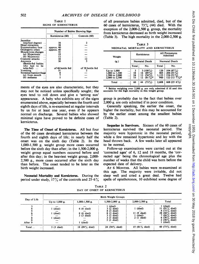
The Signs of Kernicterus. The first 24 or 48 hoursof life in the premature baby are the most critical,but once these are passed the chances of ultimatesurvival are good. The signs ofkernicterus (Table 1)develop in a baby who is jaundiced after this initialadjustment to an extra-uterine environment. Thesigns include head retraction, an expressionlessfacies, usually with oculogyric movements, changesin muscle tone, cyanotic attacks, a refusal to suck inthose bottle-fed, vomiting and, terminally, haemor-rhage, usually from the mouth.
In severe cases these signs are self evident, but inthose less affected they are easily missed. Jaundicemay be overlooked, especially in artificial light,because the plethoric colour of prematures tends toconceal the underlying icterus. If the skin, how-ever, is stretched between two fingers, jaundicebecomes immediately apparent. Oculogyric move-
iO1
copyright. on 30 M
ay 2018 by guest. Protected by
http://adc.bmj.com
/A
rch Dis C
hild: first published as 10.1136/adc.30.154.501 on 1 Decem
ber 1955. Dow
nloaded from
ARCHIVES OF DISEASE IN CHILDHOOD
TABLE 1SIGNS OF KERNICTERUS
Number of Babies Showing Sign
Kemicterus (60) Controls (60)
Jaundice(marked degree) 38 2
Head retraction 38 -
Expressionless face approx. 40 -
Oculogyric crises approx. 40 -
Muscle tone changes 31 -
(a) Hypertonic 19 -
(b) Hypotonic 12 -
Cyanotic attacks .. 34 12Anorexia 30 5
(Bottle-fed babieswho had to betube-fed) of 46 bottle fed of 38 bottle fed
Vomiting 31 2Terminal haemorrhage 8 -
(a) from mouth 7 -(b) melaena ..1
ments of the eyes are also characteristic, but theymay not be noticed unless specifically sought; theeyes tend to roll down and give a 'setting sun'appearance. A baby who exhibits any of the signsenumerated above, especially between the fourth andeighth days of life, is re-examined at regular intervalsby us for at least one year, even if he appearsnormal on discharge. Several babies who showedminimal signs have proved to be definite cases ofkernicterus.
The Time of Onset of Kernicterus. All but fourof the 60 cases developed kernicterus between thefourth and eighth days of life; in nearly half theonset was on the sixth day (Table 2). In the1,000-1,500 g. weight group more cases occurredbefore the sixth day than after; in the 1,500-2,000 g.weight group equal numbers occurred before andafter this day; in the heaviest weight group, 2,000-2,500 g., more cases occurred after the sixth daythan before. The onset tended to be later as thebirth weight increased.
Neonatal Mortality and Kernicterus. During theperiod under study, 17% of the controls and 25 6%
of all premature babies admitted, died, but of the60 cases of kernicterus, 73% (44) died. With theexception of the 2,000-2,500 g. group, the mortalityfrom kernicterus decreased as birth weight increased(Table 3). The high mortality in the 2,000-2,500 g.
TABLE 3NEONATAL MORTALITY AND KERNICTERUS
Weight Kernicterus All PrematuresWeight eritus Admitted
(g.) Neonatal Death Neonatal Death
Total No. Total No.
Up to 1,000 .. 3 3 (100%) 120 104 (87%)1,000 to 1,500 .. 18 16 (89%) 405 206 (51%)1,500 to 2,000 .. 24 13 (54%) 1,123 236 (21%)2,000 to 2,500 .. 15 12 (81 %)* 960 122 (12 7%)*
Total . 60 44 (73%) 2,608 668 (25 6%)
* Babies weighing over 2,000 g. are only admitted if ill and thisaccounts for the high mortality in this weight group.
group is probably due to the fact that babies over2,000 g. are only admitted if in poor condition.
Generally speaking, the earlier the onset, thehigher the mortality, but this may be accounted forby the earlier onset among the smallest babies(Table 2).
Sequelae in Survivors. Sixteen of the 60 cases ofkernicterus survived the neonatal period. Themajority were hypotonic in the neonatal period,while a few remained hypertonic and lay with thehead thrown back. A few weeks later all appearedto be normal.
Follow-up examinations were carried out at the'corrected ages' of 6, 12 and 18 months, the 'cor-rected age' being the chronological age plus thenumber of weeks that the child was born before theexpected date of delivery.AT 6 MONTHS. All babies were re-examined at
this age. The majority were irritable, did notsleep well and cried a great deal. Twelve hadspells of opisthotonos, 10 exhibited some degree of
TABLE 2DAY OF ONSET OF KERNICTERUS
Birth Weight GroupsDay of Life
Up to 1,000 g. 1,000-1,500 g. 1,500-2,000 g. 2,000-2,500 g. Total
3 - - 1 (died) I (died)4 1 (died) 6 (6 died) 3 (2 died) 1 (died) 11 (91 % died)5 _ - 4 (3 died) - 4 (75% died)6 1 (died) 8 (6 died) 8 (3 died) 11 (9 died) 28 (68% died)7 - 3 (3 died) 3 (2 died) 1 (died) 7 (86% died)8 1 (died) _ 3 (I died) 2 (I died) 6 (50% died)9 _ 2 (I died) 2 (50% died)
After 9 - I (died) - - 1 (died)
Total .. .. 3 (3 died) 18 (89% died) 24 (54% died) 15 (81% died) 60 (73% died)
502
copyright. on 30 M
ay 2018 by guest. Protected by
http://adc.bmj.com
/A
rch Dis C
hild: first published as 10.1136/adc.30.154.501 on 1 Decem
ber 1955. Dow
nloaded from

KERNICTERUS AND PREMATURITY
eye-rolling. All had been late in smiling, holdingtheir heads up and taking solids. When loweredinto the prone position, 13 still went into the knee-chest position, and when in the prone position, withthe hips extended and knees flexed, the babies stillresponded to stroking of the soles by hip flexion,i.e., by the withdrawal reflex. Muscle tone was
variable.AT 12 MONTHS. The three most severely affected
babies died with severe rigidity and hyperpyrexiabefore reaching this age, and three have left the area.
Of the remaining 10 babies, eight were less irritableand were sleeping better. Feeding difficulties were
still present in five, opisthotonos in six and oculo-gyric movements in four. All were backward inreaching the recognized milestones: only two were
able to sit without support, and only one couldstand alone. Hearing was probably impaired inseven. Speech was delayed in all, and so was
mental development.AT 18 MONTHS. Six babies have now reached
this age. These six appear contented. Five are
definitely hypotonic, and in two of these athetoidmovements are beginning to develop. Three stillexhibit spells of opisthotonos and oculogyric move-ments. The withdrawal reflex is still present infour. Two can sit without support; one of thesehas good head control and can stand by himself.Speech is delayed in all, one can say recognizablewords, and this is the only child whose hearingappears to be unimpaired. Mental development isdelayed in all.
Dental Development. None of the children havegreen teeth. In only one are the canines fullyerupted. In this the junction between the hypo-plastic pre-natal and well calcified post-natal enamelcan be clearly seen on the canines half way betweenthe incisal edge and the gingival margin.
TABLE 4KERNICTERUS AND BIRTH WEIGHT
AllPrematures Kernicterus
Birth AdmittedWeight
(g.) Babiesat Risk Percentage Percentage
Total (48-hour Total of of BabiesSur- Total at Risk
vivors)
Up to 1,000 .. 120 43 3 2 5 7-01,000 to 1,500 .. 405 266 18 4*4 6-81,500 to 2,000 .. 1,123 972 24 2-1 2- 52,000 to 2,500 .. 960 900 15 1-6 1-7
Total .. 2,608 2,181 60 2 3 2 8
Factors with a Bearing on Aetiology. Table 4shows (a) all prematures admitted during the four-year period 1951-54, and (b) the 60 cases of kernic-terus, divided into 500 g. birth weight groups.With the exception of those weighing less than 1,000g. at birth, the incidence of kernicterus decreased asthe birth weight increased. Because many pre-mature babies, especially those weighing less than1,000 g. at birth, die before they can developkernicterus, the incidence of this complication is alsodetermined among those surviving the first 48 hours;the incidence was highest in those weighing less than1,000 g. at birth. The incidence of kernicterus was,therefore, inversely proportional to the weight atbirth among the babies at risk.
MATURITY. The relationship of kernicterus tomaturity is shown in Table 5. The incidence of
TABLE 5KERNICTERUS AND MATURITY
AllPrematures KernicterusAdmitted
Maturityin Weeks Babies at
Risk (48- Percentage PercentageTotal hour Sur- Cases of of Babies
vivors) Total at Risk
Up to3 .. 264 109 1 1 4-2 10*131 to 32 .. .. 386 282 16 4-2 5-733 to 34 .. .. 801 685 22 2-7 3-235 to 36 .. .. 792 749 8 1 0 1*1Over 36 .. .. 365 356 3 0*8 0 8
Total .. 2,608 2,181 60 2-3 2-8
kernicterus was inversely proportional to maturityboth for all prematures and for those surviving thefirst two days.BIRTH ORDER. When the cases of kernicterus are
compared with the 60 controls and with all thepremature babies admitted during the same four-year period (Table 6), it is seen that there is noappreciable difference between the three series inregard to the percentage found in each birth rank.
TABLE 6KERNICTERUS AND BIRTH ORDER
AllKernicterus Controls Prematures
Birth AdmittedOrder
Total Percentage Total Percentage Total Percentage
1 20 33-3 19 31 7 879 33-72 13 21 7 15 25 0 725 27-83 10 16-7 9 15-0 436 16 74 8 13-3 8 13-3 175 6-75 3 5 0 5 8-3 125 4-86+ 6 10 0 4 6-7 268 10-3
Total .. 60 100*0 60 100*0 2,608 100*0
503
copyright. on 30 M
ay 2018 by guest. Protected by
http://adc.bmj.com
/A
rch Dis C
hild: first published as 10.1136/adc.30.154.501 on 1 Decem
ber 1955. Dow
nloaded from

ARCHIVES OF DISEASE IN CHILDHOOD
It can be concluded, therefore, that birth rank doesnot have any influence on the development ofkernicterus.
SEX. An excess of females is usual amongprematures because, on average, females weigh lessthan males at each stage of gestation. As expected,therefore, there was a slight excess of females amongthe 60 controls; 55 % were female. This was in veryclose agreement with the distribution among allprematures born in the City of Birmingham duringthe same four-year period, of whom 54 7Y werefemale. Among the cases of kernicterus, however,there was a preponderance of males; only 42% werefemale.
MULTIPLE BIRTH. Table 7 shows that there wasno significant excess of multiple births among the
TABLE 7KERNICTERUS AND MULTIPLE BIRTH
AllKernicterus Controls Prematures
Admitted
1st born twin 4 babies 7 babies 273 babies2nd born twin 11 babies 6 babies 277 babiesTriplet - 27 babies
All multiple born 15 = 25*0% 13 = 21*7% 577 = 22 %
affected babies. When a twin developed kernicterus,the second was more frequently affected than thefirst. This was not due to any marked differencein the weight of the baby, for the average birthweight of those affected was 1,600 g., and of thoseunaffected was 1,650 g. The sex distribution amongaffected cases was approximately equal, eight were
male and seven female; four pairs were MM, threeFF and seven MF. Only once did both twinsdevelop kernicterus; one was male, the other was
female.COMPLICATIONS OF PREGNANCY. Table 8 shows
the main complications of pregnancy for the cases ofkernicterus, the controls and all admissions.
TABLE 8COMPLICATIONS OF PREGNANCY
All PrematuresKernicterus Controls Admitted
(60) (60) (2,608)Complication I
Total Total Total
Toxaemia .. 2 (3* 3%) 9 (15*0%) 464 (1788%)Non-toxic separa-
tion of placenta 11 (18-3%) 5 (8 4%) 331 (12 7%)Maternal illnesses 4 (6*7%) 2 (3*3%) 167 (6*4%)Other .. 5 (8*3%) 4 (6*7'%) 156 (6*0%)
Totals 22 (366%) 20 (33*4%) 1,118 (42*90%)
Toxaemia, perhaps the commonest known causeof prematurity, was only infrequently associatedwith kernicterus in the infant. Non-toxic separationof the placenta, on the other hand, occurred withrather unexpected frequency.COMPLICATIONS OF LABOUR. Abnormalities of
labour did not predispose to the development ofkernicterus; the incidence of breech and forcepsdeliveries and other complications did not differsignificantly in the three groups. It was notpossible to assess accurately the incidence ofasphyxia at birth, because the majority of the babieswere delivered elsewhere, but a history of asphyxiawas obtained in 28% (17) -of the babies who develop-ed kernicterus, and in 20% (12) of the controls.POST-NATAL COMPLICATIONS. The complications
listed in Table 9 were observed before any signs of
TABLE 9POST-NATAL COMPLICATIONS
Babies with ComplicationsComplication
Kernicterus (60) Controls (60)
Jaundice(a) present .59 28(b) severe .38 2
Oedema .40 12Atelectasis (marked degree) 35 19Cyanotic attacks (after becom-
ing a good colour) 34 12Haemorrhages ..9 1
(a) From mouth I1(b) Melaena ..8 -
(c) Purpura .. -
Rectal temperature(a) On admission
92° F. or below 12 4100' F. or above 3 6
(b) Range during first threedays3' F. or less 34 355' F. or more.. 8 7
kernicterus had appeared. Jaundice, oedema, ate-lectasis, cyanotic attacks, haemorrhages and hypo-thermia on admission were all more frequentlyobserved in those who later developed kernicterusthan in the controls.
Post-natal Treatment. The following factorshaving a possible bearing on the incidence ofkernicterus were examined: the period of starvationbefore feeds were introduced, the administration ofoxygen, of sedatives, and of vitamin B, C and K(Table 10).
PERIOD OF STARVATION. On average, feeds wereintroduced no later in the children who developedkernicterus than in the controls; the former wereoffered their first feeds after an average of 2- 1, andthe latter after an average of 2 0 days. Threechildren who developed kernicterus were, however,
504
copyright. on 30 M
ay 2018 by guest. Protected by
http://adc.bmj.com
/A
rch Dis C
hild: first published as 10.1136/adc.30.154.501 on 1 Decem
ber 1955. Dow
nloaded from

KERNICTERUS AND PREMATURITY
TABLE 10POST-NATAL TREATMENT
Treatment Kernicterus (60) Controls (60)
Starvation period (died beforefed) (2) (7)
Less than one day ..I day 15 142 days 25 243 days 15 144 days ..3 -
Vitamins B and C (daily from See above for See above forfirst day of feeding) .. .. first day feeding first day feeding
Vitamin K (total dosage):10 mg. 1 (out- 1 (died
side case) early)20 mg. 1 530 mg. 10 1440 mg. 21 2550 mg. 14 1360 mg. 6 2Over 60 mg. 7 -
Sedatives (given before onsetof signs) 9 6
Administration of oxygen(before onset of signs) 42 27
not given their first feed until the fourth day, a
longer period of starvation than that experienced byany of the controls.OXYGEN. Forty-two of the babies who developed
kernicterus were given oxygen compared with only27 of the controls. Oxygen is never given as
routine treatment, but only when specificallyindicated.
SEDATIVES. Before the onset of signs of ker-nicterus, nine affected babies and only six controlsrequired sedatives (chloral or paraldehyde). Mostbabies who developed kernicterus had no sedation.
VITAMINS. The administration of vitamins Band C did not differ in the two groups. Theadministration of vitamin K did and was thereforestudied in greater detail. All vitamin K was givenin the form of 'synkavit'.
Only two of the controls, both fed late, were givenmore than 50 mg. of vitamin K, whereas 13 of thebabies who later developed kernicterus were givenmore than 50 mg. These 13 babies were given extra
vitamin K for the following reasons: haemorrhagicdiathesis (nine), suspected intracranial haemorrhage(two) and delay in the introduction of feeds (two).To clarify the relationship between the adminis-
tration of vitamin K and the development ofkernicterus the yearly incidence of kernicterus in twoof the units has been calculated for the period1945-54 and has been correlated with the dosagescheme of vitamin K in use each year (Table 11).Until 1950 it was the practice in unit S to give onlyone injection of vitamin K (1-10 mg.) on admission;after this date 10 mg. were given for the first threedays of life. During 1953 and 1954 vitamin K was
given eight hourly to babies with a tendency to bleedor with signs of increased intracranial pressure.With the increase in dose of vitamin K, the incidenceof kernicterus rose four-fold, from 1 % to 40',approximately, of all admissions. In unit C it hasbeen the practice, since 1950, to give 10 mg. ofvitamin daily until two days after the introductionof feeds. Babies in this unit have therefore beengiven, on average, 30-50 mg. of vitamin K. *Theincidence of kernicterus in this unit (2 8%) since1950 has been slightly higher than in unit S (2 2 %).
In the case of the twins, the twin receiving more
vitamin K was more likely to develop kernicterusthan the twin who received less. There were 11
pairs of twins in which both lived long enough todevelop kernicterus. In only one instance did bothdevelop kernicterus; both received 40 mg. of vita-min K. Where dosage of vitamin K differed it wasalways the twin who received more vitamin K whodeveloped kernicterus, but in four instances bothreceived the same dose of vitamin K and yet onlyone developed kernicterus. On average, the ker-nicteric twin received 52 mg. of vitamin K (range30-120 mg.) whereas the unaffected received 30 mg.(range 10-40 mg.). One affected baby received theunusually large dose of 120 mg. of vitamin K;if this child and its twin are removed from the series
TABLE 11KERNICTERUS AND VITAMIN K DOSAGE
Unit S. Unit C.
Year Kernicterus KernicterusVitamin K Vitamin K(total mg.) Admissions Total % (total mg.) Admissions Total %
1945 1-2 215 2 0 91946 1-2 234 5 2-11947 1-2 254 2 0 81948 1-2 219 1 0 5 1-5 70 0 01949 10 243 4 1-6 1-5 141 0 01950 30 252 3 1-2 30-50 138 4 2-91951 30 269 3 1*1 30-50 164 3 1*81952 30 263 1 0 4 30-50 153 6 3 91953 30+ 316 13 4-1 30-50 146 5 3-41954 30+ 334 12 3-6 30-50 173 4 2-3
505
copyright. on 30 M
ay 2018 by guest. Protected by
http://adc.bmj.com
/A
rch Dis C
hild: first published as 10.1136/adc.30.154.501 on 1 Decem
ber 1955. Dow
nloaded from

ARCHIVES OF DISEASE IN CHILDHOOD
the kernicterus babies still received more vitamin K(average 44 mg., range 30-60 mg.) than the un-affected (average 31 mg., range 10-40 mg.).
Blood Group Incompatibilities. The incidence ofthe A, B, 0 groups and the C, D, E, c and dantigens were studied in the mother, father andchild of the second group of patients. Data werenot complete in every case, but they were in themajority. The distribution of the A, B and 0groups is shown in Table 12. Although there is a
TABLE 12KERNICTERUS AND ABO DISTRIBUTION
Percentage Distribution of ABO Groups
Parents and Kernicteric Child
Father Mother Child Normal
0 44 50 46 44- 3A 50 39 5 45 42- 8B 4 5 7 6 5 9.1
AB 1-5 3 5 2-5 3-7
Cases 68 86 76
slight, but not significant, excess of group 0 amongthe mothers, there is no appreciable diminution ofgroup 0 among either the fathers or the affectedchildren. Not all the affected children were lessthan 2,500 g. at birth, and were, therefore, notstrictly speaking, premature, but if the 15 whoweighed more than 2,500 g. at birth are removedfrom the series, the distribution of the ABO group isnot materially altered. It is concluded that ABOincompatibility did not play a significant part in theproduction of kernicterus in this group.
It was considered unlikely that the less antigenicRh antigens would predispose to the development ofkernicterus in these cases; nevertheless, the dis-tribution in the parents and children of the D, dd,C, c and E antigens was investigated (Table 13).The distribution did not differ from the expected
TABLE 13KERNICTERUS AND DISTRIBUTION OF RH FACTORS
Percentage Distribution ofD, dd, C, E, and c Factors
Parents and Kernicteric Child
Father Mother Child Normal
D 83 85 80 85dd 17 15 20 15C 71 70-8 61 67-8E 30-7 29-2 26 27-7c 72-5 72- 3 85 81-4
Cases 62 72 54
to any significant degree. It is concluded that thesefactors played no part in producing kernicterus.
Bilirubin Levels in Babies with Kernicterus and inNormal Full-term and Premature Babies. *Bilirubinlevels were studied in 12 babies who developedkernicterus; in 10 the levels rose above 18 mg. per100 ml. and in two they did not; these two weremoribund when the estimations were made, and thelevels may not indicate the heights to which theserum bilirubin had risen when kernicterusdeveloped.Serum bilirubin levels were also studied in 46 full-
term and in 47 premature babies so that a clearerunderstanding of the levels in unaffected babiesmight be obtained (Fig. 1). In none did the serumbilirubin rise above 18 mg. per 100 ml. It is
2DAY OF LIFE
4 b
FIG. 1.-Mean bilirubin levels (indirect reacting) in 47 full-termand 46 premature babies. Since Fig. 1 was submitted comparativestudies have been done on premature babies who received no vitamin
K. and show generally lower curves for each weight group.
seen that the less the baby's weight at birth, thehigher the post-natal bilirubinaemia and the later thepeak. In babies weighing less than 2,000 g. atbirth, the serum bilirubin is still rising on the sixthday of life, the day when kernicterus most com-monly develops.
It appeared from these findings that all babies
* The figures given in this paper refer only to indirect reactingbilirubin.
506
copyright. on 30 M
ay 2018 by guest. Protected by
http://adc.bmj.com
/A
rch Dis C
hild: first published as 10.1136/adc.30.154.501 on 1 Decem
ber 1955. Dow
nloaded from

KERNICTERUS AND PREMATURITY
with a serum bilirubin level above 18 mg. per 100 ml.developed kernicterus. This has, however, not beenborne out by subsequent studies which are still inprogress. To date, six premature babies have beenencountered who did not develop kernicterus butin whom the serum bilirubin rose above 18 mg. per100 ml.; nevertheless, 66% of those in whom theserum bilirubin rose above 18 mg. per 100 ml.have developed kernicterus.
DiscussionAlthough kernicterus has only recently been obser-
ved to be a hazard of prematurity, we do not thinkthat it is necessarily a new disease in prematures, andthis for two reasons. First, we have seen childrenwith choreo-athetosis and a perceptive deafness whowere deeply jaundiced in infancy, who were born pre-maturely and in whom no evidence of either Rh iso-immunization or ofABO incompatibility between thechild and mother was found. Some of these probablyhad kernicterus of prematurity. Similar cases areto be found in surveys of children with cerebralpalsy (Asher and Schonell, 1950). Secondly, wehave noticed, over a number of years, a small groupof prematures who survive the first few criticalhours of life only to die unexpectedly towards theend of the first week. These children in the pastwere labelled 'late cerebrals'. Since 1945, necropsieshave been performed by Dr. H. S. Baar on themajority of the premature babies in these units;it is only since 1945 that kernicterus has beenrecognized in this group of cases (Baar, 1945). Thefirst few were diagnosed at necropsy; the majorityare now diagnosed before death, and confirmed atnecropsy.To suggest that the disease is not new does not
imply that its incidence may not have increased.With the greater care now devoted to prematuresit is possible that more survive long enough todevelop kernicterus. In addition, the introductionof new methods of treatment, e.g., vitamin K forhaemorrhagic disease in the newborn, may have ledunwittingly to an increase in its incidence. It is,nevertheless, not encountered frequently. Thehighest yearly incidence in any of the prematurebaby units in Birmingham was 4- 1% of all admis-sions, and this unit admits routinely only the moreimmature prematures, those weighing less than2,000 g. at birth. It has been found in only 6%of prematures at necropsy; its incidence in survivorshas been very much less, namely 0 8 %.The most important predisposing factor appears
to be immaturity, as evidenced by the weight of thebaby at birth, and by the period of gestation,
because the incidence of the disorder increasesconsiderably as birth weight and period of gestationdecrease; it is ten times more common, for example,in prematures delivered before the 30th week than inthose delivered after the 36th. The higher incidenceof the disease in males also underlines the impor-tance of immaturity, for of two babies with the sameweight at birth, the male is the more immature.(The female weighs less on average than the male atcorresponding periods of gestation.) The raritywith which the disease, in our practice, is associatedwith toxaemia in the mother also confirms theimportance of immaturity, for though babies oftoxaemic mothers tend to be underweight and oftenmarasmic, they are relatively mature, the majoritybeing delivered after the 36th week, when, as wehave already shown, kernicterus is unusual.Although immaturity seems the most important
predisposing factor, some post-natal conditions areencountered more frequently in babies later develop-ing kernicterus than in controls (Table 9), e.g.,jaundice, marked atelectasis, cyanotic attacks,haemorrhagic diatheses, subnormal temperaturesand asphyxia at birth. It is possible that thesecomplications may contribute to the developmentof kernicterus; on the other hand they may onlybe other evidence of immaturity.Govan and Scott (1953) have suggested that the
prime cause of the cerebral damage in prematureswith kernicterus is anoxia, and that this occurs atbirth. An analysis of our material also suggeststhat anoxia may be a predisposing factor, but theanoxia in our cases was not always due to asphyxiaat the time of delivery; in some it was ante-partum,due to premature separation of the placenta, whilein others it was post-natal and due to markedatelectasis. Our finding that the second of the twinswas more frequently affected than the first alsosuggests that asphyxia may play a part. Anoxia,however, was not invariable.
In haemolytic disease of the newborn, theincidence of kernicterus has been shown to berelated to the level of serum bilirubin. Hsia,Allen, Gellis and Diamond (1952), estimating thetotal serum bilirubin, not merely the indirectreacting, found that kernicterus commonly occurredwhen the serum bilirubin rose above 30 mg. %,but that it did not do so if the serum bilirubin waskept below 20 mg. % (Hsia, 1954). Our studies inprematures also show that kernicterus occurs mostcommonly when the serum bilirubin rises to un-usually high levels, even though there is not a criticallevel above which all babies develop kernicterus.This being so, it is reasonable to assume that anycondition which interferes with liver function (and it
507
copyright. on 30 M
ay 2018 by guest. Protected by
http://adc.bmj.com
/A
rch Dis C
hild: first published as 10.1136/adc.30.154.501 on 1 Decem
ber 1955. Dow
nloaded from

508 ARCHIVES OF DISEASE IN CHILDHOODis possible that anoxia and infection may do this),or which increases red cell destruction, will tend, ifliver function is already immature, to cause a risein the indirect reacting serum bilirubin, and, paripassu, in the incidence of kemicterus.
Allison (1955) has recently shown that vitamin K,in the form of 'synkavit', if given intramuscularlyto vitamin E-deficient rats, will cause a severehaemolytic anaemia. Moore and Sharman (1955)have confirmed his findings and have also notedthat different vitamin K analogues have, in thisrespect, varying degrees of toxicity; 'synkavit', thepreparation used in the premature units underinvestigation in Birmingham, was found by them tobe toxic. That 'synkavit' may be a precipitatingfactor is also suggested by the experience ofLaurance(1955). A review of our practice tends to confirmthis, for the incidence of kernicterus has been con-sistently higher in unit C, which has used a con-sistently higher dosage. The incidence rose four-fold in unit S when the dose of vitamin K wasincreased in 1953. In both units, when one of twinsdeveloped kemicterus it was always the twin whohad received more vitamin K who developed thiscomplication. On the other hand, and this suggeststhat vitamin K therapy is not of over-riding impor-tance, in four instances both twins were given thesame amount of vitamin K and yet only one twindeveloped kernicterus (the weights, on average of thetwins, affected and unaffected, were the same).Moreover, no indirect evidence of any haemolyticprocess has been obtained as might have beenexpected had vitamin K caused a haemolysis; thebabies who developed kernicterus were not anaemicwhen they developed kernicterus, and in only twoof the survivors did the haemoglobin eventually fallbelow 8 6 g. %. Nevertheless, it is possible thatvitamin K contributes to the development ofkemicterus, and in our units the dose is nowrestricted to one injection on the first day of life, asthis has been shown to be adequate for the preven-tion of haemorrhagic disease (Gordon, 1949).This alteration in treatment has not eradicatedkernicterus; one baby who was given only 2 mg. of'synkavit' and another who received none havesubsequently developed kernicterus.
Hepatic immaturity would appear to be the mostimportant underlying factor in the development ofkernicterus in prematures. Liver function is soimmature in these babies that the indirect-reactingbilirubin accumulates in the blood, rising to dan-gerously high levels. It is possible that substitutiontherapy with a suitable liver extract might enable theliver to metabolize and excrete bilirubin moreefficiently, but, in the light of our present knowledge
the only certain way of eliminating kernicterus inprematures would appear to be by an exchangetransfusion whenever the bilirubin threatens to reachthe danger level; by this means the indirect-reactingbilirubin is removed from the body at a time whenthe liver is unable to do this on its own account.
Summary and ConclusionsDetails have been given of the mode of onset,
mortality and signs in survivors of 60 babies deve-loping kernicterus associated with prematurity only.
Ante-natal, natal and post-natal factors whichmight have contributed to the development ofkernicterus in these cases have been analysed, anda comparison has been made with 60 controls andwith all premature babies admitted to the unitsunder investigation.
Bilirubin levels have been studied in 12 prematurebabies with kernicterus and in a series of normals.The incidence of kernicterus and its mortality
were greatest in those who were most immature atbirth, as evidenced by the birth weight and period ofgestation.The post-natal bilirubinaemia was also greatest
in the babies who were most immature, as evidencedby the weight at birth.
In 10 of 12 babies with kernicterus bilirubin levelsrose above 18 mg. %. Levels of this order wereuncommon in unaffected prematures, but they wereencountered in six instances.A history of anoxia and of excessive therapy with
vitamin K ('synkavit') was more common in babiesdeveloping kemicterus than in controls, and mayhave contributed to the development of this com-plication, but the most important underlyingaetiological factor appeared to be the immaturity ofthe infant, and, more particularly, the immaturityof the liver.We are very grateful to Dr. H. S. Baar for his help and
advice, and for his post-mortem studies; to Dr. W.Weiner for the serological studies; to Dr. A. H. Henleyfor technical assistance; and to the sisters and nursingstaff of the premature baby units, without whose help thiswork could not have been undertaken.
REFERENCESAidin, R., Corner, B. and Tovey, G. (1950). Lancet, 1, 1153.Allison, A. C. (1955). Ibid., 1, 669.Asher, P. and Schonell, F. E. (1950). Archives ofDisease in Childhood,
25, 360.Baar, H. S. (1945). Austrian med. Bull. Special issue (Oct.-Nov.),
p. 1.Gerrard, J. (1952). Brain, 75, 526.Gordon, R. G. (1949). Lancet, 1, 692.Govan, A. D. T. and Scott, J. M. (1953). Ibid., 1, 611.Hsia, D. Y. Y. (1954). Rep. 7th M. and R. Pediatric Research
Conference, p. 30.Allen, F. H., Gellis, S. S. and Diamond, L. K. (1952). NewEngl. J. Med., 247, 668.
Laurance, B. (1955). Lancet, 1, 819.Malloy, H. T. and Evelyn, K. A. (1937). J. biol. Chem., 119, 481.Moore, T. and Sharman, I. M. (1955). Lancet, 1, 819.Zuelzer, W. W. and Mudgett, R. T. (1950). Pediatrics, 6, 452.
copyright. on 30 M
ay 2018 by guest. Protected by
http://adc.bmj.com
/A
rch Dis C
hild: first published as 10.1136/adc.30.154.501 on 1 Decem
ber 1955. Dow
nloaded from