KDIGO 2016 CLINICAL PRACTICE GUIDELINE UPDATE...
62
KDIGO 2016 CLINICAL PRACTICE GUIDELINE UPDATE ON DIAGNOSIS, EVALUATION, PREVENTION AND TREATMENT OF CKD-MBD PUBLIC REVIEW DRAFT AUGUST 2016
Transcript of KDIGO 2016 CLINICAL PRACTICE GUIDELINE UPDATE...

KDIGO 2016 CLINICAL PRACTICE GUIDELINE UPDATE ON DIAGNOSIS, EVALUATION, PREVENTION AND
TREATMENT OF CKD-MBD
PUBLIC REVIEW DRAFT
AUGUST 2016

ii
TABLE OF CONTENTS
Disclaimer……………………………………………………………………................................................................. iii Work Group Membership…………………………………………………………………….…………………………… iv KDIGO Executive Committee………………….…………………………………...................................................... vi Reference Keys……………………………………………………………………………………................................. vii CKD Nomenclature………………………………………………………………………………………………………... viii Conversion Factors..……………………………………………………………….…….............................................. ix Abbreviations and Acronyms…………………………………………………………………………………………….. x Preface……………………………………………………………………………………………………………………… 1 Summary and Comparison of 2016 Updated and 2009 KDIGO CKD-MBD Recommendations…………………. 2 Chapter 3.2: Diagnosis of CKD–MBD: Bone........................................................................................................ 7 Chapter 4.1: Treatment of CKD–MBD Targeted at Lowering High Serum Phosphorus and Maintaining Serum
Calcium……………………………………………………………………………………………………...
12 Chapter 4.2: Treatment of Abnormal PTH levels in CKD–MBD..……….……………………………………………. 24 Chapter 4.3: Treatment of Bone with Bisphosphonates, Other Osteoporosis Medications, and Growth
Hormone…………………………………………………………………………………………………….
31 Chapter 5: Evaluation and Treatment of Kidney Transplant Bone Disease.……………………………………….. 32 Appendix: Methodologic Approach to the 2016 KDIGO CKD-MBD Update………………………………………… 34 References…………………………………………………………………………………………………………………. 45

iii
DISCLAIMER
SECTION I: USE OF THE CLINICAL PRACTICE GUIDELINE This Clinical Practice Guideline Update document is based upon the best information available as of August 2016. It is designed to provide information and assist decision making. It is not intended to define a standard of care, and should not be construed as one, nor should it be interpreted as prescribing an exclusive course of management. Variations in practice will inevitably and appropriately occur when clinicians take into account the needs of individual patients, available resources, and limitations unique to an institution or type of practice. Every health-care professional making use of these recommendations is responsible for evaluating the appropriateness of applying them in any particular clinical situation. The recommendations for research contained within this document are general and do not imply a specific protocol.
SECTION II: DISCLOSURE Kidney Disease: Improving Global Outcomes (KDIGO) makes every effort to avoid any actual or reasonably perceived conflicts of interest that may arise as a result of an outside relationship or a personal, professional, or business interest of a member of the Work Group. All members of the Work Group are required to complete, sign, and submit a disclosure and attestation form showing all such relationships that might be perceived as or are actual conflicts of interest. This document is updated annually and information is adjusted accordingly. All reported information will be published in its entirety in the final publication and is kept on file at KDIGO.
Note: This draft version of the KDIGO 2016 Clinical Practice Guideline Update on Diagnosis, Evaluation, Prevention and Treatment of CKD-MBD is not final.
Please do not quote or reproduce any part of this document.

iv
WORK GROUP MEMBERSHIP
Work Group Co-Chairs
Markus Ketteler, MD Klinikum Coburg Coburg, Germany
Mary B. Leonard, MD, MSCE Stanford University School of Medicine
Stanford, USA
Work Group
Geoffrey A. Block, MD Denver Nephrology Denver, USA Pieter Evenepoel, MD, PhD University Hospitals Leuven Leuven, Belgium Masafumi Fukagawa, MD, PhD Tokai University School of Medicine Isehara, Japan Charles A. Herzog, MD Hennepin County Medical Center Minneapolis, USA Linda McCann, RN, CSR, LD Eagle, USA Sharon M. Moe, MD Indiana University School of Medicine Roudebush Veterans Affairs Medical Center Indianapolis, USA
Rukshana Shroff, MD, PhD Great Ormond Street Hospital for Children NHS Foundation Trust, London, UK Marcello A. Tonelli, MD, SM, FRCPC University of Calgary Calgary, Canada Nigel D. Toussaint MBBS, FRACP, PhD Royal Melbourne Hospital University of Melbourne Melbourne, Australia Marc G. Vervloet, MD, PhD, FERA VU University Medical Center Amsterdam Amsterdam, The Netherlands

v
Evidence Review Team
Johns Hopkins University Evidence-based Practice Center
Baltimore, USA
Karen A. Robinson, PhD, Associate Professor of Medicine and Project Director
Casey M. Rebholz, PhD, MPH, MS, Co-Investigator
Lisa M. Wilson, ScM, Project Manager
Ermias Jirru, MD, MPH, Research Assistant
Marisa Chi Liu, MD, MPH, Research Assistant
Jessica Gayleard, BS, Research Assistant
Allen Zhang, BS, Research Assistant

vi
KDIGO EXECUTIVE COMMITTEE
Garabed Eknoyan, MD Norbert Lameire, MD, PhD
Founding KDIGO Co-Chairs
Bertram L. Kasiske, MD Immediate Past Co-Chair
Wolfgang C. Winkelmayer, MD, ScD KDIGO Co-Chair
David C. Wheeler, MD, FRCP KDIGO Co-Chair
Ali K. Abu-Alfa, MD
Olivier Devuyst, MD, PhD
Jürgen Floege, MD
Bertram L. Kasiske, MD
Andrew S. Levey, MD
Zhi-Hong Liu, MD
Ziad A. Massy, MD, PhD
Roberto Pecoits-Filho, MD, PhD
Brian J.G. Pereira, MBBS, MD, MBA
Yusuke Tsukamoto, MD
Angela Yee-Moon Wang, MD, PhD, FRCP
Christoph Wanner, MD
KDIGO Staff
John Davis, Chief Executive Officer
Danielle Green, Managing Director
Michael Cheung, Chief Scientific Officer
Tanya Green, Communications Director
Melissa McMahan, Programs Director

vii
REFERENCE KEYS
NOMENCLATURE AND DESCRIPTION FOR RATING GUIDELINE RECOMMENDATIONS
Within each recommendation, the strength of recommendation is indicated as Level 1, Level 2, or Not Graded, and the quality of the supporting evidence is shown as A, B, C, or D.
Grade* Implications
Patients Clinicians Policy
Level 1 “We recommend”
Most people in your situation would want the recommended course of action and only a small proportion would not.
Most patients should receive the recommended course of action.
The recommendation can be evaluated as a candidate for developing a policy or a performance measure.
Level 2 “We suggest”
The majority of people in your situation would want the recommended course of action, but many would not.
Different choices will be appropriate for different patients. Each patient needs help to arrive at a management decision consistent with her or his values and preferences.
The recommendation is likely to require substantial debate and involvement of stakeholders before policy can be determined.
* The additional category “Not Graded” was used, typically, to provide guidance based on common sense or where the topic does not allow adequate application of evidence. The most common examples include recommendations regarding monitoring intervals, counseling, and referral to other clinical specialists. The ungraded recommendations are generally written as simple declarative statements, but are not meant to be interpreted as being stronger recommendations than Level 1 or 2 recommendations.
Grade Quality of Evidence
Meaning
A High We are confident that the true effect lies close to that of the estimate of the effect.
B Moderate The true effect is likely to be close to the estimate of the effect, but there is a possibility that it is substantially different.
C Low The true effect may be substantially different from the estimate of the effect.
D Very low The estimate of effect is very uncertain, and often will be far from the truth.

viii
CURRENT CHRONIC KIDNEY DISEASE (CKD) NOMENCLATURE USED BY KDIGO
CKD is defined as abnormalities of kidney structure or function, present for > 3 months, with implications for health. CKD is classified based on Cause, GFR category (G1-G5), and Albuminuria category (A1-A3), abbreviated as CGA.
Prognosis of CKD by GFR and albuminuria category
Prognosis of CKD by GFR and Albuminuria Categories:
KDIGO 2012
Persistent albuminuria categories Description and range
A1 A2 A3
Normal to mildly
increased
Moderately increased
Severely increased
<30 mg/g <3 mg/mmol
30-300 mg/g 3-30 mg/mmol
>300 mg/g >30 mg/mmol
GF
R c
ateg
ori
es (
ml/m
in/ 1
.73
m2 )
Des
crip
tio
n a
nd
ran
ge
G1 Normal or high ≥90
G2 Mildly decreased 60-89
G3a Mildly to moderately decreased
45-59
G3b Moderately to severely decreased
30-44
G4 Severely decreased 15-29
G5 Kidney failure <15
Green: low risk (if no other markers of kidney disease, no CKD); Yellow: moderately increased risk; Orange: high risk; Red, very high risk.

ix
CONVERSION FACTORS OF CONVENTIONAL UNITS TO SI UNITS
Conventional Unit Conversion Factor SI Unit Albumin g/dl 10 g/l Bicarbonate mEq/l 1 mmol/l Calcitonin pg/ml 1 ng/l Calcium, total mg/dl 0.2495 mmol/l Calcium, ionized mg/dl 0.25 mmol/l CaXP mg2/dl2 0.0807 mmol2/l2 Cholesterol, total mg/dl 0.02586 mmol/l Creatinine mg/dl 88.4 µmol/l High-density-lipoprotein cholesterol mg/dl 0.02586 mmol/l Low-density-lipoprotein cholesterol mg/dl 0.02586 mmol/l Parathyroid hormone pg/ml 0.106 pmol/l Phosphorus (as inorganic phosphate) mg/dl 0.3229 mmol/l Protein, total g/dl 10 g/l Triglycerides mg/dl 0.01129 mmol/l Urea nitrogen mg/dl 0.357 mmol/l Vitamin D, 1,25-Dihydroxyvitamin D pg/ml 2.6 pmol/l Vitamin D, 25-Hydroxyvitamin D ng/ml 2.496 nmol/l
Note: Conventional unit x conversion factor = SI unit

x
ABBREVIATIONS AND ACRONYMS
1,25(OH)2D 1,25-Dihydroxyvitamin D 25(OH)D 25-Hydroxyvitamin D AUC Area under the curve bALP Bone-specific alkaline phosphatase BMD Bone mineral density CAC Coronary artery calcification CI Confidence interval CT Computed tomography CV Coefficient of variation DXA Dual energy X-ray absorptiometry eGFR Estimated glomerular filtration rate ERT Evidence review team FGF Fibroblast growth factor FRAX Fracture Risk Assessment Tool GFR Glomerular filtration rate GI Gastrointestinal GRADE Grades of Recommendations Assessment, Development, and Evaluation HD Hemodialysis HR Hazard ratio iPTH Intact PTH ISCD International Society of Clinical Densitometry ITT Intention-to-treat IU International unit KDIGO Kidney Disease: Improving Global Outcomes KDOQI Kidney Disease Outcomes Quality Initiative LVH Left ventricular hypertrophy LVMI Left ventricular mass index MRI Magnetic resonance imaging OR Odds ratio P1NP Amino-terminal propeptide of type 1 procollagen PTH Parathyroid hormone RCT Randomized clinical trial ROC Receiver-operating characteristic SD Standard deviation SHPT Secondary hyperparathyroidism VDR Vitamin D receptor
1
PREFACE
With the growing awareness that chronic kidney disease is an international health problem, Kidney Disease: Improving Global Outcomes (KDIGO) was established in 2003 with its stated mission to “improve the care and outcomes of kidney disease patients worldwide through promoting coordination, collaboration, and integration of initiatives to develop and implement clinical practice guidelines.”
When the KDIGO Clinical Practice Guideline for the Diagnosis, Evaluation, Prevention
and Treatment of Chronic Kidney Disease-Mineral and Bone Disorder (CKD-MBD) was originally published in 2009, the Work Group acknowledged the lack of high-quality evidence on which to base recommendations. The Guideline included specific research recommendations to encourage investigators to help fill the gaps and bolster the evidence base.
Multiple randomized controlled trials and prospective cohort studies have been published
since the 2009 Guideline and as such KDIGO recognizes the need to re-examine the currency of all of its guidelines on a periodic basis. Accordingly, KDIGO convened a Controversies Conference in 2013, titled “CKD-MBD: Back to the Future” whose objective was to determine whether sufficient new data had emerged to support a reassessment of the 2009 CKD-MBD Clinical Practice Guideline and, if so, to determine the scope of the potential revisions.
Although most of the recommendations were still considered to be current, the
conference identified a total of 12 recommendations for reevaluation based on new data. In addition, the conference prepared a table of additional topic questions to be considered by the guideline update Work Group. The conference noted that, in spite of the completion of several key clinical trials since the 2009 publication of the CKD-MBD guideline, large gaps of knowledge still remained as demonstrated by the relatively small number of recommendation statements identified for reevaluation. Interested readers should refer to the conference publication for further details regarding its processes and deliberations.1
Therefore, KDIGO commissioned an update to the CKD-MBD guideline and formed a
Work Group, led by Drs. Markus Ketteler and Mary Leonard. The Work Group convened in June 2015 to review and appraise the evidence accumulated since the 2009 Guideline. The topics addressed for revision are listed in Table 2 and included issues prompted by EVOLVE post-hoc analyses which were published after the 2013 Controversies Conference. Though seven years have passed since the 2009 CKD-MBD guideline, evidence in many areas is still lacking, which has resulted in many of the “opinion-based” recommendation statements from the original guideline document remaining unchanged.
In keeping with the standard KDIGO policy of maintaining transparency during the
guideline development process and attesting to its rigor, we are now conducting this open public review of the draft CKD-MBD guideline update. All feedback received will be reviewed and considered by the Work Group before finalizing this guideline document for publication. Your comments and suggestions will greatly assist us in shaping a final document that would be as valuable as possible to the entire nephrology community.
We wish to thank the Work Group Co-Chairs, Drs. Markus Ketteler and Mary Leonard,
along with all of the Work Group members who volunteered countless hours of their time to develop this guideline. We also thank Dr. Karen Robinson and her Evidence Review Team at Johns Hopkins University, the KDIGO staff, and many others for their support which made this project possible.
Wolfgang C. Winkelmayer, MD, ScD
David C. Wheeler, MD, FRCP KDIGO Co-Chairs

2
SUMMARY AND COMPARISON OF 2016 UPDATED AND 2009 KDIGO CKD-MBD RECOMMENDATIONS
2016 REVISED KDIGO CKD-MBD Recommendations 2009 KDIGO CKD-MBD Recommendations Brief rationale for updating 3.2.1. In patients with CKD Stages 3a-5D with evidence of CKD-MBD and/or risk factors for osteoporosis, we suggest BMD testing to assess fracture risk if results will impact treatment decisions. (2B)
3.2.2. In patients with CKD stages 3–5D with evidence of CKD–MBD, we suggest that BMD testing not be performed routinely, because BMD does not predict fracture risk as it does in the general population, and BMD does not predict the type of renal osteodystrophy (2B).
Multiple new prospective studies have documented that lower DXA BMD predicts incident fractures in patients with CKD Stages 3a-5D. The order of these first two recommendations was changed since a DXA BMD result might impact the decision to do a bone biopsy.
3.2.2. In patients with CKD Stages 3a-5, it is reasonable to perform a bone biopsy if knowledge of the type of renal osteodystrophy will impact treatment decisions. (Not Graded)
3.2.1. In patients with CKD stages 3–5D, it is reasonable to perform a bone biopsy in various settings including, but not limited to: unexplained fractures, persistent bone pain, unexplained hypercalcemia, unexplained hypophosphatemia, possible aluminum toxicity, and prior to therapy with bisphosphonates in patients with CKD–MBD (not graded).
The primary motivation for this revision was the growing experience with osteoporosis medications in patients with CKD, low BMD and a high risk of fracture. The lack of ability to perform a bone biopsy may not justify withholding antiresorptive therapy to patients at high risk of fracture.
4.1.1. In patients with CKD Stages 3a-5D, treatments of CKD-MBD should be based on serial assessments of phosphorus, calcium and PTH levels, considered together. (Not Graded)
This new recommendation was provided in order to emphasize the complexity and interaction of CKD-MBD laboratory parameters.
4.1.2. In patients with CKD Stages 3a-5D, we suggest lowering elevated phosphorus levels towards the normal range. (2C)
4.1.1. In patients with CKD stages 3–5, we suggest maintaining serum phosphorus in the normal range (2C). In patients with CKD stage 5D, we suggest lowering elevated phosphorus levels toward the normal range (2C).
There is an absence of data that efforts to maintain phosphorus in the normal range are of benefit to CKD Stage 3a-4 patients, including some safety concerns. Treatment should aim at overt hyperphosphatemia.
4.1.3. In adult patients with CKD Stages 3a-5D, we suggest avoiding hypercalcemia (2C). In children with CKD Stages 3a-5D, we suggest maintaining serum calcium in the age-appropriate normal range. (2C)
4.1.2. In patients with CKD stages 3–5D, we suggest maintaining serum calcium in the normal range (2D).
Mild and asymptomatic hypocalcemia (e.g., in the context of calcimimetic treatment) can be tolerated in order to avoid inappropriate calcium loading in adults.

3
4.1.4. In patients with CKD Stage 5D, we suggest using a dialysate calcium concentration between 1.25 and 1.50 mmol/l (2.5 and 3.0 mEq/l). (2C)
4.1.3. In patients with CKD stage 5D, we suggest using a dialysate calcium concentration between 1.25 and 1.50 mmol/l (2.5 and 3.0 mEq/l) (2D).
Additional studies of better quality are available; however, these do not allow discrimination of benefits and harms between calcium dialysate concentrations of 1.25 and 1.50 mmol/l (2.5 and 3.0 mEq/l); hence the wording is unchanged but evidence grade is upgraded from 2D to 2C.
4.1.5. In patients with CKD Stages 3a-5D, decisions about phosphate-lowering treatment should be based on progressively or persistently elevated serum phosphorus. (Not Graded)
4.1.4. In patients with CKD stages 3–5 (2D) and 5D (2B), we suggest using phosphate-binding agents in the treatment of hyperphosphatemia. It is reasonable that the choice of phosphate binder takes into account CKD stage, presence of other components of CKD–MBD, concomitant therapies, and side-effect profile (not graded).
Emphasizes the perception that early “preventive” treatment of hyperphosphatemia is currently not supported by data (see Recommendation 4.1.2).
4.1.6. In adult patients with CKD Stages 3a-5D receiving phosphate-lowering treatment, we suggest restricting the dose of calcium-based phosphate binders. (2B) In children with CKD Stages 3a-5D, it is reasonable to base the choice of phosphate-lowering treatment on serum calcium levels. (Not Graded)
4.1.5. In patients with CKD stages 3–5D and hyperphos- phatemia, we recommend restricting the dose of calcium-based phosphate binders and/or the dose of calcitriol or vitamin D analog in the presence of persistent or recurrent hypercalcemia (1B). In patients with CKD stages 3–5D and hyperphosphatemia, we suggest restricting the dose of calcium-based phosphate binders in the presence of arterial calcification (2C) and/or adynamic bone disease (2C) and/or if serum PTH levels are persistently low (2C).
New evidence from three RCTs supports a more general recommendation to restrict calcium-based phosphate binders in hyperphosphatemic patients of all stages of CKD.
4.1.8. In patients with CKD Stages 3a-5D, we suggest limiting dietary phosphate intake in the treatment of hyperphosphatemia alone or in combination with other treatments. (2D) It is reasonable to consider phosphate source (e.g., animal, vegetable, additives) in making dietary recommendations. (Not Graded)
4.1.7. In patients with CKD stages 3–5D, we suggest limiting dietary phosphate intake in the treatment of hyperphosphatemia alone or in combination with other treatments (2D).
New data on phosphate sources were felt to be included as an additional qualifier to the previous recommendation.

4
4.2.1. In patients with CKD Stages 3a-5 not on dialysis, the optimal PTH level is not known. However, we suggest that patients with levels of intact PTH progressively rising or persistently above the upper normal limit for the assay be evaluated for modifiable factors, including hyperphosphatemia, hypocalcemia, high phosphate intake, and vitamin D deficiency. (2C)
4.2.1. In patients with CKD stages 3–5 not on dialysis, the optimal PTH level is not known. However, we suggest that patients with levels of intact PTH above the upper normal limit of the assay are first evaluated for hyperphosphatemia, hypocalcemia, and vitamin D deficiency (2C). It is reasonable to correct these abnormalities with any or all of the following: reducing dietary phosphate intake and administering phosphate binders, calcium supplements, and/or native vitamin D (not graded).
The Work Group felt that modest increases in PTH may represent an appropriate adaptive response to declining kidney function and have revised this statement to include ‘persistently’ above the upper normal PTH level as well as ‘progressively rising’ PTH levels, rather than ‘above the upper normal limit.’ That is, treatment should not be based on a single elevated value.
4.2.2. In adult patients with CKD Stages 3a-5 not on dialysis, we suggest calcitriol and vitamin D analogs not be routinely used (2C). It is reasonable to reserve the use of calcitriol and vitamin D analogs for patients with CKD Stages 4-5 with severe and progressive hyperparathyroidism (Not Graded). In children, calcitriol and vitamin D analogs may be considered to maintain serum calcium levels in the age-appropriate normal range (Not Graded).
4.2.2. In patients with CKD stages 3–5 not on dialysis, in whom serum PTH is progressively rising and remains persistently above the upper limit of normal for the assay despite correction of modifiable factors, we suggest treatment with calcitriol or vitamin D analogs (2C).
Recent RCTs of vitamin D analogs failed to demonstrate improvements in clinically relevant outcomes but did demonstrate increased risk of hypercalcemia.
4.2.4. In patients with CKD Stage 5D requiring PTH-lowering therapy, we suggest calcimimetics, calcitriol, or vitamin D analogs, or a combination of calcimimetics and calcitriol, or vitamin D analogs. (2B)
4.2.4. In patients with CKD stage 5D and elevated or rising PTH, we suggest calcitriol, or vitamin D analogs, or calcimimetics, or a combination of calcimimetics and calcitriol or vitamin D analogs be used to lower PTH (2B). It is reasonable that the initial drug selection for
the treatment of elevated PTH be based on serum calcium and phosphorus levels and other aspects of CKD–MBD (not graded).
This recommendation originally had not been for updating by the KDIGO Controversies Conference in 2013. However, due to a subsequent series of secondary and post-hoc publications of the EVOLVE trial, the Work Group decided to re-evaluate Recommendation 4.2.4 as well. Although EVOLVE did not meet its primary endpoint, the majority of the Work Group were reluctant to exclude potential benefits of calcimimetics for Stage 5D patients based on subsequent pre-specified analyses. It was, however, decided not to prioritize any PTH-lowering treatment at this time since calcimimetics, calcitriol, or vitamin D analogs are all acceptable first-line options in Stage 5D patients.

5
It is reasonable that calcium or non-calcium-based phosphate binder dosage be adjusted so that treatments to control PTH do not compromise levels of phosphorus and calcium (not graded).
We recommend that, in patients with hypercalcemia,
calcitriol or another vitamin D sterol be reduced or stopped (1B).
We suggest that, in patients with
hyperphosphatemia, calcitriol or another vitamin D sterol be reduced or stopped (2D).
We suggest that, in patients with hypocalcemia,
calcimimetics be reduced or stopped depending on severity, concomitant medications, and clinical signs and symptoms (2D).
We suggest that, if the intact PTH levels fall below two times the upper limit of normal for the assay, calcitriol, vitamin D analogs, and/or calcimimetics be reduced or stopped (2C).
4.3.3. In patients with CKD Stages 3a-5D with biochemical abnormalities of CKD-MBD and low BMD and/or fragility fractures, we suggest that treatment choices take into account the magnitude and reversibility of the biochemical abnormalities and the progression of CKD, with consideration of a bone biopsy (2D).
4.3.3. In patients with CKD stage 3 with biochemical abnormalities of CKD–MBD and low BMD and/or fragility fractures, we suggest that treatment choices take into account the magnitude and reversibility of the biochemical abnormalities and the progression of CKD, with consideration of a bone biopsy (2D). 4.3.4. In patients with CKD stages 4–5D having biochemical abnormalities of CKD–MBD, and low BMD and/or fragility fractures, we suggest additional investigation with bone biopsy prior to therapy with antiresorptive agents (2C).
Recommendation 3.2.2 now addresses the indications for a bone biopsy prior to antiresorptive and other osteoporosis therapies. Therefore, Recommendation 4.3.4 has been removed and Recommendation 4.3.3 is broadened from CKD Stage 3 to CKD Stages 3a-5D.
6
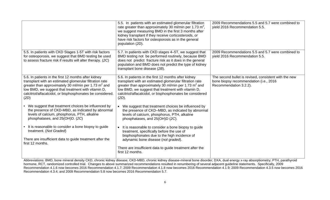
5.5. In patients with an estimated glomerular filtration rate greater than approximately 30 ml/min per 1.73 m2, we suggest measuring BMD in the first 3 months after kidney transplant if they receive corticosteroids, or have risk factors for osteoporosis as in the general population (2D).
2009 Recommendations 5.5 and 5.7 were combined to yield 2016 Recommendation 5.5.
5.5. In patients with CKD Stages 1-5T with risk factors for osteoporosis, we suggest that BMD testing be used to assess fracture risk if results will alter therapy. (2C)
5.7. In patients with CKD stages 4–5T, we suggest that BMD testing not be performed routinely, because BMD does not predict fracture risk as it does in the general population and BMD does not predict the type of kidney transplant bone disease (2B).
2009 Recommendations 5.5 and 5.7 were combined to yield 2016 Recommendation 5.5.
5.6. In patients in the first 12 months after kidney transplant with an estimated glomerular filtration rate greater than approximately 30 ml/min per 1.73 m2 and low BMD, we suggest that treatment with vitamin D, calcitriol/alfacalcidol, or bisphosphonates be considered. (2D) • We suggest that treatment choices be influenced by
the presence of CKD-MBD, as indicated by abnormal levels of calcium, phosphorus, PTH, alkaline phosphatases, and 25(OH)D. (2C)
• It is reasonable to consider a bone biopsy to guide
treatment. (Not Graded) There are insufficient data to guide treatment after the first 12 months.
5.6. In patients in the first 12 months after kidney transplant with an estimated glomerular filtration rate greater than approximately 30 ml/min per 1.73 m2 and low BMD, we suggest that treatment with vitamin D, calcitriol/alfacalcidol, or bisphosphonates be considered (2D). We suggest that treatment choices be influenced by
the presence of CKD–MBD, as indicated by abnormal levels of calcium, phosphorus, PTH, alkaline phosphatases, and 25(OH)D (2C).
It is reasonable to consider a bone biopsy to guide
treatment, specifically before the use of bisphosphonates due to the high incidence of adynamic bone disease (not graded).
There are insufficient data to guide treatment after the first 12 months.
The second bullet is revised, consistent with the new bone biopsy recommendation (i.e., 2016 Recommendation 3.2.2).
Abbreviations: BMD, bone mineral density CKD, chronic kidney disease; CKD-MBD, chronic kidney disease-mineral bone disorder; DXA, dual energy x-ray absorptiometry; PTH, parathyroid hormone, RCT, randomized controlled trial. Changes to above summarized recommendations resulted in renumbering of several adjacent guideline statements. Specifically, 2009 Recommendation 4.1.6 now becomes 2016 Recommendation 4.1.7; 2009 Recommendation 4.1.8 now becomes 2016 Recommendation 4.1.9; 2009 Recommendation 4.3.5 now becomes 2016 Recommendation 4.3.4; and 2009 Recommendation 5.8 now becomes 2016 Recommendation 5.7.

7
CHAPTER 3.2: DIAGNOSIS OF CKD–MBD: BONE
3.2.1. In patients with CKD Stages 3a-5D with evidence of CKD-MBD and/or risk factors
for osteoporosis, we suggest BMD testing to assess fracture risk if results will impact treatment decisions. (2B)
RATIONALE
It is well established that patients with CKD stages 3a-5D have increased fracture rates,
compared with the general population,2-4 and moreover, incident hip fractures are associated with substantial morbidity and mortality.5-9 At the time of the 2009 KDIGO guideline, publications addressing the ability of dual energy X-ray absorptiometry (DXA) measures of bone mineral density (BMD) to estimate fracture risk in CKD were limited to cross-sectional studies comparing BMD in CKD patients with and without a prevalent fracture. The results were variable across studies and across skeletal sites. In light of the lack of evidence that DXA BMD predicted fractures in CKD patients as it does in the general population, and the inability of DXA to indicate the histological type of bone disease, the 2009 Guideline recommended that BMD testing not be performed routinely in patients with CKD stages 3-5D with CKD-MBD. Furthermore, the lack of clinical trials in patients with low BMD and CKD also limited the enthusiasm for measuring BMD in the first place.
The current evidence-based review identified four prospective cohort studies of DXA BMD and incident fractures in adults with CKD stages 3a to 5D (Supplemental Tables 7–12). These studies demonstrated that DXA BMD predicted fractures across the spectrum from CKD stages 3a to 5D (Supplemental Tables 7–12).10-13 In the earliest study, DXA BMD was measured annually in 485 hemodialysis (HD) patients (mean age 60 years) in a single center in Japan.10 In adjusted Cox-proportional analyses, lower baseline femoral neck and total hip BMD predicted a greater risk of fracture; e.g., the hazard ratio (HR) was 0.65 (95% CI 0.47−0.90) for each standard deviation (SD) higher femoral neck BMD. In receiver-operating characteristic (ROC) analyses stratified according to PTH below or above the median value of 204 pg/ml (21.6 pmol/l), the area under the curve (AUC) for femoral neck BMD was 0.717 in the lower stratum and 0.512 in the higher stratum. Of note, higher serum bone specific alkaline phosphate levels also predicted incident fractures.
In the second study, Yenchek et al. assessed whether DXA total hip and femoral neck BMD were associated with incident non-spine fragility fractures in participants with (estimated glomerular filtration rate (eGFR) < 60 ml/min/1.73 m2) and without CKD in the Health, Aging and Body Composition Study, a prospective study of community-living individuals, 70-79 years of age at enrollment.13 A total of 587 (21%) of the 2,754 participants had CKD, and among these, 83% and 13% were CKD stage 3a and 3b, respectively. In adjusted analyses, the fracture HR for each SD lower femoral neck BMD was 2.14 (95% confidence interval [CI] 1.80−2.55) in participants without CKD, and 2.69 (95% CI 1.96−3.69) in those with CKD. Similar results were observed for total hip BMD. When limited to hip fractures, the adjusted femoral neck BMD HRs were 5.82 (95% CI 3.27−10.35) among those with CKD and 3.08 (95% CI 2.29−4.14) among those without CKD. Interaction terms demonstrated that the association of BMD with fracture did not differ in those with vs. without CKD. However, the association of femoral neck BMD with fracture was significantly less pronounced (test for interaction, p = 0.04) among those with parathyroid hormone (PTH) > 65 pg/ml [6.9 pmol/l] (HR 1.56, 95% CI 0.90− 2.70) compared with those with a PTH ≤ 65 pg/ml [6.9 pmol/l] (HR 2.41, 95% CI 2.04−2.85) in all participants combined. This is noteworthy in light of the similar pattern observed in dialysis patients, as described above.10
West et al. reported the results of a prospective cohort study of 131 predialysis participants, mean age 62 years, followed over a two year interval.12 At baseline, the proportions with CKD stages 3, 4 and 5 were 34, 40 and 26%, respectively. DXA BMD was measured in the

8
total hip, lumbar spine, and ultradistal and 1/3rd radius at baseline and two years. Low BMD at all sites, and a greater annualized percent decrease in BMD predicted fracture. For example, in multivariate models, each SD lower total hip BMD was associated with an odds ratio (OR) of fracture of 1.75 (95% CI 1.30−2.20). The ROC AUC ranged from 0.62 in the spine to 0.74 in the ultradistal radius in adjusted models.
Most recently, Naylor, et al.11 assessed the ability of the Fracture Risk Assessment Tool (FRAX) to predict a major osteoporotic fracture in 2,107 adults ≥ 40 years of age in the Canadian Multicenter Osteoporosis Study, including 320 with an eGFR ≤ 60 ml/min/1.73 m2. Of these, 72% and 24% were CKD stage 3a and 3b, respectively. FRAX with BMD, FRAX without BMD, and the femoral neck T-score all predicted fractures (AUC 0.65 to 0.71); the AUC was highest for femoral neck T-score with inclusion of fall history. Importantly, the AUCs did not differ between those with and without CKD.
There is growing evidence that DXA BMD predicts fractures in healthy children and adolescents, and those with chronic disease.14, 15 However, no studies have examined the associations among DXA BMD and fractures in children and adolescents with CKD. In light of the lack of evidence that the ability of DXA BMD to predict fracture in children with CKD is different than in adults, no specific recommendations are provided for children. However, it should be noted that children and adolescents with CKD frequently exhibit substantial growth failure. Given that DXA measures of areal BMD (g/cm2) underestimate volumetric BMD (g/cm3) in children with short stature,16 DXA results should be adjusted for bone size, consistent with the 2013 International Society of Clinical Densitometry (ISCD) Pediatric Official Positions.17 Predictions equations to adjust DXA results for height Z-score are now available,16 and the impact on DXA BMD Z-scores in children with CKD is substantial.18 Finally, a single center study in 171 children with CKD stages 2–5D reported that lower cortical volumetric BMD in the tibia, as measured by peripheral quantitative computed tomography (CT), predicted fractures over a one year interval (Supplemental Tables 7–12).19 The HR per unit lower cortical BMD Z-score was 1.75 (95% 1.15− 2.67, p < 0.01).
The evidence based review also evaluated clinical trials of the effects of osteoporosis medications on BMD in CKD stages 3a to 5D (Supplemental Tables 1–6). Prior analyses of large randomized clinical trials (RCT) evaluating medications for the treatment of postmenopausal osteoporosis (risedronate, alendronate, teriparatide, and raloxifene) were described in the 2009 Guideline. These trials specifically excluded patients with an elevated serum creatinine, hyperparathyroidism, or abnormal alkaline phosphate levels.20-23 However, post-hoc analyses found that these drugs had similar efficacy on improving BMD and reducing fracture incidence in individuals with moderately reduced eGFR, compared to those with mildly decreased or normal eGFR. Three new trials were identified. The denosumab study was also a post-hoc analysis of a RCT in women with postmenopausal osteoporosis and normal PTH levels.24 The analysis demonstrated efficacy in decreasing fracture risk and increasing BMD in 2,817 women with CKD stage 3 and 73 with CKD stage 4. The remaining two new trials on alendronate,25 and raloxifene26 were small studies (less than 60 participants) that did not exclude patients with evidence of CKD-MBD. These studies did not show consistent beneficial effects on DXA BMD.
In summary, these four prospective studies evaluating BMD testing in adults with CKD represent a substantial advance since the original guideline from 2009. Despite the fact that they were conducted across a spectrum of CKD severity, the finding that hip BMD predicted fractures was consistent across studies, and two studies demonstrated associations comparable to those seen in the absence of CKD.11, 13 Based on these insights, if a low or declining BMD will lead to additional interventions to reduce falls or use osteoporosis medications, then BMD assessment is reasonable.

9
RESEARCH RECOMMENDATIONS
RCTs are needed to determine if interventions based on DXA BMD are associated with lower fracture rates, and if the effects vary based on clinical variables such as the baseline PTH level, underlying cause of renal disease, and CKD stage.
Prospective studies are needed to determine if alternative imaging techniques, such as quantitative CT, improve fracture prediction in CKD.
Prospective studies are needed in children and adolescents to determine if DXA predicts fractures in children and to determine if the ISCD recommendations to measure whole body and spine BMD in children are the appropriate sites in the context of CKD.17 Hip and radius BMD pediatric reference data are now available and predict incident fractures in healthy children and adolescents.27, 28
3.2.2. In patients with CKD Stages 3a-5, it is reasonable to perform a bone biopsy if
knowledge of the type of renal osteodystrophy will impact treatment decisions. (Not Graded)
RATIONALE
Renal osteodystrophy is defined as abnormal bone histology and is one component of the
bone abnormalities of CKD-MBD.29 Bone biopsy is the gold standard for the diagnosis and classification for renal osteodystrophy. As detailed in the 2009 KDIGO Guideline,30 DXA BMD does not distinguish among types of renal osteodystrophy and the diagnostic utility of biochemical markers is limited by poor sensitivity and specificity. Differences in PTH assays (e.g., intact vs whole PTH) and reference ranges have contributed to differences across studies. Unfortunately, cross-sectional studies have provided conflicting information on the use of biomarkers to predict underlying bone histology. This is not surprising given the short half-lives of most of the circulating biomarkers, and the long (3 to 6 month) bone remodeling (turnover) cycle.
KDIGO recently led an international consortium to conduct a cross-sectional retrospective diagnostic study of biomarkers (all run in a single laboratory) and bone biopsies in 492 dialysis patients.31 The objective was to determine the predictive value of PTH (determined by both intact PTH [iPTH] and whole PTH assays), bone-specific alkaline phosphatase (bALP), and amino-terminal propeptide of type 1 procollagen (P1NP) as markers of bone turnover. Although iPTH, whole PTH and bALP levels were associated with bone turnover, no biomarker singly or in combination was sufficiently robust to diagnose low, normal and high bone turnover in an individual patient. The conclusion was in support of the 2009 KDIGO Guideline to use trends in PTH rather than absolute ‘target’ values when making decisions as to whether to start or stop treatments to lower PTH. Table 1 provides the sensitivity, specificity, positive and negative predictive value of PTH in helping clinicians determine therapies, demonstrating the challenges clinicians face. Thus, the Work Group encourages the continued use of trends in PTH to guide therapy, and when trends in PTH are inconsistent, a bone biopsy should be considered.

10
Table 1. Utility of KDOQI and KDIGO PTH thresholds for diagnostic decision m8aking
KDOQI* KDIGO+
Sens Spec PPV NPV Sens Spec PPV NPV
Differentiating low turnover from non-low turnover bone disease, or “When do I stop therapy?”
69% 61% 72% 58% 66% 65% 73% 57%
Differentiating high turnover from non-high turnover bone disease, or “When do I start therapy?”
58% 78% 35% 90% 37% 86% 35% 87%
Abbreviations: iPTH, intact parathyroid hormone; KDIGO, Kidney Disease: Improving Global Outcomes; KDOQI, Kidney Disease Outcomes Quality Initiative; NPV, negative predictive value; PPV, positive predictive value; PTH, parathyroid hormone; Sens, sensitivity; Spec, specificity *Using serum iPTH < 150 pg/ml (16 pmol/l) for lower and > 300 pg/ml (32 pmol/l) for upper threshold +Using serum iPTH < 130 pg/ml (14 pmol/l) for lower and > 585 pg/ml (62 pmol/l) for upper threshold (2X and 9X of upper limit
of normal for assay) Reproduced from Sprague et al.31
A bone biopsy should also be considered in patients with unexplained fractures, refractory hypercalcemia, suspicion of osteomalacia, an atypical response to standard therapies for elevated PTH, or progressive decreases in BMD despite standard therapy. The goal of a bone biopsy would be to: (a) rule out atypical or unexpected bone pathology; (b) determine if patient has high or low turnover disease which may alter the dose of medications to treat renal osteodystrophy (e.g., initiate or discontinue calcimimetics, calcitriol or vitamin D analogs); or (c) identify a mineralization defect that would alter treatment (e.g., stop intake of aluminum, or aggressively treat hypophosphatemia or vitamin D deficiency).
The 2009 Guideline recommended a bone biopsy prior to antiresorptive therapy in
patients with CKD stages 4–5D and evidence of biochemical abnormalities of CKD–MBD, low BMD and/or fragility fractures. The rationale was that low BMD may be due to CKD-MBD (e.g., high PTH) and that lowering PTH is a safer and more appropriate therapy than an anti-resorptive. In addition, there was concern that bisphosphonates would induce low-turnover bone disease. This was based on a single cross-sectional study in 13 patients with CKD stage 2 to 4 that were referred for bone biopsy after a variable duration of bisphosphonate therapy.32 To date, studies in patients with CKD have not definitively demonstrated that bisphosphonates cause adynamic bone disease. Furthermore, the concerns in patients with CKD are only theoretical, as it is well established that antiresorptive medications suppress bone formation rates, even in the absence of kidney disease. For example, in a RCT of zolendronic acid for the treatment of postmenopausal osteoporosis, bALP levels were 59% lower in the zolendronic acid group compared with the placebo group at 12 months.33
Despite these limitations, in weighing the risk-benefit ratio of bisphosphonate treatment,
the 2009 KDIGO Guideline suggested a biopsy prior to therapy. Since 2009, an additional antiresorptive treatment (denosumab) has proven to be effective in CKD stages 3 and 4, as discussed in Recommendation 3.2.1. The growing experience with osteoporosis medications in patients with CKD increases the comfort of treating patients with low BMD and a high risk of fracture with antiresorptive therapy, although definitive trials are lacking. Furthermore, additional data clearly support that the incidence of fracture is markedly increased in patients with CKD and thus the lack of ability to perform a bone biopsy may not justify withholding antiresorptive therapy to patients at high risk of fracture. Thus, the Work Group voted to remove the requirement of bone biopsy prior to the use of antiresorptive therapy for osteoporosis as the use of these drugs must be individualized in patients with CKD. However, it is still prudent that these drugs be used with caution and that the underlying renal osteodystrophy be addressed first.

11
In summary, bone biopsy is the gold standard for the assessment of renal osteodystrophy and should be considered in patients in whom the etiology of clinical symptoms and biochemical abnormalities is in question, and the results may lead to changes in therapy. Given that growing evidence that antiresorptive therapies are effective in patients with CKD stages 3 and 4, and the lack of robust evidence that these medications induce adynamic bone disease, the guideline no longer suggests a bone biopsy be performed prior to initiative of these medications.
RESEARCH RECOMMENDATION
Prospective studies of circulating biomarkers are needed to determine if they can predict changes in bone histology.

12
CHAPTER 4.1: TREATMENT OF CKD–MBD TARGETED AT LOWERING HIGH SERUM PHOSPHORUS
AND MAINTAINING SERUM CALCIUM 4.1.1. In patients with CKD Stages 3a-5D, treatments of CKD-MBD should be based on
serial assessments of phosphorus, calcium and PTH levels, considered together. (Not Graded)
RATIONALE
The previous Recommendation 4.1.1 from 2009 gave treatment directions concerning
serum phosphate levels in different stages of CKD. The accumulated evidence on this issue to date is now depicted in Supplemental Tables 49-55. Results of this evidence review can be summarized as follows: Most studies showed increasing risk of all-cause mortality with increasing levels of serum phosphate in a consistent and direct fashion, with moderate risk of bias and low quality of evidence, thus not essentially different from the study results before 2009. For GFR decline and cardiovascular event rate, results were considered less conclusive.
Serum phosphorus, calcium and PTH concentrations are all routinely measured in CKD
patients, and clinical decisions are often made based on these values. However, the results of these tests are influenced by food intake, adherence to and the timing of drug intake and dietary modifications, differences in assay methods and their intra-assay coefficient of variation (CV), and also by the interval from the last dialysis session in CKD 5D patients. Furthermore, it has recently been suggested that these markers have significant diurnal changes even in CKD patients.34, 35 Accordingly, the decision should be based not on a single result, but rather on the trends of serial results, which stands very much in accordance to 2009 Recommendation 3.1.4. In addition, recent post-hoc analyses of large dialysis cohorts suggest that the prognostic implications of individual biochemical components of CKD-MBD largely depend on their context with regard to constellations of the full array of MBD biomarkers.36 This analysis identified a wide range of CKD-MBD phenotypes, based on phosphorus, calcium and PTH measurements categorized into mutually exclusive categories low, medium and high levels using previous (Kidney Disease Outcomes Quality Initiative (KDOQI)/Kidney Disease: Improving Global Outcomes (KDIGO) guideline targets, further illustrating important potential interactions between components of CKD-MBD in terms of risk prediction for death or cardiovascular events. This analysis however did not provide guidance for treatment, because it is unknown if switching from “risk classes” parallels changes in incidence of complications or mortality over time.
Finally, therapeutic maneuvers aimed at improving one parameter often have
unintentional effects on other parameters, as exemplified by the recent EVOLVE trial.37 The guideline Work Group considered it reasonable to take the context of therapeutic interventions into account when assessing values of phosphorus, calcium and PTH, and felt that it was important to emphasize the interdependency of these biochemical parameters for clinical therapeutic decision making. Based on these assumptions, it was also decided to split previous Recommendation 4.1.1 into two new Recommendations 4.1.1 (diagnostic recommendation based on accumulated observational evidence) and 4.1.2 (therapeutic recommendation based mostly on RCTs).
RESEARCH RECOMMENDATIONS
Prospective cohort studies or RCTs to evaluate whether changes in CKD-MBD risk
marker patterns over time associate with changes in risk (e.g., multiple interventions).
13
Prospective cohort studies or RCTs studying whether or not biochemical abnormalities of CKD-MBD must be weighed differently when induced by pharmacotherapy compared to baseline values (e.g., past experience with hemoglobin as risk predictor versus active treatment to targets by erythropoiesis-stimulating agents).
Investigations contributing to the understanding of the usefulness of fibroblast growth
factor-23 (FGF23) as a (complementary) marker for treatment indications (e.g., phosphate-lowering therapies to halt CKD progression) and direct treatment target (e.g., regression of left ventricular hypertrophy [LVH]).
4.1.2. In patients with CKD Stages 3a-5D, we suggest lowering elevated phosphorus levels
towards the normal range. (2C)
RATIONALE
As outlined above, since publication of the 2009 CKD-MBD Guideline, additional high quality evidence now links higher concentrations of phosphorus with mortality among patients with CKD stages 3a-5 or post-transplantation,38-47 (Supplemental Tables 49-55) although some studies did not confirm this association.48, 49 However, trial data demonstrating that treatments which lower serum phosphorus will improve patient-centered outcome are still lacking, and therefore the strength of this recommendation remains weak (2C). The rationale of interventions therefore is still only based on epidemiological evidence as described above and biological plausibility pointing to possible phosphorus toxicity as recently summarized.50 Three recent historical cohort analyses from DOPPS, ArMORR and COSMOS were not eligible for this evidence-based review; however, it is noteworthy that these analyses suggested that those dialysis patients who had been prescribed phosphate-binder therapy showed improved survival.51-53 It is important to note that phosphate-binder prescription was associated with better nutritional status. Indeed, correction for markers of nutritional status in the DOPPS study did mitigate the strength of the association, yet a statistically significant benefit persisted. In addition, propensity scoring attempting to correct for selection bias, and subgroup analysis applied by Isakova et al.52 in the ArMORR cohort suggested robustness of the beneficial findings for those treated with phosphate binders. However, residual confounding still cannot be completely ruled out and due to the nature of the observational data, these studies did not affect the current recommendation.
Methods to prevent the development of hyperphosphatemia essentially include dietary
modification, the use of phosphate-lowering therapy, and intensified dialysis schedules for those in CKD stage 5D. In the 2009 KDIGO Guideline it was suggested to maintain serum phosphorus in the normal range in the predialysis setting and lower serum phosphorus towards the normal range in patients on dialysis. Interestingly, in the prospective observational COSMOS study cohort of HD patients (Supplemental Tables 49-55), the best patient survival was observed with serum phosphorus close to 4.4 mg/dl (1.42 mmol/l).54
The previous recommendation suggested that clinicians “maintain serum phosphorus in the normal range” for patients in CKD stages 3 and 4. The Work Group re-evaluated the evidence underlying this assumption. The majority of studies (Supplemental Table 49) found phosphorus to be consistently associated with excess mortality at levels above and below the limits of normal, but not in the normal range.39-42, 46, 47, 55, 56 This finding is in line with previously found U-shaped relation of phosphorus with mortality risk in dialysis patients.57 In addition, a recent trial comparing placebo with active phosphate-binder therapy in predialysis patients (CKD 3b-4) with a mean baseline phosphorus concentration of 4.2 mg/dl (1.36 mmol/l), found a minimal decline in serum phosphorus, no effect on FGF23 and increases in coronary calcification scores for the active treatment group58 − questioning the efficacy and safety of

14
phosphate binding in this population, with normal phosphorus concentration prior to initiation of binder treatment (Supplementary Tables 19-24). In addition, a well-executed mineral balance study in predialysis patients using calcium-containing phosphate binders, demonstrated the absence of any effect on phosphorus balance (while showing in the short-term a positive calcium balance).59
The second principal option to control phosphorus in predialysis patient is dietary
restriction, as will be addressed in reference to Recommendation 4.1.8. However, in both the NHANES and MDRD cohorts, studying the general population and advanced CKD respectively, dietary intake or intervention to reduce dietary phosphate intake as assessed by either urinary excretion or dietary recall, had only minimal effects on serum phosphorus.60, 61 It is unknown whether this minimal decline in serum phosphorus concentrations, or the more robust lower phosphorus intake translates into beneficial clinical outcome. Although a subsequent analysis of the MDRD study found no impact of low phosphorus intake as compared to higher intake on cardiovascular disease or all-cause mortality,62 the current intake of phosphorus is generally higher than at time of the MDRD study (see Recommendation 4.1.8). Taken together, the key insights from these data were: (1) the association between serum phosphorus and clinical outcome is not monotonic; (2) there is a lack of demonstrated efficacy of phosphate binders for lowering serum phosphorus in people with CKD stages 3-4; (3) the safety of phosphate binders in this population is unproven; and (4) there is an absence of data showing that dietary phosphorus restriction improves clinical outcomes. Consequently, the Work Group has abandoned the previous suggestion to maintain phosphorus in the normal range, instead suggests to focus treatment on patients with hyperphosphatemia. The Work Group recognizes that preventing, rather than treating, hyperphosphatemia may be of value in patients with CKD 3-5D, but acknowledges that current data are inadequate to support the safety or efficacy of such an approach and encourages research in this specific area.
Only two RCTs have examined phosphate-lowering therapy in children with CKD or on
dialysis;63, 64 due to the small patient numbers and short follow-up both studies did not meet preset criteria by the evidence review team (ERT). The first RCT examined biochemical endpoints only and showed equivalent phosphate control with calcium acetate and sevelamer hydrochloride in an 8-week cross-over trial.64 In the second, 29 children were randomized to different combinations of phosphate binders and vitamin D analogues: bone biopsies suggested that the sevelamer group had reduced bone formation versus baseline at 8-month follow-up, but numbers were too small for comparison versus the calcium carbonate treated group.63 Several studies in children on dialysis have shown an association between high phosphate levels and increased vessel thickness,65-67 vessel stiffness67, 68 and coronary artery calcification (CAC).66, 67,
69, 70 In young adults on dialysis the CAC score was shown to double within 20-months, and progression was associated with higher serum phosphate levels.70
RESEARCH RECOMMENDATIONS
Randomized placebo-controlled trial for controlling hyperphosphatemia in CKD stages 3a-5D using phosphate-lowering therapy strategies to test the hypothesis that lowering phosphorus induces a reduction in the incidence of patient-level endpoints (including CKD progression) in children and adults.
Prospective clinical trial in normophosphatemic patients in CKD stage 3a-5, limiting current excessive dietary phosphorus content, to test the hypothesis that an upper limit for safety exists in terms of cardiovascular outcomes.
If the feasibility of a placebo-controlled trial is threatened due to perceived lack of equipoise (despite the lack of high quality data), a prospective trial comparing two

15
different phosphorus targets in patients with stage CKD 3a-5D is encouraged in whom their physicians believe there is an indication to reduce serum phosphorus concentrations.
Prospective study testing the hypothesis that active compensatory mechanisms to counterbalance increased phosphate intake (such as increases in FGF23 and PTH) are associated with poorer clinical outcome, despite comparable serum phosphate concentration. The population should be normophosphatemic CKD patients, and the intervention is dietary phosphate restriction, phosphate-binder therapy, novel compounds to limit phosphate uptake, or a combination thereof.
4.1.3. In adult patients with CKD Stages 3a-5D, we suggest avoiding hypercalcemia. (2C)
In children with CKD Stages 3a-5D, we suggest maintaining serum calcium in the age-appropriate normal range. (2C)
RATIONALE
As is the case for phosphorus, novel epidemiological evidence linking higher calcium
concentrations to increased mortality in adults with CKD has accumulated since the 2009 KDIGO guideline on CKD-MBD (Supplemental Tables 49-55).41-43, 46, 49, 59, 71-73 Moreover, and in addition to previous observations,47 novel studies link higher concentrations of serum calcium to non-fatal cardiovascular events.74, 75 This consistency justifies the change of this recommendation from 2D to 2C, although the overall evidence base remains limited due to the lack of prospective controlled trial data.
Hypocalcemia is a classical feature of untreated CKD, in part secondary to diminished
gastrointestinal (GI) uptake of calcium due to vitamin D deficiency.76 Hypocalcemia contributes to the pathogenesis of secondary hyperparathyroidism (SHPT) and renal osteodystrophy. Therefore, the previous recommendation suggested maintaining serum calcium in the normal range, which included the correction of hypocalcemia. A more recent retrospective observational analysis of a large dialysis cohort confirmed the association between hypocalcemia and mortality risk.49 Two other recent observations however raised doubt within the KDIGO guideline Work Group about the generalizability of the suggestion to correct hypocalcemia. The first is the potential harm for some adults associated with a positive calcium balance.77, 78 The second observation is that the prevalence of hypocalcemia may have increased after the introduction of calcimimetics (cinacalcet) in patients on dialysis.37, 79, 80 The clinical implications of this increased incidence of low calcium due to the therapeutic institution of a calcimimetic is uncertain, but may be less harmful. With regard to the intention-to-treat (ITT) population of the EVOLVE trial, no negative signals were associated with the persistently low serum calcium levels in the cinacalcet arm of the trial. Retaining the 2009 KDIGO Guideline on this issue would support the concept that patients developing hypocalcemia during calcimimetic treatment require aggressive calcium treatment. Given the unproven benefits of this treatment and the potential for harm, the Work Group emphasizes an individualized approach to the treatment of hypocalcemia rather than recommending the correction of hypocalcemia for all patients.
Childhood and adolescence are critical periods for bone mass accrual: in healthy children
the calcium content of the skeleton increases from ~25 g at birth to ~1000 g in adults, and ~25% of total skeletal mass is laid down during the 2-year interval of peak height velocity.81 The mean calcium accretion rate in healthy pubertal boys and girls peaked at 359 and 284 mg/day respectively.82 The updated evidence review identified a prospective cohort study in 170 children and adolescents with CKD Stages 2-5D (Supplemental Table 49-55) that showed lower serum calcium levels were independently associated with lower cortical volumetric BMD Z-scores.19 Over a one-year follow-up in 89 children, a change in the cortical BMD Z-score positively correlated with baseline calcium (p = 0.008) and increase in calcium (p = 0.002)

16
levels, particularly in growing children. 6.5% of children sustained a fracture during the one-year follow-up. Notably, lower cortical BMD Z-score predicted future fractures: the HR for fracture was 1.75 (95% CI 1.15−2.67; p = 0.009) per SD decrease in baseline BMD.19
Thus, the Work Group recognizes the higher calcium requirements of the growing
skeleton and suggests that serum calcium levels are maintained in the age-appropriate normal range.
RESEARCH RECOMMENDATIONS
• Calcium balance study in dialysis patients at baseline versus after start of calcimimetic
treatment (with and without calcium supplementation, adaptations in dialysate calcium concentrations and/or concomitant active vitamin D analogue treatment).
• RCTs in children and adolescents with CKD to determine if calcium-based phosphate binders, as compared to calcium-free phosphate binders, promote bone accrual (as measured by bone density and structure, and fractures), and to determine the impact of phosphate binders on arterial calcification in the context of the high calcium requirement of growing bones.
4.1.4. In patients with CKD Stage 5D, we suggest using a dialysate calcium concentration
between 1.25 and 1.50 mmol/l (2.5 and 3.0 mEq/l). (2C)
RATIONALE
Based on the available evidence, the 2009 Work Group considered that a dialysate calcium concentration of 1.25 mmol/l (2.5 mEq/l) would yield neutral calcium balance, but this statement was subsequently challenged by kinetic modeling studies.83
Two relevant new RCTs are available concerning this topic (Supplemental Tables 13-
18).84, 85 In the study by Spasovski et al.,85 the effects of two different dialysate calcium concentrations were examined in patients with adynamic bone disease and found improvement of bone and mineral parameters with the lower dialysate calcium (1.25 mmol/l, or 2.5 mEq/l) as compared to the higher concentration of 1.75 mmol/l (3.5 mEq/l). Their data confirmed the results of previous papers and also supports individualization of dialysate calcium concentrations as recommended previously by the Work Group. The comparator in this study however was a high dialysate calcium concentration of 1.75 mmol/l (3.5 mEq/l), leaving open the possibility that lower levels of dialysate calcium (> 1.25 mmol/l [2.5 mEq/l] but < 1.75 mmol/l [3.5 mEq/l]) would be equally beneficial. Ok et al. randomized 425 HD patients with iPTH levels < 300 pg/ml (32 pmol/l) and baseline dialysate calcium concentrations between 1.5-1.75 mmol/l (3.0-3.5 mEq/l) to concentrations of either 1.25 mmol/l (2.5 mEq/l) or 1.75 mmol/l (3.5 mEq/l).84 Lowering dialysate calcium levels slowed the progression of CAC and improved biopsy-proven bone turnover (low bone turnover decreased from 85.0% to 41.8%) in this cohort of patients on HD. Again in this trial, comparative effects of a 1.5 mmol/l (3.0 mEq/l) calcium concentration were not addressed.
Retrospective observational data by Brunelli et al.86 however also suggested safety
concerns (i.e., heart failure events, hypotension) associated with the default use of dialysate calcium concentrations < 1.25 mmol/l (2.5 mEq/l). In turn at the high end of dialysate calcium concentration (1.75 mmol/l [3.5 mEq/l]), Kim et al.87 found increased risk for all-cause mortality and cardiovascular or infection-related hospitalization in incident HD patients for high dialysate calcium. However, by definition observational studies cannot be used to update treatment recommendations.

17
Patients with mild hypocalcemia might potentially even have a positive calcium mass transfer when dialyzed against a concentration of 1.25 mmol/l (2.5 mEq/l), but no such metabolic balance studies exist. Taken together, the Work Group felt that this recommendation remains valid as written in 2009 and that there is no new evidence justifying a change in the wording. However, additional studies of better quality are now available and as such the evidence grade has been upgraded from 2D to 2C.
RESEARCH RECOMMENDATION
Calcium balance study should be performed with non-calcium containing vs. calcium containing phosphate binders, and vitamin D sterols vs. cinacalcet in different calcium dialysate settings. These studies should include children and adolescents and assess calcium balance in the context of skeletal calcium accrual.
4.1.5. In patients with CKD Stages 3a-5D, decisions about phosphate-lowering treatment
should be based on progressively or persistently elevated serum phosphorus. (Not Graded)
RATIONALE
With regard to 2016 Recommendation 4.1.5 (formerly 2009 Recommendation 4.1.4), the
previous 2009 KDIGO CKD-MBD guideline publication commented that available phosphate binders are all effective in the treatment of hyperphosphatemia, and that there is evidence that calcium-free binders may favor halting progression of vascular calcifications (“other components of CKD-MBD”) vs. calcium-containing binders.30 Concerns about calcium balance, uncertainties about phosphate lowering in CKD patients not on dialysis, coupled with additional hard endpoint RCTs and a systematic review (effects on mortality comparing calcium-free vs. calcium-containing phosphate binders) resulted in the decision to re-evaluate this recommendation.
Based on new pathophysiological insights into phosphate regulation and the roles of FGF23 and (soluble) Klotho in early CKD, clinical studies had been initiated investigating phosphate-lowering therapies in CKD patients in whom hyperphosphatemia had not yet developed. Here, the concept of early phosphate retention, possibly represented by increases in FGF23 serum or plasma concentrations, was the focus of scientific attention. The most notable RCT in this context was performed by Block et al.58 In this study, predialysis patients (CKD 3b-4) with mean baseline serum phosphate concentrations of 4.2 mg/dl (1.36 mmol/l) were exposed to three different phosphate binders (sevelamer, lanthanum or calcium acetate) versus matching placebos, in order to explore effects on serum phosphate levels, urinary phosphate excretion, serum FGF23 levels, vascular calcification, bone density etc., with a 9-month follow-up (Supplemental Tables 19-24). While there was a small decrease of serum phosphate concentrations (for those allocated to active treatment) and a 22% decrease in urinary phosphate excretion (suggesting adherence to therapy), no changes in FGF23 levels were observed versus placebo, as already briefly outlined in the context of Recommendation 4.1.2. In contrast to the authors’ expectations, however, progression of coronary and aortic calcification was observed with active phosphate-binder treatment, while there was no progression in the placebo arm. Subgroup analysis suggested that this negative effect was accounted for by calcium acetate treatment, but neither calcium-free binders were superior to placebo with regard to this surrogate endpoint.
This study was further supported by another metabolic study in a small group of patients
in the same CKD stage range (3b-4), in whom the addition of 3 x 500 mg calcium carbonate to

18
meals containing 1 g of calcium and 1.5 g of phosphorus per day did not impact baseline neutral phosphate balance, but caused a significantly positive calcium balance,59 at least on the short-term. Due to its small patient number and short duration, this study did not fulfill the predefined inclusion criteria for full evidence review. Nevertheless, in the Work Group’s opinion, this well-performed metabolic study may have issued a plausible and relevant safety signal, and thus should be mentioned here.
Both Block et al.58 and Hill et al.59 studied subjects with essentially normal phosphorus
concentrations at baseline. Thus, there may be two key messages from these studies. First, normophosphatemia may not be an indication to start phosphate-lowering treatments. Second, the concept that not all phosphate binders are interchangeable must be noted. Whether disproportional elevations in FGF23 serum concentrations may become a signal in order to start phosphate-lowering therapies in early CKD in the future will need to be investigated in appropriate trial formats.
Considering these insights, especially regarding CKD patients not on dialysis, and as
already suggested in the rationale to Recommendation 4.1.2, the Work Group felt that the updated guideline should clarify that phosphate-lowering therapies may only be indicated in case of “progressive or persistent hyperphosphatemia.” When thinking about risk-benefit ratios, even calcium-free binders may possess a potential for predominant harm (e.g., due to side effects such as GI distress and binding of essential nutrients), if treatment does not offer concurrent benefits. The broader term “phosphate-lowering therapies” instead of phosphate-binding agents was introduced, because it appears likely that all possible approaches (i.e., binders, diet, dialysis) can be effective and because it is possible that phosphate transport inhibitors may expand the therapeutic armamentarium in the not so distant future.
There have been no additional data since 2008 with regard to “safe” phosphate level
thresholds or to hard endpoints (i.e., mortality, cardiovascular events, progression of CKD) from RCTs treating patients towards different phosphate (or FGF23) targets. The previous qualifiers (presence of other components of CKD-MBD, concomitant therapies, side-effect profile) were deleted because the Work Group thought that their consideration was self-evident. Diurnal variation of serum phosphate concentrations was discussed as another pathophysiologically relevant aspect, but while it was felt that these variations in daily phosphate levels do affect the accuracy of evaluations, this notion was considered unfeasible in clinical routine practice and therefore not included in the guideline text.
RESEARCH RECOMMENDATIONS
Prospective clinical trials studying the value of FGF23 (and possibly soluble Klotho) and serum levels as indicators for establishing phosphate-lowering therapies should be undertaken (e.g., desirable endpoints: CKD progression, cardiovascular calcification, cardiovascular events, mortality).
See research recommendations following Recommendation 4.1.2.

19
4.1.6. In adult patients with CKD Stages 3a-5D receiving phosphate-lowering treatment, we suggest restricting the dose of calcium-based phosphate binders. (2B) In children with CKD Stages 3a-5D, it is reasonable to base the choice of phosphate-lowering treatment on serum calcium levels. (Not Graded)
RATIONALE
The Work Group thought that the available novel data and the changes applied to 2009
Recommendation 4.1.4 (now 4.1.5) suggested a need to revise the 2009 Recommendation 4.1.5 (now 4.1.6). The above mentioned balance study by Hill et al.,59 supported by results published by Spiegel and Brady,78 in normophosphatemic adults in CKD stages 3b-4 suggested potential harms of liberal calcium exposure in such cohorts, but due to their study designs were not eligible for full evidence review. The RCT by Block et al.58 in a much larger, similar cohort, and two additional RCTs in hyperphosphatemic CKD patients have added hard endpoint data when prospectively comparing the calcium-free binders, mostly sevelamer, to calcium-containing binders in predialysis or dialysis adult patients, respectively (Supplemental Tables 19-24)58, 88, 89 These results were supported by results from recent systematic reviews,90-93 however, since the ERT had considered all included studies separately and individually during this update process, these two meta-analyses did not have additional bearing on the decision making.
Overall, it was felt by the Work Group that there is new evidence suggesting that excess
exposure to exogenous calcium in adults may be harmful in all stages of CKD, regardless of whether other candidate markers of risk such as hypercalcemia, arterial calcification, adynamic bone disease or low PTH levels are also present. Therefore, these previous qualifiers in the 2009 KDIGO recommendation were deleted.
Di Iorio et al. published RCTs on both predialysis and dialysis patients showing
significant survival benefits for patients treated with sevelamer vs. patients on calcium-containing binders over follow-ups of 3 years, respectively (Supplemental Tables 19-24).88, 89 Both studies were analyzed by the ERT and felt to suffer a moderate risk of bias (Supplemental Tables 23-24), leading to a 2B recommendation only. Overall, the findings from all identified studies seemed to show either a potential for benefit or an absence of harm associated with calcium-free phosphate-binding agents to treat hyperphosphatemia compared with calcium-based agents (Supplemental Tables 20, 21).
The wording in Recommendation 4.1.6 of “restricting the dose of calcium-based
phosphate binders” was retained from previous Recommendation 4.1.5; however, the qualifier that the recommendation applies to patients with persistent or recurrent hypercalcemia was removed. Given the fact of two reasonably large RCTs demonstrating mortality risks associated with calcium-containing binder treatment, it was debated within the Work Group, whether the recommendation should potentially be more explicit. However, some members of the Work Group felt that available evidence does not conclusively demonstrate that calcium-free agents are superior to calcium-based agents. In addition, none of the studies provided sufficient dose threshold information about calcium exposure and they gave no information on the potential safety of moderately dosed calcium-containing binders in combination therapies. Finally, since KDIGO guidelines are intended for a global audience and calcium-free agents are not available or affordable in all jurisdictions, recommending against the use of calcium-based binders would imply that no treatment is preferable to using calcium-based agents. Therefore, the Work Group did not make an explicit recommendation about a maximum dose of calcium-based binders, preferring to leave this to the judgment of individual physicians while acknowledging the potential existence of an upper limit of calcium dose safety.

20
The recent availability of iron-containing phosphate binders was discussed within the Work Group but did not affect the recommendations given the absence of data on patient-centered outcomes in the published phase III trials.
All of the above studies were limited to adults. Importantly, concerns regarding the
adverse effects of exogenous calcium may not be generalizable to children. Skeletal growth and development are characterized by rapid calcium accrual,82 as described in Recommendation 4.1.3 above. Furthermore, recent studies demonstrated that bone accrual continues into the third decade of life in healthy individuals, well beyond cessation of linear growth.94, 95 Of relevance to adolescents with CKD, bone accrual between ages 18 and 24 was especially pronounced among those with late puberty.96 Therefore, studies of calcium- and non-calcium- containing binders and other therapies that impact calcium balance should consider the needs of the developing skeleton. The observation that serum calcium levels were positively associated with increases in BMD in children with CKD, and this association was significantly more pronounced with greater linear growth velocity,19 illustrates the unique needs of the growing skeleton (see Recommendation 4.1.3 above). Lastly, a recent prospective cohort study in 537 children with predialysis CKD reported that phosphate-binder treatment (calcium-based in 82%) was associated with decreased risk of incident fractures (HR 0.37, 95% CI 0.15−0.91), independent of age, sex, eGFR, and PTH levels.97 Although this study did not meet the criteria for inclusion in the evidence review, it highlights the need for additional studies in children. In light of the lack of data suggesting adverse effects of exogenous calcium in children, the Work Group concluded that there was insufficient evidence to change this recommendation in children, who may be uniquely vulnerable to calcium restriction.
RESEARCH RECOMMENDATIONS
Calcium and phosphate balance studies using different calcium-based binder doses and combinations with calcium-free binders in hyperphosphatemic patients in all CKD stages should be conducted.
RCTs using iron-based phosphate binders on patient-centered and surrogate outcomes in all CKD stages should be undertaken – comparators: placebo, calcium-based binders, other calcium-free binders (“added value”?).
RCTs using phosphate transport inhibitors (e.g., nicotinamide, tenapanor) as “add-on” treatments in patients with resistant hyperphosphatemia should be investigated.
Prospective clinical and balance studies should examine on the role of magnesium as a phosphate binder, with regard to patient-centered outcomes, calcification, and cardiovascular event rates.
RCT in children and adolescents with CKD should determine if calcium-based phosphate binders, as compared to calcium-free phosphate binders, promote bone accrual (as measured by bone density and structure, and fractures), and to determine the impact of phosphate binders on arterial calcification in the context of the high calcium requirement of growing bones.

21
4.1.8. In patients with CKD Stages 3a-5D, we suggest limiting dietary phosphate intake in the treatment of hyperphosphatemia alone or in combination with other treatments. (2D) It is reasonable to consider phosphate source (e.g., animal, vegetable, additives) in making dietary recommendations. (Not Graded)
RATIONALE
There was no general controversy towards the KDIGO 2009 Guideline on dietary
phosphate restriction as an important standard of practice to lower elevated phosphate levels, but previous recommendation 4.1.7 (now 4.1.8) on limiting dietary phosphate intake was considered as vague, especially with regard to new evidences on different phosphate and phosphoprotein sources. However, as in some other contexts within this guideline update, predefined criteria on study duration and cohort size prohibited inclusion of some study reports to undergo full evidence review. Nevertheless, the Work Group felt that some of these reports sent safety signals demanding a brief discussion.
As summarized in Supplemental Tables 25-30, only two studies on this topic in dialysis patients fulfilled the evidence review criteria.98, 99 Both studies investigated the impact of intensified versus routine dietary counselling on serum phosphate levels after follow-up of 6 months. In both studies, the intensified groups more successfully reached the laboratory targets, however, no hard endpoints were documented, so the quality of evidence for outcome had to be rated as very low. Similarly, a recent Cochrane review concluded that there is low quality evidence that dietary interventions positively affect CKD-MBD biomarkers.100
The daily phosphorus intake for a typical American diet varies with age and gender. A
majority of young to middle-aged men take in more than 1600 mg/day whereas women in the same age groups take in about 1000 mg/day.101 On a global scale, there are quite significant differences in diet compositions to be considered. Estimates of dietary phosphorus from food composition tables likely underestimate the phosphorus content since they may mostly reflect the “natural” phosphorus content of foods which are highest in dairy products, meats, poultry, fish, and grains. There are actually three major sources of phosphorus; natural phosphorus (as cellular and protein constituents) contained in raw or unprocessed foods, phosphorus added to foods during processing, and phosphorus in dietary supplements/medications.
Russo et al.102 had assessed the effect of dietary phosphate restriction on the progression
of CAC. This study was not designed to compare the efficacy of phosphate binders against dietary phosphate restriction. However, they found that dietary phosphate restriction alone did not lead to a decrease in urinary phosphate excretion nor did it prevent progression of CAC, while the urine data questioned compliance with the diet, and no control group on a normal or high phosphate diet was included.
Aggressive dietary phosphate restriction is difficult since it has the potential to
compromise adequate intake of other nutrients, especially protein. Zeller et al.103 showed that the restriction of dietary protein and phosphorus could be achieved with maintenance of good nutrition status. They demonstrated that dietary protein/phosphate restriction resulted in a significant reduction in urinary phosphate excretion when compared to a control diet.
Dietary supplements and over-the-counter or prescription medications are hidden sources
of phosphorus. They may contain phosphate salts in their list of inactive ingredients. The data on the amount of phosphorus in oral medications and vitamin/mineral supplements is limited, but has the potential to contribute significantly to the phosphorus load considering the number of medications CKD patients are required to take.104, 105

22
Another consideration for modification of dietary phosphorus and control of serum phosphorus is the “bioavailability” of phosphorus in different foods based on the form – organic versus inorganic sources of phosphorus. Animal and plant based foods contain the organic form of phosphorus. Food additives contain inorganic phosphorus. About 40-60% of animal-based phosphorus is absorbed depending on the GI vitamin D receptor activations. Plant phosphorus, mostly associated with phytates, is less absorbable, generally 20-50%, in the human GI tract. It behooves the dietitian and other interdisciplinary staff to include education about the best food choices as they relate to absorbable phosphorus. Additionally, it is important for patients to be guided toward fresh and homemade foods rather than processed foods in order to avoid additives.
Organic phosphorus in such plant foods as seeds and legumes is less bioavailable because
of limited GI absorption of phytate-based phosphorus. In this context, Moe et al.34 recently demonstrated that a vegetable-based diet showed significantly lower phosphate absorption versus a meat-based diet with similar phosphate content. Inorganic phosphorus is generally more readily absorbed and its presence in additive-laden processed, preserved, or enhanced foods or soft drinks may be underreported in nutrient databases. Hence, the phosphorus burden from food additives is disproportionately high relative to natural sources that are derived from organic (animal and vegetable) food proteins, and these additives are almost completely absorbed in the GI tract. For example, Benini et al. showed that foods which contain phosphate additives have a phosphorus content nearly 70% higher that food that do not contain additives.106 Sherman and Mehta also demonstrated that phosphate contents between unprocessed and processed meat/poultry may differ by more than 60%, and thus the absorbable phosphate may even be 2-3 times higher per weight in processed food.107
In contrast, many of the foods that are traditionally labeled as high phosphorus may be
more acceptable with the knowledge that the phosphorus is absorbed more slowly and not as efficiently. For example, beans and nuts have always been listed as very high in phosphorus, however, considering their lower absorption rate they may be acceptable as protein sources, if they are not too high in other nutrients such as potassium.
The amount of phosphorus contributed by food intake is increasing with current and new
processing practices that utilize phosphorus containing ingredients in popular foods such as restructured meats (formed, pressed, rolled, and shaped for ease of preparation and ingestion), processed and spreadable cheeses, “instant” products (puddings, sauces), refrigerator bakery projects and beverages.108 Phosphorus additives are also widely used in fast foods and convenience foods that are fully or partially pre-made or instant.104
Various types of nutrition education have had mixed results for control of serum
phosphorus. Intense education focusing on phosphorus intake has been useful to reduce phosphorus retention in some studies.99, 109 A simple education tool on how to read food labels to “Look-for-PHOS” (study acronym) was successful in helping dialysis patients reduce their phosphorus intake. A magnifying glass was provided to help patients read labels,110 as well as instructions available to guide “better choices” in fast food restaurants. Other studies have had less favorable results.111
Taken together, these insights led the Work Group to the decision to not change the
principal recommendation, but to add a qualifier statement suggesting that phosphate sources should be better substantiated.

23
RESEARCH RECOMMENDATIONS
Prospective randomized trials comparing low, medium versus high phosphate intake on phosphate metabolism and homeostasis, including responses concerning FGF23, PTH, calcification, CKD progression, in patients in CKD stages 3b-4, should be performed.
In such study designs, the role of the phosphate quality should be studied: vegetable versus meat versus additive sources, respectively.
Kinetic and balance studies on the uptake of phosphate additives in dialysis patients should be performed.
Prospective trials identifying the most effective phosphate-lowering approach (benefit-risk-cost ratio) should be performed in all CKD stages – how to best combine binders, transport inhibitors and diet (plus dialysis treatment in CKD stage 5D) – in order to tackle patient-centered and surrogate endpoints, e.g., calcification, FGF23 levels, and LVH.

24
CHAPTER 4.2: TREATMENT OF ABNORMAL PTH LEVELS IN CKD–MBD
4.2.1. In patients with CKD Stages 3a-5 not on dialysis, the optimal PTH level is not
known. However, we suggest that patients with levels of intact PTH progressively rising or persistently above the upper normal limit for the assay be evaluated for modifiable factors, including hyperphosphatemia, hypocalcemia, high phosphate intake, and vitamin D deficiency. (2C)
RATIONALE
The pathogenesis of SHPT is complex and driven by several factors including vitamin D
deficiency, hypocalcemia and hyperphosphatemia. Elevated FGF23 concentrations exacerbate SHPT through further reductions in 1,25(OH)2 vitamin D (calcitriol) levels. Calcitriol deficiency results in decreased intestinal absorption of calcium and may lead to hypocalcemia, a major stimulus for PTH secretion. This leads to parathyroid cell proliferation, contributing to SHPT. The incidence and severity of SHPT increases as renal function declines and can lead to significant abnormalities in bone mineralization and turnover.
The previous 2009 KDIGO Guideline recommended addressing modifiable risk factors
for all patients with a PTH level above the upper limit of normal for the assay used.30 Unfortunately, there is still an absence of RCTs that define an optimal PTH level for patients with CKD stages 3a-5, or clinical endpoints of hospitalization, fracture or mortality. The Work Group felt that modest increases in PTH may represent an appropriate adaptive response to declining kidney function and have revised this statement to include ‘persistently’ above the upper normal PTH level as well as ‘progressively rising’ PTH levels, rather than ‘above the upper normal limit’ as in the 2009 KDIGO Guideline. Thus, treatment should not be based on a single elevated value. Although the optimal PTH is not known, the Work Group felt that rising PTH levels in CKD stages 3a-5 warrant examination of modifiable factors, such as vitamin D insufficiency /deficiency, hypocalcemia and hyperphosphatemia. In the interval since the 2009 KDIGO Guideline, one eligible RCT examined the impact of cholecalciferol supplementation (Supplemental Table 31) and three examined the impact of phosphate binders on PTH levels in the non-dialysis CKD population (Tables 20 and 31). Oksa et al.112 reported a RCT of high (20,000 international unit [IU]/week) vs low (5,000 IU/week) dose cholecalciferol supplementation in 87 adults with CKD stages 2 to 4 (Supplemental Tables 31-36). Serum 25(OH) vitamin D levels increased significantly in both groups and were significantly greater in the high dose arm at the completion of the 12 month intervention. PTH levels decreased significantly in both groups; however, the PTH levels did not differ significantly between groups at the completion of the study. Three recent RCTs in the non-dialysis CKD population evaluated phosphate binders and their effects on surrogate endpoints, such as vascular calcification, arterial compliance, left ventricular mass, and BMD, as well as calcium, phosphate and PTH levels. Two RCTs compared sevelamer with placebo (Supplemental Tables 31-36), the first in 109 non-diabetic CKD stage 3 patients113 and the second in 117 CKD patients with a mean eGFR of 36 ± 17 ml/min/1.73 m2.114 The studies were conducted over 36 weeks and 24 months, respectively, and neither study demonstrated significant differences in changes in PTH levels between sevelamer and placebo groups. Another RCT involving 148 CKD patients (eGFR 20-45 ml/min/1.73 m2) compared placebo with three different phosphate binders (calcium-based, lanthanum and sevelamer) over a 9-month period and reported that PTH levels remained stable in those on

25
active therapy (combined phosphate-binder groups) but increased by 21% in the placebo group (p = 0.002)58 (Supplemental Table 20). In the updated recommendation, an additional modifiable risk factor, ‘high phosphate intake,’ was added because of the increasing recognition that excess phosphate intake does not always result in hyperphosphatemia, especially in early stages of CKD, and that high phosphate intake may promote SHPT. While dietary phosphate, whether from food or additives, is modifiable, better methods for assessment of dietary phosphate intake are required.
4.2.2. In adult patients with CKD Stages 3a-5 not on dialysis, we suggest calcitriol and
vitamin D analogs not be routinely used. (2C) It is reasonable to reserve the use of calcitriol and vitamin D analogs for patients with CKD Stages 4-5 with severe and progressive hyperparathyroidism. (Not Graded)
In children, calcitriol and vitamin D analogs may be considered to maintain serum calcium levels in the age-appropriate normal range. (Not Graded)
RATIONALE
Prevention and treatment of SHPT is important because imbalances in mineral metabolism are associated with renal bone disease and higher PTH levels are associated with increased morbidity and mortality in CKD patients. Calcitriol and other vitamin D analogs have been the mainstay of treatment of SHPT in individuals with CKD for many decades. The 2009 KDIGO Guideline summarized multiple studies demonstrating that administration of calcitriol or vitamin D analogs (such as paricalcitol, doxercalciferol and alfacalcidol) resulted in suppression of PTH levels.30 However, there was a notable lack of trials demonstrating improvements in patient-centered outcomes.
Multiple well-conducted RCTs cited in the 2009 KDIGO CKD MBD Guideline reported benefits of calcitriol or vitamin D analogs in treating SHPT in patients with CKD stages 3-5; two primarily involved biochemical endpoints,115, 116 and two evaluated bone histomorphometry.117,
118 Despite the lack of hard clinical endpoints, these data led to the original recommendation to treat elevated PTH with calcitriol or vitamin D analogs early in CKD to prevent parathyroid hyperplasia and its skeletal consequences (level 2C). Although benefits were predominantly related to suppression of SHPT, adverse effects of hypercalcemia were noted to be of concern in the 2009 KDIGO Guideline.30
The effects of vitamin D therapy on biochemical endpoints in CKD have been previously documented, especially in regard to reduction of PTH levels. Numerous previous studies have reported significant reductions of PTH levels with calcitriol or vitamin D analogs in CKD stages 3 and 4 when compared to placebo116, 118, 119 and recent RCTs have also demonstrated vitamin D treatment effectively lowers PTH levels in CKD stages 3-5.120, 121 Additional RCTs of calcitriol or vitamin D analog therapy have been published since the 2009 KDIGO CKD-MBD Guideline (Supplemental Tables 37-42). Two, in particular, demonstrated significantly increased risk of hypercalcemia in patients treated with paricalcitol, compared with placebo, in the absence of beneficial effects on surrogate cardiac endpoints, as detailed below.120, 121 These results, combined with the opinion that moderate PTH elevations may represent an appropriate adaptive response, led the Work Group to conclude that the risk-benefit ratio of treating moderate PTH elevations was no longer favorable and that the use of calcitriol or vitamin D analogs should be reserved for only severe and progressive SHPT.

26
The two recent RCTs were designed to detect potential benefits of calcitriol or vitamin D analogs on cardiac structure and function, as measured by magnetic resonance imaging (MRI), in adults with CKD (Supplemental Tables 37-42). The rationale for these studies is that calcitriol and vitamin D analogs act through the vitamin D receptor (VDR) to exert their benefits to inhibit PTH secretion, and the VDR is also present in many tissues and organs including vascular smooth muscle, endothelial cells, and the heart. The key evidence with regard to changes in Recommendation 4.2.2 predominantly came from these trials. The first study was a double-blind RCT by Thadhani et al. (the PRIMO study), where participants with CKD stages 3-4, mild to moderate LVH, and PTH levels between 50 and 300 pg/ml (5.3 – 32 pmol/l) were assigned to placebo (n = 112) or paricalcitol (n = 115) to test the primary hypothesis that paricalcitol will reduce left ventricular mass index (LVMI) over a 48 month interval.121 Paricalcitol was administered at a dose of 2 µg/day, with protocol-specified dose reduction to 1 µg/day, if the serum calcium was > 11 mg/dl (2.75 mmol/l). Baseline PTH levels were approximately 1.5 times the upper limit of normal. The ITT analysis revealed that paricalcitol did not reduce LVMI, nor did it modify diastolic function. Of subjects on paricalcitol, the mean serum calcium increased by 0.32 mg/dl [0.08 mmol/l) (95% CI 0.19−0.45 mg/dl; 0.05-0.11 mmol/l) versus a decrease by 0.25 mg/dl [0.06 mmol/l] (95% CI −0.37- −0.12 mg/dl; −0.09—0.03 mmol/l) in the placebo group. Hypercalcemia was defined as two consecutive measurements of serum calcium > 10.5 mg/dl (> 2.63 mmol/l), and the number of patients requiring dose reductions from 2 µg/day to 1 µg/day and episodes of hypercalcemia were more frequent in the paricalcitol group (20.9%) compared with the placebo (0.9%) group. In the second key study, a double-blind RCT by Wang et al. (the OPERA study), subjects with CKD stages 3-5, LVH, and PTH ≥ 55 pg/ml (5.83 pmol/l) were randomly assigned to receive paricalcitol (n = 30) or placebo (n = 30).120 The primary endpoint was change in LVMI over 52 weeks. Baseline PTH levels were approximately twice the upper limit of normal. Change in LVMI did not differ significantly between groups, nor did secondary outcomes such as measures of systolic and diastolic function. The median (interquartile range) changes in serum calcium were 0.08 mmol/l [0.32 mg/dl] (0.02-0.16 mmol/l; 0.08-0.64 mg/dl) and 0.01 mmol/l [0.04 mg/dl] (-0.06-0.05 mmol/l; -0.24-0.2 mg/dl) in the paricalcitol and placebo arms, respectively. Hypercalcemia, defined as any serum calcium > 2.55 mmol/l (> 10.2 mg/dl), occurred in 43.3% and 3.3% of participants in the paricalcitol and placebo arms, respectively. Of note, 70% of those who were hypercalcemic received concomitant calcium-based phosphate binders, and generally the hypercalcemia was mild and could be corrected by stopping the binder without changing the paricalcitol dose. Recent meta-analyses were largely confirmatory and supported the hypercalcemia risk association with calcitriol and vitamin D analogs.122, 123 As specified in the evidence-review process, meta-analyses were not eligible for inclusion and guideline decision-making.
The evidence review identified two RCTs comparing paricalcitol to calcitriol (Supplemental Tables 37-42); neither demonstrated differences in the incidence of hypercalcemia.124, 125 Coyne et al.124 compared calcitriol (0.25 µg/day) to paricalcitol (1 µg/day) in 110 patients with CKD stages 3 and 4 and PTH > 120 pg/ml (12.7 pmol/l). The change in PTH was comparable in the two arms (a decline of 52% vs 46%) over the six month trial and the incidence of hypercalcemia was very low in both groups (only 3 with paricalcitol and 1 with calcitriol). Further details regarding changes in biochemical parameters are provided in the Supplemental Tables 37-42. An alternative to calcitriol and its analogs is “nutritional” vitamin D supplementation (calcidiol) which can also suppress PTH and decrease hypercalcemia because the normal homeostatic loops that suppress the CYP27B remain intact. However, no studies of sufficient duration were identified in this evidence review and thus this therapy remains unproven.

27
All of the above studies were conducted in adults. A recent Cochrane review examined vitamin D therapy for bone disease in children with CKD stages 2–5 and on dialysis.126 Bone disease, as assessed by changes in PTH levels, was improved by all vitamin D preparations regardless of preparation or route or frequency of administration. The prospective cohort study referenced in section 4.1.3 demonstrated that high PTH levels were independently associated with reduced cortical BMD Z-scores at baseline (p = 0.002) and one year follow-up (p < 0.001).19 High PTH levels are associated with CAC in children on dialysis.66, 67 The Cochrane review has not shown any significant difference in hypercalcemia risk with vitamin D preparations compared with placebo, but one study showed a significantly greater risk of hypercalcemia with intravenous calcitriol administration.126 No difference in growth rates was detected between different vitamin D analogs or use of oral or intravenous vitamin D treatments.126 As noted in Recommendation 4.1.3, the Work Group recommended that serum calcium should be maintained within age-appropriate reference range in children, and given the association of high PTH levels with reduced bone mineralization and increased vascular calcification, children are likely to require calcitriol or other active vitamin D analog therapy.
In summary, the PRIMO and OPERA studies failed to demonstrate improvements in clinically relevant outcomes but did demonstrate increased risk of hypercalcemia. Accordingly, the recommendation no longer recommends routine use of calcitriol or its analogs in CKD stages 3a to 5. This was not a uniform consensus among the Work Group. It should be noted that the participants in the PRIMO and OPERA trials only had moderately increased PTH levels, thus therapy with calcitriol and vitamin D analogs may be considered in those with progressive and severe SHPT.
There are still no RCTs demonstrating beneficial effects of calcitriol or vitamin D analogs on patient-level outcomes, such as cardiac events or mortality, and the optimal level of PTH in CKD stages 3a-5 is not known. Furthermore, therapy with these agents may have additional harmful effects related to increases in serum phosphate and FGF23 levels. If initiated for severe and progressive SHPT, calcitriol or vitamin D analogs should be started with low doses, independent of the initial PTH concentration, and then titrated based on the PTH response. Hypercalcemia should be avoided.
RESEARCH RECOMMENDATIONS
Multi-center RCTs should be conducted in children and adults to determine the benefits or harms of calcitriol or vitamin D analogs in patients with CKD stages 3a-5; patient-level outcomes including falls, fractures, sarcopenia, muscle strength, physical function, progression to end-stage kidney disease, cardiovascular events, hospitalizations, and mortality should be assessed. Additional important patient-level outcomes to include are bone pain, pruritus, and health-related quality of life. Studies should also include patients with more severe SHPT and should determine the impact of reducing PTH to different target levels, such as the normal range vs higher levels.
Studies should determine the effect of calcitriol and vitamin D analogs on other potential newer biomarkers of bone and mineral metabolism, as current reliance on PTH levels in guiding initiation and titration of calcitriol or vitamin D analogs is unsatisfactory. This will also require the identification of biomarkers associated with clinically significantly outcomes.

28
4.2.4. In patients with CKD Stage 5D requiring PTH-lowering therapy, we suggest calcimimetics, calcitriol, or vitamin D analogs, or a combination of calcimimetics and calcitriol, or vitamin D analogs. (2B)
RATIONALE
New data becoming available since the 2013 Madrid Controversies Conference prompted
the Work Group to reappraise the use of PTH-lowering therapies in patients with CKD stage 5D. As shown in Supplemental Table 43, the ERT identified 5 trials evaluating treatment with cinacalcet versus placebo and one trial evaluating calcitriol versus a vitamin D analog. One open label clinical trial was conducted evaluating the effect of cinacalcet on bone histomorphometry.
The Work Group discussed the EVOLVE trial at length. EVOLVE evaluated the effect of cinacalcet versus placebo on patient-level outcomes in 3,883 HD patients using a composite endpoint of all-cause mortality, non-fatal myocardial infarction, hospitalization for unstable angina, congestive heart failure and peripheral vascular events. Secondary endpoints included individual components of the primary endpoint, clinical fracture, stroke, parathyroidectomy and cardiovascular events and cardiovascular death.
The results of EVOLVE have proven controversial. The unadjusted primary composite
endpoint showed a non-significant reduction (HR 0.93, p = 0.112) with cinacalcet use. However, analyses adjusted for imbalances in baseline characteristics demonstrated a nominally significant reduction in the primary composite endpoint (HR 0.88, p = 0.007) as did sensitivity analyses accounting for patient non-adherence to randomized study medication (HR 0.77, 95% CI 0.70−0.92) or when patients were censored at the time of kidney transplant, parathyroidectomy or the use of commercial cinacalcet (HR 0.84, p ≤ 0.001). Further challenging the interpretation of the non-significant reduction in risk seen with the unadjusted primary endpoint was a significant treatment-age interaction (p = .04), leading to speculation that cinacalcet may be effective predominantly in older dialysis patients. Approximately 1/3 of the EVOLVE participants were under the age of 55 and pre-specified analyses which evaluated subjects above or below age 65 demonstrated a significant reduction in risk associated with use of cinacalcet for both the primary endpoint (HR 0.74, p ≤ 0.001) and all-cause mortality (HR 0.73, p ≤ 0.001) for those aged above 65.
The Work Group also considered additional pre-specified and post-hoc analyses from
EVOLVE. These included a demonstrated significant reduction in the risk of severe unremitting SHPT (defined as those with 2 consecutive PTH values over 1,000 pg/ml (106 pmol/l) with a serum calcium > 10.5 mg/dl [2.63 mmol/l], and the administration of cinacalcet, or parathyroidectomy). Cinacalcet appeared to consistently reduce the risk of this endpoint regardless of baseline PTH (HR 0.31, p ≤ 0.001 for those with baseline PTH 300-600 pg/ml [32-64 pmol/l]; HR 0.49, p ≤ 0.001 for those with baseline PTH 600-900 pg/ml [64-95 pmol/l]; HR 0.41, p < 0.001 for those with PTH > 900 pg/ml [95 pmol/l]). Cinacalcet had no effect on the risk of clinical fractures in unadjusted analyses (HR 0.93, p = 0.111) and showed a nominally significant reduction in risk of fracture when adjusted for age (HR 0.88, p = 0.007).
Thus, EVOLVE did not meet its primary endpoint that cinacalcet reduces the risk of
death or clinically important vascular events in CKD stage 5D patients. However, the results of secondary analyses suggest that cinacalcet may yet be beneficial in this population or a subset. There was a lack of uniform consensus among the Work Group in their interpretation of these data with regard to establishing cinacalcet as the recommended first-line therapy for patients with CKD stage 5D requiring PTH-lowering therapy. While some felt that only the primary analysis should be used to interpret the outcome, others were equally convinced that the secondary analyses strongly suggested a benefit of treatment with cinacalcet on important patient level outcomes.

29
Despite these differences in interpretation, there was agreement among Work Group members that the higher cost of cinacalcet was also a relevant consideration given its uncertain clinical benefits. There was also agreement that the documented association between good clinical outcomes and the extent of FGF23 reduction with cinacalcet warrants further study.127
No trials demonstrated the benefits of combination therapy (cinacalcet plus another
agent) on clinically relevant outcomes. However, several additional RCTs were identified that studied the effect of combination therapy on putative surrogate outcomes (summarized in Supplemental Tables 43-48). Two trials evaluated the use of cinacalcet with low dose active vitamin D versus standard therapy. Urena-Torres et al.128 demonstrated improved PTH lowering efficacy in cinacalcet/low dose active vitamin D treated subjects, while Raggi et al.129 found that cinacalcet with low dose vitamin D attenuated the progression of coronary artery calcium accumulation when assessed using calcium volume scores (p = 0.009) though not when using the more common Agatston score (p = 0.07). Two open label trials of cinacalcet were considered important in reaching consensus for Recommendation 4.2.4. The PARADIGM trial compared a cinacalcet based treatment strategy with an active vitamin D based strategy in 312 HD patients and demonstrated similar reductions in PTH in both treatment arms. The BONAFIDE trial evaluated bone histomorphometry in 77 paired bone biopsy samples in cinacalcet treated subjects with proven high turnover bone disease and demonstrated reductions in bone formation rates and substantial increase in the number of subjects with normal bone histology (from 0 at baseline to 20 after 6-12 months of therapy).130 Two subjects developed adynamic bone disease, both of whom had PTH values < 150 pg/ml (16 pmol/l) and one patient developed osteomalacia coincident with overt hypophosphatemia. Despite being a prospective interventional trial, the BONAFIDE trial did not fulfil full evidence review criteria, because there was no control group and only longitudinal assessments were available, and thus is not listed in the Supplemental Tables.
It was recognized by the Work Group that newer, intravenous calcimimetic agents have
undergone clinical trial investigation. However, no data were available for inclusion in the KDIGO systematic review, and it was agreed that without patient-level outcomes it was unlikely to modify the current recommendation.
In summary, the Work Group was divided as to whether the EVOLVE data are sufficient
to recommend cinacalcet as first-line therapy for all patients with SHPT and CKD stage 5D requiring PTH lowering. Given the lack of uniform consensus among the Work Group and the higher acquisition cost of cinacalcet, it was decided to modify the 2009 recommendation to list calcimimetic therapy now first, in alphabetical order, among acceptable treatment options while still recognizing the utility and efficacy of active vitamin D compounds.
To date, studies of cinacalcet in children are limited to case-reports,131 case series,132, 133 a
single center experience (with 4 to 28 patients),134 and an open label study of a single dose in 12 children on dialysis.135 In recognition of the unique calcium demands of the growing skeleton, PTH-lowering therapies should be used with caution in children to avoid hypocalcemia. Future studies are needed in children before making pediatric specific recommendations.
RESEARCH RECOMMENDATIONS
The Work Group explicitly endorses the presence of clinical equipoise and the need to conduct placebo controlled trials with calcimimetics versus standard therapy for the treatment of SHPT in patients with CKD stage 5D with emphasis on those at greatest risk (e.g., older, presence of cardiovascular disease).

30
Prospective RCTs aiming at patient-centered surrogate outcomes (primary endpoints: mortality, cardiovascular events; secondary endpoints: FGF23, LVH progression, calcification) should be performed with the new parenteral calcimimetic compound (e.g., etelcalcitide).
Given the disparate effects of calcimimetic and active vitamin D therapies on FGF23 and data suggesting a clinical benefit from FGF23 reduction, RCTs evaluating the specific reduction of FGF23 as a therapeutic endpoint should be undertaken.

31
CHAPTER 4.3: TREATMENT OF BONE WITH BISPHOSPHONATES, OTHER OSTEOPOROSIS MEDICATIONS, AND GROWTH HORMONE
4.3.3. In patients with CKD Stages 3a-5D with biochemical abnormalities of CKD-MBD
and low BMD and/or fragility fractures, we suggest that treatment choices take into account the magnitude and reversibility of the biochemical abnormalities and the progression of CKD, with consideration of a bone biopsy (2D).
RATIONALE
Recommendation 3.2.2 now addresses the indications for a bone biopsy prior to
antiresorptive and other osteoporosis therapies. Therefore, the original guideline Recommendation 4.3.4 has now been removed and Recommendation 4.3.3 has broadened from CKD stage 3 to CKD stages 3a-5D.

32
CHAPTER 5: EVALUATION AND TREATMENT OF KIDNEY TRANSPLANT BONE DISEASE
5.5. In patients with CKD Stages 1-5T with risk factors for osteoporosis, we suggest that
BMD testing be used to assess fracture risk if results will alter therapy. (2C)
RATIONALE
Fracture risk is four-fold higher in patients with end-stage renal disease136 as compared to the general population and further increases in the early post-transplant period.137 A 2002 study examined the risk of hip fracture in kidney transplant recipients and estimated the fracture rate at 3.3 events per 1000 person-years, a 34% higher risk compared with patients receiving dialysis who were waitlisted for transplantation.3 Bone disease in transplant recipients is complex and heterogeneous.138 Essentially, transplant bone disease is the composite of preexisting damage to the bone acquired during the period of renal insufficiency and damage to the bone starting in the period of transplantation. Of note and as opposed to older studies,139 recent cohort studies showed minimal BMD losses in the early post-transplant period, which moreover seem to be restricted to sites rich in cortical bone such as the distal radius.140 A low cumulative steroid exposure along with persistent hyperparathyroidism most probably accounts for this shift.140 The widespread implementation of steroid minimization protocols may explain the favorable trend in fracture risk in renal transplant recipients observed over the last two decades.141-143 The 2009 KDIGO Guideline30 recommended BMD testing in the first three months following transplantation in patients with an eGFR greater than 30 ml/min per 1.73 m2, if they receive corticosteroids or have risk factors for osteoporosis (previous Recommendation 5.5), but recommend that DXA BMD not be performed in those with CKD stages 4-5T (previous Recommendation 5.7). As detailed in the new DXA guideline Recommendation 3.2.1 above, there is growing evidence that DXA BMD predicts fractures across the spectrum of CKD severity, including four prospective cohort studies in patients with CKD stages 3-5D10-13 (Supplemental Tables 7-12). To date, there are no prospective studies addressing the ability of DXA to predict fractures in transplant recipients. However, a retrospective cohort study conducted in 238 renal transplant recipients with CKD stages 1-5T examined the associations of DXA BMD with fracture events.144 Lumbar spine and total hip BMD results were expressed as T-scores and categorized as normal (T-score ≥ −1), osteopenic (T-score < −1 and > −2.5), or osteoporotic (T-score ≤ −2.5). A total of 46 incident fractures were recorded in 53 patients. In a multivariate Cox analysis of DXA BMD results in the total hip, osteopenia (HR 2.7, 95% CI 1.6−4.6) and osteoporosis (HR 3.5, 95% CI 1.8−6.4) were associated with significantly increased risk of fracture compared with normal BMD, independent of age, sex and diabetes. Multivariate models were not provided for the lumbar spine BMD T-score results; however, unadjusted analyses suggested spine BMD provided less fracture prediction, compared with total hip BMD. Although this DXA study in renal transplant recipients was not eligible for the evidence-based review, based on its retrospective design, the Work Group concluded that the findings were consistent with the other studies in CKD stages 3-5D described above.
In summary, there is growing evidence that DXA BMD predicts fractures in patients with CKD across the spectrum of CKD data, with limited data suggesting these findings extend to transplant recipients. The revised guideline statements recommend BMD testing in transplant recipients, as in those with CKD stages 3a-5D, if the results will impact treatment decisions.

33
RESEARCH RECOMMENDATIONS
The research recommendations outlined for Recommendation 3.2.1 should be expanded to include studies in renal transplant recipients.
Prospective studies in patients with CKD stages 1-5T should be performed to determine the value of BMD and bone biomarkers as predictors of fractures.
5.6. In patients in the first 12 months after kidney transplant with an estimated
glomerular filtration rate greater than approximately 30 ml/min per 1.73 m2 and low BMD, we suggest that treatment with vitamin D, calcitriol/alfacalcidol, or bisphosphonates be considered. (2D)
We suggest that treatment choices be influenced by the presence of CKD-MBD, as indicated by abnormal levels of calcium, phosphorus, PTH, alkaline phosphatases, and 25(OH)D. (2C)
It is reasonable to consider a bone biopsy to guide treatment. (Not Graded) There are insufficient data to guide treatment after the first 12 months.
RATIONALE The rationale for revised guideline Recommendation 3.2.2 now addresses the indications
for a bone biopsy prior to antiresorptive and other osteoporosis therapies. Therefore, the second bullet statement above concerning bone biopsies has been modified.

34
APPENDIX: METHODOLOGIC APPROACH TO THE 2016 KDIGO CKD-MBD UPDATE
PURPOSE
In 2009, KDIGO developed a clinical practice guideline on the diagnosis, evaluation, prevention, and treatment of CKD-MBD.30 Because of the limited evidence, many of the recommendations were deliberately vague. In October of 2013, KDIGO held a Controversies Conference to determine if there was sufficient new evidence to support updating any of the recommendations. Based on the discussions at the conference, the participants opted for a “selective update” of the guideline.1 The purpose of this chapter is to describe the methods used to conduct the evidence review and to develop and update the recommendations and statements.
OVERVIEW OF THE PROCESS The process of updating the guideline consisted of the following steps:
Convening of a Controversies Conference to determine if sufficient new data to support a reassessment of the guideline
Appointing a Work Group and an ERT
Refining the research questions
Developing the search strategy, inclusion/exclusion criteria, and data extraction tables
Drafting the Evidence Matrices and Evidence Profiles
Revising the recommendations
Grading the quality of the evidence
Grading the strength of the recommendation
CONTROVERSIES CONFERENCE
In October 2013, KDIGO held a Controversies Conference entitled, “CKD-MBD: Back
to the Future” in Madrid, Spain.1 The purpose of the conference was to determine if there was sufficient new evidence to support updating any of the recommendations from the 2009 KDIGO guideline on the diagnosis, evaluation, prevention, and treatment of CKD-MBD. Seventy-four experts in adult, pediatric, and transplant nephrology, endocrinology, cardiology, bone histomorphometry pathology, and epidemiology attended the conference.
Four topic areas were considered: 1) vascular calcification; 2) bone quality; 3) calcium and phosphate; and 4) vitamin D and PTH. Each participant was assigned to one of the four topics based on their area of expertise. Participants identified new studies in their topic area and answered a set of questions to determine which recommendations required re-evaluation.1
The result was a list of recommendations to be addressed in a selected update (i.e., to use specific methods to update only those parts of the guideline in need of update). There was a public review of the scope of work for the guideline.
35
APPOINTMENT OF GUIDELINE WORK GROUP AND EVIDENCE REVIEW TEAM
The KDIGO Co-Chairs appointed two Chairs for the Guideline Work Group, who then assembled the Work Group to be responsible for the development of the guideline. The Work Group comprised domain experts, including individuals with expertise in adult and pediatric nephrology, bone disease, cardiology, and nutrition. The Johns Hopkins University in Baltimore, MD, USA was contracted as the ERT to provide expertise in guideline development methodology and systematic evidence review. KDIGO support staff provided administrative assistance and facilitated communication.
The ERT consisted of methodologists with expertise in nephrology and internal medicine, and research associates and assistants. The ERT and the Work Group worked closely throughout the project. In January 2015, the ERT and the Work Group had a 2-day meeting in Baltimore, MD to discuss the guideline development and systematic review processes and to refine the key questions.
The ERT performed systematic reviews for each of the questions conducting literature searches, abstract and full-text screening, data extraction, risk of bias assessment, and synthesis. The ERT provided suggestions and edits on the wording of recommendations, and on the use of specific grades for the strength of the recommendations and the quality of evidence. The Work Group took on the primary role of writing the recommendations and rationale, and retained final responsibility for the content of the recommendations and for the accompanying narrative. REFINEMENT OF THE RESEARCH QUESTIONS
The first task was to define the overall topics and goals for the guideline. Using the recommendations identified during the Controversies Conference, the ERT drafted research questions and identified the population, interventions, comparison, and outcomes (PICO elements) for each research question. The questions were further refined at a 2-day meeting with the ERT and the Work Group Co-Chairs.
The ERT recruited a technical expert panel to review the research questions. The technical expert panel included internal and external clinicians and researchers in nephrology and CKD. During a conference call, the technical expert panel provided feedback on the research questions.
With feedback from the technical expert panel and with public comment on the recommendations, the Work Group Co-Chairs and the ERT refined the research questions during a 2-day meeting in Baltimore, MD. During this meeting decisions were also made about outcomes, including those considered most important for decision making that would be graded (key outcomes). The finalized research questions and outcomes are presented in Table 2.

36
Table 2. Research questions
Section 2009 Rec #
Research Question Key Outcomes Additional Outcomes
Bone quality
3.2.1 In patients with CKD stages 3-5D, what is the effect on bone quality of bisphosphonates, teriparatide, denosumab and raloxifene?
TMV (Bone turnover, mineralization, volume as measured by bone biopsy)
BMD/bone mineral content
Fracture
4.3.4 In patients with CKD stages 4-5D, what is the effect on bone quality of bisphosphonates, teriparatide, denosumab and raloxifene?
TMV (Bone turnover, mineralization, volume as measured by bone biopsy)
BMD/bone mineral content
Fracture
3.2.2 (a) In patients with CKD stages 3-5D, how well do BMD results predict fractures?
(b) In patients with CKD stages 3-5D, how well do BMD results predict renal osteodystrophy?
(a) Fracture (b) TMV (Bone
turnover, mineralization, volume)
5.5 In patients with CKD stages 1-3T, how well do BMD results predict fractures?
Fracture
5.7 In patients with CKD stages 4-5T, how well do BMD results predict fractures?
Fracture
Calcium and phosphate
4.1.1 In patients with CKD stages 3-5 or 5D, what is the evidence for benefit or harm in maintaining serum phosphate in the normal range compared with other targets of serum phosphate in terms of biochemical outcomes, other surrogate outcomes, and patient-centered outcomes?
Mortality GFR decline Cardiovascular and
cerebrovascular events
Phosphorus Bone histology, BMD Vascular and valvular
calcification imaging Hospitalizations Quality of life Kidney or kidney graft
failure Fracture Parathyroidectomy Clinical adverse events Growth, skeletal
deformities, bone accrual Calciphylaxis/CUA
4.1.3 In patients with CKD stage 5D, what is the evidence for benefit or harm in using a dialysate calcium concentration between 1.25 and 1.50 mmol/l (2.5 and 3.0 mEq/l) compared with other concentrations of dialysate calcium in terms of biochemical outcomes, other surrogate outcomes, and patient-centered outcomes?
Mortality Cardiovascular and
cerebrovascular events
Calcium Bone histology, BMD Vascular and valvular
calcification imaging Measures of GFR Hospitalizations Quality of life Kidney or kidney graft
failure Fracture Parathyroidectomy Clinical adverse events Growth, skeletal
deformities, bone accrual Calciphylaxis/CUA
4.1.2 In patients with CKD stages 3-5D, what is the evidence for benefit or harm in maintaining serum calcium in the normal range compared with other targets of serum calcium in terms of biochemical outcomes, other surrogate outcomes, and patient-centered outcomes?
Mortality Cardiovascular and
cerebrovascular events
Calcium Bone histology, BMD Vascular and valvular
calcification imaging Measures of GFR Hospitalizations Quality of life Kidney or kidney graft
failure Fracture Parathyroidectomy Clinical adverse events Growth, skeletal
deformities, bone accrual Calciphylaxis/CUA

37
Section 2009 Rec #
Research Question Key Outcomes Additional Outcomes
4.1.4 In patients with CKD stages 3-5 or 5D with hyperphosphatemia, what is the evidence for benefit or harm in using calcium-containing phosphate-binding agents to treat hyperphosphatemia compared with calcium-free phosphate-binding agents in terms of biochemical outcomes, other surrogate outcomes, and patient-centered outcomes?
Mortality Cardiovascular and
cerebrovascular events
Phosphorus Bone histology, BMD Vascular and valvular
calcification imaging Measures of GFR Hospitalizations Quality of life Kidney or kidney graft
failure Fracture Parathyroidectomy Clinical adverse events Growth, skeletal
deformities, bone accrual Calciphylaxis/CUA
4.1.7 In patients with CKD stages 3-5D with hyperphosphatemia, what is the evidence for benefit or harm in limiting dietary phosphorous compared with a standard diet in terms of biochemical outcomes, other surrogate outcomes, and patient-centered outcomes?
Mortality Cardiovascular and
cerebrovascular events
Vascular and valvular calcification
Phosphorus Bone histology, BMD Measures of GFR Hospitalizations Quality of life Kidney or kidney graft
failure Fracture Parathyroidectomy Clinical adverse events Growth, skeletal
deformities, bone accrual Calciphylaxis/CUA
Vitamin D and PTH
4.2.1 In patients with CKD stages 3-5 with levels of intact PTH above the upper normal limit of the assay, what is the evidence for benefit or harm in reducing dietary phosphorous intake or treating with phosphate-binding agents, calcium supplements, or native vitamin D in terms of biochemical outcomes, other surrogate outcomes, and patient-centered outcomes?
Mortality Cardiovascular and
cerebrovascular events
GFR decline
Calcium Phosphorus Parathyroid hormone 25-hydroxyvitamin D
[25(OH)D] 1,25-dihydroxyvitamin D
[1,25(OH)2D] Alkaline phosphatases Bone-specific alkaline
phosphatase Bicarbonate FGF23 Bone histology, BMD Vascular and valvular
calcification imaging Measures of GFR Hospitalizations Quality of life Kidney or kidney graft
failure Fracture Parathyroidectomy Clinical adverse events Growth, skeletal
deformities, bone accrual Calciphylaxis/CUA
4.2.2 In patients with CKD stages 3-5 not on dialysis, in whom serum PTH is progressively rising and remains persistently above the upper limit of normal for the assay despite correction of modifiable factors, what is the evidence for benefit or harm in treating with vitamin D analogs compared with placebo or active control in terms of biochemical outcomes, other surrogate outcomes, and patient-centered outcomes?
LVH Hypercalcemia Mortality Cardiovascular and
cerebrovascular events
Calcium Phosphorus Parathyroid hormone 25-hydroxyvitamin D
[25(OH)D] 1,25-dihydroxyvitamin D
[1,25(OH)2D] Alkaline phosphatases Bone-specific alkaline
phosphatase Bicarbonate FGF23 Bone histology, BMD Vascular and valvular
calcification imaging Measures of GFR Hospitalizations Quality of life Kidney or kidney graft
failure Fracture Parathyroidectomy Clinical adverse events

38
Section 2009 Rec #
Research Question Key Outcomes Additional Outcomes
Growth, skeletal deformities, bone accrual
Calciphylaxis/CUA 4.2.4 In patients with CKD stage 5D
what is the what is the evidence for benefit or harm in treating with calcitriol, vitamin D analogs, calcimimetics or combination thereof compared with placebo or active control in terms of biochemical outcomes, other surrogate outcomes, and patient-centered outcomes?
Mortality Cardiovascular and
cerebrovascular events
Fracture Vascular and
valvular calcification imaging
Calcium Phosphorus Parathyroid hormone 25-hydroxyvitamin D
[25(OH)D] 1,25-dihydroxyvitamin D
[1,25(OH)2D] Alkaline phosphatases Bone-specific alkaline
phosphatase Bicarbonate FGF23 Bone histology, BMD Vascular and valvular
calcification imaging Measures of GFR Hospitalizations Quality of life Kidney or kidney graft
failure Fracture Parathyroidectomy Clinical adverse events Growth, skeletal
deformities, bone accrual Calciphylaxis/CUA
Abbreviations: BMD, bone mineral density; CUA, calcific uremic arteriolopathy; GFR, glomerular filtration rate; FGF-23, fibroblast growth factor-23; LVH, left ventricular hypertrophy;TMV, bone turnover mineralization volume;
SEARCH STRATEGY
The ERT searched MEDLINE and the Cochrane Central Register of Controlled Trials (CENTRAL) from December 2006 through September 2015. The December 2006 date provided the recommended 1-year overlap with the end of the previous search.145
The search strategy included MeSH and text terms for CKD and the interventions and markers of interest (Appendix A) and was limited to the English language. The ERT also reviewed the list of references that were suggested during the Controversies Conference.
All studies that were previously included in the prior guideline were re-reviewed to ensure that they met the eligibility criteria. Inclusion/exclusion criteria
With input from the Work Group, the ERT defined the eligibility criteria a priori. The eligibility criteria for all studies were: (1) original data published in English, (2) followed at least 10 patients with CKD for at least 6 months, and (3) addressed one of the questions. The minimum mean duration of follow-up of 6 months was chosen on the basis of clinical reasoning, accounting for the hypothetical mechanisms of action. For treatments of interest, the proposed effects on patient-centered outcomes require long-term exposure and typically would not be evident before several months of follow-up. The question-specific eligibility criteria are provided in Table 3.
Two reviewers independently screened titles and abstracts and full-text articles for inclusion. Differences on inclusion were resolved through consensus adjudication.
Any study not meeting the inclusion criteria could be cited in the narrative but was not considered part of the body of evidence for a particular recommendation.

39
Table 3. Question-specific eligibility criteria 2009 Recommendation #
Exposure or Intervention Eligibility Criteria
3.2.1, 4.3.4 Bisphosphonates, teriparatide, denosumab, raloxifene
RCT with at least 10 participants per arm Evaluates bone quality, bone mineral density, or fracture
3.2.2, 5.5, 5.7 Predictive value of BMD results
RCT with at least 25 participants per arm or a prospective cohort study with 50 participants
Evaluates fractures or renal osteodystrophy 4.1.1 or 4.1.2 Serum phosphate or serum
calcium levels RCT with at least 25 participants per arm or a prospective
cohort study with 50 participants Evaluates mortality, GFR decline, cardiovascular or
cerebrovascular events, phosphate levels, calcium levels, bone histology, bone mineral density, bone volume, vascular and valvular calcification imaging, hospitalizations, quality of life, kidney or kidney graft failure, fractures, parathyroidectomy, clinical adverse events, growth, skeletal deformities, bone accrual, calciphylaxis/calcific uremic arteriolopathy
4.1.3 Dialysate calcium concentrations
RCT with at least 25 participants per arm Evaluates mortality, cardiovascular or cerebrovascular
events, calcium levels, bone histology, bone mineral density, bone volume, vascular and valvular calcification imaging, measures of GFR, hospitalizations, quality of life, kidney or kidney graft failure, fractures, parathyroidectomy, clinical adverse events, growth, skeletal deformities, bone accrual, calciphylaxis/calcific uremic arteriolopathy
4.1.4, 4.1.7, 4.2.1, 4.2.2, 4.2.4
Dietary phosphate intake, phosphate binding agents, calcium supplements, native vitamin D, vitamin D analogs, calcitriol, or calcimimetics
RCT with at least 25 participants per arm Evaluates mortality, cardiovascular or cerebrovascular
events, GFR decline, calcium levels, phosphate levels, parathyroid hormone levels, 25-hydroxyvitamin D or 1,25-dihydroxyvitamin D levels, alkaline phosphatases, bone-specific alkaline phosphatase, bicarbonate, FGF23, bone histology, bone mineral density, bone volume, vascular and valvular calcification imaging, measures of GFR, hospitalizations, quality of life, kidney or kidney graft failure, fractures, parathyroidectomy, clinical adverse events, growth, skeletal deformities, bone accrual, calciphylaxis/calcific uremic arteriolopathy for any of the above interventions or LVH or hypercalcemia for vitamin D analogs
Abbreviations: BMD, bone mineral density; FGF-23, fibroblast growth factor-23; GFR, glomerular filtration rate; LVH, left ventricular hypertrophy; RCT, randomized controlled trial. DATA EXTRACTION
The ERT modified the online supplemental tables from the prior guideline. One reviewer abstracted data directly into the modified tables and a second reviewer confirmed the data abstraction. The ERT abstracted data on general study characteristics, participant characteristics, interventions and co-interventions, and outcome measures, including measures of variability.
Two reviewers independently assessed individual study quality using the Cochrane Collaboration’s tool146 for assessing risk of bias for RCTs and using the Quality in Prognosis Studies tool147 for the observational studies.
The Work Group critically reviewed draft tables, and tables were revised, as appropriate. EVIDENCE MATRICES AND EVIDENCE PROFILES
The ERT created evidence matrices for each of the key outcomes for each research question. For each key outcome, the matrix lists the individual studies, their sample size, follow-up duration, and the individual study quality. The ERT also drafted evidence profiles to display the total number and overall quality of the studies addressing each key outcome for each research question.
40
REVISING RECOMMENDATIONS
In June 2015, the Work Group and the ERT had a 3-day meeting in Baltimore, MD, USA to review the summary tables, Evidence Profiles, and Evidence Matrices; to decide if and how the recommendations should be revised; and to determine a grade that described the quality of the overall evidence and a grade for the strength of a recommendation. GRADING
A structured approach, modeled after GRADE,148-151 and facilitated by the use of Evidence Profiles and Evidence Matrices, was used to determine a grade that described the quality of the overall evidence and a grade for the strength of a recommendation. For each topic, the discussion on grading of the quality of evidence was led by the ERT, and the discussion regarding the strength of the recommendations was led by the Work Group Chairs. GRADING THE QUALITY OF EVIDENCE FOR EACH OUTCOME
The ‘quality of a body of evidence’ refers to the extent to which our confidence in an estimate of effect is sufficient to support a particular recommendation. Following Grades of Recommendations Assessment, Development, and Evaluation (GRADE), the quality of a body of evidence pertaining to a particular outcome of interest is initially categorized on the basis of study design. For questions of interventions, the initial quality grade is ‘High’ if the body of evidence consists of RCTs, or ‘Low’ if it consists of observational studies, or ‘Very Low’ if it consists of studies of other study designs. For questions of interventions, the Work Group graded only RCTs. The grade for the quality of evidence for each intervention/ outcome pair was then decreased if there were serious limitations to the methodological quality of the aggregate of studies; if there were important inconsistencies in the results across studies; if there was uncertainty about the directness of evidence including a limited applicability of findings to the population of interest; if the data were imprecise or sparse; or if there was thought to be a high likelihood of bias. The final grade for the quality of evidence for an intervention/outcome pair could be one of the following four grades: ‘High’, ‘Moderate’, ‘Low’, or ‘Very Low’ (Table 4).
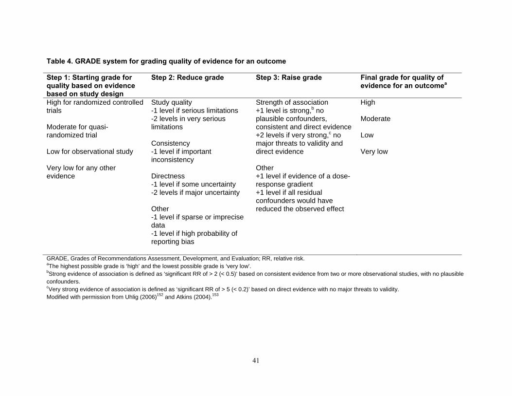
41
Table 4. GRADE system for grading quality of evidence for an outcome
Step 1: Starting grade for quality based on evidence based on study design
Step 2: Reduce grade Step 3: Raise grade Final grade for quality of evidence for an outcomea
High for randomized controlled trials Moderate for quasi-randomized trial Low for observational study Very low for any other evidence
Study quality -1 level if serious limitations -2 levels in very serious limitations Consistency -1 level if important inconsistency Directness -1 level if some uncertainty -2 levels if major uncertainty Other -1 level if sparse or imprecise data -1 level if high probability of reporting bias
Strength of association +1 level is strong,b no plausible confounders, consistent and direct evidence +2 levels if very strong,c no major threats to validity and direct evidence Other +1 level if evidence of a dose-response gradient +1 level if all residual confounders would have reduced the observed effect
High Moderate Low Very low
GRADE, Grades of Recommendations Assessment, Development, and Evaluation; RR, relative risk. aThe highest possible grade is ‘high’ and the lowest possible grade is ‘very low’. bStrong evidence of association is defined as ‘significant RR of > 2 (< 0.5)’ based on consistent evidence from two or more observational studies, with no plausible confounders. cVery strong evidence of association is defined as ‘significant RR of > 5 (< 0.2)’ based on direct evidence with no major threats to validity. Modified with permission from Uhlig (2006)152 and Atkins (2004).153

42
GRADING THE OVERALL QUALITY OF EVIDENCE
The quality of the overall body of evidence was then determined on the basis of the quality grades for all outcomes of interest, taking into account explicit judgments about the relative importance of each outcome. The resulting four final categories for the quality of overall evidence were ‘A’, ‘B’, ‘C’, or ‘D’ (Table 5). This grade for overall evidence is indicated behind the strength of recommendations. The summary of the overall quality of evidence across all outcomes proved to be very complex. Thus, as an interim step, the evidence profiles recorded the quality of evidence for each of three outcome categories: patient-centered outcomes, other bone and vascular surrogate outcomes, and laboratory outcomes. The overall quality of evidence was determined by the Work Group and is based on an overall assessment of the evidence. It reflects that, for most interventions and tests, there is no high-quality evidence for net benefit in terms of patient-centered outcomes. Table 5. Final grade for overall quality of evidence
Grade Quality of evidence Meaning A High We are confident that the true effect lies close to that of the
estimate of the effect. B Moderate The true effect is likely to be close to the estimate of the effect, but
there is a possibility that it is substantially different. C Low The true effect may be substantially different from the estimate of
the effect. D Very low The estimate of effect is very uncertain, and often will be far from
the truth.
ASSESSMENT OF THE NET HEALTH BENEFIT ACROSS ALL IMPORTANT CLINICAL OUTCOMES
Net health benefit was determined on the basis of the anticipated balance of benefits and harm across all clinically important outcomes. The assessment of net medical benefit was affected by the judgment of the Work Group and ERT. The assessment of net health benefit is summarized in one of the following statements: (i) There is net benefit from intervention when benefits outweigh harm; (ii) there is no net benefit; (iii) there are tradeoffs between benefits and harm when harm does not altogether offset benefits, but requires consideration in decision making; or (iv) uncertainty remains regarding net benefit (Table 6). Table 6. Balance of benefits and harms
When there was evidence to determine the balance of medical benefits and harm of an intervention to a patient, conclusions were categorized as follows: Net benefits The intervention clearly does more good than harm. Trade-offs There are important trade-offs between the benefits and harm. Uncertain trade-offs It is not clear whether the intervention does more good than harm. No net benefits The intervention clearly does not do more good than harm. GRADING THE RECOMMENDATIONS
The ‘strength of a recommendation’ indicates the extent to which one can be confident that adherence to the recommendation will do more good than harm. The strength of a recommendation is graded as Level 1 or Level 2.154 Table 7 shows the nomenclature for grading the strength of a recommendation and the implications of each level for patients, clinicians, and policy makers. Recommendations can be for or against doing something. Table 8 shows that the strength of a recommendation is determined not just by the quality of evidence, but also by other, often complex judgments regarding the size of the net medical benefit, values and preferences, and costs.

43
Table 7. Implications of the strength of a recommendation
Implications Grade Patients Clinicians Policy Level 1 ‘We recommend’
Most people in your situation would want the recommended course of action and only a small proportion would not.
Most patients should receive the recommended course of action.
Most patients should receive the recommended course of action.
Level 2 ‘We suggest’
The majority of people in your situation would want the recommended course of action, but many would not.
Different choices will be appropriate for different patients. Each patient needs help to arrive at a management decision consistent with her or his values and preferences.
The recommendation is likely to require debate and involvement of stakeholders before policy can be determined.
Table 8. Determinants of strength of recommendation
Factor CommentBalance between desirable and undesirable effects
The larger the difference between the desirable and undesirable effects, the more likely a strong recommendation is warranted. The narrower the
gradient, the more likely a weak recommendation is warranted. Quality of the evidence The higher the quality of evidence, the more likely a strong
recommendation is warranted. Values and preferences The more variability in values and preferences, or the more uncertainty in
values and preferences, the more likely a weak recommendation is warranted.
Costs (resource allocation) The higher the costs of an intervention—that is, the more resources consumed—the less likely a strong recommendation is warranted.
UNGRADED STATEMENTS
The Work Group felt that having a category that allows it to issue general advice would be useful. For this purpose, the Work Group chose the category of a recommendation that was not graded. Typically, this type of ungraded statement met the following criteria: it provides guidance on the basis of common sense; it provides reminders of the obvious; and it is not sufficiently specific enough to allow an application of evidence to the issue, and therefore it is not based on a systematic review. Common examples include recommendations regarding the frequency of testing, referral to specialists, and routine medical care. The ERT and Work Group strove to minimize the use of ungraded recommendations. LIMITATIONS OF APPROACH
Although the literature searches were intended to be comprehensive, they were not exhaustive. MEDLINE and Cochrane CENTRAL were the only databases searched, and the search was limited to English language publications. Hand searches of journals were not performed, and review articles and textbook chapters were not systematically searched. However, Work Group members did identify additional or new studies for consideration.
Nonrandomized studies were not systematically reviewed for studies of interventions. The ERT and Work Group resources were devoted to review of randomized trials, as these were deemed to most likely provide data to support treatment recommendations with higher quality evidence.
Evidence for patient-relevant clinical outcomes was low. Usually, low-quality evidence required a substantial use of expert judgment in deriving a recommendation from the evidence reviewed.

44
FORMULATION AND VETTING OF RECOMMENDATIONS
Recommendations were drafted to be clear and actionable, and the wording also considered the ability of concepts to be translated accurately into other languages. The final wording of recommendations and corresponding grades for the strength of the recommendations and the quality of evidence, were voted upon by the Work Group, and required a majority to be accepted.
The process of peer review included an external review by the public to ensure widespread input from numerous stakeholders, including patients, experts, and industry and national organizations.
FORMAT FOR CHAPTERS
Each chapter contains one or more specific ‘recommendations’. Within each recommendation, the strength of the recommendation is indicated as level 1 or level 2, and the quality of the overall supporting evidence is shown as A, B, C, or D. The recommendations are followed by a section that describes the body of evidence and rationale for the recommendations. In relevant sections, research recommendations suggest future research to resolve current uncertainties.

45
REFERENCES
1. Ketteler M, Elder GJ, Evenepoel P, et al. Revisiting KDIGO clinical practice guideline on chronic
kidney disease-mineral and bone disorder: a commentary from a Kidney Disease: Improving Global Outcomes controversies conference. Kidney Int 2015; 87: 502-528.
2. Alem AM, Sherrard DJ, Gillen DL, et al. Increased risk of hip fracture among patients with end-
stage renal disease. Kidney Int 2000; 58: 396-399. 3. Ball AM, Gillen DL, Sherrard D, et al. Risk of hip fracture among dialysis and renal transplant
recipients. JAMA 2002; 288: 3014-3018. 4. Nickolas TL, McMahon DJ, Shane E. Relationship between moderate to severe kidney disease and
hip fracture in the United States. J Am Soc Nephrol 2006; 17: 3223-3232. 5. Beaubrun AC, Kilpatrick RD, Freburger JK, et al. Temporal trends in fracture rates and
postdischarge outcomes among hemodialysis patients. J Am Soc Nephrol 2013; 24: 1461-1469. 6. Lin ZZ, Wang JJ, Chung CR, et al. Epidemiology and mortality of hip fracture among patients on
dialysis: Taiwan National Cohort Study. Bone 2014; 64: 235-239. 7. Maravic M, Ostertag A, Torres PU, et al. Incidence and risk factors for hip fractures in dialysis
patients. Osteoporos Int 2014; 25: 159-165. 8. Mittalhenkle A, Gillen DL, Stehman-Breen CO. Increased risk of mortality associated with hip
fracture in the dialysis population. Am J Kidney Dis 2004; 44: 672-679. 9. Tentori F, McCullough K, Kilpatrick RD, et al. High rates of death and hospitalization follow bone
fracture among hemodialysis patients. Kidney Int 2014; 85: 166-173.
10. Iimori S, Mori Y, Akita W, et al. Diagnostic usefulness of bone mineral density and biochemical markers of bone turnover in predicting fracture in CKD stage 5D patients--a single-center cohort study. Nephrol Dial Transplant 2012; 27: 345-351.
11. Naylor KL, Garg AX, Zou G, et al. Comparison of fracture risk prediction among individuals with
reduced and normal kidney function. Clin J Am Soc Nephrol 2015; 10: 646-653. 12. West SL, Lok CE, Langsetmo L, et al. Bone mineral density predicts fractures in chronic kidney
disease. J Bone Miner Res 2015; 30: 913-919. 13. Yenchek RH, Ix JH, Shlipak MG, et al. Bone mineral density and fracture risk in older individuals
with CKD. Clin J Am Soc Nephrol 2012; 7: 1130-1136. 14. Bianchi ML, Leonard MB, Bechtold S, et al. Bone health in children and adolescents with chronic
diseases that may affect the skeleton: the 2013 ISCD Pediatric Official Positions. J Clin Densitom 2014; 17: 281-294.
15. Bishop N, Arundel P, Clark E, et al. Fracture prediction and the definition of osteoporosis in
children and adolescents: the ISCD 2013 Pediatric Official Positions. J Clin Densitom 2014; 17: 275-280.
16. Zemel BS, Leonard MB, Kelly A, et al. Height adjustment in assessing dual energy x-ray
absorptiometry measurements of bone mass and density in children. J Clin Endocrinol Metab 2010; 95: 1265-1273.
17. Crabtree NJ, Arabi A, Bachrach LK, et al. Dual-energy X-ray absorptiometry interpretation and
reporting in children and adolescents: the revised 2013 ISCD Pediatric Official Positions. J Clin Densitom 2014; 17: 225-242.
18. Griffin LM, Kalkwarf HJ, Zemel BS, et al. Assessment of dual-energy X-ray absorptiometry
measures of bone health in pediatric chronic kidney disease. Pediatr Nephrol 2012; 27: 1139-1148. 19. Denburg MR, Tsampalieros AK, de Boer IH, et al. Mineral metabolism and cortical volumetric
bone mineral density in childhood chronic kidney disease. J Clin Endocrinol Metab 2013; 98: 1930-1938.
46
20. Ishani A, Blackwell T, Jamal SA, et al. The effect of raloxifene treatment in postmenopausal women with CKD. J Am Soc Nephrol 2008; 19: 1430-1438.
21. Jamal SA, Bauer DC, Ensrud KE, et al. Alendronate treatment in women with normal to severely
impaired renal function: an analysis of the fracture intervention trial. J Bone Miner Res 2007; 22: 503-508.
22. Miller PD, Roux C, Boonen S, et al. Safety and efficacy of risedronate in patients with age-related
reduced renal function as estimated by the Cockcroft and Gault method: a pooled analysis of nine clinical trials. J Bone Miner Res 2005; 20: 2105-2115.
23. Miller PD, Schwartz EN, Chen P, et al. Teriparatide in postmenopausal women with osteoporosis
and mild or moderate renal impairment. Osteoporos Int 2007; 18: 59-68. 24. Jamal SA, Ljunggren O, Stehman-Breen C, et al. Effects of denosumab on fracture and bone
mineral density by level of kidney function. J Bone Miner Res 2011; 26: 1829-1835. 25. Toussaint ND, Lau KK, Strauss BJ, et al. Effect of alendronate on vascular calcification in CKD
stages 3 and 4: a pilot randomized controlled trial. Am J Kidney Dis 2010; 56: 57-68. 26. Haghverdi F, Farbodara T, Mortaji S, et al. Effect of raloxifene on parathyroid hormone in
osteopenic and osteoporotic postmenopausal women with chronic kidney disease stage 5. Iran J Kidney Dis 2014; 8: 461-466.
27. Wren TA, Kalkwarf HJ, Zemel BS, et al. Longitudinal tracking of dual-energy X-ray
absorptiometry bone measures over 6 years in children and adolescents: persistence of low bone mass to maturity. J Pediatr 2014; 164: 1280-1285.
28. Zemel BS, Kalkwarf HJ, Gilsanz V, et al. Revised reference curves for bone mineral content and
areal bone mineral density according to age and sex for black and non-black children: results of the bone mineral density in childhood study. J Clin Endocrinol Metab 2011; 96: 3160-3169.
29. Moe S, Drueke T, Cunningham J, et al. Definition, evaluation, and classification of renal
osteodystrophy: a position statement from Kidney Disease: Improving Global Outcomes (KDIGO). Kidney Int 2006; 69: 1945-1953.
30. Kidney Disease: Improving Global Outcomes (KDIGO) CKD–MBD Work Group. KDIGO clinical
practice guideline for the diagnosis, evaluation, prevention, and treatment of chronic kidney disease–mineral and bone disorder (CKD–MBD). Kidney Int 2009; 76 (Suppl 113): S1–S130.
31. Sprague SM, Bellorin-Font E, Jorgetti V, et al. Diagnostic Accuracy of Bone Turnover Markers
and Bone Histology in Patients With CKD Treated by Dialysis. Am J Kidney Dis 2016; 67:559-566. 32. Amerling R, Harbord NB, Pullman J, et al. Bisphosphonate use in chronic kidney disease:
association with adynamic bone disease in a bone histology series. Blood Purif 2010; 29: 293-299. 33. Black DM, Delmas PD, Eastell R, et al. Once-yearly zoledronic acid for treatment of
postmenopausal osteoporosis. N Engl J Med 2007; 356: 1809-1822. 34. Moe SM, Zidehsarai MP, Chambers MA, et al. Vegetarian compared with meat dietary protein
source and phosphorus homeostasis in chronic kidney disease. Clin J Am Soc Nephrol 2011; 6: 257-264.
35. Viaene L, Meijers B, Vanrenterghem Y, et al. Daytime rhythm and treatment-related fluctuations of
serum phosphorus concentration in dialysis patients. Am J Nephrol 2012; 35: 242-248. 36. Block GA, Kilpatrick RD, Lowe KA, et al. CKD-mineral and bone disorder and risk of death and
cardiovascular hospitalization in patients on hemodialysis. Clin J Am Soc Nephrol 2013; 8: 2132-2140.
37. Chertow GM, Block GA, Correa-Rotter R, et al. Effect of cinacalcet on cardiovascular disease in
patients undergoing dialysis. N Engl J Med 2012; 367: 2482-2494. 38. Chartsrisak K, Vipattawat K, Assanatham M, et al. Mineral metabolism and outcomes in chronic
kidney disease stage 2-4 patients. BMC Nephrol 2013; 14: 14. 39. Connolly GM, Cunningham R, McNamee PT, et al. Elevated serum phosphate predicts mortality in
renal transplant recipients. Transplantation 2009; 87: 1040-1044.

47
40. Eddington H, Hoefield R, Sinha S, et al. Serum phosphate and mortality in patients with chronic kidney disease. Clin J Am Soc Nephrol 2010; 5: 2251-2257.
41. Fouque D, Roth H, Pelletier S, et al. Control of mineral metabolism and bone disease in
haemodialysis patients: which optimal targets? Nephrol Dial Transplant 2013; 28: 360-367. 42. Fukagawa M, Kido R, Komaba H, et al. Abnormal mineral metabolism and mortality in
hemodialysis patients with secondary hyperparathyroidism: evidence from marginal structural models used to adjust for time-dependent confounding. Am J Kidney Dis 2014; 63: 979-987.
43. Lacson E, Jr., Wang W, Hakim RM, et al. Associates of mortality and hospitalization in
hemodialysis: potentially actionable laboratory variables and vascular access. Am J Kidney Dis 2009; 53: 79-90.
44. McGovern AP, de Lusignan S, van Vlymen J, et al. Serum phosphate as a risk factor for
cardiovascular events in people with and without chronic kidney disease: a large community based cohort study. PLoS One 2013; 8: e74996.
45. Moore J, Tomson CR, Tessa Savage M, et al. Serum phosphate and calcium concentrations are
associated with reduced patient survival following kidney transplantation. Clin Transplant 2011; 25: 406-416.
46. Nakai S, Akiba T, Kazama J, et al. Effects of serum calcium, phosphorous, and intact parathyroid
hormone levels on survival in chronic hemodialysis patients in Japan. Ther Apher Dial 2008; 12: 49-54.
47. Tentori F, Blayney MJ, Albert JM, et al. Mortality risk for dialysis patients with different levels of
serum calcium, phosphorus, and PTH: the Dialysis Outcomes and Practice Patterns Study (DOPPS). Am J Kidney Dis 2008; 52: 519-530.
48. Fliser D, Kollerits B, Neyer U, et al. Fibroblast growth factor 23 (FGF23) predicts progression of
chronic kidney disease: the Mild to Moderate Kidney Disease (MMKD) Study. J Am Soc Nephrol 2007; 18: 2600-2608.
49. Floege J, Kim J, Ireland E, et al. Serum iPTH, calcium and phosphate, and the risk of mortality in a
European haemodialysis population. Nephrol Dial Transplant 2011; 26: 1948-1955. 50. Gross P, Six I, Kamel S, et al. Vascular toxicity of phosphate in chronic kidney disease: beyond
vascular calcification. Circ J 2014; 78: 2339-2346. 51. Cannata-Andia JB, Fernandez-Martin JL, Locatelli F, et al. Use of phosphate-binding agents is
associated with a lower risk of mortality. Kidney Int 2013; 84: 998-1008.
52. Isakova T, Gutierrez OM, Chang Y, et al. Phosphorus binders and survival on hemodialysis. J Am Soc Nephrol 2009; 20: 388-396.
53. Lopes AA, Tong L, Thumma J, et al. Phosphate binder use and mortality among hemodialysis
patients in the Dialysis Outcomes and Practice Patterns Study (DOPPS): evaluation of possible confounding by nutritional status. Am J Kidney Dis 2012; 60: 90-101.
54. Fernandez-Martin JL, Martinez-Camblor P, Dionisi MP, et al. Improvement of mineral and bone
metabolism markers is associated with better survival in haemodialysis patients: the COSMOS study. Nephrol Dial Transplant 2015; 30: 1542-1551.
55. Scialla JJ, Astor BC, Isakova T, et al. Mineral metabolites and CKD progression in African
Americans. J Am Soc Nephrol 2013; 24: 125-135. 56. Silva AP, Fragoso A, Pinho A, et al. Phosphorus as an early marker of morbidity and mortality in
type 2 chronic kidney disease diabetic patients. J Diabetes Complications 2013; 27: 328-332. 57. Kalantar-Zadeh K, Kuwae N, Regidor DL, et al. Survival predictability of time-varying indicators
of bone disease in maintenance hemodialysis patients. Kidney Int 2006; 70: 771-780. 58. Block GA, Wheeler DC, Persky MS, et al. Effects of phosphate binders in moderate CKD. J Am
Soc Nephrol 2012; 23: 1407-1415. 59. Hill KM, Martin BR, Wastney ME, et al. Oral calcium carbonate affects calcium but not
phosphorus balance in stage 3-4 chronic kidney disease. Kidney Int 2013; 83: 959-966.

48
60. de Boer IH, Rue TC, Kestenbaum B. Serum phosphorus concentrations in the third National Health and Nutrition Examination Survey (NHANES III). Am J Kidney Dis 2009; 53: 399-407.
61. Newsome B, Ix JH, Tighiouart H, et al. Effect of protein restriction on serum and urine phosphate
in the modification of diet in renal disease (MDRD) study. Am J Kidney Dis 2013; 61: 1045-1046. 62. Selamet U, Tighiouart H, Sarnak MJ, et al. Relationship of dietary phosphate intake with risk of
end-stage renal disease and mortality in chronic kidney disease stages 3-5: The Modification of Diet in Renal Disease Study. Kidney Int 2016; 89: 176-184.
63. Salusky IB, Goodman WG, Sahney S, et al. Sevelamer controls parathyroid hormone-induced bone
disease as efficiently as calcium carbonate without increasing serum calcium levels during therapy with active vitamin D sterols. J Am Soc Nephrol 2005; 16: 2501-2508.
64. Pieper AK, Haffner D, Hoppe B, et al. A randomized crossover trial comparing sevelamer with
calcium acetate in children with CKD. Am J Kidney Dis 2006; 47: 625-635. 65. Litwin M, Wuhl E, Jourdan C, et al. Evolution of large-vessel arteriopathy in paediatric patients
with chronic kidney disease. Nephrol Dial Transplant 2008; 23: 2552-2557. 66. Oh J, Wunsch R, Turzer M, et al. Advanced coronary and carotid arteriopathy in young adults with
childhood-onset chronic renal failure. Circulation 2002; 106: 100-105. 67. Shroff RC, Donald AE, Hiorns MP, et al. Mineral metabolism and vascular damage in children on
dialysis. J Am Soc Nephrol 2007; 18: 2996-3003.
68. Mitsnefes MM, Kimball TR, Kartal J, et al. Cardiac and vascular adaptation in pediatric patients with chronic kidney disease: role of calcium-phosphorus metabolism. J Am Soc Nephrol 2005; 16: 2796-2803.
69. Civilibal M, Caliskan S, Adaletli I, et al. Coronary artery calcifications in children with end-stage
renal disease. Pediatr Nephrol 2006; 21: 1426-1433. 70. Goodman WG, Goldin J, Kuizon BD, et al. Coronary-artery calcification in young adults with end-
stage renal disease who are undergoing dialysis. N Engl J Med 2000; 342: 1478-1483. 71. Coen G, Pierantozzi A, Spizzichino D, et al. Risk factors of one year increment of coronary
calcifications and survival in hemodialysis patients. BMC Nephrol 2010; 11: 10. 72. Fein PA, Asadi S, Singh P, et al. Relationship between alkaline phosphatase and all-cause mortality
in peritoneal dialysis patients. Adv Perit Dial 2013; 29: 61-63. 73. Markaki A, Kyriazis J, Stylianou K, et al. The role of serum magnesium and calcium on the
association between adiponectin levels and all-cause mortality in end-stage renal disease patients. PLoS One 2012; 7: e52350.
74. Gallieni M, Caputo F, Filippini A, et al. Prevalence and progression of cardiovascular calcifications
in peritoneal dialysis patients: A prospective study. Bone 2012; 51: 332-337. 75. Nakano C, Hamano T, Fujii N, et al. Combined use of vitamin D status and FGF23 for risk
stratification of renal outcome. Clin J Am Soc Nephrol 2012; 7: 810-819. 76. Rutherford WE, Blondin J, Hruska K, et al. Effect of 25-hydroxycholecalciferol on calcium
absorption in chronic renal disease. Kidney Int 1975; 8: 320-324. 77. Raggi P, Bommer J, Chertow GM. Valvular calcification in hemodialysis patients randomized to
calcium-based phosphorus binders or sevelamer. J Heart Valve Dis 2004; 13: 134-141. 78. Spiegel DM, Brady K. Calcium balance in normal individuals and in patients with chronic kidney
disease on low- and high-calcium diets. Kidney Int 2012; 81: 1116-1122. 79. Chonchol M, Locatelli F, Abboud HE, et al. A randomized, double-blind, placebo-controlled study
to assess the efficacy and safety of cinacalcet HCl in participants with CKD not receiving dialysis. Am J Kidney Dis 2009; 53: 197-207.
80. St Peter WL, Li Q, Liu J, et al. Cinacalcet use patterns and effect on laboratory values and other
medications in a large dialysis organization, 2004 through 2006. Clin J Am Soc Nephrol 2009; 4: 354-360.

49
81. Baxter-Jones AD, Faulkner RA, Forwood MR, et al. Bone mineral accrual from 8 to 30 years of age: an estimation of peak bone mass. J Bone Miner Res 2011; 26: 1729-1739.
82. Bailey DA, Martin AD, McKay HA, et al. Calcium accretion in girls and boys during puberty: a
longitudinal analysis. J Bone Miner Res 2000; 15: 2245-2250. 83. Gotch FA, Kotanko P, Thijssen S, et al. The KDIGO guideline for dialysate calcium will result in
an increased incidence of calcium accumulation in hemodialysis patients. Kidney Int 2010; 78: 343-350.
84. Ok E, Asci G, Bayraktaroglu S, et al. Reduction of Dialysate Calcium Level Reduces Progression
of Coronary Artery Calcification and Improves Low Bone Turnover in Patients on Hemodialysis. J Am Soc Nephrol 2016 7: 2475-2486.
85. Spasovski G, Gelev S, Masin-Spasovska J, et al. Improvement of bone and mineral parameters
related to adynamic bone disease by diminishing dialysate calcium. Bone 2007; 41: 698-703. 86. Brunelli SM, Sibbel S, Do TP, et al. Facility Dialysate Calcium Practices and Clinical Outcomes
Among Patients Receiving Hemodialysis: A Retrospective Observational Study. Am J Kidney Dis 2015; 66: 655-665.
87. Kim HW, Kim SH, Kim YO, et al. Impact of Dialysate Calcium Concentration on Clinical
Outcomes in Incident Hemodialysis Patients. Medicine (Baltimore) 2015; 94: e1694. 88. Di Iorio B, Bellasi A, Russo D. Mortality in kidney disease patients treated with phosphate binders:
a randomized study. Clin J Am Soc Nephrol 2012; 7: 487-493. 89. Di Iorio B, Molony D, Bell C, et al. Sevelamer versus calcium carbonate in incident hemodialysis
patients: results of an open-label 24-month randomized clinical trial. Am J Kidney Dis 2013; 62: 771-778.
90. Jamal SA, Vandermeer B, Raggi P, et al. Effect of calcium-based versus non-calcium-based
phosphate binders on mortality in patients with chronic kidney disease: an updated systematic review and meta-analysis. Lancet 2013; 382: 1268-1277.
91. Palmer SC, Gardner S, Tonelli M, et al. Phosphate-binding agents in adults with CKD: A network
meta-analysis of randomized trials. Am J Kidney Dis doi: 10.1053/j.ajkd.2016.05.015. 92. Patel L, Bernard LM, Elder GJ. Sevelamer versus calcium-based binders for treatment of
hyperphosphatemia in CKD: A meta-analysis of randomized controlled trials. Clin J Am Soc Nephrol 2016; 11: 232-244.
93. Sekercioglu N, Thabane L, Diaz Martinez JP, et al. Comparative effectiveness of phosphate binders
in patients with chronic kidney disease: A systematic review and network meta-analysis. PLoS One 2016; 11: e0156891.
94. Ohlsson C, Darelid A, Nilsson M, et al. Cortical consolidation due to increased mineralization and
endosteal contraction in young adult men: a five-year longitudinal study. J Clin Endocrinol Metab 2011; 96: 2262-2269.
95. Berger C, Goltzman D, Langsetmo L, et al. Peak bone mass from longitudinal data: implications for
the prevalence, pathophysiology, and diagnosis of osteoporosis. J Bone Miner Res 2010; 25: 1948-1957.
96. Darelid A, Ohlsson C, Nilsson M, et al. Catch up in bone acquisition in young adult men with late
normal puberty. J Bone Miner Res 2012; 27: 2198-2207. 97. Denburg MR, Kumar J, Jemielita T, et al. Fracture Burden and Risk Factors in Childhood CKD:
Results from the CKiD Cohort Study. J Am Soc Nephrol 2016; 27: 543-550. 98. Karavetian M, de Vries N, Elzein H, et al. Effect of behavioral stage-based nutrition education on
management of osteodystrophy among hemodialysis patients, Lebanon. Patient Educ Couns 2015; 98: 1116-1122.
99. Lou LM, Caverni A, Gimeno JA, et al. Dietary intervention focused on phosphate intake in
hemodialysis patients with hyperphosphoremia. Clin Nephrol 2012; 77: 476-483.

50
100. Liu Z, Su G, Guo X, et al. Dietary interventions for mineral and bone disorder in people with chronic kidney disease. Cochrane Database Syst Rev 2015: CD010350.
101. Calvo MS, Park YK. Changing phosphorus content of the U.S. diet: potential for adverse effects on
bone. J Nutr 1996; 126: 1168S-1180S. 102. Russo D, Miranda I, Ruocco C, et al. The progression of coronary artery calcification in predialysis
patients on calcium carbonate or sevelamer. Kidney Int 2007; 72: 1255-1261. 103. Zeller K, Whittaker E, Sullivan L, et al. Effect of restricting dietary protein on the progression of
renal failure in patients with insulin-dependent diabetes mellitus. N Engl J Med 1991; 324: 78-84. 104. Anderson JJB, Sell ML, Garner S, Calvo MS. Phosphorus. In: Present Knowledge of Nutrition.
Washington, DC: ILSI Press, 2001. 105. Sherman RA, Ravella S, Kapoian T. The phosphate content of prescription medication: A new
consideration. Therapeutic Innovation & Regulatory Science 2015; 49: 886-889. 106. Benini O, D'Alessandro C, Gianfaldoni D, et al. Extra-phosphate load from food additives in
commonly eaten foods: a real and insidious danger for renal patients. J Ren Nutr 2011; 21: 303-308.
107. Sherman RA, Mehta O. Phosphorus and potassium content of enhanced meat and poultry products:
implications for patients who receive dialysis. Clin J Am Soc Nephrol 2009; 4: 1370-1373. 108. Calvo MS. Dietary considerations to prevent loss of bone and renal function. Nutrition 2000; 16:
564-566. 109. Caldeira D, Amaral T, David C, et al. Educational strategies to reduce serum phosphorus in
hyperphosphatemic patients with chronic kidney disease: systematic review with meta-analysis. J Ren Nutr 2011; 21: 285-294.
110. Sullivan C, Sayre SS, Leon JB, et al. Effect of food additives on hyperphosphatemia among
patients with end-stage renal disease: a randomized controlled trial. JAMA 2009; 301: 629-635. 111. Durose CL, Holdsworth M, Watson V, et al. Knowledge of dietary restrictions and the medical
consequences of noncompliance by patients on hemodialysis are not predictive of dietary compliance. J Am Diet Assoc 2004; 104: 35-41.
112. Oksa A, Spustova V, Krivosikova Z, et al. Effects of long-term cholecalciferol supplementation on
mineral metabolism and calciotropic hormones in chronic kidney disease. Kidney Blood Press Res 2008; 31: 322-329.
113. Chue CD, Townend JN, Moody WE, et al. Cardiovascular effects of sevelamer in stage 3 CKD. J
Am Soc Nephrol 2013; 24: 842-852. 114. Lemos MM, Watanabe R, Carvalho AB, et al. Effect of rosuvastatin and sevelamer on the
progression of coronary artery calcification in chronic kidney disease: a pilot study. Clin Nephrol 2013; 80: 1-8.
115. Coyne D, Acharya M, Qiu P, et al. Paricalcitol capsule for the treatment of secondary
hyperparathyroidism in stages 3 and 4 CKD. Am J Kidney Dis 2006; 47: 263-276.
116. Coburn JW, Maung HM, Elangovan L, et al. Doxercalciferol safely suppresses PTH levels in patients with secondary hyperparathyroidism associated with chronic kidney disease stages 3 and 4. Am J Kidney Dis 2004; 43: 877-890.
117. Nordal KP, Dahl E. Low dose calcitriol versus placebo in patients with predialysis chronic renal
failure. J Clin Endocrinol Metab 1988; 67: 929-936. 118. Hamdy NA, Kanis JA, Beneton MN, et al. Effect of alfacalcidol on natural course of renal bone
disease in mild to moderate renal failure. BMJ 1995; 310: 358-363. 119. Fishbane S, Chittineni H, Packman M, et al. Oral paricalcitol in the treatment of patients with
CKD and proteinuria: a randomized trial. Am J Kidney Dis 2009; 54: 647-652. 120. Wang AY, Fang F, Chan J, et al. Effect of paricalcitol on left ventricular mass and function in
CKD--the OPERA trial. J Am Soc Nephrol 2014; 25: 175-186.

51
121. Thadhani R, Appelbaum E, Pritchett Y, et al. Vitamin D therapy and cardiac structure and function in patients with chronic kidney disease: the PRIMO randomized controlled trial. JAMA 2012; 307: 674-684.
122. Li XH, Feng L, Yang ZH, et al. The effect of active vitamin D on cardiovascular outcomes in
predialysis chronic kidney diseases: A systematic review and meta-analysis. Nephrology (Carlton) 2015 10.1111/nep.12505.
123. Xu L, Wan X, Huang Z, et al. Impact of vitamin D on chronic kidney diseases in non-dialysis
patients: a meta-analysis of randomized controlled trials. PLoS One 2013; 8: e61387. 124. Coyne DW, Goldberg S, Faber M, et al. A randomized multicenter trial of paricalcitol versus
calcitriol for secondary hyperparathyroidism in stages 3-4 CKD. Clin J Am Soc Nephrol 2014; 9: 1620-1626.
125. Riccio E, Sabbatini M, Bruzzese D, et al. Effect of paricalcitol vs calcitriol on hemoglobin levels
in chronic kidney disease patients: a randomized trial. PLoS One 2015; 10: e0118174. 126. Hahn D, Hodson EM, Craig JC. Interventions for metabolic bone disease in children with chronic
kidney disease. Cochrane Database Syst Rev 2015: CD008327. 127. Moe SM, Chertow GM, Parfrey PS, et al. Cinacalcet, Fibroblast Growth Factor-23, and
Cardiovascular Disease in Hemodialysis: The Evaluation of Cinacalcet HCl Therapy to Lower Cardiovascular Events (EVOLVE) Trial. Circulation 2015; 132: 27-39.
128. Urena-Torres P, Bridges I, Christiano C, et al. Efficacy of cinacalcet with low-dose vitamin D in
incident haemodialysis subjects with secondary hyperparathyroidism. Nephrol Dial Transplant 2013; 28: 1241-1254.
129. Raggi P, Chertow GM, Torres PU, et al. The ADVANCE study: a randomized study to evaluate
the effects of cinacalcet plus low-dose vitamin D on vascular calcification in patients on hemodialysis. Nephrol Dial Transplant 2011; 26: 1327-1339.
130. Behets GJ, Spasovski G, Sterling LR, et al. Bone histomorphometry before and after long-term
treatment with cinacalcet in dialysis patients with secondary hyperparathyroidism. Kidney Int 2015; 87: 846-856.
131. Nichols KR, Knoderer CA, Johnston B, et al. Cinacalcet administration by gastrostomy tube in a
child receiving peritoneal dialysis. J Pediatr Pharmacol Ther 2014; 19: 202-205. 132. Dotis J, Printza N, Ghogha C, et al. Short- and middle-term continuous use of cinacalcet in
children on peritoneal dialysis. J Pediatr Endocrinol Metab 2013; 26: 39-43. 133. Platt C, Inward C, McGraw M, et al. Middle-term use of Cinacalcet in paediatric dialysis patients.
Pediatr Nephrol 2010; 25: 143-148. 134. Alharthi AA, Kamal NM, Abukhatwah MW, et al. Cinacalcet in pediatric and adolescent chronic
kidney disease: a single-center experience. Medicine (Baltimore) 2015; 94: e401. 135. Padhi D, Langman CB, Fathallah-Shaykh S, et al. An open-label study to evaluate a single-dose of
cinacalcet in pediatric dialysis subjects. Pediatr Nephrol 2012; 27: 1953-1959. 136. Jadoul M, Albert JM, Akiba T, et al. Incidence and risk factors for hip or other bone fractures
among hemodialysis patients in the Dialysis Outcomes and Practice Patterns Study. Kidney Int 2006; 70: 1358-1366.
137. Vautour LM, Melton LJ, 3rd, Clarke BL, et al. Long-term fracture risk following renal
transplantation: a population-based study. Osteoporos Int 2004; 15: 160-167. 138. Evenepoel P. Recovery versus persistence of disordered mineral metabolism in kidney transplant
recipients. Semin Nephrol 2013; 33: 191-203. 139. Heaf JG. Bone disease after renal transplantation. Transplantation 2003; 75: 315-325. 140. Iyer SP, Nikkel LE, Nishiyama KK, et al. Kidney transplantation with early corticosteroid
withdrawal: paradoxical effects at the central and peripheral skeleton. J Am Soc Nephrol 2014; 25: 1331-1341.
52
141. Naylor KL, Jamal SA, Zou G, et al. Fracture Incidence in Adult Kidney Transplant Recipients. Transplantation 2016; 100: 167-175.
142. Nikkel LE, Mohan S, Zhang A, et al. Reduced fracture risk with early corticosteroid withdrawal
after kidney transplant. Am J Transplant 2012; 12: 649-659.
143. Sukumaran Nair S, Lenihan CR, Montez-Rath ME, et al. Temporal trends in the incidence, treatment and outcomes of hip fracture after first kidney transplantation in the United States. Am J Transplant 2014; 14: 943-951.
144. Akaberi S, Simonsen O, Lindergard B, et al. Can DXA predict fractures in renal transplant
patients? Am J Transplant 2008; 8: 2647-2651. 145. Tsertsvadze A, Maglione M, Chou R, et al. Updating comparative effectiveness reviews: current
efforts in AHRQ's Effective Health Care Program. J Clin Epidemiol 2011; 64: 1208-1215. 146. Higgins JPT. Cochrane handbook for systemic reviews of interventions Version 5.1.0. The
Cochrane Collaboration; 2011. 147. Hayden JA, van der Windt DA, Cartwright JL, et al. Assessing bias in studies of prognostic factors.
Ann Intern Med 2013; 158: 280-286. 148. Guyatt GH, Thorlund K, Oxman AD, et al. GRADE guidelines: 13. Preparing summary of findings
tables and evidence profiles-continuous outcomes. J Clin Epidemiol 2013; 66: 173-183. 149. Guyatt G, Oxman AD, Akl EA, et al. GRADE guidelines: 1. Introduction-GRADE evidence
profiles and summary of findings tables. J Clin Epidemiol 2011; 64: 383-394. 150. Guyatt GH, Oxman AD, Santesso N, et al. GRADE guidelines: 12. Preparing summary of findings
tables-binary outcomes. J Clin Epidemiol 2013; 66: 158-172. 151. Guyatt G, Oxman AD, Sultan S, et al. GRADE guidelines: 11. Making an overall rating of
confidence in effect estimates for a single outcome and for all outcomes. J Clin Epidemiol 2013; 66: 151-157.
152. Uhlig K, Macleod A, Craig J, et al. Grading evidence and recommendations for clinical practice
guidelines in nephrology. A position statement from Kidney Disease: Improving Global Outcomes (KDIGO). Kidney Int 2006; 70: 2058-2065.
153. Atkins D, Best D, Briss PA, et al. Grading quality of evidence and strength of recommendations.
BMJ 2004; 328: 1490. 154. Guyatt GH, Oxman AD, Kunz R, et al. Going from evidence to recommendations. BMJ 2008; 336:
1049-1051.