KDIGO Clinical Practice Guideline for Management of Blood Pressure in CKD
KDIGO CLINICAL PRACTICE GUIDELINE ON THE EVALUATION AND MANAGEMENT OF
CANDIDATES FOR KIDNEY TRANSPLANTATION
PUBLIC REVIEW DRAFT
OCTOBER 2018
CONFIDENTIAL: DO NOT DISTRIBUTE

ii
TABLE OF CONTENTS
Disclaimer……………………………………………………………………................................................................ iii Work Group Membership…………………………………………………………………….…………………………… iv KDIGO Executive Committee…………………….…………………………………................................................... vi Reference Keys……………………………………………………………………………………................................. vii Abbreviations and Acronyms…………………………………………………………………………………………….. x Preface……………………………………………………………………………………………………………………… xiii Summary of Recommendatons………………………………………………………………………………………….. xvi Chapter 1: Access to Transplantation………….................................................................................................... 1 Chapter 2: Age………………..…………………………..….………….…................................................................ 6 Chapter 3: Pediatric Issues………………………………………………………………………....……….…………... 8 Chapter 4: Psychosocial Assessment…………………………….…………………………..………………………… 10 Chapter 5: Adherence………………………………………………………….…………………………………………. 16 Chapter 6: Tobacco…………………………………………………..…………………………………………………… 23 Chapter 7: Surgical Issues including Obesity………………………………………………………………..…………. 26 Chapter 8: Diabetes…………………………………………....…………………………………………………………. 35 Chapter 9: Cause of End-Stage Kidney Disease……………………………………………………………………… 37 Chapter 10: Infections………………………………………………………………………..………………………..…. 56 Chapter 11: Malignancy………………………………………………………………….……………………………….. 78 Chapter 12: Pulmonary Disease……………………………………..………………………………………………….. 88 Chapter 13: Cardiac Disease……………………………………………………...…………………………………….. 90 Chapter 14: Peripheral Arterial Disease.......…………..……………………………………………………………….. 101 Chapter 15: Neurologic Disease……………………..………………………………………………………………….. 105 Chapter 16: Gastrointestinal and Liver Disease……………………………………………………………………… 109 Chapter 17: Hematological Disorders…………………………………………………………………………………… 115 Chapter 18: Bone and Mineral Metabolism…………………………………………………………………………….. 119 Chapter 19: Immunological Assessment…………….…………………………………………………………………. 122 Methods for Guideline Development……………………………………………………………………………………. 129 References…………………………………………………………………………………………………………………. 146
iii
DISCLAIMER
SECTION I: USE OF THE CLINICAL PRACTICE GUIDELINE This Clinical Practice Guideline document is based upon the best information available as of July 2018. It is designed to assist decision making. It is not intended to define a standard of care, and should not be interpreted as prescribing an exclusive course of management. Variations in practice will inevitably and appropriately occur when clinicians consider the needs of individual patients, available resources, and limitations unique to an institution or type of practice. Health-care professional using these recommendations should decide how to apply them to their own clinical practice.
SECTION II: DISCLOSURE Kidney Disease: Improving Global Outcomes (KDIGO) makes every effort to avoid any actual or reasonably perceived conflicts of interest that may arise from an outside relationship or a personal, professional, or business interest of a member of the Work Group. All members of the Work Group are required to complete, sign, and submit a disclosure and attestation form showing all such relationships that might be perceived as or are actual conflicts of interest. This document is updated annually and information is adjusted accordingly. All reported information will be published in its entirety in the final publication and is kept on file at KDIGO.
Note: This draft version of the KDIGO Clinical Practice Guideline on the Evaluation and Management of Candidates for Kidney Transplantation is not final.
Please do not quote or reproduce any part of this document.

iv
WORK GROUP MEMBERSHIP
Work Group Co-Chairs
Steven J. Chadban, BMed (hons), PhD, FRACP Royal Prince Alfred Hospital
University of Sydney Sydeny, Australia
Gregory A. Knoll, MD, MSc, FRCPC Department of Medicine
The Ottawa Hospital and Ottawa Hospital Research Institute Ottawa, Canada
Work Group
Curie Ahn, MD Seoul National University Seoul, South Korea David A. Axelrod, MD, MBA Lahey Hospital & Medical Center Burlington, MA, USA Bethany J. Foster, MD, MSCE McGill Children's Hospital Montreal, Canada Bertram L. Kasiske, MD Hennepin County Medical Center Minneapolis, MN, USA Vijay Kher, MBBS, MD, DM, DNB Medanta Kidney and Urology Institute Haryana, India Deepali Kumar, MD, MSC, FRCPC University of Toronto Toronto, Canada Rainer Oberbauer, MD, MSc Medical University of Vienna Vienna, Austria
Julio Pascual, MD Hospital del Mar Barcelona, Spain Helen L. Pilmore, MB ChB, FRACP Auckland City Hospital Auckland, New Zealand James R. Rodrigue, PhD Beth Israel Deaconess Medical Center Boston, MA, USA Dorry L. Segev, MD, PhD Johns Hopkins University Baltimore, MD, USA Neil S. Sheerin, PhD, FRCP University of Newcastle Newcastle, United Kingdom Kathryn J. Tinckam, MD, MMSc, FRCPC, FAST University of Toronto Toronto, Canada Germaine Wong, PhD University of Sydney Sydney, Australia

v
Evidence Review Team
Center for Evidence Synthesis in Health, Brown University School of Public Health
Providence, RI, USA
Ethan M. Balk, MD, MPH, Project Director, Evidence Review Team Director
Craig E. Gordon, MD, MS, Assistant Project Director, Evidence Review Team Associate Director
Amy Earley, BS, Research Associate
Mengyang Di, MD, PhD, Physician Researcher

vi
KDIGO EXECUTIVE COMMITTEE
Garabed Eknoyan, MD Norbert Lameire, MD, PhD
Founding KDIGO Co-Chairs
Bertram L. Kasiske, MD Immediate Past Co-Chair
David C. Wheeler, MD, FRCP KDIGO Co-Chair
Wolfgang C. Winkelmayer, MD, MPH, ScD KDIGO Co-Chair
Ali K. Abu-Alfa, MD
Geoffrey A. Block, MD
Jürgen Floege, MD
John S. Gill, MD, MS
Kunitoshi Iseki, MD
Zhi-Hong Liu, MD, PhD
Magdalena Madero, MD
Ziad A. Massy, MD, PhD
Ikechi G. Okpechi, MBBS, FWACP, PhD
Brian J.G. Pereira, MBBS, MD, MBA
Rukshana Shroff, MD, FRCPCH, PhD
Paul E. Stevens, MB, FRCP
Marcello A. Tonelli, MD, SM, FRCPC
Suzanne Watnick, MD
Angela C. Webster, MBBS, MM (Clin Epi), PhD
Christina M. Wyatt, MD
KDIGO Staff
John Davis, Chief Executive Officer
Danielle Green, Executive Director
Michael Cheung, Chief Scientific Officer
Tanya Green, Communications Director
Melissa Thompson, Implementation Director
vii
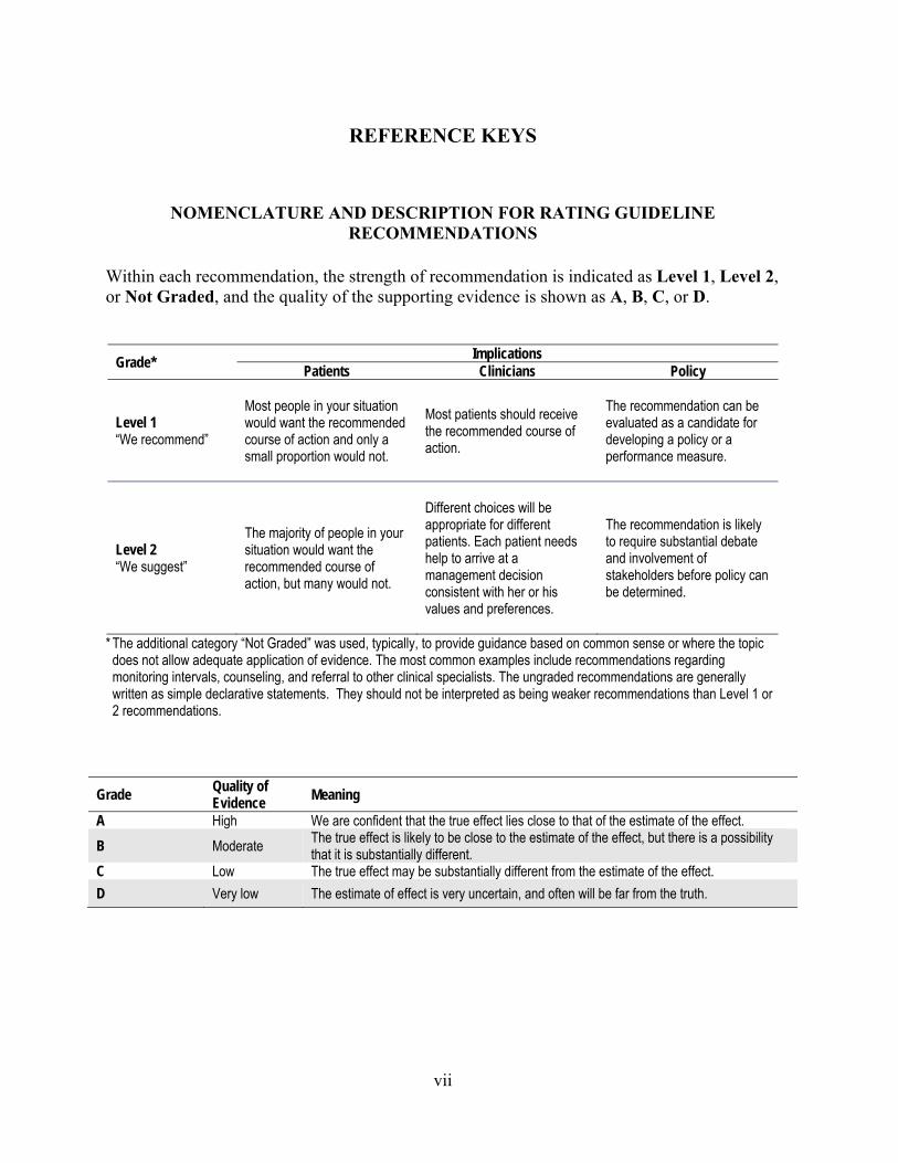
REFERENCE KEYS
NOMENCLATURE AND DESCRIPTION FOR RATING GUIDELINE RECOMMENDATIONS
Within each recommendation, the strength of recommendation is indicated as Level 1, Level 2, or Not Graded, and the quality of the supporting evidence is shown as A, B, C, or D.
Grade* Implications
Patients Clinicians Policy
Level 1 “We recommend”
Most people in your situation would want the recommended course of action and only a small proportion would not.
Most patients should receive the recommended course of action.
The recommendation can be evaluated as a candidate for developing a policy or a performance measure.
Level 2 “We suggest”
The majority of people in your situation would want the recommended course of action, but many would not.
Different choices will be appropriate for different patients. Each patient needs help to arrive at a management decision consistent with her or his values and preferences.
The recommendation is likely to require substantial debate and involvement of stakeholders before policy can be determined.
* The additional category “Not Graded” was used, typically, to provide guidance based on common sense or where the topic does not allow adequate application of evidence. The most common examples include recommendations regarding monitoring intervals, counseling, and referral to other clinical specialists. The ungraded recommendations are generally written as simple declarative statements. They should not be interpreted as being weaker recommendations than Level 1 or 2 recommendations.
Grade Quality of Evidence
Meaning
A High We are confident that the true effect lies close to that of the estimate of the effect.
B Moderate The true effect is likely to be close to the estimate of the effect, but there is a possibility that it is substantially different.
C Low The true effect may be substantially different from the estimate of the effect.
D Very low The estimate of effect is very uncertain, and often will be far from the truth.

viii
CURRENT CHRONIC KIDNEY DISEASE (CKD) NOMENCLATURE USED BY KDIGO
CKD is defined as abnormalities of kidney structure or function, present for > 3 months, with implications for health. CKD is classified based on Cause, GFR category (G1-G5), and Albuminuria category (A1-A3), abbreviated as CGA.
Prognosis of CKD by GFR and albuminuria category
Prognosis of CKD by GFR and Albuminuria Categories:
KDIGO 2012
Persistent albuminuria categories Description and range
A1 A2 A3
Normal to mildly
increased
Moderately increased
Severely increased
<30 mg/g <3 mg/mmol
30-300 mg/g 3-30 mg/mmol
>300 mg/g >30 mg/mmol
GF
R c
ateg
ori
es (
ml/m
in/ 1
.73
m2 )
Des
crip
tio
n a
nd
ran
ge
G1 Normal or high ≥90
G2 Mildly decreased 60-89
G3a Mildly to moderately decreased
45-59
G3b Moderately to severely decreased
30-44
G4 Severely decreased 15-29
G5 Kidney failure <15
Green: low risk (if no other markers of kidney disease, no CKD); Yellow: moderately increased risk; Orange: high risk; Red, very high risk.

ix
CONVERSION FACTORS OF CONVENTIONAL UNITS TO SI UNITS
Conventional Unit Conversion Factor SI Unit Creatinine mg/dl 88.4 µmol/l
Note: Conventional unit x conversion factor = SI unit
ALBUMINURIA CATEGORIES IN CKD
Category AER
(mg/24 hours) ACR (approximate equivalent)
(mg/mmol) (mg/g) Terms
A1 < 30 < 3 < 30 Normal to mildly increased
A2 30-300 3-30 30-300 Moderately increased*
A3 > 300 > 30 > 300 Severely increased**
ACR, albumin-to-creatinine ratio; AER, albumin excretion rate; CKD, chronic kidney disease *Relative to young adult level **Including nephrotic syndrome (albumin excretion usually > 2200 mg/24 hours [ACR > 2200 mg/g; > 220 mg/mmol]
x
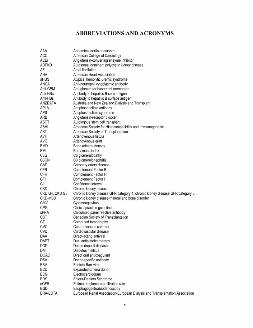
ABBREVIATIONS AND ACRONYMS
AAA Abdominal aortic aneurysm ACC American College of Cardiology ACEi Angiotensin-converting enzyme inhibitor ADPKD Autosomal dominant polycystic kidney disease AF Atrial fibrillation AHA American Heart Association aHUS Atypical hemolytic uremic syndrome ANCA Anti-neutrophil cytoplasmic antibody Anti-GBM Anti-glomerular basement membrane Anti-HBc Antibody to hepatitis B core antigen Anti-HBs Antibody to hepatitis B surface antigen ANZDATA Australia and New Zealand Dialysis and Transplant APLA Antiphospholipid antibody APS Antiphospholipid syndrome ARB Angiotensin-receptor blocker ASCT Autologous stem cell transplant ASHI American Society for Histocompatibility and Immunogenetics AST American Society of Transplantation AVF Arteriovenous fistula AVG Arteriovenous graft BMD Bone mineral density BMI Body mass index C3G C3 glomerulopathy C3GN C3 glomerulonephritis CAD Coronary artery disease CFB Complement Factor B CFH Complement Factor H CFI Complement Factor I CI Confidence interval CKD Chronic kidney disease CKD G4, CKD G5 Chronic kidney disease GFR category 4; chronic kidney disease GFR category 5 CKD-MBD Chronic kidney disease-mineral and bone disorder CMV Cytomeaglovirus CPG Clinical practice guideline cPRA Calculated panel reactive antibody CST Canadian Society of Transplantation CT Computed tomography CVC Central venous catheter CVD Cardiovascular disease DAA Direct-acting antiviral DAPT Dual antiplatelet therapy DDD Dense deposit disease DM Diabetes mellitus DOAC Direct oral anticoagulant DSA Donor-specific antibody EBV Epstein-Barr virus ECD Expanded-criteria donor ECG Electrocardiogram EDS Ehlers-Danlers Syndrome eGFR Estimated glomerular filtration rate EGD Esophagogastroduodenoscopy ERA-EDTA European Renal Association-European Dialysis and Transplantation Association

xi
ERT Evidence Review Team ESC European Society of Cardiology ESKD End-stage kidney disease FBG Fasting blood glucose FI Frailty Index FSGS Focal segmental glomerulosclerosis FVL Factor V Leiden GFR Glomerular filtration rate GRADE Grades of Recommendation, Assessment, Development and Evaluation HBsAg Hepatitis B surface antigen HBV Hepatitis B virus HCDD Heavy chain deposition disease HCV Hepatitis C virus HDV Hepatitis D virus HIT Heparin-induced thrombocytopenia HIV Human immunodeficiency virus HLA Human leukocyte antigen HR Hazard ratio HSV Herpes simplex virus HTLV Human T-cell lymphotropic virus HUS Hemolytic uremic syndrome ICA Intracranial aneurysm IC-MPGN Immune complex-mediated MPGN IgAN IgA nephropathy IgAV IgA vasculitis IGT Impaired glucose tolerance IU International unit KDIGO Kidney Disease: Improving Global Outcomes KDOQI Kidney Disease Outcomes Quality Initiative KHA-CARI Kidney Health Australia-Caring for Australasians with Renal Impairment KRT Kidney replacement therapy KTC Kidney transplant candidate LCDD Light chain deposition disease LVEF Left ventricular ejection fraction LHCDD Light and heavy chain deposition disease LN Lupus nephritis MET Metabolic equivalent MGUS Monoclonal gammopathy of undetermined significance MI Myocardial infarction MIDD Monoclonal immunoglobulin deposition disease MDRO Multidrug resistant organisms MMR Measles, mumps, or rubella MN Membranous nephropathy MPGN Membranoproliferative glomerulonephritis NAT Nucleic acid test(ing) NODAT New-onset diabetes after transplantation NYHA New York Heart Association OGTT Oral glucose tolerance test OR Odds ratio PAD Peripheral arterial disease PASP Pulmonary artery systolic pressure PCD Plasma cell dyscrasia PF4 Platelet factor 4 PKD Polycystic kidney disease PLA2R Phospholipase A2 receptor PPD Purified protein derivative
xii
PRA Panel reactive antibody PTH Parathyroid hormone RCT Randomized controlled trial RR Relative risk SLE Systemic lupus erythematosus SPK Simultaneous pancreas-kidney STEC-HUS Shiga toxin–associated hemolytic uremic syndrome T1DM Diabetes mellitus, Type 1 T2DM Diabetes mellitus, Type 2 TB Tuberculosis TIA Transient ischemic attack TSANZ Transplantation Society of Australia and New Zealand UK United Kingdom UNOS United Network for Organ Sharing US United States UTI Urinary tract infection VZV Varicella-zoster virus WHR Waist-to-hip circumference ratio
xiii
PREFACE Introduction
Transplantation is the kidney replacement therapy (KRT) of choice for suitable patients with end-stage kidney disease (ESKD). However, not all patients are suitable candidates for transplantation, and suitability is often determined by the perceived risks of transplantation relative to the risks of not receiving a transplant. Estimation of risk is therefore a key part of the transplant candidate evaluation. Should a decision to proceed to transplantation be made, consideration of how to minimize risks and maximize the chances of a successful outcome are additional aspects of the candidate evaluation process.
This guideline systematically examines current evidence concerning the risks of transplantation associated with specific conditions and provides recommendations as to how clinicians may wish to deal with specific risk factors in isolation. In practice, patients are frequently complex and exhibit multiple risk factors for poor transplant outcomes. Ultimately the clinician will be required to synthesize the total risk burden that each candidate presents in deciding on suitability for transplantation. Scope
This guideline addresses the evaluation and management of possible candidates for kidney transplantation alone, from either a deceased or living donor. It covers the time period from the first consideration of the need for KRT to kidney transplant surgery. It considers adult and pediatric candidates. Education of the candidate and their family is beyond the scope of this guideline, however we do wish to highlight the essential role of patient education in parallel with the evaluation process, as it is required to enable shared-decision making and consent regarding the decision to proceed to transplantation or not. This guideline does not address candidates for combined transplantation of a kidney and another organ. Inasmuch we attempt to be as comprehensive as possible to address as many types of infections, malignancies, etc, relevant to the evaluation of a kidney transplant candidate, our systematic review is not an exhaustive one; as such absence of a statement on a particular infection, malignancy, etc. should not imply its lack of importance. Please consult chapter on Methods for Guideline Development for further details. Target Audience
This guideline is intended for caregivers who refer and/or evaluate patients for possible kidney transplantation.
xiv
Background and Principles Underpinning the Guideline Ethics
Kidney transplantation, using organs obtained from either living or deceased donors, should be conducted in accordance with the Declaration of Istanbul,1 which provides clear guidelines on ethical practice in this area. Local considerations
As a global guideline, Kidney Disease: Improving Global Outcomes (KDIGO) necessarily seeks and considers all available evidence in producing guidelines which are of global relevance. However, the fact that the practice and outcomes of transplantation vary enormously across the globe− between continents, countries and even jurisdictions− requires the reader to consider their local practices and outcomes in interpreting and implementing the guideline. In particular, considerations should include:
1. Superiority of transplantation over dialysis for the provision of KRT. Existing data clearly demonstrate that on average, transplantation achieves superior medical outcomes (i.e., survival and quality of life) at lower cost as compared to dialysis, and transplantation is therefore considered to be the medically desirable and economically dominant therapy. However, this conclusion is based upon data from high income countries with good access to both transplantation and dialysis.2 This conclusion is likely to hold true for low- and middle-income countries from a medical perspective, though whether transplantation is cheaper than dialysis in this context is less certain and remains to be proven.
2. Access to dialysis and transplantation. In some areas, access to dialysis and/or transplantation may be restricted or absent. This may be due to the lack (or absence) of necessary infrastructure or services, cost of services to the patient, geographical inaccessibility, or other factors. Thus, access must be considered when interpreting these guidelines.
3. Outcomes of dialysis and transplantation. The decision to pursue transplantation in preference to dialysis for any given patient is based upon an expectation of superior outcomes following transplantation. To make this decision, knowledge of expected outcomes from dialysis and transplantation, at a local level, is required. For example, if local transplant outcomes yield a 60% patient survival at 2 years, whereas dialysis yields 70% survival, then transplantation may not be justified for an average patient with ESKD. In the absence of local data describing the outcomes of dialysis and transplantation, the decision to transplant or not must be made by adaptation of available data to the local context.

xv
4. Local risks involved in transplantation. Regional and geographical variation in risk is evident following transplantation and should be considered in implementing the Guideline. The risk of infection after transplantation exhibits marked regional variation in type, frequency and severity. For example, the risk of post-transplant reactivation of latent tuberculosis (TB) is high among those from endemic areas, yet profoundly low among those from temperate climates. Cancer incidence is also affected by geography, genetics and lifestyle. For example, skin cancer is a common cause of death among Caucasian kidney transplant recipients in Australia, particularly among those residents with high sun exposure, yet skin cancers are far less common and are a rare cause of death in other areas of the world. Thus, local knowledge of likely risks and benefits are required to place the recommendations made within this Guideline into local context.

xvi
SUMMARY OF RECOMMENDATION STATEMENTS
CHAPTER 1: ACCESS TO TRANSPLANTATION 1.1: We recommend that all patients with CKD G4-G5 (GFR < 30 ml/min/1.73
m2) who are expected to reach ESKD, regardless of socioeconomic status, sex, or race/ethnicity, be informed of, educated about, and considered for kidney transplantation. (1D) 1.1.1: Refer potential kidney transplant candidates (KTCs) for evaluation at
least 6 to 12 months before anticipated dialysis initiation to facilitate identification/work-up of living donors and plan for possible pre-emptive transplantation. (Not Graded)
1.1.2: Refer potential KTCs already on dialysis when medically stable and
kidney failure deemed irreversible. (Not Graded) 1.1.3: We recommend not referring patients for transplant evaluation with
the following conditions (1D):
An active psychiatric or ongoing substance use disorder that affects decision-making or puts the candidate at a level of post-transplant risk that is higher than acceptable to the transplant program (Recs 4.2 and 4.3);
Ongoing, health-compromising nonadherent behavior despite education and adherence-based counseling (Rec 5.4);
Multiple myeloma with cast nephropathy except for those receiving potentially curative treatment or under stable remission (Rec 9.13.1.1);
Light chain deposition disease (LCDD) or light and heavy chain deposition disease (LHCDD) (Recs 9.13.2.1 and 9.13.2.3);
Active malignancy except for those with indolent and low-grade cancers (Rec 11.2.1);
Severe irreversible obstructive or restrictive lung disease (Rec 12.5);
Systemic amyloidosis with cardiac amyloid (Rec 13.11);
Non-healing extremity wounds with active infection until fully resolved (Rec 14.5);
Progressive neurodegenerative disease (Rec 15.4).

xvii
1.1.3.1: Document the reason(s) for not referring patients for transplant evaluation. (Not Graded)
1.1.3.2: Inform patients about the reason(s) for not referring for
transplant evaluation. (Not Graded) 1.2: Use a multidisciplinary team, which includes at a minimum a transplant
physician and a transplant surgeon, to evaluate and decide about suitability for kidney transplantation. (Not Graded)
1.3: Approve patients for kidney transplantation that have an estimated survival
which is acceptable according to local practice. (Not Graded) 1.3.1: Inform patients of their option to seek a second opinion from another
transplant center if they are declined. (Not Graded) 1.4: We recommend pre-emptive transplantation with a living kidney donor as
the preferred treatment for transplant-eligible CKD patients. (1A) 1.4.1: We recommend pre-emptive transplantation (living or deceased
donor) in adults when the eGFR is < 10 ml/min/1.73 m2 or earlier with symptoms. (1D)
1.4.2: We recommend pre-emptive transplantation (living or deceased
donor) in children when the eGFR is < 15 ml/min/1.73 m2 or earlier with symptoms. (1D)
CHAPTER 2: AGE 2.1: Consider age when deciding about suitability for kidney transplantation.
(Not Graded) 2.2: We recommend not excluding patients from kidney transplantation because
of advanced age alone. (1A)

xviii
CHAPTER 3: PEDIATRIC ISSUES 3.1: We suggest performing a neurocognitive assessment in pediatric KTCs. (2D) 3.2: We suggest performing an academic assessment in pediatric KTCs of school
age. (2D)
CHAPTER 4: PSYCHOSOCIAL ASSESSMENT 4.1: We suggest performing a psychosocial assessment in all KTCs. (2D)
4.1.1: Refer KTCs to a health care professional experienced in the psychosocial dimensions of kidney transplantation to perform this assessment. (Not Graded)
4.1.2: Use measurement tools completed by the patient and/or evaluating
clinician to supplement the assessment. (Not Graded)
4.1.2.1: We suggest not using measurement tools in isolation to determine transplant candidacy. (2D)
4.1.3: Refer KTCs with a diagnosable psychiatric or psychological
condition, substance use disorder or nonadherence for pre-transplant counseling and services to enhance the likelihood of a favorable post-transplant outcome. (Not Graded)
4.2: We recommend not transplanting patients with an active psychiatric
disorder that affects decision-making or puts the candidate at a level of post-transplant risk that is higher than acceptable to the transplant program. (1C)
4.3: We recommend not transplanting patients with ongoing substance use
disorder that affects decision-making or puts the candidate at a level of post-transplant risk that is higher than acceptable to the transplant program. (1C)
4.4: We suggest that patients without social support be considered for kidney
transplantation if they are able to independently care for themselves. (2D)
xix
CHAPTER 5: ADHERENCE 5.1: Assess adherence and adherence barriers pre-transplantation to allow for
appropriate education, counseling and post-transplant surveillance. (Not Graded)
5.2: Refer KTCs with a history of nonadherence or identified adherence barriers
for adherence-based education and counseling pre-transplant. (Not Graded)
5.3: We suggest that KTCs with a history of graft loss due to nonadherence undergo adherence-based counseling prior to re-transplantation. (2D)
5.4: We recommend not excluding candidates with a history of nonadherence from kidney transplantation except if there is ongoing, health-compromising nonadherent behavior despite education and adherence-based counseling. (1D)
CHAPTER 6: TOBACCO 6.1: Assess past and present use of tobacco products at evaluation and while on
the waiting list. (Not Graded) 6.2: We suggest not excluding smokers from kidney transplantation. (2B) 6.3: We recommend counseling all KTCs to avoid use of tobacco products, both
before and indefinitely after transplantation. (1B) 6.4: We recommend that potential KTCs who are smoking tobacco products be
offered a tobacco cessation program. (1B) 6.5: We recommend that KTCs abstain from tobacco use, at a minimum 1 month
prior to living donor transplantation. (1B) 6.6: We suggest chest computed tomography (CT) for current or former heavy
tobacco users (≥ 30 pack-year), per local guidelines, to screen for occult lung cancer. (2C)

xx
CHAPTER 7: SURGICAL ISSUES INCLUDING OBESITY 7.1: We recommend KTCs be evaluated for obesity using body mass index (BMI)
or waist-to-hip circumference ratio (WHR) at the time of listing and while on the waiting list. (1B)
7.1.1: We suggest that KTCs not be excluded from transplantation because
of obesity, per se. (2B) 7.1.2: We suggest weight loss interventions prior to transplantation be
offered in patients with obesity, including gastric sleeve bariatric surgery for morbid obesity. (2D)
7.2: We suggest that patients be evaluated for frailty at listing and while on the
waiting list to inform risk and enable optimization strategies. (2C) 7.3: We suggest KTCs be assessed for medical conditions that inhibit wound
healing, including obesity, undernutrition, tobacco abuse, and prior abdominal surgeries, to inform risks of delayed wound healing and hernia formation. (2B)
7.4: KTCs should not be excluded from consideration for kidney transplantation
because of their need for anticoagulation, anti-platelet therapy or a history of heparin-induced thrombocytopenia (HIT). (Not Graded)
7.4.1: Antiplatelet agents (e.g., aspirin, clopidogrel, ticagrelor) can be
continued while waiting for deceased donor transplant. (Not Graded) 7.4.2: All antiplatelet agents except aspirin should be stopped 5 days prior to
living donor transplant (unless cessation is contraindicated) and during the perioperative period for deceased donor transplantation. (Not Graded)
7.4.3: KTCs treated with direct oral anticoagulant agents should not be waitlisted for deceased donor transplant nor committed to living donor transplantation. Switch to an alternative anticoagulant prior to waitlisting or prior to proceeding to living donor transplantation. (Not Graded)

xxi
7.4.4: Ascertain the history of HIT and utilize non-heparin based agents for perioperative and intraoperative anticoagulation in positive patients. (Not Graded)
7.5: Assess vascular anatomy and patency for patients with significant peripheral
vascular disease (See Chapter 14), prior transplant procedures, venous dialysis catheters, pelvic surgery, or deep venous thrombosis. (Not Graded)
7.6: Consider alternative approaches, including transperitoneal organ placement
and the need for urologic evaluation, in candidates with prior pelvic surgery including previous kidney transplantation. (Not Graded)
7.7: Evaluate native kidney size in patients with polycystic liver/kidney disease.
(Not Graded)
7.7.1: We suggest staged or simultaneous native nephrectomy and transplantation for candidates with polycystic kidney disease (PKD) that is symptomatic, there is a suspicion of malignancy, or if the patient has insufficient room for a transplant. (2D)
7.8: Referral for evaluation by a transplant urologist is indicated for patients with
a history or high risk of urologic malignancy, recurrent urinary tract infections, dysfunctional voiding, prior bladder augmentation/division, an ileal conduit, any congenital anomalies of the kidneys or urinary tract, or nephrolithiasis. (Not Graded)
7.8.1: We suggest that patients with a history of cyclophosphamide use
undergo cystoscopy. (2D) 7.8.2: We suggest that pre-transplant unilateral or bilateral nephrectomy be
considered for pediatric candidates with high urine volumes (> 2.5 ml/kg/hour) or heavy proteinuria associated with hypoalbuminemia. (2D)

xxii
CHAPTER 8: DIABETES 8.1: We recommend that KTCs with diabetes mellitus, Type 1 (T1DM) or Type 2
(T2DM), not be excluded from kidney transplantation per se. (1B) 8.2: We suggest KTCs with ESKD and T1DM be considered for simultaneous
pancreas-kidney transplantation. (2A) 8.3: We suggest testing for abnormal glucose metabolism by oral glucose
tolerance test in KTCs who are not known to be diabetic. (2A)
CHAPTER 9: CAUSE OF END-STAGE KIDNEY DISEASE (ESKD) 9.1 Cause of ESKD and kidney transplantation
9.1.1: We recommend that the cause of ESKD in KTCs be determined, where possible, to inform risks and management after kidney transplantation. (1A)
9.1.2: Advise KTCs about the disease-specific risk of recurrence and
resultant risk of graft loss. (Not Graded) 9.2 Focal segmental glomerulosclerosis (FSGS)
9.2.1: We recommend not excluding candidates with primary focal segmental glomerulosclerosis (FSGS) from kidney transplantation, however the risk of recurrence should be considered and discussed with the candidate. (1B)
9.2.1.1: Loss of a prior graft due to recurrent FSGS indicates a high
risk of recurrence upon subsequent transplantation and this factor should be a major consideration in determining candidacy. (Not Graded)
9.2.2: We suggest genetic testing for the etiology of primary FSGS be
performed in children and young adults to inform the risk of recurrence. (2C)

xxiii
9.2.3: We suggest avoiding routine use of pre-transplant plasma exchange or rituximab to reduce the risk of recurrent FSGS. (2D)
9.3 Membranous nephropathy (MN)
9.3.1: We recommend not excluding candidates with membranous nephropathy (MN) from kidney transplantation, however the risk of recurrence should be considered and discussed with the candidate. (1B)
9.3.1.1 Loss of a prior graft due to recurrent MN indicates a high
risk of recurrence upon subsequent transplantation and this should be a major consideration in determining candidacy. (Not Graded)
9.3.2: We suggest that pre-transplant testing for autoantibodies to
phospholipase A2 receptor (PLA2R) be done to inform the risk of recurrence. (2C)
9.3.3: We suggest avoiding routine use of rituximab or alkylating agents to
reduce the risk of recurrent MN. (2D) 9.4 IgA nephropathy (IgAN)
9.4.1: We recommend not excluding candidates with IgA nephropathy (IgAN) from kidney transplantation, however the risk of recurrence should be considered and discussed with the candidate. (1B)
9.5 IgA vasculitis (IgAV)
9.5.1: We recommend not excluding candidates with IgA vasculitis (IgAV) from kidney transplantation, however the risk of recurrence should be considered and discussed with the candidate. (1B)

xxiv
9.6 Immune complex-mediated membranoproliferative glomerulonephritis (IC-MPGN) and C3 glomerulopathy (C3G) 9.6.1 Immune complex-mediated membranoproliferative
glomerulonephritis (IC MPGN)
9.6.1.1: We recommend not excluding candidates with IC MPGN from kidney transplantation, however the risk of recurrence should be considered and discussed with the candidate. (1B)
9.6.1.2: We recommend investigation for an infective, autoimmune,
or paraprotein-mediated cause of IC MPGN prior to transplantation to guide treatment and inform risk of recurrence. (1C)
9.6.1.3: We suggest that, when possible, the cause of the IC MPGN be
treated prior to transplantation. (2C)
9.6.2 C3 glomerulopathy (C3G), including dense deposit disease (DDD) and C3 glomerulonephritis (C3GN) 9.6.2.1: We recommend not excluding candidates with C3G from
kidney transplantation, however the risk of recurrence should be considered and discussed with the candidate. (1B)
9.6.2.2: We suggest that transplant candidates with C3G be screened for genetic or acquired causes for the dysregulation of the complement alternative pathway to guide treatment and inform risk of recurrence. (2C)
9.6.2.3: Loss of a prior graft due to recurrent C3G indicates a high risk of recurrence upon subsequent transplantation and this should be a major consideration in determining candidacy. (Not Graded)
9.7 Lupus nephritis (LN)
9.7.1: We recommend not excluding candidates with lupus nephritis (LN) from kidney transplantation, however the risk of recurrence should be considered and discussed with the candidate. (1B)

xxv
9.7.2: We recommend that lupus activity should be clinically quiescent on no or minimal immunosuppression prior to transplantation. (1D)
9.7.3: We recommend evaluation for secondary antiphospholipid antibody
syndrome prior to transplantation to inform perioperative management. (1C)
9.8 Antiphospholipid antibody syndrome (APS)
9.8.1: We recommend not excluding candidates with antiphospholipid antibody syndrome (APS) from kidney transplantation, however the risks of post-transplant thrombosis and peri-operative anticoagulant therapies should be considered and discussed with the candidate. (1B)
9.8.2: We suggest that APS should be clinically quiescent prior to
transplantation. (2D) 9.8.3: Continue aspirin and/or warfarin at the time of activation on the
transplant wait list. (Not Graded) 9.9 Anti-neutrophil cytoplasmic antibody (ANCA)-associated vasculitis
9.9.1: We recommend not excluding candidates with anti-neutrophil
cytoplasmic antibody (ANCA)-associated vasculitis from kidney transplantation, however the risk of recurrence should be considered and discussed with the candidate. (1B)
9.9.2: We suggest that ANCA-vasculitis should be clinically quiescent prior
to transplantation. (2D) 9.10 Anti-glomerular basement membrane (anti-GBM) disease
9.10.1: We recommend not excluding candidates with anti-glomerular basement membrane disease (anti-GBM disease) from kidney transplantation. (1B)
9.10.2: We recommend that anti-GBM antibody titers be measured in KTCs
and that transplantation is only performed when antibodies are undetectable. (1D)

xxvi
9.11 Hemolytic uremic syndrome (HUS)
9.11.1: We recommend not excluding candidates with hemolytic uremic syndrome (HUS) due to infection with a Shiga-toxin producing organism, usually E. coli (STEC-HUS), from kidney transplantation. (1A)
9.11.2: We recommend assessment of a KTC with suspected atypical HUS
(aHUS) for a genetic or acquired defect in complement regulation or other genetic causes of aHUS to inform risk of recurrence. (1B)
9.11.3: We recommend not excluding candidates with aHUS from kidney
transplantation, however the risk of recurrence should be considered and discussed with the candidate. (1B)
9.11.3.1: We recommend that if the candidate has an abnormality in
complement regulation placing them at high risk of recurrence, kidney transplantation should not proceed unless a complement inhibitor can be administered or combined liver-kidney transplant can be performed. (1B)
9.12 Systemic sclerosis
9.12.1: We recommend not excluding candidates with systemic sclerosis from
kidney transplantation, in the absence of severe pulmonary, gastrointestinal, or other life threatening non-renal disease. (1C)
9.13 Plasma cell dyscrasias (PCDs)
9.13.1 Multiple myeloma/cast nephropathy
9.13.1.1: We suggest that candidates with multiple myeloma with cast
nephropathy be excluded from kidney transplantation (1D), unless they have received a potentially curative treatment regimen and are in stable remission from multiple myeloma. (2D)
9.13.1.2: We suggest that HLA-matched combined kidney and bone
marrow transplantation be considered for patients with multiple myeloma. (2C)

xxvii
9.13.2 Monoclonal immunoglobulin deposition disease (MIDD) 9.13.2.1: We suggest that candidates with light chain deposition
disease (LCDD) be excluded from kidney transplantation, outside of a curative treatment regimen. (2C)
9.13.2.2: We suggest not excluding candidates with heavy chain
deposition disease (HCDD) from kidney transplantation, however the significant risk of recurrence causing graft loss should be considered and discussed with the candidate. (2D)
9.13.2.3: We suggest that candidates with light and heavy chain
deposition disease (LHCDD) be excluded from kidney transplantation, outside of a curative treatment regimen. (2D)
9.13.3 AL amyloidosis
9.13.3.1 We recommend not excluding candidates with AL
amyloidosis from kidney transplantation, in the absence of myeloma or significant non-renal organ involvement. (2C)
9.14 Amyloidosis
9.14.1: We recommend not excluding candidates with AA amyloidosis from
kidney transplantation after adequate treatment of the underlying cause and in the absence of severe non-renal organ involvement. (1D)
9.14.2: See 9.13.3 above re AL amyloidosis
9.15 Fibrillary/immunotactoid glomerulonephritis
9.15.1: We recommend not excluding candidates with fibrillary or
immunotactoid glomerulonephritis from kidney transplantation, however the risk of recurrence should be considered and discussed with the candidate. (1D)
9.16 Hyperoxaluria (oxalosis), primary and secondary
9.16.1: We suggest that KTCs with primary hyperoxaluria type 1 be considered for combined or sequential liver-kidney transplantation. (2C)

xxviii
9.16.2: We suggest genetic testing to identify the cause of primary hyperoxaluria to inform treatment decisions. (2C)
9.16.3: We suggest not excluding candidates with correctable
hyperoxaluria—pyridoxine-responsive or secondary— from kidney transplantation alone, however the risk of recurrence should be considered and discussed with the candidate. (2D)
9.16.4: We recommend the use of strategies to lower total body oxalate
burden prior to transplantation in patients with hyperoxaluria, including intensive dialysis, diet modification, and pyridoxine treatment as appropriate on a case-by-case basis. (1D)
9.17 Cystinosis
9.17.1: We recommend not excluding candidates with cystinosis from kidney
transplantation in the absence of severe non-renal manifestations. (1C)
9.18 Fabry disease
9.18.1: We recommend not excluding candidates with Fabry disease from
kidney transplantation in the absence of severe cardiac or other systemic non-renal involvement. (1C)
9.19 Sickle cell disease
9.19.1: We recommend not excluding candidates with sickle cell disease from kidney transplantation in the absence of active, severe non-renal sickle cell disease. (1C)
9.20 Sarcoidosis
9.20.1: We recommend not excluding candidates with renal sarcoidosis from kidney transplantation in the absence of severe non-renal disease. (1C)
9.21 Alport syndrome
9.21.1: We recommend not excluding candidates with Alport syndrome from kidney transplantation. (1C)
xxix
CHAPTER 10: INFECTIONS 10.1 Active infections
10.1.1: We recommend that kidney transplantation be delayed until active
infections (bacterial, fungal, viral, parasitic) are treated. (1C) 10.2 Colonization
10.2.1: Follow local protocols for detection and management of colonization with drug-resistant organisms. (Not Graded)
10.2.2: We recommend not excluding patients from kidney transplantation
with asymptomatic bacterial or fungal colonization. (1C) 10.3 Specific Infections
10.3.1 Urinary tract infections (UTIs)
10.3.1.1: We recommend treating symptomatic urinary tract
infections (UTI) prior to kidney transplantation. (1B) 10.3.1.2: We suggest not routinely performing prophylactic
nephrectomy for recurrent pyelonephritis or cyst infections. (2D)
10.3.2 Tuberculosis (TB)
10.3.2.1: We suggest complete treatment of active tuberculosis (TB) prior to kidney transplantation, as per World Health Organization or local guidelines. (2C)
10.3.2.2: We recommend pre-transplant screening for latent TB in low
TB prevalence areas with a chest radiograph along with a purified protein derivative (PPD) skin test or interferon-gamma release assay. (1C)
10.3.2.3: We suggest starting treatment of latent TB prior to or
immediately following kidney transplantation in low TB prevalence areas. (2C)

xxx
10.3.2.4: We suggest pre-transplant screening for latent TB as per local guidelines in intermediate and high TB prevalence areas with post-transplantation vigilance for active TB. (2C)
10.4 Screening for periodontal disease
10.4.1: We suggest dental evaluation, as per local general population guidelines, to screen for dental/periodontal disease prior to kidney transplantation. (2C)
10.5 Screening for viral infections (see Table 1)
10.5.1 Human immunodeficiency virus (HIV) 10.5.1.1: We recommend screening all patients for human
immunodeficiency virus (HIV) infection, using HIV serology, at the time of evaluation for kidney transplantation. (1A)
10.5.1.2: We recommend not excluding patients with controlled HIV
infection for kidney transplantation. (1C) KTCs with HIV should be managed in a center with experience in this area. (Not Graded)
10.5.2 Hepatitis C virus (HCV) [This section is largely adapted from 2018
KDIGO HCV Guideline] 10.5.2.1: We recommend screening all patients for hepatitis C virus
(HCV) infection at the time of evaluation for kidney transplantation. (1A) (KDIGO HCV Guideline Recommendation 1.1.4)
10.5.2.2: We recommend using an immunoassay followed by nucleic
acid testing (NAT) if immunoassay is positive. (1A) (KDIGO HCV Guideline Recommendation 1.1.1.1)
10.5.2.3: We recommend kidney transplantation as the best
therapeutic option for patients with CKD G5, irrespective of presence of HCV infection. (1A) (KDIGO HCV Guideline Recommendation 4.1.1)

xxxi
10.5.2.4: We suggest that all HCV-infected KTCs be evaluated for severity of liver disease and presence of portal hypertension (if indicated) prior to acceptance for kidney transplantation (see Figure 2 below). (2D) (KDIGO HCV Guideline Recommendation 4.1.2) 10.5.2.4.1: We recommend that HCV-infected patients with
compensated cirrhosis (without portal hypertension) undergo isolated kidney transplantation. (1B) (KDIGO HCV Guideline Recommendation 4.1.2.1)
10.5.2.4.2: We recommend referring HCV-infected patients
with decompensated cirrhosis for combined liver-kidney transplantation (1B) and deferring HCV treatment until after transplantation. (1D) (KDIGO HCV Guideline Recommendation 4.1.2.2)
10.5.2.5: Timing of HCV treatment in relation to kidney
transplantation (before vs. after) should be based on donor type (living vs. deceased donor), wait-list times by donor type, center-specific policies governing the use of kidneys from HCV-infected deceased donors, HCV genotype, and severity of liver fibrosis. (Not Graded) (KDIGO HCV Guideline Recommendation 4.1.3) 10.5.2.5.1: We recommend that all HCV-infected patients
who are candidates for kidney transplantation be considered for direct-acting antiviral (DAA) therapy, either before or after transplantation. (1A) (KDIGO HCV Guideline Recommendation 4.1.3.1)
10.5.2.5.2: We suggest that HCV-infected KTCs with a living
kidney donor can be considered for treatment before or after transplantation according to HCV genotype and anticipated timing of transplantation. (2B) (KDIGO HCV Guideline Recommendation 4.1.3.2)

xxxii
10.5.2.5.3: We suggest that if receiving a kidney from an HCV-positive donor improves the chances for transplantation, the HCV NAT-positive patient can undergo transplantation with an HCV-positive kidney and be treated for HCV infection after transplantation. (2B) (KDIGO HCV Guideline Recommendation 4.1.3.3)
10.5.3 Hepatitis B virus (HBV) [See Section 10.7 for related
recommendations on HBV vaccinations]
10.5.3.1 We recommend pre-transplant screening for hepatitis B virus (HBV) infection with HBsAg, anti-HBs, and anti-HBc in KTCs. (1A)
10.5.3.2: We recommend pre-transplant screening with HBV DNA for patients with a positive HBsAg or anti-HBc antibody. (1A)
10.5.3.3: We recommend pre-transplant screening with hepatitis D
virus (HDV) serology in HDV endemic areas for patients with a positive HBsAg or anti-HBc antibody. (1A)
10.5.3.4: We recommend that HBsAg positive and/or HBV DNA
positive KTCs be referred to a specialist with expertise in the management of liver disease and HBV infection to determine proper antiviral treatment. (1D)
10.5.3.4.1: We recommend that HBsAg positive and/or HBV
DNA positive KTCs undergo isolated kidney transplantation if deemed to have compensated cirrhosis and are stable on antiviral therapy after specialist evaluation. (1B)
10.5.3.5: We recommend not excluding anti-HBc antibody positive
(HBsAg negative) patients from kidney transplantation. (1C)
10.5.3.5.1: We recommend that anti-HBc antibody positive (HBsAg negative) patients not receive antiviral prophylaxis given that the risk of reactivation is low. (1D)

xxxiii
10.5.3.5.2: We suggest that anti-HBc antibody positive (HBsAg negative) patients have a plan in place for post-transplant monitoring of HBsAg and HBV DNA for a minimum of 1-year post-transplantation. (2C)
10.5.4 Cytomegalovirus (CMV)
10.5.4.1: We recommend pre-transplant screening for cytomegalovirus (CMV) with CMV IgG in KTCs. (1C)
10.5.5 Epstein-Barr virus (EBV)
10.5.5.1: We recommend pre-transplant screening for Epstein-Barr virus (EBV) with EBV antivirus capsid antigen (VCA) IgG and/or EBV nuclear antigen (EBNA) IgG in KTCs. (1C)
10.5.6 Herpes simplex virus (HSV)
10.5.6.1: We suggest pre-transplant screening for herpes simplex virus (HSV) with HSV IgG in KTCs. (2C)
10.5.7 Varicella-zoster virus (VZV)
10.5.7.1: We recommend pre-transplant screening for varicella-zoster virus (VZV) with VZV IgG in KTCs. (1C)
10.5.7.1.1: We recommend varicella immunization for VZV
seronegative KTCs at least 4 weeks prior to transplantation if using a live vaccine. (1C)
10.5.8 Measles, mumps, and rubella (MMR)
10.5.8.1: We suggest pre-transplant screening for measles, mumps, and rubella (MMR) using IgG serology in KTCs. (2C)
10.5.8.1.1: We suggest MMR immunization for MMR
seronegative KTCs at least 4 weeks prior to transplantation. (2C)
xxxiv
10.5.9 BK virus
10.5.9.1: We recommend not screening for BK virus infection in KTCs. (1C)
10.5.9.1.1: We recommend not excluding patients for repeat
transplantation if a previous graft was lost due to BK nephropathy. (1C)
10.5.10 Human T-cell lymphotropic virus (HTLV)
10.5.10.1: We recommend pre-transplant screening for HTLV 1/2 with IgG serology in KTCs from endemic areas as per WHO. (1C)
10.6 Screening for non-viral infections
10.6.1 Syphilis
10.6.1.1: We recommend pre-transplant screening for syphilis (Treponema pallidum) in KTCs and treatment prior to transplantation if infection is identified. (1C)
10.6.2 Strongyloides
10.6.2.1: We suggest pre-transplant screening for strongyloidiasis in KTCs from endemic areas, and treatment prior to transplantation if infection is identified. (2C)
10.6.3 Chagas
10.6.3.1: We recommend pre-transplant screening for Chagas disease in KTCs from endemic areas, and treatment prior to transplantation if infection is identified. (1C)
10.6.4 Malaria
10.6.4.1: We recommend pre-transplant screening for malaria in KTCs who have recently travelled to endemic areas and treatment prior to transplantation if infection is identified. (1C)

xxxv
10.7 Vaccinations
10.7.1: We recommend that the vaccination series be commenced using an accelerated schedule, if necessary, prior to kidney transplantation for any inactivated vaccines (Table 2). (1B)
10.7.1.1: We suggest not excluding candidates who do not complete an
inactivated vaccine series prior to kidney transplantation. (2D)
10.7.2: We recommend that the vaccination series be completed prior to
kidney transplantation for any live attenuated vaccines (Table 2). (1B)
10.7.2.1: We recommend a 4-week delay in kidney transplantation if a live vaccine is administered (e.g., MMR, VZV, shingles, yellow fever, oral typhoid, oral polio vaccine). (1B)
10.7.3: We recommend that splenectomized KTCs or those at increased risk
for post-transplant splenectomy receive pre-transplant pneumococcal, hemophilus, and meningococcal vaccines. (1B)
10.7.4: We recommend that KTCs requiring complement inhibitors
perioperatively or post-transplant be first given the meningococcal vaccine. (1B)
10.7.5: We suggest administering the following vaccines to KTCs who, due to
age, direct exposure, residence or travel to endemic areas, or other epidemiological risk factors, are at increased risk for the specific diseases:
Rabies (2D)
Tick-borne meningoencephalitis (2D)
Japanese encephalitis (inactivated) (2D)
Meningococcus (2D)
Salmonella typhi (inactivated) (2D)
Yellow fever (2D)

xxxvi
CHAPTER 11: CANCER 11.1 Cancer screening
11.1.1: We recommend KTCs undergo routine cancer screening, as per local guidelines for the general population (Table 3). (1D) 11.1.1.1: We suggest chest imaging prior to transplantation in all
KTCs. (2C) (Same as Rec 12.2) 11.1.1.2: We suggest chest CT for current or former tobacco users
with > 30 pack-year history, as per local guidelines, and chest radiograph for other KTCs. (2C) (Same as Rec 12.2.1)
11.1.2: We recommend screening for renal cell carcinoma with
ultrasonography for KTCs at increased risk, such as long time on dialysis, family history of renal cancer, acquired cystic disease, and analgesic nephropathy. (1D)
11.1.3: We recommend screening for bladder carcinoma using urine cytology
or cystoscopy for KTCs at increased risk, such as previous cyclophosphamide use or history of heavy smoking (> 30 pack-year). (1D)
11.1.4: We recommend screening for hepatocellular carcinoma in KTCs with
cirrhosis prior to transplantation using techniques (e.g., ultrasound, α-fetoprotein, etc.) and frequency as per local guidelines. (1C)
11.1.5: We recommend screening for bowel cancer in KTCs with
inflammatory bowel disease as per local guidelines. (1C) 11.2 Potential KTCs with a prior cancer
11.2.1: We recommend that candidates with active malignancy be excluded
from kidney transplantation except for those with indolent and low-grade cancers such as prostate cancer (Gleason score ≤ 6) and basal cell carcinoma, and renal incidentaloma ≤ 1 cm in maximum diameter). (1B)
11.2.2: We suggest that the waiting time period for kidney transplantation
begins upon completion of potentially curative treatment. (2D)

xxxvii
11.2.3: Timing of kidney transplantation after potentially curative treatment for cancer is dependent on the cancer type and stage at initial diagnosis. (Not Graded)
11.2.4: We recommend no waiting time for KTCs with curatively treated
(surgically or otherwise) non-melanoma skin cancers, small renal cell carcinoma (< 3 cm), prostate cancer (Gleason score ≤ 6), carcinoma in situ (ductal carcinoma in situ [DCIS], cervical, others), thyroid cancer (follicular/papillary < 2 cm of low grade histology), and superficial bladder cancer. (1C) 11.2.4.1: For other cancers, we suggest following waiting time
parameters as outlined in Table 4. (2D)
11.2.5: We recommend not excluding candidates with a prior history of metastatic cancer from kidney transplantation, however the risk of recurrence should be a major consideration and discussed with the candidate. (1D)
11.2.6: For relevant cancers, use genomic profiling, other molecular genomic
tests, and phenotyping to predict patient-specific risk of progression and/or recurrence. (Not Graded)
11.2.7: Decisions about transplantation for KTCs in remission from cancer
should be made collaboratively with oncologists, transplant nephrologists, patients, and their caregivers. (Not Graded)

xxxviii
11.3 Hematological malignancy (see Chapter 17.7-17.9)
CHAPTER 12: PULMONARY DISEASE 12.1: Assess KTCs with lung disease in collaboration with a pulmonary specialist
to determine suitability for transplantation. (Not Graded) 12.2: We suggest chest imaging prior to transplantation in all KTCs. (2C) (Same as
Rec 11.1.1.1) 12.2.1 We suggest chest CT for current or former heavy tobacco users (> 30
pack-year), as per local guidelines, and chest radiograph for other KTCs. (2C) (Same as Rec 11.1.1.2)
17.7 Acute leukemia and high-grade lymphoma
17.7.1: We suggest avoidance of kidney transplantation until patient has received curative therapy, achieved remission and remained cancer free for a period to be determined in consultation with the patient, a hematologist/oncologist and the transplant program. (Not Graded)
17.8 Myelodysplasias, chronic leukemia and chronic/low-grade lymphoma
17.8.1: Decisions about kidney transplantation in patients with myelodysplasia should be made in collaboration with a hematologist. (Not Graded)
17.8.2: Advise consultation with a hematologist with transplant
experience in determining transplant candidacy since many lesions may be deemed to be at high risk of accelerated progression or transformation post-transplant. (Not Graded)
17.9: Decisions about kidney transplantation in patients with a prior history
of hematological malignancy who are now in remission should be made in collaboration with a hematologist. (Not Graded)

xxxix
12.3: We recommend pulmonary function testing in KTCs with impaired functional capacity, respiratory symptoms, or known pulmonary disease. (1C)
12.4: We recommend counseling all KTCs to avoid use of tobacco products, both
before and indefinitely after transplantation. (1B) (Same as Rec 6.3) 12.5 We recommend that candidates with severe irreversible obstructive or
restrictive lung disease be excluded from kidney transplantation. (1C)
CHAPTER 13: CARDIAC DISEASE 13.1: All patients evaluated for kidney transplantation should undergo assessment
for the presence and severity of cardiac disease with history, physical examination, and electrocardiogram (ECG). (Not Graded)
13.2: Patients with signs or symptoms of active cardiac disease (e.g., angina,
arrhythmia, heart failure, symptomatic valvular heart disease) should undergo assessment by a cardiologist and be managed according to current local cardiac guidelines prior to further consideration for a kidney transplant. (Not Graded)
13.3: We suggest that asymptomatic KTCs at high risk for coronary artery disease
(CAD) or with poor functional capacity undergo non-invasive CAD screening. (2C)
13.3.1: We recommend that asymptomatic KTCs with known CAD not be
revascularized exclusively to reduce perioperative cardiac events. (1B) 13.3.2: We suggest not excluding candidates with advanced triple vessel
coronary disease from kidney transplantation, however the risk of a post-transplant major cardiac event should be a major consideration and discussed with the candidate. (2D)
13.4: We suggest that maintenance aspirin, β-blockers, angiotensin-converting
enzyme inhibitors/angiotensin-receptor blockers (ACE-inhibitors/ARBs), and statins be continued while on the waiting list and perioperatively, according to cardiac and local guidelines. (2A)
xl
13.5: We suggest that kidney transplantation be delayed for at least one month after myocardial infarction. (2B)
13.6: We suggest that kidney transplantation be deferred for at least one month
after placement of a bare metal stent and six months after insertion of a drug eluting stent. (2B)
13.7: We suggest that asymptomatic KTCs who have been on dialysis for at least
two years or have risk factors for pulmonary hypertension undergo echocardiography. (2D)
13.8: Patients with severe valvular heart disease should be evaluated and managed
by a cardiologist according to cardiac and local guidelines. (Not Graded) 13.9: We suggest that candidates with uncorrectable, symptomatic (NYHA III/IV)
heart disease including severe CAD, cardiac dysfunction (ejection fraction < 30%), and severe valvular disease, should not be excluded from kidney transplantation per se, however the cardiac prognosis should be evaluated and considered by the clinical team and the patient in determining candidacy for transplantation. (2D)
13.9.1: Patients with severe heart failure (NYHA III/IV) who are otherwise
suitable for kidney transplantation should be assessed by a cardiologist and considered for combined/simultaneous heart and kidney transplantation. (Not Graded)
13.10: Patients with an estimated pulmonary systolic pressure greater than 45 mm
Hg should be assessed by a cardiologist. (Not Graded)
13.10.1: We recommend not excluding candidates with uncorrectable pulmonary artery systolic pressure greater than 60 mm Hg from kidney transplantation, however the risks of sudden deterioration or progression after transplantation should be a major consideration and discussed with the candidate. (1C)
13.11: Perform cardiac imaging in patients with systemic amyloidosis. Exclude
such patients from kidney transplantation if cardiac amyloid is confirmed. (Not Graded)

xli
CHAPTER 14: PERIPHERAL ARTERIAL DISEASE (PAD) 14.1: Evaluate all patients for presence and severity of peripheral arterial disease
(PAD) with history and physical examination. (Not Graded) 14.2: We suggest candidates without clinically apparent PAD, but who are at high
risk for PAD, undergo non-invasive vascular testing. (2D) 14.3: We suggest KTCs with clinically apparent PAD undergo imaging and
management of their vasculature in consultation with a vascular surgeon (2D)
14.4: For patients with clinically apparent PAD, abnormal non-invasive testing, or
prior vascular procedures, we suggest non-contrast CT of the abdomen and pelvis to evaluate arterial calcification and improve operative planning. (2D)
14.5: Non-healing extremity wounds with active infection preclude kidney
transplantation until the infection is resolved. (Not Graded) 14.6: We suggest not excluding patients with severe aorto-iliac disease from kidney
transplantation. We suggest not excluding patients with prior aorto-iliac procedures including iliac artery stent placement from kidney transplantation if there is sufficient native artery available for vascular anastomosis. (2D)
14.7: We suggest not excluding candidates with advanced diabetic distal vascular
disease (e.g., major lower extremity amputation) from kidney transplantation, however the risks of progression after transplantation should be considered and discussed with the candidate. (2D)
CHAPTER 15: NEUROLOGIC DISEASE 15.1: We suggest waiting at least 6 months after a stroke or 3 months after a
transient ischemic attack (TIA) before kidney transplantation. (2D) 15.2: We suggest not screening asymptomatic KTCs for carotid artery disease.
(2C)

xlii
15.3: We suggest screening KTCs with autosomal dominant polycystic kidney (ADPKD) disease for intracranial aneurysms only if they are at high risk due to prior history of or a family history of subarachnoid hemorrhage. (2D)
15.4: Patients with progressive neurodegenerative disease should not undergo
kidney transplantation if survival and quality of life are not expected to be substantially improved by transplantation. (Not Graded)
15.5: Assess mental status in KTCs with known or suspected cognitive
impairment. (Not Graded)
15.5.1: We recommend not excluding candidates from kidney transplantation because of non-progressive intellectual, developmental, or cognitive disability. (1D)
CHAPTER 16: GASTROINTESTINAL AND LIVER DISEASE 16.1 Peptic ulcer disease
16.1.1: Assess KTCs for peptic ulcer disease. (Not Graded)
16.1.2: We recommend that candidates with symptoms suggestive of active peptic ulcer disease undergo esophagogastroscopy and H. pylori testing prior to kidney transplantation. (1C)
16.1.3: Delay kidney transplantation in candidates with endoscopically-
proven peptic ulcer disease until symptoms have resolved. (Not Graded)
16.1.4: We recommend not screening KTCs with a history of peptic ulcer disease with esophagogastroscopy. (1C)
16.1.5: We recommend not excluding candidates from kidney transplantation
because of a history of peptic ulcer disease. (1D) 16.2 Diverticulitis
16.2.1: Assess KTCs for diverticulitis. (Not Graded)

xliii
16.2.2: Delay kidney transplantation in candidates with active diverticulitis until symptoms have resolved. (Not Graded)
16.2.3: We recommend not screening for diverticulosis in asymptomatic
KTCs. (1C) 16.2.4: We recommend not performing prophylactic colectomy in patients
with a history of diverticulitis or asymptomatic diverticulosis. (1C) 16.2.5: We recommend not excluding candidates from kidney transplantation
because of a history of diverticulitis. (1C) 16.3 Pancreatitis
16.3.1: Assess KTCs for pancreatitis. (Not Graded)
16.3.2: Delay kidney transplantation in candidates with acute pancreatitis a minimum of three months after symptoms have resolved. (Not Graded)
16.3.3: We recommend not excluding candidates from kidney transplantation
because of a history of acute or chronic pancreatitis. (1C) 16.4 Cholelithiasis
16.4.1: Assess KTCs for cholelithiasis. (Not Graded) 16.4.2: Delay kidney transplantation in candidates with symptomatic
gallstone or gallbladder disease until symptoms have resolved. (Not Graded)
16.4.3: We recommend that candidates with a history of cholecystitis undergo
cholecystectomy before kidney transplantation. (1C) 16.4.4: We recommend not screening for cholelithiasis in asymptomatic
KTCs. (1C) 16.4.5: We recommend not performing prophylactic cholecystectomy in
KTCs with asymptomatic cholelithiasis. (1C)
xliv
16.4.6: We recommend not excluding candidates from kidney transplantation because of asymptomatic cholelithiasis. (1A)
16.5 Inflammatory bowel disease
16.5.1: Assess KTCs for inflammatory bowel disease. (1D) 16.5.2: Delay kidney transplantation in candidates with active symptomatic
inflammatory bowel disease. (Not Graded)
16.5.2.1: Determine timing of transplantation in consultation with a gastroenterologist. (Not Graded)
16.5.3: We recommend screening for bowel cancer in patients with
inflammatory bowel disease as per local guidelines. (1C) 16.5.4: We recommend not excluding candidates from kidney transplantation
because of a history of inflammatory bowel disease. (1D)
16.6 Liver disease
16.6.1: Screen KTCs for evidence of liver disease with appropriate history and physical exam, total bilirubin, alanine aminotransferase (ALT), international normalized ratio (INR), and albumin. (Not Graded)
16.6.2: Delay kidney transplantation until acute hepatitis, of any cause, has
resolved and a long-term strategy for managing liver disease has been implemented. (Not Graded)
16.6.3: We recommend that KTCs with cirrhosis or suspected cirrhosis be
referred to a specialist with expertise in combined liver-kidney transplantation for evaluation. (1B) 16.6.3.1: We recommend that patients undergo isolated kidney
transplantation if deemed to have compensated cirrhosis after specialist evaluation. (1B)
For liver disease associated with Hepatitis B or C infection see
Chapter 10.5

xlv
16.6.4: We recommend screening for hepatocellular carcinoma in KTCs with cirrhosis prior to transplantation using techniques (e.g., ultrasound, alpha-fetoprotein, etc.) and frequency as per local guidelines. (1C)
CHAPTER 17: HEMATOLOGICAL DISORDERS 17.1: We recommend not routinely screening for thrombophilia in KTCs. (1C)
17.1.1: We suggest screening for thrombophilia only in KTCs who have
experienced a venous thromboembolic event, recurrent arteriovenous access thromboses, non-atherosclerotic arterial thrombosis, or family history of venous thromboembolism to identify candidates at higher risk of graft thrombosis. (2C)
17.2: We suggest testing for antiphospholipid antibodies (APLAs) in patients with
systemic lupus erythematosus (SLE) or features of antiphospholipid syndrome (APS). (2C)
17.3: We suggest candidates receiving dual antiplatelet therapy not be excluded
from kidney transplantation when the transplant team deems the benefit of transplantation to exceed risk of bleeding. (2D) Where risk is assessed to exceed potential benefits, we suggest that transplant surgery be delayed for the mandated period of treatment with dual antiplatelet treatment. (2C) 17.3.1: Evaluate the risk of stopping dual antiplatelet therapy to allow kidney
transplantation on a case-by-case basis by a multidisciplinary team including transplant surgeon and cardiologist. (Not Graded)
17.3.2: We suggest stopping a P2Y12 inhibitor (e.g., clopidogrel) for at least 5
days prior to living donor transplantation. (2C) 17.4: We recommend that candidates receiving anticoagulation with warfarin not
be excluded from kidney transplantation. (1B) 17.5: In the presence of significant cytopenias, evaluate suitability for kidney
transplantation based on cause and severity. (Not Graded)

xlvi
17.6: We recommend that candidates with monoclonal gammopathy of undetermined significance (MGUS), sickle cell disease, or thalassemia not be excluded from kidney transplantation [see sections on recurrent disease: plasma cell dyscrasias, Chapter 9.13 and sickle cell disease, Chapter 9.19 and hematology malignancy, Chapter 17.7-17.9]. (1C)
17.7 Acute leukemia and high-grade lymphoma
17.7.1: We suggest avoidance of kidney transplantation until patient has received curative therapy, achieved remission and remained cancer free for a period to be determined in consultation with the patient, a hematologist/oncologist and the transplant program. (Not Graded)
17.8 Myelodysplasias, chronic leukemia and chronic/low-grade lymphoma
17.8.1: Decisions about kidney transplantation in patients with myelodysplasia should be made in collaboration with a hematologist. (Not Graded)
17.8.2: Advise consultation with a hematologist with transplant experience in
determining transplant candidacy since many lesions may be deemed to be at high risk of accelerated progression or transformation post-transplant. (Not Graded)
17.9: Decisions about kidney transplantation in patients with a prior history of
hematological malignancy who are now in remission should be made in collaboration with a hematologist. (Not Graded)
CHAPTER 18: BONE AND MINERAL METABOLISM
18.1: Measure serum parathyroid hormone (PTH) at the time of transplant evaluation. (Not Graded)
18.2: We suggest not transplanting patients with severe hyperparathyroidism until
they are adequately treated (medically or surgically) as per KDIGO Chronic Kidney Disease-Mineral and Bone Disorder (CKD-MBD) guideline. (2D)
18.3: Bone mineral density (BMD) should not be measured as part of the
transplant evaluation. (Not Graded)
xlvii
CHAPTER 19: HLA TESTING 19.1: Communicate all sensitizing events (e.g., blood product transfusion,
including platelets, pregnancy or miscarriage) or clinical events that can impact panel reactive antibody (PRA) (e.g., vaccination, withdrawal of immunosuppression, transplant nephrectomy, significant infection) to the human leukocyte antigen (HLA) laboratory. (Not Graded)
19.2: Perform HLA antibody testing at transplant evaluation, at regular intervals
prior to transplantation and a minimum of 2 weeks after a sensitizing event or a clinical event that can impact PRA. (Not Graded)
19.3: We recommend that HLA antibody testing be performed using solid phase
assays. (1B) 19.4: We recommend HLA typing of KTCs at evaluation using molecular methods,
optimally at all loci. (1D) 19.5: We suggest not routinely testing KTCs for non-HLA antibodies. (2C) 19.6: We suggest not routinely testing KTCs for complement-binding HLA
antibodies. (2C) 19.7: We suggest informing KTCs about their access to transplantation based on
blood type and histocompatibility testing results. (2C)
19.7.1: We recommend offering KTCs with immunologically-reduced access to transplant access to a larger deceased donor pool, kidney exchange programs, and/or desensitization. (1C)
19.7.2: We suggest that antibody avoidance (e.g., kidney exchange programs
or deceased donor acceptable mismatch allocation) be considered before desensitization. (2C)

1
CHAPTER 1: ACCESS TO TRANSPLANTATION
1.1: We recommend that all patients with CKD G4-G5 (GFR < 30 ml/min/1.73 m2) who are expected to reach ESKD, regardless of socioeconomic status, sex, or race/ethnicity, be informed of, educated about, and considered for kidney transplantation. (1D) 1.1.1: Refer potential kidney transplant candidates (KTCs) for evaluation at
least 6 to 12 months before anticipated dialysis initiation to facilitate identification/work-up of living donors and plan for possible pre-emptive transplantation. (Not Graded)
1.1.2: Refer potential KTCs already on dialysis when medically stable and
kidney failure deemed irreversible. (Not Graded) 1.1.3: We recommend not referring patients for transplant evaluation with
the following conditions (1D):
An active psychiatric or ongoing substance use disorder that affects decision-making or puts the candidate at a level of post-transplant risk that is higher than acceptable to the transplant program (Recs 4.2 and 4.3);
Ongoing, health-compromising nonadherent behavior despite education and adherence-based counseling (Rec 5.4);
Multiple myeloma with cast nephropathy except for those receiving potentially curative treatment or under stable remission (Rec 9.13.1.1);
Light chain deposition disease (LCDD) or light and heavy chain deposition disease (LHCDD) (Recs 9.13.2.1 and 9.13.2.3);
Active malignancy except for those with indolent and low-grade cancers (Rec 11.2.1);
Severe irreversible obstructive or restrictive lung disease (Rec 12.5);
Systemic amyloidosis with cardiac amyloid (Rec 13.11);
Non-healing extremity wounds with active infection until fully resolved (Rec 14.5);
Progressive neurodegenerative disease (Rec 15.4).
1.1.3.1: Document the reason(s) for not referring patients for transplant evaluation. (Not Graded)

2
1.1.3.2: Inform patients about the reason(s) for not referring for transplant evaluation. (Not Graded)
1.2: Use a multidisciplinary team, which includes at a minimum a transplant
physician and a transplant surgeon, to evaluate and decide about suitability for kidney transplantation. (Not Graded)
1.3: Approve patients for kidney transplantation that have an estimated survival
which is acceptable according to local practice. (Not Graded) 1.3.1: Inform patients of their option to seek a second opinion from another
transplant center if they are declined. (Not Graded) 1.4: We recommend pre-emptive transplantation with a living kidney donor as
the preferred treatment for transplant-eligible CKD patients. (1A) 1.4.1: We recommend pre-emptive transplantation (living or deceased
donor) in adults when the eGFR is < 10 ml/min/1.73 m2 or earlier with symptoms. (1D)
1.4.2: We recommend pre-emptive transplantation (living or deceased
donor) in children when the eGFR is < 15 ml/min/1.73 m2 or earlier with symptoms. (1D)
BACKGROUND
For suitable candidates kidney transplantation is the preferred form of KRT
because it improves survival and quality of life and is less costly than dialysis.2-6 Therefore, all patients with advanced chronic kidney disease (CKD) should be informed about options for KRT, including transplantation. However, in most industrialized countries the majority of patients with end-stage kidney disease (ESKD) are older patients with many comorbidities. As such, in most regions less than 30% of prevalent dialysis patients are on the transplant wait-list but there is considerable variability.7, 8 Given the organ shortage, it is reasonable to match patient survival with anticipated graft survival in order to avoid futility and maximize utility. In fact, such an algorithm has been implemented for deceased donor kidney transplantation in some regions of the world.9, 10 Therefore, a reasonable estimated life expectancy, according to local standards, should be considered a prerequisite in order to proceed with transplant evaluation. The situation is different in living donor kidney transplantation. In this scenario, there is no waiting-time, surgery is planned and ‘borderline’ recipients can be
3
optimized pre-transplantation. The decision to proceed in such cases requires an open discussion with both the donor and recipient regarding anticipated outcomes.
RATIONALE
Kidney transplantation improves survival and quality of life and is less costly compared to dialysis.
Patients with advanced CKD who are expected to reach ESKD have the right to be informed of all treatment options, including transplantation.
There is an organ shortage and thus candidacy for deceased donor transplantation
needs careful evaluation.
Initiation of the transplant evaluation process depends on the patient’s subjective well-being, underlying kidney disease and rate of glomerular filtration rate (GFR) loss; number of comorbid conditions; and the anticipated need for specialized testing (e.g., coronary angiography).
Depending on the patient and region, the transplant evaluation process may take
weeks to several months to complete.
Pre-emptive transplantation is the preferred treatment option but requires sufficient time to ensure a complete evaluation.
The timing of pre-emptive living donor transplantation needs individual decision
making depending on patient’s symptoms and estimated glomerular filtration rate (eGFR).
Access to transplantation
Patients with progressive CKD (e.g., CKD G4-G5) who are expected to reach ESKD should be informed about all treatment options. This also includes the option of conservative management in cases with limited life expectancy or severe comorbidities. Discussions regarding treatment options, including transplantation, should occur regardless of the patient’s age, sex, socioeconomic status or race/ethnicity. This does not mean that all CKD patients should be referred for transplant evaluation. Rather, patients should receive appropriate information to facilitate a discussion regarding transplantation. Indeed, some factors such as progressive dementia, severe, uncorrectable cardiac dysfunction or certain cancers are common reasons for patients not to be considered for transplant evaluation.

4
Not all patients who may benefit from transplantation will actually receive a kidney transplant due to the shortage of donor organs. Some regions have limited access to deceased donor kidney transplants based on anticipated survival.11-14 However, the threshold or estimated survival needed for transplant candidacy is not consistent.11-14 In Australia and New Zealand, for example, patients must have an anticipated 80% likelihood of survival at five years post-transplantation to be eligible for deceased donor wait-listing.11 While the method used to estimate survival is not explicitly stated, prediction models have been created to guide clinicians.15-17 These tools, while not perfect, can be used to inform decision-making regarding eligibility for deceased donor transplantation. One of these prediction models15 has been adopted for use in New Zealand.11 The United Kingdom (UK) Renal Association guidelines on transplant eligibility state that patient survival must not be compromised by transplantation14 and that graft survival should not be limited by premature death (maximum benefit obtained from a limited resource).14 These statements imply that clinical judgment, although subjective, is needed to ensure that appropriate candidates are referred for transplantation while those not likely to benefit should not proceed with evaluation.
Given the difficult decisions regarding candidacy in some patients, it is advisable to use a multidisciplinary team to evaluate and decide about suitability for transplantation. Since some comorbid conditions are only relative contraindications and can improve over time, a re-evaluation of patients initially denied may be advisable. Similarly, since much of this decision making is subjective in nature, patients should be informed of their option to seek a second opinion from another transplant center if they are declined.
Potential candidates should begin the evaluation process at least 6 to 12 months before the anticipated start of KRT. Earlier evaluation may render some of the diagnostic tests outdated while a delay might lead to an incomplete work-up and miss the opportunity for pre-emptive transplantation. When a live donor is available or where pre-emptive deceased donor transplantation is possible, cases should proceed when the eGFR is < 10 ml/min/1.73 m2 (10 to 15 ml/min/1.73 m2 in pediatrics). Optimal timing, however, depends on factors other than GFR such as the pace of renal decline, presence of symptoms and living donor preferences. What prior guidelines recommend
Prior guidelines from Kidney Health Australia-Caring for Australasians with Renal Impairment (KHA-CARI) do not specifically address the topic of access to transplantation.18 In the 2013 update, the KHA-CARI guidelines focused on the evaluation of pediatric patients and those with specific comorbidities (cardiovascular disease [CVD], diabetes mellitus [DM], viral infections, malignancies, obesity). The

5
American Society of Transplantation (AST) evaluation guideline does not have specific recommendations on access to kidney transplantation.19 The Canadian Society of Transplantation (CST), however, has published consensus guidelines on eligibility for kidney transplantation in 2005.20 Similar to our current KDIGO guideline, the CST guideline strongly recommends (Grade A) to consider all ESKD patients without absolute contraindication for kidney transplantation. The European Renal Association-European Dialysis and Transplantation Association (ERA-EDTA) endorsed the 2009 KDIGO guidelines on management of the kidney transplant recipient but no specific statements are given regarding access or eligibility for kidney transplantation.21, 22 The UK Renal Association guidelines have a detailed section on access to transplantation with several specific recommendations, some of which are similar to this current guideline.14 Important recommendations include a statement about equity of access to transplant regardless of gender or ethnicity; that all patients predicted to have an increased life expectancy with transplant should be evaluated; all transplant programs should have written criteria for transplant eligibility; and that patients should be active on the wait list within six months of their anticipated dialysis start date.14
RESEARCH RECOMMENDATIONS
Randomized controlled trials (RCTs) should be conducted on early versus late pre-emptive transplantation to determine whether important clinical outcomes are improved with earlier transplantation after accounting for lead-time bias.
RCTs should be conducted on prediction-model guided evaluation process versus usual care to determine if the proportion of appropriate candidates referred would increase with a reduction in inappropriate referrals and improvement in post-transplant survival.
RELEVANT SUPPLEMENTAL MATERIAL
Summary table of registry studies: Categorical outcomes Summary table of registry studies: Quality assessment Evidence profile: Pre-transplant predictors of post-transplant outcomes other than death and graft loss Evidence profile: Pre-transplant predictors of post-transplant mortality Evidence profile: Effect of pre-emptive transplantation on post-transplant outcomes Evidence profile: Pre-transplant predictors of graft loss Summary table: Kidney transplantation vs waitlisting Summary table: Kidney transplantation vs waitlisting (quality assessment) Evidence profile: Kidney transplantation vs waitlisting

6
CHAPTER 2: AGE 2.1: Consider age when deciding about suitability for kidney transplantation.
(Not Graded) 2.2: We recommend not excluding patients from kidney transplantation because
of advanced age alone. (1A)
RATIONALE
In adjusted analyses, kidney transplantation is associated with greater survival compared to similar patients on the wait-list – this is also true for elderly patients.
This survival advantage is maintained for elderly patients that receive advanced age donor kidneys, expanded-criteria donor (ECD) kidneys or high kidney donor profile index (KDPI) kidneys.23
Estimated biological age together with several other risk factors for mortality
should be taken into account when deciding about transplantation.
Patients aged 65 years and older represent the fastest growing group on the United States (US) wait-list with the numbers increasing from 6,991 (12.9%) in 2003 to 21.2% of the wait list in 2014.24 This trend, although encouraging, fails to highlight the overall low rate of elderly patients wait-listed or transplanted. For instance, less than 5% of dialysis patients > 65 years are on the waiting list in the UK and only 10% are transplanted in the first 5 years.25 The elderly population brings with them a unique set of problems, including frailty, cognitive impairment, and comorbidities less commonly seen in the other age groups.26 All these factors have been associated with morbidity and mortality after transplantation,27-30 although the trend has improved.31
Despite these issues, a number of studies have shown improvement in overall life expectancy (mortality risk 40-60% lower) for those who have received a transplant compared to similar wait-listed patients who have remained on dialysis.32-42 This survival advantage persists despite a significantly higher incidence of early mortality in some reports.31, 32, 36, 37, 43 A number of European and American studies44-63 have confirmed that transplantation in advanced age patients is associated with prolonged graft survival, since patient survival is often the limiting factor.44, 46-50, 53-55, 57, 58, 60, 61, 64 On the contrary, other studies have shown higher mortality and worse death censored graft survival in older recipients using ECD kidneys.25, 45, 50, 52, 59, 62, 65

7
Most elderly patients listed for transplantation will receive an ECD kidney, often from an older donor. Consequently, it is important to clarify if there is a survival using these kidneys compared to remaining on dialysis.32, 33, 35-41, 43, 66, 67 In an attempt to minimize confounding factors, a paired-matched analysis has recently been published, comparing 823 recipients from donors over 65 years and counterparts listed with the same comorbidity.33 The risk for death was 2.66-fold higher in the dialysis group.33 In another analysis, the outcomes using donors ≥ 75 years were examined. Even using these extreme aged kidneys, the survival benefit was clear with a 60% reduction in mortality for those transplanted compared to the patients remaining on dialysis.38 What prior guidelines recommend
The CST eligibility guidelines state that advanced age per se is not a contraindication to kidney transplantation (Grade B level of evidence).20 The UK Renal Association guideline recommends that age is not a contraindication to transplantation but recognizes that age-related comorbidity is an important limiting factor (1B level of evidence).14
The AST,19 ERA-EDTA68 and KHA-CARI18 evaluation guidelines do not have specific recommendations regarding age and access to kidney transplantation.
RESEARCH RECOMMENDATIONS
Future investigations on tools for evaluation of the impact of age and the combination of comorbidities and advanced age on kidney transplantation outcomes are needed.
Prospective studies to evaluate the utility of formally measuring fraility as part of the transplant evaluation process.

8
CHAPTER 3: PEDIATRIC ISSUES 3.1: We suggest performing a neurocognitive assessment in pediatric KTCs. (2D) 3.2: We suggest performing an academic assessment in pediatric KTCs of school
age. (2D)
RATIONALE
Neurocognitive assessment evaluates all aspects of cognitive function including global intelligence, language, problem-solving, visual-spatial perception, attention, memory, processing speed, motor function, emotion, and executive functions. This is distinguished from academic assessment, which evaluates academic performance in relation to expected performance based on age and on neurocognitive abilities. Neurocognitive and academic assessments are suggested for the following reasons:
Abnormalities in cognitive function and academic performance are common in
pediatric kidney transplant recipients, but may be unrecognized without formal testing.
Identification of cognitive deficits will facilitate specialized academic services if needed.
Planning of transition to adult care and expectations for self-care may be modified
by results of cognitive assessment.
Children with CKD are at high risk for abnormal neurodevelopment due to a combination of factors including the impact of uremic toxins on the developing brain, anemia, malnutrition, hypertension, and impaired interactions with the environment due to illness and frequent medical procedures.69 Cognitive deficits result in impaired academic performance and may also influence self-care abilities. While the intelligence of the majority of pediatric kidney transplant recipients is in the average range, a greater than expected proportion are in the impaired, borderline, or low average range compared with healthy children.70 Memory deficits have been reported consistently in the pediatric CKD population; attention problems are also common.69, 71 However, cognitive deficits may be unrecognized; the proportion of pediatric kidney transplant recipients receiving special educational services is lower than expected given the level of cognitive impairment.71 Academic performance may be lower than expected for age for many reasons including frequent illnesses and school absences, chronic fatigue, and cognitive developmental delays and dysfunction.
9
Assessment of cognitive and academic function will help set appropriate expectations for patients, parents, and educational professionals, and will guide provision of appropriate services, including accommodations and supports.70 Furthermore, cognitive assessment may uncover deficits in executive functions (e.g., planning, organization, problem-solving) that could influence the patient’s ability to engage in self-care behaviors such as medication adherence.70
The specific cognitive deficits identified in children with CKD and kidney transplants vary somewhat across studies. There are several potential reasons for these inconsistencies, including changes in the severity of deficits over time due to improvements in care, heterogeneity of the populations studied, small sample sizes, and inclusion or exclusion of children with co-morbid neurological conditions. Children with moderate to severe CKD pre-transplant have consistently shown poorer cognitive function than healthy children or sibling controls.69, 72 There is some evidence that cognitive function improves following kidney transplant.72-74 Kidney transplant recipients have better cognitive function than children with moderate to severe CKD pre-transplant,69, 72, 73 but still show deficits compared with healthy children.71, 75 Improvements in attention and memory following transplant were observed in one longitudinal study.74 Younger age at onset of ESKD, longer duration of dialysis, and older age at transplant were associated with poorer cognitive function.71, 75
Neurocognitive and academic performance assessment must be done by a qualified psychologist. Results are effort-dependent; assessment tools may not be available in all languages and some may be difficult to interpret in children from non-Western cultural backgrounds. No studies have examined the impact of pre-transplant neurocognitive and/or academic performance assessment on long-term outcomes. Therefore, the value of such assessments in improving academic, occupational, quality of life or self-care (and therefore graft) outcomes is unknown. What prior guidelines recommend
To our knowledge, no prior guidelines addressed the issue of neurocognitive or academic assessment in pediatric transplant candidates.
RESEARCH RECOMMENDATIONS
Studies are needed to assess the frequency with which pre-transplant neurocognitive and academic assessments lead to implementation of specialized education programs, educational accommodations or modifications in self-care training or expectations, as well as whether pre-transplant assessments lead to improved educational, vocational and graft outcomes. Economic analyses or cost-benefit studies would also be helpful, especially in resource-limited regions.
10
CHAPTER 4: PSYCHOSOCIAL ASSESSMENT 4.1: We suggest performing a psychosocial assessment in all KTCs. (2D)
4.1.1: Refer KTCs to a health care professional experienced in the psychosocial dimensions of kidney transplantation to perform this assessment. (Not Graded)
4.1.2: Use measurement tools completed by the patient and/or evaluating
clinician to supplement the assessment. (Not Graded)
4.1.2.1: We suggest not using measurement tools in isolation to determine transplant candidacy. (2D)
4.1.3: Refer KTCs with a diagnosable psychiatric or psychological
condition, substance use disorder or nonadherence for pre-transplant counseling and services to enhance the likelihood of a favorable post-transplant outcome. (Not Graded)
4.2: We recommend not transplanting patients with an active psychiatric
disorder that affects decision-making or puts the candidate at a level of post-transplant risk that is higher than acceptable to the transplant program. (1C)
4.3: We recommend not transplanting patients with ongoing substance use
disorder that affects decision-making or puts the candidate at a level of post-transplant risk that is higher than acceptable to the transplant program. (1C)
4.4: We suggest that patients without social support be considered for kidney
transplantation if they are able to independently care for themselves. (2D)
RATIONALE
The psychosocial assessment of potential kidney transplant candidates (KTCs) typically occurs within a multidisciplinary context. It provides an opportunity to assess the patient’s psychological, behavioral health, and social network strengths and limitations that may facilitate or hinder adaptation to the complexities and challenges of chronic illness, transplantation, lifestyle modifications, and long-term survivorship. Moreover, a comprehensive psychosocial assessment allows for identification of factors that may adversely impact the success of transplantation and for targeted interventions to be implemented, thereby enhancing the likelihood of a favorable outcome for the patient. Published guidelines, consensus statements, transplant center protocols, regulatory
11
requirements, and clinical practice articles representing several countries were reviewed for content pertaining to the psychosocial assessment.18-20, 68, 76-83 While most guidelines stress the relative importance of a psychosocial assessment, we concluded that there is wide variability in practice with respect to this component of the transplant evaluation process. Psychosocial evaluation is mandatory in some regions, at the discretion of transplant centers in other regions, or not performed in some parts of the world due to lack of qualified mental health professionals. Additionally, even when a psychosocial assessment is performed as part of the transplant evaluation, there is no empirical evidence on who should conduct the assessment, how the assessment should be conducted, what factors are most essential to evaluate, and how to handle psychosocial issues that are uncovered during the assessment.76-78 Recommendations regarding these elements of the psychosocial assessment are based on expert opinion. Evidence is limited and generally weak regarding the predictive role of pre-transplant psychosocial factors on post-transplant outcomes. Consequently, recommendations put forth regarding the psychosocial assessment, like prior guidelines, are based largely on expert opinion. Should all KTCs have a psychosocial assessment?
Our suggestion is consistent with prior guidelines, regulations in some countries, and expert opinion, which describe the psychosocial assessment as an important and essential part of the evaluation of each potential transplant candidate.18-20, 68, 76-82 However, we recognize that in certain regions of the world, there may be limited or no qualified health care professionals available to conduct such assessments on behalf of the transplant program. Who should perform the psychosocial assessment?
The psychosocial assessment should be conducted by a qualified health care professional. The type of health care professional (e.g., social worker, psychologist, psychiatrist, psychiatric nurse practitioner, etc.) may vary from center to center and region to region; however, the health care professional should be knowledgeable of and experienced in the psychosocial aspects of transplantation. How should the psychosocial assessment be performed?
There is considerable variability in how psychosocial assessments are performed across transplant programs and regions. The different formats of the psychosocial assessment and their relationship to post-transplant outcomes have not been the focus of clinical investigation. However, consistent with sound clinical practice, the psychosocial assessment should be conducted face-to-face with the transplant candidate. In addition to conducting an interview, it may be important in some instances to obtain collateral or corroborating information from one or more members of the patient’s identified social

12
network who will provide caregiving assistance throughout the transplant process. In rare instances, it may not be possible to conduct a face-to-face interview assessment of the patient (e.g., medically incapacitated and unable to participate reliably in interview), thus requiring the clinician to rely heavily on collateral sources (e.g., family member, primary care physician, etc.) for information to complete the psychosocial assessment.
The psychosocial elements considered essential to examine in a transplant candidate also vary considerably based on availability of qualified mental health professionals, cultural factors, regulatory requirements, different health care systems, and other factors. Elements of the psychosocial assessment should include: a mental status examination; cognitive evaluation to ensure valid decision-making capacity and ability to provide informed consent for transplantation; understanding of the transplant process; motivation for transplantation; expectations of the outcomes (including graft/patient survival, symptom relief, and quality of life); ability and willingness to form a collaborative relationship with the transplant team; past and current psychiatric/psychological disorders; past and current substance use (e.g., alcohol, tobacco, drugs); past and current adherence to recommendations regarding medical treatment and lifestyle modifications; social history (e.g., education, occupation, financial resources, important relationships, living circumstances, etc.); cultural factors relevant to chronic illness and transplantation; and availability and stability of the social network as it pertains to meeting any caregiving needs of the patient. Assessment of these elements may allow the clinician to make an informed conceptualization of the patient’s relative personal strengths and limitations that may be relevant to favorable psychosocial adjustment throughout the transplant continuum of care.76-78, 83-85
Clinician rating scales (e.g., Psychosocial Assessment of Candidates for Transplantation, Stanford Integrated Psychosocial Assessment for Transplant, Transplant Evaluation Rating Scale, INTERMED, Psychosocial Assessment Tool, Psychosocial Transplant Evaluation Scale, etc.) may be used to supplement the psychosocial assessment. These instruments aid in the identification of patient strengths and limitations as they pertain to psychosocial readiness for transplantation.86-93 However, we suggest that such tools not be used in isolation to determine candidacy for transplantation. There is insufficient evidence regarding their validity and reliability, and they may have limited applicability beyond the US. What psychosocial criteria preclude listing for transplantation?
In our evidence review, we found limited and generally weak evidence regarding the utility of specific psychosocial elements in predicting post-transplant outcomes (psychosocial or medical) [see summary table and evidence profile: psychosocial]. While some prior reports and guidelines suggest that certain psychiatric conditions, severe

13
developmental disorders, substance use, lack of social support, and a history of nonadherence may be contraindications to transplantation, the literature was very inconsistent about the presence of these factors pre-transplant and the association with poor post-transplant outcomes. Similarly, the absence of these psychosocial risk factors was not consistently associated with favorable post-transplant outcomes.78, 84, 85 A history of affective disturbances such as anxiety or depression is not uncommon among transplant candidates.94-98 While there is evidence that these affective disorders may be associated with graft function and mortality, such distress that occurs early post-transplant is more strongly associated with mortality than depression and anxiety that was present prior to transplantation.95, 99-104 Therefore, we recommend that these affective conditions not necessarily exclude transplantation. Rather, identifying the presence of these factors provides the transplant center with an opportunity to recommend or provide appropriate treatment or additional support to remove these potential barriers and to optimize outcomes.
While the primary goal of the psychosocial assessment is to identify areas necessitating additional support or intervention, some conditions may interfere with a patient’s ability to engage in self-care activities at a level necessary to achieve favorable transplant outcomes. Substance use disorder – which may include alcohol and/or drugs – has been found to be an independent risk factor for medication nonadherence and associated graft failure.105-108 However, the definition of substance abuse or dependency, the duration and frequency of use, and the abstinence duration prior to transplantation have been variably applied in the literature. As such, there is weak evidence regarding which patients, if any, with a history of substance abuse should be precluded from transplantation. Moreover, while much has been written about the relationship between alcohol abuse and outcomes, very little is known about the association between drug use, abuse, or dependency (e.g., marijuana, cocaine, prescription drugs) and post-transplant psychosocial and medical outcomes. Patients with recent or current substance use disorder should be further evaluated by a substance abuse specialist and, as appropriate, offered or referred for counseling or treatment. Given the high relapse rate both in and beyond the transplant population, written policies regarding abstinence expectations, toxicology screening, and how relapses will be managed by the transplant program while the patient is on the waiting list are advisable.109 We recommend that patients with ongoing substance use disorder (as defined in the Diagnostic and Statistical Manual of Mental Disorders)110 despite appropriate treatment, that adversely impacts decision-making or increases the level of post-transplant risk that is higher than acceptable to the transplant program not be accepted for transplantation.
14
An available and stable support system that provides patients with both instrumental and practical assistance throughout the transplant process is often considered an integral component of the evaluation process.19, 81, 82 While the presence of a caregiver is based on sound clinical judgment, there is little evidence suggesting that the absence of social support is an absolute contraindication to transplantation.111 However, in light of the complexities of progressive kidney failure, its treatment, and the associated demands of post-transplant recovery and rehabilitation, we recommend that patients who are unable to engage independently in self-care activities have an identified support system in place prior to transplantation. What prior guidelines recommend
Prior guidelines from the CST and the AST suggest or recommend a psychosocial evaluation of all transplant candidates,19, 20 while other guidelines are either silent about the need for such evaluation (KHA-CARI); Transplantation Society of Australia and New Zealand (TSANZ)] or fall short of suggesting psychosocial assessment for all transplant candidates (ERA-EDTA).18, 79, 80
The CST and AST guidelines indicate that mental illness alone is not a contraindication to transplantation and that patients with psychiatric or psychological disorders should be referred for treatment.19, 20 The ERA-EDTA states that transplant candidates with a history of suicide attempt and psychosis are “poor candidates,” while the KHA-CARI and TSANZ guidelines are silent on evaluation and/or selection of candidates with a psychiatric or psychological disorder.18, 79, 80
All prior guidelines from the CST, AST, ERA-EDTA, KHA-CARI, and TSANZ considered ongoing or active substance abuse to be a contraindication to transplantation.18-20, 79, 80 The CST and AST guidelines further suggested delaying transplantation until patients with a history of substance abuse have received appropriate treatment and achieved a minimum abstinence period of six months.19, 20
The CST, AST, ERA-EDTA, and KHA-CARI guidelines are silent about the role of social support in determining transplant eligibility.18-20, 79 The TSANZ guidelines suggest that patients with cognitive or neuropsychiatric deficits may not be appropriate transplant candidates if they do not have a caregiver to facilitate post-transplant medication adherence.80
RESEARCH RECOMMENDATIONS
RCTs are needed to examine the effectiveness of different evaluation strategies designed to reliably identify psychosocial risk factors predictive of post-transplant outcomes.
15
Multicenter prospective studies are needed to assess the validity and reliability of existing and emerging clinician rating scales for identifying psychosocial risk factors during the evaluation process.
Multicenter prospective studies and psychosocial risk-prediction modeling are needed to isolate the unique contribution of psychosocial factors on different post-transplant outcomes (i.e., psychosocial functioning, nonadherence, rehospitalization rates, complications, healthcare utilization, graft survival, patient survival).
RCTs are needed to test interventions given during the pre-transplant period that will reduce the risk of poor post-transplant psychosocial and medical outcomes.
RELEVANT SUPPLEMENTAL MATERIAL
Summary table: Psychosocial Summary table: Psychosocial (quality assessment) Evidence profile: Psychosocial testing

16
CHAPTER 5: ADHERENCE 5.1: Assess adherence and adherence barriers pre-transplantation to allow for
appropriate education, counseling and post-transplant surveillance. (Not Graded)
5.2: Refer KTCs with a history of nonadherence or identified adherence barriers
for adherence-based education and counseling pre-transplant. (Not Graded)
5.3: We suggest that KTCs with a history of graft loss due to nonadherence undergo adherence-based counseling prior to re-transplantation. (2D)
5.4: We recommend not excluding candidates with a history of nonadherence from kidney transplantation except if there is ongoing, health-compromising nonadherent behavior despite education and adherence-based counseling. (1D)
RATIONALE
Non-adherence is defined as “deviation from the prescribed medication regimen
sufficient to adversely influence the regimen’s intended effect.”112 Although the exact degree of deviation required to result in a poor outcome is unknown, even minor deviations have been linked to inferior outcomes among kidney transplant recipients.113 Although some have suggested that a history of poor adherence should exclude patients from transplant candidacy, our ability to predict future adherence behavior from past behavior is imperfect. Furthermore, not all adherence behaviors are equivalent; poor adherence in one domain (i.e., dietary and fluid restriction) does not necessarily predict poor adherence in another (i.e., medication adherence). In addition, adherence may change over time, particularly among developing adolescents and young adults. The recommendations provided are based on the following:
Poor adherence to immunosuppressive medication is one of the most important factors limiting graft survival.
Identification of patients at high risk for post-transplant non-adherence may allow more intensive monitoring and intervention to promote better adherence.
Identification of patients’ barriers to adherence before transplant may permit pre-transplant intervention to address these barriers.

17
Pre-transplant nonadherence modestly predicts post-transplant nonadherence, but not all adherence behaviors are equal; evidence that nonadherence to dialysis treatments or dietary restrictions predicts post-transplant medication nonadherence is lacking.
Adherence behavior may change over time.
Denying patients who admit non-adherence a chance for another transplant will ‘punish honesty’ and may lead to more covert non-adherence and undermine the therapeutic relationship
Pre-transplant adherence assessment
Medication non-adherence is estimated to be responsible for at least 15% of graft failures and about 50% of late acute rejections.114 Solid organ transplant recipients who reported non-adherence pre-transplant have been shown to have a 3.1 to 7.9 times higher likelihood of non-adherence post-transplant than those who did not report nonadherence.111, 115 However, these may represent overestimates of the ability of pre-transplant non-adherence to predict post-transplant non-adherence. Patients willing to report nonadherence pre-transplant may also be more likely to report nonadherence post-transplant.
Important stakeholders, including members of the general community, patients, and transplant healthcare professionals have expressed the view that adherence behavior should be considered in organ allocation decisions.116-118 However, very few transplant centers have an objective protocol in place to assess adherence pre-transplant. A survey of 79 US transplant centers found that only 51% of respondents had any knowledge of a protocol to evaluate adherence pre-transplant, and of these, only 10% used a standardized assessment questionnaire.119 The most commonly used means of assessing pre-transplant adherence was the number of missed hemodialysis sessions. However, it is not known if missed hemodialysis sessions predicts poor medication adherence post-transplant; transportation problems were reported as the most frequent reason for missing hemodialysis sessions.119 In contrast, the reason for medication non-adherence post-transplant most frequently cited by survey respondents was an inability to pay for medications (73%). When assessing pre-transplant adherence, it is important to consider the likelihood that non-adherence in one domain of treatment will predict non-adherence in another. For example, failure to adhere to dietary and fluid restrictions (i.e., to NOT do something) may be a poor predictor of a patient’s ability to take medication on a strict schedule (i.e., to DO something). Furthermore, the complexity and burden of tasks required for self-care pre-transplant (e.g., dietary and fluid restrictions, regular dialysis treatments, erythropoiesis stimulating agent injections, phosphate binders, numerous

18
other medications three or more times per day etc.) may be overwhelming compared with the tasks post-transplant.
Pre-transplant adherence assessment should include not only evaluation of the patient’s adherence to treatment, but assessment of personal barriers to medication adherence, and identification of risk factors for poor adherence post-transplant. Such a comprehensive assessment will permit identification of high risk patients for more intensive monitoring and potential interventions, and will allow care providers to address adherence barriers before problems arise. Adherence as a criterion for transplant
Although pre-transplant non-adherence is a risk factor for post-transplant non-adherence,111, 115 concordance is not perfect. A study of 924 kidney transplant recipients found 30% to have self-reported non-adherence pre-transplant. The proportion reporting non-adherence at 6 months post-transplant was only 10%, and at 3 years post-transplant was 20%.115 However, survival bias may have resulted in underestimation of the prevalence of non-adherence as non-adherent patients are likely to lose their grafts before adherent patients and therefore be less likely to contribute to the prevalence of non-adherence over time. Whether the patients exhibiting non-adherence post-transplant had also been non-adherent pre-transplant was not reported. It must also be recognized that accurate adherence assessment is difficult; many patients with suboptimal adherence may not be detected. It would be difficult to base such a critical decision as access to transplantation on a questionable measure such as perceived adherence. Furthermore, poor adherence does not universally lead to poor outcomes [see summary table and evidence profile: nonadherence]. Patients with excellent human leukocyte antigen (HLA) matching may tolerate some non-adherence, and have shown outcomes similar to those of adherent patients with poorer HLA matching.120
Although we advise that pre-transplant non-adherence should not disqualify patients from transplant candidacy, we do not suggest that pre-transplant non-adherence be ignored. The preparation for and timing of transplantation should be carefully considered for patients at high risk for post-transplant non-adherence. Transplantation before adherence barriers are addressed, or before there is some evidence of willingness to adhere to treatment may not be in the patient’s best interest. Post-transplant non-adherence will likely increase the risk of sensitization, limiting options for another transplant should one be needed. Non-adherence post-transplant may also lead to repeated and intensive immunosuppression to treat rejection, increasing the risks of infectious and malignant complications. Patients should be informed of the substantial risks associated with post-transplant non-adherence, including limited opportunity for another transplant due to sensitization. Preparation for transplant should include efforts

19
to identify and address each patient’s personal barriers to adherence. Re-transplant following graft loss due to non-adherence
Greater controversy surrounds the question of whether a patient who has lost a graft to non-adherence should be offered another transplant. The general community, patients, and transplant healthcare professionals often react strongly to non-adherent behavior, considering non-adherent individuals less deserving of an organ than adherent individuals.116-118 The scarcity of organs, along with the poorer outcomes observed following re-transplantation, has been cited as justification for denying repeat transplants to patients who lost a graft to non-adherence.114 A strict utilitarian approach would exclude patients with prior graft loss due to non-adherence from re-transplantation, directing organs preferentially to low risk patients with the longest potential graft survival. A comparison of 35 patients re-transplanted after graft loss following overt non-adherence with 552 patients re-transplanted without non-adherence showed a trend towards poorer graft and patient survival for the non-adherent group.121 Although survival differences were not statistically significant, study power was limited. Such differences, if true, would support excluding non-adherent patients from re-transplant under utilitarianism. However, strict utilitarianism is not applied to other decisions regarding transplant candidacy. For example, patients at high risk of disease recurrence (such as focal segmental glomerulosclerosis [FSGS]), or at high immunologic risk, are routinely accepted for transplantation. If we are to be consistent in our decisions, strict utilitarianism cannot be applied to the non-adherent.
The difficulty in accurately identifying non-adherence also makes the exclusion from re-transplantation problematic. Only when a patient admits non-adherence can it be confirmed. An open dialogue between patients and healthcare professionals is critical to high quality care and is important to promoting good adherence. If patients fear that honesty about non-adherence will reduce their opportunities for re-transplantation, they may be less likely to report it.
In a study of 114 kidney transplant recipients who lost a graft to non-adherence, adolescent issues and financial problems were the most common reasons given for non-adherence; 29% were pediatric recipients, the majority of whom lost their grafts during adolescence or early young adulthood.121 Interestingly, pediatric recipients showed a lower rate of non-adherence after re-transplantation than adult recipients (38% vs 55%).121 These data show that behavior change is possible. Indeed, among pediatric recipients, behavior change is expected as a part of normal neurodevelopment. Neuroscientists hypothesize that the risk-taking behavior common among adolescents and young adults may reflect relatively rapid development of the limbic system (associated with reward-seeking and emotion) paired with slow maturation of the
20
prefrontal cortex (associated with impulse control, planning, and organization).122 Brain development continues well into the twenties.123, 124 The coincident decrease in graft failure risk after the age of about 25 years may reflect better adherence associated with brain maturity.125
Excluding patients who have lost a graft to non-adherence from re-transplant may particularly discriminate against pediatric recipients. Not only do pediatric recipients likely have a higher risk of non-adherence when they reach adolescence than other age groups – possibly due to brain immaturity – but they also require graft function for many more years than older recipients. Denying an individual who lost a graft to non-adherence during adolescence any hope of re-transplant effectively condemns him or her to a dramatically shortened life expectancy and an inferior quality of life. Furthermore, such an approach would necessitate prolonged high-cost dialysis, rather than relatively economical transplant.
Proceeding with re-transplantation for a patient who has lost a graft to non-adherence should be undertaken carefully. A protocol for selective retransplantation was proposed in 2009 (Figure 1).121 Although there is no evidence that this protocol results in better outcomes than would be seen without the protocol, the approach is reasonable and has the potential to be beneficial.
What prior guidelines recommend
Prior guidelines from KHA-CARI,18 AST,19 CST,20 and ERA-EDTA68 all suggested a pre-transplant assessment aimed at identifying risk factors for nonadherence in order to target high-risk patients for adherence education and counselling. KHA-CARI guidelines specifically discussed adherence only in relation to pediatric patients, and did not recommend delaying transplant due to nonadherence.18 The AST guidelines, which discussed adherence for both adult and pediatric candidates, suggested considering delaying transplant for patients who continue to demonstrate poor adherence despite intervention.19 The CST guidelines were more specific, recommending that transplantation be delayed until adherence has been demonstrated for at least 6 months.20 Although the ERA-EDTA guidelines stated that those with a history of poor adherence are ‘poor candidates’ for transplant, the guidelines recommended against excluding those with a past history of nonadherence from repeat transplantation.

21
Figure 1: Reevaluation protocol after graft loss to nonadherence
RESEARCH RECOMMENDATIONS
Studies examining trajectories of adherence from pre- to post-transplant would be helpful in understanding the true value of pre-transplant non-adherence in predicting post-transplant non-adherence. Clinical trials are needed to test the value of pre-transplant adherence evaluation and selective re-transplant protocols, such as the one shown above, in improving clinical outcomes for those transplanted following graft failure due to non-adherence.

22
RELEVANT SUPPLEMENTAL MATERIAL Summary table: Nonadherence Summary table: Nonadherence (quality assessment) Evidence profile: Nonadherence

23
CHAPTER 6: TOBACCO 6.1: Assess past and present use of tobacco products at evaluation and while on
the waiting list. (Not Graded) 6.2: We suggest not excluding smokers from kidney transplantation. (2B) 6.3: We recommend counseling all KTCs to avoid use of tobacco products, both
before and indefinitely after transplantation. (1B) 6.4: We recommend that potential KTCs who are smoking tobacco products be
offered a tobacco cessation program. (1B) 6.5: We recommend that KTCs abstain from tobacco use, at a minimum 1 month
prior to living donor transplantation. (1B) 6.6: We suggest chest computed tomography (CT) for current or former heavy
tobacco users (≥ 30 pack-year), per local guidelines, to screen for occult lung cancer. (2C)
BACKGROUND
Smoking after transplantation is associated with poor outcomes in both the short
and long term after kidney transplantation.
RATIONALE
There is high quality evidence that smokers have an increased risk of peri-operative respiratory complications.
There is high quality evidence that people who smoke have an increased risk of CVD disease, non-skin malignancy, and death after kidney transplantation compared to non-smokers.
There is high quality evidence that smoking cessation programs are more likely to result in patients stopping smoking compared to no intervention.
There is moderate quality evidence that an annual low-dose computed tomography (CT) scan of the chest versus a chest x-ray for 3 consecutive years reduces the risk of death from lung cancer and all-cause mortality in patients in

24
the general population who have at least a 30 pack-year history of smoking.
Current smokers have an increased risk of peri-operative respiratory complications with the risk depending on several factors including duration of smoking, the presence of respiratory symptoms and a history of chronic lung disease. Recent evidence has suggested that smoking discontinuation as recently as 4 weeks prior to surgery can decrease post-operative complications.126
In the long-term there is an increased risk of CVD, non-skin malignancies and death. A recent systematic review examined 43 studies of kidney transplant recipients127 and reported that younger individuals, males and those with a lower body mass index (BMI) were more likely to smoke. There was an increased risk of new CVD occurring after transplantation (odds ratio [OR] 1.41, 95% confidence interval [CI] 1.02 – 1.95, p = 0.036) in smokers compared with non-smokers. Additionally there was a more than two-fold risk of non-skin malignancies in smokers compared with non-smokers (OR 2.58; 95% CI 1.26 – 5.29; p = 0.01) and a significantly shorter survival time (OR 0.59; 95% CI 0.44 – 0.79; p < 0.001) while patient mortality was significantly higher in smokers (OR 1.74: 95% CI 1.21 – 2.48; p = 0.003). Other studies have shown similar results with an increase in malignancy and death in kidney transplant recipients who smoke in addition to reduced graft survival.128, 129
Smoking cessation programs should be offered to patients who are current smokers. There is high quality evidence in the general population demonstrating efficacy of smoking cessation measures compared with no intervention.130, 131
Due to the increased mortality associated with smoking after transplantation, smoking may be considered an additional risk factor that along with other co-morbidities, may preclude transplantation suitability.
The National Lung Screening Trial (NLST) was a large RCT in which current and former were randomized to annual screening for three years with either low-dose CT scans or a chest x-ray.132 53,454 individuals aged between 55 – 74 who had a history of cigarette smoking of at least 30 pack-years, and, if former smokers, had quit within the previous 15 years, were randomized to undergo 3 annual screenings with either CT or chest x-ray. Compared with a plain chest x-ray, CT reduced the risk of death from lung cancer by 20% and the overall risk of death by 6.7%.
However, there were a number of important issues raised in the study. Firstly
there were a large number of false positive tests in the CT screening arm with around a quarter of patients having a positive finding on one of the CT scans – of these 96.4%

25
were false positives. Hence screening did lead to increased follow up investigations with potential complications arising from these. Additionally individuals in this study were otherwise healthy and did not have kidney failure.
Screening is recommended for high-risk smokers by a number of organizations including the American Association of Thoracic Surgery, American Cancer Society, American College of Chest Physicians/American Society of Clinical Oncology, the Canadian Task Force on the Periodic Health examination, the National Comprehensive Cancer Network and the US Preventative Services Task Force. What prior guidelines recommend
The Work Group agrees with the European, UK, American, KHA-CARI and Canadian Guidelines, all of which recommend smoking cessation prior to transplantation and recommend the offering of a smoking cessation program to current smokers. Canadian guidelines also argue that patients who continue to smoke may be eligible for kidney transplantation with full informed consent regarding their increased risk.20
RESEARCH RECOMMENDATION
Further studies should examine the efficacy of screening for lung cancer in KTCs.

26
CHAPTER 7: SURGICAL ISSUES INCLUDING OBESITY 7.1: We recommend KTCs be evaluated for obesity using body mass index (BMI)
or waist-to-hip circumference ratio (WHR) at the time of listing and while on the waiting list. (1B)
7.1.1: We suggest that KTCs not be excluded from transplantation because
of obesity, per se. (2B) 7.1.2: We suggest weight loss interventions prior to transplantation be
offered in patients with obesity, including gastric sleeve bariatric surgery for morbid obesity. (2D)
7.2: We suggest that patients be evaluated for frailty at listing and while on the
waiting list to inform risk and enable optimization strategies. (2C) 7.3: We suggest KTCs be assessed for medical conditions that inhibit wound
healing, including obesity, undernutrition, tobacco abuse, and prior abdominal surgeries, to inform risks of delayed wound healing and hernia formation. (2B)
7.4: KTCs should not be excluded from consideration for kidney transplantation
because of their need for anticoagulation, anti-platelet therapy or a history of heparin-induced thrombocytopenia (HIT). (Not Graded)
7.4.1: Antiplatelet agents (e.g., aspirin, clopidogrel, ticagrelor) can be
continued while waiting for deceased donor transplant. (Not Graded) 7.4.2: All antiplatelet agents except aspirin should be stopped 5 days prior to
living donor transplant (unless cessation is contraindicated) and during the perioperative period for deceased donor transplantation. (Not Graded)
7.4.3: KTCs treated with direct oral anticoagulant agents should not be
waitlisted for deceased donor transplant nor committed to living donor transplantation. Switch to an alternative anticoagulant prior to waitlisting or prior to proceeding to living donor transplantation. (Not Graded)

27
7.4.4: Ascertain the history of HIT and utilize non-heparin based agents for perioperative and intraoperative anticoagulation in positive patients. (Not Graded)
7.5: Assess vascular anatomy and patency for patients with significant peripheral
vascular disease (See Chapter 14), prior transplant procedures, venous dialysis catheters, pelvic surgery, or deep venous thrombosis. (Not Graded)
7.6: Consider alternative approaches, including transperitoneal organ placement
and the need for urologic evaluation, in candidates with prior pelvic surgery including previous kidney transplantation. (Not Graded)
7.7: Evaluate native kidney size in patients with polycystic liver/kidney disease.
(Not Graded)
7.7.1: We suggest staged or simultaneous native nephrectomy and transplantation for candidates with polycystic kidney disease (PKD) that is symptomatic, there is a suspicion of malignancy, or if the patient has insufficient room for a transplant. (2D)
7.8: Referral for evaluation by a transplant urologist is indicated for patients
with a history or high risk of urologic malignancy, recurrent urinary tract infections, dysfunctional voiding, prior bladder augmentation/division, an ileal conduit, any congenital anomalies of the kidneys or urinary tract, or nephrolithiasis. (Not Graded)
7.8.1: We suggest that patients with a history of cyclophosphamide use
undergo cystoscopy. (2D) 7.8.2: We suggest that pre-transplant unilateral or bilateral nephrectomy be
considered for pediatric candidates with high urine volumes (> 2.5 ml/kg/hour) or heavy proteinuria associated with hypoalbuminemia. (2D)
RATIONALE
Definitions
Body mass index (BMI) is defined at weight in kilograms squared divided the height in meters. Obesity is defined as a BMI > 30 kg/m2 and super obese as > 35 kg/m2. For candidates of Asian ethnicity, the definition for obesity is changed to

28
BMI > 27.5 kg/m2.
Waist-to-hip ratio (WHR) is defined as the ratio of the circumference of the waist to that of the hips. WHR ratios > 0.85 for women or > 0.9 for men is considered obese by the World Health Organization.
Frailty characterized by a loss of function in 5 domains: (1) shrinkage (unintentional weight loss and sarcopenia), (2) muscular weakness, (3) exhaustion and lack of endurance, (4) slow gait, and (5) physical inactivity. (Refer to Frailty Index [FI]133, 134)
Direct oral anticoagulant (DOAC) are oral thrombin inhibitors commonly used for long-term anticoagulation and are difficult to reverse, except in centers with hematological expertise in DOAC monitoring and reversal.
Obesity
Morbid obesity is highly prevalent across high-income countries and increasingly so across low- and low-middle income countries. In the US, nearly 70% of the adult population is overweight or obese, while 6.7% are extremely obese.135 Obesity in the context of metabolic syndrome is a strong risk factor for the development of ESKD. In the Reason for Geographic and Racial Differences in Stroke (REGARDS) study which prospectively evaluated 30,239 black and white adults in the US, the overall incidence of obesity (BMI > 30 kg/m2) was 38%, of whom 66% had metabolic syndrome. In the presence of metabolic syndrome, obesity increased the risk of ESKD (hazard ratio [HR] 2.29, P < 0.001). However, there was no independent association of obesity and ESKD in the absence of metabolic syndrome. Despite the clear association of obesity with peripheral vascular disease, coronary artery disease (CAD), and steatohepatitis, obesity is often associated with a lower hazard of death among patients receiving maintenance dialysis.136, 137
The impact of obesity on kidney transplant outcomes is complex. When compared to remaining on dialysis, obese and extremely obese patients who undergo kidney transplant experience prolonged survival.138, 139 Gill et al. demonstrated a 48% reduction in mortality after transplantation, of a standard criteria donor kidney, compared to remaining on dialysis among obese or severely obese patients. However, a recent meta-analysis including more than 200,000 recipients comparing outcomes in obese and non-obese recipients, demonstrated that obesity (BMI > 30 kg/m2) conveys an increased risk of death (relative risk [RR] = 1.52), delayed graft function (RR = 1.52), acute rejection (RR = 1.17), wound infection or dehiscence (RR = 3.13; RR = 4.85), and prolonged hospital stay (2.31 days). Consequently, the Work Group recommends

29
assessment of all candidates for obesity using either BMI or WHR criteria. Morbid obesity is a relative contraindication to kidney transplantation. Patients found to be obese (BMI > 35 kg/m2 or WHR > 0.85 for women or 0.9 for men) or super obese, class II or class III (BMI > 35 kg/m2) should be considered for dietary counseling, physical fitness, or bariatric surgery. The Work Group did not establish a firm BMI cutoff, but encourages each transplant program to consider their own resources and skills in caring for this population. For example, early experience with robotically assisted transplantation has demonstrated improved outcomes among morbidly obese patients.140 Pre-transplant panniculectomy may be useful in reducing BMI and improving wound outcomes following transplant.141 Transplantation in patients with a BMI > 40 kg/m2 should be approached with caution and patient counseling related to the increased risk of post-operative complications is recommended. Frailty
Frailty is a constellation of symptoms resulting in reduced physiological reserve which progresses with aging and chronic disease. In the ESKD population, the incidence of frailty in a European cohort increased from 27.5% in patients < 65 to 43.6% in patients > 65 as identified using the FI.133, 134 Similar rates have been documented in the US using the FI. Frailty was 3.3 times more frequent in women and appears to increase over time among patients on dialysis. Higher FI has been associated with greater risks of mortality, morbidity, and hospitalization among ESKD patients.142
Recent prospective studies have evaluated the independent impact of frailty on kidney transplant outcomes. Patients determined to be frail at the time of transplant have greater rates of delayed graft function, longer length of stay, and a greater incidence of risk adjusted graft loss and mortality.143 Furthermore, frailty appears to increase immediately after transplant, returning to baseline values after 3 months. Assessment of frailty at the time of listing is crucial to assess physiologic reserve and the potential for perioperative complications. However, frailty alone should not be a contraindication to transplantation as average survival after transplantation is superior to long-term dialysis. The Work Group believes that patients with significant frailty should be referred for rehabilitation and conditioning prior to transplantation and should be counselled regarding the risk of significant complications including perioperative mortality.

30
Wound healing and hernia management
All kidney transplant procedures have a risk of wound complications including infection and hernia formation due, in part, to the impact of immunosuppressive medications on wound healing. Comorbid conditions which increase this risk include diabetes, PKD, prior surgical procedures (including transplantation or hernia repairs), and tobacco abuse. The reported incidence of incisional hernia is approximately 7% at 10 years, and is increased 2-fold in patients who are active or former smokers.144-146 Technical factors which increase the rate of hernia include closure of the myofascial wall in one layer, the development of a lymphocele, need for re-exploration, or the development of a wound infection. Patients with risk factors for hernia formation should be advised of the potential need for surgical repair after transplant and tobacco cessation should be strongly advised. Repair of incisional hernias can be performed using open or laparoscopic approaches and the use of prosthetic mesh is safe and effective in transplant patients.
Wound healing is also affected by the development of superficial and deep tissue infections. Risk factors for post-transplant wound infections include obesity, diabetes, peripheral vascular disease, rheumatologic conditions (including lupus), and prior narcotic abuse. Significant wound infections occur in approximately 15% of kidney transplant recipients. Perioperative antibiotics and chlorhexidine-based skin preparation should be administered per surgical guidelines. Among patients with significant infections, open packing followed by vacuum assisted closure devices has been demonstrated to be safe and effective in promoting healing.147 Collagen vascular disease/Ehlers-Danlers Syndrome (EDS)
Collagen vascular diseases are an uncommon spectrum of disease that affect the formation and cross linking of collagen. Collagen vascular diseases contribute to transplant morbidity including an elevated risk of hernia formation.148 A history of collagen vascular diseases may be a contraindication to transplant in patients with other risks for poor wound healing. Ehlers-Danlers Syndrome (EDS), specifically, is the result of abnormal fibrillary collagen formation due to in deficiencies in collagen-processing enzymes, dominant negative effects of mutant collagen α-chains, and haploinsufficiency. Type IV or vascular type EDS is an autosomal dominant defect in type III collagen synthesis. Affected individuals have an increased risk of arterial and hollow organ rupture, arterial dissection, and aneurysm formation resulting an average life expectancy of less than 50 years. While endovascular techniques have been used to prevent exsanguination, these arteries frequently fail to hold sutures, making vascular anastomoses quite treacherous. Alternative surgical techniques can be considered including the use of pledgetted sutures, fibrin glue, and end-to-end anastomosis with the internal iliac artery rather than end to side to the common or external iliac. However, any

31
vascular surgery in this population caries a high risk of morbidity and mortality. Anticoagulation
Patients with ESKD are frequently exposed to anticoagulants during dialysis treatment, as treatment for comorbid conditions including atrial fibrillation (AF), ischemic heart disease and peripheral vascular disease, or as adjunctive therapy to preserve patency of vascular accesses. Among dialysis patients, 11.6% develop AF and many are placed on warfarin despite a lack of data confirming clinical benefit in the ESKD population.149, 150 Given long waiting times and a high rate of comorbidities, the proportion of ESKD patients taking anticoagulation and antiplatelet agents is likely to increase.
The Work Group does not believe that the use of warfarin, dipyridamole, or
aspirin should be considered as a contraindication to proceeding with listing for or receiving a kidney transplant. In the case of living donor transplant, most clinicians recommend stopping warfarin for a period of 5 days, dipyridamole for 7 days, and continuing aspirin throughout the transplant period. For deceased donors, anticoagulation can be reversed successfully with fresh frozen plasma, vitamin K, and platelet transfusions prior to transplant or after reperfusion of the kidney. However, transplantation of patients receiving warfarin (OR 8.2, P < 0.001) and antiplatelet therapy (OR 2.9, p = 0.001) markedly increases the likelihood of receiving a blood transfusion when compared to patients on no therapy.151 The impact of newer, DOACs on transplant outcomes has yet to be reported. All are at least partially renally excreted and this, compounded by the limited access to and expertise in the use of agents to reverse their effect, renders their use inappropriate for candidates on the waiting list for deceased donor transplantation. Unlike warfarin-based therapy, they cannot be readily reversed with fresh frozen plasma or platelets. It is recommended that DOACs be stopped at least 48-72 hours prior to elective surgery, particularly in patients with kidney failure.152 In emergent cases, the use of prothrombin complex concentrate may be useful in addressing ongoing bleeding.
The development of heparin-induced thrombocytopenia (HIT type II) is the result of immunization against soluble heparin/platelet complexes which bind to protein platelet factor 4 (PF4). Aggregates of antibody/heparin/PF4 complexes can activate platelets with the Fc receptor, resulting in a prothrombotic state, increased thrombin generation, and excessive clot formation. HIT type II can be reliably diagnosed using a combination of clinical signs (heparin exposure, thrombocytopenia, evidence of thrombosis) and serologic evaluation to demonstrate the presence of anti-heparin/PF4 platelet activating antibodies.153 There are only six published case reports of HIT in kidney transplantation, mostly demonstrating graft loss. In patients with established HIT, the use of heparin-free

32
anticoagulation (e.g., argatroban or hirudin) as a bridge to warfarin is recommended. In addition, in studies of other solid organ transplant recipients, the use of heparin during organ recovery did not appear to precipitate HIT recurrence in patients who were free from heparin for at least 100 days. Surgical planning
Kidney transplantation requires completion of vascular anastomoses to provide appropriate arterial inflow and venous outflow. The kidney transplant is traditionally placed in the iliac fossa, which is extra-peritoneal, reducing risk of intra-abdominal infection and facilitating ureteral reconstruction given the shorter ureter and risk of ischemia due to a poor ureteral blood supply. Arterial inflow is generally obtained from the iliac artery (external, common, internal) and venous outflow provided into the iliac vein. Alternative placement includes use of the distal aorta and vena cava, portal venous drainage, and an orthotopic transplant with recipient nephrectomy.154 Significant peripheral vascular disease should be assessed and the surgical plan adjusted as described in Chapter 14. Patients with extensive past surgical interventions or vascular procedures should be evaluated with cross-sectional imaging prior to listing.
Appropriate pre-transplant anatomic evaluation is crucial to identify the optimal location for vascular anastomoses and plan for the recipient’s incision. In the case of prior kidney transplant, the optimal approach is generally to avoid previously violated tissue planes and not performing transplant nephrectomies if possible. For the initial re-transplant procedure, this can be accomplished using the contralateral iliac fossa. Subsequent kidney transplant can be performed a midline incision mobilizing the right colon, and using the distal aorta and inferior vena cava. Alternatively, the superior mesenteric vein can be used for drainage.
Prolonged exposure to hemodialysis has led to the exhaustion of upper extremity vascular access options for a growing population of ESKD patients. Lower extremity options for dialysis access including arteriovenous fistulas (AVF), arteriovenous AV grafts (AVG), and central venous catheters (CVC).155 Ipsilateral lower extremity AVF and AVG may contribute to venous hypertension and potential graft dysfunction, but do not pose a contraindication to transplantation. In the case of hemodynamically significant venous hypertension, the AVG/AVF should be ligated after the transplant procedure. Ipsilateral CVCs have a high incidence of femoral and iliac venous thrombosis and infection. Patients with a history of dialysis access procedures in the lower extremity should have perioperative imaging to confirm venous patency. Imaging options include CT with intravenous contrast, magnetic resonance imaging with time-of-flight sequences, vascular Doppler ultrasonography, or venography. Transplant using an iliac vein with an indwelling CVC is generally contraindicated, especially without pre-

33
operative imaging confirming patency of the vein. In addition, CVCs in the femoral vein have lower patency rates and higher mortality rates than patients who are managed with a lower extremity AVG or AVF, suggesting the permanent lower extremity access is preferred over lower extremity CVCs among waitlist patients.
Patients with PKD should undergo a non-contrast CT scan of the abdomen and pelvis to determine if they would benefit from simultaneous or staged native nephrectomy. The indications for pre-transplant nephrectomy include bleeding, recurrent infection, renal mass precluding safe transplant into the iliac fossa, suspicion of renal cell carcinoma, and constraint syndrome resulting in poor oral intake and pain. The options for surgical interventions include pre-transplant bilateral laparoscopic nephrectomy, simultaneous bilateral nephrectomy/transplant, or post-transplant nephrectomy (open or laparoscopic). Each approach can be performed safely, suggesting that patient symptomatology and kidney size should dictate the timing of this procedure.156-158 Native nephrectomy for pediatric candidates
High urine output is relatively common among children with ESKD because many of the conditions leading to ESKD involve significant tubular dysfunction (e.g., renal hypoplasia, nephronophthesis, cystinosis). These high urine volumes from the native kidneys may persist following transplantation making fluid management challenging. Infants and very young children in particular may have difficulty maintaining adequate perfusion of an adult donor kidney – which may result in a drop on GFR and accelerated fibrosis.159, 160 Polyuria increases the risk of volume depletion in the recipient. Some have advocated unilateral or bilateral native nephrectomy prior to transplant, or at the time of transplant, to facilitate maintenance of adequate volume status and improve perfusion of the graft.160, 161
Heavy proteinuria has also been proposed as an indication for native nephrectomy pretransplant due to the associated increased risk of graft thrombosis among patients losing anti-thrombotic factors in the urine.160, 162, 163 Pre-transplant nephrectomy for patients with nephrotic syndrome and persistent hypoalbuminemia may allow recovery of normal levels of anticoagulation factors prior to the transplant.160 What prior guidelines recommend
Prior guidelines have identified obesity (BMI > 30 kg/m2) as a risk factor for preoperative complications, post-transplant diabetes, and decreased graft outcomes. However, data do demonstrate improved survival for obese patients with transplant when compared with dialysis. For these reasons, the AST guidelines suggest that a BMI > 30 kg/m2 should not considered an absolute contraindication, though weight loss is

34
recommended. The CST reviewed additional data from the US Renal Data System. While stopping short of declaring a high BMI as an absolute contraindication, the CST states, the increased risk of death post-transplant first becomes significant when BMI is 34-36 kg/m2. The RR of death is even greater when BMI at transplant is above 36 kg/m2. These data suggest that transplantation at this level of BMI may be associated with unacceptably higher risk and will need careful consideration. The CST further recommends monitored weight loss programs and consideration of bariatric surgical options to achieve a BMI < 30 kg/m2. The ERA-EDTA reports similar conclusions. This body suggests that there is no clear evidence that denying obese patients transplant is in the best interest of the patient regardless of the the reduction in post-transplant outcomes. They suggest dietary modification and do not endorse pharmacologic or surgical weight loss interventions. Finally, the KHA-CARI guidelines suggest that a BMI < 40 kg/m2 not be considered a contraindication to transplant, provided the patient’s comorbid conditions are not prohibitive. In patients with the BMI > 40 kg/m2, the guideline appears to question the benefit of transplant compared to dialysis, given the risk of complications and graft loss.
RESEARCH RECOMMENDATIONS
Studies should examine the impact of pre-transplant rehabilitation on post-operative outcomes for frail patients who present for pre-transplant assessment.
Studies should investigate the impact of pre-transplant bariatric surgery (sleeve gastrectomy) on outcomes after kidney transplantation.
35
CHAPTER 8: DIABETES 8.1: We recommend that KTCs with diabetes mellitus, Type 1 (T1DM) or Type 2
(T2DM), not be excluded from kidney transplantation per se. (1B) 8.2: We suggest KTCs with ESKD and T1DM be considered for simultaneous
pancreas-kidney transplantation. (2A) 8.3: We suggest testing for abnormal glucose metabolism by oral glucose
tolerance test in KTCs who are not known to be diabetic. (2A)
RATIONALE
DM, Type 2 (T2DM) is the most common cause of ESKD globally. Candidates with DM, Type 1 (T1DM) and T2DM are, however, less likely to be listed for transplantation and less likely to be transplanted than people with ESKD from causes such as glomerulonephritis and PKD, due to the higher prevalence of comorbidities among those with diabetes.164 Inferior patient and kidney survival rates for those with diabetes have been evident for many years, attributed to a higher burden of vascular, surgical and infective complications. Several single-center studies have reported substantial improvement in recent eras,165, 166 however this was not matched in a recent registry analysis from Australia.167 Nonetheless, survival after kidney transplantation is superior to remaining on dialysis for the majority of those candidates with ESKD due to diabetes.6 Therefore, diabetes per se should not be seen as a contraindication to transplantation, but rather an indication to closely evaluate and manage associated complications.
People with ESKD and T1DM may benefit more from simultaneous pancreas-kidney (SPK) transplantation over kidney-alone transplantation. Discussion of the merits of SPK are beyond the scope of this guideline, however referral to and evaluation by a center with expertise in SPK is warranted where available.
New-onset diabetes after transplantation (NODAT) is a common complication of kidney transplantation, occurring in 10-40% of recipients.168 NODAT is associated with reduced survival after kidney transplantation, principally due to an increase in cardiovascular mortality, and an increase in comorbidity and cost.168, 169 Pre-transplant assessment of the risk of a candidate developing NODAT is therefore indicated to enable implementation of strategies to reduce risk, such as steroid minimization, choice of cyclosporine over tacrolimus or early post-transplant use of insulin, and to inform the candidate and their medical team of this risk.170-172 In addition to recognized risk factors

36
for the development of NODAT, including obesity, family history of diabetes and older age, demonstration of impaired glucose tolerance (IGT) is strongly predictive.173-176
Screening for undiagnosed DM and IGT may be performed by fasting blood glucose (FBG), HbA1c or oral glucose tolerance test (OGTT). FBG is an insensitive test for DM among ESKD patients and for the diagnosis of NODAT,177, 178 however, elevated FBG has been advocated as an indication for OGTT during candidate assessment. Performance characteristics of HbA1c for the diagnosis of DM or the prediction of NODAT development have not been formally assessed in transplant candidates, however the altered performance of HbA1c in advanced kidney disease and the poor sensitivity of HbA1c for NODAT imply the utility of this test is likely to be reduced in ESKD as compared to the general population.178-180 The use of OGTT to predict risk of NODAT has been assessed in several studies of moderate to good quality, which have found moderate to good performance characteristics for the prediction of NODAT [see summary table and evidence profile: DM testing].173-176 Caillard et al. reported a cumulative incidence of NODAT of 50% (n = 11) among candidates with IGT as compared to 20% (n = 20) candidates with normal glucose tolerance, as determined by pre-transplant OGTT. In that study, IGT, older recipient age and autosomal dominant polycystic kidney disease (ADPKD) as cause of ESKD were significantly predictive of NODAT in a multivariate model. Thus use of OGTT may be considered the gold standard for demonstration of pre-transplant glucose metabolic status and prediction of NODAT, despite the cost, inconvenience and potential for day-to-day variability of this test.173
RESEARCH RECOMMENDATION
Studies should determine the impact of demonstrating IGT by OGTT on post-transplant outcomes.
RELEVANT SUPPLEMENTAL MATERIAL Summary table: DM testing Summary table: DM testing (quality assessment) Evidence profile: Glucose tolerance testing pre-transplantation
37
CHAPTER 9: CAUSE OF END-STAGE KIDNEY DISEASE (ESKD) 9.1 Cause of ESKD and kidney transplantation
9.1.1: We recommend that the cause of ESKD in KTCs be determined, where possible, to inform risks and management after kidney transplantation. (1A)
9.1.2: Advise KTCs about the disease-specific risk of recurrence and
resultant risk of graft loss. (Not Graded)
RATIONALE Many causes of ESKD can recur after transplantation and affect the survival of
the transplant and the patient. Primary disease can recur in up to 20% of transplants and has been reported as the cause of graft loss in 8.4% of grafts 10 years after transplantation, representing the third most common cause of graft loss.181 182 Despite the risk of recurrence, transplantation is the treatment of choice in eligible patients. However, patients should be made aware of the risk of recurrence of the primary disease and the implication this would have for transplant survival. There is a significant proportion of patients for whom the cause of ESKD is not known and therefore no information can be provided on recurrence risk. 9.2 Focal segmental glomerulosclerosis (FSGS)
9.2.1: We recommend not excluding candidates with primary focal segmental glomerulosclerosis (FSGS) from kidney transplantation, however the risk of recurrence should be considered and discussed with the candidate. (1B)
9.2.1.1: Loss of a prior graft due to recurrent FSGS indicates a high
risk of recurrence upon subsequent transplantation and this factor should be a major consideration in determining candidacy. (Not Graded)
9.2.2: We suggest genetic testing for the etiology of primary FSGS be
performed in children and young adults to inform the risk of recurrence. (2C)

38
9.2.3: We suggest avoiding routine use of pre-transplant plasma exchange or rituximab to reduce the risk of recurrent FSGS. (2D)
RATIONALE
There is a significant risk of recurrence of primary FSGS after transplantation,
reported in 10-56% of transplants (average 30%).183-186 A 2016 report of 736 patients with FSGS from the Australian and New Zealand Dialysis and Transplant Registry (ANZDATA) suggested a recurrence rate of biopsy proven FSGS of 10.3%.187
When disease recurs, graft loss attributed to recurrence is reported in 30-50% of cases. Therefore, in KTCs with primary FSGS, approximately 10-20% of grafts will be lost due to recurrent disease, with a reported RR of graft loss of 2.03 compared to other glomerular diseases.181 In the ANZDATA data, 5-year graft survival was 52% in patients with recurrent FSGS compared to 83% in patients without recurrent disease.187
Factors associated with recurrence of FSGS include: young age, non-white ethnicity, live donor transplant, mesangial hypercellularity, rapid progression to ESKD, high levels of pre-transplant proteinuria and recurrence of FSGS in a previous graft.184,
186, 187 However, clinical assessment of recurrence risk lacks specificity. Soluble urokinase plasminogen activator receptors have been proposed as a biomarker of recurrence, but this has not been confirmed in other studies.188, 189
Despite living donation being an independent risk factor for disease recurrence, allograft survival is generally equivalent to or superior to deceased donor grafts.187 Living donation is therefore not contraindicated. Registry data suggest that outcome is better in zero mismatched grafts.190
Most reports suggest that genetic forms of the disease have a lower rate of recurrence although recurrence has been reported.191-193 The low rate of recurrence reported by most authors would suggest that genetic screening is indicated to inform risk prior to transplantation in younger patients with a history of steroid resistant nephrotic syndrome.
The risk of recurrence in KTCs who have previously lost a transplant due to recurrent disease is high, in the order of 80%.183 The benefits of re-transplantation with likely recurrence compared with long-term, maintenance dialysis should be considered on a case-by-case basis.

39
Plasma exchange is frequently used to treat recurrent disease. Case reports and case series have suggested efficacy of pre-transplant rituximab184, 185 or plasma exchange186, 194 to prevent FSGS recurrence, however the absence of RCTs and the presence of negative case reports186, 195 demonstrate uncertainty [see summary table and evidence profile: recurrence FSGS]. Thus neither therapy can be recommended at this stage.184 9.3 Membranous nephropathy (MN)
9.3.1: We recommend not excluding candidates with membranous nephropathy (MN) from kidney transplantation, however the risk of recurrence should be considered and discussed with the candidate. (1B)
9.3.1.1 Loss of a prior graft due to recurrent MN indicates a high
risk of recurrence upon subsequent transplantation and this should be a major consideration in determining candidacy. (Not Graded)
9.3.2: We suggest that pre-transplant testing for autoantibodies to
phospholipase A2 receptor (PLA2R) be done to inform the risk of recurrence. (2C)
9.3.3: We suggest avoiding routine use of rituximab or alkylating agents to
reduce the risk of recurrent MN. (2D)
RATIONALE
There is a significant risk of recurrent primary membranous nephropathy (MN) following transplantation. The reported rate of recurrence is between 10-50%.196-198 This wide range of reported recurrence rate is due to different follow-up periods and reporting of clinical recurrence versus histological recurrence on ‘for cause’ or protocol biopsy.
The effect of recurrent primary MN on allograft outcome is unclear with reports of worse or equivalent outcomes in patients with recurrent primary MN.197, 199 This difference may reflect whether disease is detected on protocol or ‘for cause’ biopsy and the use of newer treatment strategies. It is clear that recurrent primary MN can lead to graft failure and when it does recur, 50% of death censored graft losses can be attributed to recurrent disease.197

40
Our understanding of the pathogenesis of primary MN has advanced significantly since the identification of autoantibodies to the phospholipase A2 receptor (PLA2R). Approximately 70% of patients with primary MN have anti-PLA2R antibodies. Patients who are anti-PLA2R antibody positive have a higher risk of recurrence (60-83%) compared to those patients who are antibody negative (28-53%).197, 200, 201 Insufficient data are available to understand the relevance to transplantation of other auto-antibodies. Heavy proteinuria prior to transplantation is also a risk factor for recurrence.197
There is accumulating evidence for the use of anti-CD20 therapy for the treatment of recurrent primary MN. Complete or partial clinical remission has been reported in 80% of cases treated with rituximab.197, 198, 202 There is currently insufficient data to determine whether the presence of anti-PLA2R antibodies is predictive of the response to anti-CD20 treatment. Alkylating agents have also been used to treat recurrent primary MN similar to the treatment of native kidney disease. However, there is no evidence at present for the pre-emptive treatment of the KTCs with either rituximab or alkylating agents to prevent recurrent primary MN. 9.4 IgA nephropathy (IgAN)
9.4.1: We recommend not excluding candidates with IgA nephropathy (IgAN) from kidney transplantation, however the risk of recurrence should be considered and discussed with the candidate. (1B)
RATIONALE
There is significant variability in the reported rate of recurrence of IgA
nephropathy (IgAN) after transplantation. This relates to the criteria for biopsy (protocol or for cause) and the duration of follow-up. Clinical recurrence occurs in approximately 30% of cases.203 Histological recurrence is more common and probably occurs in > 50% or cases, with this percentage increasing the longer the period between transplantation and biopsy.183, 204
Generally the outcome of transplantation for those with IgAN is equivalent to or better than other primary diagnoses.203, 205 However, despite good outcome overall in patients with IgAN, recurrence is associated with a higher risk of allograft failure.206 Early recurrence of IgAN is unusual but this may be more common in younger KTCs with rapidly progressive, crescentic disease in their native kidneys.207

41
9.5 IgA vasculitis (IgAV)
9.5.1: We recommend not excluding candidates with IgA vasculitis (IgAV) from kidney transplantation, however the risk of recurrence should be considered and discussed with the candidate. (1B)
RATIONALE
A primary diagnosis of IgA vasculitis (IgAV), previously referred to as Henoch-
Schönlein purpura (HSP), is associated with a similar death-censored graft survival compared to other diagnoses.208 The risk of recurrence is lower than for IgAN with a rate of recurrence of 11.5% at 10 years reported in a multicenter European study.208 The proportion of graft losses attributed to recurrent disease was 7.5-13.6% in the European series and United Network for Organ Sharing (UNOS) database study.208, 209 9.6 Immune complex-mediated membranoproliferative glomerulonephritis (IC-
MPGN) and C3 glomerulopathy (C3G) 9.6.1 Immune complex-mediated membranoproliferative
glomerulonephritis (IC-MPGN)
9.6.1.1: We recommend not excluding candidates with IC-MPGN from kidney transplantation, however the risk of recurrence should be considered and discussed with the candidate. (1B)
9.6.1.2: We recommend investigation for an infective, autoimmune,
or paraprotein-mediated cause of IC-MPGN prior to transplantation to guide treatment and inform risk of recurrence. (1C)
9.6.1.3: We suggest that, when possible, the cause of the IC-MPGN
be treated prior to transplantation. (2C)
9.6.2 C3 glomerulopathy (C3G), including dense deposit disease (DDD) and C3 glomerulonephritis (C3GN) 9.6.2.1: We recommend not excluding candidates with C3G from
kidney transplantation, however the risk of recurrence should be considered and discussed with the candidate. (1B)
42
9.6.2.2: We suggest that transplant candidates with C3G be screened for genetic or acquired causes for the dysregulation of the complement alternative pathway to guide treatment and inform risk of recurrence. (2C)
9.6.2.3: Loss of a prior graft due to recurrent C3G indicates a high risk of recurrence upon subsequent transplantation and this should be a major consideration in determining candidacy. (Not Graded)
RATIONALE
Recent progress in our understanding of the pathogenesis of
membranoproliferative glomerulonephritis (MPGN) has led to a revision of the classification depending on the presence of immunoglobulin containing immune complexes (IC-MPGN) or dominant C3 (C3G). The assessment of the KTCs and the risk of recurrent disease is dependent on the type of MPGN and therefore studies that do not differentiate between the different types of MPGN have to be interpreted with caution. Overall the rate of recurrence is high and recurrence is associated with inferior graft outcomes.182, 210, 211
Using protocol biopsies, Lorenz and colleagues reported a risk of recurrent IC-MPGN of 41%, with a higher risk in those patients with monoclonal IgG deposition.210,
212 Recurrence of MPGN with monoclonal deposition is associated with a poor graft prognosis. Only a minority of patients will have a detectable paraprotein (30%) and there is a low risk of progression to multiple myeloma. The risk of recurrent disease in cases with polyclonal IgG deposition, including secondary cryoglobulinemia, is lower provided the underlying cause is adequately treated.
C3 glomerulopathy (C3G) is divided into two diseases depending primarily on appearances under electron microscopy: dense deposit disease (DDD) and C3 glomerulonephritis (C3GN). The rate of recurrence of both subtypes of C3G is high, 70% in C3GN213-215 and 50-100% in DDD.213, 215
Recurrence of C3G has a negative impact on transplant survival. A study using data from the North American Pediatric Renal Transplant Cooperative Study reported a 5-year graft survival of only 50% in patients with a primary diagnosis of DDD compared with 74% for the database as a whole.216 This 5-year survival is consistent with other reports in the literature.216, 217 When DDD recurs, the proportion of graft losses attributable to recurrence is > 50%.217 A similar 5-year allograft survival is reported for

43
patients with C3GN.213 Nevertheless, in patients with either C3GN or DDD 5-year survival of > 50% are expected, therefore transplantation is a realistic option for this patient cohort despite the risk of recurrence.
The cause of C3G should be determined when testing is available as it may affect future treatment in case of recurrence. Insufficient data are available to comment on whether the cause of complement dysregulation (genetic or acquired) predicts risk of recurrence. Several factors have been reported to predict a higher risk of recurrence and poor outcome including low complement (C3 and C4) levels at the time of transplant in some210, 218 but not all reports,216, 219 young age, heavy proteinuria and crescentic primary disease.216 9.7 Lupus nephritis (LN)
9.7.1: We recommend not excluding candidates with lupus nephritis (LN) from kidney transplantation, however the risk of recurrence should be considered and discussed with the candidate. (1B)
9.7.2: We recommend that lupus activity should be clinically quiescent on
no or minimal immunosuppression prior to transplantation. (1D) 9.7.3: We recommend evaluation for secondary antiphospholipid antibody
syndrome prior to transplantation to inform perioperative management. (1C)
RATIONALE
The reported incidence of systemic lupus erythematosus (SLE) recurrence after
transplantation varies widely, ranging from 2.5-54%, depending on whether clinical or biopsy recurrence is reported.220-223 A retrospective analysis of the UNOS database suggested a low rate of clinical recurrence, affecting 2.4% of patients.224 This is in contrast to a recurrence rate of 54% in one study where surveillance biopsies were performed.225 Clinically relevant recurrence is likely to be in the range reported from registry data (< 5%).
From the UNOS data, the risk of graft failure is increased in patients who develop recurrence, four fold higher than patients without recurrence.224 However, only 7% of graft losses were attributed to recurrent disease. Although some studies have suggested that transplant outcome is worse in patients with lupus nephritis (LN),226 most studies report a low rate graft loss due to recurrent LN and equivalent transplant survival in

44
patients with LN compared to patients with other primary diseases.220, 222, 223, 227, 228 The UNOS data suggest that black race, female gender and young age increase
the risk of recurrence.224 Similar risk factors are identified in other reports.
There are cases of successful transplantation in patients with serologically active lupus. However, the risk of recurrence is higher in patients with clinical or serological disease activity at the time of transplantation.229, 230 Therefore, it is generally accepted that disease should be quiescent, or at least stable, on no or minimal immunosuppression prior to transplantation. There is no relationship between time on dialysis before transplantation and risk of recurrence.220 Although a period on dialysis prior to transplantation has been suggested to reduce recurrence risk, there is insufficient evidence to support this.231
A proportion of patients with LN exhibit features of antiphospholipid syndrome (APS). Because of the implications of APS in kidney transplantation, we suggest that kidney transplant recipients with a primary diagnosis of LN be screened for the presence of antiphospholipid antibodies (APLAs). 9.8 Antiphospholipid antibody syndrome (APS)
9.8.1: We recommend not excluding candidates with antiphospholipid antibody syndrome (APS) from kidney transplantation, however the risks of post-transplant thrombosis and peri-operative anticoagulant therapies should be considered and discussed with the candidate. (1B)
9.8.2: We suggest that APS should be clinically quiescent prior to
transplantation. (2D) 9.8.3: Continue aspirin and/or warfarin at the time of activation on the
transplant wait list. (Not Graded)
RATIONALE
Primary or secondary (most commonly in association with SLE) APS can cause intrarenal vascular disease and thrombotic microangiopathy, ultimately leading to ESKD. APS is associated with arterial and venous thrombosis and bleeding at the time of transplant, the recurrence of nephropathy or castatrophic APS. Consequently the presence of APLAS is associated with worse allograft and patient survival.232 The relevance of isolated positive antibody tests, particularly anti-cardiolipin, in the absence of clinical

45
features of APS, is less clear as anti-cardiolipin antiibodies can be found in up to one-third of dialysis patients and may not increase thrombotic risk.233 9.9 Anti-neutrophil cytoplasmic antibody (ANCA)-associated vasculitis
9.9.1: We recommend not excluding candidates with anti-neutrophil
cytoplasmic antibody (ANCA)-associated vasculitis from kidney transplantation, however the risk of recurrence should be considered and discussed with the candidate. (1B)
9.9.2: We suggest that ANCA-vasculitis should be clinically quiescent prior
to transplantation. (2D)
RATIONALE
The reported rate of relapse varies from 9-36.8% with a pooled analysis of reported cases finding that recurrence in the graft occurred in 17% of those transplanted.234, 235 The variation may be explained by the different treatment regimens used to treat primary disease and the criteria used for diagnosis of recurrence. A more recent study, with patients on modern immunosuppression, reported a lower rate of recurrence (9%).236 The only relapses that occurred were extrarenal and no detrimental effect on graft function was identified.
Both allograft and patient survival is good in recipients with a primary diagnosis of ANCA-associated vasculitis, with 10-year patient and death censored graft survival of 87% and 70-84%, respectively.235, 237, 238
The risk of relapse is not influenced by the pattern of original disease (granulomatosis with polyangiitis or microscopic polyarteritis) or ANCA type.234 ANCA positivity at the time of transplant did not increase risk of allograft loss,234, 238 but high titer antibodies at the time of transplant may be associated with early recurrence.239 There is some evidence that the risk of relapse is increased if transplantation is performed within 1 year of clinical remission and therefore a period of 1 year of clinical remission prior to transplantation has been recommended in previous guidelines.20, 238

46
9.10 Anti-glomerular basement membrane (anti-GBM) disease
9.10.1: We recommend not excluding candidates with anti-glomerular basement membrane disease (anti-GBM disease) from kidney transplantation. (1B)
9.10.2: We recommend that anti-GBM antibody titers be measured in KTCs
and that transplantation is only performed when antibodies are undetectable. (1D)
RATIONALE
The exact rate of anti-glomerular basement membrane (anti-GBM) disease
recurrence after transplantation is not known but is estimated to be < 10% and is more likely if anti-GBM antibodies are detectable at the time of transplantation.240 Therefore, to reduce the risk of recurrence, we suggest that serological remission is confirmed. The evidence to support how long a KTC is in serological remission is sparse, and although 9-12 months has previously been suggested, there is insufficient evidence to recommend this.241 9.11 Hemolytic uremic syndrome (HUS)
9.11.1: We recommend not excluding candidates with hemolytic uremic syndrome (HUS) due to infection with a Shiga-toxin producing organism, usually E. coli (STEC-HUS), from kidney transplantation. (1A)
9.11.2: We recommend assessment of a KTC with suspected atypical HUS
(aHUS) for a genetic or acquired defect in complement regulation or other genetic causes of aHUS to inform risk of recurrence. (1B)
9.11.3: We recommend not excluding candidates with aHUS from kidney
transplantation, however the risk of recurrence should be considered and discussed with the candidate. (1B)
9.11.3.1: We recommend that if the candidate has an abnormality in
complement regulation placing them at high risk of recurrence, kidney transplantation should not proceed unless a complement inhibitor can be administered or combined liver-kidney transplant can be performed. (1B)
47
RATIONALE
Hemolytic uremic syndrome (HUS) is most commonly due to infection with a Shiga-toxin producing E. coli (STEC-HUS, 90% of cases). STEC-HUS is a self-limiting illness that only rarely results in ESKD, although lesser degrees of CKD are common. STEC-HUS recurs very rarely after transplantation (0-1%) and therefore this diagnosis is not a contraindication to transplantation.232 The low rate of ESKD in patients with STEC-HUS raises the possibility of an alternative diagnosis when ESKD occurs, particularly an atypical, complement-mediated form of disease. In this situation, consideration should be given to testing for of a genetic or acquired defect in complement regulation.233
When presumed STEC-HUS has recurred after transplantation, again an alternative diagnosis such as atypical hemolytic uremic syndrome (aHUS) should be considered. Alberti et al. described genetic defects in complement regulation in 2 patients with recurrent HUS, despite evidence of STEC infection during the initial presentation.233
Unlike STEC-HUS, the renal prognosis of aHUS is poor with 50% of patients developing ESKD.242 The risk of recurrence and subsequent graft loss is high. Patients with a pathological variant of Complement Factor H (CFH), Complement Factor I (CFI), C3, Complement Factor B (CFB) or high titer anti-CFH autoantibodies have an 80-90% risk of recurrence and, without treatment with a complement inhibitor, most grafts are lost following recurrence.243, 244 Patients with a variant Membrane Co-factor Protein or low titer or historical anti-CFH antibodies can be considered for transplantation as the recurrence risk is low.243, 244 KTCs in whom no cause of aHUS is identified are at an intermediate risk of recurrent disease.245
KTCs at risk of recurrent aHUS should be counseled about the pre-emptive use of a complement inhibitor or the need to start treatment if aHUS occurs post-transplant [See summary table and evidence profile: recurrence aHUS].246 Transplant candidates with a genetic defect in proteins primarily synthesized in the liver (CFH, CFI, C3 and CFB) could be considered for combined liver and kidney transplantation.247 9.12 Systemic sclerosis
9.12.1: We recommend not excluding candidates with systemic sclerosis from
kidney transplantation, in the absence of severe pulmonary, gastrointestinal, or other life threatening non-renal disease. (1C)

48
RATIONALE
Transplantation should be considered for KTCs with systemic sclerosis as a cause of ESKD provided that the severity of non-renal manifestation of the disease does not preclude transplantation. UNOS database studies suggested that although transplantation improved the outcome of patients with systemic sclerosis, survival was less favorable than for other KTCs (68% 1-year graft survival).248, 249 More recently a French Registry study reported the outcome of 36 transplants in 34 patients with a primary diagnosis of systemic sclerosis. Patient survival was 82.5% at 5 years, with death-censored graft survival of 92.8% at 5 years.250 There were 3 cases of renal crisis, and cardiac and gastrointestinal disease worsened in 45 and 26% of patients, respectively. 9.13 Plasma cell dyscrasias (PCDs)
9.13.1 Multiple myeloma/cast nephropathy
9.13.1.1: We suggest that candidates with multiple myeloma with cast
nephropathy be excluded from kidney transplantation (1D), unless they have received a potentially curative treatment regimen and are in stable remission from multiple myeloma. (2D)
9.13.1.2: We suggest that HLA-matched combined kidney and bone
marrow transplantation be considered for patients with multiple myeloma. (2C)
9.13.2 Monoclonal immunoglobulin deposition disease (MIDD)
9.13.2.1: We suggest that candidates with light chain deposition
disease (LCDD) be excluded from kidney transplantation, outside of a curative treatment regimen. (2C)
9.13.2.2: We suggest not excluding candidates with heavy chain
deposition disease (HCDD) from kidney transplantation, however the significant risk of recurrence causing graft loss should be considered and discussed with the candidate. (2D)
9.13.2.3: We suggest that candidates with light and heavy chain
deposition disease (LHCDD) be excluded from kidney transplantation, outside of a curative treatment regimen. (2D)
49
9.13.3 AL amyloidosis 9.13.3.1 We recommend not excluding candidates with AL
amyloidosis from kidney transplantation, in the absence of myeloma or significant non-renal organ involvement. (2C)
RATIONALE
Renal manifestations of plasma cell dyscrasias (PCDs) are common and are
present in approximately 25% of cases at the time of presentation and in 50% of patients at some stage.251, 252 The most common renal manifestations of PCD are cast nephropathy, monoclonal immunoglobulin deposition disease (MIDD) and AL amyloidosis. In patients with PCD these are found in 40-63%, 19-26% and 7-30%, respectively. Patient survival is dependent on the type of kidney disease present, with a median survival of 6, 48 and 22 months for cast nephropathy, MIDD and AL amyloidosis, respectively, and secondly on kidney function at presentation, with impaired function predicting a poor survival.
There have been advances in the treatment of PCD which have led to a significant improvement in remission rates and survival. Hence, older reports should be interpreted with caution.
Multiple myeloma with cast nephropathy has been regarded as a contraindication to transplantation because of the high risk of recurrence and poor survival due to the underlying multiple myeloma.20 However, there are a number of case series and reports describing short and medium term survival after kidney transplantation in patients with multiple myeloma.253 In a series of nine patients with multiple myeloma who received a kidney transplant, patients survived between 14 and 114 months (report from 1996 prior to the introduction of new treatment strategies).254 Three patients died of recurrent disease and 3 from sepsis. No graft was lost due to recurrent cast nephropathy. The ERA-EDTA registry identified 35 cases of patients with multiple myeloma undergoing transplantation with a median survival of 9.6 years.255 There is no information about disease or patient characteristics, but this is likely to represent a highly selected group of patients. There is no evidence to inform the wait time between induction of multiple myeloma remission and transplantation. A multidisciplinary approach to transplant candidate with multiple myeloma, involving hematologists and nephrologists, is advised.
Successful outcomes have been reported after HLA matched, combined kidney and stem cell transplantation. In a series of 7 cases reported in 2011, 4 remained disease free after 4 years.256

50
Light chain deposition disease (LCDD) is the most common form of MIDD and has been considered as a contraindication to transplant.79 LCDD occurs in association with monoclonal gammopathy of undetermined significance (MGUS, 20%) or multiple myeloma (60%) and, as with cast nephropathy, poor outcomes have been reported after kidney transplantation. In a series reported by Leung et al., 7 patients received a transplant with a median allograft survival of 37 months. Disease recurred in 5 patients, 4 of whom died.257 The use of myeloablative therapy with autologous stem cell transplant (ASCT) can induce hematological remission in a high proportion of patients treated (90%), although relapse is common. Successful kidney transplant outcomes have been reported after ASCT, but further research is required to determine allograft and patient survival.258, 259
Light and heavy chain deposition disease (LHCDD) is the second most common form of MIDD, representing 10% of cases, but is still rare. As with LCDD, underlying multiple myeloma is common, present in about 50% of cases. Experience of transplantation is limited but, based on similarities with LCDD, transplant outcome is likely to be poor in the absence of effective treatment of the underlying PCD.
Heavy chain deposition disease (HCDD) is very rare with a review in 2013 identifying only 37 cases in the literature. Therefore, there is limited experience of kidney transplantation in this patient group. Renal prognosis is poor, with case reports of response to corticosteroids and chemotherapy. The proportion of patients with multiple myeloma is lower than with LCDD (20%). Of two patients who received a kidney transplant, one developed recurrent disease.260
There have been two case series of patients with AL amyloidosis reporting kidney allograft survival in 41 patients from 18 to 72 months without evidence of disease recurrence. These patients had received treatment for their PCD consisting of chemotherapy with or without ASCT and had maintained good functional status without significant extrarenal amyloid deposition. A study from the UK National Amyloidosis Centre reported outcome of 25 patients with AL amyloidosis who received a kidney transplant. Median patient survival was 7.3 years and median graft survival was 5.8 years. No graft was lost due to recurrent AL amyloidosis. Survival was improved if there was at least a partial response to treatment aimed at suppression of the precursor fibril load (median survival 8.9 vs 5.2 years in those patients with no response).
Other manifestations of monoclonal deposition are considered in the sections on MPGN and fibrillary glomerulonephritis.

51
9.14 Amyloidosis 9.14.1: We recommend not excluding candidates with AA amyloidosis from
kidney transplantation after adequate treatment of the underlying cause and in the absence of severe non-renal organ involvement. (1D)
9.14.2: See 9.13.3 above re AL amyloidosis
RATIONALE
There are conflicting data on the outcome of kidney transplantation in candidates
with a primary diagnosis of AA amyloidosis, with both equivalent and inferior graft and patient survival reported.261, 262 A multicenter study reported inferior 10-year patient survival for AA amyloid versus non-amyloid ESKD (62% vs 83%) but equivalent death censored graft survival suggesting an effect of non-renal manifestations of AA amyloidosis on patient survival.263 9.15 Fibrillary/immunotactoid glomerulonephritis
9.15.1: We recommend not excluding candidates with fibrillary or
immunotactoid glomerulonephritis from kidney transplantation, however the risk of recurrence should be considered and discussed with the candidate. (1D)
RATIONALE
Fibrillary glomerulonephritis can recur after transplantation.264 A case series
reported recurrence of fibrillary glomerulonephritis in 43% of cases, and this was more common in patients with a monoclonal gamopathy.265, 266 Fibrillary glomerulonephritis with a monoclonal gammopathy is associated with a high risk of allograft loss suggesting that treatment of the underlying PCD is required prior to kidney transplantation.266 9.16 Hyperoxaluria (oxalosis), primary and secondary
9.16.1: We suggest that KTCs with primary hyperoxaluria type 1 be considered for combined or sequential liver-kidney transplantation. (2C)
9.16.2: We suggest genetic testing to identify the cause of primary
hyperoxaluria to inform treatment decisions. (2C)
52
9.16.3: We suggest not excluding candidates with correctable hyperoxaluria—pyridoxine-responsive or secondary— from kidney transplantation alone, however the risk of recurrence should be considered and discussed with the candidate. (2D)
9.16.4: We recommend the use of strategies to lower total body oxalate
burden prior to transplantation in patients with hyperoxaluria, including intensive dialysis, diet modification, and pyridoxine treatment as appropriate on a case-by-case basis. (1D)
RATIONALE
Primary hyperoxaluria casues kidney injury due to crystal deposition in the
kidneys, and this can lead to ESKD. As kidney disease progresses, oxalate production exceeds excretion and tissue accumulation occurs. This continues while on dialysis, which does not remove sufficient oxalate to prevent accumulation. After transplantation, in primary hyperoxaluria the kidney is exposed to both new oxalate produced in the liver and tissue oxalate that is mobilized on restoration of kidney function, and this may cause early graft failure.
A study of the outcome of kidney transplantation in patients with primary hyperoxaluria published in 1990 from the EDTA registry reported a 3-year graft survival of 23% from living donors and 17% from deceased donors.267 More recently a publication from the International Primary Hyperoxaluria Registry reported a 5-year survival of 45%.268
Liver transplantation will reverse the metabolic abnormality responsible for primary hyperoxaluria type 1. Less is known about the benefit in other types. Combined liver-kidney transplantation offers superior death censored graft survival compared with kidney transplant alone.268, 269 Although the metabolic defect is corrected, high oxalate levels may persist after transplantation due to mobilization of tissues stores.270 Sequential liver and kidney transplantation can be performed in order to minimize oxalate accumulation in the transplanted kidney and may be considered.271 If this is not possible, strategies to reduce oxalte burden, including intensive dialysis and a low oxalate diet, should be started early, even with a GFR above 20 ml/min/1.73 m2.267

53
9.17 Cystinosis 9.17.1: We recommend not excluding candidates with cystinosis from kidney
transplantation in the absence of severe non-renal manifestations. (1C)
RATIONALE
Cystinosis does not recur in the kidney allograft and transplantation represents the
best treatment for patients with cystinosis and ESKD, provided that extrarenal manifestations do not represent an unacceptable risk.272 9.18 Fabry disease
9.18.1: We recommend not excluding candidates with Fabry disease from
kidney transplantation in the absence of severe cardiac or other systemic non-renal involvement. (1C)
RATIONALE
Fabry disease does not recur after transplantation.273 Reports suggest that
allograft and patient survival is good after transplantation in patients with Fabry disease, although perhaps worse than in patients with other primary diseases due to extrarenal disease.273-275 Therefore kidney trannsplantation is an option for most transplant candidates with Fabry disease. In some patients the severity of cardiac or cerebrovascular disease may preclude transplantation. 9.19 Sickle cell disease
9.19.1: We recommend not excluding candidates with sickle cell disease from kidney transplantation in the absence of active, severe non-renal sickle cell disease. (1C)
RATIONALE
Sickle cell disease can recurr in the allograft but currently there are insufficient
data to determine the rate of recurrence.276
Earlier reports suggested a poor allograft survival in patients with sickle cell disease, but more recent studies report similar graft and patient survival compared to

54
patients with normal hemoglobin genotype.277 A review of US Renal Data System reported that transplant survival was similar at 1 year in patients with a primary diagnosis of sickle cell disease compared to black patients with other primary diagnoses.278 However, longer-term patient and allograft survival was inferior in sickle cell patients, with a RR of graft failure of 1.60 and 2.95 of death. Although mortality is higher in sickle cell patients after transplant, it is lower than in sickle cell patients who remain on dialysis.
There are insufficient data available to predict the effect of bone marrow transplantation on outcomes after kidney transplantation. 9.20 Sarcoidosis
9.20.1: We recommend not excluding candidates with renal sarcoidosis from kidney transplantation in the absence of severe non-renal disease. (1C)
RATIONALE
Sarcoidosis can recur in the kidney allograft. There are case reports and one
series of 18 KTCs with sarcoidosis, 10 of whom had renal sarcoid diagnosed prior to transplantation. Sarcoidosis recurred in the grafts of 3 of the 10 patients who had renal sarcoid in their native kidneys.279, 280 Graft loss was not seen in patients with recurrent renal sarcoid but kidney function was inferior. 9.21 Alport syndrome
9.21.1: We recommend not excluding candidates with Alport syndrome from kidney transplantation. (1C)
RATIONALE
The outcome of transplantation is equivalent to or better in patients with Alport
syndrome compared to other causes of ESKD. The development of post-transplant anti-GBM disease has been recognized and occurs in 3-5% of recipients and KTCs should be aware of this potential outcome. It is more likely to occur in patients with large gene deletions. This outcome was not seen in a recent report of 51 patients with Alport syndrome undergoing kidney transplant, suggesting that modern immunosuppressive regimens may be protective against this occurrence.281

55
RESEARCH RECOMMENDATIONS
Studies should evaluate the efficacy of pre- and post-transplant interventions to prevent or treat post-transplant FSGS recurrence.
Studies should evaluate the efficacy of pre-transplant rituximab to prevent recurrence of MN, including effect on anti-PLA2R positive and negative KTCs.
RCTs should compare pre-transplant complement inhibition versus post-transplant therapy only on the presence of aHUS recurrence.
Cohort studies should be conducted to assess the outcomes of kidney transplantation for candidates successfully treated with novel agents for PCDs.
Further studies should assess the impact of new treatments for PCD on kidney transplant outcomes.
RELEVANT SUPPLEMENTAL MATERIAL Summary table: Recurrence aHUS Summary table: Recurrence FSGS Evidence profile: Treatments to prevent kidney disease recurrence

56
CHAPTER 10: INFECTIONS 10.1 Active infections
10.1.1: We recommend that kidney transplantation be delayed until active
infections (bacterial, fungal, viral, parasitic) are treated. (1C)
RATIONALE
Patients awaiting kidney transplantation are at risk for a variety of infectious diseases due to underlying immunologic abnormalities from CKD, diabetes, and the process of dialysis itself. All infections should be treated and attempted to be cured. Clinical and radiologic improvement should occur before transplantation. Microbiologic eradication should be documented in situations where cultures can be obtained. Any active infection at the time of transplant surgery can increase the risk of sepsis and wound infection. In addition, the infection can also become more difficult to resolve due to post-transplant immunosuppression. Ideally, the patient should complete the full course of therapy for an active infection prior to transplantation. Although not ideal, transplantation can be considered prior to completion of the course of therapy as long as clinical improvement has occurred, cultures have become negative and the patient will continue on the antimicrobials post-transplant.
Common infections in dialysis patients include CVC-related, soft tissue and bloodstream infections. These are infections are usually caused by Staphylococcus aureus or coagulase-negative Staphylococci although Gram-negative organisms can also be isolated. Infection source, such as catheters, should be removed especially in the case of bloodstream infections from Staphylococcus aureus, Candida spp., Pseudomonas spp., and other multidrug resistant Gram-negative bacteria where antimicrobial options are limited.282 Infection of the peritoneal dialysis catheter can also occur and lead to the development of peritonitis. Culture negativity, a decrease in peritoneal dialysis fluid leukocyte count as well as clinical improvement should be documented before transplantation. In some cases, infection of the peritoneal dialysis catheter can recur or become chronic. In such cases, infection is not possible to completely cure and transplantation with simultaneous removal of the catheter is the best treatment option. Skin and soft tissue infections in diabetic patients may develop in KTCs and are often polymicrobial. In chronic infections or ulcers, an underlying osteomyelitis needs to be ruled out. Surgical management may be necessary for severe cases prior to transplantation. In the ideal situation, an ulcer should not be actively infected and healing should be complete or nearing completion prior to transplantation.

57
10.2 Colonization
10.2.1: Follow local protocols for detection and management of colonization with drug-resistant organisms. (Not Graded)
10.2.2: We recommend not excluding patients from kidney transplantation
with asymptomatic bacterial or fungal colonization. (1C)
RATIONALE
Transplant candidates may harbor drug-resistant microbes. Knowledge of colonization with specific organisms can help in management and selection of antimicrobials for peri- and post-operative infections. Many healthcare facilities have implemented screening practices to detect and manage colonization with drug resistant organisms such as methicillin resistant Staphylococcus aureus, vancomycin-resistant enterococci, carbapenem-resistant enterobacteriaceae, etc. Although active screening for multidrug resistant organisms (MDRO) is not required for transplantation, candidates may test positive during routine screening or have a prior history of MDRO infection. In such cases, consideration can be given to modification of perioperative and post-transplant prophylaxis to cover the organisms found during screening. Transplant candidates may have a history of fungal or bacterial colonization. Colonization without evidence of infection is not a contraindication for transplant. However, there is greater risk of progression to infection and strategies to mitigate progression such as antimicrobial prophylaxis should be considered at the time of transplant. 10.3 Specific Infections
10.3.1 Urinary tract infections (UTIs)
10.3.1.1: We recommend treating symptomatic urinary tract
infections (UTIs) prior to kidney transplantation. (1B) 10.3.1.2: We suggest not routinely performing prophylactic
nephrectomy for recurrent pyelonephritis or cyst infections. (2D)
58
RATIONALE
For transplant candidates with recurrent urinary tract infections (UTIs), anatomic abnormalities need to be ruled out. In the specific case of PKD, recurrent UTIs with the same organism may be indicative of a renal cyst infection. One study reported on 73 PKD patients, 30 of whom underwent pretransplant nephrectomy while 43 did not. Complications, especially cyst infections, were more frequent in those without nephrectomy although the overall rate was not significantly different.283 Some experts suggest native nephrectomy at the time of transplant in patients with a history of cyst infection although this has not shown to reduce post-transplant UTI or to reduce the risk of graft loss [see summary table and evidence profile: nephrectomy].284 In select situations, patients with chronic pyelonephritis have also undergone nephrectomy prior to transplantation with significant post-operative complications.285, 286 One study that determined the effect of bilateral nephrectomy in patients with vesicoureteral reflux showed no significant difference in the rates of UTIs at 3 years in those with or without nephrectomy.287
10.3.2 Tuberculosis (TB)
10.3.2.1: We suggest complete treatment of active tuberculosis (TB) prior to kidney transplantation, as per World Health Organization or local guidelines. (2C)
10.3.2.2: We recommend pre-transplant screening for latent TB in low
TB prevalence areas with a chest radiograph along with a purified protein derivative (PPD) skin test or interferon-gamma release assay. (1C)
10.3.2.3: We suggest starting treatment of latent TB prior to or
immediately following kidney transplantation in low TB prevalence areas. (2C)
10.3.2.4: We suggest pre-transplant screening for latent TB as per
local guidelines in intermediate and high TB prevalence areas with post-transplantation vigilance for active TB. (2C)
RATIONALE
One specific infection that may occur in persons with CKD is active TB,
especially in persons living in endemic areas. Therapy for active TB involves a
59
multidrug regimen for at least 6 months with longer durations for more complex disease.288, 289 Overall, multidrug resistant TB makes up approximately 2-5% of cases; however, in some areas, resistance rates to the primary anti-tuberculous drugs approaches > 20%.289 The World Health Organization recommends at least 20 months of treatment for multidrug resistant TB. In a meta-analysis, cure rates for multidrug resistant-TB were only 65%.290 Ideally, therapy for TB should be completed prior to transplantation. However, studies have shown that transplantation can successfully occur after 3-6 months of therapy for active TB with completion of therapy in the post-transplant setting [see summary table and evidence profile: TB treatment].291-294 At a minimum, the patient should be documented as culture-negative, and have clinical as well as radiologic improvement. In some situations, it may not be feasible to wait for therapy completion before transplantation (e.g., lack of access to dialysis); in such cases, the benefit of transplantation should be weighed against the risk of recurrent TB or non-completion of therapy.
Latent TB is a significant worldwide problem and it is estimated that 1 in 4 people are infected. Post-transplant, there is a 20-55 fold increase in the risk of TB reactivation compared to the general population. In many non-endemic countries (< 20 cases per 100,000 population annually), public health measures such as contact tracing and ensuring completion of therapy are used to control transmission of TB. Therefore, many guidelines recommend screening and subsequent treatment for latent TB. Screening can be performed using either purified protein derivative (PPD) skin test or an interferon-gamma release assay as well as a chest radiograph.295 One study showed that a positive PPD test and previously healed TB on chest radiograph were significant risk factors for post-transplant TB.296 Where TB screening is performed, it should be repeated annually if there is ongoing risk of exposure while awaiting transplantation. If the patient is determined to have latent TB, there are several treatment regimens that can be used.291,
297, 298 There is no consensus as to the duration of treatment that needs to be completed prior to transplantation; however, it is reasonable that once the patient is clinically tolerating the therapy, transplantation can be performed. Since the majority of reactivation occurs within the first year post-transplant, therapy for latent TB should be instituted no later than 1-2 weeks post-transplant if it was not started in the pre-transplant period.299, 300
While TB screening in low prevalence countries is generally performed, the same may not be feasible in intermediate or high prevalence countries where there is a high rate of positivity and resistance to first-line anti-tuberculous agents. Therefore, in TB-endemic regions, screening strategies or universal therapy for latent TB may not prevent post-transplant TB since there is risk for ongoing exposure. In such situations, local screening guidelines should be followed [see summary table and evidence profile: TB
60
testing]. At a minimum, a chest radiograph should be performed to rule out active TB and the clinician should remain vigilant for the development of post-transplant TB. 10.4 Screening for periodontal disease
10.4.1: We suggest dental evaluation, as per local general population guidelines, to screen for dental/periodontal disease prior to kidney transplantation. (2C)
RATIONALE
Dental screening is important prior to transplant in order to screen for and prevent
post-transplant oral infections.301-303 Although not mandated prior to transplantation, a dental evaluation may be especially important in diabetics who appear to have a greater risk of periodontal disease. 10.5 Screening for viral infections (see Table 1)
10.5.1 Human immunodeficiency virus (HIV) 10.5.1.1: We recommend screening all patients for human
immunodeficiency virus (HIV) infection, using HIV serology, at the time of evaluation for kidney transplantation. (1A)
10.5.1.2: We recommend not excluding patients with controlled HIV
infection for kidney transplantation. (1C) KTCs with HIV should be managed in a center with experience in this area. (Not Graded)
10.5.2 Hepatitis C virus (HCV) [This section is largely adapted from 2018
KDIGO HCV Guideline] 10.5.2.1: We recommend screening all patients for hepatitis C virus
(HCV) infection at the time of evaluation for kidney transplantation. (1A) (KDIGO HCV Guideline Recommendation 1.1.4)
10.5.2.2: We recommend using an immunoassay followed by nucleic
acid testing (NAT) if immunoassay is positive. (1A) (KDIGO HCV Guideline Recommendation 1.1.1.1)
61
10.5.2.3: We recommend kidney transplantation as the best therapeutic option for patients with CKD G5, irrespective of presence of HCV infection. (1A) (KDIGO HCV Guideline Recommendation 4.1.1)
10.5.2.4: We suggest that all HCV-infected KTCs be evaluated for
severity of liver disease and presence of portal hypertension (if indicated) prior to acceptance for kidney transplantation (see Figure 2 below). (2D) (KDIGO HCV Guideline Recommendation 4.1.2) 10.5.2.4.1: We recommend that HCV-infected patients with
compensated cirrhosis (without portal hypertension) undergo isolated kidney transplantation. (1B) (KDIGO HCV Guideline Recommendation 4.1.2.1)
10.5.2.4.2: We recommend referring HCV-infected patients
with decompensated cirrhosis for combined liver-kidney transplantation (1B) and deferring HCV treatment until after transplantation. (1D) (KDIGO HCV Guideline Recommendation 4.1.2.2)
10.5.2.5: Timing of HCV treatment in relation to kidney
transplantation (before vs. after) should be based on donor type (living vs. deceased donor), wait-list times by donor type, center-specific policies governing the use of kidneys from HCV-infected deceased donors, HCV genotype, and severity of liver fibrosis (Not Graded). (KDIGO HCV Guideline Recommendation 4.1.3) 10.5.2.5.1: We recommend that all HCV-infected patients
who are candidates for kidney transplantation be considered for direct-acting antiviral (DAA) therapy, either before or after transplantation. (1A) (KDIGO HCV Guideline Recommendation 4.1.3.1)

62
10.5.2.5.2: We suggest that HCV-infected KTCs with a living kidney donor can be considered for treatment before or after transplantation according to HCV genotype and anticipated timing of transplantation. (2B) (KDIGO HCV Guideline Recommendation 4.1.3.2)
10.5.2.5.3: We suggest that if receiving a kidney from an
HCV-positive donor improves the chances for transplantation, the HCV NAT-positive patient can undergo transplantation with an HCV-positive kidney and be treated for HCV infection after transplantation. (2B) (KDIGO HCV Guideline Recommendation 4.1.3.3)
10.5.3 Hepatitis B virus (HBV) [See Section 10.7 for related
recommendations on HBV vaccinations]
10.5.3.1 We recommend pre-transplant screening for hepatitis B virus (HBV) infection with HBsAg, anti-HBs, and anti-HBc in KTCs. (1A)
10.5.3.2: We recommend pre-transplant screening with HBV DNA for
patients with a positive HBsAg or anti-HBc antibody. (1A) 10.5.3.3: We recommend pre-transplant screening with hepatitis D
virus (HDV) serology in HDV endemic areas for patients with a positive HBsAg or anti-HBc antibody. (1A)
10.5.3.4: We recommend that HBsAg positive and/or HBV DNA
positive KTCs be referred to a specialist with expertise in the management of liver disease and HBV infection to determine proper antiviral treatment. (1D)
10.5.3.4.1: We recommend that HBsAg positive and/or HBV
DNA positive KTCs undergo isolated kidney transplantation if deemed to have compensated cirrhosis and are stable on antiviral therapy after specialist evaluation. (1B)
63
10.5.3.5: We recommend not excluding anti-HBc antibody positive (HBsAg negative) patients from kidney transplantation. (1C)
10.5.3.5.1: We recommend that anti-HBc antibody positive
(HBsAg negative) patients not receive antiviral prophylaxis given that the risk of reactivation is low. (1D)
10.5.3.5.2: We suggest that anti-HBc antibody positive
(HBsAg negative) patients have a plan in place for post-transplant monitoring of HBsAg and HBV DNA for a minimum of 1-year post-transplantation. (2C)
10.5.4 Cytomegalovirus (CMV)
10.5.4.1: We recommend pre-transplant screening for cytomegalovirus (CMV) with CMV IgG in KTCs. (1C)
10.5.5 Epstein-Barr virus (EBV)
10.5.5.1: We recommend pre-transplant screening for Epstein-Barr virus (EBV) with EBV antivirus capsid antigen (VCA) IgG and/or EBV nuclear antigen (EBNA) IgG in KTCs. (1C)
10.5.6 Herpes simplex virus (HSV)
10.5.6.1: We suggest pre-transplant screening for herpes simplex virus (HSV) with HSV IgG in KTCs. (2C)
10.5.7 Varicella-zoster virus (VZV)
10.5.7.1: We recommend pre-transplant screening for varicella-zoster virus (VZV) with VZV IgG in KTCs. (1C)
10.5.7.1.1: We recommend varicella immunization for VZV
seronegative KTCs at least 4 weeks prior to transplantation if using a live vaccine. (1C)
64
10.5.8 Measles, mumps, and rubella (MMR)
10.5.8.1: We suggest pre-transplant screening for measles, mumps, and rubella (MMR) using IgG serology in KTCs. (2C)
10.5.8.1.1: We suggest MMR immunization for MMR
seronegative KTCs at least 4 weeks prior to transplantation. (2C)
10.5.9 BK virus
10.5.9.1: We recommend not screening for BK virus infection in KTCs. (1C)
10.5.9.1.1: We recommend not excluding patients for repeat
transplantation if a previous graft was lost due to BK nephropathy. (1C)
10.5.10 Human T-cell lymphotropic virus (HTLV)
10.5.10.1: We recommend pre-transplant screening for HTLV 1/2 with IgG serology in KTCs from endemic areas as per WHO. (1C)
BACKGROUND
Viral infections are one of the most common opportunistic infections post-
transplant. Therefore, pre-transplant risk stratification using viral serology can help to define post-transplant prophylaxis and pre-emptive strategies to mitigate infections (Table 1). Standard serologic testing is generally available for the following viruses: HIV, HCV, HBV, CMV, EBV, HSV, VZV, HTLV, MMR. Assays with sufficient sensitivity for testing in KTCs should be used. If negative at initial screening, serology for HIV, HCV, and HBV should be repeated annually and at the time of transplantation. For other viruses such as CMV, EBV, VZV, HSV, testing should be repeated at the time of transplantation (Table 1).
RATIONALE
If the candidate is HIV positive, this does not preclude transplantation (see summary table and evidence profile: HIV).304-308 However, the patient will need further

65
testing for viral load, T-cell counts and viral resistance to determine an appropriate immunosuppressive regimen and post-transplant anti-retrovirals. HIV positive transplant candidates should be considered if: (a) CD4+ T-cell count is ≥ 200/µl and stable for the past 3 months; (b) the viral load is undetectable; (c) no opportunistic infections in the past 6 months; (d) compliant with antiretroviral regimen; (e) no cognitive impairment; (f) no history of progressive multifocal leukoencephalopathy and (g) no history of central nervous system lymphoma.309 Re-transplantation has been performed in HIV positive candidates but has been associated with an increased risk of death and graft loss.310 Evaluation of HIV positive transplant candidates should be done in collaboration with an HIV specialist.
If the candidate is HCV seropositive, this does not preclude transplantation.
However, HCV RNA and liver imaging should be performed to rule out hepatocellular carcinoma. The patient should be assessed for chronic liver disease and treatment with direct-acting antivirals (DAAs) to eradicate HCV should be considered (Figure 2). Please consult the 2018 KDIGO HCV guideline for further details.311
The prevalence of HBV infections ranges from 0-7% of patients on hemodialysis.312, 313 A positive hepatitis B serology (HBsAg and/or anti-HBc antibody) does not preclude transplantation but does require further evaluation. Positivity of hepatitis B surface antigen (HBsAg) denotes actively replicating virus and this should be further quantified using HBV DNA. In such cases, the patient should be assessed for chronic liver disease. Liver imaging should be performed to rule out hepatocellular carcinoma and expert consultation should be sought to determine antiviral therapy prior to transplantation (see summary table and evidence profile: HBV treatment). Positivity of hepatitis B core antibody (anti-HBc) with a negative HBsAg is evidence of prior infection. Active replication should be ruled out with HBV DNA testing. Patients with isolated anti-HBc antibody positivity (with or without a positive anti-HBs) can undergo transplantation. Post-transplant there is a small risk of reactivation (< 5%) and monitoring of HBsAg and HBV DNA is required at regular intervals up to one year post-transplant.314, 315 Since hepatitis D virus (HDV) can co-infect those with HBV and HDV is endemic in Asia and Africa, transplant candidates from these regions who have serologic evidence of HBV infection should also have HDV serology performed.
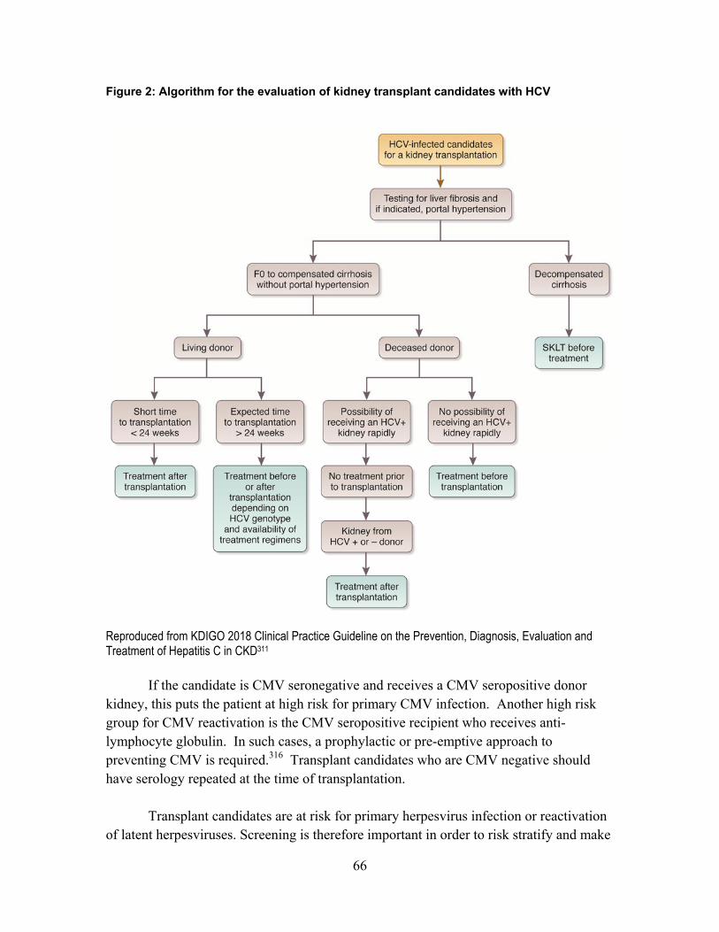
66
Figure 2: Algorithm for the evaluation of kidney transplant candidates with HCV
Reproduced from KDIGO 2018 Clinical Practice Guideline on the Prevention, Diagnosis, Evaluation and Treatment of Hepatitis C in CKD311
If the candidate is CMV seronegative and receives a CMV seropositive donor kidney, this puts the patient at high risk for primary CMV infection. Another high risk group for CMV reactivation is the CMV seropositive recipient who receives anti-lymphocyte globulin. In such cases, a prophylactic or pre-emptive approach to preventing CMV is required.316 Transplant candidates who are CMV negative should have serology repeated at the time of transplantation.
Transplant candidates are at risk for primary herpesvirus infection or reactivation of latent herpesviruses. Screening is therefore important in order to risk stratify and make

67
decisions for post-transplant prevention. If the candidate is EBV seronegative and receives an EBV seropositive donor kidney, this increases the risk of primary EBV infection and post-transplant lymphoproliferative disease.317 If the candidate is VZV seronegative, varicella immunization is recommended. Since varicella vaccine is live-attenuated, the candidate should defer transplant for at least 4 weeks after immunization. Immunization should not occur pre-transplantation if patient is immunosuppressed for another indication (e.g., treatment of underling kidney disease with steroids). If the candidate is HSV seropositive and corticosteroids are used, there is increased risk of local and disseminated HSV infection. There may also be risk for primary infection in HSV seronegative recipients of seropositive donors and antiviral prophylaxis may be indicated.
If the candidate is MMR seronegative, consideration should be given to MMR immunization prior to kidney transplant [see summary table and evidence profile: vaccines, measles]. Those born after the introduction of MMR vaccine in their region may be seronegative since circulation of wild-type virus decreased. Since MMR vaccine is live-attenuated, the candidate should defer transplant for at least 4 weeks after immunization. Live virus immunization should not occur pre-transplantation if patient is immunosuppressed for another indication (e.g., treatment of underling kidney disease with steroids).
For other viruses such as BK, serology assays may be available for use in the research setting. It is unknown whether BK viremia or viruria pre-transplant affects graft outcomes post-transplant.318, 319 There are also limited data on graft nephrectomy and the risk of subsequent BK nephropathy. In one study, 7 of 10 patients that underwent retransplantation for BK virus-associated nephropathy had nephrouretectomy of the first graft; only one patient had recurrent BK virus-associated nephropathy [see summary table and evidence profile: nephrectomy].320
HTLV is endemic in many parts of the world including the Caribbean, Japan and South America. If the candidate is HTLV seropositive, this does not preclude transplantation. However, the patient should be counseled as to the increased risk of HTLV-associated disease post-transplant such as T-cell leukemia and myelopathy/spastic paraparesis.321, 322 In addition, there should be a high-index of suspicion for these conditions post-transplant.
Although the above recommendations describe established viruses in the population, the clinician should be cognizant of emerging viral infections such as new respiratory viruses (e.g., new coronaviruses), flaviviruses (e.g., Zika, Chikungunya virus) and hemorrhagic fever viruses (e.g., Ebola), their incubation periods and disease manifestations. Transplant candidates with symptomatic disease from these viruses

68
should await resolution. Table 1. Screening for viral and non-viral pathogens in kidney transplant candidates
Pathogen Test Repeat testing
Viral infections
HIV IgG Annually if negative and at
time of transplant
HCV IgG Annually if negative and at
time of transplant
HBV Anti-HBs, Anti-HBc, HBsAg Annually if negative and at
time of transplant
CMV IgG At time of transplant
EBV VCA IgG or EBNA IgG At time of transplant
HSV IgG At time of transplant
VZV IgG At time of transplant and 4
weeks post-vaccination
Measles, Mumps, Rubella IgG At time of transplant and 4
weeks post-vaccination
HTLV IgG None unless ongoing risk
of exposure
Non-Viral infections
Syphilis IgG with confirmatory testing if IgG positive
None
Strongyloides IgG None
Chagas IgG None
Tuberculosis (in low prevalence areas)
Tuberculin skin test or Interferon-gamma release
assay (IGRA)
Annually if ongoing risk of exposure
Malaria Blood smear if clinically
indicated None
Anti-HBc, hepatitis B core antibody; Anti-HBs, hepatitis B surface antibody; CMV, cytomegalovirus; EBNA, EBV nuclear antigen; EBV, Epstein-Barr virus; HBV, hepatitis B virus; HBsAg, hepatitis B surface antigen; HCV, hepatitis C virus; HIV, human immunodeficiency virus; HSV, herpes simplex virus; HTLV, human T-lymphotropic virus; IgG, immunoglobulin G; VCA, EBV antivirus capsid antigen; VZV, varicella zoster virus.
69
10.6 Screening for non-viral infections 10.6.1 Syphilis
10.6.1.1: We recommend pre-transplant screening for syphilis (Treponema pallidum) in KTCs and treatment prior to transplantation if infection is identified. (1C)
10.6.2 Strongyloides
10.6.2.1: We suggest pre-transplant screening for strongyloidiasis in KTCs from endemic areas, and treatment prior to transplantation if infection is identified. (2C)
10.6.3 Chagas
10.6.3.1: We recommend pre-transplant screening for Chagas disease in KTCs from endemic areas, and treatment prior to transplantation if infection is identified. (1C)
10.6.4 Malaria
10.6.4.1: We recommend pre-transplant screening for malaria in KTCs who have recently travelled to endemic areas and treatment prior to transplantation if infection is identified. (1C)
RATIONALE
Syphilis is often asymptomatic but could progress with cardiac and neurologic
disease post-transplant. Therefore, serology should be routinely performed in patients awaiting transplantation and the patient treated if a confirmatory test for syphilis is positive. Lumbar puncture can be done if neurologic or ocular involvement is suspected. The ideal treatment is three doses of benzathine penicillin one week apart. In penicillin-allergic patients, ceftriaxone or doxycycline can be used.
Testing for endemic infections and tropical diseases should only be done in transplant candidates at risk. The worldwide distribution of endemic zones for various infections is readily available on the World Health Organization website (www.who.int). Strongyloides infection may be asymptomatic and lead to hyperinfection post-transplant. Therefore, screening for strongyloides is recommended in those who have lived in or

70
travelled to strongyloides endemic areas.323 Screening should be done using serology. Malaria testing should be performed if a transplant candidate has returned within the past month from a malaria endemic area and did not use malaria prophylaxis. For patients living in endemic areas, testing should be performed if clinical symptoms suggest disease. Chagas disease is endemic in Latin America and is caused by the protozoan parasite, Trypanosoma cruzi. This infection is transmitted by an insect vector and can establish clinical latency for decades. After kidney transplantation, reactivation generally occurs in the first year as asymptomatic parasitemia or fever with skin, heart or brain involvement.324, 325 Screening for Chagas is by serology. In the case of seropositivity, most experts recommend to monitor for reactivation post-transplant using polymerase chain reaction rather than treatment of the asymptomatic phase. The clinical utility for detection of endemic fungal infection in an otherwise asymptomatic transplant candidate is low as the serology-based tests lack sensitivity.326 Please see Table 1 for a summary of screenings for non-viral infections. 10.7 Vaccinations
10.7.1: We recommend that the vaccination series be commenced using an
accelerated schedule, if necessary, prior to kidney transplantation for any inactivated vaccines (Table 2). (1B) 10.7.1.1: We suggest not excluding candidates who do not complete an
inactivated vaccine series prior to kidney transplantation. (2D)
10.7.2: We recommend that the vaccination series be completed prior to
kidney transplantation for any live attenuated vaccines (Table 2). (1B)
10.7.2.1: We recommend a 4-week delay in kidney transplantation if a live vaccine is administered (e.g., MMR, VZV, shingles, yellow fever, oral typhoid, oral polio vaccine). (1B)
10.7.3: We recommend that splenectomized KTCs or those at increased risk
for post-transplant splenectomy receive pre-transplant pneumococcal, hemophilus, and meningococcal vaccines. (1B)

71
10.7.4: We recommend that KTCs requiring complement inhibitors perioperatively or post-transplant be first given the meningococcal vaccine. (1B)
10.7.5: We suggest administering the following vaccines to KTCs who, due to
age, direct exposure, residence or travel to endemic areas, or other epidemiological risk factors, are at increased risk for the specific diseases:
Rabies (2D)
Tick-borne meningoencephalitis (2D)
Japanese encephalitis (inactivated) (2D)
Meningococcus (2D)
Salmonella typhi (inactivated) (2D)
Yellow fever (2D)
RATIONALE
Vaccine preventable diseases are an important cause of morbidity after kidney transplantation. Vaccine immunogenicity is generally reduced in both CKD and post-transplant settings. However, data suggest that some vaccines are more immunogenic when given pre-transplant rather than post-transplant. In addition, live-attenuated vaccines can be only be given prior to transplantation. Therefore, assessment of vaccination status is an integral part of the pre-transplant evaluation. Childhood vaccinations should be updated as per local guidelines. Accelerated schedules can be used.327, 328 Inactivated vaccines can be given pre or post-transplantation (see KDIGO post-transplant guidelines). Vaccines should be updated as per local guidelines for diphtheria, polio, tetanus, pertussis, and Hemophilus influenzae. Transplant recipients have an increased risk for developing invasive pneumococcal disease. Transplant candidates should receive the conjugated pneumococcal vaccine followed by the polysaccharide pneumococcal vaccine at least 8 weeks later.329 Transplant candidates should receive the influenza vaccine annually while awaiting transplantation. Depending on availability, the MF59 adjuvanted or the high-dose influenza vaccine can be used in transplant candidates ≥ 65 years of age. Hepatitis B vaccine is recommended for those with CKD (see summary table: HBV vaccination).330 A 40 µg preparation (‘dialysis dose’) should be used with a 3-dose interval.331, 332 Anti-HBs titer should be measured 4-6 weeks after series completion. Titers of anti-HBs should be checked at regular intervals as they may decline over time.333 If titers have declined to < 10 IU/ml, a repeat hepatitis B vaccine series can be given. Meningococcal conjugate vaccine should be given to children as per local guidelines. In adults, meningococcal conjugate vaccine should be given to those with risk factors including functional or anatomic asplenia, travelers to

72
meningococcus endemic areas (e.g., sub-Saharan Africa, travelers to Hajj in Mecca etc) or those requiring complement inhibitors perioperatively or post-transplant. In adults, two doses of quadrivalent vaccine at least 8 weeks apart can be given. In candidates who may receive eculizumab or other complement inhibitors, two doses of quadrivalent meningococcal vaccine (for serogroups A,C,Y,W-135) as well as Meningitis serogroup B vaccine should be provided. Human papillomavirus vaccine is also inactivated and can be given using the 3-dose schedule to males and females over age 9 years. In endemic areas (www.who.int), hepatitis A vaccine should be given to all candidates before transplantation. Please refer to Table 2 for a summary of routine vaccinations for KTCs. A recombinant subunit inactivated vaccine is available to prevent herpes zoster and can be used in transplant candidates ≥ 50 years of age. In the general population, efficacy of this vaccine is > 97% and it is recommended for those ≥ 50 years;334 however, there are no specific data on its efficacy or effectiveness in those with CKD.
For inactivated vaccines, no specific wait period is required pre-transplantation and candidates can remain active if on a deceased donor wait list; however, at least two weeks is required for establishment of vaccine immunity. Nevertheless, due to lack of data, there are no recommendations for reimmunization if transplantation occurs within days after vaccination. Vaccine series that are not completed pre-transplant can be generally resumed post-transplant. Please refer to the KDIGO Care of the Transplant Recipient guideline for post-transplant guidance on vaccination.21
Live attenuated vaccines include MMR, varicella, herpes zoster, yellow fever, oral typhoid and oral polio vaccine. Transplant candidates who do not have documented immunity to MMR and have not previously received MMR vaccine should receive MMR vaccination since the vaccine is immunogenic and immunity is shown to be retained post-transplant.335 Since viremia can occur after vaccination, transplantation should be delayed by at least 4 weeks. Varicella vaccine is indicated for persons who are VZV IgG negative.336 Herpes zoster vaccine is effective for the prevention of shingles in those ≥ 50 years of age that are VZV IgG positive. Herpes zoster vaccine is beneficial in CKD and can reduce the risk of zoster by approximately 2-fold.337 However, since this is a live-attenuated vaccine, a period of 4 weeks should elapse before transplantation occurs in order to clear the viremia. Limited data show that vaccine titers persist post-transplant although the duration of persistence is unclear. In general, the inactivated herpes zoster vaccine is preferred over the live zoster vaccine since its efficacy in the general population is higher than that of live vaccine and candidates can remain active on the waitlist. Yellow fever vaccine is also a live-attenuated vaccine. For transplant candidates at increased risk of developing yellow fever, vaccination must be given at least 4 weeks before transplantation.

73
Transplant candidates should also receive specific travel vaccines if travel to endemic areas is anticipated. Based on exposure risk, transplant candidates can receive any travel vaccines including both inactivated and live vaccines.

74
Table 2. Summary of routine vaccinations for kidney transplant candidates
Routine Vaccines Dosing Guidelines* Comment
Inactive Vaccines
Diphtheria, Pertussis, Polio, Tetanus, HiB
Generally given in childhood; Ensure these are up-to-date
Pneumococcal Vaccination:
PCV13
PPV23
One dose of PCV13 followed by one dose of PPV23 with a minimum of 8-week interval between
One booster of PPV23 five years from previous PPV23
Influenza One dose annually
Hepatitis B Three doses at 0, 1, 6 months Check anti-HBs titer
Monitor annually and give booster dose if titers decline <10 IUs/ml
Hepatitis A Two doses at 0, 2 months Check titers; If not immune, give vaccination again (i.e., repeat if no response to first series)
Human Papillomavirus Three doses in both males and females if not previously given (ages 9 to 45)
No boosters
Meningococcal quadrivalent conjugate
(Serogroups A,C,Y,W-135)
Two doses given 8 weeks apart; Indicated for travel to endemic areas, prior or planned splenectomy or planned use of eculizumab
Repeat one dose every five years in patients at risk
Meningococcal B vaccine One dose if planned use of eculizumab
Shingles (Herpes Zoster Subunit)
Two doses at 0, 2-6 months for those age ≥ 50 years and VZV IgG positive
Unknown if benefit in less than 50 years of age
No boosters
Live Vaccines
Measles, Mumps, Rubella Two doses given 4 weeks apart. Considered immune after two doses regardless of seroconversion.
Check serology and provide vaccination if negative
Varicella Two doses given 4 weeks apart. Considered immune after two doses regardless of seroconversion.
Check serology and provide vaccination if negative
Shingles (Herpes Zoster Live)**
One dose in those age ≥ 50 years and VZV IgG positive
Unknown if benefit in less than 50 years of age
No boosters

75
*Duration and doses are suggestive only as they may be variable in different regions. Please check your local guidelines. **The Herpes Zoster subunit inactivated vaccine is preferred over the Herpes Zoster live vaccine. If the Herpes zoster live vaccine has already been administered, the transplant candidate can be reimmunized with the inactivated vaccine a minimum of one year after the live vaccine. Anti-HBs, hepatitis B surface antibodies; HiB, hemophilus influenzae type b; IgG, immunoglobulin G; IU, international unit; PCV13, pneumococcal conjugate vaccine-13 valent; PPV23, pneumococcal polysaccharide vaccine-23 valent; VZV, varicella zoster virus.
76
What prior guidelines recommend
Most prior guidelines recommend to delay transplantation in a KTC with an active infection. All guidelines also recommend screening for HIV, HCV, and HBV prior to transplantation. HIV infection is not a contraindication for transplant in all previous guidelines. Only the AST and CST guidelines address screening for TB and recommend that all transplant candidates be screened and treated. In the current KDIGO guidelines, we recognize that treatment may not be feasible in TB-endemic countries performing kidney transplants and therefore make separate recommendations for regions with low and high TB prevalence. We address screening for geographically restricted infections (e.g., strongyloides, Chagas, malaria) which are not addressed in other guidelines. The AST, CST, and ERA-EDTA guidelines address pre-transplant immunization to varying extents. The AST recommends annual influenza vaccine, polysaccharide pneumococcal vaccine, and routine childhood immunizations whereas the CST guidelines additionally recommend hepatitis B and varicella immunization. ERA-EDTA specifically addresses only pre-transplant varicella vaccination. Our KDIGO guideline recommendations address pre-transplant screening and immunizations in a comprehensive manner. The CST guidelines make a recommendation to consider retransplantation of KTCs with prior BK nephropathy but do not outline a consensus on pre-transplant nephrectomy prior to retransplantation for BK.
RESEARCH RECOMMENDATIONS
Studies should determine the post-transplant infection rates, morbidity, and mortality of transplant candidates colonized with MDROs.
Studies should determine newer strategies to increase the immunogenicity of vaccines in transplant candidates including influenza, shingles, pneumococcal, and hepatitis B vaccines. With newer high-dose influenza vaccines and adjuvanted influenza vaccines, comparative trials can be performed with immunogenicity or efficacy as an endpoint. Similarly, inactivated shingles vaccine should be evaluated in this population.
Studies should examine whether pre-transplant vaccinations affect the incidence of post-transplant disease, specifically where the disease outcome is measurable (e.g., varicella zoster).
Studies should examine whether it is ideal to treat HCV-positive transplant
candidates pre- or post-transplant.

77
RELEVANT SUPPLEMENTAL MATERIAL Summary table: Nephrectomy Summary table: Nephrectomy (quality assessment) Evidence profile: Transplantation outcomes after pre-transplant nephrectomy for UTI or
BK-associated nephropathy Summary table: TB treatment Summary table: TB treatment (quality assessment) Evidence profile: TB treatment, short vs. full course Summary table: TB testing Summary table: TB testing (quality assessment) Evidence profile: TB testing Summary table: HBV vaccination Summary table: HBV vaccination (quality assessment) Summary table: Vaccines measles Summary table: Vaccines measles (quality assessment) Evidence profile: Pre-transplant vaccination Summary table: HIV Summary table: HIV (quality assessment) Evidence profile: Transplantation outcomes in patients with HIV Summary table: HBV treatment Summary table: HBV treatment (quality assessment) Evidence profile: HBV treatment (lamivudine)

78
CHAPTER 11: CANCER 11.1 Cancer screening
11.1.1: We recommend KTCs undergo routine cancer screening, as per local guidelines for the general population (Table 3). (1D) 11.1.1.1: We suggest chest imaging prior to transplantation in all
KTCs. (2C) (Same as Rec 12.2) 11.1.1.2: We suggest chest CT for current or former tobacco users
with > 30 pack-year history, as per local guidelines, and chest radiograph for other KTCs. (2C) (Same as 12.2.1)
11.1.2: We recommend screening for renal cell carcinoma with
ultrasonography for KTCs at increased risk, such as long time on dialysis, family history of renal cancer, acquired cystic disease, and analgesic nephropathy. (1D)
11.1.3: We recommend screening for bladder carcinoma using urine cytology
or cystoscopy for KTCs at increased risk, such as previous cyclophosphamide use or history of heavy smoking (> 30 pack-year). (1D)
11.1.4: We recommend screening for hepatocellular carcinoma in KTCs with
cirrhosis prior to transplantation using techniques (e.g., ultrasound, α-fetoprotein, etc.) and frequency as per local guidelines. (1C)
11.1.5: We recommend screening for bowel cancer in KTCs with
inflammatory bowel disease as per local guidelines. (1C) 11.2 Potential KTCs with a prior cancer
11.2.1: We recommend that candidates with active malignancy be excluded
from kidney transplantation except for those with indolent and low-grade cancers such as prostate cancer (Gleason score ≤ 6) and basal cell carcinoma, and renal incidentaloma ≤ 1 cm in maximum diameter). (1B)
11.2.2: We suggest that the waiting time period for kidney transplantation
begins upon completion of potentially curative treatment. (2D)

79
11.2.3: Timing of kidney transplantation after potentially curative treatment for cancer is dependent on the cancer type and stage at initial diagnosis. (Not Graded)
11.2.4: We recommend no waiting time for KTCs with curatively treated
(surgically or otherwise) non-melanoma skin cancers, small renal cell carcinoma (< 3 cm), prostate cancer (Gleason score ≤ 6), carcinoma in situ (ductal carcinoma in situ [DCIS], cervical, others), thyroid cancer (follicular/papillary < 2 cm of low grade histology), and superficial bladder cancer. (1C) 11.2.4.1: For other cancers, we suggest following waiting time
parameters as outlined in Table 4. (2D)
11.2.5: We recommend not excluding candidates with a prior history of metastatic cancer from kidney transplantation, however the risk of recurrence should be a major consideration and discussed with the candidate. (1D)
11.2.6: For relevant cancers, use genomic profiling, other molecular genomic
tests, and phenotyping to predict patient-specific risk of progression and/or recurrence. (Not Graded)
11.2.7: Decisions about transplantation for KTCs in remission from cancer
should be made collaboratively with oncologists, transplant nephrologists, patients, and their caregivers. (Not Graded)
80
11.3 Hematological malignancy (see Chapter 17.7-17.9)
17.7 Acute leukemia and high-grade lymphoma
17.7.1: We suggest avoidance of kidney transplantation until patient has received curative therapy, achieved remission and remained cancer free for a period to be determined in consultation with the patient, a hematologist/oncologist and the transplant program. (Not Graded)
17.8 Myelodysplasias, chronic leukemia and chronic/low-grade lymphoma
17.8.1: Decisions about kidney transplantation in patients with myelodysplasia should be made in collaboration with a hematologist. (Not Graded)
17.8.2: Advise consultation with a hematologist with transplant
experience in determining transplant candidacy since many lesions may be deemed to be at high risk of accelerated progression or transformation post-transplant. (Not Graded)
17.9: Decisions about kidney transplantation in patients with a prior history
of hematological malignancy who are now in remission should be made in collaboration with a hematologist. (Not Graded)
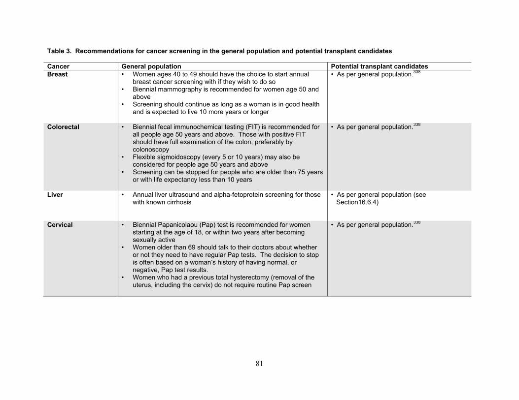
81
Table 3. Recommendations for cancer screening in the general population and potential transplant candidates Cancer General population Potential transplant candidates Breast • Women ages 40 to 49 should have the choice to start annual
breast cancer screening with if they wish to do so • Biennial mammography is recommended for women age 50 and
above • Screening should continue as long as a woman is in good health
and is expected to live 10 more years or longer
• As per general population.338
Colorectal • Biennial fecal immunochemical testing (FIT) is recommended for all people age 50 years and above. Those with positive FIT should have full examination of the colon, preferably by colonoscopy
• Flexible sigmoidoscopy (every 5 or 10 years) may also be considered for people age 50 years and above
• Screening can be stopped for people who are older than 75 years or with life expectancy less than 10 years
• As per general population.338
Liver • Annual liver ultrasound and alpha-fetoprotein screening for those with known cirrhosis
• As per general population (see Section16.6.4)
Cervical • Biennial Papanicolaou (Pap) test is recommended for women starting at the age of 18, or within two years after becoming sexually active
• Women older than 69 should talk to their doctors about whether or not they need to have regular Pap tests. The decision to stop is often based on a woman’s history of having normal, or negative, Pap test results.
• Women who had a previous total hysterectomy (removal of the uterus, including the cervix) do not require routine Pap screen
• As per general population.338

82
Lung • Routine screening for lung cancer using chest radiography and low-dose computed tomography (LDCT) is not recommended for average risk individuals
• However, there is some evidence to suggest annual screening for people at high risk of lung cancer using LDCT. Individuals at high risk are adults aged 55 to 80 years who have a smoking history of at least 30 pack-years and currently smoke or have quit within the past 15 years.
• LDCT of the chest may be recommended for individuals who are at high risk of lung cancer, including a prolonged heavy smoking history.
Prostate • Routine screening using prostate specific antigen for prostate cancer is not recommended for average risk individuals
• As per general population.338
Renal • Routine screening for renal cell cancer is not recommended for average risk individuals
• Ultrasonographic screening of the native kidneys may be recommended for individuals who have a family history of renal cancer, a personal history of acquired cystic disease, analgesic nephropathy, long-term smoking and/or prolonged waiting time on dialysis.339
Bladder • Routine screening for bladder cancer is not recommended for
average risk individuals
• Urine cytology and cystoscopies may be recommended for individuals who had been previously exposed to chemotherapeutic agents such as cyclophosphamide, regular users of compound analgesics and for heavy smokers (> 30 pack-year history).
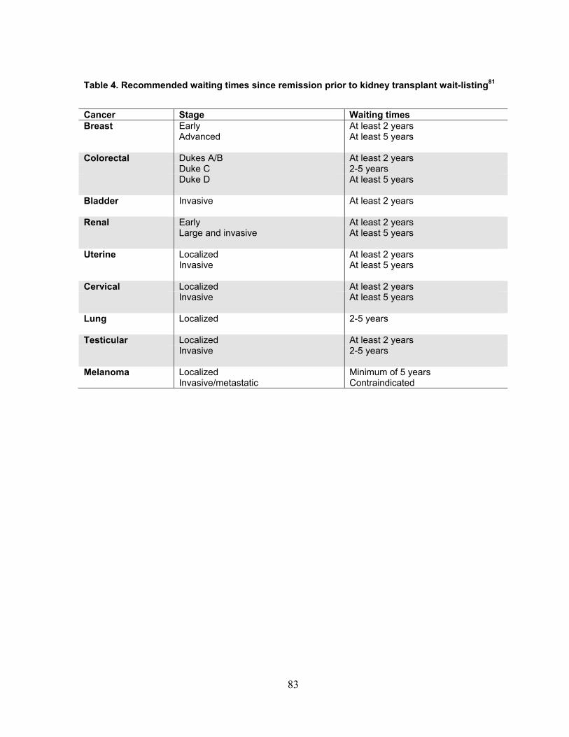
83
Table 4. Recommended waiting times since remission prior to kidney transplant wait-listing81
Cancer Stage Waiting times Breast Early At least 2 years Advanced At least 5 years
Colorectal Dukes A/B At least 2 years Duke C 2-5 years Duke D At least 5 years
Bladder Invasive At least 2 years
Renal Early At least 2 years Large and invasive At least 5 years
Uterine Localized At least 2 years Invasive At least 5 years
Cervical Localized At least 2 years Invasive At least 5 years
Lung Localized 2-5 years
Testicular Localized At least 2 years Invasive 2-5 years
Melanoma Localized Minimum of 5 years Invasive/metastatic Contraindicated

84
RATIONALE Cancer screening
Cancer is common in patients with ESKD. Evidence from observational studies and registry data reported a two-fold increase in overall cancer incidence among patients on dialysis, with kidney-related (such as urogenital cancers), endocrine-related malignancy such as thyroid cancer, and solid organ cancers such as colorectal cancer seen in excess compared to the general population.340, 341 Cancer is also a major cause of mortality and morbidity in patients with advanced stage kidney disease. Registry and linked data analyses reported at least a 1.5-fold increase in risk of cancer related death in patients on dialysis compared to the age-matched general population.342 Early detection through screening and eradication of pre-cancerous lesions is one of the few strategies proven to reduce the risk of cancer-related morbidity and mortality in the general population. Trials have reported significant reductions in cancer mortality, of at least 20% for solid organ cancers such as colorectal cancer, in the screened versus unscreened arms.343
Despite the increased risk of cancer and cancer-related death in potential transplant candidates, cancer screening uptake in those with ESKD is much lower than those without kidney disease.344 The rationale behind the reduced screening uptake is unclear, but may reflect patients’ preferences for preventive medicine in the context of chronic illness.345, 346 Also, potential candidates may experience a lower likelihood of benefits from screening even if cancer is diagnosed early because of the reduced life expectancy compared to the general population. Prior modeling analyses reported the projected gains in life years to be gained by applying screening mammography, colorectal and cervical cancer screening of patients on dialysis were at least 50% less than expected in the general population, largely because of the risk of competing events in this high-risk population including risk of death from CVD.339, 347-351 Uncertainties also exist in the test performance characteristics of individual screening tests, patient preferences, and the choice of the screening tool.352 Currently, there are no quality primary data to inform cancer screening practices specifically in the ESKD population [see summary table and evidence profile: cancer screening]. As such, it would be appropriate for potential transplant candidates to follow the current cancer screening practices for common cancer types such as colorectal, breast, cervical, lung and prostate cancers as per the general population (Table 3).350, 352 For other common cancer types that are specific to the ESKD populations, such as cancers of the urinary tract system, previous research has indicated some benefits of routine ultrasonographic screening for renal cell cancers and urinary cytology/cystoscopies for bladder cancers among high-risk individuals.339

85
Potential candidates with a prior cancer
Patients with ESKD and a cancer history in need of a transplant typically pose a challenge for transplant health professionals. While the long-term overall risk of cancer recurrence after transplantation may be low (between 5-10%), cancer prognoses after recurrence are poor.353 A recent systematic review reported an increased risk of cancer-related mortality by at least 3-fold in patients with a pre-transplant cancer history compared to recipients without prior cancers. Recipients with prior cancer also have an increased risk of developing de novo malignancy after transplantation.354
Although a prior cancer history on dialysis is not an absolute contraindication for transplantation, waiting time between two and five years for most cancer types has been recommended by clinical practice guidelines (CPGs).81 This recommendation arises from several large registry analyses indicating that the risk of cancer recurrence was maximal within the first five years after kidney transplantation. The highest risk of recurrences occurs among symptomatic renal cell carcinomas, sarcomas, melanocytic skin cancers, invasive bladder cancers and multiple myeloma.355 Consequently, a waiting period of five years or more between cancer remission and kidney transplantation has been recommended for these cancers. Other solid organ tumors such as breast, prostate and colorectal cancers confer a lesser risk, with a recommended minimum waiting period before transplantation of two years. More recently, data from Norway found no association between waiting time and all-cause mortality after kidney transplantation for those with prior cancer. However, an increased risk of cancer-related death was observed among recipients with a prior history of kidney, prostate, breast, lung or plasma cell cancers compared to those without a cancer history.356 Given the findings, the authors recommended a shorter waiting time (one year) to transplantation from disease remission, particularly for those with localized cancer. In a recent case series study, prostate cancer recurrence risks were shown to be related to the stage of disease at initial diagnosis, with the recurrence rates of stage I and II diseases, 14% and 16% respectively, significantly lower than stage III disease at 33%, suggesting a longer waiting time may be necessary for poorer stage disease.357 Analyses using the ANZDATA registry found a much lower rate of cancer recurrence compared to the US study. Between the years 1963 and 1999, the overall cancer recurrence rate in 210 kidney transplant recipients with a prior cancer history was only 5%, with a much higher rate of death among those whose prior cancers were diagnosed after commencement of dialysis compared to those diagnosed before dialysis.355 Differences between the two registries, probably due to selection bias of recipients, ascertainment bias of cancer diagnoses and unadjusted residual confounders, imply further unbiased analyses are necessary to address these unresolved issues in detail.

86
Recent analyses from the ANZDATA registry reported the overall survival for recipients who developed cancer after transplantation was generally poor, with less than 50% surviving five years after cancer diagnosis. For those that did not die from cancer, less than 20% survived more than 10 years after cancer diagnosis. Cancer of the digestive, respiratory and urinary tract systems were the three most common causes of cancer death regardless of cancer types (first cancer, recurrence and second primary). However, there were no significant differences in the risk of cancer-specific and all-cause mortality between patients who developed their first cancer after transplantation and those with cancer recurrence and those with second primary cancers.353
When considering the prospect of re-transplantation in potential candidates with a prior cancer, clinicians must balance the risk of death and associated morbidities against the reduced life expectancy and quality of life while waiting on dialysis instead of receiving a kidney transplant. To better define and stratify the risk of disease recurrence in a potential transplant candidate, genomic profiling may represent a novel application that distinguishes between breast cancers that are likely to result in early recurrence versus those that are unlikely to recur. Currently, there are two commercially available assays including the Oncotype DX Breast Recurrence Score (Genomic Health Inc., Redwood City, CA) and Mamma-Print (Agendia, Amsterdam, Netherlands). These assays can calculate a Breast Cancer Recurrence Score that correlates with the risk of cancer recurrence 10 years after transplantation, thus representing a potentially effective prognostic tool to guide treatment and future management.358 What prior guidelines recommend
Most CPGs recommended that potential transplant candidates should undergo age- and gender-specific cancer screening consistent with what is recommended for the general population. For potential transplant recipients with a prior history of cancer, clinical guidelines generally recommend a waiting time of between two and five years prior to transplantation, largely due to the fear of recurrent disease.
Instead of imposing a strict waiting time-period, we have provided a suggested list of waiting-time parameters in Table 4. These recommendations are based on previous studies which showed a reduction in cancer recurrence with time.355 Approximately 50% of cancer recurrences occurred in patients treated for cancer within 2 years of transplantation and only 13% in patients treated more than 5 years prior to transplantation.
Given the rapid advancement in cancer genome sequencing, we also suggest the use of genomic profiling assays, which may help to better assess potential transplant candidate’s risks of cancer recurrence and the timing of transplant eligibility. Assays are

87
now commercially available for early stage breast cancer and similar assays are also under investigation for other cancers such as early colorectal cancer and lung cancer.
RESEARCH RECOMMENDATIONS
There is a lack of trial based evidence of cancer screening in the transplant population; therefore, reliance has been placed on evidence from observational cohort and registry studies and modeling analyses. Given variations in the accuracy of screening tests in kidney transplant recipients and differing prognoses and life expectancies for individual transplant patients, future research that focuses on a personalized approach to shared-decision making for cancer screening, which takes into consideration a patient’s individual risks of cancer, the competing priorities of other comorbidities and the patient’s preferences towards cancer screening should be encouraged.
Emerging evidence has shown that prior cancer site, histology and stage are key factors that determine the risk of post-transplant cancer recurrence for most potential candidates with prior cancers. However, often the risk of death from cardiovascular causes outweighs the projected risk of cancer recurrence. Future work is needed to model the tradeoff for early transplantation versus remaining on dialysis for these patients.
RELEVANT SUPPLEMENTAL MATERIAL Summary table: Cancer screening Summary table: Cancer screening (quality assessment) Evidence profile: Cancer screening Summary table: Prostatectomy Summary table: Prostatectomy (quality assessment) Evidence profile: Prostatectomy Summary table: Treatment of active cancer Summary table: Treatment of active cancer (quality assessment) Evidence profile: Pretransplant cancer treatment

88
CHAPTER 12: PULMONARY DISEASE 12.1: Assess KTCs with lung disease in collaboration with a pulmonary specialist
to determine suitability for transplantation. (Not Graded) 12.2: We suggest chest imaging prior to transplantation in all KTCs. (2C) (Same as
Rec 11.1.1.1)
12.2.1 We suggest chest CT for current or former heavy tobacco users (> 30 pack-year), as per local guidelines, and chest radiograph for other KTCs. (2C) (Same as Rec 11.1.1.2)
12.3: We recommend pulmonary function testing in KTCs with impaired
functional capacity, respiratory symptoms, or known pulmonary disease. (1C)
12.4: We recommend counseling all KTCs to avoid use of tobacco products, both
before and indefinitely after transplantation. (1B) (Same as Rec 6.3) 12.5 We recommend that candidates with severe irreversible obstructive or
restrictive lung disease be excluded from kidney transplantation. (1C)
RATIONALE
There are very little data on pre-transplant evaluation of patients with pulmonary disease. As such, the recommendations are based on evidence from the general population who undergo preoperative pulmonary assessment for non-transplant surgery.359, 360 Post-operative pulmonary complications prolong hospital stay and results in increased morbidity and mortality.19, 361 Preoperative chest radiographs have not been shown to be of benefit in routine non-pulmonary surgery.359, 361 However, in KTCs a routine chest x-ray might demonstrate localized fluid collections or volume overload.362,
363 The American Cancer Society recommends that patients who have at least a 30 pack-year smoking history and who currently smoke or have quit within the past 15 years undergo lung cancer screening with a chest CT.364 It seems reasonable to apply these recommendations to transplant candidates as well.
Pulmonary function tests are not needed in most transplant candidates without significant pulmonary disease or symptoms given the lack of benefit seen with the use of these tests in the preoperative setting in the general population. However, preoperative pulmonary function tests may offer benefit in patients with impaired functional capacity,

89
known pulmonary disease, or unexplained dyspnea.
Cigarette smoking increases the risk of cancer and CVD in the general population. In kidney transplant recipients, a smoking history of more than 25 pack-years was associated with a 30% higher risk of graft failure (RR 1.30; 95% CI 1.04 to 1.63; P < 0.021), mainly due to an increased risk of death.365 For patients who quit smoking > 5 years before transplantation, the RR of graft failure was reduced by 34% (RR 0.66; 95% CI 0.52 to 0.85; P < 0.001). Given the evidence in the general population and transplant recipients, transplant candidates must be advised to stop smoking.365
KTCs with underlying pulmonary disease should be assessed and evaluated in collaboration with a pulmonary specialist. The benefit of kidney transplantation in patients with severe pulmonary disease will be offset by poor outcomes related to their lung pathology.366, 367 Given the poor prognosis, patients with the following conditions should not be candidates for kidney transplantation: lung disease requiring home oxygen therapy; uncontrolled asthma; severe cor-pulmonale; irreversible moderate to severe pulmonary hypertension; and severe chronic obstructive pulmonary disease, pulmonary fibrosis or restrictive disease.20 Patients with underlying bronchiectasis and previously treated pulmonary TB may need additional pulmonary assessments for consideration of impact of long-term immunosuppression on these diseases (see also Chapter 10 on pre-transplant infectious disease assessment). What prior guidelines recommend
The European Renal Best Practice and the UK Renal Association evaluation guideline recommend tobacco cessation pre-transplant but no other specific statements are made regarding pulmonary evaluation.14, 368 In a review by Bunnapradist and Danovitch, they have recommended evaluation to include assessment for general anesthetic risk and cessation of smoking prior to transplantation.369 Both the AST and the CST evaluation guidelines make several suggestions regarding pulmonary assessment that are very similar to our recommendations with no notable discrepancies.19, 20 The KHA-CARI guidelines make no specific mention of pulmonary assessment pre-transplantation.18

90
CHAPTER 13: CARDIAC DISEASE 13.1: All patients evaluated for kidney transplantation should undergo assessment
for the presence and severity of cardiac disease with history, physical examination, and electrocardiogram (ECG). (Not Graded)
13.2: Patients with signs or symptoms of active cardiac disease (e.g., angina,
arrhythmia, heart failure, symptomatic valvular heart disease) should undergo assessment by a cardiologist and be managed according to current local cardiac guidelines prior to further consideration for a kidney transplant. (Not Graded)
13.3: We suggest that asymptomatic KTCs at high risk for coronary artery disease
(CAD) or with poor functional capacity undergo non-invasive CAD screening. (2C)
13.3.1: We recommend that asymptomatic KTCs with known CAD not be
revascularized exclusively to reduce perioperative cardiac events. (1B) 13.3.2: We suggest not excluding candidates with advanced triple vessel
coronary disease from kidney transplantation, however the risk of a post-transplant major cardiac event should be a major consideration and discussed with the candidate. (2D)
13.4: We suggest that maintenance aspirin, β-blockers, angiotensin-converting
enzyme inhibitors/angiotensin-receptor blockers (ACE-inhibitors/ARBs), and statins be continued while on the waiting list and perioperatively, according to cardiac and local guidelines. (2A)
13.5: We suggest that kidney transplantation be delayed for at least one month
after myocardial infarction. (2B) 13.6: We suggest that kidney transplantation be deferred for at least one month
after placement of a bare metal stent and six months after insertion of a drug eluting stent. (2B)
13.7: We suggest that asymptomatic KTCs who have been on dialysis for at least
two years or have risk factors for pulmonary hypertension undergo echocardiography. (2D)

91
13.8: Patients with severe valvular heart disease should be evaluated and managed by a cardiologist according to cardiac and local guidelines. (Not Graded)
13.9: We suggest that candidates with uncorrectable, symptomatic (New York
Heart Association [NYHA] III/IV) heart disease including severe CAD, cardiac dysfunction (ejection fraction < 30%), and severe valvular disease, should not be excluded from kidney transplantation per se, however the cardiac prognosis should be evaluated and considered by the clinical team and the patient in determining candidacy for transplantation. (2D)
13.9.1: Patients with severe heart failure (NYHA III/IV) who are otherwise
suitable for kidney transplantation should be assessed by a cardiologist and considered for combined/simultaneous heart and kidney transplantation. (Not Graded)
13.10: Patients with an estimated pulmonary systolic pressure greater than 45 mm
Hg should be assessed by a cardiologist. (Not Graded)
13.10.1: We recommend not excluding candidates with uncorrectable pulmonary artery systolic pressure greater than 60 mm Hg from kidney transplantation, however the risks of sudden deterioration or progression after transplantation should be a major consideration and discussed with the candidate. (1C)
13.11: Perform cardiac imaging in patients with systemic amyloidosis. Exclude
such patients from kidney transplantation if cardiac amyloid is confirmed. (Not Graded)
Definitions
Coronary angiogram: Imaging modality of coronary arteries by injection of contrast medium usually by selective catheterization of coronary arteries.
Coronary artery disease (CAD): CAD is a narrowing or blockage of the arteries supplying the heart caused by atherosclerosis.
Heart failure: The pathophysiological state in which an abnormality of cardiac function is responsible for the failure of the heart to pump blood at a rate sufficient for the requirements of the body.
Metabolic equivalents (MET): The ratio of the work metabolic rate to the resting

92
metabolic rate. One MET is defined as 1 kcal/kg/hour and is roughly equivalent to the energy cost of sitting quietly.
Myocardial infarction (MI): Myocardial necrosis in a clinical setting consistent with acute myocardial ischemia.
Perioperative: Around the time of surgery
Pulmonary hypertension: A mean pulmonary arterial pressure (mPAP) ≥ 25 mm Hg at rest usually confirmed by right heart catheterization.
Valvular heart disease: Any disease process involving one or more of the four valves of the heart (the aortic and mitral valves on the left and the pulmonary and tricuspid valves on the right)
BACKGROUND
Cardiac disease is the most common cause of death in dialysis patients and the
incidence of cardiac events increases with worsening stages of CKD. Patients with ESKD being assessed for kidney transplantation have an increased risk of CAD, impaired left ventricular function, pulmonary hypertension and valvular heart disease compared to the general population. These risks are further increased in patients with older age, DM, and previous vascular events. Risks are also elevated in smokers and those with a longer duration of dialysis. Additionally, patients with cardiac disease have a higher risk of death and cardiac events in the peri-transplant and post-transplant periods. Kidney transplantation is generally classified as intermediate risk surgery, however many patients have co-morbidities that increase the risk for cardiac events. For these reasons, assessment for cardiac disease is important in the evaluation of KTCs.
RATIONALE
There is high quality evidence that patients with ESKD have a higher risk of cardiac disease than the general population.
There is high quality evidence that positive non-invasive stress testing is predictive of significant CAD, cardiac events and death in patients assessed for kidney transplantation. However, evidence that screening for CAD results in improved survival or a reduction in CAD events is lacking.
93
There is no evidence that revascularization of coronary artery stenoses exclusively to reduce perioperative events is beneficial.
There is moderate quality evidence that the risk of death is highest in the first month after a MI.
There is high quality evidence that dual antiplatelet therapy (DAPT) should be maintained for at least one month after insertion of a bare metal stent.
There is high quality evidence that DAPT should be maintained for at least six months after insertion of a drug eluting stent.
There is high quality evidence from the general population that patients benefit from continuing cardioprotective medication in the perioperative period.
There is moderate quality evidence that echocardiography does not accurately measure right heart pressures in patients with severe pulmonary hypertension.
There is moderate quality evidence that patients with an ejection fraction of less than 30% are at increased risk of death after kidney transplantation.
Patients with CKD G5 and those on dialysis (G5D) have a significantly higher
incidence of CAD than those of the general population.370 The diagnosis of CAD is challenging as many patients are asymptomatic with no clinical evidence of cardiac ischemia. There are a number of guidelines and consensus statements in the literature regarding cardiac assessment for patients prior to both general and kidney transplant surgery.19, 20, 371-373
The goal of a perioperative assessment is to establish whether there is active cardiac disease present. Active conditions include unstable coronary syndromes, significant heart failure, arrhythmias and valvular heart disease. Hence, a thorough history and full physical examination should be undertaken in all patients assessed for kidney transplantation. The updated American College of Cardiology/American Heart Association (ACC/AHA) guidelines on perioperative cardiovascular evaluation and care for non-cardiac surgery suggests consideration of a 12-lead electrocardiogram (ECG) in asymptomatic patients without known CAD except for those undergoing low risk surgery.371 Statements from the AHA/ACC scientific statement on cardiac evaluation for kidney and liver transplantation recommend a 12-lead ECG in potential KTCs with known CAD, peripheral vascular disease, or any cardiovascular symptoms and suggest that a 12-lead ECG is reasonable in candidates without known CVD.372

94
Due to the high risk of CAD in patients with ESKD, non-invasive stress testing of asymptomatic patients has become commonplace in patients assessed for kidney transplantation with the aim of diagnosing occult CAD and thereby reducing peri-transplant cardiac events and mortality. While multiple studies have demonstrated reasonable sensitivity and specificity for the detection of significant CAD with non-invasive stress testing in addition to reasonable positive predictive value for death and major adverse cardiac events, there are no studies demonstrating a survival benefit in patients assessed for kidney transplantation undergoing stress testing for asymptomatic CAD.374, 375 Patients with a positive stress test are however less likely to be listed for kidney transplantation.376 In the diabetic population, the DIAD trial did not show a benefit in survival or cardiac events in patients randomized to non-invasive screening versus medical management, with 7 nonfatal MIs and 8 cardiac deaths (2.7%) in the screened group and 10 nonfatal MIs and 7 cardiac deaths (3.0%) among the not screened group (HR 0.88; 95% CI 0.44–1.88; P = 0.73).377
In the general population, patients with excellent functional capacity (> 10 METS) have a low risk of cardiac events and recommendations from the ACC/AHA state that it is reasonable to forgo exercise testing in this population but suggests that cardiac stress testing be considered in patients with poor (< 4 METS) or unknown functional capacity.371 Similarly in the European Society of Cardiology (ESC)/ European Society of Anaesthesiology (ESA) guidelines on non-cardiac surgery, cardiovascular management and assessment recommend stress testing in patients who have poor functional capacity (< 4 METS) and greater than 2 risk factors for CAD.373
As patients assessed for kidney transplant have at least one clinical risk factor for CAD (kidney failure) and there is a high incidence of additional risk factors in this population, the AHA/ACC scientific statement recommends that non-invasive stress testing be considered for KTCs with 3 or more CAD risk factors regardless of functional status.372, 378 Relevant risk factors include DM, prior CVD, a duration of dialysis of ˃ 1 year, older age, smoking, hypertension and dyslipidemia.
There is little evidence to support periodically screening asymptomatic KTCs while on the waiting list although this is common practice. This practice is currently the subject of a RCT (CARSK, ANZCTR Number ACTRN 12616000736448).
Coronary revascularization exclusively to reduce perioperative cardiac events is not recommended in general prior to surgery. The CARP trial randomly assigned over 500 patients with stable CAD requiring elective vascular surgery to either medical therapy alone or medical therapy plus revascularization and found no difference in mortality between the two groups.379 Similar findings were found in the DECREASE V

95
trial where 101 patients with significant stress-induced ischemia on dobutamine stress echocardiography were randomized to medical therapy or revascularization prior to elective vascular surgery.380 In guidelines for the general population it is not recommended that that coronary revascularization be undertaken prior to non-cardiac surgery exclusively to reduce perioperative events in low and intermediate risk surgery.371, 373
In patients in whom revascularization is recommended according to existing CPGs, this should occur prior to transplantation.372
There is one randomized trial of revascularization in patients assessed for kidney transplantation.381 Twenty six patients with insulin-dependent DM and clinically significant CAD were randomized to medical therapy or revascularization prior to kidney transplantation. The outcome for those managed medically was markedly inferior to that of those who were revascularized with only 2 of 13 revascularized patients reaching a cardiovascular endpoint in 8.4 months of follow-up compared to 10 of 13 who were managed medically. This trial however, was limited by the use of short-acting calcium channel blockers in the medically managed group, sub-optimal use of aspirin, small sample size, and short follow-up [see summary table and evidence profile: CABG and cardiac revascularization pre-transplantation].
There have been a number of publications including systematic reviews examining the role of perioperative medical therapy. Continuation of β-blockade has been shown to be beneficial in multiple observational studies in the general population382-
384 and continuation has been recommended by the ACC/AHA and ESC.371-373 Similarly these guidelines recommend continuation of statins in the perioperative period. The KDIGO guideline for lipid management in CKD recommends statin treatment in kidney transplant recipients to reduce cardiac death and non-fatal MI and therefore maintaining statin use in those about to be transplanted is reasonable.385 There is an increased risk of rhabdomyolysis with the use of calcineurin inhibitors in particular cyclosporine and hence, surveillance for this rare but important side effect is warranted.386 There are no RCTs evaluating the efficacy of aspirin to prevent CVD in dialysis and CKD patients. However observational studies suggest that aspirin is associated with a reduction in mortality in patients with a previous MI and hence maintaining aspirin in patients with known vascular disease is reasonable.387, 388 There are similar recommendations from the ACC/AHA regarding ACE-inhibitors.371
In patients prescribed anticoagulant therapy, the risk of bleeding needs to be weighed against the risk of thrombosis. Vitamin K antagonists such as warfarin are commonly used in patients with AF or prosthetic heart valves. In patients with AF

96
without mechanical heart valves requiring interruption of anticoagulation for procedures, guidelines from the AHA/ACC state that decisions on bridging therapy should balance the risks of stroke and bleeding.389 In patients with prosthetic heart valves, bridging anticoagulation with either intravenous unfractionated heparin or low molecular weight heparin is recommended in the perioperative period in patients with a mechanical aortic valve replacement and any thromboembolic risk factor, older generation mechanical aortic valve replacement or mechanic mitral valve replacement.390 The use of oral direct thrombin inhibitors or anti-Xa agents in patients with mechanical valves is not recommended, due to the role of kidney function in drug clearance and the difficulties involved in reversing anticoagulation in the case of excess bleeding at the time of transplantation.
There is an increased risk of mortality in patients having surgery after a recent MI. The ACC/AHA task force recommends waiting for 4-6 weeks after a MI prior to undertaking elective surgery.390 A study using discharge data showed that the post-operative MI rate decreased substantially as the length of time from MI to operation increased from 32.8% at less than 30 days after MI to 5.9% at 90-180 days after MI. Similarly 30-day post-operative mortality was highest in the first month after MI.391 Both the ESC and AHA/ACC guidelines recommend that in the setting of an acute coronary syndrome, guidelines for treatment for ST-segment elevation MI or non-ST-segment elevation MI should be followed. In those patients with a MI who have been treated with revascularization and DAPT, guidelines for duration of antiplatelet therapy should be followed.
Coronary artery revascularization using percutaneous angioplasty and coronary artery stenting after both MI and in patients with stable CAD generally requires the use of DAPT. DAPT is associated with an increased risk of bleeding which is likely to be increased in the CKD population.392 Additionally there is an increased risk of cardiac events in the first six months after coronary artery stenting.393 The ACC/AHA recommends delaying non-cardiac surgery for a duration of at least 14 days after balloon angioplasty and at least 30 days after insertion of a bare metal stent.371 Similarly they recommend delaying elective surgery for at least a year after insertion of a drug eluting stent although more recent data has suggested that surgery after 6 months may be possible with no increase in risk.394, 395 Guidelines recommend delaying elective non-cardiac surgery until completion of a full course of DAPT has been completed to reduce the risk of perioperative bleeding and requirement for transfusion.396 In patients who have had coronary artery stenting, both the ESC and ACC/AHA guidelines recommend continuation of aspirin at a dose of 75-100 mg daily.

97
Valvular heart disease is common in the setting of ESKD with an incidence in dialysis patients that is five times greater than that of the general population.397 Additionally, survival after valve replacement surgery is significantly lower than that of the general population with a 2-year mortality of 39.5-60% as previously reported.398, 399 Similarly the incidence of pulmonary hypertension increases with worsening stages of CKD with an incidence of 32.8% reported in patients with CKD G5 in the CRIC study participants.400 Pulmonary hypertension as defined by a pulmonary artery systolic pressure (PASP) > 35 mm Hg and or Tricuspid Regurgitant Velocity (TRV) > 2.5 m/s had an adjusted 38% increased risk of all-cause mortality and 23% risk for cardiac events with a significantly higher risk in patients with a PASP > 55 mm Hg. In patients assessed for kidney transplantation, pulmonary hypertension has been shown to be associated with an increased risk of cardiac events and death.401 As volume status may impact on right heart pressure estimates, the National Kidney Foundation’s Kidney Disease Outcomes Quality Initiative (KDOQI) recommends that echocardiograms should be performed once “dry weight” has been achieved.402 Echocardiographic estimates of PASP may be inaccurate and hence, the 2012 AHA/ACC scientific statement on evaluation of cardiac disease in kidney and liver transplant candidates recommends consideration of right heart catheterization in KTCs with PASP ≥ 50 mm Hg.372 Severe pulmonary hypertension is defined as PASP > 60 mm Hg. There are a number of therapeutic and management strategies that may be beneficial in patients with severe pulmonary hypertension although these have not been rigorously tested in the ESKD population. Therefore patients with severe pulmonary hypertension who are at a satisfactory dry weight should be referred to a cardiologist for assessment and management. Despite the association of pulmonary hypertension with increased mortality and morbidity, there is some evidence that regression of elevated pulmonary pressure may occur after transplantation. Thus, assessment of this risk should be integrated with other known risk factors when deciding if an individual will benefit from kidney transplantation.403
In the general population, the European guidelines recommend that patients with established or suspected heart failure scheduled for high or intermediate risk surgery undergo evaluation of left ventricular function with echocardiography while the ACC/AHA guidelines suggest it is reasonable for patients with dyspnea of unknown origin or heart failure to undergo echocardiography.371, 373 The KDOQI guidelines for CVD in dialysis patients recommend a resting echocardiogram in all patients at the initiation of dialysis once the patient has achieved a dry weight.402 Impaired left ventricular function has been shown to be a strong predictor of mortality in both the general population and KTCs [see summary table and evidence profile: echocardiography].404, 405 In a large series of hemodialysis patients, the risk of cardiovascular death in patients with a left ventricular ejection fraction (LVEF) of < 30% was more than nine times that of those with a LVEF of ≥ 60%.406 Due to the high risk of

98
mortality with severe impairment of left ventricular function, dialysis treatment to improve fluid overload and consideration of carvedilol which has been shown to reduce mortality in the general population and in a small cohort of dialysis patients, may be beneficial.407 Patients with severe heart failure (NYHA III/IV) or with a persistently low ejection fraction < 30% despite adequate fluid removal on dialysis who are otherwise suitable for kidney transplantation should be referred to a heart transplant service for assessment for combined heart-kidney transplantation.
There are a number of cardiology guidelines recommending optimal investigation and treatment of valvular heart disease, and patients with ESKD should be evaluated according to up-to-date guidelines unless evidence emerges to the contrary.389, 407
Systemic amyloidosis is a rare multisystem disease that can result in ESKD. Registry data have shown that patients with amyloid have inferior survival both on dialysis and after kidney transplantation. However in carefully selected cases, successful transplantation has been undertaken.408, 409 Cardiac involvement is a leading cause of mortality and morbidity and can occur in amyloidosis of all etiologies. In particular cardiac involvement is most common in primary light chain AL amyloid.410 Cardiac amyloid is a restrictive cardiomyopathy which causes progressive diastolic and later biventricular dysfunction. Additionally, myocardial ischemia can result from amyloid deposits in the microvasculature. There is no consistent ECG finding in cardiac amyloid although low QRS voltages occur in up to 50% of patients with cardiac AL amyloidosis. Recommendations from amyloid centers are that all patients with amyloidosis undergo echocardiography. Findings of advanced disease have prognostic significance and these patients are unlikely to be suitable for kidney transplantation. Cardiac magnetic resonance imaging is superior for the evaluation of diastolic abnormalities however this requires gadolinium which has been shown to cause nephrogenic systemic fibrosis in patients with ESKD. Assessment should be undertaken by a cardiologist with expertise in amyloidosis. What prior guidelines recommend
Our Work Group is in general agreement with multiple guidelines outlining recommendations for assessment and management of cardiac disease in KTCs. Specifically the Work Group agrees with guidelines which recommend that candidates be assessed for cardiac disease and that patients with significant risk of CAD be assessed with non-invasive testing prior to acceptance for transplantation. The Work Group also agrees with guidelines suggesting that non-invasive testing is not necessary in asymptomatic patients at low risk of CAD. We differ with previous guidelines which recommend periodic non-invasive screening for occult CAD after admission to a waitlist, due to the lack of evidence. There is no evidence that angiography is required in

99
asymptomatic patients who have a negative non-invasive stress test. We are also in general concordance with most guidelines that recommend assessing transplant candidates for left ventricular dysfunction, valvular heart disease and pulmonary hypertension, initially by echocardiography.
The Work Group agrees with most guidelines that recommend continuing maintenance cardioprotective medications while waiting for kidney transplantation. In terms of revascularization, the Work Group agrees with the AHA/ACC Scientific Statement on cardiac disease evaluation and management among kidney and liver candidates, that routine prophylactic coronary revascularization is not recommended in patients with stable CAD who have no symptoms and have no survival indication for revascularization.
Our recommendations on timing of transplantation after MI and coronary artery stenting differ slightly from other guidelines but overall the Work Group is in general agreement with guidance provided by the recent ACC/AHA Guideline on Perioperative Cardiovascular Evaluation and Management of Patients Undergoing Noncardiac Surgery.371
RESEARCH RECOMMENDATIONS
RCTs should be conducted to examine the costs and benefits of non-invasive cardiac testing for CAD in patients being assessed for kidney transplantation, and similarly for periodic screening of patients already listed for transplantation.
RCTs should be conducted to compare revascularization versus optimal medication management prior to kidney transplantation in patients with severe but asymptomatic CAD.
Further research on the development of valid prediction scores for survival after kidney transplantation for cardiac disease, including combinations of cardiac co-morbidities, should be encouraged.
Studies should examine the efficacy of treatment options for pulmonary hypertension in patients with ESKD.

100
RELEVANT SUPPLEMENTAL MATERIAL Summary table: CABG Summary table: CABG (quality assessment) Evidence profile: Cardiac revascularization pre-transplantation Summary table: Echocardiography Summary table: Echocardiography (quality assessment) Evidence profile: Echocardiography pre-transplantation

101
CHAPTER 14: PERIPHERAL ARTERIAL DISEASE (PAD) 14.1: Evaluate all patients for presence and severity of peripheral arterial disease
(PAD) with history and physical examination. (Not Graded) 14.2: We suggest candidates without clinically apparent PAD, but who are at high
risk for PAD, undergo non-invasive vascular testing. (2D) 14.3: We suggest KTCs with clinically apparent PAD undergo imaging and
management of their vasculature in consultation with a vascular surgeon. (2D)
14.4: For patients with clinically apparent PAD, abnormal non-invasive testing, or
prior vascular procedures, we suggest non-contrast CT of the abdomen and pelvis to evaluate arterial calcification and improve operative planning. (2D)
14.5: Non-healing extremity wounds with active infection preclude kidney
transplantation until the infection is resolved. (Not Graded) 14.6: We suggest not excluding patients with severe aorto-iliac disease from kidney
transplantation. We suggest not excluding patients with prior aorto-iliac procedures including iliac artery stent placement from kidney transplantation if there is sufficient native artery available for vascular anastomosis. (2D)
14.7: We suggest not excluding candidates with advanced diabetic distal vascular
disease (e.g., major lower extremity amputation) from kidney transplantation, however the risks of progression after transplantation should be considered and discussed with the candidate. (2D)
RATIONALE
Prevalence of PAD in transplant candidates
Peripheral arterial disease (PAD) is highly prevalent in the ESKD population due to high rates of hypertension, diabetes, tobacco abuse, and altered calcium and phosphorus balance. Population-based estimates of dialysis-dependent patients demonstrate that 24% of patients with CKD have evidence of PAD using non-invasive studies.411 Among dialysis patients, 24% have clinical evidence of PAD (e.g., claudication, rest pain, or tissue loss), 35% have evidence of an abnormal ankle-brachial index, and nearly 46% have health care claims related to peripheral vascular disease.
102
Overall survival among ESKD patients who develop critical limb ischemia is less than 23% at five years.
The incidence of PAD among transplant candidates is lower as patients with advanced disease are excluded. While reporting of PAD to registries may be incomplete, one registry analysis reported that only 7% of KTCs in UNOS (listed 1994-2008) were listed as having PAD. PAD was a risk factor for waitlist mortality (HR 1.47 p < 0.001) and subsequent allograft loss (HR 2.01, p < 0.001).412 Furthermore, the degree of iliac artery calcification increases with length of dialysis prior to evaluation and listing.413 However, when compared to remaining on dialysis, kidney transplant in patient with PAD was associated with 50% reduction in mortality at five years (68.1% vs. 34.5%, p < 0.0001). For this reason, the Work Group believes that PAD is not an absolute contraindication to transplantation. Candidates with evidence PAD should be counseled that cessation of tobacco use is mandatory prior to transplantation. Evaluation of PAD
Previous KDIGO guideline have emphasized the need for appropriate assessment of PAD among patients with CKD.414 Characterization of PAD in transplant candidates relies on history, physical examination and imaging studies. The Work Group believes that all patients with risk factors for PAD (e.g., diabetes, tobacco use, history of CAD and long-term dialysis dependence) or clinical evidence of limb ischemia (e.g., claudication, rest pain, or prior amputations) should be screened for PAD. In addition, a complete history of all prior open and endovascular interventions should be obtained prior to the determination of candidacy.
Assessment of the severity of PAD can be accomplished through lower extremity segmental flow and pressure studies and non-invasive duplex evaluation.415 These tests have been demonstrated to be reliable and correlate with post-transplant outcomes.416 In patients with established PAD, arteriography (with CO2 or iodinated contrast dye) or CT scan without contrast can provide important information on the degree of proximal iliac artery and aortic calcification which assists with preoperative planning.413 Andres et al., in a prospective evaluation of 114 helical CT scans of pre-transplant candidates with risk factors for iliac stenosis, reported a 29% rate of iliac artery calcification sufficient to preclude transplantation.417 Infrainguinal arterial disease is best assessed using angiography. Severe aortoiliac disease is a relative contraindication to kidney transplant
Advanced aortoiliac disease is a relative contraindication to kidney transplantation.418 High-grade, calcific stenosis precludes kidney transplant in the ipsilateral iliac fossa, if there is an insufficient length of soft artery to allow safe clamp

103
placement and anastomosis. Selected patients can be considered for placement of an interposition graft with donor iliac artery (when available) or prosthetic with immediate or staged transplantation.419-421 Small clinical series report successful outcomes from both approaches with a low rate of vascular graft infection or allograft loss. Patients with common iliac artery disease or aortic/iliac aneurysms can be considered for pre-transplant endovascular repair provided the external iliac arteries are not overly diseased and there is room for a vascular clamp below the level of the stent. Infrainguinal vascular disease in transplant candidates
PAD below the inguinal ligament is common in patients with advanced CKD and ESKD who are candidates for kidney transplant.422 The manifestations of distal PAD include claudication, rest pain, tissue loss, infection, and amputation. Successful transplant has the potential to stabilize distal disease and reduce arterial stiffness.423 There is no evidence that kidney transplant to the ipsilateral iliac artery worsens steal syndrome or increases the risk of tissue loss.424 However, pre-transplant correction of PAD should be considered to reduce potential post-transplant exposure to iodinated contrast dye and other complications. Aortic aneurysmal disease
Patients being evaluated for kidney transplant should be evaluated for abdominal aortic aneurysm (AAA) if they have established risk factors (e.g., males, advanced age, tobacco abuse, chronic obstructive pulmonary disease, PAD, prior MI, prior transient ischemic attack [TIA]). Endovascular repair of AAA does not preclude transplant provided the iliac limbs are not extended into the external iliac arteries bilaterally. What prior guidelines recommend
Prior guidelines point to peripheral vascular disease as a marker for general cardiovascular morbidity as well as a risk factor for technical complications. The AST guidelines suggest that peripheral vascular occlusive disease alone is not a contraindication, though patients should be carefully screened for associated cardiovascular and cerebrovascular disease. No specific imaging modality was recommended, though routine angiography was unlikely to be beneficial. The presence of large unrepaired aortic aneurysms, advanced aortoiliac disease, active atheroembolic disease, or gangrene should be considered as absolute contraindications until treated and resolved. Patients with advanced aortoiliac occlusive disease should not be considered for transplant as the risk of graft loss is excessive in patients with inadequate arterial inflow. The CST similarly classified peripheral vascular occlusive disease as a risk factor for poor outcomes though not as an absolute contraindication unless symptomatic. Patients with symptomatic, recurrent peripheral vascular occlusive disease experienced

104
markedly lower post-transplant survival (5-year survival 26% vs. 80%) and may not benefit from transplantation.425 The use of arterial grafts for arterial inflow should be seen as a last resort as higher complication rates have been reported. The ERA-EDTA guidelines state only the patient should be screened for peripheral vascular occlusive disease and symptomatic or clinical significant disease should be treated as soon as possible and preferably prior to transplantation as these conditions are associated with poor long-term patient survival.

105
CHAPTER 15: NEUROLOGIC DISEASE 15.1: We suggest waiting at least 6 months after a stroke or 3 months after a
transient ischemic attack (TIA) before kidney transplantation. (2D) 15.2: We suggest not screening asymptomatic KTCs for carotid artery disease.
(2C) 15.3: We suggest screening KTCs with autosomal dominant polycystic kidney
(ADPKD) disease for intracranial aneurysms only if they are at high risk due to prior history of or a family history of subarachnoid hemorrhage. (2D)
15.4: Patients with progressive neurodegenerative disease should not undergo
kidney transplantation if survival and quality of life are not expected to be substantially improved by transplantation. (Not Graded)
15.5: Assess mental status in KTCs with known or suspected cognitive
impairment. (Not Graded)
15.5.1: We recommend not excluding candidates from kidney transplantation because of non-progressive intellectual, developmental, or cognitive disability. (1D)
Definitions
Transient ischemic attack (TIA): Episode of temporary and focal cerebral dysfunction of vascular origin, rapid in onset which commonly last 2-15 minutes but occasionally up to 24 hours with no permanent neurologic deficit.426
Carotid artery disease: Stenosis of carotid arteries, generally caused by atherosclerosis and only rarely caused by radiation therapy, vasculitis, dissection, or fibromuscular dysplasia.
Neurodegenerative disease: Neurologic diseases that cause diminished quality of life and survival despite treatment (e.g., Alzheimer’s disease and other progressive dementias, Parkinson’s disease, Huntington’s disease, and motor neuron diseases, etc.)

106
RATIONALE Waiting period
There are no data to guide decisions on when it is safe for CKD patients who have had a stroke or TIA to undergo transplantation. Observational data from the general population indicate that the risk of poorer outcomes after elective non-cardiac surgery is increased if surgery is performed within 12 months of a stroke or TIA.427, 428 However, since the risk of death is substantially higher on dialysis compared to transplant, waiting too long may increase the patients overall risk of death. The Work Group agreed that waiting for at least 6 months after a stroke or 3 months after a TIA seemed reasonable, based on expert opinion. This suggestion assumes there is not a quality-of-life-limiting neurologic deficit from the stroke, such as vascular dementia, dense hemiplegia, etc. Screening in patients with a history of stroke or TIA
It is good medical practice to screen for treatable causes of stroke or TIA when they occur. This includes echocardiography to determine if there is valvular heart disease that might be the source of emboli; ECG to rule out AF; and carotid artery imaging to rule out a treatable cause of stroke or TIA. Therefore, the Work Group concluded that these tests should be done at some time before transplantation based on expert opinion. Screening for carotid stenosis
A systematic review of evidence from the general population found no trials comparing screening versus no screening, or carotid stenting versus medical therapy.429 The specificity of ultrasonography for detecting carotid artery stenosis was found to be low, so that many false positives could be expected. A study of patients undergoing kidney transplantation found no association between pre-transplant carotid stenosis found on duplex ultrasonography and post-transplantation risk of stroke or TIA [see summary table and evidence profile: carotid screening].430 For carotid endarterectomy versus medical management the absolute reduction of non-perioperative strokes was 5.5% (95% CI, 3.9-7.0%) in 3 trials with 5223 participants with approximately 5 years of follow-up. However, the 30-day rates of stroke or death after carotid endarterectomy in trials and cohort studies were 2.4% (CI, 1.7-3.1%) in 6 trials with 3435 participants, and 3.3% (CI, 2.7-3.9%) in 7 studies with 17,474 participants. Other harms of interventions included MI, nerve injury, and hematoma. The authors of the systematic review concluded that the evidence did not indicate an overall benefit of carotid endarterectomy, stenting, or intensification of medical therapy.429 Based on this evidence, the US Preventative Services Task Force recommended against screening for asymptomatic carotid stenosis.431
107
There have been no trials investigating the potential benefits and harms of screening and intervention for asymptomatic extracranial disease in CKD. Similarly, there have there been no trials comparing intervention with no intervention or medical management for carotid artery stenosis in patients with CKD. However, there is no reason to believe that screening in CKD would be more specific than screening in the general population, or that the prevalence of carotid stenosis would be greater in advanced CKD than in the general population. In a recent series of 882 transplant candidates, only 1.5% had evidence of significant stenosis on screening carotid ultrasound.432 Therefore, given these factors, it is unlikely that the benefits would outweigh the harms of screening for asymptomatic carotid artery stenosis in transplant candidates. Screening for intracranial aneurysms in ADPKD
Intracranial aneurysms (ICAs) occur in 9-12% of patients with ADPKD433, 434 compared with 2-3% in the general population.435 From studies in the general population, ICAs less than 7 mm in diameter are more often identified with screening but are lower risk for rupture compared to larger ICAs. Patients with ADPKD and a family history of ICA rupture may be at higher risk of rupture. However, surgical repair of asymptomatic ICA is associated with a high incidence of morbidity and mortality.436
A 2014 KDIGO Controversies Conference did not recommend routine screening for ICA.437 However, screening could be considered in patients with a family history of ICAs or subarachnoid hemorrhage, previous ICA rupture, high-risk professions (e.g., airline pilots), and increased patient anxiety438 [see summary table and evidence profile: ADPKD-related cerebral aneurysm]. The Conference participants concluded that time-of-flight magnetic resonance imaging without gadolinium enhancement is the method of choice if screening is undertaken. Individuals with ICAs should be reevaluated every 6-24 months.433, 439, 440 Patients with a family history of ICA but no ICA on screening should be rescreened at 5 to 10-year intervals.439 What prior guidelines recommend
The US Preventative Services Task Force and several other guideline organizations recommend against screening for asymptomatic carotid artery stenosis in the general population.431 These guidelines are consistent with our recommendation against screening in asymptomatic transplant candidates. KHA-CARI ADPKD guidelines are consistent with our recommendation of screening for ICA only in transplant candidates at increased risk.438 The CST transplant eligibility guidelines make no distinction between stroke and TIA; a delay of at least 6 months is suggested for each condition.20

108
RELEVANT SUPPLEMENTAL MATERIAL Summary table: Carotid screening Summary table: Carotid screening (quality assessment) Evidence profile: Carotid artery testing Summary table: ADPKD-related cerebral aneurysm Summary table: ADPKD-related cerebral aneurysm (quality assessment) Evidence profile: Intracranial imaging in patients with ADPKD
109
CHAPTER 16: GASTROINTESTINAL AND LIVER DISEASE 16.1 Peptic ulcer disease
16.1.1: Assess KTCs for peptic ulcer disease. (Not Graded)
16.1.2: We recommend that candidates with symptoms suggestive of active peptic ulcer disease undergo esophagogastroscopy and H. pylori testing prior to kidney transplantation. (1C)
16.1.3: Delay kidney transplantation in candidates with endoscopically-
proven peptic ulcer disease until symptoms have resolved. (Not Graded)
16.1.4: We recommend not screening KTCs with a history of peptic ulcer disease with esophagogastroscopy. (1C)
16.1.5: We recommend not excluding candidates from kidney transplantation
because of a history of peptic ulcer disease. (1D) 16.2 Diverticulitis
16.2.1: Assess KTCs for diverticulitis. (Not Graded) 16.2.2: Delay kidney transplantation in candidates with active diverticulitis
until symptoms have resolved. (Not Graded) 16.2.3: We recommend not screening for diverticulosis in asymptomatic
KTCs. (1C) 16.2.4: We recommend not performing prophylactic colectomy in patients
with a history of diverticulitis or asymptomatic diverticulosis. (1C) 16.2.5: We recommend not excluding candidates from kidney transplantation
because of a history of diverticulitis. (1C) 16.3 Pancreatitis
16.3.1: Assess KTCs for pancreatitis. (Not Graded)

110
16.3.2: Delay kidney transplantation in candidates with acute pancreatitis a minimum of three months after symptoms have resolved. (Not Graded)
16.3.3: We recommend not excluding candidates from kidney transplantation
because of a history of acute or chronic pancreatitis. (1C) 16.4 Cholelithiasis
16.4.1: Assess KTCs for cholelithiasis. (Not Graded) 16.4.2: Delay kidney transplantation in patients with symptomatic gallstone
or gallbladder disease until symptoms have resolved. (Not Graded) 16.4.3: We recommend that candidates with a history of cholecystitis undergo
cholecystectomy before kidney transplantation. (1C) 16.4.4: We recommend not screening for cholelithiasis in asymptomatic
KTCs. (1C) 16.4.5: We recommend not performing prophylactic cholecystectomy in
KTCs with asymptomatic cholelithiasis. (1C)
16.4.6: We recommend not excluding candidates from kidney transplantation because of asymptomatic cholelithiasis. (1A)
16.5 Inflammatory bowel disease
16.5.1: Assess KTCs for inflammatory bowel disease. (1D) 16.5.2: Delay kidney transplantation in candidates with active symptomatic
inflammatory bowel disease. (Not Graded) 16.5.2.1: Determine timing of transplantation in consultation with a
gastroenterologist. (Not Graded) 16.5.3: We recommend screening for bowel cancer in patients with
inflammatory bowel disease as per local guidelines. (1C)

111
16.5.4: We recommend not excluding candidates from kidney transplantation because of a history of inflammatory bowel disease. (1D)
16.6 Liver disease
16.6.1: Screen KTCs for evidence of liver disease with appropriate history and physical exam, total bilirubin, alanine aminotransferase (ALT), international normalized ratio (INR), and albumin. (Not Graded)
16.6.2: Delay kidney transplantation until acute hepatitis, of any cause, has
resolved and a long-term strategy for managing liver disease has been implemented. (Not Graded)
16.6.3: We recommend that KTCs with cirrhosis or suspected cirrhosis be
referred to a specialist with expertise in combined liver-kidney transplantation for evaluation. (1B) 16.6.3.1: We recommend that patients undergo isolated kidney
transplantation if deemed to have compensated cirrhosis after specialist evaluation. (1B)
For liver disease associated with HBV or HCV infection see Chapter 10.5
16.6.4: We recommend screening for hepatocellular carcinoma in KTCs with
cirrhosis prior to transplantation using techniques (e.g., ultrasound, alpha-fetoprotein, etc.) and frequency as per local guidelines. (1C)
RATIONALE
Purpose of the evaluation
To provide an accurate assessment of the risk factors for perioperative morbidity and post-transplant complications related to gastrointestinal organs
To determine the severity of the comorbid gastrointestinal conditions as a contraindication to transplantation Peptic ulcer disease is the most common post-transplant gastrointestinal
complication.441, 442 One study conducted in the 1990s reported a 3.7% incidence of post-transplant peptic ulcer disease, including 1.3% with serious complications (1.0% bleeding and 0.3% perforation).441 Peptic ulcer disease was present in 16.9% of patients in a post-
112
transplant esophagogastroduodenoscopy (EGD) study, which was 1.7-fold higher than that of the general gastroenterology patients.441, 442 Although the incidence and severity of peptic ulcer disease after kidney transplantation has been reduced,443, 444 treatment of active peptic ulcer disease and eradication of H. pylori infection prior to transplantation is recommended. These recommendations are based on the relatively higher incidence of early post-transplant peptic ulcer disease, which is often serious and requiring surgical treatment.19, 20, 445
There is little evidence to support pre-transplant H. pylori screening for all transplant candidates. Observational studies have reported a 20% to 60% prevalence of H. pylori in KTCs, which is similar to rates found in the general population.442 Eradication of H. pylori has been shown to significantly reduce the incidence of post-transplant peptic ulcer disease and mucosa-associated lymphoid tissue (MALT) lymphoma.445, 446 However, the association of pre-transplant H. pylori with the occurrence of peptic ulcer disease within the first year post-transplant has not been proven.442, 447
Post-transplant immunosuppression leads to an increased risk of colonic perforation and may mask typical signs and symptoms of diverticulitis.448 As such, evaluation for diverticulosis and consideration of pre-transplant partial colectomy have been previously recommended.20 However, a recent systematic review found that the incidence of post-transplant diverticulitis (0.8%) and complicated diverticulitis (1%) were both relatively low.449 These incidence rates do not support routine screening for diverticulosis and pre-transplant colectomy in KTCs. Moreover, there is a lack of evidence for prophylactic colectomy and elective resection is not totally benign with a reported mortality rate of 1.9% and a major complication rate of 25%.450
Post-transplant acute pancreatitis is relatively uncommon (1 to 2%) but is associated with an increased risk for both local complications and death.451 There is no evidence to support the routine pre-transplant evaluation of the pancreas in asymptomatic patients. However, patients with a history of pancreatitis should be evaluated for traditional risk factors (e.g., gallstones, hyperlipidemia etc) and, if present, manage these prior to transplantation.
Cholecystectomy for asymptomatic transplant candidates is a controversial issue. The incidence of post-transplant emergency cholecystectomy (1%) and mortality (1%) are low. Observational studies have not definitively shown benefit of elective, pre-transplant cholecystectomy on post-transplant morbidity or mortality.452-455 Prophylactic cholecystectomy for selective high-risk patients (e.g., older, obese, previous gallstone pancreatitis etc) could be considered, although supportive data are lacking.456, 457

113
Approximately 30% of patients with inflammatory bowel disease will develop an acute exacerbation following transplantation.458 In a liver transplant study, active inflammatory bowel disease at the time of transplant was a risk factor for a post-transplant flare of disease activity.459 The use of tacrolimus might be a risk factor for inflammatory bowel disease relapse, although the causal relationship is unclear.460-462 Anti-TNF (tumor necrosis factor) therapy is now an option for transplant patients with inflammatory bowel disease who previously were treated with escalating doses of steroid.463
The decision to proceed with isolated kidney transplantation or combined liver-kidney transplantation in the setting of liver disease and CKD is complex and practice is highly variable worldwide. Discussion of the merits of combined organ transplantation is beyond the scope of the guideline. We have, however, recommended the involvement of specialists with expertise in combined liver-kidney transplantation for evaluation of patients with known or suspected cirrhosis. This recommendation follows standard clinical practice in most regions of the world. Although there are exceptions, most transplant candidates without decompensated cirrhosis or severe portal hypertension can safely and successfully undergo isolated kidney transplantation.464 What prior guidelines recommend
Both the AST and the CST evaluation guidelines suggest that patients with a prior history of peptic ulcer disease be considered for screening with EGD.19, 20 We have recommended against this practice as there is no evidence to support EGD in the absence of symptoms.
The AST evaluation guidelines suggest that diabetic patients be screened for cholelithiasis and offered a pre-transplant cholecystectomy if gallstones are found.19 We have recommended against routine screening and prophylactic cholecystectomy for all patients except those with a history of cholecystitis. This recommendation is based on the relatively low incidence of post-transplant acute cholecystitis and the lack of measurable impact of prophylactic cholecystectomy on clinical outcomes.
The CST guidelines suggest that patients with a history of diverticulitis be evaluated and considered for partial colectomy before transplant.20 We have advised against this practice. Similar to cholecystectomy, there is little supporting evidence that prophylactic colectomy alters the post-transplant course in patients with diverticulitis or diverticulosis.
The CST guidelines recommend a 6-month remission period following acute pancreatitis and a 12-month remission for those with chronic pancreatitis before

114
proceeding with transplantation.20 These recommendations were based on expert opinion at the time of publication in 2005. Given improvements in overall medical care for pancreatitis and the known benefits of kidney transplantation, we have recommended only a 3-month wait following acute pancreatitis. Similar to the CST guideline, this recommendation is based on expert opinion with little supporting evidence.
Similar to our recommendations, the UK Renal Association guideline14 suggests that there is no evidence to support routine screening for diverticular disease, peptic ulceration or gallbladder stones in asymptomatic transplant candidates but makes no mention of liver disease. The KHA-CARI evaluation guideline and the ERA-EDTA evaluation guideline do not specifically address issues related to the gastrointestinal system or liver disease with the exception of viral hepatitis.18, 68
RESEARCH RECOMMENDATION
Future studies should determine the incidence of post-transplant diverticulitis among those with at least one episode of diverticulitis prior to transplantation.

115
CHAPTER 17: HEMATOLOGICAL DISORDERS 17.1: We recommend not routinely screening for thrombophilia in KTCs. (1C)
17.1.1: We suggest screening for thrombophilia only in KTCs who have
experienced a venous thromboembolic event, recurrent arteriovenous access thromboses, non-atherosclerotic arterial thrombosis, or family history of venous thromboembolism to identify candidates at higher risk of graft thrombosis. (2C)
17.2: We suggest testing for antiphospholipid antibodies (APLAs) in patients with
systemic lupus erythematosus (SLE) or features of antiphospholipid syndrome (APS). (2C)
17.3: We suggest candidates receiving dual antiplatelet therapy not be excluded
from transplantation when the transplant team deems the benefit of transplantation to exceed risk of bleeding. (2D) Where risk is assessed to exceed potential benefits, we suggest that transplant surgery be delayed for the mandated period of treatment with dual antiplatelet treatment. (2C) 17.3.1: Evaluate the risk of stopping dual antiplatelet therapy to allow kidney
transplantation on a case-by-case basis by a multidisciplinary team including transplant surgeon and cardiologist. (Not Graded)
17.3.2: We suggest stopping a P2Y12 inhibitor (e.g., clopidogrel) for at least 5
days prior to living donor transplantation. (2C) 17.4: We recommend that candidates receiving anticoagulation with warfarin not
be excluded from kidney transplantation. (1B) 17.5: In the presence of significant cytopenias, evaluate suitability for kidney
transplantation based on cause and severity. (Not Graded) 17.6: We recommend that candidates with monoclonal gammopathy of
undetermined significance (MGUS), sickle cell disease, or thalassemia not be excluded from kidney transplantation [see sections on recurrent disease: plasma cell dyscrasias, Chapter 9.13 and sickle cell disease, Chapter 9.19 and hematology malignancy, Chapter 17.7-17.9]. (1C)

116
17.7 Acute leukemia and high-grade lymphoma
17.7.1: We suggest avoidance of kidney transplantation until patient has received curative therapy, achieved remission and remained cancer free for a period to be determined in consultation with the patient, a hematologist/oncologist and the transplant program. (Not Graded)
17.8 Myelodysplasias, chronic leukemia and chronic/low-grade lymphoma
17.8.1: Decisions about kidney transplantation in patients with myelodysplasia should be made in collaboration with a hematologist. (Not Graded)
17.8.2: Advise consultation with a hematologist with transplant experience in
determining transplant candidacy since many lesions may be deemed to be at high risk of accelerated progression or transformation post-transplant. (Not Graded)
17.9: Decisions about kidney transplantation in patients with a prior history of
hematological malignancy who are now in remission should be made in collaboration with a hematologist. (Not Graded)
RATIONALE
Arterial or venous thrombosis represents an important cause of early graft loss, leading to loss of approximately 2% of grafts.465 There are inherited and acquired risk factors that predispose to thrombosis. Inherited factors include Factor V Leiden (FVL), prothrombin variants and deficiencies in antithrombin III and Protein C or S with acquired defects including APS and hyperhomocysteinemia. FVL is most common and can be found in 5-8% of European populations, 20% of patients who have a thrombotic episode and up to 50% of patients with recurrent thromboses and FVL is associated with a 4-fold increased risk of graft vein thrombosis.466, 467 Although other inherited deficiencies are reported to increase thrombotic risk, data definitively linking them to graft thrombosis is lacking.
Low-titer APLAs are found commonly in healthy populations and more commonly in ESKD populations. They are found in 10-26% of patients with a clinical thrombosis and in up to 50% of patients with SLE. The outcome of transplantation in patients with APS (as opposed to APLAs without clinical manifestations) is poor with 100% graft loss reported in one study without anticoagulation.468 However, in patients

117
without clinical manifestations, APLAs did not predict graft thrombosis. Other acquired risk factors for thrombosis are common in the ESKD population, for example hyperhomocysteinemia, acquired protein C and S deficiency, but their impact on graft thrombosis is unknown.
Screening all KTCs for thrombophilia is likely have a high false-positive rate and may lead to unnecessary use of perioperative anticoagulation and higher risk of bleeding. There is insufficient evidence for untargeted screening and it is therefore not recommended [See summary table and evidence profile: thrombophilia testing]. Screening patients with a history of venous, arterial or fistula thrombosis, particularly if recurrent, features of APS or a family history of recurrent thrombosis is more likely to identify clinically significant thrombophilia and is the approach suggested. Screening should include coagulation tests (activated partial thromboplastin time and prothrombin time), FVL, prothrombin variants, Protein C and S, antithrombin III and APLAs/anticardiolipin. This will allow use of anticoagulation in candidates most at risk of graft thrombosis. This strategy is anecdotal however, with current evidence being sparse and inconsistent.469-471
CAD is common in KTCs and may have been treated with drug-eluting stents. DAPT is frequently used in this situation, combining aspirin with a P2Y12 inhibitor such as clopidogrel, ticagrelor and prasugrel.472 There is a risk of in-stent thrombosis if antiplatelet therapy is discontinued before full stent endothelialization. Continuing dual therapy will increase the risk of perioperative bleeding. There are different considerations for a living donor, when the date of transplant is known, and a deceased donor transplant, which would require the candidate to be off DAPT for longer periods. Newer P2Y12 inhibitors with shorter duration of action may provide greater flexibility. The complex balance of risk and benefit to the transplant candidate requires careful consideration by a multidisciplinary team involving transplant surgeons, hematologists and cardiologists.371
The ESC recommends avoiding elective surgery in patients on DAPT for the mandated period of treatment, usually 6 months for stable CAD or 12 months for acute coronary syndrome.473 When surgery is being considered in transplant candidates on aspirin and clopidogrel, standard advice is to withdraw clopidogrel more than 5 days prior to surgery. Testing platelet function may allow a shorter period of withdrawal.473 Withdrawal of ticagrelor for 5 days and prasugrel for 7 days is recommended.473 Aspirin should be continued through the procedure.
Oral anticoagulation with the vitamin K antagonist warfarin is not a contraindication to transplantation as the effect can be reversed. Direct thrombin

118
inhibitors are difficult to reverse, not licensed for use in CKD G4 or G5 in many jurisdictions and we suggest they should not be administered to KTCs.
Significant cytopenias require investigation and the impact on kidney transplantation depends on the cause and severity. Myelodysplastic syndromes have the potential to progress to hematological malignancy. The risk of this transformation should be considered prior to kidney transplantation in consultation with a hematologist. Specific considerations are required when transplanting patients with sickle cell disease.277 Patients with forms of thalassemia who develop ESKD can be considered for transplantation. Monoclonal gammopathy that is not the cause of kidney disease, is not a contraindication to kidney transplantation.474-476
It is in the Work Group's opinion that patients with acute leukemia and high-grade lymphomas should avoid transplantation until the potential candidate has received potentially curative therapy, achieved remission, and remained cancer free for a period to be determined in consultation with the patient, treating oncologist and the transplant program. For patients with myelodysplasias, chronic leukemia and chronic/low-grade lymphomas, the Work Group advises consultation with a hematologist with transplant experience in determining transplant candidacy since many lesions may be deemed to be at high risk for accelerated progression or transformation post-transplant.
RELEVANT SUPPLEMENTAL MATERIAL
Summary table: Thrombophilia Summary table: Thrombophilia (quality assessment) Evidence profile: Thrombophilia testing

119
CHAPTER 18: BONE AND MINERAL METABOLISM
18.1: Measure serum parathyroid hormone (PTH) at the time of transplant evaluation. (Not Graded)
18.2: We suggest not transplanting patients with severe hyperparathyroidism until
they are adequately treated (medically or surgically) as per KDIGO Chronic Kidney Disease-Mineral and Bone Disorder (CKD-MBD) guideline. (2D)
18.3: Bone mineral density (BMD) should not be measured as part of the
transplant evaluation. (Not Graded)
BACKGROUND
Most patients with advanced CKD have disorders of bone and mineral metabolism to some extent. Studies showed that up to 30% of bone mineral density (BMD) is lost within the first six months after kidney transplantation.477, 478 Recent studies have shown that despite this persistent decrease in BMD, trabecular microarchitecture remains normal in long-term transplant recipients suggesting that there is bone recovery occurring late post-transplantation.479
No intervention has been proven to prevent fractures after transplantation. Thus, prevention of bone loss is of key importance in this population. The overriding risk for fractures can be appreciated from large registry data. Recent data from Canada suggest that kidney transplant recipients have a 10-year cumulative incidence of hip fracture of approximately 2%, which is lower than previously reported.480 The same group, however, previously reported in a systematic review that the 5-year cumulative incidence for fracture varied from 0.9% to 27%.481 American registry data showed that the median 5-year fracture rate was 23%.482 The variability in reported fracture rate suggests that individual parameters such as age, gender, dialysis vintage and immunosuppressive regimen, have a substantial impact on fracture occurrence. Preventive measures of bone disease and fractures after kidney transplantation include interventions such as vitamin D, bisphosphonates, denosumab and calcitonin. However, the preferred intervention and timing of intervention have yet to be determined.483
RATIONALE
Kidney transplantation causes considerable bone loss within the first months after transplantation.

120
Most patients evaluated for transplantation already have a reduced BMD.
Risk factors for bone loss and fracture included age, sex, frailty, previous fractures, hyperparathyroidism and cumulative steroid exposure.
Post-transplant interventions for prevention of bone loss/fracture include vitamin D, bisphosphonates, denosumab and calcitonin which should be used according to individual risk.
Pre-transplant measurement of BMD does not help in decision-making regarding the use of post-transplant preventative therapies.
Severe hyperparathyroidism needs to be treated before transplantation. Access to transplantation
All patients with progressive CKD suffer from some degree of mineral and bone disorder (CKD-MBD). Treatment of the original kidney disease with steroids, dialysis vintage as well as previous transplants are key risk factors for CKD-MBD. After transplantation, the complexity of bone disease increases further due to immunosuppression.484 Bone disorders in transplant candidates are complex and span the whole spectrum from high-turnover to adynamic bone disease.
In general, serum biomarkers of bone turnover in patients with advanced CKD or on dialysis have low diagnostic accuracy when compared to the gold standard of bone histology on biopsy.485 Nevertheless, intact parathyroid hormone (PTH) is determined at routine intervals in most CKD patients because values in the extremes, when used in combination with alkaline phosphatase, potentially help to guide treatment decisions before transplantation. As per recent KDIGO CKD-MBD update,486 patients requiring PTH-lowering therapy should first receive medical therapy in the form of calcimimetics, calcitriol, or vitamin D analogs. Patients who fail to respond to medical therapy should undergo parathyroidectomy before transplantation. Several reports have shown worsening kidney function if parathyroidectomy is performed after transplantation,487, 488 however, this finding has not been universal.489 Patients with adynamic bone disease represent an even more challenging population because no intervention has been shown to be effective. Small studies on the use of recombinant PTH for this indication, either on dialysis or after transplantation, were inconclusive.490, 491 What prior guidelines recommend
Prior 2013 guidelines from KHA-CARI do not specifically address the topic of bone and mineral metabolism as a part of recipient assessment prior to transplantation.18

121
The AST evaluation guideline provides no specific recommendations on bone and mineral disease status among KTCs.19 The 2009 KDIGO guideline on the management of the kidney transplant recipient also does not make any recommendations regarding bone and mineral metabolism in the transplant candidate.21 Similarly, the recent 2017 KDIGO CPG update for the diagnosis, evaluation, prevention, and treatment of CKD-MBD do not have a specific bone disease recommendations for transplant candidates.486
The 2005 CST consensus guideline on eligibility for kidney transplantation suggests that calcium, phosphorus and PTH levels should be measured as part of the pre-transplant evaluation (Grade A) and that parathyroidectomy should be considered for those who have failed medical management or have severe, persistent complications of hyperparathyroidism (Grade B).20 The ERA-EDTA recommended in 2013 that a deceased donor allograft should not be refused only because of uncontrolled hyperparathyroidism in the recipient (Level 1D).68 The UK Renal Association and the British Transplant Society have no specific directions on bone and mineral disease in their 2011 guidelines about the assessment of the potential kidney transplant recipient.14
RESEARCH RECOMMENDATIONS
An adequately powered RCT should be conducted to examine the effect of teriparatide (recombinant PTH) on BMD and fracture risk in transplant candidates with adynamic bone disease.
A large, multicenter cohort study should be conducted to examine the association between pre-transplant PTH level and clinically important post-transplant outcomes.

122
CHAPTER 19: IMMUNOLOGICAL ASSESSMENT 19.1: Communicate all sensitizing events (e.g., blood product transfusion,
including platelets, pregnancy or miscarriage) or clinical events that can impact panel reactive antibody (PRA) (e.g., vaccination, withdrawal of immunosuppression, transplant nephrectomy, significant infection) to the human leukocyte antigen (HLA) laboratory. (Not Graded)
19.2: Perform HLA antibody testing at transplant evaluation, at regular intervals
prior to transplantation and a minimum of 2 weeks after a sensitizing event or a clinical event that can impact PRA. (Not Graded)
19.3: We recommend that HLA antibody testing be performed using solid phase
assays. (1B) 19.4: We recommend HLA typing of KTCs at evaluation using molecular methods,
optimally at all loci. (1D) 19.5: We suggest not routinely testing KTCs for non-HLA antibodies. (2C) 19.6: We suggest not routinely testing KTCs for complement-binding HLA
antibodies. (2C) 19.7: We suggest informing KTCs about their access to transplantation based on
blood type and histocompatibility testing results. (2C)
19.7.1: We recommend offering KTCs with immunologically-reduced access to transplant access to a larger deceased donor pool, kidney exchange programs, and/or desensitization. (1C)
19.7.2: We suggest that antibody avoidance (e.g., kidney exchange programs
or deceased donor acceptable mismatch allocation) be considered before desensitization. (2C)
BACKGROUND
Sensitizing events including pregnancy, blood transfusion and prior transplant can lead to the formation of HLA antibodies in transplant candidates. These antibodies, depending on donor HLA typing and donor rates, may significantly limit a candidate’s access to donors. The goal of HLA testing during candidate evaluation and while waitlisted is to estimate the risk of reduced access to potential donors based upon HLA
123
antibodies/HLA typing. In addition, up-to-date testing will ensure the ready availability of the necessary recipient information required to facilitate allocation, perform transplant decision making and donor-recipient immunologic risk assessment at the time of transplant. This section contains clinical recommendations for histocompatibility testing, basic technical interpretation and actions as they relate to immunologic risk assessment of the potential transplant recipient during workup and while waitlisted. The spectrum of potential use of the testing results in allocation and transplant decision making, as well as HLA testing for potential kidney donors, are beyond the scope of this guideline. HLA testing of living and deceased donors, testing to guide allocation or or the interpretation of the testing for specific donor-recipient transplant decision making or risk assessment are outside of the scope of this guideline. Definitions
HLA antibody: Any antibody to any HLA antigen or allelic variant of an antigen
PRA: Panel reactive antibody, the presence of any detectable HLA antibody
cPRA: Calculated PRA, an estimate of the percentage of donors in a population to whom a transplant candidate has at least one HLA antibody specificity directed
RATIONALE
Sensitizing events (blood product transfusions including platelets,
pregnancy/miscarriage, and prior transplant) as well as clinical events that can impact PRA (including vaccination, significant infection, withdrawal of immunosuppression/non adherence and nephrectomy) should be communicated to the HLA laboratory in a timely fashion.492-501 A sensitization history is essential for HLA laboratory staff to interpret testing results where antibody levels can be dynamic over time and not always captured with PRA testing while on the waitlist. Documenting and reporting a reliable clinical history is an ungraded recommendation as there are no specific studies addressing the impact of this practice, however it is low cost, of high benefit, and universally accepted as necessary for good clinical practice. Equally importantly, patients with a history of a sensitizing event, even without circulating HLA antibodies detected, should be considered as having potential for memory responses after transplant.502-504 As such, the immunologic history is also critical for perioperative management of patients.
The optimal frequency of HLA antibody testing has not been specifically studied but it is rather extrapolated from clinical observations within laboratories regarding the cadence of potential for significant change in results in their unique patient populations and to most completely capture the potential for immunologic memory. Protocols widely in use vary in testing frequency from 4 to 24 weeks to have greater reassurance that test results used in allocation (e.g., virtual crossmatching, donor-specific antibody [DSA]
124
assessment) are representative of the patient`s immunologic state at the time of transplant. The Work Group acknowledges that both fiscal and clinical considerations (e.g., history reliably negative for sensitizing events, whether HLA antibody specificity is used to guide allocation) may reduce the frequency of testing without clinical impact in certain settings. This recommendation for testing frequency is made with the intent that the clinical team liaise with their respective laboratories about the testing frequency that can be supported at their site, which would provide adequate immunologic risk assessment for a given patient. Indeed, testing frequency may also vary between patients at a given center depending on the relevant clinical circumstances. Additional testing at a minimum six weeks after interval sensitizing events is recommended in all patients to accurately document de novo as well as memory responses which may in some cases be transient and not readily detectable at the time of the next routinely schedule clinical test. De novo HLA IgG antibodies may take up to 6 weeks to form, whereas memory responses can occur within 7-14 days. The timing of testing after a sensitizing event may be sooner than six weeks depending on clinical need. Where financial considerations may prevent regular testing, we encourage a baseline test and repeat testing 2 to 6 weeks after sensitizing events. Where live donors and recipients are reliably shown to be HLA identical at all loci, testing may also be reduced without impacting clinical risk assessment.
There are two basic assays for detecting HLA antibodies: cytotoxic and solid phase. In the former, serum from the recipient is mixed with a panel of cells derived from a population that is immunogenetically comparable to the donor population of interest. The proportion of different cells lysed in the presence of complement estimates the percentage of donors in the population to whom the recipient would be expected to have cytotoxic DSA. These assays are insensitive505-507 (i.e., can miss clinically relevant low level antibody including antibodies to HLA-C508 and DP509-512 antigens) and nonspecific with immunologically irrelevant non-HLA or IgM antibodies able to trigger a false positive result.513, 514 Conversely, solid phase assays are engineered to specifically detect HLA antibodies and are significantly more sensitive ensuring lower level and other clinically relevant antibodies are not missed.505-512, 514-520 Although far more specific than cytotoxic assays, recent data suggest that some non-specificity may occur with solid phase assays as well.521, 522 Furthermore, where resources permit the use of single antigen bead assays, full delineation of antibody specificities should be performed. This will permit the calculation of a cPRA and a list of antibody specificities can be compiled for comparison to all future potential donors.523, 524
Notwithstanding regulatory requirements of any particular jurisdiction, complete HLA typing by molecular methods is optimal for interpreting HLA antibody results and describing donor-recipient mismatch with chronic rejection, de novo DSA and graft loss.

125
At a minimum, typing should be completed at loci required to interpret any detectable HLA antibodies (i.e., corresponding to the loci of the detected antibody). Optimally, HLA typing should be completed at all loci (HLA-A, B, C; DRB1, 3, 4, 5; DQA1, DQB1; DPA1, DPB1).
Serologic (cell-based complement dependent) methods of HLA typing do not provide sufficient resolution to adjudicate allele-specific HLA antibodies as DSA, nor to reliably and routinely identify antigens from HLA-C, DQA, DPA1 and DPB1. There are increasing data that antibodies to these loci may also be deleterious after transplant requiring that they be fully characterized in recipients; this will provide a robust antibody analysis as well as quantify mismatches with future donors.508-512, 525, 526 Although no direct comparisons have been made with serologic testing, studies using molecular methods for HLA typing have identified more meaningful metrics associated with transplant outcomes of interest including the ability to more specifically identify donor and recipient differences with the greatest immunologic relevance.527-533
Histocompatibility-based quantification of access to transplant lies at the complex intersection of breadth of sensitization (cPRA or PRA) to the local donor pool;534 the absolute (not relative) number of ABO compatible deceased donors available; the allocation prioritization given to sensitized candidates; the HLA phenotypes and frequencies in the accessible donor population; the potential for living donors; and the access to specialty programs (e.g., acceptable mismatch programs,535, 536 prioritization for highly sensitized patients, kidney paired donation, desensitization). It is imperative to utilize a region’s own data to determine what level of cPRA (or equivalent) antibody metric is associated with reduced HLA-based access to transplantation.523, 537, 538 No specific PRA, cPRA or equivalent other local metric (such as calculated reaction frequency utilized in the UK) threshold should be defined as “highly sensitized” across different populations. HLA-based access to transplant is indeed a continuum of risk and the cPRA level above which access is considered reduced must be considered not only in the context of the metric, but also wait times and waitlist mortality for a given degree of sensitization in the local region.539 We specifically note that cPRA (or equivalent) itself is not a measure of rejection risk. In regions where a DSA positive donor may be allocated, the cPRA is representative of an increased risk of having DSA whereas it is the presence of DSA that confers the immunologic risk.536, 540, 541 We also note the importance of race in HLA phenotype determination and allele frequency,542-545 and the resultant importance of cPRA (or equivalent metric) being determined in a population with comparable racial/HLA distribution to the recipient’s local donor population. Finally, we acknowledge the importance of the loci included in the cPRA calculator in determining the calculated value. It is imperative to include all loci where DSA at those loci would influence transplant decision-making as this will provide the best estimate of

126
transplant access.524 Despite associations reported between non-HLA antibodies (e.g., anti-angiotensin
II type 1 receptor antibody,546-548 major histocompatibility complex class I chain-related gene A (MICA) antibody,548-551 anti-endothelial antibodies552-554 and others), with rejection and or graft loss, the role of these antibodies independent of HLA antibodies in identifying humoral risk pre-transplant remains controversial. We note that these antibodies may augment the effect of HLA DSA in some,547, 555 but not all, patients. In patients where history or clinical status indicates that these antibodies may have clinical relevance, testing should be performed on a case-by-case basis. However, routine pre-transplant measurement of non-HLA antibodies cannot be recommended.
Complement binding single antigen bead assays test for the presence of high titer anti-HLA IgG1 and IgG3 antibodies capable of binding C1q or C3 in vitro, and not a unique property of the antibody itself.556-558 Complement-based assays do not accurately quantify antibody titer. Serum dilution can abrogate a positive assay and serum concentration can change a previously negative assay to positive.556 Additionally, the assay cannot account for variation in target antigen expression on endothelium which may also impact complement activation in vivo. For all antibodies of unique specificity detected in a serum, the occurrence of isolated weak/non-complement-binding HLA DSA is rare, estimated to be in the range of 1-5%.559-562 Readily available single antigen bead metrics (e.g., mean fluorescent intensity after serum dilution) may also estimate complement binding capacity in many cases. Conflicting data exist as to the relationship between complement binding assay results and transplant outcomes.563-565 In the largest study to date566 pre-transplant DSA conferred higher odds of graft loss compared to pre-transplant C1q assay positivity.556-567 For the reasons noted above, routine testing in all patients for complement binding HLA antibodies cannot be recommended with the current level of data, but may have a role in specific patient testing algorithms.
For transplant candidates in whom histocompatibility testing indicates a general reduction in transplant access (high cPRA or equivalent) or a specific barrier to a living donor (known DSA), offering increased access to a larger donor pool (e.g., national or regional deceased donor sharing or living kidney paired donation) is recommended to increase the chance of finding a DSA-negative donor. Indeed, such HLA antibody avoidance is associated with improved graft survival (comparable to unsensitized recipients) in comparison to transplantation with DSA present.568-580 However, in those with very high cPRA or fewer absolute donors available in their jurisdiction, desensitization should be explored as an option to achieve transplantation.581-587 Compared to remaining on dialysis, desensitization has been associated with improved patient survival in the US but not in studies from the UK; the role of desensitization must

127
be considered in any region in the context of the competing risks of additional time on dialysis to wait for a DSA-negative organ.580, 588 No specific desensitization protocol can be recommended based upon the available data; factors in success, regardless of protocol, are the ability of the patient to tolerate immunosuppression, antibody titer, and center experience. Desensitization with anti-B cell agents (e.g., rituximab), proteasome inhibitors (e.g., bortezomib), alone or in combination with other protocols, may increase transplant opportunities in the short term but, depending on antibody strength, can be associated with shortened long-term survival.570, 581, 586, 589, 590 Therefore, antibody avoidance is still the preferred strategy where patient characteristics and available resources permit.
The KDIGO recommendations presented here are not intended to supplant or replace any local accreditation standards. The American Society for Histocompatibility and Immunogenetics (ASHI) Accreditation Standards should be consulted for those labs under its jurisdiction (http://c.ymcdn.com/sites/www.ashi-hla.org/resource/resmgr/docs/Standards/152017_CMS_Approved_2016_ASH.pdf ). The corresponding standards from European Federation of Immunogenetics may be found at: http://www.efiweb.eu/fileadmin/user_upload/Website_documenten/EFI_Committees/Standards_Committee/Standardv6.3.pdf
For additional technical recommendations not included in this document, the reader is referred to the relevant sections of ASHI-AST STAR Guideline and The Transplantation Society 2013 Consensus Guideline for antibody testing and clinical management.591, 592 What prior guidelines recommend
The most recent comparable guideline for HLA antibody testing are Consensus Guidelines on the Testing and Clinical Management Issues Associated with HLA and Non-HLA Antibodies in Transplantation.592 In comparison, the current guidance provides specific recommendations as to the nature, frequency and implementation of testing specifically during workup and on the waitlist, and gives updated context for complement binding assay application. The former guidance recommend best practices, with the current guideline providing alternatives to best practices in certain circumstances, mindful of international differences in patient populations and resources.

128
RESEARCH RECOMMENDATIONS
Future research should determine the optimal frequency of testing on the waitlist in patients with different risks of sensitization.
Future research should determine at what resolution of typing is optimal in solid organ transplantation to best quantify donor and recipient mismatch and associated outcomes.
Future research should determine in which groups of waitlisted patients are non-HLA antibody tests of the greatest incremental benefit in predicting transplant outcomes.

129
METHODS FOR GUIDELINE DEVELOPMENT
AIM
The overall aim of this project was to develop an evidence-based CPG for the management of patients being evaluated for kidney transplantation. The guideline consists of recommendation statements, rationale text, and a summary of systematically generated evidence on relevant pre-defined clinical topics. The general guideline development method is described at below.
OVERVIEW OF PROCESS
The development process for the KDIGO 2018 Clinical Practice Guideline on the Evaluation and Management of Candidates for Kidney Transplantation included the following steps:
• Appointing Work Group members and the evidence review team (ERT) • Discussing process, methods, and results • Developing and refining topics • Identifying populations, interventions or predictors, and outcomes of
interest • Selecting topics for systematic evidence review • Standardizing quality assessment methodology • Developing and implementing literature search strategies • Screening abstracts and retrieving full-text articles on the basis of pre-
defined eligibility criteria • Creating data extraction forms • Extracting data and performing critical appraisal of the literature • Grading the methodology and outcomes in individual studies • Tabulating data from individual studies into summary tables • Grading quality of evidence for each outcome across studies, and
assessing the overall quality of evidence across outcomes with the aid of evidence profiles
• Grading the strength of recommendations on the basis of the quality of evidence and other considerations
• Finalizing guideline recommendations and supporting rationales • Sending the guideline draft for for public review in October 2018 • Editing the guideline • Publishing the final version of the guideline
130
The overall process for conducting the systematic reviews and developing the CPG follow international standards, including those from the Institute of Medicine.593, 594
The Work Group Co-Chairs and ERT met for a two-day meeting to review the guideline development process, evidence review topics, and systematic review findings. Following this, the Work Group, ERT, and KDIGO support staff met for two separate meetings to review the available evidence, formulate recommendation statements, evaluate the quality of the evidence and strength of recommendations, deliberate on rationale for recommendations, and develop consensus. The draft CPG will then undergo public review, after which revisions to recommendations and text will be made where appropriate.
Commissioning of Work Group and ERT
The KDIGO Co-Chairs appointed the Work Group Co-Chairs, who then assembled the Work Group of domain experts, including individuals with expertise in adult and pediatric nephrology, transplant nephrology, transplantation surgery, transplantation medicine, transplant immunology, and cancer epidemiology. The Brown University Center for Evidence Synthesis in Health in Providence, Rhode Island, USA, was contracted as the ERT to conduct systematic evidence review and provide expertise in guideline development methodology. The ERT consisted of physician–methodologists with expertise in nephrology and evidence-based CPG development and experienced research associates. Defining scope and topics
The Work Group Co-Chairs and the ERT defined the overall scope and goals of the guideline including lists of populations, interventions, predictors, comparators, outcomes, and analyses of interest. Together, they then drafted a preliminary list of topics and key clinical questions. The Work Group, as a whole, with the ERT further developed and refined each topic and specified screening criteria, literature search strategies, and data extraction forms.
Establishing the process for guideline development
The ERT performed systematic literature searches and organized abstract and article screening. The ERT also coordinated the methodological and analytical processes and defined and standardized the methodology for performing literature searches, data extraction, and summarizing the evidence. The Work Group took the primary role of

131
writing and grading the recommendation statements and rationale text and retained final responsibility for their content. Formulating questions of interest
Questions of interest were formulated according to the PICOTS criteria (Population, Intervention/Predictor, Comparator, Outcome, Timing, and Study Design). Details of the PICOTS criteria are presented in Table M1.
Table M1. Systematic review topics and screening criteria Clinical outcomes: Transpant vs. continued waitlist Population Adult or child eligible for potential kidney transplant Intervention Kidney transplantation (de novo, retransplant, any donor) Comparator Continuation on waitlist for kidney transplantation.
Exclude if include patients not on transplant waitlist (not awaiting transplantation).
Predictors Age subgroups, obesity subgroups, HIV, HBV Outcome Mortality (all cause), HIV or HBV outcomes as relevant Study design Multivariate (adults, HBV), Any design (pediatrics, HIV) Minimum duration of follow-up None Minimum N of Subjects 100 (adults), Any (pediatrics) Prediction model studies Population Received kidney transplant, in large registry or national database or equivalent.
Exclude multi-organ transplantation. Predictors Pre-transplantation (or at time of transplant) variables only: eGFR, albumin,
BMI (particularly at extremes), SGA or other nutrition markers, malnutrition, age (particularly at extremes), tobacco use, PRA, history of cardiac disease, heart disease status/measures, diabetes, aortoiliac disease, diabetic peripheral vascular disease, pulmonary disease, specific CKD, cancer history, morbidity indexes, substance use disorder, intellectual disability. Exclude organ donor factors.
Outcome All predictors: Mortality (all cause), graft failure/loss Predictor-specific: Mortality (cause-specific), cancer recurrence, new-onset diabetes
Design Registry study (or equivalent), multivariable analyses Minimum N of subjects 100 Registry dates Latest enrollment in registry in or after 2007 CKD recurrence after transplantation Population Kidney transplantation due to known, specific (listed) causes of CKD Predictor Specific causes of CKD Outcome CKD recurrence after transplantation (percentage with recurrence) Design Longitudinal Minimum duration of follow-up None Minimum N of subjects Variable based on population frequency of specific causes of CKD

132
Prevention of CKD recurrence Population Kidney transplantation due to FSGS, HUS, membranous nephropathy, or
MPGN Intervention Treatments for CKD at or around time of transplantation, including plasma
exchange/plasmapheresis, rituximab, eculizumab, immunoabsorption, and immunosuppression
Outcome Mortality (all-cause), Graft failure/loss, GFR, Proteinuria, Recurrent disease (by biopsy)
Design Longitudinal Minimum duration of follow-up None Minimum N of subjects None Tuberculosis Population CKD G4-5 with active tuberculosis Intervention Short course tuberculosis treatment Comparator Long (typical) course tuberculosis treatment (or no comparator) Outcome Mortality (all-cause and TB), TB reactivation, graft failure/loss Study design Longitudinal Minimum duration of follow-up None Minimum N of Subjects 50 Nephrectomy Population CKD G4-5 with recurrent UTI or
Kidney transplant recipient with failed/failing graft due to BK virus Intervention Nephrectomy (native or allograft kidney) Comparator No nephrectomy (or no comparator) Outcome Mortality (all-cause), Graft failure/loss, GFR, Recurrent UTI or BK nephropathy Study design Any Minimum duration of follow-up None Minimum N of Subjects None HIV Population Kidney transplant candidates who receive transplants Intervention HIV+ Comparator HIV- Outcome Mortality (all-cause), Graft failure/loss, HIV and infectious outcomes, GFR Study design Comparative (HIV+ vs. HIV-) Minimum duration of follow-up None Minimum N of Subjects 100 Tuberculosis testing Population CKD G4-5 who receive transplants Intervention Any TB test (pre-transplantation) Outcome Test performance characteristics, Post-transplant TB outcomes Study design Any Minimum duration of follow-up None Minimum N of Subjects 20 Vaccination Population CKD G4-5 who receive transplants Intervention Vaccination for/with Pneumovax (Prevnar 13 f/b), Influenza, HBV, Measles,
Shingles Outcome Immunogenicity, Post-transplant vaccine effectiveness (disease incidence) Study design Any Minimum duration of follow-up None

133
Minimum N of Subjects 20 Prostate cancer Population Kidney transplant candidate with nonmetastatic prostate cancer who receive
transplants Intervention Prostatectomy (at time of kidney transplantation) Comparator None needed Outcome Mortality (all-cause), Graft failure/loss, Prostate cancer outcomes Study design Longitudinal Minimum duration of follow-up None Minimum N of Subjects 10 Cancer, active Population Kidney transplant candidates with known, specific, treated cancer who receive
transplants Predictor Wait-time for transplantation after cancer cure or treatment Outcome Mortality (all-cause, cancer), Graft failure/loss, Cancer recurrence Study design Longitudinal Minimum duration of follow-up None Minimum N of Subjects 100 Cancer screening Population Kidney transplant candidates with no known cancer who receive transplants Intervention Cancer screening (any cancer, method) Outcome Mortality (all-cause, cancer), Graft failure/loss, Cancer Study design Longitudinal Minimum duration of follow-up None Minimum N of Subjects 100 Echocardiography Population Kidney transplant candidates asymptomatic for CHF, valvular disease, or other
indications for echocardiography who receive transplants Intervention Echocardiography measures Outcome Mortality (all-cause, cardiac), Graft failure/loss, Cardiac disease, Pulmonary
hypertension, Left ventricular function (overall or categorical, not specific measures)
Study design Longitudinal Minimum duration of follow-up None Minimum N of Subjects 100 (adults), any (pediatrics) Cardiac revascularization Population CKD G5 (dialysis) with severe CAD who receive transplants Intervention Cardiac revascularization Outcome Mortality (all-cause, cardiac), Graft failure/loss Study design Longitudinal Minimum duration of follow-up None Minimum N of Subjects 10 Cerebrovascular disease screening Population CKD G4-5 who receive transplants Intervention Extracranial cerebrovascular testing (as screening) Outcome Mortality (all-cause, cerebrovascular), Graft failure/loss, Stroke Study design Longitudinal Minimum duration of follow-up None Minimum N of Subjects 100

134
ADPKD-related cerebral aneurysm screening Population ADPKD Intervention Intracranial aneurysm screen/test Outcome Mortality (all-cause, cerebrovascular), Stroke, Intracranial aneurysm Study design Any Minimum duration of follow-up None Minimum N of Subjects None Hepatitis B treatment Population CKD G5 (dialysis) with HBV who receive transplant Intervention HBV treatment Outcome HBV cure (HBV DNA-) Study design Longitudinal Minimum duration of follow-up None Minimum N of Subjects 10 Perioperative testing, diabetes Population Undergoing kidney transplantation Intervention Diabetes testing (OGTT, FBG/FPG, RBG) Outcome Perioperative complications, NODAT, Change in perioperative management Study design Any Minimum duration of follow-up None Minimum N of Subjects 100 Perioperative testing, thrombophilia Population Kidney transplant candidates or CKD G5 (dialysis) with history of VTE,
recurrent AV access thrombosis, or arterial thrombosis, or family history of VTE Intervention Thrombophilia tests Outcome Mortality (all-cause, thrombosis-related), Graft loss/faiure, VTE, Perioperative
complications, Change in perioperative management Study design Any Minimum duration of follow-up None Minimum N of Subjects 200 Psychosocial testing Population Kidney transplant candidates who receive transplants Intervention Psychosocial scales/instruments, including: Psychosocial Assessment of
Candidates for Transplantation (PACT), Stanford Integrated Psychosocial Assessment for Transplant (SIPAT), Transplant Evaluation Rating Scale (TERS)
Outcome Mortality (all-cause), Graft failure/loss, Adherence Study design Longitudinal Minimum duration of follow-up Any Minimum N of Subjects 10 Retransplantation with history of nonadherence Population History of graft failure/loss due to nonadherence Intervention Retransplantation Comparator None necessary Outcome Mortality (all cause), Graft failure/loss Design Longitudinal Minimum duration of follow-up None Minimum N of subjects 100

135
Chest CT Population CKD G4-5 Intervention Low-radiation chest CT Outcome Mortality (all-cause, lung cancer), Lung cancer diagnosis Study design Any Minimum duration of follow-up Any Minimum N of Subjects 10 Dual antiplatelet agents Population Kidney transplant candidates who receive transplants Intervention Dual antiplatelet treatment Comparator Single antiplatelet treatment Outcome Perioperative complications, Thombosis outcomes Study design Comparative Minimum duration of follow-up None Minimum N of Subjects 10/arm Hyperparathyroid Population Kidney transplant candidates who receive transplants with
hyperparathyroidisms (with or without hypercalcemia) Intervention Parathyroidectomy Comparator No surgery (or no comparator) Outcome Mortality (all-cause), Graft failure/loss, Parathyroidectomy post-transplanty Study design Any Minimum duration of follow-up None Minimum N of Subjects 20 Peripheral artery disease testing Population CKD G4-5 with clinically-apparent PAD who receive transplant Intervention Peripheral artery disease testing Outcome Perioperative complications, Change in management, PAD post-transplantation Study design Any Minimum duration of follow-up Any Minimum N of Subjects 10
ADPKD, autosomal dominant polycystic kidney disease; AV, arteriovenous; BMI, body mass index; CAD, coronary artery disease; CHF, congestive heart failure; CKD, chronic kidney disease; CT, computed tomography; eGFR, estimated glomerular filtration rate; FBG/FPG, fasting blood/plasma glucose; FSGS, focal segmental glomerulosclerosis; HBV, hepatitis B infection (DNA+, surface antigen +); HIV, human immunodeficiency virus infection; HUS, hemolytic uremic syndrome; MPGN, membranoproliferative glomerulonephritis; NODAT, new-onset diabetes after transplantation; OGTT, oral glucose tolerance test; PAD, peripheral artery testing; PRA, panel reactive antibodies; RBG, random blood glucose; SGA, subjective global assessment (nutrition assessment tool); TB, tuberculosis; UTI, urinary tract infection; VTE, venous thromboembolism. Ranking of outcomes
The Work Group ranked outcomes of interest on the basis of their importance for informing clinical decision making (Table M2).

136
Table M2. Hierarchy of outcomes
Hierarchy Outcome Critical importance Mortality, Graft loss, Intracranial aneurysm rupture, Stroke High importance Graft loss (cause specific), Cancer, Infection, Intracranial aneurysm, LV
function, Recurrent kidney disease Moderate importance NODAT, Nonadherence, Uncomplicated UTI LV, left ventrical; NODAT, new-onset diabetes after transplantation; UTI, urinary tract infection. Literature searches and article selection
Systematic search strategies were developed by the ERT with input from the Work Group Co-Chairs. Modules were created for kidney transplantation, study designs, and terms for each of the systematic review topics. Separate searches were conducted for each topic (or sets of related topics). Searches were conducted in Medline (via PubMed), EMBASE, Cochrane Central Register of Controlled Trials, and Cochrane Database of Systematic Reviews. No date or language restrictions were entered into the searches. The full literature search strategies are provided in the Supplemental Appendix 1. The final searches were conducted on June 2, 2017. In addition, the search was supplemented by articles provided by Work Group members through December 2017.
For selection of studies, all members of the ERT screened each set of abstracts in duplicate using an open-source, on-line screening program Abstrackr (http://abstrackr.cebm.brown.edu/). To establish relevance and consensus among reviewers, the entire team screened and achieved consensus on a series of initial batches of 100 abstracts. A total of 36,183 citations were screened. Journal articles reporting original data or systematic reviews were selected for evidence review, based on a priori criteria for eligible evidence. Of these, 762 were selected for consideration for inclusion. After review of the full text articles, 178 were included, as enumerated in Table M3.
Data extraction
Data extraction was done by ERT research associates. Extracted data from each study was reviewed by another ERT member to confirm accuracy. The ERT designed forms to capture data on design, methodology, eligibility criteria, study participant characteristics, interventions, comparators, predictors, outcomes, and results of individual studies. Methodology and outcomes were also systematically assessed for risk of bias (see the section on risk of bias assessment below) and recorded during the data extraction process.

137
Summary tables
Summary tables were developed for each reviewed topic with eligible studies. Summary tables contain outcomes of interest, relevant population characteristics, description of intervention and comparator (or predictor), results, and quality grading for each outcome. Categorical and continuous outcomes were tabulated separately.
Work Group members reviewed and confirmed all summary table data and quality assessments. Summary tables are available at www.kdigo.org.
Evidence profiles
Evidence profiles were constructed to assess the quality and record quality grades and descriptions of effect (or association) for each outcome across studies, as well as the quality of overall evidence and description of net benefits or harms of the intervention or comparator across all outcomes. These profiles aim to make the evidence synthesis process transparent. Decisions in the evidence profiles were based on data from the primary studies listed in corresponding summary tables and on judgments of the ERT and Work Group. Each evidence profile was initially constructed by the ERT and then reviewed, edited, and confirmed by the Work Group and/or Work Group Chairs. The work products created by the ERT for summarizing the evidence base are listed in Table M3, together with the number of included studies.

138
Table M3. Work products for the guideline
Topics Topics Searched
Citations Screened
Included Studies,
n
Summary Tables / Evidence Profiles
1. Access to Transplantation Txp vs. WtL Pre-emptive
1832 *
8 *
+ +
2. Age as a factor * * * * 3. Pediatric issues † † † 4. Psychosocial assessment Psychosocial 449 2 + 5. Adherence issues Nonadherence 1137 1 + 6. Tobacco use Tobacco Cess’n 407 0 7. Obesity and related surgical issues
Bariatric 2838 0
8. Diabetes Testing 738 7 + 9. Cause of ESKD Recurrence
Recur Tx 2285 231
86 0
+ −
10. Infection TB Tx Nephrectomy
HIV HBV
TB screen/Vac
925 1528 1138 622
1319
4 2 7 3 5
+ + + + +
11. Malignancy Cancer Tx Prostatectomy
Screening
1001 440 699
2 2 4
+ + +
12. Pulmonary disease Chest CT 673 0 − 13. Cardiac disease Revasc
Echo 1144 2824
2 6
+ +
14. Peripheral artery disease PAD 1400 0 − 15. Neurologic disease ADPKD
Carotic Doppler 364 988
4 1
+ +
16. GI and liver disease − 0 − 17. Hematologic disorders Thrombophilia
Dual antiPlt 546
3028 6 0
+ −
18. Bone and mineral metabolism
Parath’omy 1371 0 −
19. HLA testing Crossmatch 1342 0 − Predictors of outcomes * Registries 3248 26 + ADPKD, autosomal dominant polycystic kidney disease; antiPlt, antiplatelet drugs; Cess’n, cessation; CT, computed tomography; Echo, echocardiography; ESKD, end-stage kidney disease; GI, gastrointestinal; HBV, hepatitis B virus; HIV, human immunodeficiency virus; HLA, human leukocyte antigens; PAD, peripheral artery disease; Parath’omy, parathyroidectomy; Recur, recurrence; Revasc, (cardiac) revascularization; TB, tuberculosis; Tx, treatment; Txp, (kidney) transplant; Vac, vaccination (all vaccinations); WtL, waitlist. * Topics were covered by searches for registry studies. † Covered within other topic searches and tables. Grading of quality of evidence for outcomes of individual studies
Methodological quality (internal validity) refers to the design, conduct, and reporting of outcomes of a clinical study. A previously devised three-level classification

139
system for quality assessment was used to grade the overall study quality and quality of all relevant outcomes in the study (Table M4). Grading of individual studies was done by one of the reviewers, then confirmed by another, with discrepancies discussed in conference.
We based the methodological quality of each study on predefined criteria. For
RCTs and other comparative studies, the ERT used the Cochrane risk of bias tool,595 which asks about risk of selection bias, performance bias, detection bias, attrition bias, reporting bias, and other potential biases. For observational studies, we also used selected questions from the Newcastle Ottawa Scale about comparability of cohorts, representativeness of the population, and adjustment for different lengths of follow-up.596 Based on these characteristics an overall assessment was made whether the study was of good, fair, or poor quality (Table M4).
Each reported outcome was then evaluated and given an individual grade
depending on the quality of reporting and methodological issues specific to that outcome. However, the quality grade of an individual outcome could not exceed the quality grade for the overall study.
Table M4. Classification of study quality
Good quality Low risk of bias and no obvious reporting errors; complete reporting of data. Must be prospective. If study of intervention, must be RCT.
Fair quality Moderate risk of bias, but problems with study or paper are unlikely to cause major bias. If study of intervention, must be prospective.
Poor quality High risk of bias or cannot rule out possible significant biases. Poor methods, incomplete data, reporting errors. Prospective or retrospective.
RCT, randomized controlled trial
Grading the quality of evidence and the strength of a guideline recommendation
A structured approach, based on ‘Grades of Recommendation, Assessment, Development and Evaluation’ (GRADE)597-599 and facilitated by the use of evidence profiles was used to grade the quality of the overall evidence and the strength of recommendations. For each topic, the discussion on grading of the quality of the evidence was led by the ERT, and the discussion regarding the strength of the recommendations was led by the Work Group Co-Chairs. The “strength of a recommendation” indicates the extent to which one can be confident that adherence to the recommendation will do more good than harm. The “quality of a body of evidence” refers to the extent to which our confidence in an estimate of effect is sufficient to support a particular recommendation.598

140
Grading the quality of evidence for each outcome across studies. Following GRADE, the quality of a body of evidence pertaining to a particular outcome of interest was initially categorized on the basis of study design. For each outcome, the potential grade for the quality of evidence for each intervention–outcome pair started at “high” but was then lowered if there were serious limitations to the methodological quality of the aggregate of studies, if there were important inconsistencies in the results across studies, if there was uncertainty about the directness of evidence including limited applicability of the findings to the population of interest, if the data were imprecise (a low event rate [0 or 1 event] in either arm or a CI spanning a range >1) or sparse (only 1 study or total N < 500), or if there was thought to be a high likelihood of bias. The final grade for the quality of the evidence for an intervention–outcome pair could be one of the following four grades: “High”, “Moderate”, “Low” or “Very Low” (Table M5).

141
Table M5. GRADE system for grading quality of evidence
Step 1: Starting grade for quality of evidence based on study design
Step 2: Reduce grade Step 3: Raise grade Final grade for quality of evidence and definition
Randomized trials = High Study quality −1 level if serious limitations −2 levels if very serious limitations
Consistency −1 level if important inconsistency
Directness −1 level if some uncertainty −2 levels if major uncertainty
Other −1 level if sparse or imprecise datac −1 level if high probability of reporting bias
Strength of association +1 level if stronga, no plausible confounders +2 levels if very strongb, no major threats to validity
Other +1 level if evidence of a dose–response gradient
+1 level if all residual plausible confounders would have reduced the observed effect
High = Further research is unlikely to change confidence in the estimate of the effect
Observational study = Low
Moderate = Further research is likely to have an important impact on confidence in the estimate of effect, and may change the estimate
Low = Further research is very likely to have an important impact on confidence in the estimate, and may change the estimate
Any other evidence = Very Low
Very Low = Any estimate of effect is very uncertain
Abbreviations: GRADE, Grading of Recommendations Assessment, Development and Evaluation. a Strong evidence of association is defined as “significant relative risk of >2 (<0.5)” based on consistent evidence from two or more observational studies, with no plausible confounders. b Very strong evidence of association is defined as “significant relative risk of >5 (<0.2)” based on direct evidence with no major threats to validity. c Sparse if there is only one study or if total N <500. Imprecise if there is a low event rate (0 or 1 event) in either arm or confidence interval spanning a range >1. Adapted by permission from Uhlig K, Macleod A, Craig J et al.599
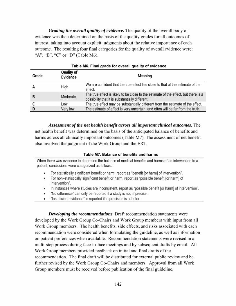
142
Grading the overall quality of evidence. The quality of the overall body of evidence was then determined on the basis of the quality grades for all outcomes of interest, taking into account explicit judgments about the relative importance of each outcome. The resulting four final categories for the quality of overall evidence were: “A”, “B”, “C” or “D” (Table M6).
Table M6. Final grade for overall quality of evidence
Grade Quality of Evidence Meaning
A High We are confident that the true effect lies close to that of the estimate of the effect.
B Moderate The true effect is likely to be close to the estimate of the effect, but there is a possibility that it is substantially different.
C Low The true effect may be substantially different from the estimate of the effect. D Very low The estimate of effect is very uncertain, and often will be far from the truth.
Assessment of the net health benefit across all important clinical outcomes. The net health benefit was determined on the basis of the anticipated balance of benefits and harms across all clinically important outcomes (Table M7). The assessment of net benefit also involved the judgment of the Work Group and the ERT.
Table M7. Balance of benefits and harms When there was evidence to determine the balance of medical benefits and harms of an intervention to a patient, conclusions were categorized as follows:
For statistically significant benefit or harm, report as “benefit [or harm] of intervention”. For non–statistically significant benefit or harm, report as “possible benefit [or harm] of
intervention”. In instances where studies are inconsistent, report as “possible benefit [or harm] of intervention”. “No difference” can only be reported if a study is not imprecise. “Insufficient evidence” is reported if imprecision is a factor.
Developing the recommendations. Draft recommendation statements were developed by the Work Group Co-Chairs and Work Group members with input from all Work Group members. The health benefits, side effects, and risks associated with each recommendation were considered when formulating the guideline, as well as information on patient preferences when available. Recommendation statements were revised in a multi-step process during face-to-face meetings and by subsequent drafts by email. All Work Group members provided feedback on initial and final drafts of the recommendation. The final draft will be distributed for external public review and be further revised by the Work Group Co-Chairs and members. Approval from all Work Group members must be received before publication of the final guideline.

143
Grading the strength of the recommendations. The strength of a recommendation is graded as level 1 or level 2. Table M8 shows the KDIGO nomenclature for grading the strength of a recommendation and the implications of each level for patients, clinicians, and policy makers. Recommendations can be for or against doing something. Each recommendation includes an explicit link between the quality of the available evidence and the strength of that recommendation. However, Table M9 shows that the strength of a recommendation is determined not only by the quality of the evidence but also by other, often complex judgments regarding the size of the net medical benefit (potential risks vs benefit), values, and preferences, and costs. Formal decision analyses including cost analysis were not conducted.
Table M8. KDIGO nomenclature and description for grading recommendations
Grade* Implications
Patients Clinicians Policy
Level 1 “We recommend”
Most people in your situation would want the recommended course of action, and only a small proportion would not.
Most patients should receive the recommended course of action.
The recommendation can be evaluated as a candidate for developing a policy or a performance measure.
Level 2 “We suggest”
The majority of people in your situation would want the recommended course of action, but many would not.
Different choices will be appropriate for different patients. Each patient needs help to arrive at a management decision consistent with her or his values and preferences.
The recommendation is likely to require substantial debate and involvement of stakeholders before policy can be determined.
*The additional category “Not Graded” is used, typically, to provide guidance based on common sense or where the topic does not allow adequate application of evidence. The most common examples include recommendations regarding monitoring intervals, counseling, and referral to other clinical specialists. The ungraded recommendations are generally written as simple declarative statements. They should not be interpreted as being weaker recommendations than Level 1 or 2 recommendations.

144
Table M9. Determinants of strength of recommendation Factor Comment Balance between desirable and undesirable effects
The larger the difference between the desirable and undesirable effects, the more likely a strong recommendation is warranted. The narrower the gradient, the more likely a weak recommendation is warranted.
Quality of the evidence The higher the quality of evidence, the more likely a strong recommendation is warranted.
Values and preferences The more variability in values and preferences, or the more uncertainty in values and preferences, the more likely a weak recommendation is warranted. Values and preferences were obtained from the literature where possible or were assessed in the judgment of the Work Group where robust evidence was not identified.
Costs (resource allocation) The higher the costs of an intervention—that is, the more resources consumed—the less likely a strong recommendation is warranted.
Ungraded statements. This category was designed to allow the Work Group to
issue general advice. Typically an ungraded statement meets the following criteria: it provides guidance based on common sense; it provides reminders of the obvious; and it is not sufficiently specific to allow for application of evidence to the issue and therefore it is not based on systematic evidence review. As such, ungraded statements may be considered to be relatively strong recommendations; they should not be interpreted as weak recommendations based on limited or poor evidence. Common examples include recommendations about frequency of testing, referral to specialists, and routine medical care. We strove to minimize the use of ungraded recommendations.
This grading scheme, with two levels for the strength of a recommendation together with four levels of grading the quality of the evidence, as well as the option of an ungraded statement for general guidance, was adopted by the KDIGO Board in December 2008. The Work Group took on the primary role of writing the recommendations and rationale statements and retained final responsibility for the content of the guideline statements and the accompanying narrative. The ERT reviewed draft recommendations and grades for consistency with the conclusions of the evidence review.
Format for guideline recommendations. Each chapter contains one or more specific recommendations. Within each recommendation, the strength of recommendation is indicated as level 1 or level 2 and the quality of the supporting evidence is shown as A, B, C, or D. The recommendation statements and grades are followed by the rationale text summarizing the key points of the evidence base and the judgments supporting the recommendation. In relevant sections, considerations of the guideline statements in international settings and suggested audit criteria are also

145
provided where applicable. Important key points and research recommendations suggesting future research to resolve current uncertainties are also outlined at the conclusion of each chapter.
Review of guideline development process
Several tools and checklists have been developed to assess the quality of the methodological process for systematic review and guideline development. These include the Appraisal of Guidelines for Research and Evaluation (AGREE 2) criteria, the Conference on Guideline Standardization (COGS) checklist,600 and the Institute of Medicine’s recent Standards for Systematic Reviews and Clinical Practice Guidelines We Can Trust.593, 594

146
REFERENCES 1. The Declaration of Istanbul on organ trafficking and transplant tourism. Kidney Int. 2008;
74: 854-859.
2. Tonelli M, Wiebe N, Knoll G, et al. Systematic review: kidney transplantation compared with dialysis in clinically relevant outcomes. Am J Transplant. 2011; 11: 2093-2109.
3. Haller M, Gutjahr G, Kramar R, et al. Cost-effectiveness analysis of renal replacement therapy in Austria. Nephrol Dial Transplant. 2011; 26: 2988-2995.
4. Klarenbach SW, Tonelli M, Chui B, et al. Economic evaluation of dialysis therapies. Nat Rev Nephrol. 2014; 10: 644-652.
5. Laupacis A, Keown P, Pus N, et al. A study of the quality of life and cost-utility of renal transplantation. Kidney Int. 1996; 50: 235-242.
6. Wong G, Howard K, Chapman JR, et al. Comparative survival and economic benefits of deceased donor kidney transplantation and dialysis in people with varying ages and co-morbidities. PLoS One. 2012; 7: e29591.
7. Bayat S, Macher MA, Couchoud C, et al. Individual and regional factors of access to the renal transplant waiting list in france in a cohort of dialyzed patients. Am J Transplant. 2015; 15: 1050-1060.
8. Plantinga LC, Pastan SO, Wilk AS, et al. Referral for Kidney Transplantation and Indicators of Quality of Dialysis Care: A Cross-sectional Study. Am J Kidney Dis. 2017; 69: 257-265.
9. Israni AK, Salkowski N, Gustafson S, et al. New national allocation policy for deceased donor kidneys in the United States and possible effect on patient outcomes. J Am Soc Nephrol. 2014; 25: 1842-1848.
10. Smith JM, Schnitzler MA, Gustafson SK, et al. Cost Implications of New National Allocation Policy for Deceased Donor Kidneys in the United States. Transplantation. 2016; 100: 879-885.

147
11. The Transplantation Society of Australia and New Zealand, Clinical Guidelines for Organ Transplantation from Deceased Donors. 97pp (2017). http://www.donatelife.gov.au/sites/default/files/TSANZ%20Clinical%20Guidelines%20for%20Organ%20Transplantation%20from%20Deceased%20Donors_Version%201.0_April%202016.pdf (Accessed Dec 12, 2017).
12. BC Transplant. Clinical Guidelines for Kidney. 66pp. (2017). http://www.transplant.bc.ca/Documents/Health%20Professionals/Clinical%20guidelines/Clinical%20Guidelines%20for%20Kidney%20Transplantation.pdf (Accessed Dec 12, 2017).
13. Trillium Gift of Life Network. Ontario’s Referral and Listing Criteria for Adult Kidney Transplantation. 8pp. (2015). https://www.giftoflife.on.ca/resources/pdf/1_Adult_Kidney_Tx_Referl_Listng_Criteria_1030.pdf (Accessed Dec 12, 2017).
14. Dudley C, Harden P. Renal Association Clinical Practice Guideline on the assessment of the potential kidney transplant recipient. Nephron Clin Pract. 2011; 118 Suppl 1: c209-224.
15. van Walraven C, Austin PC, Knoll G. Predicting potential survival benefit of renal transplantation in patients with chronic kidney disease. CMAJ. 2010; 182: 666-672.
16. Molnar MZ, Nguyen DV, Chen Y, et al. Predictive Score for Posttransplantation Outcomes. Transplantation. 2017; 101: 1353-1364.
17. Patzer RE, Basu M, Larsen CP, et al. iChoose Kidney: A Clinical Decision Aid for Kidney Transplantation Versus Dialysis Treatment. Transplantation. 2016; 100: 630-639.
18. Campbell S, Pilmore H, Gracey D, et al. KHA-CARI guideline: recipient assessment for transplantation. Nephrology (Carlton). 2013; 18: 455-462.
19. Kasiske BL, Cangro CB, Hariharan S, et al. The evaluation of renal transplantation candidates: clinical practice guidelines. Am J Transplant. 2001; 1 Suppl 2: 3-95.
20. Knoll G, Cockfield S, Blydt-Hansen T, et al. Canadian Society of Transplantation consensus guidelines on eligibility for kidney transplantation. CMAJ. 2005; 173: 1181-1184.
21. KDIGO clinical practice guideline for the care of kidney transplant recipients. Am J Transplant. 2009; 9 Suppl 3: S1-155.

148
22. Heemann U, Abramowicz D, Spasovski G, et al. Endorsement of the Kidney Disease Improving Global Outcomes (KDIGO) guidelines on kidney transplantation: a European Renal Best Practice (ERBP) position statement. Nephrol Dial Transplant. 2011; 26: 2099-2106.
23. Rao PS, Schaubel DE, Guidinger MK, et al. A comprehensive risk quantification score for deceased donor kidneys: the kidney donor risk index. Transplantation. 2009; 88: 231-236.
24. Hart A, Smith JM, Skeans MA, et al. Kidney. Am J Transplant. 2016; 16 Suppl 2: 11-46.
25. Stevens KK, Woo YM, Clancy M, et al. Deceased donor transplantation in the elderly--are we creating false hope? Nephrol Dial Transplant. 2011; 26: 2382-2386.
26. Ponticelli C, Podesta MA, Graziani G. Renal transplantation in elderly patients. How to select the candidates to the waiting list? Transplant Rev. 2014; 28: 188-192.
27. Garonzik-Wang JM, Govindan P, Grinnan JW, et al. Frailty and delayed graft function in kidney transplant recipients. Arch Surg. 2012; 147: 190-193.
28. Karim A, Farrugia D, Cheshire J, et al. Recipient age and risk for mortality after kidney transplantation in England. Transplantation. 2014; 97: 832-838.
29. McAdams-Demarco MA, Grams ME, Hall EC, et al. Early hospital readmission after kidney transplantation: patient and center-level associations. Am J Transplant. 2012; 12: 3283-3288.
30. McAdams-DeMarco MA, Ying H, Olorundare I, et al. Individual Frailty Components and Mortality in Kidney Transplant Recipients. Transplantation. 2017; 101: 2126-2132.
31. McAdams-DeMarco MA, James N, Salter ML, et al. Trends in kidney transplant outcomes in older adults. J Am Geriatr Soc. 2014; 62: 2235-2242.
32. Gill JS, Schaeffner E, Chadban S, et al. Quantification of the early risk of death in elderly kidney transplant recipients. Am J Transplant. 2013; 13: 427-432.
33. Lloveras J, Arcos E, Comas J, et al. A paired survival analysis comparing hemodialysis and kidney transplantation from deceased elderly donors older than 65 years. Transplantation. 2015; 99: 991-996.
149
34. Macrae J, Friedman AL, Friedman EA, et al. Live and deceased donor kidney transplantation in patients aged 75 years and older in the United States. Int Urol Nephrol. 2005; 37: 641-648.
35. Massie AB, Luo X, Chow EK, et al. Survival benefit of primary deceased donor transplantation with high-KDPI kidneys. Am J Transplant. 2014; 14: 2310-2316.
36. Merion RM, Ashby VB, Wolfe RA, et al. Deceased-donor characteristics and the survival benefit of kidney transplantation. JAMA. 2005; 294: 2726-2733.
37. Ojo AO, Hanson JA, Meier-Kriesche H, et al. Survival in recipients of marginal cadaveric donor kidneys compared with other recipients and wait-listed transplant candidates. J Am Soc Nephrol. 2001; 12: 589-597.
38. Perez-Saez MJ, Arcos E, Comas J, et al. Survival Benefit From Kidney Transplantation Using Kidneys From Deceased Donors Aged >/=75 Years: A Time-Dependent Analysis. Am J Transplant. 2016; 16: 2724-2733.
39. Rao PS, Merion RM, Ashby VB, et al. Renal transplantation in elderly patients older than 70 years of age: results from the Scientific Registry of Transplant Recipients. Transplantation. 2007; 83: 1069-1074.
40. Savoye E, Tamarelle D, Chalem Y, et al. Survival benefits of kidney transplantation with expanded criteria deceased donors in patients aged 60 years and over. Transplantation. 2007; 84: 1618-1624.
41. Schold JD, Howard RJ, Scicchitano MJ, et al. The expanded criteria donor policy: an evaluation of program objectives and indirect ramifications. Am J Transplant. 2006; 6: 1689-1695.
42. Wolfe RA, Ashby VB, Milford EL, et al. Comparison of mortality in all patients on dialysis, patients on dialysis awaiting transplantation, and recipients of a first cadaveric transplant. N Engl J Med. 1999; 341: 1725-1730.
43. Kauffman HM, McBride MA, Cors CS, et al. Early mortality rates in older kidney recipients with comorbid risk factors. Transplantation. 2007; 83: 404-410.
44. Al-Shraideh Y, Farooq U, Farney AC, et al. Influence of recipient age on deceased donor kidney transplant outcomes in the expanded criteria donor era. Clin Transplant. 2014; 28: 1372-1382.

150
45. Cacho DT, Cusi LI, Pique AA, et al. Elderly donor kidney transplant: factors involved in graft survival. Transplant Proc. 2005; 37: 3690-3692.
46. Cohen B, Smits JM, Haase B, et al. Expanding the donor pool to increase renal transplantation. Nephrol Dial Transplant. 2005; 20: 34-41.
47. Fabrizii V, Winkelmayer WC, Klauser R, et al. Patient and graft survival in older kidney transplant recipients: does age matter? J Am Soc Nephrol. 2004; 15: 1052-1060.
48. Foley DP, Patton PR, Meier-Kriesche HU, et al. Long-term outcomes of kidney transplantation in recipients 60 years of age and older at the University of Florida. Clin Transplant. 2005: 101-109.
49. Foss A, Tuvin D, Leivestad T, et al. Should kidneys from older cadaveric donors be age-matched to the recipient? Transplant Proc. 2005; 37: 3280-3282.
50. Frei U, Noeldeke J, Machold-Fabrizii V, et al. Prospective age-matching in elderly kidney transplant recipients--a 5-year analysis of the Eurotransplant Senior Program. Am J Transplant. 2008; 8: 50-57.
51. Heldal K, Hartmann A, Leivestad T, et al. Clinical outcomes in elderly kidney transplant recipients are related to acute rejection episodes rather than pretransplant comorbidity. Transplantation. 2009; 87: 1045-1051.
52. Huang E, Poommipanit N, Sampaio MS, et al. Intermediate-term outcomes associated with kidney transplantation in recipients 80 years and older: an analysis of the OPTN/UNOS database. Transplantation. 2010; 90: 974-979.
53. Humar A, Denny R, Matas AJ, et al. Graft and quality of life outcomes in older recipients of a kidney transplant. Exp Clin Transplant. 2003; 1: 69-72.
54. Ma MK, Lim WH, Craig JC, et al. Mortality among Younger and Older Recipients of Kidney Transplants from Expanded Criteria Donors Compared with Standard Criteria Donors. Clin J Am Soc Nephrol. 2016; 11: 128-136.
55. Meier-Kriesche HU, Schold JD, Gaston RS, et al. Kidneys from deceased donors: maximizing the value of a scarce resource. Am J Transplant. 2005; 5: 1725-1730.

151
56. Mezrich JD, Pirsch JD, Fernandez LA, et al. Differential outcomes of expanded-criteria donor renal allografts according to recipient age. Clin J Am Soc Nephrol. 2012; 7: 1163-1171.
57. Molnar MZ, Streja E, Kovesdy CP, et al. Age and the associations of living donor and expanded criteria donor kidneys with kidney transplant outcomes. Am J Kidney Dis. 2012; 59: 841-848.
58. Nyberg SL, Matas AJ, Kremers WK, et al. Improved scoring system to assess adult donors for cadaver renal transplantation. Am J Transplant. 2003; 3: 715-721.
59. Rose C, Schaeffner E, Frei U, et al. A Lifetime of Allograft Function with Kidneys from Older Donors. J Am Soc Nephrol. 2015; 26: 2483-2493.
60. Shah T, Bunnapradist S, Hutchinson I, et al. The evolving notion of "senior" kidney transplant recipients. Clin Transplant. 2008; 22: 794-802.
61. Smits JM, Persijn GG, van Houwelingen HC, et al. Evaluation of the Eurotransplant Senior Program. The results of the first year. Am J Transplant. 2002; 2: 664-670.
62. Sola R, Guirado L, Lopez-Navidad A, et al. Is it appropriate to implant kidneys from elderly donors in young recipients? Transplantation. 2010; 90: 286-291.
63. Tullius SG, Tran H, Guleria I, et al. The combination of donor and recipient age is critical in determining host immunoresponsiveness and renal transplant outcome. Ann Surg. 2010; 252: 662-674.
64. Swanson SJ, Hypolite IO, Agodoa LY, et al. Effect of donor factors on early graft survival in adult cadaveric renal transplantation. Am J Transplant. 2002; 2: 68-75.
65. Rao PS, Ojo A. The alphabet soup of kidney transplantation: SCD, DCD, ECD--fundamentals for the practicing nephrologist. Clin J Am Soc Nephrol. 2009; 4: 1827-1831.
66. Haller MC, Kainz A, Baer H, et al. Dialysis Vintage and Outcomes after Kidney Transplantation: A Retrospective Cohort Study. Clin J Am Soc Nephrol. 2017; 12: 122-130.
152
67. Miles CD, Schaubel DE, Jia X, et al. Mortality experience in recipients undergoing repeat transplantation with expanded criteria donor and non-ECD deceased-donor kidneys. Am J Transplant. 2007; 7: 1140-1147.
68. European Renal Best Practice Transplantation Guideline Development G. ERBP Guideline on the Management and Evaluation of the Kidney Donor and Recipient. Nephrol Dial Transplant. 2013; 28 Suppl 2: ii1-71.
69. Gerson AC, Butler R, Moxey-Mims M, et al. Neurocognitive outcomes in children with chronic kidney disease: Current findings and contemporary endeavors. Ment Retard Dev Disabil Res Rev. 2006; 12: 208-215.
70. Reed-Knight B, Lee JL, Cousins LA, et al. Intellectual and academic performance in children undergoing solid organ pretransplant evaluation. Pediatr Transplant. 2015; 19: 229-234.
71. Haavisto A, Korkman M, Holmberg C, et al. Neuropsychological profile of children with kidney transplants. Nephrol Dial Transplant. 2012; 27: 2594-2601.
72. Icard P, Hooper SR, Gipson DS, et al. Cognitive improvement in children with CKD after transplant. Pediatr Transplant. 2010; 14: 887-890.
73. Gulleroglu K, Baskin E, Bayrakci US, et al. Neurocognitive functions in pediatric renal transplant patients. Transplant Proc. 2013; 45: 3511-3513.
74. Mendley SR, Zelko FA. Improvement in specific aspects of neurocognitive performance in children after renal transplantation. Kidney Int. 1999; 56: 318-323.
75. Johnson RJ, Warady BA. Long-term neurocognitive outcomes of patients with end-stage renal disease during infancy. Pediatr Nephrol. 2013; 28: 1283-1291.
76. Olbrisch ME, Benedict SM, Ashe K, et al. Psychological assessment and care of organ transplant patients. J Consult Clin Psychol. 2002; 70: 771-783.
77. Levenson JL, Olbrisch ME. Psychosocial evaluation of organ transplant candidates. A comparative survey of process, criteria, and outcomes in heart, liver, and kidney transplantation. Psychosomatics. 1993; 34: 314-323.
78. Dew MA, Switzer GE, DiMartini AF, et al. Psychosocial assessments and outcomes in organ transplantation. Prog Transplant. 2000; 10: 239-259.
153
79. EBPG (European Expert Group on Renal Transplantation); European Renal Association (ERA-EDTA); European Society for Organ Transplantation (ESOT). European Best Practice Guidelines for Renal Transplantation (part 1). Nephrol Dial Transplant. 2000; 15 Suppl 7: 1-85.
80. The Transplantation Socieity of Australia and New Zealand. Organ transplantation from deceased donors: Consensus statement on eligibility criteria and allocation protocols. Australian Government Organ and Tissue Authority, June 2011.
81. Batabyal P, Chapman JR, Wong G, et al. Clinical practice guidelines on wait-listing for kidney transplantation: consistent and equitable? Transplantation. 2012; 94: 703-713.
82. Segall L, Nistor I, Pascual J, et al. Criteria for and Appropriateness of Renal Transplantation in Elderly Patients With End-Stage Renal Disease: A Literature Review and Position Statement on Behalf of the European Renal Association-European Dialysis and Transplant Association Descartes Working Group and European Renal Best Practice. Transplantation. 2016; 100: e55-65.
83. Dobbels F, De Geest S, Cleemput I, et al. Psychosocial and behavioral selection criteria for solid organ transplantation. Prog Transplant. 2001; 11: 121-130; quiz 131-122.
84. Faeder S, Moschenross D, Rosenberger E, et al. Psychiatric aspects of organ transplantation and donation. Curr Opin Psychiatry. 2015; 28: 357-364.
85. Kuntz K, Weinland SR, Butt Z. Psychosocial Challenges in Solid Organ Transplantation. J Clin Psychol Med Settings. 2015; 22: 122-135.
86. Maldonado JR, Dubois HC, David EE, et al. The Stanford Integrated Psychosocial Assessment for Transplantation (SIPAT): a new tool for the psychosocial evaluation of pre-transplant candidates. Psychosomatics. 2012; 53: 123-132.
87. Maldonado JR, Sher Y, Lolak S, et al. The Stanford Integrated Psychosocial Assessment for Transplantation: A Prospective Study of Medical and Psychosocial Outcomes. Psychosom Med. 2015; 77: 1018-1030.
88. Pai AL, Tackett A, Ittenbach RF, et al. Psychosocial Assessment Tool 2.0_General: validity of a psychosocial risk screener in a pediatric kidney transplant sample. Pediatr Transplant. 2012; 16: 92-98.

154
89. Olbrisch, ME, Levenson, JL, Hamer, R. The PACT: A rating scale for the study of clinical decision-making in psychosocial screening of organ transplant candidates. Clin Transplant. 1989; 6: 164-169.
90. Mori DL, Gallagher P, Milne J. The Structured Interview for Renal Transplantation--SIRT. Psychosomatics. 2000; 41: 393-406.
91. Twillman RK, Manetto C, Wellisch DK, et al. The Transplant Evaluation Rating Scale. A revision of the psychosocial levels system for evaluating organ transplant candidates. Psychosomatics. 1993; 34: 144-153.
92. Greene GM. Description of a psychosocial assessment instrument and risk criteria to support social work recommendations for kidney transplant candidates. Soc Work Health Care. 2013; 52: 370-396.
93. Michaud L, Ludwig G, Berney S, et al. Immunosuppressive therapy after solid-organ transplantation: does the INTERMED identify patients at risk of poor adherence? Ph Pharm Pract (Granada). 2016; 14: 822.
94. Chilcot J, Spencer BW, Maple H, et al. Depression and kidney transplantation. Transplantation. 2014; 97: 717-721.
95. Muller HH, Englbrecht M, Wiesener MS, et al. Depression, Anxiety, Resilience and Coping Pre and Post Kidney Transplantation - Initial Findings from the Psychiatric Impairments in Kidney Transplantation (PI-KT)-Study. PLoS One. 2015; 10: e0140706.
96. Corruble E, Barry C, Varescon I, et al. Report of depressive symptoms on waiting list and mortality after liver and kidney transplantation: a prospective cohort study. BMC Psychiatry. 2011; 11: 182.
97. Kuntz KK, Bonfiglio DB. Psychological distress in patients presenting for initial renal transplant evaluation. J Clin Psychol Med Settings. 2011; 18: 307-311.
98. Mucsi I, Bansal A, Jeannette M, et al. Mental Health and Behavioral Barriers in Access to Kidney Transplantation: A Canadian Cohort Study. Transplantation. 2017; 101: 1182-1190.
99. Dew MA, Rosenberger EM, Myaskovsky L, et al. Depression and Anxiety as Risk Factors for Morbidity and Mortality After Organ Transplantation: A Systematic Review and Meta-Analysis. Transplantation. 2015; 100: 988-1003.
155
100. Bunzel B, Laederach-Hofmann K. Solid organ transplantation: are there predictors for posttransplant noncompliance? A literature overview. Transplantation. 2000; 70: 711-716.
101. Corbett C, Armstrong MJ, Parker R, et al. Mental health disorders and solid-organ transplant recipients. Transplantation. 2013; 96: 593-600.
102. Calia R, Lai C, Aceto P, et al. Preoperative psychological factors predicting graft rejection in patients undergoing kidney transplant: a pilot study. Transplant Proc. 2011; 43: 1006-1009.
103. Dobbels F, Skeans MA, Snyder JJ, et al. Depressive disorder in renal transplantation: an analysis of Medicare claims. Am J Kidney Dis. 2008; 51: 819-828.
104. Rosenberger EM, Dew MA, Crone C, et al. Psychiatric disorders as risk factors for adverse medical outcomes after solid organ transplantation. Curr Opin Organ Transplant. 2012; 17: 188-192.
105. Parker R, Armstrong MJ, Corbett C, et al. Alcohol and substance abuse in solid-organ transplant recipients. Transplantation. 2013; 96: 1015-1024.
106. Lentine KL, Lam NN, Xiao H, et al. Associations of pre-transplant prescription narcotic use with clinical complications after kidney transplantation. Am J Nephrol. 2015; 41: 165-176.
107. Lentine KL, Yuan H, Tuttle-Newhall JE, et al. Quantifying prognostic impact of prescription opioid use before kidney transplantation through linked registry and pharmaceutical claims data. Transplantation. 2015; 99: 187-196.
108. Garg J, Karim M, Tang H, et al. Social adaptability index predicts kidney transplant outcome: a single-center retrospective analysis. Nephrol Dial Transplant. 2012; 27: 1239-1245.
109. Dew MA, DiMartini AF, Steel J, et al. Meta-analysis of risk for relapse to substance use after transplantation of the liver or other solid organs. Liver Transpl. 2008; 14: 159-172.
110. The Diagnostic and Statistical Manual of Mental Disorders, 5th ed. (DSM-5). American Psychiatric Association, 2013.

156
111. Dobbels F, Vanhaecke J, Dupont L, et al. Pretransplant predictors of posttransplant adherence and clinical outcome: an evidence base for pretransplant psychosocial screening. Transplantation. 2009; 87: 1497-1504.
112. Fine RN, Becker Y, De Geest S, et al. Nonadherence consensus conference summary report. Am J Transplant. 2009; 9: 35-41.
113. Pinsky BW, Takemoto SK, Lentine KL, et al. Transplant outcomes and economic costs associated with patient noncompliance to immunosuppression. Am J Transplant. 2009; 9: 2597-2606.
114. Dobbels F, Hames A, Aujoulat I, et al. Should we retransplant a patient who is non-adherent? A literature review and critical reflection. Pediatr Transplant. 2012; 16: 4-11.
115. De Geest S, Burkhalter H, Bogert L, et al. Describing the evolution of medication nonadherence from pretransplant until 3 years post-transplant and determining pretransplant medication nonadherence as risk factor for post-transplant nonadherence to immunosuppressives: the Swiss Transplant Cohort Study. Transpl Int. 2014; 27: 657-666.
116. Howard K, Jan S, Rose J, et al. Community Preferences for the Allocation & Donation of Organs--the PAraDOx Study. BMC Public Health. 2011; 11: 386.
117. Tong A, Jan S, Wong G, et al. Rationing scarce organs for transplantation: healthcare provider perspectives on wait-listing and organ allocation. Clin Transplant. 2013; 27: 60-71.
118. Tong A, Jan S, Wong G, et al. Patient preferences for the allocation of deceased donor kidneys for transplantation: a mixed methods study. BMC Nephrol. 2012; 13: 18.
119. Brar A, Babakhani A, Salifu MO, et al. Evaluation of non-adherence in patients undergoing dialysis and kidney transplantation: United States transplantation practice patterns survey. Transplant Proc. 2014; 46: 1340-1346.
120. Wiebe C, Nevins TE, Robiner WN, et al. The Synergistic Effect of Class II HLA Epitope-Mismatch and Nonadherence on Acute Rejection and Graft Survival. Am J Transplant. 2015; 15: 2197-2202.
121. Dunn TB, Browne BJ, Gillingham KJ, et al. Selective retransplant after graft loss to nonadherence: success with a second chance. Am J Transplant. 2009; 9: 1337-1346.

157
122. Casey BJ, Jones RM, Hare TA. The adolescent brain. Ann N Y Acad Sci. 2008; 1124: 111-126.
123. Reyna VF, Farley F. Risk and Rationality in Adolescent Decision Making: Implications for Theory, Practice, and Public Policy. Psychol Sci Public Interest. 2006; 7: 1-44.
124. Sowell ER, Thompson PM, Holmes CJ, et al. In vivo evidence for post-adolescent brain maturation in frontal and striatal regions. Nat Neurosci. 1999; 2: 859-861.
125. Foster BJ, Dahhou M, Zhang X, et al. Association between age and graft failure rates in young kidney transplant recipients. Transplantation. 2011; 92: 1237-1243.
126. Thomsen T, Villebro N, Moller AM. Interventions for preoperative smoking cessation. Cochrane Database Syst Rev. 2014: CD002294.
127. Duerinckx N, Burkhalter H, Engberg SJ, et al. Correlates and Outcomes of Posttransplant Smoking in Solid Organ Transplant Recipients: A Systematic Literature Review and Meta-Analysis. Transplantation. 2016; 100: 2252-2263.
128. Nourbala MH, Nemati E, Rostami Z, et al. Impact of cigarette smoking on kidney transplant recipients: a systematic review. Iran J Kidney Dis. 2011; 5: 141-148.
129. Corbett C, Armstrong MJ, Neuberger J. Tobacco smoking and solid organ transplantation. Transplantation. 2012; 94: 979-987.
130. Suls JM, Luger TM, Curry SJ, et al. Efficacy of smoking-cessation interventions for young adults: a meta-analysis. Am J Prev Med. 2012; 42: 655-662.
131. Suissa K, Lariviere J, Eisenberg MJ, et al. Efficacy and Safety of Smoking Cessation Interventions in Patients With Cardiovascular Disease: A Network Meta-Analysis of Randomized Controlled Trials. Circ Cardiovasc Qual Outcomes. 2017; 10. pii: e002458.
132. National Lung Screening Trial Research T, Aberle DR, Adams AM, et al. Reduced lung-cancer mortality with low-dose computed tomographic screening. N Engl J Med. 2011; 365: 395-409.
133. Mitnitski AB, Mogilner AJ, Rockwood K. Accumulation of deficits as a proxy measure of aging. Scientific World Journal. 2001; 1: 323-336.

158
134. Drost D, Kalf A, Vogtlander N, et al. High prevalence of frailty in end-stage renal disease. Int Urol Nephrol. 2016; 48: 1357-1362.
135. NIH National Institute of Diabetes and Digestive and Kidney Diseases. Overweight & Obesity Statistics. (2017). https://www.niddk.nih.gov/health-information/health-statistics/Pages/overweight-obesity-statistics.aspx (Accessed Mar 02, 2018).
136. Schold JD, Srinivas TR, Guerra G, et al. A "weight-listing" paradox for candidates of renal transplantation? Am J Transplant. 2007; 7: 550-559.
137. Molnar MZ, Streja E, Kovesdy CP, et al. Associations of body mass index and weight loss with mortality in transplant-waitlisted maintenance hemodialysis patients. Am J Transplant. 2011; 11: 725-736.
138. Gill JS, Lan J, Dong J, et al. The survival benefit of kidney transplantation in obese patients. Am J Transplant. 2013; 13: 2083-2090.
139. Krishnan N, Higgins R, Short A, et al. Kidney Transplantation Significantly Improves Patient and Graft Survival Irrespective of BMI: A Cohort Study. Am J Transplant. 2015; 15: 2378-2386.
140. Oberholzer J, Giulianotti P, Danielson KK, et al. Minimally invasive robotic kidney transplantation for obese patients previously denied access to transplantation. Am J Transplant. 2013; 13: 721-728.
141. Troppmann C, Santhanakrishnan C, Kuo JH, et al. Impact of panniculectomy on transplant candidacy of obese patients with chronic kidney disease declined for kidney transplantation because of a high-risk abdominal panniculus: A pilot study. Surgery. 2016; 159: 1612-1622.
142. McAdams-DeMarco MA, Law A, Salter ML, et al. Frailty as a novel predictor of mortality and hospitalization in individuals of all ages undergoing hemodialysis. J Am Geriatr Soc. 2013; 61: 896-901.
143. McAdams-DeMarco MA, Law A, King E, et al. Frailty and mortality in kidney transplant recipients. Am J Transplant. 2015; 15: 149-154.
144. Broggi E, Bruyere F, Gaudez F, et al. Risk factors of severe incisional hernia after renal transplantation: a retrospective multicentric case-control study on 225 patients. World J Urol. 2017; 35: 1111-1117.

159
145. Ooms LS, Verhelst J, Jeekel J, et al. Incidence, risk factors, and treatment of incisional hernia after kidney transplantation: An analysis of 1,564 consecutive patients. Surgery. 2016; 159: 1407-1411.
146. Smith CT, Katz MG, Foley D, et al. Incidence and risk factors of incisional hernia formation following abdominal organ transplantation. Surg Endosc. 2015; 29: 398-404.
147. Shrestha BM. Systematic review of the negative pressure wound therapy in kidney transplant recipients. World J Transplant. 2016; 6: 767-773.
148. Harrison B, Sanniec K, Janis JE. Collagenopathies-Implications for Abdominal Wall Reconstruction: A Systematic Review. Plast Reconstr Surg Glob Open. 2016; 4: e1036.
149. Zimmerman D, Sood MM, Rigatto C, et al. Systematic review and meta-analysis of incidence, prevalence and outcomes of atrial fibrillation in patients on dialysis. Nephrol Dial Transplant. 2012; 27: 3816-3822.
150. Garg L, Chen C, Haines DE. Atrial fibrillation and chronic kidney disease requiring hemodialysis - Does warfarin therapy improve the risks of this lethal combination? Int J Cardiol. 2016; 222: 47-50.
151. Marzouk K, Lawen J, Kiberd BA. Blood transfusion in deceased donor kidney transplantation. Transplant Res. 2013; 2: 4.
152. Peacock WF, Rafique Z, Singer AJ. Direct-Acting Oral Anticoagulants: Practical Considerations for Emergency Medicine Physicians. Emerg Med Int. 2016; 1781684.
153. Assfalg V, Huser N. Heparin-induced thrombocytopenia in solid organ transplant recipients: The current scientific knowledge. World J Transplant.2016; 6: 165-173.
154. Izquierdo L, Peri L, Piqueras M, et al. Third and fourth kidney transplant: still a reasonable option. Transplant Proc. 2010; 42: 2498-2502.
155. Parekh VB, Niyyar VD, Vachharajani TJ. Lower Extremity Permanent Dialysis Vascular Access. Clin J Am Soc Nephrol. 2016; 11: 1693-1702.
156. Veroux M, Zerbo D, Basile G, et al. Simultaneous Native Nephrectomy and Kidney Transplantation in Patients With Autosomal Dominant Polycystic Kidney Disease. PLoS One. 2016; 11: e0155481.

160
157. Chebib FT, Prieto M, Jung Y, et al. Native Nephrectomy in Renal Transplant Recipients with Autosomal Dominant Polycystic Kidney Disease. Transplant Direct. 2015; 1: e43.
158. Ahmad SB, Inouye B, Phelan MS, et al. Live Donor Renal Transplant With Simultaneous Bilateral Nephrectomy for Autosomal Dominant Polycystic Kidney Disease Is Feasible and Satisfactory at Long-term Follow-up. Transplantation. 2016; 100: 407-415.
159. Salvatierra O, Jr., Singh T, Shifrin R, et al. Successful transplantation of adult-sized kidneys into infants requires maintenance of high aortic blood flow. Transplantation. 1998; 66: 819-823.
160. Ghane Sharbaf F, Bitzan M, Szymanski KM, et al. Native nephrectomy prior to pediatric kidney transplantation: biological and clinical aspects. Pediatr Nephrol. 2012; 27: 1179-1188.
161. Kravarusic D, Sigalet DL, Hamiwka LA, et al. Persistent post-transplant polyuria managed by bilateral native-kidney laparoscopic nephrectomy. Pediatr Nephrol. 2006; 21: 880-882.
162. Lau SO, Tkachuck JY, Hasegawa DK, et al. Plasminogen and antithrombin III deficiencies in the childhood nephrotic syndrome associated with plasminogenuria and antithrombinuria. J Pediatr. 1980; 96: 390-392.
163. Cochat P, Offner G. European Best Practice Guidelines for Renal Transplantation (Part 2). IV.11 Paediatrics (specific problems). Nephrol Dial Transplant. 2002; 17: 55-58.
164. Chadban SJ, Staplin ND. Is it time to increase access to transplantation for those with diabetic end-stage kidney disease? Kidney Int. 2014; 86: 464-466.
165. Keddis MT, El Ters M, Rodrigo E, et al. Enhanced posttransplant management of patients with diabetes improves patient outcomes. Kidney Int. 2014; 86: 610-618.
166. Boucek P, Saudek F, Pokorna E, et al. Kidney transplantation in type 2 diabetic patients: a comparison with matched non-diabetic subjects. Nephrol Dial Transplant. 2002; 17: 1678-1683.
167. Lim WH, Wong G, Pilmore HL, et al. Long-term outcomes of kidney transplantation in people with type 2 diabetes: a population cohort study. Lancet Diabetes Endocrinol. 2017; 5: 26-33.

161
168. Yates CJ, Fourlanos S, Hjelmesaeth J, et al. New-onset diabetes after kidney transplantation-changes and challenges. Am J Transplant. 2012; 12: 820-828.
169. Cole EH, Johnston O, Rose CL, et al. Impact of acute rejection and new-onset diabetes on long-term transplant graft and patient survival. Clin J Am Soc Nephrol. 2008; 3: 814-821.
170. Hjelmesaeth J, Hartmann A, Kofstad J, et al. Glucose intolerance after renal transplantation depends upon prednisolone dose and recipient age. Transplantation. 1997; 64: 979-983.
171. Vincenti F, Friman S, Scheuermann E, et al. Results of an international, randomized trial comparing glucose metabolism disorders and outcome with cyclosporine versus tacrolimus. Am J Transplant. 2007; 7: 1506-1514.
172. Hecking M, Haidinger M, Doller D, et al. Early basal insulin therapy decreases new-onset diabetes after renal transplantation. J Am Soc Nephrol. 2012; 23: 739-749.
173. Caillard S, Eprinchard L, Perrin P, et al. Incidence and risk factors of glucose metabolism disorders in kidney transplant recipients: role of systematic screening by oral glucose tolerance test. Transplantation. 2011; 91: 757-764.
174. Mathew JT, Rao M, Job V, et al. Post-transplant hyperglycaemia: a study of risk factors. Nephrol Dial Transplant. 2003; 18: 164-171.
175. Tokodai K, Amada N, Haga I, et al. The 5-time point oral glucose tolerance test as a predictor of new-onset diabetes after kidney transplantation. Diabetes Res Clin Pract. 2014; 103: 298-303.
176. Ramesh Prasad GV, Huang M, Bandukwala F, et al. Pre-transplantation glucose testing for predicting new-onset diabetes mellitus after renal transplantation. Clin Nephrol. 2009; 71: 140-146.
177. Armstrong KA, Prins JB, Beller EM, et al. Should an oral glucose tolerance test be performed routinely in all renal transplant recipients? Clin J Am Soc Nephrol. 2006; 1: 100-108.
178. Valderhaug TG, Jenssen T, Hartmann A, et al. Fasting plasma glucose and glycosylated hemoglobin in the screening for diabetes mellitus after renal transplantation. Transplantation. 2009; 88: 429-434.
162
179. Sharif A, Baboolal K. Diagnostic application of the A(1c) assay in renal disease. J Am Soc Nephrol. 2010; 21: 383-385.
180. Eide IA, Halden TA, Hartmann A, et al. Limitations of hemoglobin A1c for the diagnosis of posttransplant diabetes mellitus. Transplantation. 2015; 99: 629-635.
181. Briganti EM, Russ GR, McNeil JJ, et al. Risk of renal allograft loss from recurrent glomerulonephritis. N Engl J Med. 2002; 347: 103-109.
182. Allen PJ, Chadban SJ, Craig JC, et al. Recurrent glomerulonephritis after kidney transplantation: risk factors and allograft outcomes. Kidney Int. 2017; 92: 461-469.
183. Cosio FG, Cattran DC. Recent advances in our understanding of recurrent primary glomerulonephritis after kidney transplantation. Kidney Int. 2017; 91: 304-314.
184. Dantal J, Baatard R, Hourmant M, et al. Recurrent nephrotic syndrome following renal transplantation in patients with focal glomerulosclerosis. A one-center study of plasma exchange effects. Transplantation. 1991; 52: 827-831.
185. Ingulli E, Tejani A. Incidence, treatment, and outcome of recurrent focal segmental glomerulosclerosis posttransplantation in 42 allografts in children--a single-center experience. Transplantation. 1991; 51: 401-405.
186. Hickson LJ, Gera M, Amer H, et al. Kidney transplantation for primary focal segmental glomerulosclerosis: outcomes and response to therapy for recurrence. Transplantation. 2009; 87: 1232-1239.
187. Francis A, Trnka P, McTaggart SJ. Long-Term Outcome of Kidney Transplantation in Recipients with Focal Segmental Glomerulosclerosis. Clin J Am Soc Nephrol. 2016; 11: 2041-2046.
188. Franco Palacios CR, Lieske JC, Wadei HM, et al. Urine but not serum soluble urokinase receptor (suPAR) may identify cases of recurrent FSGS in kidney transplant candidates. Transplantation. 2013; 96: 394-399.
189. Spinale JM, Mariani LH, Kapoor S, et al. A reassessment of soluble urokinase-type plasminogen activator receptor in glomerular disease. Kidney Int. 2015; 87: 564-574.
190. Cibrik DM, Kaplan B, Campbell DA, et al. Renal allograft survival in transplant recipients with focal segmental glomerulosclerosis. Am J Transplant. 2003; 3: 64-67.

163
191. Vincenti F, Ghiggeri GM. New insights into the pathogenesis and the therapy of recurrent focal glomerulosclerosis. Am J Transplant. 2005; 5: 1179-1185.
192. Ruf RG, Lichtenberger A, Karle SM, et al. Patients with mutations in NPHS2 (podocin) do not respond to standard steroid treatment of nephrotic syndrome. J Am Soc Nephrol. 2004; 15: 722-732.
193. Bertelli R, Ginevri F, Caridi G, et al. Recurrence of focal segmental glomerulosclerosis after renal transplantation in patients with mutations of podocin. Am J Kidney Dis. 2003; 41: 1314-1321.
194. Gohh RY, Yango AF, Morrissey PE, et al. Preemptive plasmapheresis and recurrence of FSGS in high-risk renal transplant recipients. Am J Transplant. 2005; 5: 2907-2912.
195. Yabu JM, Ho B, Scandling JD, et al. Rituximab failed to improve nephrotic syndrome in renal transplant patients with recurrent focal segmental glomerulosclerosis. Am J Transplant. 2008; 8: 222-227.
196. Dabade TS, Grande JP, Norby SM, et al. Recurrent idiopathic membranous nephropathy after kidney transplantation: a surveillance biopsy study. Am J Transplant. 2008; 8: 1318-1322.
197. Grupper A, Cornell LD, Fervenza FC, et al. Recurrent Membranous Nephropathy After Kidney Transplantation: Treatment and Long-Term Implications. Transplantation. 2016; 100: 2710-2716.
198. El-Zoghby ZM, Grande JP, Fraile MG, et al. Recurrent idiopathic membranous nephropathy: early diagnosis by protocol biopsies and treatment with anti-CD20 monoclonal antibodies. Am J Transplant. 2009; 9: 2800-2807.
199. Cosyns JP, Couchoud C, Pouteil-Noble C, et al. Recurrence of membranous nephropathy after renal transplantation: probability, outcome and risk factors. Clin Nephrol. 1998; 50: 144-153.
200. Kattah A, Ayalon R, Beck LH, Jr., et al. Anti-phospholipase A(2) receptor antibodies in recurrent membranous nephropathy. Am J Transplant. 2015; 15: 1349-1359.
201. Quintana LF, Blasco M, Seras M, et al. Antiphospholipase A2 Receptor Antibody Levels Predict the Risk of Posttransplantation Recurrence of Membranous Nephropathy. Transplantation. 2015; 99: 1709-1714.
164
202. Ruggenenti P, Chiurchiu C, Brusegan V, et al. Rituximab in idiopathic membranous nephropathy: a one-year prospective study. J Am Soc Nephrol. 2003; 14: 1851-1857.
203. Ponticelli C, Traversi L, Banfi G. Renal transplantation in patients with IgA mesangial glomerulonephritis. Pediatr Transplant. 2004; 8: 334-338.
204. Ortiz F, Gelpi R, Koskinen P, et al. IgA nephropathy recurs early in the graft when assessed by protocol biopsy. Nephrol Dial Transplant. 2012; 27: 2553-2558.
205. Andresdottir MB, Hoitsma AJ, Assmann KJ, et al. Favorable outcome of renal transplantation in patients with IgA nephropathy. Clin Nephrol. 2001; 56: 279-288.
206. Moroni G, Longhi S, Quaglini S, et al. The long-term outcome of renal transplantation of IgA nephropathy and the impact of recurrence on graft survival. Nephrol Dial Transplant. 2013; 28: 1305-1314.
207. Soler MJ, Mir M, Rodriguez E, et al. Recurrence of IgA nephropathy and Henoch-Schonlein purpura after kidney transplantation: risk factors and graft survival. Transplant Proc. 2005; 37: 3705-3709.
208. Kanaan N, Mourad G, Thervet E, et al. Recurrence and graft loss after kidney transplantation for henoch-schonlein purpura nephritis: a multicenter analysis. Clin J Am Soc Nephrol. 2011; 6: 1768-1772.
209. Samuel JP, Bell CS, Molony DA, et al. Long-term outcome of renal transplantation patients with Henoch-Schonlein purpura. Clin J Am Soc Nephrol. 2011; 6: 2034-2040.
210. Lorenz EC, Sethi S, Leung N, et al. Recurrent membranoproliferative glomerulonephritis after kidney transplantation. Kidney Int. 2010; 77: 721-728.
211. Angelo JR, Bell CS, Braun MC. Allograft failure in kidney transplant recipients with membranoproliferative glomerulonephritis. Am J Kidney Dis. 2011; 57: 291-299.
212. Nasr SH, Sethi S, Cornell LD, et al. Proliferative glomerulonephritis with monoclonal IgG deposits recurs in the allograft. Clin J Am Soc Nephrol. 2011; 6: 122-132.
213. Medjeral-Thomas NR, O'Shaughnessy MM, O'Regan JA, et al. C3 glomerulopathy: clinicopathologic features and predictors of outcome. Clin J Am Soc Nephrol. 2014; 9: 46-53.
165
214. Zand L, Lorenz EC, Cosio FG, et al. Clinical findings, pathology, and outcomes of C3GN after kidney transplantation. J Am Soc Nephrol. 2014; 25: 1110-1117.
215. Servais A, Noel LH, Roumenina LT, et al. Acquired and genetic complement abnormalities play a critical role in dense deposit disease and other C3 glomerulopathies. Kidney Int. 2012; 82: 454-464.
216. Braun MC, Stablein DM, Hamiwka LA, et al. Recurrence of membranoproliferative glomerulonephritis type II in renal allografts: The North American Pediatric Renal Transplant Cooperative Study experience. J Am Soc Nephrol. 2005; 16: 2225-2233.
217. Lu DF, Moon M, Lanning LD, et al. Clinical features and outcomes of 98 children and adults with dense deposit disease. Pediatr Nephrol. 2012; 27: 773-781.
218. Berthoux FC, Ducret F, Colon S, et al. Renal transplantation in mesangioproliferative glomerulonephritis (MPGN): relationship between the high frquency of recurrent glomerulonephritis and hypocomplementemia. Kidney Int Suppl. 1975: 323-327.
219. Leibowitch J, Halbwachs L, Wattel S, et al. Recurrence of dense deposits in transplanted kidney: II. Serum complement and nephritic factor profiles. Kidney Int. 1979; 15: 396-403.
220. Goral S, Ynares C, Shappell SB, et al. Recurrent lupus nephritis in renal transplant recipients revisited: it is not rare. Transplantation. 2003; 75: 651-656.
221. Nyberg G, Blohme I, Persson H, et al. Recurrence of SLE in transplanted kidneys: a follow-up transplant biopsy study. Nephrol Dial Transplant.1992; 7: 1116-1123.
222. Mojcik CF, Klippel JH. End-stage renal disease and systemic lupus erythematosus. Am J Med. 1996; 101: 100-107.
223. Burgos PI, Perkins EL, Pons-Estel GJ, et al. Risk factors and impact of recurrent lupus nephritis in patients with systemic lupus erythematosus undergoing renal transplantation: data from a single US institution. Arthritis Rheum. 2009; 60: 2757-2766.
224. Contreras G, Mattiazzi A, Guerra G, et al. Recurrence of lupus nephritis after kidney transplantation. J Am Soc Nephrol. 2010; 21: 1200-1207.
225. Norby GE, Strom EH, Midtvedt K, et al. Recurrent lupus nephritis after kidney transplantation: a surveillance biopsy study. Ann Rheum Dis. 2010; 69: 1484-1487.

166
226. Stone JH, Amend WJ, Criswell LA. Outcome of renal transplantation in systemic lupus erythematosus. Semin Arthritis Rheum. 1997; 27: 17-26.
227. Bunnapradist S, Chung P, Peng A, et al. Outcomes of renal transplantation for recipients with lupus nephritis: analysis of the Organ Procurement and Transplantation Network database. Transplantation. 2006; 82: 612-618.
228. Grimbert P, Frappier J, Bedrossian J, et al. Long-term outcome of kidney transplantation in patients with systemic lupus erythematosus: a multicenter study. Groupe Cooperatif de Transplantation d'ile de France. Transplantation. 1998; 66: 1000-1003.
229. Mejia G, Zimmerman SW, Glass NR, et al. Renal transplantation in patients with systemic lupus erythematosus. Arch Intern Med. 1983; 143: 2089-2092.
230. Goss JA, Cole BR, Jendrisak MD, et al. Renal transplantation for systemic lupus erythematosus and recurrent lupus nephritis. A single-center experience and a review of the literature. Transplantation. 1991; 52: 805-810.
231. Roth D, Milgrom M, Esquenazi V, et al. Renal transplantation in systemic lupus erythematosus: one center's experience. Am J Nephrol. 1987; 7: 367-374.
232. Ferraris JR, Ramirez JA, Ruiz S, et al. Shiga toxin-associated hemolytic uremic syndrome: absence of recurrence after renal transplantation. Pediatr Nephrol. 2002; 17: 809-814.
233. Alberti M, Valoti E, Piras R, et al. Two patients with history of STEC-HUS, posttransplant recurrence and complement gene mutations. Am J Transplant. 2013; 13: 2201-2206.
234. Nachman PH, Segelmark M, Westman K, et al. Recurrent ANCA-associated small vessel vasculitis after transplantation: A pooled analysis. Kidney Int. 1999; 56: 1544-1550.
235. Moroni G, Torri A, Gallelli B, et al. The long-term prognosis of renal transplant in patients with systemic vasculitis. Am J Transplant. 2007; 7: 2133-2139.
236. Gera M, Griffin MD, Specks U, et al. Recurrence of ANCA-associated vasculitis following renal transplantation in the modern era of immunosupression. Kidney Int. 2007; 71: 1296-1301.

167
237. Geetha D, Eirin A, True K, et al. Renal transplantation in antineutrophil cytoplasmic antibody-associated vasculitis: a multicenter experience. Transplantation. 2011; 91: 1370-1375.
238. Little MA, Hassan B, Jacques S, et al. Renal transplantation in systemic vasculitis: when is it safe? Nephrol Dial Transplant. 2009; 24: 3219-3225.
239. Lobbedez T, Comoz F, Renaudineau E, et al. Recurrence of ANCA-positive glomerulonephritis immediately after renal transplantation. Am J Kidney Dis. 2003; 42: E2-6.
240. Deegens JK, Artz MA, Hoitsma AJ, et al. Outcome of renal transplantation in patients with pauci-immune small vessel vasculitis or anti-GBM disease. Clin Nephrol. 2003; 59: 1-9.
241. Netzer KO, Merkel F, Weber M. Goodpasture syndrome and end-stage renal failure--to transplant or not to transplant? Nephrol Dial Transplant.1998; 13: 1346-1348.
242. Noris M, Remuzzi G. Atypical hemolytic-uremic syndrome. N Engl J Med. 2009; 361: 1676-1687.
243. Bresin E, Daina E, Noris M, et al. Outcome of renal transplantation in patients with non-Shiga toxin-associated hemolytic uremic syndrome: prognostic significance of genetic background. Clin J Am Soc Nephrol. 2006; 1: 88-99.
244. Noris M, Remuzzi G. Thrombotic microangiopathy after kidney transplantation. Am J Transplant. 2010; 10: 1517-1523.
245. Le Quintrec M, Zuber J, Moulin B, et al. Complement genes strongly predict recurrence and graft outcome in adult renal transplant recipients with atypical hemolytic and uremic syndrome. Am J Transplant. 2013; 13: 663-675.
246. Zuber J, Fakhouri F, Roumenina LT, et al. Use of eculizumab for atypical haemolytic uraemic syndrome and C3 glomerulopathies. Nat Rev Nephrol. 2012; 8: 643-657.
247. Saland JM, Ruggenenti P, Remuzzi G, et al. Liver-kidney transplantation to cure atypical hemolytic uremic syndrome. J Am Soc Nephrol. 2009; 20: 940-949.

168
248. Gibney EM, Parikh CR, Jani A, et al. Kidney transplantation for systemic sclerosis improves survival and may modulate disease activity. Am J Transplant. 2004; 4: 2027-2031.
249. Bleyer AJ, Donaldson LA, McIntosh M, et al. Relationship between underlying renal disease and renal transplantation outcome. Am J Kidney Dis. 2001; 37: 1152-1161.
250. Bertrand D, Dehay J, Ott J, et al. Kidney transplantation in patients with systemic sclerosis: a nationwide multicentre study. Transpl Int 2017; 30: 256-265.
251. Bansal T, Garg A, Snowden JA, et al. Defining the role of renal transplantation in the modern management of multiple myeloma and other plasma cell dyscrasias. Nephron Clin Pract. 2012; 120: c228-235.
252. Eleutherakis-Papaiakovou V, Bamias A, Gika D, et al. Renal failure in multiple myeloma: incidence, correlations, and prognostic significance. Leuk Lymphoma.2007; 48: 337-341.
253. Walker F, Bear RA. Renal transplantation in light-chain multiple myeloma. Am J Nephrol. 1983; 3: 34-37.
254. van Bommel EF. Multiple myeloma treatment in dialysis-dependent patients: to transplant or not to transplant? Nephrol Dial Transplant.1996; 11: 1486-1487.
255. Tsakiris DJ, Stel VS, Finne P, et al. Incidence and outcome of patients starting renal replacement therapy for end-stage renal disease due to multiple myeloma or light-chain deposit disease: an ERA-EDTA Registry study. Nephrol Dial Transplant. 2010; 25: 1200-1206.
256. Spitzer TR, Sykes M, Tolkoff-Rubin N, et al. Long-term follow-up of recipients of combined human leukocyte antigen-matched bone marrow and kidney transplantation for multiple myeloma with end-stage renal disease. Transplantation. 2011; 91: 672-676.
257. Leung N, Lager DJ, Gertz MA, et al. Long-term outcome of renal transplantation in light-chain deposition disease. Am J Kidney Dis. 2004; 43: 147-153.
258. Girnius S, Seldin DC, Quillen K, et al. Long-term outcome of patients with monoclonal Ig deposition disease treated with high-dose melphalan and stem cell transplantation. Bone Marrow Transplant. 2011; 46: 161-162.

169
259. Lorenz EC, Gertz MA, Fervenza FC, et al. Long-term outcome of autologous stem cell transplantation in light chain deposition disease. Nephrol Dial Transplant. 2008; 23: 2052-2057.
260. Oe Y, Soma J, Sato H, et al. Heavy chain deposition disease: an overview. Clin Exp Nephrol. 2013; 17: 771-778.
261. Sherif AM, Refaie AF, Sobh MA, et al. Long-term outcome of live donor kidney transplantation for renal amyloidosis. Am J Kidney Dis. 2003; 42: 370-375.
262. Heering P, Hetzel R, Grabensee B, et al. Renal transplantation in secondary systemic amyloidosis. Clin Transplant.1998; 12: 159-164.
263. Kofman T, Grimbert P, Canoui-Poitrine F, et al. Renal transplantation in patients with AA amyloidosis nephropathy: results from a French multicenter study. Am J Transplant. 2011; 11: 2423-2431.
264. Calls Ginesta J, Torras A, Ricart MJ, et al. Fibrillary glomerulonephritis and pulmonary hemorrhage in a patient with renal transplantation. Clin Nephrol. 1995; 43: 180-183.
265. Samaniego M, Nadasdy GM, Laszik Z, et al. Outcome of renal transplantation in fibrillary glomerulonephritis. Clin Nephrol. 2001; 55: 159-166.
266. Czarnecki PG, Lager DJ, Leung N, et al. Long-term outcome of kidney transplantation in patients with fibrillary glomerulonephritis or monoclonal gammopathy with fibrillary deposits. Kidney Int. 2009; 75: 420-427.
267. Broyer M, Brunner FP, Brynger H, et al. Kidney transplantation in primary oxalosis: data from the EDTA Registry. Nephrol Dial Transplant.1990; 5: 332-336.
268. Bergstralh EJ, Monico CG, Lieske JC, et al. Transplantation outcomes in primary hyperoxaluria. Am J Transplant. 2010; 10: 2493-2501.
269. Harambat J, van Stralen KJ, Espinosa L, et al. Characteristics and outcomes of children with primary oxalosis requiring renal replacement therapy. Clin J Am Soc Nephrol. 2012; 7: 458-465.
270. Ruder H, Otto G, Schutgens RB, et al. Excessive urinary oxalate excretion after combined renal and hepatic transplantation for correction of hyperoxaluria type 1. Eur J Pediatr. 1990; 150: 56-58.

170
271. Malla I, Lysy PA, Godefroid N, et al. Two-step transplantation for primary hyperoxaluria: cadaveric liver followed by living donor related kidney transplantation. Pediatr Transplant. 2009; 13: 782-784.
272. Langman CB, Barshop BA, Deschenes G, et al. Controversies and research agenda in nephropathic cystinosis: conclusions from a "Kidney Disease: Improving Global Outcomes" (KDIGO) Controversies Conference. Kidney Int. 2016; 89: 1192-1203.
273. Shah T, Gill J, Malhotra N, et al. Kidney transplant outcomes in patients with Fabry disease. Transplantation. 2009; 87: 280-285.
274. Cybulla M, Kurschat C, West M, et al. Kidney transplantation and enzyme replacement therapy in patients with Fabry disease. J Nephrol. 2013; 26: 645-651.
275. Mignani R, Feriozzi S, Schaefer RM, et al. Dialysis and transplantation in Fabry disease: indications for enzyme replacement therapy. Clin J Am Soc Nephrol. 2010; 5: 379-385.
276. Miner DJ, Jorkasky DK, Perloff LJ, et al. Recurrent sickle cell nephropathy in a transplanted kidney. Am J Kidney Dis. 1987; 10: 306-313.
277. Okafor UH, Aneke E. Outcome and challenges of kidney transplant in patients with sickle cell disease. J Transplant. 2013; 2013: 614610.
278. Ojo AO, Govaerts TC, Schmouder RL, et al. Renal transplantation in end-stage sickle cell nephropathy. Transplantation. 1999; 67: 291-295.
279. Mann DM, Fyfe B, Osband AJ, et al. Sarcoidosis within a renal allograft: a case report and review of the literature. Transplant Proc. 2013; 45: 838-841.
280. Aouizerate J, Matignon M, Kamar N, et al. Renal transplantation in patients with sarcoidosis: a French multicenter study. Clin J Am Soc Nephrol. 2010; 5: 2101-2108.
281. Kelly YP, Patil A, Wallis L, et al. Outcomes of kidney transplantation in Alport syndrome compared with other forms of renal disease. Ren Fail. 2017; 39: 290-293.
282. Mermel LA, Allon M, Bouza E, et al. Clinical practice guidelines for the diagnosis and management of intravascular catheter-related infection: 2009 Update by the Infectious Diseases Society of America. Clin Infect Dis. 2009; 49: 1-45.

171
283. Rozanski J, Kozlowska I, Myslak M, et al. Pretransplant nephrectomy in patients with autosomal dominant polycystic kidney disease. Transplant Proc. 2005; 37: 666-668.
284. Kanaan N, Devuyst O, Pirson Y. Renal transplantation in autosomal dominant polycystic kidney disease. Nat Rev Nephrol. 2014; 10: 455-465.
285. Darby CR, Cranston D, Raine AE, et al. Bilateral nephrectomy before transplantation: indications, surgical approach, morbidity and mortality. Br J Surg. 1991; 78: 305-307.
286. Shoma AM, Eraky I, El-Kappany HA. Pretransplant native nephrectomy in patients with end-stage renal failure: assessment of the role of laparoscopy. Urology. 2003; 61: 915-920.
287. Erturk E, Burzon DT, Orloff M, et al. Outcome of patients with vesicoureteral reflux after renal transplantation: the effect of pretransplantation surgery on posttransplant urinary tract infections. Urology. 1998; 51: 27-30.
288. Zumla A, Chakaya J, Centis R, et al. Tuberculosis treatment and management--an update on treatment regimens, trials, new drugs, and adjunct therapies. Lancet Respir Med. 2015; 3: 220-234.
289. World Health Organization. Treatment of tuberculosis: guidelines – 4th ed. 160pp (2010). http://apps.who.int/iris/bitstream/10665/44165/1/9789241547833_eng.pdf?ua=1&ua=1 (Accessed Feb 2, 2018).
290. Weiss P, Chen W, Cook VJ, et al. Treatment outcomes from community-based drug resistant tuberculosis treatment programs: a systematic review and meta-analysis. BMC Infect Dis. 2014; 14: 333.
291. Simkins J, Abbo LM, Camargo JF, et al. Twelve-Week Rifapentine Plus Isoniazid Versus 9-Month Isoniazid for the Treatment of Latent Tuberculosis in Renal Transplant Candidates. Transplantation. 2017; 101: 1468-1472.
292. Malhotra KK, Dash SC, Dhawan IK, et al. Tuberculosis and renal transplantation--observations from an endemic area of tuberculosis. Postgrad Med J. 1986; 62: 359-362.
293. Vachharajani T, Abreo K, Phadke A, et al. Diagnosis and treatment of tuberculosis in hemodialysis and renal transplant patients. Am J Nephrol. 2000; 20: 273-277.
172
294. de Castilla DL, Rakita RM, Spitters CE, et al. Short-course isoniazid plus rifapentine directly observed therapy for latent tuberculosis in solid-organ transplant candidates. Transplantation. 2014; 97: 206-211.
295. Rogerson TE, Chen S, Kok J, et al. Tests for latent tuberculosis in people with ESRD: a systematic review. Am J Kidney Dis. 2013; 61: 33-43.
296. Jung JY, Joo DJ, Lee CH, et al. Pre-transplant risk factors for tuberculosis after kidney transplant in an intermediate burden area. Int J Tuberc Lung Dis. 2012; 16: 248-254.
297. Getahun H, Matteelli A, Abubakar I, et al. Management of latent Mycobacterium tuberculosis infection: WHO guidelines for low tuberculosis burden countries. Eur Respir J. 2015; 46: 1563-1576.
298. Knoll BM, Nog R, Wu Y, et al. Three months of weekly rifapentine plus isoniazid for latent tuberculosis treatment in solid organ transplant candidates. Infection. 2017; 45: 335-339.
299. Singh N, Paterson DL. Mycobacterium tuberculosis infection in solid-organ transplant recipients: impact and implications for management. Clin Infect Dis. 1998; 27: 1266-1277.
300. Subramanian AK, Morris MI, Practice ASTIDCo. Mycobacterium tuberculosis infections in solid organ transplantation. Am J Transplant. 2013; 13 Suppl 4: 68-76.
301. Nylund KM, Meurman JH, Heikkinen AM, et al. Oral health in patients with renal disease: a longitudinal study from predialysis to kidney transplantation. Clin Oral Investig. 2018; 22: 339-347.
302. Nylund K, Meurman JH, Heikkinen AM, et al. Oral health in predialysis patients with emphasis on periodontal disease. Quintessence Int.2015; 46: 899-907.
303. Veisa G, Tasmoc A, Nistor I, et al. The impact of periodontal disease on physical and psychological domains in long-term hemodialysis patients: a cross-sectional study. Int Urol Nephrol. 2017; 49: 1261-1266.
304. Roland ME, Barin B, Huprikar S, et al. Survival in HIV-positive transplant recipients compared with transplant candidates and with HIV-negative controls. AIDS. 2016; 30: 435-444.

173
305. Sawinski D, Forde KA, Eddinger K, et al. Superior outcomes in HIV-positive kidney transplant patients compared with HCV-infected or HIV/HCV-coinfected recipients. Kidney Int. 2015; 88: 341-349.
306. Abbott KC, Swanson SJ, Agodoa LY, et al. Human immunodeficiency virus infection and kidney transplantation in the era of highly active antiretroviral therapy and modern immunosuppression. J Am Soc Nephrol. 2004; 15: 1633-1639.
307. Malat G, Jindal RM, Mehta K, et al. Kidney donor risk index (KDRI) fails to predict kidney allograft survival in HIV (+) recipients. Transplantation. 2014; 98: 436-442.
308. Locke JE, Mehta S, Reed RD, et al. A National Study of Outcomes among HIV-Infected Kidney Transplant Recipients. J Am Soc Nephrol. 2015; 26: 2222-2229.
309. Stock PG, Barin B, Murphy B, et al. Outcomes of kidney transplantation in HIV-infected recipients. N Engl J Med. 2010; 363: 2004-2014.
310. Shelton BA, Mehta S, Sawinski D, et al. Increased Mortality and Graft Loss With Kidney Retransplantation Among Human Immunodeficiency Virus (HIV)-Infected Recipients. Am J Transplant. 2017; 17: 173-179.
311. Kidney Disease: Improving Global Outcomes (KDIGO) Hepatitis C Work Group. KDIGO 2018 Clinical Practice Guideline on the Prevention, Diagnosis, Evaluation, and Treatment of Hepatitis C in Chronic Kidney Disease. Kidney Int Suppl. 2018;8:91-165 .
312. Levitsky J, Doucette K, Practice ASTIDCo. Viral hepatitis in solid organ transplantation. Am J Transplant. 2013; 13 Suppl 4: 147-168.
313. Burdick RA, Bragg-Gresham JL, Woods JD, et al. Patterns of hepatitis B prevalence and seroconversion in hemodialysis units from three continents: the DOPPS. Kidney Int. 2003; 63: 2222-2229.
314. Knoll A, Pietrzyk M, Loss M, et al. Solid-organ transplantation in HBsAg-negative patients with antibodies to HBV core antigen: low risk of HBV reactivation. Transplantation. 2005; 79: 1631-1633.
315. Chen GD, Gu JL, Qiu J, et al. Outcomes and risk factors for hepatitis B virus (HBV) reactivation after kidney transplantation in occult HBV carriers. Transpl Infect Dis. 2013; 15: 300-305.

174
316. Kotton CN, Kumar D, Caliendo AM, et al. Updated international consensus guidelines on the management of cytomegalovirus in solid-organ transplantation. Transplantation. 2013; 96: 333-360.
317. Allen UD, Preiksaitis JK, Practice ASTIDCo. Epstein-Barr virus and posttransplant lymphoproliferative disorder in solid organ transplantation. Am J Transplant. 2013; 13 Suppl 4: 107-120.
318. Bicalho CS, Oliveira RR, Pierrotti LC, et al. Pre-transplant shedding of BK virus in urine is unrelated to post-transplant viruria and viremia in kidney transplant recipients. Clin Transplant.2016; 30: 796-801.
319. Mitterhofer AP, Tinti F, Pietropaolo V, et al. Role of BK virus infection in end-stage renal disease patients waiting for kidney transplantation--viral replication dynamics from pre- to post-transplant. Clin Transplant. 2014; 28: 299-306.
320. Ramos E, Vincenti F, Lu WX, et al. Retransplantation in patients with graft loss caused by polyoma virus nephropathy. Transplantation. 2004; 77: 131-133.
321. Montesdeoca Andrade MJ, Correa Diaz EP, Buestan ME. HTLV-1-associated myelopathy in a solid organ transplant recipient. BMJ Case Rep. 2016; pii: bcr2016215243.
322. Yoshizumi T, Takada Y, Shirabe K, et al. Impact of human T-cell leukemia virus type 1 on living donor liver transplantation: a multi-center study in Japan. J Hepatobiliary Pancreat Sci. 2016; 23: 333-341.
323. Schar F, Trostdorf U, Giardina F, et al. Strongyloides stercoralis: Global Distribution and Risk Factors. PLoS Negl Trop Dis. 2013; 7: e2288.
324. Riarte A, Luna C, Sabatiello R, et al. Chagas' disease in patients with kidney transplants: 7 years of experience 1989-1996. Clin Infect Dis. 1999; 29: 561-567.
325. Kocher C, Segerer S, Schleich A, et al. Skin lesions, malaise, and heart failure in a renal transplant recipient. Transpl Infect Dis. 2012; 14: 391-397.
326. Wheat LJ. Approach to the diagnosis of the endemic mycoses. Clin Chest Med. 2009; 30: 379-389.

175
327. Imam MH. The accelerated hepatitis B virus vaccination schedule among hemodialysis patients, does it work? A randomized controlled trial. J Nephrol. 2017; 30: 803-809.
328. Jin H, Tan Z, Zhang X, et al. Comparison of Accelerated and Standard Hepatitis B Vaccination Schedules in High-Risk Healthy Adults: A Meta-Analysis of Randomized Controlled Trials. PLoS One. 2015; 10: e0133464.
329. Vandecasteele SJ, Ombelet S, Blumental S, et al. The ABC of pneumococcal infections and vaccination in patients with chronic kidney disease. Clin Kidney J. 2015; 8: 318-324.
330. Lin SY, Liu JH, Wang SM, et al. Association of response to hepatitis B vaccination and survival in dialysis patients. BMC Nephrol. 2012; 13: 97.
331. Chow KM, Lo SH, Szeto CC, et al. Extra-high-dose hepatitis B vaccination does not confer longer serological protection in peritoneal dialysis patients: a randomized controlled trial. Nephrol Dial Transplant. 2010; 25: 2303-2309.
332. Potsangbam G, Yadav A, Chandel N, et al. Challenges in containing the burden of hepatitis B infection in dialysis and transplant patients in India. Nephrology (Carlton). 2011; 16: 383-388.
333. Tsouchnikas I, Dounousi E, Xanthopoulou K, et al. Loss of hepatitis B immunity in hemodialysis patients acquired either naturally or after vaccination. Clin Nephrol. 2007; 68: 228-234.
334. Lal H, Cunningham AL, Godeaux O, et al. Efficacy of an adjuvanted herpes zoster subunit vaccine in older adults. N Engl J Med. 2015; 372: 2087-2096.
335. Mori K, Kawamura K, Honda M, et al. Responses in children to measles vaccination associated with perirenal transplantation. Pediatr Int. 2009; 51: 617-620.
336. Kho MM, Zuijderwijk JM, van der Eijk AA, et al. Humoral and cellular response after varicella vaccination in VZV IgG seronegative kidney transplant candidates. Vaccine. 2017; 35: 71-76.
337. Tseng HF, Luo Y, Shi J, et al. Effectiveness of Herpes Zoster Vaccine in Patients 60 Years and Older With End-stage Renal Disease. Clin Infect Dis.2016; 62: 462-467.
176
338. Smith RA, Manassaram-Baptiste D, Brooks D, et al. Cancer screening in the United States, 2015: a review of current American cancer society guidelines and current issues in cancer screening. CA Cancer J Clin. 2015; 65: 30-54.
339. Wong G, Howard K, Webster AC, et al. Screening for renal cancer in recipients of kidney transplants. Nephrol Dial Transplant. 2011; 26: 1729-1739.
340. Wong G, Staplin N, Emberson J, et al. Chronic kidney disease and the risk of cancer: an individual patient data meta-analysis of 32,057 participants from six prospective studies. BMC Cancer. 2016; 16: 488.
341. Shebl FM, Warren JL, Eggers PW, et al. Cancer risk among elderly persons with end-stage renal disease: a population-based case-control study. BMC Nephrol. 2012; 13: 65.
342. Vajdic CM, McDonald SP, McCredie MR, et al. Cancer incidence before and after kidney transplantation. JAMA. 2006; 296: 2823-2831.
343. Atkin W, Wooldrage K, Parkin DM, et al. Long term effects of once-only flexible sigmoidoscopy screening after 17 years of follow-up: the UK Flexible Sigmoidoscopy Screening randomised controlled trial. Lancet. 2017; 389: 1299-1311.
344. Wong G, Hayward JS, McArthur E, et al. Patterns and Predictors of Screening for Breast and Cervical Cancer in Women with CKD. Clin J Am Soc Nephrol. 2017; 12: 95-104.
345. Williams NC, Tong A, Howard K, et al. Knowledge, beliefs and attitudes of kidney transplant recipients regarding their risk of cancer. Nephrology (Carlton). 2012; 17: 300-306.
346. James LJ, Wong G, Craig JC, et al. Beliefs and Attitudes to Bowel Cancer Screening in Patients with CKD: A Semistructured Interview Study. Clin J Am Soc Nephrol. 2017; 12: 568-576.
347. Kiberd BA, Keough-Ryan T, Clase CM. Screening for prostate, breast and colorectal cancer in renal transplant recipients. Am J Transplant. 2003; 3: 619-625.
348. Wong G, Li MW, Howard K, et al. Health benefits and costs of screening for colorectal cancer in people on dialysis or who have received a kidney transplant. Nephrol Dial Transplant. 2013; 28: 917-926.

177
349. Wong G, Howard K, Webster A, et al. The health and economic impact of cervical cancer screening and human papillomavirus vaccination in kidney transplant recipients. Transplantation. 2009; 87: 1078-1091.
350. Wong G, Chapman JR, Craig JC. Cancer screening in renal transplant recipients: what is the evidence? Clin J Am Soc Nephrol. 2008; 3 Suppl 2: S87-S100.
351. Wong G, Howard K, Chapman JR, et al. Cost-effectiveness of breast cancer screening in women on dialysis. Am J Kidney Dis. 2008; 52: 916-929.
352. Acuna SA, Huang JW, Scott AL, et al. Cancer Screening Recommendations for Solid Organ Transplant Recipients: A Systematic Review of Clinical Practice Guidelines. Am J Transplant. 2017; 17: 103-114.
353. Viecelli AK, Lim WH, Macaskill P, et al. Cancer-Specific and All-Cause Mortality in Kidney Transplant Recipients With and Without Previous Cancer. Transplantation. 2015; 99: 2586-2592.
354. Acuna SA, Huang JW, Daly C, et al. Outcomes of Solid Organ Transplant Recipients With Preexisting Malignancies in Remission: A Systematic Review and Meta-Analysis. Transplantation. 2017; 101: 471-481.
355. Chapman JR, Sheil AG, Disney AP. Recurrence of cancer after renal transplantation. Transplant Proc. 2001; 33: 1830-1831.
356. Dahle DO, Grotmol T, Leivestad T, et al. Association Between Pretransplant Cancer and Survival in Kidney Transplant Recipients. Transplantation. 2017; 101: 2599-2605.
357. Woodle ES, Gupta M, Buell JF, et al. Prostate cancer prior to solid organ transplantation: the Israel Penn International Transplant Tumor Registry experience. Transplant Proc. 2005; 37: 958-959.
358. Mukhtar RA, Piper ML, Freise C, et al. The Novel Application of Genomic Profiling Assays to Shorten Inactive Status for Potential Kidney Transplant Recipients With Breast Cancer. Am J Transplant. 2017; 17: 292-295.
359. Smetana GW. Preoperative pulmonary evaluation. N Engl J Med. 1999; 340: 937-944.
360. Fan ST, Lau WY, Yip WC, et al. Prediction of postoperative pulmonary complications in oesophagogastric cancer surgery. Br J Surg. 1987; 74: 408-410.
178
361. Qaseem A, Snow V, Fitterman N, et al. Risk assessment for and strategies to reduce perioperative pulmonary complications for patients undergoing noncardiothoracic surgery: a guideline from the American College of Physicians. Ann Intern Med. 2006; 144: 575-580.
362. Rucker L, Frye EB, Staten MA. Usefulness of screening chest roentgenograms in preoperative patients. JAMA. 1983; 250: 3209-3211.
363. Archer C, Levy AR, McGregor M. Value of routine preoperative chest x-rays: a meta-analysis. Can J Anaesth. 1993; 40: 1022-1027.
364. Wender R, Fontham ET, Barrera E, Jr., et al. American Cancer Society lung cancer screening guidelines. CA Cancer J Clin. 2013; 63: 107-117.
365. Kasiske BL, Klinger D. Cigarette smoking in renal transplant recipients. J Am Soc Nephrol. 2000; 11: 753-759.
366. Hansen EF, Phanareth K, Laursen LC, et al. Reversible and irreversible airflow obstruction as predictor of overall mortality in asthma and chronic obstructive pulmonary disease. Am J Respir Crit Care Med. 1999; 159: 1267-1271.
367. Chailleux E, Fauroux B, Binet F, et al. Predictors of survival in patients receiving domiciliary oxygen therapy or mechanical ventilation. A 10-year analysis of ANTADIR Observatory. Chest. 1996; 109: 741-749.
368. Abramowicz D, Cochat P, Claas FH, et al. European Renal Best Practice Guideline on kidney donor and recipient evaluation and perioperative care. Nephrol Dial Transplant. 2015; 30: 1790-1797.
369. Bunnapradist S, Danovitch GM. Evaluation of adult kidney transplant candidates. Am J Kidney Dis. 2007; 50: 890-898.
370. Gill JS, Ma I, Landsberg D, et al. Cardiovascular events and investigation in patients who are awaiting cadaveric kidney transplantation. J Am Soc Nephrol. 2005; 16: 808-816.
371. Fleisher LA, Fleischmann KE, Auerbach AD, et al. 2014 ACC/AHA guideline on perioperative cardiovascular evaluation and management of patients undergoing noncardiac surgery: a report of the American College of Cardiology/American Heart Association Task Force on practice guidelines. J Am Coll Cardiol. 2014; 64: e77-137.

179
372. Lentine KL, Costa SP, Weir MR, et al. Cardiac disease evaluation and management among kidney and liver transplantation candidates: a scientific statement from the American Heart Association and the American College of Cardiology Foundation: endorsed by the American Society of Transplant Surgeons, American Society of Transplantation, and National Kidney Foundation. Circulation. 2012; 126: 617-663.
373. Kristensen SD, Knuuti J, Saraste A, et al. 2014 ESC/ESA Guidelines on non-cardiac surgery: cardiovascular assessment and management: The Joint Task Force on non-cardiac surgery: cardiovascular assessment and management of the European Society of Cardiology (ESC) and the European Society of Anaesthesiology (ESA). Eur Heart J. 2014; 35: 2383-2431.
374. Wang LW, Fahim MA, Hayen A, et al. Cardiac testing for coronary artery disease in potential kidney transplant recipients. Cochrane Database Syst Rev. 2011: CD008691.
375. Wang LW, Masson P, Turner RM, et al. Prognostic value of cardiac tests in potential kidney transplant recipients: a systematic review. Transplantation. 2015; 99: 731-745.
376. Patel RK, Mark PB, Johnston N, et al. Prognostic value of cardiovascular screening in potential renal transplant recipients: a single-center prospective observational study. Am J Transplant. 2008; 8: 1673-1683.
377. Young LH, Wackers FJ, Chyun DA, et al. Cardiac outcomes after screening for asymptomatic coronary artery disease in patients with type 2 diabetes: the DIAD study: a randomized controlled trial. JAMA. 2009; 301: 1547-1555.
378. Lee TH, Marcantonio ER, Mangione CM, et al. Derivation and prospective validation of a simple index for prediction of cardiac risk of major noncardiac surgery. Circulation. 1999; 100: 1043-1049.
379. McFalls EO, Ward HB, Moritz TE, et al. Coronary-artery revascularization before elective major vascular surgery. N Engl J Med. 2004; 351: 2795-2804.
380. Poldermans D, Schouten O, Vidakovic R, et al. A clinical randomized trial to evaluate the safety of a noninvasive approach in high-risk patients undergoing major vascular surgery: the DECREASE-V Pilot Study. J Am Coll Cardiol. 2007; 49: 1763-1769.
381. Manske CL, Wang Y, Rector T, et al. Coronary revascularisation in insulin-dependent diabetic patients with chronic renal failure. Lancet. 1992; 340: 998-1002.

180
382. Lindenauer PK, Pekow P, Wang K, et al. Perioperative beta-blocker therapy and mortality after major noncardiac surgery. N Engl J Med. 2005; 353: 349-361.
383. Wallace AW, Au S, Cason BA. Association of the pattern of use of perioperative beta-blockade and postoperative mortality. Anesthesiology. 2010; 113: 794-805.
384. Andersson C, Merie C, Jorgensen M, et al. Association of beta-blocker therapy with risks of adverse cardiovascular events and deaths in patients with ischemic heart disease undergoing noncardiac surgery: a Danish nationwide cohort study. JAMA Intern Med. 2014; 174: 336-344.
385. Kidney Disease: Improving Global Outcomes (KDIGO) Lipid Work Group. KDIGO Clinical Practice Guideline for Lipid Management in Chronic Kidney Disease. Kidney Int Suppl. 2013; 3: 259–305.
386. Hurst FP, Neff RT, Jindal RM, et al. Incidence, predictors and associated outcomes of rhabdomyolysis after kidney transplantation. Nephrol Dial Transplant. 2009; 24: 3861-3866.
387. Berger AK, Duval S, Krumholz HM. Aspirin, beta-blocker, and angiotensin-converting enzyme inhibitor therapy in patients with end-stage renal disease and an acute myocardial infarction. J Am Coll Cardiol. 2003; 42: 201-208.
388. McCullough PA, Sandberg KR, Borzak S, et al. Benefits of aspirin and beta-blockade after myocardial infarction in patients with chronic kidney disease. Am Heart J. 2002; 144: 226-232.
389. Nishimura RA, Otto CM, Bonow RO, et al. 2014 AHA/ACC Guideline for the Management of Patients With Valvular Heart Disease: executive summary: a report of the American College of Cardiology/American Heart Association Task Force on Practice Guidelines. Circulation. 2014; 129: 2440-2492.
390. Livhits M, Ko CY, Leonardi MJ, et al. Risk of surgery following recent myocardial infarction. Ann Surg. 2011; 253: 857-864.
391. Palmer SC, Di Micco L, Razavian M, et al. Effects of antiplatelet therapy on mortality and cardiovascular and bleeding outcomes in persons with chronic kidney disease: a systematic review and meta-analysis. Ann Int Med. 2012; 156: 445-459.

181
392. Wijeysundera DN, Wijeysundera HC, Yun L, et al. Risk of elective major noncardiac surgery after coronary stent insertion: a population-based study. Circulation. 2012; 126: 1355-1362.
393. Evidence Review Committee M, Bittl JA, Baber U, et al. Duration of Dual Antiplatelet Therapy: A Systematic Review for the 2016 ACC/AHA Guideline Focused Update on Duration of Dual Antiplatelet Therapy in Patients With Coronary Artery Disease: A Report of the American College of Cardiology/American Heart Association Task Force on Clinical Practice Guidelines. Circulation. 2016; 134: e156-178.
394. Holcomb CN, Graham LA, Richman JS, et al. The incremental risk of noncardiac surgery on adverse cardiac events following coronary stenting. J Am Coll Cardiol. 2014; 64: 2730-2739.
395. Task Force on Myocardial Revascularization of the European Society of C, the European Association for Cardio-Thoracic S, European Association for Percutaneous Cardiovascular I, et al. Guidelines on myocardial revascularization. Eur Heart J. 2010; 31: 2501-2555.
396. Herzog CA. Kidney disease in cardiology. Nephrol Dial Transplant. 2011; 26: 46-50.
397. Sharma A, Gilbertson DT, Herzog CA. Survival of kidney transplantation patients in the United States after cardiac valve replacement. Circulation. 2010; 121: 2733-2739.
398. Herzog CA, Ma JZ, Collins AJ. Long-term survival of dialysis patients in the United States with prosthetic heart valves: should ACC/AHA practice guidelines on valve selection be modified? Circulation. 2002; 105: 1336-1341.
399. Navaneethan SD, Roy J, Tao K, et al. Prevalence, Predictors, and Outcomes of Pulmonary Hypertension in CKD. J Am Soc Nephrol. 2016; 27: 877-886.
400. Stallworthy EJ, Pilmore HL, Webster MW, et al. Do echocardiographic parameters predict mortality in patients with end-stage renal disease? Transplantation. 2013; 95: 1225-1232.
401. Workgroup KD. K/DOQI clinical practice guidelines for cardiovascular disease in dialysis patients. Am J Kidney Dis. 2005; 45: S1-153.
402. Lentine KL, Villines TC, Axelrod D, et al. Evaluation and Management of Pulmonary Hypertension in Kidney Transplant Candidates and Recipients: Concepts and Controversies. Transplantation. 2017; 101: 166-181.
182
403. Solomon SD, Anavekar N, Skali H, et al. Influence of ejection fraction on cardiovascular outcomes in a broad spectrum of heart failure patients. Circulation. 2005; 112: 3738-3744.
404. de Mattos AM, Siedlecki A, Gaston RS, et al. Systolic dysfunction portends increased mortality among those waiting for renal transplant. J Am Soc Nephrol. 2008; 19: 1191-1196.
405. Yamada S, Ishii H, Takahashi H, et al. Prognostic value of reduced left ventricular ejection fraction at start of hemodialysis therapy on cardiovascular and all-cause mortality in end-stage renal disease patients. Clin J Am Soc Nephrol. 2010; 5: 1793-1798.
406. Cice G, Ferrara L, D'Andrea A, et al. Carvedilol increases two-year survivalin dialysis patients with dilated cardiomyopathy: a prospective, placebo-controlled trial. J Am Coll Cardiol. 2003; 41: 1438-1444.
407. Vahanian A, Alfieri O, Andreotti F, et al. Guidelines on the management of valvular heart disease (version 2012): the Joint Task Force on the Management of Valvular Heart Disease of the European Society of Cardiology (ESC) and the European Association for Cardio-Thoracic Surgery (EACTS). Eur J Cardiothorac Surg. 2012; 42: S1-44.
408. Tang W, McDonald SP, Hawley CM, et al. End-stage renal failure due to amyloidosis: outcomes in 490 ANZDATA registry cases. Nephrol Dial Transplant. 2013; 28: 455-461.
409. Sattianayagam PT, Gibbs SD, Pinney JH, et al. Solid organ transplantation in AL amyloidosis. Am J Transplant. 2010; 10: 2124-2131.
410. Banypersad SM, Moon JC, Whelan C, et al. Updates in cardiac amyloidosis: a review. J Am Heart Assoc. 2012; 1: e000364.
411. Jones DW, Dansey K, Hamdan AD. Lower Extremity Revascularization in End-Stage Renal Disease. Vasc Endovascular Surg. 2016; 50: 582-585.
412. Brar A, Jindal RM, Elster EA, et al. Effect of peripheral vascular disease on kidney allograft outcomes: a study of U.S. Renal data system. Transplantation. 2013; 95: 810-815.
413. Kahn J, Ram LM, Eberhard K, et al. Calcification score evaluation in patients listed for renal transplantation. Clin Transplant. 2017; 31.

183
414. Kidney Disease: Improving Global Outcomes (KDIGO) CKD Work Group. KDIGO 2012 Clinical Practice Guideline for the Evaluation and Management of Chronic Kidney Disease. Kidney Int Suppl. 2013; 3: 1–150.
415. Chen J, Mohler ER, 3rd, Garimella PS, et al. Ankle Brachial Index and Subsequent Cardiovascular Disease Risk in Patients With Chronic Kidney Disease. J Am Heart Assoc. 2016; 5: pii: e003339.
416. Wu SW, Lin CK, Hung TW, et al. Subclinical peripheral arterial disease in renal transplantation. Am J Med Sci. 2014; 347: 267-270.
417. Andres A, Revilla Y, Ramos A, et al. Helical computed tomography angiography is the most efficient test to assess vascular calcifications in the iliac arterial sector in renal transplant candidates. Transplant Proc. 2003; 35: 1682-1683.
418. Tozzi M, Franchin M, Soldini G, et al. Treatment of aortoiliac occlusive or dilatative disease concomitant with kidney transplantation: how and when? Int J Surg. 2013; 11 Suppl 1: S115-119.
419. Matia I, Adamec M, Varga M, et al. Aortoiliac reconstruction with allograft and kidney transplantation as a one-stage procedure: long term results. Eur J Vasc Endovasc Surg. 2008; 35: 353-357.
420. Galazka Z, Grochowiecki T, Jakimowicz T, et al. Is severe atherosclerosis in the aortoiliac region a contraindication for kidney transplantation? Transplant Proc. 2011; 43: 2908-2910.
421. Gallagher KA, Ravin RA, Schweitzer E, et al. Outcomes and timing of aortic surgery in renal transplant patients. Ann Vasc Surg. 2011; 25: 448-453.
422. Snyder JJ, Kasiske BL, Maclean R. Peripheral arterial disease and renal transplantation. J Am Soc Nephrol. 2006; 17: 2056-2068.
423. Ro H, Kim AJ, Chang JH, et al. Can Kidney Transplantation Improve Arterial Stiffness in End-Stage Renal Patients? Transplant Proc. 2016; 48: 884-886.
424. Northcutt A, Zibari G, Tan TW, et al. Does kidney transplantation to iliac artery deteriorate ischemia in the ipsilateral lower extremity with peripheral arterial disease? Vascular. 2015; 23: 490-493.

184
425. Sung RS, Althoen M, Howell TA, et al. Peripheral vascular occlusive disease in renal transplant recipients: risk factors and impact on kidney allograft survival. Transplantation. 2000; 70: 1049-1054.
426. A classification and outline of cerebrovascular diseases. II. Stroke. 1975; 6: 564-616.
427. Sanders RD, Bottle A, Jameson SS, et al. Independent preoperative predictors of outcomes in orthopedic and vascular surgery: the influence of time interval between an acute coronary syndrome or stroke and the operation. Ann Surg. 2012; 255: 901-907.
428. Jorgensen ME, Torp-Pedersen C, Gislason GH, et al. Time elapsed after ischemic stroke and risk of adverse cardiovascular events and mortality following elective noncardiac surgery. JAMA. 2014; 312: 269-277.
429. Jonas DE, Feltner C, Amick HR, et al. Screening for asymptomatic carotid artery stenosis: a systematic review and meta-analysis for the U.S. Preventive Services Task Force. Ann Intern Med. 2014; 161: 336-346.
430. Aull-Watschinger S, Konstantin H, Demetriou D, et al. Pre-transplant predictors of cerebrovascular events after kidney transplantation. Nephrol Dial Transplant. 2008; 23: 1429-1435.
431. LeFevre ML, Force USPST. Screening for asymptomatic carotid artery stenosis: U.S. Preventive Services Task Force recommendation statement. Ann Intern Med. 2014; 161: 356-362.
432. Rossitter CW, Vigo RB, Gaber AO, et al. Evaluation of Carotid Ultrasonography Screening Among Kidney Transplant Candidates: A Single-Center, Retrospective Study. Transplant Direct 2017; 3: e135.
433. Irazabal MV, Huston J, 3rd, Kubly V, et al. Extended follow-up of unruptured intracranial aneurysms detected by presymptomatic screening in patients with autosomal dominant polycystic kidney disease. Clin J Am Soc Nephrol. 2011; 6: 1274-1285.
434. Xu HW, Yu SQ, Mei CL, et al. Screening for intracranial aneurysm in 355 patients with autosomal-dominant polycystic kidney disease. Stroke. 2011; 42: 204-206.
435. Vlak MH, Algra A, Brandenburg R, et al. Prevalence of unruptured intracranial aneurysms, with emphasis on sex, age, comorbidity, country, and time period: a systematic review and meta-analysis. Lancet Neurol. 2011; 10: 626-636.

185
436. Rozenfeld MN, Ansari SA, Mohan P, et al. Autosomal Dominant Polycystic Kidney Disease and Intracranial Aneurysms: Is There an Increased Risk of Treatment? AJNR AJNR Am J Neuroradiol. 2016; 37: 290-293.
437. Chapman AB, Devuyst O, Eckardt KU, et al. Autosomal-dominant polycystic kidney disease (ADPKD): executive summary from a Kidney Disease: Improving Global Outcomes (KDIGO) Controversies Conference. Kidney Int. 2015; 88: 17-27.
438. Lee VW, Dexter MA, Mai J, et al. KHA-CARI Autosomal Dominant Polycystic Kidney Disease Guideline: Management of Intracranial Aneurysms. Semin Nephrol. 2015; 35: 612-617.
439. Schrier RW, Belz MM, Johnson AM, et al. Repeat imaging for intracranial aneurysms in patients with autosomal dominant polycystic kidney disease with initially negative studies: a prospective ten-year follow-up. J Am Soc Nephrol. 2004; 15: 1023-1028.
440. Jiang T, Wang P, Qian Y, et al. A follow-up study of autosomal dominant polycystic kidney disease with intracranial aneurysms using 3.0 T three-dimensional time-of-flight magnetic resonance angiography. Eur J Radiol. 2013; 82: 1840-1845.
441. Sarkio S, Halme L, Kyllonen L, et al. Severe gastrointestinal complications after 1,515 adult kidney transplantations. Transpl Int. 2004; 17: 505-510.
442. Telkes G, Peter A, Tulassay Z, et al. High frequency of ulcers, not associated with Helicobacter pylori, in the stomach in the first year after kidney transplantation. Nephrol Dial Transplant. 2011; 26: 727-732.
443. Logan AJ, Morris-Stiff GJ, Bowrey DJ, et al. Upper gastrointestinal complications after renal transplantation: a 3-yr sequential study. Clin Transplant. 2002; 16: 163-167.
444. Troppmann C, Papalois BE, Chiou A, et al. Incidence, complications, treatment, and outcome of ulcers of the upper gastrointestinal tract after renal transplantation during the cyclosporine era. J Am Coll Surg. 1995; 180: 433-443.
445. Ueda Y, Chiba T. Helicobacter pylori in solid-organ transplant recipient. Curr Opin Organ Transplant. 2008; 13: 586-591.
446. Cocchiara G, Romano M, Buscemi G, et al. Advantage of eradication therapy for Helicobacter pylori before kidney transplantation in uremic patients. Transplant Proc. 2007; 39: 3041-3043.

186
447. Sarkio S, Rautelin H, Kyllonen L, et al. Should Helicobacter pylori infection be treated before kidney transplantation? Nephrol Dial Transplant. 2001; 16: 2053-2057.
448. Coccolini F, Catena F, Di Saverio S, et al. Colonic perforation after renal transplantation: risk factor analysis. Transplant Proc. 2009; 41: 1189-1190.
449. Oor JE, Atema JJ, Boermeester MA, et al. A systematic review of complicated diverticulitis in post-transplant patients. J Gastrointest Surg. 2014; 18: 2038-2046.
450. Klarenbeek BR, Veenhof AA, Bergamaschi R, et al. Laparoscopic sigmoid resection for diverticulitis decreases major morbidity rates: a randomized control trial: short-term results of the Sigma Trial. Ann Surg. 2009; 249: 39-44.
451. Slakey DP, Johnson CP, Cziperle DJ, et al. Management of severe pancreatitis in renal transplant recipients. Ann Surg. 1997; 225: 217-222.
452. Graham SM, Flowers JL, Schweitzer E, et al. The utility of prophylactic laparoscopic cholecystectomy in transplant candidates. Ann Surg. 1995; 169: 44-48.
453. Jackson T, Treleaven D, Arlen D, et al. Management of asymptomatic cholelithiasis for patients awaiting renal transplantation. Surg Endosc. 2005; 19: 510-513.
454. Meka M, Potdar S, Benotti P, et al. Role of ultrasound screening for gallbladder disease in pretransplant patients. Am Surg. 2008; 74: 832-833.
455. Melvin WS, Meier DJ, Elkhammas EA, et al. Prophylactic cholecystectomy is not indicated following renal transplantation. Am J Surg. 1998; 175: 317-319.
456. Brito AT, Azevedo LS, Nahas WC, et al. Cholelithiasis in patients on the kidney transplant waiting list. Clinics (Sao Paulo.) 2010; 65: 389-391.
457. Sarkio S, Salmela K, Kyllonen L, et al. Complications of gallstone disease in kidney transplantation patients. Nephrol Dial Transplant. 2007; 22: 886-890.
458. Schnitzler F, Friedrich M, Stallhofer J, et al. Solid Organ Transplantation in Patients with Inflammatory Bowel Diseases (IBD): Analysis of Transplantation Outcome and IBD Activity in a Large Single Center Cohort. PLoS One. 2015; 10: e0135807.
187
459. Verdonk RC, Dijkstra G, Haagsma EB, et al. Inflammatory bowel disease after liver transplantation: risk factors for recurrence and de novo disease. Am J Transplant. 2006; 6: 1422-1429.
460. Dvorchik I, Subotin M, Demetris AJ, et al. Effect of liver transplantation on inflammatory bowel disease in patients with primary sclerosing cholangitis. Hepatology. 2002; 35: 380-384.
461. Haagsma EB, Van Den Berg AP, Kleibeuker JH, et al. Inflammatory bowel disease after liver transplantation: the effect of different immunosuppressive regimens. Aliment Pharmacol Ther. 2003; 18: 33-44.
462. Indriolo A, Ravelli P. Clinical management of inflammatory bowel disease in the organ recipient. World J Gastroenterol. 2014; 20: 3525-3533.
463. Garrouste C, Anglicheau D, Kamar N, et al. Anti-TNFalpha therapy for chronic inflammatory disease in kidney transplant recipients: Clinical outcomes. Medicine (Baltimore). 2016; 95: e5108.
464. Eason JD, Gonwa TA, Davis CL, et al. Proceedings of Consensus Conference on Simultaneous Liver Kidney Transplantation (SLK). Am J Transplant. 2008; 8: 2243-2251.
465. Phelan PJ, O'Kelly P, Tarazi M, et al. Renal allograft loss in the first post-operative month: causes and consequences. Clin Transplant. 2012; 26: 544-549.
466. Kujovich JL. Thrombophilia and thrombotic problems in renal transplant patients. Transplantation. 2004; 77: 959-964.
467. Irish AB, Green FR, Gray DW, et al. The factor V Leiden (R506Q) mutation and risk of thrombosis in renal transplant recipients. Transplantation. 1997; 64: 604-607.
468. Vaidya S, Sellers R, Kimball P, et al. Frequency, potential risk and therapeutic intervention in end-stage renal disease patients with antiphospholipid antibody syndrome: a multicenter study. Transplantation. 2000; 69: 1348-1352.
469. Friedman GS, Meier-Kriesche HU, Kaplan B, et al. Hypercoagulable states in renal transplant candidates: impact of anticoagulation upon incidence of renal allograft thrombosis. Transplantation. 2001; 72: 1073-1078.
188
470. Esfandiar N, Otukesh H, Sharifian M, et al. Protective effect of heparin and aspirin against vascular thrombosis in pediatric kidney transplants. Iran J Kidney Dis. 2012; 6: 141-145.
471. Murashima M, Konkle BA, Bloom RD, et al. A single-center experience of preemptive anticoagulation for patients with risk factors for allograft thrombosis in renal transplantation. Clin Nephrol. 2010; 74: 351-357.
472. Dalal A. Organ transplantation and drug eluting stents: Perioperative challenges. World J Transplant. 2016; 6: 620-631.
473. Kolh P, Windecker S, Alfonso F, et al. 2014 ESC/EACTS Guidelines on myocardial revascularization: the Task Force on Myocardial Revascularization of the European Society of Cardiology (ESC) and the European Association for Cardio-Thoracic Surgery (EACTS). Developed with the special contribution of the European Association of Percutaneous Cardiovascular Interventions (EAPCI). Eur J Cardiothorac Surg. 2014; 46: 517-592.
474. Bancu I, Canas L, Juega FJ, et al. Outcomes of Monoclonal Gammopathy of Undetermined Significance in Patients Who Underwent Kidney Transplantation. Transplant Proc. 2015; 47: 2344-2345.
475. Cuellar-Garcia C, Sevillano Ruiz-Mateos C, Mazuecos Blanca MA, et al. Follow-up monoclonal gammopathy of undetermined significance in kidney transplant. Transplant Proc. 2015; 47: 78-80.
476. Naina HV, Harris S, Dispenzieri A, et al. Long-term follow-up of patients with monoclonal gammopathy of undetermined significance after kidney transplantation. Am J Nephrol. 2012; 35: 365-371.
477. Coco M, Pullman J, Cohen HW, et al. Effect of risedronate on bone in renal transplant recipients. J Am Soc Nephrol. 2012; 23: 1426-1437.
478. Haas M, Leko-Mohr Z, Roschger P, et al. Zoledronic acid to prevent bone loss in the first 6 months after renal transplantation. Kidney Int. 2003; 63: 1130-1136.
479. Perez-Saez MJ, Herrera S, Prieto-Alhambra D, et al. Bone density, microarchitecture, and material strength in chronic kidney disease patients at the time of kidney transplantation. Osteoporos Int. 2017; 28: 2723-2727.

189
480. Naylor KL, Jamal SA, Zou G, et al. Fracture Incidence in Adult Kidney Transplant Recipients. Transplantation. 2016; 100: 167-175.
481. Naylor KL, Li AH, Lam NN, et al. Fracture risk in kidney transplant recipients: a systematic review. Transplantation. 2013; 95: 1461-1470.
482. Nikkel LE, Hollenbeak CS, Fox EJ, et al. Risk of fractures after renal transplantation in the United States. Transplantation. 2009; 87: 1846-1851.
483. Palmer SC, McGregor DO, Strippoli GF. Interventions for preventing bone disease in kidney transplant recipients. Cochrane Database Syst Rev. 2007: CD005015.
484. Bouquegneau A, Salam S, Delanaye P, et al. Bone Disease after Kidney Transplantation. Clin J Am Soc Nephrol. 2016; 11: 1282-1296.
485. Sprague SM, Bellorin-Font E, Jorgetti V, et al. Diagnostic Accuracy of Bone Turnover Markers and Bone Histology in Patients With CKD Treated by Dialysis. Am J Kidney Dis. 2016; 67: 559-566.
486. Kidney Disease: Improving Global Outcomes (KDIGO) CKD-MBD Update Work Group. KDIGO 2017 Clinical Practice Guideline Update for the Diagnosis, Evaluation, Prevention, and Treatment of Chronic Kidney Disease–Mineral and Bone Disorder (CKD-MBD). Kidney Int Suppl. 2017;7:1–59.
487. Callender GG, Malinowski J, Javid M, et al. Parathyroidectomy prior to kidney transplant decreases graft failure. Surgery. 2017; 161: 44-50.
488. Parikh S, Nagaraja H, Agarwal A, et al. Impact of post-kidney transplant parathyroidectomy on allograft function. Clin Transplant. 2013; 27: 397-402.
489. Cruzado JM, Moreno P, Torregrosa JV, et al. A Randomized Study Comparing Parathyroidectomy with Cinacalcet for Treating Hypercalcemia in Kidney Allograft Recipients with Hyperparathyroidism. J Am Soc Nephrol. 2016; 27: 2487-2494.
490. Hod T, Riella LV, Chandraker A. Recombinant PTH therapy for severe hypoparathyroidism after kidney transplantation in pre-transplant parathyroidectomized patients: review of the literature and a case report. Clin Transplant. 2015; 29: 951-957.
491. Cejka D, Haas M. Should teriparatide ever be used for adynamic bone disease? Semin Dial. 2011; 24: 431-433.
190
492. De Clippel D, Baeten M, Torfs A, et al. Screening for HLA antibodies in plateletpheresis donors with a history of transfusion or pregnancy. Transfusion. 2014; 54: 3036-3042.
493. Magee BA, Martin J, Cole MP, et al. Effects of HLA-matched blood transfusion for patients awaiting renal transplantation. Transplantation. 2012; 94: 1111-1116.
494. Jia Y, Li W, Liu N, et al. Prevalence of platelet-specific antibodies and efficacy of crossmatch-compatible platelet transfusions in refractory patients. Transfus Med. 2014; 24: 406-410.
495. Honger G, Fornaro I, Granado C, et al. Frequency and determinants of pregnancy-induced child-specific sensitization. Am J Transplant. 2013; 13: 746-753.
496. Geneugelijk K, Honger G, van Deutekom HW, et al. Predicted Indirectly Recognizable HLA Epitopes Presented by HLA-DRB1 Are Related to HLA Antibody Formation During Pregnancy. Am J Transplant. 2015; 15: 3112-3122.
497. Arnold ML, Dechant M, Doxiadis, II, et al. Prevalence and specificity of immunoglobulin G and immunoglobulin A non-complement-binding anti-HLA alloantibodies in retransplant candidates. Tissue Antigens. 2008; 72: 60-66.
498. Arnold ML, Pei R, Spriewald B, et al. Anti-HLA class II antibodies in kidney retransplant patients. Tissue Antigens. 2005; 65: 370-378.
499. Scornik JC, Kriesche HU. Human leukocyte antigen sensitization after transplant loss: timing of antibody detection and implications for prevention. Hum Immunol. 2011; 72: 398-401.
500. Hyun J, Park KD, Yoo Y, et al. Effects of different sensitization events on HLA alloimmunization in solid organ transplantation patients. Transplant Proc. 2012; 44: 222-225.
501. Gralla J, Tong S, Wiseman AC. The impact of human leukocyte antigen mismatching on sensitization rates and subsequent retransplantation after first graft failure in pediatric renal transplant recipients. Transplantation. 2013; 95: 1218-1224.
502. Lucia M, Luque S, Crespo E, et al. Preformed circulating HLA-specific memory B cells predict high risk of humoral rejection in kidney transplantation. Kidney Int. 2015; 88: 874-887.
191
503. Mulder A, Kardol MJ, Kamp J, et al. Determination of the frequency of HLA antibody secreting B-lymphocytes in alloantigen sensitized individuals. Clin Exp Immunol. 2001; 124: 9-15.
504. Hricik DE, Rodriguez V, Riley J, et al. Enzyme linked immunosorbent spot (ELISPOT) assay for interferon-gamma independently predicts renal function in kidney transplant recipients. Am J Transplant. 2003; 3: 878-884.
505. Bray RA, Nickerson PW, Kerman RH, et al. Evolution of HLA antibody detection: technology emulating biology. Immunol Res. 2004; 29: 41-54.
506. Tait BD, Hudson F, Brewin G, et al. Solid phase HLA antibody detection technology--challenges in interpretation. Tissue Antigens. 2010; 76: 87-95.
507. Montgomery RA, Leffell MS, Zachary AA. Transplantation of the sensitized patient: histocompatibility testing. Methods Mol Biol. 2013; 1034: 117-125.
508. Bachelet T, Martinez C, Del Bello A, et al. Deleterious Impact of Donor-Specific Anti-HLA Antibodies Toward HLA-Cw and HLA-DP in Kidney Transplantation. Transplantation. 2016; 100: 159-166.
509. Mierzejewska B, Schroder PM, Baum CE, et al. Early acute antibody-mediated rejection of a negative flow crossmatch 3rd kidney transplant with exclusive disparity at HLA-DP. Hum Immunol. 2014; 75: 703-708.
510. Cippa PE, Gaspert A, Etter C, et al. Late antibody-mediated rejection by de novo donor HLA-DP-specific antibody after renal transplantation: a case report. Hum Immunol.2014; 75: 462-465.
511. Jolly EC, Key T, Rasheed H, et al. Preformed donor HLA-DP-specific antibodies mediate acute and chronic antibody-mediated rejection following renal transplantation. Am J Transplant. 2012; 12: 2845-2848.
512. Billen EV, Christiaans MH, Doxiadis, II, et al. HLA-DP antibodies before and after renal transplantation. Tissue Antigens. 2010; 75: 278-285.
513. Cicciarelli J, Helstab K, Mendez R. Flow cytometry PRA, a new test that is highly correlated with graft survival. Clin Transplant. 1992; 6: 159-164.

192
514. Pei R, Lee JH, Shih NJ, et al. Single human leukocyte antigen flow cytometry beads for accurate identification of human leukocyte antigen antibody specificities. Transplantation. 2003; 75: 43-49.
515. Zachary AA, Kopchaliiska D, Jackson AM, et al. Immunogenetics and immunology in transplantation. Immunol Res. 2010; 47: 232-239.
516. Taylor CJ, Kosmoliaptsis V, Summers DM, et al. Back to the future: application of contemporary technology to long-standing questions about the clinical relevance of human leukocyte antigen-specific alloantibodies in renal transplantation. Hum Immunol. 2009; 70: 563-568.
517. Zachary AA, Vega RM, Lucas DP, et al. HLA antibody detection and characterization by solid phase immunoassays: methods and pitfalls. Methods Mol Biol. 2012; 882: 289-308.
518. Haarberg KM, Tambur AR. Detection of donor-specific antibodies in kidney transplantation. Br Med Bull. 2014; 110: 23-34.
519. Pei R, Lee J, Chen T, et al. Flow cytometric detection of HLA antibodies using a spectrum of microbeads. Hum Immunol. 1999; 60: 1293-1302.
520. Pei R, Wang G, Tarsitani C, et al. Simultaneous HLA Class I and Class II antibodies screening with flow cytometry. Hum Immunol. 1998; 59: 313-322.
521. Morales-Buenrostro LE, Terasaki PI, Marino-Vazquez LA, et al. "Natural" human leukocyte antigen antibodies found in nonalloimmunized healthy males. Transplantation. 2008; 86: 1111-1115.
522. Grenzi PC, de Marco R, Silva RZ, et al. Antibodies against denatured HLA class II molecules detected in luminex-single antigen assay. Hum Immunol. 2013; 74: 1300-1303.
523. Cecka JM. Calculated PRA (CPRA): the new measure of sensitization for transplant candidates. Am J Transplant. 2010; 10: 26-29.
524. Tinckam KJ, Liwski R, Pochinco D, et al. cPRA Increases With DQA, DPA, and DPB Unacceptable Antigens in the Canadian cPRA Calculator. Am J Transplant. 2015; 15: 3194-3201.

193
525. Duquesnoy RJ, Awadalla Y, Lomago J, et al. Retransplant candidates have donor-specific antibodies that react with structurally defined HLA-DR,DQ,DP epitopes. Transpl Immunol. 2008; 18: 352-360.
526. Vaidya S, Hilson B, Sheldon S, et al. DP reactive antibody in a zero mismatch renal transplant pair. Hum Immunol. 2007; 68: 947-949.
527. Wiebe C, Pochinco D, Blydt-Hansen TD, et al. Class II HLA epitope matching-A strategy to minimize de novo donor-specific antibody development and improve outcomes. Am J Transplant. 2013; 13: 3114-3122.
528. Sapir-Pichhadze R, Tinckam K, Quach K, et al. HLA-DR and -DQ eplet mismatches and transplant glomerulopathy: a nested case-control study. Am J Transplant. 2015; 15: 137-148.
529. Duquesnoy RJ, Kamoun M, Baxter-Lowe LA, et al. Should HLA mismatch acceptability for sensitized transplant candidates be determined at the high-resolution rather than the antigen level? Am J Transplant. 2015; 15: 923-930.
530. Bray RA, Gebel HM. Allele-specific HLA alloantibodies: Implications for organ allocation. [Abstract 1306] Am J Transplant. 2005; 5(s11): 488.
531. Wiebe C, Gibson IW, Blydt-Hansen TD, et al. Rates and determinants of progression to graft failure in kidney allograft recipients with de novo donor-specific antibody. Am J Transplant. 2015; 15: 2921-2930.
532. Hricik DE, Formica RN, Nickerson P, et al. Adverse Outcomes of Tacrolimus Withdrawal in Immune-Quiescent Kidney Transplant Recipients. J Am Soc Nephrol. 2015; 26: 3114-3122.
533. Wiebe C, Gibson IW, Blydt-Hansen TD, et al. Evolution and clinical pathologic correlations of de novo donor-specific HLA antibody post kidney transplant. Am J Transplant. 2012; 12: 1157-1167.
534. Bostock IC, Alberu J, Arvizu A, et al. Probability of deceased donor kidney transplantation based on % PRA. Transpl Immunol. 2013; 28: 154-158.
535. Friedewald JJ, Samana CJ, Kasiske BL, et al. The kidney allocation system. Surg Clin North Am. 2013; 93: 1395-1406.

194
536. Claas FH, Witvliet MD, Duquesnoy RJ, et al. The acceptable mismatch program as a fast tool for highly sensitized patients awaiting a cadaveric kidney transplantation: short waiting time and excellent graft outcome. Transplantation. 2004; 78: 190-193.
537. Cecka JM, Kucheryavaya AY, Reinsmoen NL, et al. Calculated PRA: initial results show benefits for sensitized patients and a reduction in positive crossmatches. Am J Transplant. 2011; 11: 719-724.
538. Leffell MS. The calculated panel reactive antibody policy: an advancement improving organ allocation. Curr Opin Organ Transplant. 2011; 16: 404-409.
539. Sapir-Pichhadze R, Tinckam KJ, Laupacis A, et al. Immune Sensitization and Mortality in Wait-Listed Kidney Transplant Candidates. J Am Soc Nephrol. 2016; 27: 570-578.
540. Huber L, Lachmann N, Niemann M, et al. Pretransplant virtual PRA and long-term outcomes of kidney transplant recipients. Transpl Int. 2015; 28: 710-719.
541. Wehmeier C, Honger G, Cun H, et al. Donor Specificity but Not Broadness of Sensitization Is Associated With Antibody-Mediated Rejection and Graft Loss in Renal Allograft Recipients. Am J Transplant. 2017; 17: 2092-2102.
542. Gonzalez-Galarza FF, Christmas S, Middleton D, et al. Allele frequency net: a database and online repository for immune gene frequencies in worldwide populations. Nucleic Acids Res. 2011; 39: D913-919.
543. Gonzalez-Galarza FF, Takeshita LY, Santos EJ, et al. Allele frequency net 2015 update: new features for HLA epitopes, KIR and disease and HLA adverse drug reaction associations. Nucleic Acids Res. 2015; 43: D784-788.
544. Takeshita LY, Jones AR, Gonzalez-Galarza FF, et al. Allele frequencies database. Transfus Med Hemother. 2014; 41: 352-355.
545. Takeshita LY, Gonzalez-Galarza FF, dos Santos EJ, et al. A database for curating the associations between killer cell immunoglobulin-like receptors and diseases in worldwide populations. Database (Oxford). 2013; bat021.
546. Giral M, Foucher Y, Dufay A, et al. Pretransplant sensitization against angiotensin II type 1 receptor is a risk factor for acute rejection and graft loss. Am J Transplant. 2013; 13: 2567-2576.

195
547. Taniguchi M, Rebellato LM, Cai J, et al. Higher risk of kidney graft failure in the presence of anti-angiotensin II type-1 receptor antibodies. Am J Transplant. 2013; 13: 2577-2589.
548. Alvarez-Marquez A, Aguilera I, Gentil MA, et al. Donor-specific antibodies against HLA, MICA, and GSTT1 in patients with allograft rejection and C4d deposition in renal biopsies. Transplantation. 2009; 87: 94-99.
549. Terasaki PI, Ozawa M, Castro R. Four-year follow-up of a prospective trial of HLA and MICA antibodies on kidney graft survival. Am J Transplant. 2007; 7: 408-415.
550. Zou Y, Stastny P, Susal C, et al. Antibodies against MICA antigens and kidney-transplant rejection. N Engl J Med. 2007; 357: 1293-1300.
551. Mizutani K, Terasaki PI, Shih RN, et al. Frequency of MIC antibody in rejected renal transplant patients without HLA antibody. Hum Immunol. 2006; 67: 223-229.
552. Praprotnik S, Blank M, Meroni PL, et al. Classification of anti-endothelial cell antibodies into antibodies against microvascular and macrovascular endothelial cells: the pathogenic and diagnostic implications. Arthritis Rheum. 2001; 44: 1484-1494.
553. Zitzner JR, Shah S, Jie C, et al. A prospective study evaluating the role of donor-specific anti-endothelial crossmatch (XM-ONE assay) in predicting living donor kidney transplant outcome. Hum Immunol. 2013; 74: 1431-1436.
554. Xavier P, Aires P, Sampaio S, et al. XM-ONE detection of endothelium cell antibodies identifies a subgroup of HLA-antibody negative patients undergoing acute rejection. Transplant Proc. 2011; 43: 91-94.
555. Mizutani K, Terasaki P, Rosen A, et al. Serial ten-year follow-up of HLA and MICA antibody production prior to kidney graft failure. Am J Transplant. 2005; 5: 2265-2272.
556. Yell M, Muth BL, Kaufman DB, et al. C1q Binding Activity of De Novo Donor-specific HLA Antibodies in Renal Transplant Recipients With and Without Antibody-mediated Rejection. Transplantation. 2015; 99: 1151-1155.
557. Otten HG, Verhaar MC, Borst HP, et al. Pretransplant donor-specific HLA class-I and -II antibodies are associated with an increased risk for kidney graft failure. Am J Transplant. 2012; 12: 1618-1623.
196
558. Crespo M, Torio A, Mas V, et al. Clinical relevance of pretransplant anti-HLA donor-specific antibodies: does C1q-fixation matter? Transpl Immunol. 2013; 29: 28-33.
559. Lowe D, Higgins R, Zehnder D, et al. Significant IgG subclass heterogeneity in HLA-specific antibodies: Implications for pathogenicity, prognosis, and the rejection response. Hum Immunol. 2013; 74: 666-672.
560. Lefaucheur C, Viglietti D, Bentlejewski C, et al. IgG Donor-Specific Anti-Human HLA Antibody Subclasses and Kidney Allograft Antibody-Mediated Injury. J Am Soc Nephrol. 2016; 27: 293-304.
561. Arnold ML, Ntokou IS, Doxiadis, II, et al. Donor-specific HLA antibodies: evaluating the risk for graft loss in renal transplant recipients with isotype switch from complement fixing IgG1/IgG3 to noncomplement fixing IgG2/IgG4 anti-HLA alloantibodies. Transpl Int. 2014; 27: 253-261.
562. Guidicelli G, Guerville F, Lepreux S, et al. Non-Complement-Binding De Novo Donor-Specific Anti-HLA Antibodies and Kidney Allograft Survival. J Am Soc Nephrol. 2016; 27: 615-625.
563. Ginevri F, Nocera A, Comoli P, et al. Posttransplant de novo donor-specific hla antibodies identify pediatric kidney recipients at risk for late antibody-mediated rejection. Am J Transplant. 2012; 12: 3355-3362.
564. Wiebe C, Gareau AJ, Pochinco D, et al. Evaluation of C1q Status and Titer of De Novo Donor-Specific Antibodies as Predictors of Allograft Survival. Am J Transplant. 2017; 17: 703-711.
565. Comoli P, Cioni M, Tagliamacco A, et al. Acquisition of C3d-Binding Activity by De Novo Donor-Specific HLA Antibodies Correlates With Graft Loss in Nonsensitized Pediatric Kidney Recipients. Am J Transplant. 2016; 16: 2106-2116.
566. Loupy A, Lefaucheur C, Vernerey D, et al. Complement-binding anti-HLA antibodies and kidney-allograft survival. N Engl J Med. 2013; 369: 1215-1226.
567. Tambur AR, Herrera ND, Haarberg KM, et al. Assessing Antibody Strength: Comparison of MFI, C1q, and Titer Information. Am J Transplant. 2015; 15: 2421-2430.
568. Gloor JM, DeGoey S, Ploeger N, et al. Persistence of low levels of alloantibody after desensitization in crossmatch-positive living-donor kidney transplantation. Transplantation. 2004; 78: 221-227.

197
569. Stegall MD, Park WD, Larson TS, et al. The histology of solitary renal allografts at 1 and 5 years after transplantation. Am J Transplant. 2011; 11: 698-707.
570. Gloor JM, Winters JL, Cornell LD, et al. Baseline donor-specific antibody levels and outcomes in positive crossmatch kidney transplantation. Am J Transplant. 2010; 10: 582-589.
571. Ferrari P, de Klerk M. Paired kidney donations to expand the living donor pool. J Nephrol. 2009; 22: 699-707.
572. Ferrari P, Fidler S, Wright J, et al. Virtual crossmatch approach to maximize matching in paired kidney donation. Am J Transplant. 2011; 11: 272-278.
573. Ferrari P, Woodroffe C, Christiansen FT. Paired kidney donations to expand the living donor pool: the Western Australian experience. Med J Aust. 2009; 190: 700-703.
574. Lucan M. Five years of single-center experience with paired kidney exchange transplantation. Transplant Proc. 2007; 39: 1371-1375.
575. Montgomery RA, Gentry SE, Marks WH, et al. Domino paired kidney donation: a strategy to make best use of live non-directed donation. Lancet. 2006; 368: 419-421.
576. Roodnat JI, Zuidema W, van de Wetering J, et al. Altruistic donor triggered domino-paired kidney donation for unsuccessful couples from the kidney-exchange program. Am J Transplant. 2010; 10: 821-827.
577. Roth AE, Sonmez T, Unver MU, et al. Utilizing list exchange and nondirected donation through 'chain' paired kidney donations. Am J Transplant. 2006; 6: 2694-2705.
578. Waki K, Terasaki PI. Paired kidney donation by shipment of living donor kidneys. Clin Transplant. 2007; 21: 186-191.
579. Ferrari P, Weimar W, Johnson RJ, et al. Kidney paired donation: principles, protocols and programs. Nephrol Dial Transplant. 2015; 30: 1276-1285.
580. Manook M, Koeser L, Ahmed Z, et al. Post-listing survival for highly sensitised patients on the UK kidney transplant waiting list: a matched cohort analysis. Lancet. 2017; 389: 727-734.

198
581. Orandi BJ, Garonzik-Wang JM, Massie AB, et al. Quantifying the risk of incompatible kidney transplantation: a multicenter study. Am J Transplant. 2014; 14: 1573-1580.
582. Montgomery RA, Lonze BE, King KE, et al. Desensitization in HLA-incompatible kidney recipients and survival. N Engl J Med. 2011; 365: 318-326.
583. Marfo K, Lu A, Ling M, et al. Desensitization protocols and their outcome. Clin J Am Soc Nephrol. 2011; 6: 922-936.
584. Montgomery RA. Renal transplantation across HLA and ABO antibody barriers: integrating paired donation into desensitization protocols. Am J Transplant. 2010; 10: 449-457.
585. Sivakumaran P, Vo AA, Villicana R, et al. Therapeutic plasma exchange for desensitization prior to transplantation in ABO-incompatible renal allografts. J Clin Apher. 2009; 24: 155-160.
586. Vo AA, Lukovsky M, Toyoda M, et al. Rituximab and intravenous immune globulin for desensitization during renal transplantation. N Engl J Med. 2008; 359: 242-251.
587. Stegall MD, Gloor J, Winters JL, et al. A comparison of plasmapheresis versus high-dose IVIG desensitization in renal allograft recipients with high levels of donor specific alloantibody. Am J Transplant. 2006; 6: 346-351.
588. Orandi BJ, Luo X, Massie AB, et al. Survival Benefit with Kidney Transplants from HLA-Incompatible Live Donors. N Engl J Med. 2016; 374: 940-950.
589. Woodle ES, Shields AR, Ejaz NS, et al. Prospective iterative trial of proteasome inhibitor-based desensitization. Am J Transplant. 2015; 15: 101-118.
590. Gloor JM, Moore SB, Schneider BA, et al. The effect of antithymocyte globulin on anti-human leukocyte antigen antibody detection assays. Transplantation. 2007; 84: 258-264.
591. Tambur AR, Campbell P, Claas FH, et al. Sensitization in Transplantation: Assessment of Risk (STAR) 2017 Working Group Meeting Report. Am J Transplant. 2018; 18: 1604-1614.
592. Tait BD, Susal C, Gebel HM, et al. Consensus guidelines on the testing and clinical management issues associated with HLA and non-HLA antibodies in transplantation. Transplantation. 2013; 95: 19-47.

199
593. IOM (Institute of Medicine). 2011. Finding What Works in Health Care: Standards for Systematic Reviews. Washington, DC: The National Academies Press.
594. IOM (Institute of Medicine). 2011. Clinical Practice Guidelines We Can Trust. Washington, DC: The National Academies Press.
595. Higgins JP, Altman DG, Gotzsche PC, et al. The Cochrane Collaboration's tool for assessing risk of bias in randomised trials. BMJ. 2011; 343: d5928.
596. Wells GAS, B.;O'Connell, D.;Peterson, J.; et al. The Newcastle-Ottawa Scale (NOS) for assessing the quality of nonrandomised studies in meta-analyses. http://www.ohri.ca/programs/clinical_epidemiology/oxford.asp Accessed October 12, 2018.
597. Atkins D, Best D, Briss PA, et al. Grading quality of evidence and strength of recommendations. BMJ. 2004; 328: 1490.
598. Guyatt GH, Oxman AD, Kunz R, et al. Going from evidence to recommendations. BMJ. 2008; 336: 1049-1051.
599. Uhlig K, Macleod A, Craig J, et al. Grading evidence and recommendations for clinical practice guidelines in nephrology. A position statement from Kidney Disease: Improving Global Outcomes (KDIGO). Kidney Int. 2006; 70: 2058-2065.
600. Shiffman RN, Shekelle P, Overhage JM, et al. Standardized reporting of clinical practice guidelines: a proposal from the Conference on Guideline Standardization. Ann Intern Med. 2003; 139: 493-498.