Kandidatuppsats version 2 - DiVA portal790945/FULLTEXT01.pdf · the disease to mind. Cardinal...
25
Örebro University School of Medicine Degree project, 15 ECTS January 2015 A retrospective journal -based study of patients diagnosed with secondary hemophagocytic lymphohistiocytosis at USÖ during 2000-2014 Version 2 Author: Henrietta Lind Supervisor: Magdalena Kättström, MD. Dep. of Internal medicine, Örebro University Hospital Örebro, Sweden
Transcript of Kandidatuppsats version 2 - DiVA portal790945/FULLTEXT01.pdf · the disease to mind. Cardinal...
Örebro University
School of Medicine
Degree project, 15 ECTS
January 2015
A retrospective journal-based study of
patients diagnosed with secondary hemophagocytic lymphohistiocytosis at USÖ
during 2000-2014
Version 2
Author: Henrietta Lind
Supervisor: Magdalena Kättström, MD.
Dep. of Internal medicine, Örebro University Hospital
Örebro, Sweden
2
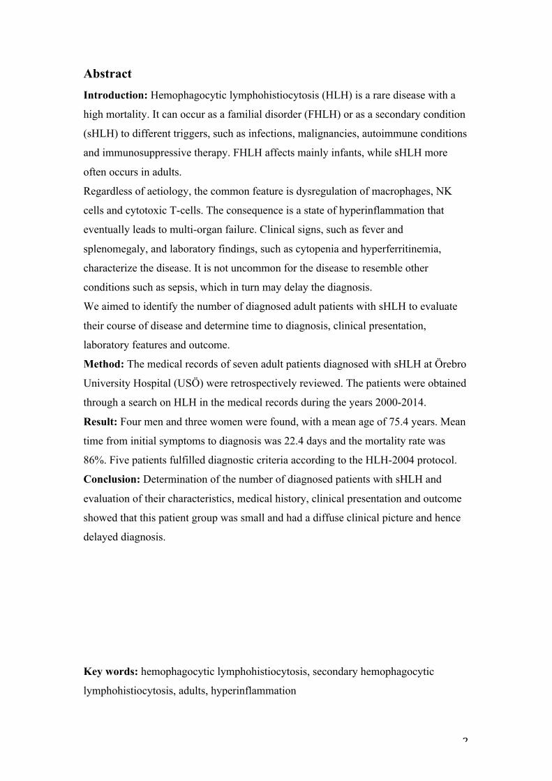
Abstract Introduction: Hemophagocytic lymphohistiocytosis (HLH) is a rare disease with a
high mortality. It can occur as a familial disorder (FHLH) or as a secondary condition
(sHLH) to different triggers, such as infections, malignancies, autoimmune conditions
and immunosuppressive therapy. FHLH affects mainly infants, while sHLH more
often occurs in adults.
Regardless of aetiology, the common feature is dysregulation of macrophages, NK
cells and cytotoxic T-cells. The consequence is a state of hyperinflammation that
eventually leads to multi-organ failure. Clinical signs, such as fever and
splenomegaly, and laboratory findings, such as cytopenia and hyperferritinemia,
characterize the disease. It is not uncommon for the disease to resemble other
conditions such as sepsis, which in turn may delay the diagnosis.
We aimed to identify the number of diagnosed adult patients with sHLH to evaluate
their course of disease and determine time to diagnosis, clinical presentation,
laboratory features and outcome.
Method: The medical records of seven adult patients diagnosed with sHLH at Örebro
University Hospital (USÖ) were retrospectively reviewed. The patients were obtained
through a search on HLH in the medical records during the years 2000-2014.
Result: Four men and three women were found, with a mean age of 75.4 years. Mean
time from initial symptoms to diagnosis was 22.4 days and the mortality rate was
86%. Five patients fulfilled diagnostic criteria according to the HLH-2004 protocol.
Conclusion: Determination of the number of diagnosed patients with sHLH and
evaluation of their characteristics, medical history, clinical presentation and outcome
showed that this patient group was small and had a diffuse clinical picture and hence
delayed diagnosis.
Key words: hemophagocytic lymphohistiocytosis, secondary hemophagocytic
lymphohistiocytosis, adults, hyperinflammation
3
Abbreviations
AAHS – autoimmune-associated hemophagocytic lymphohistiocytosis
AOSD – adult onset Still´s disease
APC – antigen presenting cell
ASCT – allogeneic stem cell transplantation
CMV – cytomegalovirus
CTLs – cytotoxic T-cells
DIC – disseminated vascular coagulation
EBV – Epstein-Barr virus
FHLH – familial hemophagocytic lymphohistiocytosis
HIV – human immunodeficiency virus
HLH – hemophagocytic lymphohistiocytosis
HPS – hemophagocytic syndrome
ICU – intensive care unit
ICD – International Classification of Disease
IFN-γ – interferon-γ
IL-1 – interleukin-1
IL-6 – interleukin-6
IL-10 – interleukin-10
IL-12 – interleukin-12
JIA – juvenile idiopathic arthritis
MAS – macrophage activation syndrome
MAHS – malignancy-associated hemophagocytic syndrome
MHC – major histocompatibility comples
NK cells – natural killer cells
RA – rheumatoid arthritis
sCD25 – soluble CD25
sHLH – secondary hemophagocytic lymphohistiocytosis
SLE – systemic lupus erythematosus
TNF-α – tumour necrosis factor-α
USÖ – Örebro University Hospital
4
Contents
1. Introduction .............................................................................................................. 5
1.1 Pathophysiology ................................................................................................... 5
1.2 Genetic or acquired ............................................................................................. 6
1.3 Epidemiology ....................................................................................................... 8
1.4 Clinical presentation ............................................................................................ 8
1.5 Diagnostic criteria ............................................................................................... 9
1.6 Treatment ........................................................................................................... 10
1.7 Aim ..................................................................................................................... 11
2. Method and Material ............................................................................................. 11
2.1 Ethics .................................................................................................................. 12
3. Results ..................................................................................................................... 12
3.1 Patients .............................................................................................................. 14
4. Discussion ................................................................................................................ 17
4.1 Conclusion ......................................................................................................... 21
5. Acknowledgement .................................................................................................. 21
6. References ............................................................................................................... 22
5
1. Introduction Hemophagocytic lymphohistiocytosis is an uncommon, aggressive and potentially
fatal disease. The name hemophagocytic lymphohistiocytosis (HLH) originally
derived from the Writing Group of the Histiocyte Society in 1987 [1]. HLH consists
of two subtypes, a familial (FHLH) and a secondary (sHLH) variant. FHLH is most
frequently seen in paediatric patients whilst sHLH is the variant of HLH most
common in adults [2].
HLH, in both forms, are sometimes referred to as hemophagocytic syndrome (HPS)
due to the fact that abnormal hemophagocytosis can be a feature in the disease,
although this is not specific for HLH and occurs in other diseases as well [3].
1.1 Pathophysiology
The pathophysiological events in HLH are not certain. It is characterised by a
hyperinflammatory state and a persistent hypercytokinemia, which is thought to be
caused by an uncontrolled stimulation of natural killer cells (NK cells), cytotoxic T-
cells (CTLs) and histiocytes (dendritic cells and macrophages) [4].
The cell types involved in HLH and their normal function include the following:
• Macrophages – Macrophages are derived from circulating monocytes and are
found in essentially all tissues, either as a stationary or circulating cell. They
play an important role in the innate immunity where they phagocytize
microbes and apoptotic cells. They are also active in the adaptive immune
system where they act as professional antigen presenting cells (APC), which
means that they present foreign antigens to lymphocytes. Macrophages also
produce cytokines such as tumour necrosis factor-α (TNF-α), interleukin-1
(IL-1), interleukin-6 (IL-6), interleukin-10 (IL-10) and interleukin-12 (IL-12).
• Natural killer cells (NK cells) - NK cells are a lymphocyte in the innate
immune system that eliminate damaged, stressed or infected host cells such as
macrophages, typically in response to viral infection or malignancy. This is
mediated through perforin-dependent cytotoxicity. NK cells are able to
recognize and eliminate harmful or stressed cells in the absence of antibodies
or expression of major histocompatibility complex (MHC). This is an
important feature because otherwise these cells would never be detected since
no other immune cells possess this characteristic. NK cells can secrete
interferon-γ (IFN-γ) in response to microbes.
6
• Cytotoxic lymphocytes (CTLs) - CTLs are a CD8+ T lymphocyte that has
bound to an antigen/MHC I complex expressed on the surface of an APC and
therefore been activated. This leads to the ability of the CTLs to kill target
cells by the act of the cytotoxins perforin and granzymes. These enter the
cytoplasm and trigger the caspase cascade in the cell, which in turn induces
apoptosis. CTLs may also work through a cell-surface interaction between the
target cell and the CTL leading to apoptosis through the Fas system. CTLs can
produce IL-10 and IFN-γ.
The normal elimination of activated macrophages is mediated by CTLs and NK cells
and induced through perforin-dependent cytotoxicity [5]. In HLH, this process fails
due to impaired cytotoxic function of NK cells and CTLs, resulting in excessive
activation of macrophages. The defective activation of macrophages, NK cells and
CTLs leads to hypersecretion of proinflammatory cytokines (cytokine storm), such as
TNF-α, IFN-γ, IL-1, IL-6, IL-10 and IL-12. Which in turn leads to the exaggerated
inflammatory response.
Patients with HLH may present a single episode with HLH or they can have relapsing
episodes of disease activity. To better understand the pathophysiology in individual
patients different triggers have been described. These are broadly categorized into
those that cause immune activation (e.g. infection) and those that lead to immune
deficiency (e.g. malignancies, immunosuppressive medication, rheumatologic
disorders) [6,7].
In summary, a combination of different factors lead to the clinical and laboratory
features of HLH and contribute to the tissue damage and multi-organ failure
associated with the high morbidity and mortality of the disease.
1.2 Genetic or acquired
Hemophagocytic lymphohistiocytosis is classified into a primary (familial) and
secondary (sometimes referred to as reactive or acquired) form depending on the
underlying cause [3].
The primary form (FHLH) afflicts mainly infants and children, particularly under the
age of two, and is inherited in an autosomal recessive manner (i.e. mutation at both
alleles is required for disease). The majority of identified mutations are in genes
involved in encoding of components in perforin-dependent cytotoxicity:
7
• PRF1 gene – encodes for perforin, which is delivered in cytolytic granules and
creates pores in the membrane of the target cell [8].
• UNC13D gene – encodes for Munc 13-4, a protein that is essential for
cytoloytic granules fusion [9].
• STX11 gene – encodes for Syntaxin 11, which control granule exocytosis
[10].
• STXBP2 gene – encodes for Munc 18-2, which is a protein that binds to
Syntaxin 11 and enhances the release of cytotoxic granules [11].
The secondary form is seen in combination with varying underlying conditions such
as autoimmune-, malignant- or infectious diseases. The aetiology to these patients is
seldom linked to family history or of genetic origin. It usually affects adults, often
with an underlying immunosuppressive disorder. However, some patients acquire this
disease without having a history of immunosuppression [12,13].
Different autoimmune conditions where sHLH can appear are systemic lupus
erythematosus (SLE), rheumatoid arthritis (RA) and adult-onset Still´s disease
(AOSD) and it can be referred to the name autoimmune-associated hemophagocytic
lymphohistiocytosis (AAHS) [14]. A variant of sHLH, with a connection to
autoimmunity, is the macrophage activation syndrome (MAS). MAS is rare but can
be a fatal complication of systemic rheumatic disorders, for example juvenile
idiopathic arthritis (JIA) [15,16].
Malignancy-associated hemophagocytic syndrome (MAHS) is another form of sHLH,
reported in both children and adults. This form is associated with a highly impaired
prognosis and therefore it is essential to diagnose an underlying malignancy as soon
as possible, since early initiation of treatment is of great importance [12,13,17].
Secondary hemophagocytic lymphohistiocytosis can also appear in patients treated
with immunosuppressive therapy [14].
Infections and infectious triggers is an important factor to consider and various
pathogens can be involved. Most commonly are virus infections, bacterial infections
comes second and infections due to fungi are least common [18]. A correlation
between sHLH and Epstein-Barr virus (EBV), herpes virus or cytomegalovirus
(CMV) has been seen, although several other viruses are believed to function as a
trigger, for instance human immunodeficiency virus (HIV). A mapping of the
8
geographical distribution of the disease showed that HIV was more common as
trigger or associated infection in Europe, whilst EBV dominated in USA and Asia [7].
Worth mentioning is that during recent years the number of adult patients with an
identified genetic cause has increased. With improved genetic techniques it has been
shown that patients with sHLH in fact can have an underlying genetic disorder similar
to those seen in FHLH [19].
1.3 Epidemiology
HLH in its secondary form is a rare disease and figuring out the incidence for adults
with the condition is challenging. For the primary form, which occurs more often in
children, there is more data available. According to a study made in Sweden during
the period of 1971-1986, the estimated incidence for children with FHLH was
0.12/100 000 children per year and the prevalence 1 per 50 000 live borns [20].
A recent study, also this performed in Sweden but during the years of 1987-2006,
confirms this though shows that the incidence is slightly elevated (0.12-0.15/100 000
children per year) and the prevalence slightly reduced (0.9 per 50 000 live borns)
[21]. Calculated mean age at diagnosis for sHLH is reported to be almost 50 years [7].
1.4 Clinical presentation
The first clinical signs of sHLH are often nonspecific and do not immediately bring
the disease to mind. Cardinal symptoms such as persistent high fever,
hepatosplenomegaly and cytopenias may raise suspicion of sHLH but it can take
weeks before a diagnosis is set since the initial symptoms are similar to, for instance,
an infection. The intensive care unit (ICU) is a common first contact with the health
care for the patients, especially for those with no previous diagnosis relevant for
sHLH [22,23].
The hyperinflammation and increased hemophagocytosis in the bone marrow seen in
HLH leads to the characteristic cytopenia seen in patients with both newly discovered
disease but also in those with more advanced disease [24]. Another distinct
characteristic is hyperferritinemia and 90% of adult patients with sHLH have a ferritin
concentration that fulfils the criteria according to protocol HLH-2004 (protocol from
the Histiocyte Society with diagnostic criteria) [7,25]. Ferritin levels have an
important role in the differential diagnostic from other systemic processes, such as
sepsis. Since it is both cost-effective and provides helpful clinical aspects, it is an
essential tool in the investigation [26]. The level in common sepsis patients seldom
9
reaches the levels that can be seen in sHLH. How ferritin concentration can reach
levels up to >100 000 mikrogram/L [27] remains unclear, although several hypotheses
might be involved. A combination of release from erythrophagocytosis and cell
damage in spleen and liver, increased secretion from macrophages and hepatocytes
and an impaired clearance due to down regulation of ferritin receptors may contribute
with some explanation [28]. Frequently involved internal organs are the liver and
spleen. Hepatic involvement is manifested as altered liver tests, which is seen in
nearly 60% of the patients [7]. The hepatic dysfunction can cause coagulation
abnormalities and disseminated intravascular coagulopathy (DIC). DIC might
increase the risk of mortality and hence an important parameter to take in
consideration [18,29].
Hypertriglyceridemia and hypofibrinogenemia are two other features that should be
evaluated, although not seen as frequent as hyperferritinemia [7,26]. Elevated levels
of triglycerides are a consequence of the persistent inflammation and liver
dysfunction [30].
Soluble CD25 (sCD25) is a part of the receptor of the interleukin-2 receptor (IL-2
receptor) found on activated T-cells and high concentrations in serum are seen in 79%
of adult patients with sHLH [7]. The activity of NK-cells is regarded as a part of the
investigation for sHLH. Results that indicate low or absent activity in these cells are
an important factor since it correlates with the postulated pathophysiology of the
disease [31].
Neurological symptoms can be part in the progress, although this is not a
characteristic seen in every patient. It is a heterogeneous group of symptoms that
includes meningitis, cerebral haemorrhage, irritability, coma and seizures.
Neuropathology is more studied in paediatric patients [14,20,32]. Cutaneous
involvement may also be a feature in the symptomatology [33].
A progressive multi-organ failure due to the involvement of several internal organ
systems is something that makes almost half of the patients in need of intensive care
[7,34].
1.5 Diagnostic criteria
In 1991, the Histiocyte Society suggested diagnostic criteria for HLH in the protocol
HLH-94 [2] and these criteria were revised in 2004 (HLH-2004) [25]. These criteria
were initially developed for paediatric patients. Although, since more diagnosed
10
patients today are adults this has led to the use of these criteria for setting a diagnosis
also in non-paediatric patients, despite the fact that these have not been validated in
adult populations [13].
A molecular (i.e. gene mutation) diagnosis of HLH or five out of the following eight
criteria must be met to be diagnosed: fever, splenomegaly, cytopenias (affecting ≥2 of
3 cell lineages in peripheral blood), hypertriglyceridemia (fasting triglycerides ≥3.0
mmol/L) and/or hypofibrinogenemia (≤1.5 g/L), hyperferritinemia (≥500
microgram/L), elevated sCD25 (>2400 U/ml), low or absent NK cells activity
(according to local laboratory reference) and hemophagocytosis in bone marrow,
spleen or lymph nodes [25].
A modification of the diagnostic criteria has been proposed, which has a lower
threshold for diagnosis. Diagnosis according to these is set if three of four clinical
findings (fever, splenomegaly, cytopenias, hepatitis) and one of four immunological
markers (hemophagocytosis, increased ferritin, increased sCD25, low/absent NK cell
function) are present [35].
1.6 Treatment
Treatment for hemophagocytic lymphohistiocytosis is divided into initial therapy and
continuation therapy. First, focus is directed at stopping the hemophagocytosis and
the uncontrolled hyperinflammation. After this attention is directed at treating
possible underlying infection or malignancy.
The initial therapy covers the first eight weeks with the goal to get the disease in
remission. A combination of Etoposide (chemotherapy that inhibits monocytes and
macrophages by initiating apoptosis) and Dexamethasone (steroids that acts
immunosuppressive, anti-inflammatory and pro-apoptotic) in high dose is
administered. Cyclosporin A (immunosuppressive agent, reduces T-cell activity) may
be added to this combination. Intrathecal injections of Methotrexate
(chemotherapeutic drug) can be given if signs of progressive neurological symptoms
are present.
Continuation therapy consists of the same drugs, although the goal is to lower the
doses. The major aim with this therapy is to decrease or cause a complete remission in
disease activity. For patients with primary HLH the only cure is an allogeneic stem
cell transplantation (ASCT) and this can also be considered in patients with secondary
HLH. It is most preferable to do when remission is achieved. In secondary
11
hemophagocytic lymphohiostiocytosis it is most important to identify the underlying
cause as this affects both treatment and prognosis [25,36].
1.7 Aim of the study
The aim of this study is to evaluate adult patients diagnosed with sHLH at Örebro
University Hospital by retrospectively reviewing their medical records. The number
of patients, different characteristics, their medical history and outcome was
determined. In addition the initial symptoms, the diagnostic approach, performed
investigations and laboratory results were reviewed. Determination of time between
the first symptoms until diagnosis, concomitant diseases and possible identified
causes was also documented. In addition, we investigated if they fulfilled the
diagnostic criteria and if treatment was initiated.
2. Method and Material To obtain the patients in the study a search on the diagnostic code of hemophagocytic
lymphohistiocytosis in the medical record system at Örebro University Hospital
(USÖ) was performed. The search was made in January 2014, and the determined
time period was January 1, 2000 to November 30, 2014. A second search in
November 2014 supplemented the initial one. The clinics included in the search were
the Department of Infectious diseases, Rheumatology, Oncology, Surgery and
Internal medicine. Diagnosis code according to International Classification of Disease
(ICD-10) for HLH (D76.1) was used. A search for macrophage activation syndrome
(MAS) was also made in the Department of Rheumatology because it is a condition
similar to HLH but more closely connected to juvenile idiopathic arthritis (JIA) and
SLE, conditions treated within the field of rheumatology.
The search resulted in seven patients with the diagnosis HLH, all were included in the
study. Three patients were treated in the Department of Internal medicine, the section
of hematology, and four in both the section of hematology and the Department of
Infectious diseases. No patients with HLH were found in the list of diagnosis from the
other clinics mentioned.
The paediatric clinic was not included in the search since this study focuses on the
secondary form of the disease and its progression in adults and not on the primary
form, which occurs more often in children.
12
The medical records were analysed retrospectively and clinical data such as age, sex,
initial symptoms, time of onset, time to diagnosis, possible identified cause and
concomitant diseases, met diagnostic criteria, results of bone marrow samples,
treatment and outcome for the patient were documented. The obtained data was
compared to the diagnostic criteria according to HLH-2004. In the event of an
autopsy the pathologist report were reviewed in order to examine if there were any
findings that would strengthen the diagnosis even more, for example splenomegaly.
Positive findings in the bone marrow were sometimes mentioned in the clinicians text
of the medical chart but not in the written pathology report of the bone marrow
sample. In terms of lab values before treatment the highest or lowest ones were
selected to mirror the grade of disease. Mean values were calculated for age at
diagnosis and time from initial symptoms to diagnosis. The mortality rate was also
determined.
2.1 Ethics
A request for viewing the medical records was approved by the head of the
Department of Internal medicine and a registration number was given that was
documented in the files read. All collected data was anonymized and was only
handled by the author and supervisor in a confidential manner. Only the data needed
for the study was collected and this did not affect the patients care in any way. All but
one of the patients was deceased, but the integrity of their medical records must still
be preserved and protected.
The patient group consisted of a small number of participants, since the diagnosis is
unusual. This increases the risk of recognition by staff or relatives, despite the de-
identification of all patient material.
3. Results In this study the medical records of seven patients diagnosed with secondary
hemophagocytic lymphohistiocytosis at USÖ were reviewed. Three were women and
four were men. The mean age at diagnosis was 75.4 years (range 68 to 84 years)
(table 1). The diagnosis was based on the criteria according to HLH-2004 and all
patients but two (patient three and seven) fulfilled at least five out of eight criteria,
which is the requirement for diagnosis (table 3).
13
The primary symptoms of all patients were fever and/or cytopenia in at least two out
of three cell lines in peripheral blood (erythrocytes, platelets, leukocytes) (table 3).
The mean duration from onset of symptoms to diagnosis of sHLH was 22.4 days
(range 6-49 days) (table 1).
Possible causes and concomitant diseases were identifiable in the records as far as this
was documented, such as infections, malignancies, treatment for other diseases and
autoimmune conditions among others (table 2).
Two patients did not receive any treatment for their sHLH (table 1). Remaining
patients started treatment according to protocol HLH-2004, with the ambition that it
could be completed and get the disease in remission.
For one out of seven patients the disease is in remission, patient number four being
the one still alive and this making the mortality rate for the group 86% (table 1).
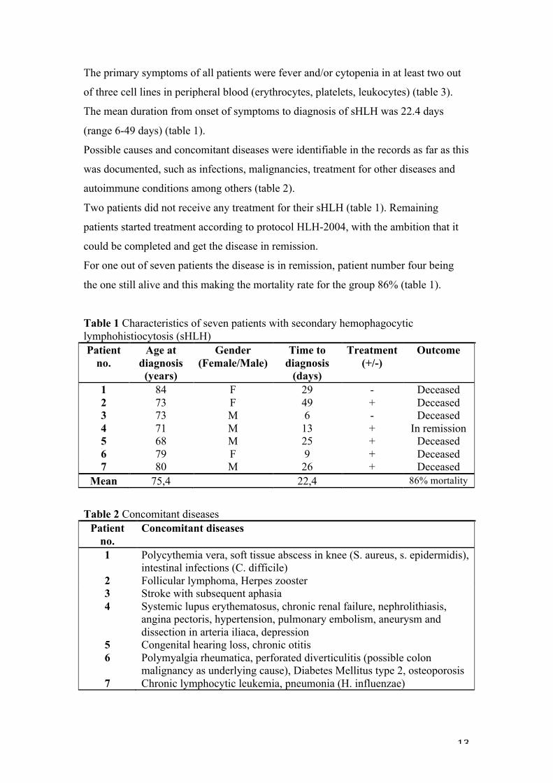
Table 1 Characteristics of seven patients with secondary hemophagocytic lymphohistiocytosis (sHLH) Patient
no. Age at
diagnosis (years)
Gender (Female/Male)
Time to diagnosis
(days)
Treatment (+/-)
Outcome
1 84 F 29 - Deceased 2 73 F 49 + Deceased 3 73 M 6 - Deceased 4 71 M 13 + In remission 5 68 M 25 + Deceased 6 79 F 9 + Deceased 7 80 M 26 + Deceased
Mean 75,4 22,4 86% mortality
Table 2 Concomitant diseases Patient
no. Concomitant diseases
1 Polycythemia vera, soft tissue abscess in knee (S. aureus, s. epidermidis), intestinal infections (C. difficile)
2 Follicular lymphoma, Herpes zooster 3 Stroke with subsequent aphasia 4 Systemic lupus erythematosus, chronic renal failure, nephrolithiasis,
angina pectoris, hypertension, pulmonary embolism, aneurysm and dissection in arteria iliaca, depression
5 Congenital hearing loss, chronic otitis 6 Polymyalgia rheumatica, perforated diverticulitis (possible colon
malignancy as underlying cause), Diabetes Mellitus type 2, osteoporosis 7 Chronic lymphocytic leukemia, pneumonia (H. influenzae)
14
Table 3 Fulfilled diagnostic criteria of the seven patients according to the HLH-2004 protocol [25] Patient no.
Diagnostic criteria 1 2 3 4 5 6 7 Fever + + + + + + + Splenomegaly - nd - - + - - Cytopenia (affected cell lineages)
+ (3/3)
+ (3/3)
+ (3/3)
+ (3/3)
+ (3/3)
+ (2/3)
+ (3/3)
Hypertriglyceridemia/ hypofibrinogenemia
+ - + + + - -
Hemophagocytosis (in bone marrow)
+ + - + - + +
NK cells activity nd nd nd nd - nd nd Hyperferritinemia (maximum, microg/L)
+ (3692)
+ (7262)
+ (1406)
+ (>40000)
+ (4079)
+ (642)
+ (32374)
Soluble CD25 (>2400 U/ml)
nd + (21793)
nd nd + (13008)
+ (5431)
nd
Abbreviations: nd, not done
3.1 Patients
Case 1
This 84-year-old woman had fever and therefore came to the emergency room at
USÖ. She was admitted to the infectious diseases ward and was there diagnosed with
a soft tissue abscess in one of her knees and was treated with antibiotics. Ten days
later she had developed a cytopenia and still had a high fever. Due to this the
suspicion of HLH was raised and she was transferred to the hematology ward for
further examination and evaluation. She was diagnosed with HLH after six days (for a
total of 29 days from first symptoms) and was estimated to be too fragile to be able to
begin treatment. The patient passed away on that same day.
Case 2
This 73-year-old woman had been treated with chemotherapy several times the last
year against follicular lymphoma and was according to the oncologist in remission.
She presented herself with initial symptoms such as fever, tiredness and anaemia and
was admitted to the infectious diseases ward. The patient developed cytopenia which
led to further investigation with a bone marrow sample. The bone marrow sample
showed dysplasia and inflammatory granulomas and she was admitted to the
hematology ward. Further examinations was made and she was diagnosed with HLH
15
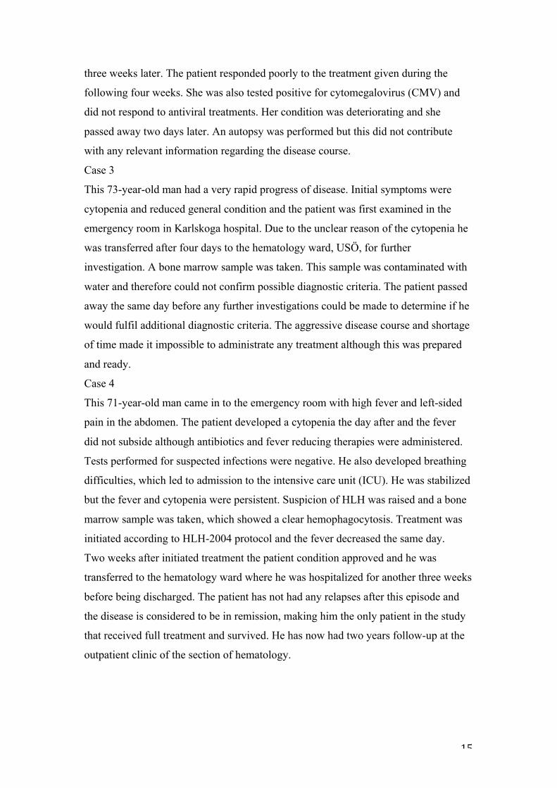
three weeks later. The patient responded poorly to the treatment given during the
following four weeks. She was also tested positive for cytomegalovirus (CMV) and
did not respond to antiviral treatments. Her condition was deteriorating and she
passed away two days later. An autopsy was performed but this did not contribute
with any relevant information regarding the disease course.
Case 3
This 73-year-old man had a very rapid progress of disease. Initial symptoms were
cytopenia and reduced general condition and the patient was first examined in the
emergency room in Karlskoga hospital. Due to the unclear reason of the cytopenia he
was transferred after four days to the hematology ward, USÖ, for further
investigation. A bone marrow sample was taken. This sample was contaminated with
water and therefore could not confirm possible diagnostic criteria. The patient passed
away the same day before any further investigations could be made to determine if he
would fulfil additional diagnostic criteria. The aggressive disease course and shortage
of time made it impossible to administrate any treatment although this was prepared
and ready.
Case 4
This 71-year-old man came in to the emergency room with high fever and left-sided
pain in the abdomen. The patient developed a cytopenia the day after and the fever
did not subside although antibiotics and fever reducing therapies were administered.
Tests performed for suspected infections were negative. He also developed breathing
difficulties, which led to admission to the intensive care unit (ICU). He was stabilized
but the fever and cytopenia were persistent. Suspicion of HLH was raised and a bone
marrow sample was taken, which showed a clear hemophagocytosis. Treatment was
initiated according to HLH-2004 protocol and the fever decreased the same day.
Two weeks after initiated treatment the patient condition approved and he was
transferred to the hematology ward where he was hospitalized for another three weeks
before being discharged. The patient has not had any relapses after this episode and
the disease is considered to be in remission, making him the only patient in the study
that received full treatment and survived. He has now had two years follow-up at the
outpatient clinic of the section of hematology.
16
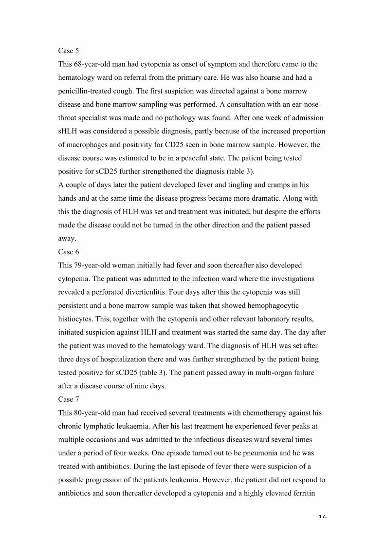
Case 5
This 68-year-old man had cytopenia as onset of symptom and therefore came to the
hematology ward on referral from the primary care. He was also hoarse and had a
penicillin-treated cough. The first suspicion was directed against a bone marrow
disease and bone marrow sampling was performed. A consultation with an ear-nose-
throat specialist was made and no pathology was found. After one week of admission
sHLH was considered a possible diagnosis, partly because of the increased proportion
of macrophages and positivity for CD25 seen in bone marrow sample. However, the
disease course was estimated to be in a peaceful state. The patient being tested
positive for sCD25 further strengthened the diagnosis (table 3).
A couple of days later the patient developed fever and tingling and cramps in his
hands and at the same time the disease progress became more dramatic. Along with
this the diagnosis of HLH was set and treatment was initiated, but despite the efforts
made the disease could not be turned in the other direction and the patient passed
away.
Case 6
This 79-year-old woman initially had fever and soon thereafter also developed
cytopenia. The patient was admitted to the infection ward where the investigations
revealed a perforated diverticulitis. Four days after this the cytopenia was still
persistent and a bone marrow sample was taken that showed hemophagocytic
histiocytes. This, together with the cytopenia and other relevant laboratory results,
initiated suspicion against HLH and treatment was started the same day. The day after
the patient was moved to the hematology ward. The diagnosis of HLH was set after
three days of hospitalization there and was further strengthened by the patient being
tested positive for sCD25 (table 3). The patient passed away in multi-organ failure
after a disease course of nine days.
Case 7
This 80-year-old man had received several treatments with chemotherapy against his
chronic lymphatic leukaemia. After his last treatment he experienced fever peaks at
multiple occasions and was admitted to the infectious diseases ward several times
under a period of four weeks. One episode turned out to be pneumonia and he was
treated with antibiotics. During the last episode of fever there were suspicion of a
possible progression of the patients leukemia. However, the patient did not respond to
antibiotics and soon thereafter developed a cytopenia and a highly elevated ferritin
17
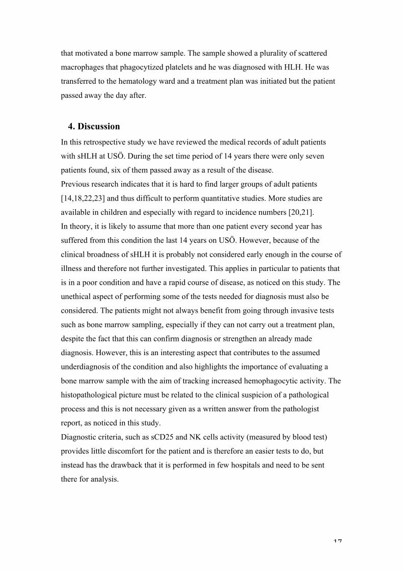
that motivated a bone marrow sample. The sample showed a plurality of scattered
macrophages that phagocytized platelets and he was diagnosed with HLH. He was
transferred to the hematology ward and a treatment plan was initiated but the patient
passed away the day after.
4. Discussion In this retrospective study we have reviewed the medical records of adult patients
with sHLH at USÖ. During the set time period of 14 years there were only seven
patients found, six of them passed away as a result of the disease.
Previous research indicates that it is hard to find larger groups of adult patients
[14,18,22,23] and thus difficult to perform quantitative studies. More studies are
available in children and especially with regard to incidence numbers [20,21].
In theory, it is likely to assume that more than one patient every second year has
suffered from this condition the last 14 years on USÖ. However, because of the
clinical broadness of sHLH it is probably not considered early enough in the course of
illness and therefore not further investigated. This applies in particular to patients that
is in a poor condition and have a rapid course of disease, as noticed on this study. The
unethical aspect of performing some of the tests needed for diagnosis must also be
considered. The patients might not always benefit from going through invasive tests
such as bone marrow sampling, especially if they can not carry out a treatment plan,
despite the fact that this can confirm diagnosis or strengthen an already made
diagnosis. However, this is an interesting aspect that contributes to the assumed
underdiagnosis of the condition and also highlights the importance of evaluating a
bone marrow sample with the aim of tracking increased hemophagocytic activity. The
histopathological picture must be related to the clinical suspicion of a pathological
process and this is not necessary given as a written answer from the pathologist
report, as noticed in this study.
Diagnostic criteria, such as sCD25 and NK cells activity (measured by blood test)
provides little discomfort for the patient and is therefore an easier tests to do, but
instead has the drawback that it is performed in few hospitals and need to be sent
there for analysis.
18
Initial symptoms such as fever and/or cytopenia were seen in all patients in this study
(table 3). Mean time from initial symptoms to diagnosis is 22.4 days (table 1). A
possible reason for this relatively long time to diagnosis is the combination of these
nonspecific initial symptoms and the variety and complexity in the clinical
presentation. Four patients in this study was first admitted to the infectious diseases
ward due to signs of infection, which later on proved to be difficult to treat. The time
to diagnoses in three of these patients (patients one, two and seven) were the longest
in the study and lasted between 26-49 days. Interestingly, the fourth patient (patient
number six) in this group was also admitted to the infectious diseases ward but only
had nine days from initial symptoms to diagnosis. What distinguished her from the
others was that she showed an earlier sign of severe cytopenia that probably led to a
more timely diagnosis. The other three patients all had haematological diseases in
their medical history which might have confused and contributed to the prolonged
time to their diagnosis. This applies especially to patient number two who had the
longest time of all (49 days) in combination with follicular lymphoma.
Additional aspects is that patient number five only had one day shorter to diagnosis
than patient number seven (25 respectively 26 days), but this diagnostic delay was
due to the initial assessment of a peaceful disease course and not an initial admission
to the infectious diseases ward.
Another reason for delayed diagnosis is that the progression of sHLH can be very fast
and might mimic other causes, such as sepsis [7,23,29]. This complicates the clinical
evaluation and meanwhile the patients are deteriorating. In addition are the majority
of patients older (table 1) and have multiple concomitant diseases that require
multidisciplinary efforts which makes it challenging just to consider the diagnosis. As
a consequence, initiation of treatment might be delayed. However, as seen in this
study, several patients might also be in such a bad condition to begin with that
treatment for sHLH is not a possible option.
Studies that indicate the difficulty in diagnosing HLH [7,13,37] proves that this is not
an uncommon situation for the patients and indicates a complex and unspecific
symptomology and little experience of this condition to the overall clinician in many
fields.
Defined criteria are an essential tool in setting the diagnosis and further reason for
delayed diagnosis is that patients can fulfil fewer criteria or develop criteria later in
19
the disease course [37] but still have a clinically active disease. Examinations done at
different times can give different results due to this dynamic disease course, which
must be considered during the investigation.
In this study patient number three and seven only fulfilled four out of eight criteria
(five is needed for diagnosis) (table 3), but all diagnostic tests was not done on these
patients (tests for sCD25 and NK cells activity were not performed on either patient).
Moreover, one of the patients lacking one criterion had for example a bone marrow
sample made but it was not of any diagnostic value because of water contamination.
Both patients had been given the diagnosis in the medical records anyway because of
strong suspicion, hence included in this study. However, these two patients would not
fulfil a diagnosis according to the proposed modified diagnostic criteria either, despite
these having a lower limit for diagnosis [35].
The diagnostic criteria that apply in the clinics today were originally developed for
children with FHLH but these are also used for adult cases due to lack of criteria
developed for sHLH [7]. Benefits with the modified criteria are their lowering of the
threshold for diagnosis and the consideration of more aspects. This is advantageous
since individual patients can differ from another and present a broad clinical picture
that is hard to assess. In order to identify patients with sHLH earlier in their disease
course a combination of the HLH-2004 criteria and the modified criteria could be
taken in consideration.
Another suggestion for earlier diagnosis is access to a wider diagnostic kit early in the
course of disease to be used on suspected cases. Markers such as ferritin, triglycerides
and fibrinogen are easily measured and should be more routinely performed in
clinical cases showing symptoms that could be early signs of this hyperinflammatory
state. These markers need to be followed to trace the dynamic course during the
disease. This applies in particular to ferritin [37], which also is a marker in other
inflammatory states, although it seldom reaches the high levels seen in HLH. If the
ferritin level deviates and shows these unusual high levels, the diagnosis can be
targeted for HLH at an earlier stage. Other reasons of high levels of ferritin must
obviously be considered, e.g. multiple blood transfusions and hemochromatosis.
It is not uncommon for patients to seek initial care in the emergency room and being
admitted to the ICU [23,34]. An extension of this study would be to do measurement
of ferritin on suspected cases directly in patients that comes to the emergency room or
20
the ICU. This can lead to the identification of more patients that later on would be
followed-up to see if there was any suspicion against HLH or if a diagnosis was
made.
When considering possible causes of sHLH we discovered that several of the reported
concomitant diseases for each patient (table 2) could play a part in the aetiology of the
patients disease. Six out of seven patients had occurrence of infections, malignancies,
autoimmune conditions or a combination of several of them. Although, none of the
patients had a defined cause documented in the medical records. When studying the
medical records retrospectively one could jump to conclusion that a certain
concomitant disease would have been the cause but the pathogenesis is a complex
combination of factors that is difficult to examine due to the lack of larger patient
groups. However, research made in recent years show that genetic defects also occurs
in adults diagnosed with sHLH [19]. This is not something that is targeted in the
investigations today. Although, we can speculate that the diagnostic tools in the future
of these patients would include genetic testing and that it could play a role in the
diagnostic information in the group that is now called secondary HLH.
To summarize, the retrospective design of this study is a limiting factor. As is the low
number of patients, which is also the reason why there is so few statistics presented.
All patients were collected in one hospital and only from the clinics that was most
likely to have treated patients with the disease. However, all patients that were found
were included in the study and an additional search for MAS was made in the Dept. of
Rheumatology. To be able to expand the patient group a search in other hospitals can
be done to increase the number of cases to evaluate.
The determined time period for the search was long and therefore brings some
strength to this study. Although the finding of so few patients during these 14 years
indicates that it would take a long time to collect material for a prospective study.
Worth mentioning is that the medical records was not computerised before the year of
2000 and this was therefore a limitation when determining the time period for this
study.
21
4.1 Conclusion
In this retrospective study we have determined the number of patients diagnosed with
sHLH at USÖ and evaluated their characteristics, medical history, clinical
presentation and outcome. From this we conclude that sHLH is an under diagnosed
disease for reasons such as diffuse clinical picture, similarity to other conditions and
hence delayed diagnosis. The cause is multifactorial and the optimal would be to as
soon as possible consider sHLH as a potential differential diagnosis in suspected
cases. To do so a combination of currently used criteria and modified criteria could be
used to broaden the possibility of diagnosis. Finally, this kind of mapping on patients
with sHLH and their disease course is important through a quality perspective. It
raises the awareness among clinicians in all areas where patients that have an
increased risk for developing sHLH are treated.
5. Acknowledgement I would like to thank my supervisor Magdalena Kättström for always being available
and helpful when needed. You have given me great support throughout this process,
both in medicine but also mentally. Thank you for all of that and for always meeting
me with a smile!
22
6. References
1. Histiocytosis syndromes in children. Writing Group of the Histiocyte Society. Lancet 1987 Jan 24;1(8526):208-209.
2. Henter JI, Elinder G, Ost A. Diagnostic guidelines for hemophagocytic lymphohistiocytosis. The FHL Study Group of the Histiocyte Society. Semin Oncol 1991 Feb;18(1):29-33.
3. Favara BE, Feller AC, Pauli M, Jaffe ES, Weiss LM, Arico M, et al. Contemporary classification of histiocytic disorders. The WHO Committee On Histiocytic/Reticulum Cell Proliferations. Reclassification Working Group of the Histiocyte Society. Med Pediatr Oncol 1997 Sep;29(3):157-166.
4. Filipovich AH. Hemophagocytic lymphohistiocytosis and other hemophagocytic disorders. Immunol Allergy Clin North Am 2008 May;28(2):293-313, viii.
5. Abbas A, Lichtman A, Pillai S. Cellular and Molecular Immunology. 7th ed. Philadelphia: Saunders, Elsevier; 2012.
6. Filipovich A, McClain K, Grom A. Histiocytic disorders: recent insights into pathophysiology and practical guidelines. Biol Blood Marrow Transplant 2010 Jan;16(1 Suppl):S82-9.
7. Ramos-Casals M, Brito-Zeron P, Lopez-Guillermo A, Khamashta MA, Bosch X. Adult haemophagocytic syndrome. Lancet 2014 Apr 26;383(9927):1503-1516.
8. Goransdotter Ericson K, Fadeel B, Nilsson-Ardnor S, Soderhall C, Samuelsson A, Janka G, et al. Spectrum of perforin gene mutations in familial hemophagocytic lymphohistiocytosis. Am J Hum Genet 2001 Mar;68(3):590-597.
9. Feldmann J, Callebaut I, Raposo G, Certain S, Bacq D, Dumont C, et al. Munc13-4 is essential for cytolytic granules fusion and is mutated in a form of familial hemophagocytic lymphohistiocytosis (FHL3). Cell 2003 Nov 14;115(4):461-473.
10. Rudd E, Goransdotter Ericson K, Zheng C, Uysal Z, Ozkan A, Gurgey A, et al. Spectrum and clinical implications of syntaxin 11 gene mutations in familial haemophagocytic lymphohistiocytosis: association with disease-free remissions and haematopoietic malignancies. J Med Genet 2006 Apr;43(4):e14.
11. Cote M, Menager MM, Burgess A, Mahlaoui N, Picard C, Schaffner C, et al. Munc18-2 deficiency causes familial hemophagocytic lymphohistiocytosis type 5 and impairs cytotoxic granule exocytosis in patient NK cells. J Clin Invest 2009 Dec;119(12):3765-3773.
12. Janka G, Imashuku S, Elinder G, Schneider M, Henter JI. Infection- and malignancy-associated hemophagocytic syndromes. Secondary hemophagocytic lymphohistiocytosis. Hematol Oncol Clin North Am 1998 Apr;12(2):435-444.
23
13. Riviere S, Galicier L, Coppo P, Marzac C, Aumont C, Lambotte O, et al. Reactive hemophagocytic syndrome in adults: a retrospective analysis of 162 patients. Am J Med 2014 Nov;127(11):1118-1125.
14. Fukaya S, Yasuda S, Hashimoto T, Oku K, Kataoka H, Horita T, et al. Clinical features of haemophagocytic syndrome in patients with systemic autoimmune diseases: analysis of 30 cases. Rheumatology (Oxford) 2008 Nov;47(11):1686-1691.
15. Sawhney S, Woo P, Murray KJ. Macrophage activation syndrome: a potentially fatal complication of rheumatic disorders. Arch Dis Child 2001 Nov;85(5):421-426.
16. Hadchouel M, Prieur AM, Griscelli C. Acute hemorrhagic, hepatic, and neurologic manifestations in juvenile rheumatoid arthritis: possible relationship to drugs or infection. J Pediatr 1985 Apr;106(4):561-566.
17. Machaczka M, Vaktnas J, Klimkowska M, Hagglund H. Malignancy-associated hemophagocytic lymphohistiocytosis in adults: a retrospective population-based analysis from a single center. Leuk Lymphoma 2011 Apr;52(4):613-619.
18. Tseng YT, Sheng WH, Lin BH, Lin CW, Wang JT, Chen YC, et al. Causes, clinical symptoms, and outcomes of infectious diseases associated with hemophagocytic lymphohistiocytosis in Taiwanese adults. J Microbiol Immunol Infect 2011 Jun;44(3):191-197.
19. Zhang K, Jordan MB, Marsh RA, Johnson JA, Kissell D, Meller J, et al. Hypomorphic mutations in PRF1, MUNC13-4, and STXBP2 are associated with adult-onset familial HLH. Blood 2011 Nov 24;118(22):5794-5798.
20. Henter JI, Elinder G, Soder O, Ost A. Incidence in Sweden and clinical features of familial hemophagocytic lymphohistiocytosis. Acta Paediatr Scand 1991 Apr;80(4):428-435.
21. Meeths M, Horne A, Sabel M, Bryceson YT, Henter JI. Incidence and clinical presentation of primary hemophagocytic lymphohistiocytosis in Sweden. Pediatr Blood Cancer 2014 Nov 8.
22. Takahashi N, Chubachi A, Kume M, Hatano Y, Komatsuda A, Kawabata Y, et al. A clinical analysis of 52 adult patients with hemophagocytic syndrome: the prognostic significance of the underlying diseases. Int J Hematol 2001 Aug;74(2):209-213.
23. Padhi S, Varghese RG, Ramdas A, Phansalkar MD, Sarangi R. Hemophagocytic lymphohistiocytosis: critical reappraisal of a potentially under-recognized condition. Front Med 2013 Dec;7(4):492-498.
24. Zoller EE, Lykens JE, Terrell CE, Aliberti J, Filipovich AH, Henson PM, et al. Hemophagocytosis causes a consumptive anemia of inflammation. J Exp Med 2011 Jun 6;208(6):1203-1214.
24
25. Henter JI, Horne A, Arico M, Egeler RM, Filipovich AH, Imashuku S, et al. HLH-2004: Diagnostic and therapeutic guidelines for hemophagocytic lymphohistiocytosis. Pediatr Blood Cancer 2007 Feb;48(2):124-131.
26. Switala JR, Hendricks M, Davidson A. Serum ferritin is a cost-effective laboratory marker for hemophagocytic lymphohistiocytosis in the developing world. J Pediatr Hematol Oncol 2012 Apr;34(3):e89-92.
27. Emmenegger U, Frey U, Reimers A, Fux C, Semela D, Cottagnoud P, et al. Hyperferritinemia as indicator for intravenous immunoglobulin treatment in reactive macrophage activation syndromes. Am J Hematol 2001 Sep;68(1):4-10.
28. Knovich MA, Storey JA, Coffman LG, Torti SV, Torti FM. Ferritin for the clinician. Blood Rev 2009 May;23(3):95-104.
29. Stephan F, Thioliere B, Verdy E, Tulliez M. Role of hemophagocytic histiocytosis in the etiology of thrombocytopenia in patients with sepsis syndrome or septic shock. Clin Infect Dis 1997 Nov;25(5):1159-1164.
30. Henter JI, Carlson LA, Soder O, Nilsson-Ehle P, Elinder G. Lipoprotein alterations and plasma lipoprotein lipase reduction in familial hemophagocytic lymphohistiocytosis. Acta Paediatr Scand 1991 Jun-Jul;80(6-7):675-681.
31. Chung HJ, Park CJ, Lim JH, Jang S, Chi HS, Im HJ, et al. Establishment of a reference interval for natural killer cell activity through flow cytometry and its clinical application in the diagnosis of hemophagocytic lymphohistiocytosis. Int J Lab Hematol 2010 Apr;32(2):239-247.
32. Horne A, Trottestam H, Arico M, Egeler RM, Filipovich AH, Gadner H, et al. Frequency and spectrum of central nervous system involvement in 193 children with haemophagocytic lymphohistiocytosis. Br J Haematol 2008 Feb;140(3):327-335.
33. Fardet L, Galicier L, Vignon-Pennamen MD, Regnier S, Noguera ME, de Labarthe A, et al. Frequency, clinical features and prognosis of cutaneous manifestations in adult patients with reactive haemophagocytic syndrome. Br J Dermatol 2010 Mar;162(3):547-553.
34. Buyse S, Teixeira L, Galicier L, Mariotte E, Lemiale V, Seguin A, et al. Critical care management of patients with hemophagocytic lymphohistiocytosis. Intensive Care Med 2010 Oct;36(10):1695-1702.
35. Filipovich AH. Hemophagocytic lymphohistiocytosis (HLH) and related disorders. Hematology Am Soc Hematol Educ Program 2009:127-131.
36. Henter JI, Tondini C, Pritchard J. Histiocyte disorders. Crit Rev Oncol Hematol 2004 May;50(2):157-174.
25
37. Okabe T, Shah G, Mendoza V, Hirani A, Baram M, Marik P. What intensivists need to know about hemophagocytic syndrome: an underrecognized cause of death in adult intensive care units. J Intensive Care Med 2012 Feb;27(1):58-64.
![Kandidatuppsats - Marc Albrecht & Sandra Lindh[2]hh.diva-portal.org/smash/get/diva2:1393058/FULLTEXT02.pdf · Kandidatuppsats Bygg och fastighetsekonomiprogrammet 180hp Franchisetagarens](https://static.fdocuments.net/doc/165x107/5ecdd57b33857467c61f69bc/kandidatuppsats-marc-albrecht-sandra-lindh2hhdiva-1393058fulltext02pdf.jpg)