JUN 2 2 1987
241
X-RAY PHOTOELECTRON SPECTROSCOPY OF SILICATE GLASSES by G. WILLIAM TASKER B.S., Ceramic Science, Pennsylvania State University 1972 SUBMITTED IN PARTIAL FULFILLMENT OF THE REQUIREMENTS FOR THE DEGREE OF DOCTOR OF PHILOSOPHY IN CERAMICS at the Massachusetts Institute of Technology June 1987 Q Massachusetts Institute of Technology 1987 Signature of Author Department of MateriaTls Science and Engineering May 1 , 1987 Certified by D. K. Uhlmann Thesis Supervisor Accepted by S. M. Allen Chairman, Departmental Committee on Graduate Students MA$S-CIIUSETT$ WSTIT E JUN 2 2 1987 LBRAR:ES
Transcript of JUN 2 2 1987

X-RAY PHOTOELECTRON SPECTROSCOPY
OF SILICATE GLASSES
by
G. WILLIAM TASKER
B.S., Ceramic Science, Pennsylvania State University
1972
SUBMITTED IN PARTIAL FULFILLMENT
OF THE REQUIREMENTS FOR THE
DEGREE OF
DOCTOR OF PHILOSOPHY
IN CERAMICS
at the
Massachusetts Institute of Technology
June 1987
Q Massachusetts Institute of Technology 1987
Signature of Author
Department of MateriaTls Science and EngineeringMay 1 , 1987
Certified byD. K. Uhlmann
Thesis Supervisor
Accepted byS. M. Allen
Chairman, Departmental Committee on Graduate Students
MA$S-CIIUSETT$ WSTIT E
JUN 2 2 1987
LBRAR:ES

X-RAY PHOTOELECTRON SPECTROSCOPY
OF SILICATE GLASSES
by
G. WILLIAM TASKER
Submitted to the Department of Materials Science and Engineering
on May 1 , 1987 in partial fulfillment of the requirements for
the Degree of Doctor of Philosophy in Ceramics.
ABSTRACT
X-ray photoelectron spectroscopy (XPS) is a powerful techniquecapable of providing unique information about the structural chemistry
of glasses. However, inappropriate procedures for the preparation of
sample surfaces or the lack of reliable methods to overcome electro-
static charging effects during analysis of dielectric materials can
cause spurious features to be introduced or intrinsic features to be
suppressed in photoelectron spectra which are nominally indicative
of bulk phenomena. An adaptation of a biased metal-dot (BMD) energy
reference for insulators in XPS to the analysis of fracture surfaces
prepared in ultra-high vacuum is described that largely eliminates
these difficulties. Based on a study of the charging behavior of
SiO glass under conditions of positive and negative bias, a referenceof he binding energy scale for an insulator to its surface Fermi
level is proposed and shown to be consistent with Schottky barrier
formation at metal-insulator interfaces.
The utility of this analytical procedure is demonstrated by com-
parisons of core and valence level spectra acquired from surfaces of
fire-polished Si0 2 glass and vacuum-fractured SiO 2 glass and a-quartz.
The similarity of core level spectra obtained from fire-polished and
vacuum-fractured surfaces of vitreous SiO2 supports a view that extensive
surface reconstruction of siloxane bonds occurs at fracture surfaces.
Since no significant differences in core level spectra are found between
crystalline and vitreous samples of SiO , it is also suggested that the
covalent-ionic nature of the Si-O bond goes not change appreciably with
the Si-O-Si bridging angle.
A structural investigation by XPS of R2 O-SiO glasses and Na 20-
Al 0 -SiO2 glasses was accomplished by analysis o core level spectra
which were acquired from vacuum-fracturedsurfaces and corrected for
charging effects by the BMD method. The 0 ls spectrum for Si0 2 glass
does not indicate the existence of doubly-bonded oxygens or strained
siloxane bonds in detectable concentrations. This result calls into

question the assertions of recent crystalline cluster models for glass
structure. In accord with earlier XPS work, an analysis of 0 ls spectra
for R O-SiO2 glasses agrees with conventional theory for the accommodation
of aliali atoms in a vitreous silicon-oxygen network at mole fractions of
alkali oxide XR<0.30 but departs negatively from this theory for XR>0.30.
The merits of various explanations for this discrepancy are critically
appraised. Trends in the binding energies and widths of various core
level spectra for Na20-SiO2 glasses support a contention that while single-
phase R20-SiO2 glasses do not exhibit any long-range order, chemical
details of the short-range atomic structure bear a close resemblance
to those in R20-SiO 2 crystals.
The proportions of various bridging and nonbridging components of
0 ls spectra for three series of Na20-Al 203-SiO glasses are shown to be..2 .2 3-SO lse r hw ob
directly related to glass composition in a predictable and rather general
way within two adjacent compositional regions: 0<Al/Na<1.0 and
1.0<Al/Na<1.5. In contrast to prior XPS studies, an analysis of O ls
spectra for glasses with 0<Al/Na<1.0 gives strong support to the classical
model for incorporation of aluminum atoms in tetrahedral sites as network
formers. An analysis of 0 ls spectra for glasses with 1.O<Al/Na<1.5
is consistent with Lacy's tricluster model for the incorporation of
aluminum atoms in excess of sodium atoms in tetrahedral sites as network
formers. Trends in the binding energies and widths of various core level
spectra for Na20-Al20 -SiO 2 glasses lend collateral support to the classical
model for Al/Na<l.0 and the tricluster model for Al/Na>1.0.
Thesis Supervisor: Donald R. Uhlmann
Title: Cabot Professor of Materials

TABLE OF CONTENTS
Chapter Page
TITLE PAGE 1
ABSTRACT 2
TABLE OF CONTENTS 4
LIST OF FIGURES 8
LIST OF TABLES 11
ACKNOWLEDGEMENTS 12
FOREWORD 14
1 EXPERIMENTAL METHODS FOR XPS OF INSULATORS 15
1.1. Introduction 15
1.2. Literature Survey 18
1.2.1. Charging Correction of Insulators 18
1.2.2. Preparation of Insulator Surfaces 30
1.3. Experimental Procedures 39
1.3.1. Description of Spectrometer, Data 39
Acquisition, and Data Refinement
1.3.2. Spectrometer Calibration 42
1.3.3. Biased Metal-Dot (BMD) Method 42
1.3.4. Adaptation of BMD Method to 46
Vacuum-fractured Surfaces
1.4. Results 47
1.4.1. BMD Studies with Gold and 47
Platinum Calibrants
1.4.2. Core and Valence Level Spectra 49
for Si0 2

5
TABLE OF CONTENTS (Cont'd.)
Chapter Page
1.5. Discussion 56
1.5.1. Metal-Insulator Interfaces 56
1.5.2. A Model for the BMD Method 62
1.5.3. Photoemission Studies of Si0 2 66Surfaces and Bulk
1.6. Summary 73
References 75
2 STRUCTURE OF ALKALI ALUMINOSILICATE GLASSES: 85
XPS OF ALKALI SILICATE GLASSES
2.1. Introduction 85
2.2. Literature Survey 90
2.2.1. Structural Models for SiO 2 Glass 90
2.2.2. Structural Models for R20 O-SiO 2 94Glasses
2.2.3. Photoemission Studies of SiO 2 108and R20 O-SiO 2 Glasses
2.3. Experimental Procedures 114
2.3.1. Sample Preparation 114
2.3.2. Photoemission Measurements 114
2.4. Results and Analysis 117
2.4.1. Spectral Artifacts 117
2.4.2. Core Level Spectra for SiO 2 122
and R20-Si0 2 Glasses

TABLE OF CONTENTS (Cont'd.)
Chapter Page
2.5. Discussion 129
2.5.1. SiO02 Glass 129
2.5.2. R20-SiO2 Glasses 131
2.6. Summary 139
References 141
3 STRUCTURE OF ALKALI ALUMINOSILICATE GLASSES: 149
XPS OF SODIUM ALUMINOSILICATE GLASSES
3.1. Introduction 149
3.2. Literature Survey 153
3.2.1. Structural Models for R20-Al 203- 153Si 20 Glasses
2
3.2.2. Photoemission Studies of Na20- 167
Al 203-SiO2 Glasses
3.3. Experimental Procedures 174
3.3.1. Sample Preparation 174
3.3.2. Photoemission Measurements 174
3.4. Results and Analysis 179
3.4.1. Core Level Spectra for Na20- 179
Al 203 -SiO 2 Glasses
3.4.2. Test of Structural Models for 197Na20-Al 203-SiO 2 Glasses
3.5. Discussion 204
3.5.1. Glasses with O<Al/Na<1.0 204
3.5.2. Glasses with 1.0<Al/Na<1.5 210
3.5.3. Additional Implications for 213
Glass Structure
3.6. Summary 220
References 221

TABLE OF CONTENTS (Cont'd.)
Chapter
4
5
Appendix 1
Appendix 2
CONCLUSIONS
SUGGESTIONS FOR FUTURE WORK
XPS of Polycrystalline Al 203
Hydration of Fire-polished Surfaces of
Fused Quartz
BIOGRAPHICAL NOTE
Page
228
231
233
237
241

LIST OF FIGURES
Figure Title Page
1.1. Schematic of photoemission experiment. 19
1.2. Energy-level diagram of Stephenson and Binkowski 29
for BMD method.
1.3. Comparison of optical reflectance and low-energy 32
electron-loss spectra for Si0 2.
1.4. Effect of ion-sputtering on thermally grown 35
vitreous SiO 2.
1.5. Effect of ion-sputtering on lithium silicate 35
glass.
1.6. Schematic of test chamber. 40
1.7. Orientation of sample relative to CMA. 45
1.8. BMD studies with gold and platinum calibrants: 48
BE F vs. Q.
1.9. Core level spectra of fire-polished fused quartz. 50
1.10. Survey spectra of vacuum-fractured fused quartz. 52
1.11. Core level spectra of vacuum-fractured fused quartz. 54
1.12. Valence band spectra for SiO 2 . 55
1.13. Metal-Si02 contacts: qPBn vs. X and q Bn vs AHR . 61
1.14. Revised energy-level diagram for BMD method. 64
2.1. Si-0 bond lengths for SiO 2 and RS crystals. 97
2.2. Bonding Relationships in a-Na2Si205 and Na2SiO3 . 99
2.3. 2 9Si NMR spectra for SiO 2 and RS glasses. 105
2.4. Prior XPS results for SiO 2 and NS glasses. 110
2.5. Prior XPS results for RS glasses (XR=0.16). 110
2.6. Survey spectra of vacuum- vs. air-fractured 118
NS glass.

LIST OF FIGURES (Cont'd.)
Figure Title Page
2.7. O is spectra of vacuum- vs. air-fractured 119NS glass.
2.8. FWHM of raw vs. deconvoluted 0 is spectra 121for SiO 2 and RS glasses.
2.9. Individual 0 ls spectra of SiO 2 and NS glasses. 123
2.10. Individual 0 ls spectra of RS glasses (XR=0.3 3 ). 124
2.11. BEF vs. n(theo) for SiO 2 and RS glasses. 126
2.12 FWHM vs. n(theo) for Si0 2 and RS glasses. 128
3.1. Physical properties of NAS glasses. 155
3.2. Bonding configurations for NAS glasses with 159Al/Na>1.0.
3.3. Summary of Al Ka-XES results for NAS glasses. 162
3.4. Al-XANES results for aluminosilicate crystalline 165compounds and NAS glasses with XSi=0.75.
3.5. Compositional lines of XPS studies on NAS glasses. 168
3.6. Prior XPS results for NAS glasses. 169
3.7. Survey spectra of NAS glasses. 180
3.8. Composite 0 ls spectra of NAS glasses with 181A=0.57 and X Si=0.67 for AI/Na<1.0.
Si
3.9. Individual 0 ls spectra of NAS glasses with 187-A=0.57. 188
3.10. Individual 0 ls spectra of NAS glasses with 189-XSi=0.67. 190
3.11. Individual O ls spectra of NAS glasses with 192
Al/Na=1.0.
3.12. Binding energy differences between various core 194levels of NAS glasses vs. Al/Na.

LIST OF FIGURES (Cont'd.)
Figure Title Page
3.13. FWHM vs. n(theo) for NAS glasses with Al/Na<1.0. 196
3.14. f (XPS) vs. f BO(theo) for NAS glasses with 198
A7Na<1.0. N
3.15. r(XPS) vs. r(theo) for NAS glasses with 200
Al/Na<1.0.
3.16. r'(XPS) vs. r'(theo) for NAS glasses with 202
Al/Na>l. 0.
3.17. Lines of constant fNBO for NAS glasses. 215
3.18. Lines of constant r and r' for NAS glasses. 216
Al.1. Core level spectra of vacuum-fractured 235
polycrystalline Al 2 0 3.
Al.2. Valence band spectrum for vacuum-fractured 236
polycrystalline Al 2 03 .
A2.1. BEF of core levels for fire-polished fused 240
quartz vs. hydration time.

11
LIST OF TABLES
Table Title Page
1.1. Binding Energies of Calibration Lines. 43
1.2. Binding Energies and FWHM of Electronic Levels 51
for Si0 2.
2.1. Si0 2 and R2 0-SiO 2 (RS) Glasses. 115
3.1. Physical Properties of RAS Glasses. 156
3.2. (a) Na20-Al2 03-SiO2(NAS) Glasses (A=0.57) 175
(b) Na20-Al2 03-SiO2(NAS) Glasses (XSi=0.67) 176
(c) Na20-Al2 03 -SiO2(NAS) Glasses (A1/Na=1.0) 177
3.3. Oxygen Chemical States for NAS glasses with 183
O<Al/Na<1.0.
3.4. Oxygen Chemical States for NAS Glasses with 185
1.0<Al/Na<1.5.

ACKNOWLEDGEMENTS
My pursuit of a doctoral degree at M.I.T. has been a challenging and
formative experience as well as an all too enduring one. Lately, I have
come to understand that such endeavors in life are not truly undertaken alone
but instead involve the generous support and collaboration of family, colleagues,
and friends. It is my considerable pleasure to acknowledge such con-
tributions at this time. It is also my hope that this document will
vindicate and reward the steadfast confidence of the following persons in
the author.
To members of my family, Marcia, Patricia, Harveyand especially my
Mother, your love provided the reserves of fortitude and inspiration
without which this Thesis could not have been completed.
To my advisor Professor D. R. Uhlmann, your substantial support of
this work, occasional scientific and professional advice, and constructive-
ly critical reading of the manuscript are all sincerely appreciated.
To the other members of my Thesis Committee,Professors H.L. Tuller
and R. Burns, I extend my gratitude for helpful discussions and suggestions
offered during the course of this work.
To Drs. P.I.K. Onorato and R. Wong, special thanks are due for the
preparation and chemical analyses of most of the glasses investigated
in this Thesis. Also, it is a delight to acknowledge the collaboration
of Drs. Onorato, M.N. Alexander,and C.W. Struck in certain aspects of the
structural studies on sodium aluminosilicate glasses.
To Dr. R.M. Cannon and Professors M.W. Barsoum and D. P. Birnie,
I am indebted for stimulating discussions about metal-insulator junctions,
the fracture of brittle solids, and other topics pertinent to this Thesis.

13
To Mike Barsoum, Rowland Cannon, Joe Dynys, Chas Gasdaska, Akihiro
Hishinuma, Pat Kearney, John Martin, Thao Nguyen, Hue Song, and other
notable denizens of Building 13 past and present, thank you for your
help and friendship but above all for the memories of the times we
shared together.
Finally, to Linda Sayegh and John Mara, I wish to express my
appreciation for her careful typing of the manuscript and for his
outstanding technical artistry.

FOREWORD
This Thesis is organized into three major sections, Chapters 1, 2,
and 3, to facilitate their prompt publication as separate scientific
papers with a minimum of additional editing. Each of these Chapters is
thus a nominally self-contained entity with individual sets of references.
However, cross-references between these Chapters are made to illustrate
certain points or to clarify the discussion.
Chapter 1 elaborates upon the experimental requirements for
acquisition of bulk physicochemical information from insulating samples
in X-ray photoelectron spectroscopy. The experimental methods that are
demonstrated in Chapter 1 by a photoemission study of crystalline and
vitreous SiO 2 are then applied to structural investigations of R2 0-SiO 2
glasses in Chapter 2 and Na20-Al203 -SiO 2 glasses in Chapter 3. A paper
based on the work in Chapter 1 will be submitted for publication to the
Journal of Electron Spectroscopy and Related Phenomena. The contents
of Chapters 2 and 3 will form companion papers to be submitted to the
Journal of Non-Crystalline Solids.
For the convenience of the reader, the conclusions of this Thesis
are listed in Chapter 4. Finally, suggestions for future work are
given in Chapter 5.

CHAPTER 1
EXPERIMENTAL METHODS FOR XPS OF INSULATORS
1.1. Introduction:
Although X-ray photoelectron spectroscopy (XPS) [1-6] is perhaps best
known as a powerful technique for chemical analysis of solid-state surfaces
and interfaces, it has increasingly been applied to investigations of bulk
physicochemical properties. The often-unique capabilities of XPS for probing
electronic energy-levels in solids have contributed substantially to the
understanding of crystalline and vitreous silicates from both theoretical
and applied perspectives. Several applications of XPS to the study of these
materials serve to illustrate both its value and potentially vexing experimen-
tal difficulties. The present Chapter focuses attention on such difficulties.
Photoemission spectra of core and valence electronic levels in Si0 2
have been used to establish an empirical framework of self-consistent initial
states for the various electronic transitions which are measured in optical
reflectance, X-ray absorption and emission, and electron-energy-loss spec-
troscopies. Comparison of such transitions on a universal energy-level
diagram leads to an illuminating overview of the electronic structure for
Si0 2 , as shown by Griscom [7]. Valence band spectra for silicates have
provided useful standards (with allowances made for differing cross-sections
for photoemission) by which the relative abilities of diverse theoretical
methods to simulate an empirical density of states can be ascertained [7-14].
In principle, the sensitivity of XPS to differences in chemical environment
among oxygen atoms in multicomponent silicate glasses enables the relative
numbers of bridging oxygens (BO's) and nonbridging oxygens (NBO's) to be
determined quantitatively as a function of composition by analysis of the 0 ls

16
core level [15,16]. BO's are oxygen atoms in Si-0-Si linkages which impart
connectivity to the vitreous silicate network; NBO's are oxygens in Si-O-$R.JJ
arrangements (e.g., where R is an alkali ion) which break up the connectivity
of the network. Such information directly addresses fundamental questions
about glass structure as emphasized in this Thesis. It is also important in
a practical sense since the compositional dependences of many physical pro-
perties for silicate glasses can be correlated with changes in structural
parameters such as the fraction of NBO's.
An implicit assumption in each of the examples cited above is that the
photoemission spectra are representative of bulk electronic structure and
chemistry. For the soft X-ray sources commonly used in XPS (e.g., Mg Ka and
Al Ka excitation), the effective sampling depth for core and valence levels in
silicates is < 10 nm. This inherent constraint requires the surface region
of a given sample under analysis to have a structural and chemical integrity
closely approaching that of the bulk. Methods of surface preparation which
are inappropriate for a particular sample can cause spurious features to be
introduced or intrinsic features to be suppressed in photoelectron spectra.
It follows that models for glass structure which are based upon such data
must be viewed with caution.
Spectrometer calibration and electrostatic charging effects for insulating
samples in XPS are other important areas of experimental concern. In order to
establish a binding energy scale for a sample with realistic separations
among electronic energy-levels, it is obvious that the energy scale of the
spectrometer must be well defined in magnitude, linear, and accurately
positioned with respect to the Fermi level = OeV of the instrument. Analysis
of silicates poses additional complications due to the tendency of dielectric
samples to acquire a positive static charge under X-irradiation. This well-
known effect results in a confounding shift of the binding energy scale for
an insulator relative to the instrumental zero. Furthermore, differential

charging over the surface of an insulator can seriously degrade the energy
resolution of photoelectron spectra. These charging effects can act to
obscure or distort trends in XPS data upon which chemical and structural
interpretations are based. The demonstration of a reliable method to compen-
sate for static charging and to minimize differential charging is thus clearly
a collateral if not essential requirement for further elucidation of silicate
glass structure by XPS.
This Chapter considers experimental methods for obtaining accurately
calibrated XPS data for insulating materials, as typified by SiO 2 , which are
indicative of bulk physicochemical properties. Previous experience with
electrostatic charging of insulators and procedures for surface preparation
in XPS are assessed in Section 1.2. Descriptions of the spectrometer used
in this work and the methods employed to surmount or mitigate each of the
experimental problems mentioned above are given in Section 1.3. The efficacy
of these procedures is demonstrated in Section 1.4: first, with the results
of an investigation of charging correction by a biased metal-dot (BMD) method
for fire-polished surfaces of SiO 2 glass; and second, with photoemission
spectra for crystalline and vitreous SiO 2 which were acquired from vacuum-
fractured surfaces and corrected for static charging by this BMD method. A
model for charging correction by the BMD method is presented in Section 1.5
as well as a critical comparison of this work with earlier photoemission
studies of SiO 2 . Finally, the major conclusions of this chapter are
summarized in Section 1.6.

1.2. Literature Survey:
1.2.1. Charging Correction of Insulators
The Fermi level for a good conductor such as elemental gold is character-
ized in photoemission studies by the onset of occupied states within the over-
lapping valence and conduction bands [17]. When such a sample is grounded to
the spectrometer in XPS, its Fermi level equilibrates with that of the
spectrometer and thus becomes a logical choice for a binding energy reference,
as illustrated in Fig. 1.1(a). Hence, the familiar relation in photoelectron
spectroscopy
BEF = hv - KE'VAC - qsp (1.1)
= BE'F
gives the binding energy of an electronic level, e.g. BEF(ECORE), for a
conductor with respect to a common Fermi level reference. In Fig. 1.1,
hv=energy of X-ray photons; BEF=binding energy referred to EF, the Fermi level
of the sample; BE'F=apparent binding energy referred to E'F, the Fermi level
of the spectrometer (i.e., the instrumental zero); KEVAC=kinetic energy of the
photoemitted electrons relative to EVAC, the vacuum level of the sample;
KE'VAC=kinetic energy of the photoelectrons measured relative to E'VAC, the
vacuum level of the spectrometer; q4=vacuum work function of the sample;
q sp=work function of the spectrometer; and, q=magnitude of electronic charge.
In contrast, the location of the Fermi level for a typical insulator
like SiO 2 is rather ambiguously defined from an experimental standpoint. Its
position is idealized at the middle of a large bandgap, Eg , between the valence
band maximum, E, and the conduction band minimum, EC. However, this midgap
position can be modified either by the presence of uncompensated impurity or

(a)Sample (conductor)
Electron analyzer
Electron analyzer
E - 0 eVEp qQ [
B BEF BEF
-- CORE -A.
Sample (insulator) Electron analyzerSchematic of XPS experiment: (a) conductor grounded to
spectrometer (BE' = BEF) and (b) insulator positvely
charged (BE # BEF). Symbols are defined in text.
(b)
Fig. 1.1:

defect states within the bandgap, or by band-bending effects near surfaces and
interfaces. The negligible electrical conductivity of thick insulating samples
in XPS further complicates the specification of an empirical energy reference
by giving rise to sample charging effects. When electrostatic charging of the
analyzed surface occurs, the Fermi level for an insulator (or an electrically
isolated conductor) is offset from the instrumental zero such that
BE' = hv - KE' - q4sp (1.2)
#BE F )
as depicted in Fig. 1.1(b). Therefore, only erroneous binding energies BE'F
are observed; and a reliable method of correction for static charging is
subsequently required to recover the actual binding energies BEF.
A fundamental reference for the potential energy scale of a solid is
the average internal electrostatic potential, E. Citran and Hamann [18] have
discussed the relation between E and two empirically accessible references,
EF and EVAC . For comparisons with theory, the choice between the latter rests
upon the relative confidence with which the contributions of the chemical
potential, qq , and surface dipole, q D, to q# in Fig. 1.1 can be calculated.
Construction of an energy scale referred to EVAC, i.e. BEVAC, can be achieved
for conductors in Fig. 1.1(a) by adding q to BEF and for insulators in
Fig. 1.1(b) either by adding E plus the electron affinity (i.e., the energeticg
separation between EC and EVAC),qX, to binding energies referred to the valence
band maximum of the sample, BEV, or by adding the photoemission threshold, qp ,
to BEV . Aside from compilations of vacuum work functions [19,20] for clean
elemental metals, q M' however, the necessary data for other materials are
generally unavailable.

A reference to EVA C can also be determined directly in X-ray or ultra-
violet photoemission experiments by adding hv to the secondary electron emission
edge [21-24]. The difficulty with this procedure is that delineation of the
secondary electron emission edge can be problematic due to truncation errors
and other experimentally-related distortions of this feature. Apart from
the difficulties in specifying EVAC, the propriety of using this empirical
reference to approximate the desired theoretical reference E depends on the
often-uncertain relative contributions of ql and q D to q4 [25]. Since EF
can be more accurately and routinely accessed than EVAC, BEF is usually taken
as the energy scale for conductors in XPS. The problem to be explored shortly
is how the confounding condition of BE' #BEF for a charged insulator in
Eq. (1.2) can be corrected to the useful condition of BE'F=BE F as for a
grounded conductor in Eq. (1.1). Before proceeding, it is instructive to
summarize prior experience with charging phenomena for insulators in general
and silicates in particular.
Electrostatic charging of dielectric samples in XPS is a complex process
and has been described in earlier work [22, 26-29] as representing a dynamic
equilibrium
i = i + i (1.3)1 2 3
among the following electron currents: il = photo-, Auger, and secondary
electrons emitted from the surface of an insulator under X-irradiation; i2 =
slow electrons from the surroundings impinging upon the analyzed surface;
and, i3=electrons flowing from ground to the analyzed surface of the insulator
via a bulk or surface conduction mechanism. The magnitudes of these currents
are regulated by often-uncontrolled factors including the spectrometer
configuration (e.g., monochromatic vs. nonmonochromatic excitation), X-ray

intensity and power distribution across the analyzed surface, kinetic energy
distributions within currents i1 and i2 , and the relative photoyield,
resistivity, bandgap, and geometry of the sample.
When steady state conditions are established for an insulator under
irradiation in XPS, a positive charging potential is typically observed. The
displacement of EF for the insulator with respect to the instrumental zero is
the apparent surface potential, Q, which is hypothetically defined in
Fig. 1.1(b) as
qQ = BE'F(EF). (1.4)
Attempts to circumvent ambiguities associated with Q by normalization of
spectra for a series of silicate glasses to either an arbitrary core level
position (e.g., the "bridging oxygen peak" [30]) or a prominent feature in
the valence band (e.g., EV [31]) frequently obscure underlying energetic and
structural relationships of interest. Furthermore, instabilities or non-
uniformities in the various factors affecting il, i2 , and i3 are capable of
producing oscillations or lateral gradients in Q across the analyzed surface
of an insulator. This differential charging of the surface is manifest by
broadening or in extreme cases by splitting of photoemission lines. The
attendant loss of spectral resolution and especially its dependence on
surface or bulk resistivity as a function of glass composition can preclude
an adequate analysis [32].
Several important conclusions may be drawn from previous investigations
of charging effects for insulators in XPS. For thick (> 100 nm) samples,
i2 is comparable to i1 under steady state conditions (see Eq. (1.3)); and
these currents are much greater than i3 [22, 27, 28]. Photoconductivity in
the irradiated region corresponding to the penetration depth of X-rays
(103-10 nm) apparently does not play a major role in establishing the charging

potential Q for X-ray intensities below a threshold which is thought to increase
with E of the sample [33]. In the case of nonmonochromatic excitation,g
secondary electrons emitted from the radiation window between the anode of the
X-ray gun and the sample account for a large fraction of i2 [26]. The absence
of such an abundant source of secondary electrons and the confinement of the
X-ray flux to the insulator surface when a focusing monochrometer is used often
requires an auxiliary source of slow electrons (e.g., a "flood-gun" [29, 34])
to supplement the smaller ambient contribution to i2 . Moreover, it is difficult
to achieve exact compensation for the charging current il, either by biasing
the sample mount [22, 27, 28] or by employing a flood-gun [28, 29] to vary
the discharging current i2 , so that Q=0 V during data acquisition. It has been
estimated that these currents must be balanced to within % 1 pA in order to
obtain charging shifts <0.1eV [27]. Uncorrected spectra for insulators are
instead typically characterized by some nontrivial -10 V<Q10 V as fixed by the
experimental conditions.
Circumstances do exist where the insulator current i3 is significant.
For thin (<< 100 nm) dielectric films on a grounded conductive support,
positive charging can be limited by photoinjection [35] or tunneling [36, 37]
of electrons from the conductor into the conduction band of the insulator.
Negative charging of a14 nm layer of SiO 2 on a silicon substrate under
illumination by a flood-gun is reportedly reduced at sufficiently high flood-
gun voltages by hole generation due to impact ionization [38]. These processes
can induce appreciable currents within thin films as a consequence of the
large electric fields (> 106 V/cm) attained at both extremes of charging
behavior in the examples cited.

24
The standard procedure [39, 40] for rectifying the binding energy scale
of a statically charged insulator in XPS involves the use of an electrically
isolated calibrant on the analyzed surface to evaluate Q. This procedure is
predicated on the following argument: A state of electronic equilibrium exists
between the calibrant and insulator (i.e., the respective Fermi levels are
coincident), such that the onset in the density of filled states for the
calibrant corresponding to EF enables Q to be monitored through Eq. (1.4).
Thus, Q is now operationally defined by the displacement of the common EF
across the calibrant-insulator junction relative to the instrumental zero.
However, Q is more readily determined in practice by noting the equivalent
offset for a core level of the calibrant with Eq. (1.2), BE'F(ECORE), relative
to its position when the calibrant material is grounded to the spectrometer
from Eq. (1.1), BEF(ECORE). If gold, platinum, or carbon is employed as a
calibrant then Q is specified for each case as follows:
qQ = BE'F(ECORE) - BEF(ECORE)
= BE' F (Au 4f 7 / 2 ) - 84.00 eV (l.5a)
= BE'F(Pt 4f7/ 2 ) - 71.15 eV (l.5b)
= BE'F(C ls) - %284.8 eV. (1.5c)
With Q evaluated in this manner, the erroneous BE' scale for a chargedF
insulator in Eq. (1.2) can in principle be corrected to the actual BEF scale
of the sample by
BEF = hv - KE'VAC - qsp-qQ (1.6)
= BE' - qQ .F
Binding energies for an insulator are thus said to have been corrected to
the grounded position of a calibrant core level via Eqs. (1.5) and (1.6).

Justification for employing an external calibrant (e.g., adventitious
carbon or vacuum-deposited gold or platinum) to monitor Q critically hinges
on several criteria:
1. During photoemission experiments, the calibrant must be sufficiently
conductive to equilibrate with a charged insulating sample and also with the
spectrometer when grounded. In addition, this material must have an accessible
core level that is both consistently well defined in shape (see 2) and suitably
intense so that its position and hence Q can be determined accurately through
Eq. (1.5).
2. Ideally, the calibrant should not chemically react or interdiffuse
with the sample, as both of these processes can cause a chemical shift in
the positions of core levels for the calibrant and sample. The amount
(thickness) of the calibrant should also be sufficient to avoid changes in
final states for photoemission due to screening effects by extra-atomic
relaxation of electronic charge within the calibrant or polarization of charge
across the interface with the sample.
3. It must be demonstrated that corrected binding energies for an
insulating sample are invariant over some extended range of Q. This behavior
indicates that a stable electronic contact between the calibrant and sample
has been established. The positions of EC, EV, and the various ECORE for
the sample are thus fixed relative to the common EF at the calibrant-
insulator junction under these conditions.
Although adventitious carbon is widely used for correction of static
charging in routine surface analytical work [40], serious reservations can
be raised regarding its applicability and validity in studies of bulk
physicochemical properties by photoemission. The obscure chemical nature of

the carbonaceous species contributing to the C is photopeak [41-43], the
effects of electronic screening on the final states for photoemission
[43-45], and the virtual absence of carbon contamination on vacuum-frac-
tured [15,46] surfaces of insulators in UHV (ultra-high vacuum, 10- 7 Pa)
systems inveigh against the use of adventitious carbon as a calibrant in
more basic work.
On the other hand, the use of noble metals to provide a correction for
static charging as practiced in conventional vacuum-deposition procedures
[39] is similarly open to strong criticism. The requirement that the metal-
insulator junction be characterized by stable electronic properties is often
assumed without adequate experimental confirmation. Binding energies of
core levels for insulators have been observed to vary relative to the
Au 4f7/ 2 photopeak with surface coverage or thickness of a gold calibrant
[47,48]. The effects of extra-atomic relaxation or polarization of electronic
charge, whereby the energetic separation of the Au 4f7/2 level from the Fermi
level is different in small vacuum-deposited gold clusters than in bulk gold,
are a likely explanation for this behavior [49]. Although gold tends not
to be chemically reactive with oxide materials including silicates, there
have been reports to the contrary with halide compounds [49-51]. Assuming
surface reactions can be avoided in most cases, the shape of the Au 4f7/2
photopeak is well defined and its cross-section for photoemission is large
in contrast to the C ls photopeak for adventitious carbon. However, this
advantage becomes a liability in conventional practices of "gold decoration",
since even if gold is deposited in very small amounts upon the analyzed
surface of the insulator, the high background level and intense photopeaks
of the calibrant are superimposed on the photoelectron spectrum of the
sample. One is reasonably led to conclude that in photoemission studies

seeking bulk structural and chemical information for insulators by analysis
of clean representative surfaces, a reliance on any external calibrant
overlaying the analyzed surface is questionable at best and potentially
self-defeating.
Against this generally unsatisfactory state of the science for correction
of statically charged dielectric samples in XPS, Stephenson and Binkowski -
(SB) [52] described a novel biased metal-dot (BID) method for recovering the
binding energy scale BEF of an insulator. A thick deposit of gold 1 mm in
diameter is vacuum-evaporated onto the surface of an insulating sample, which
is then positioned inside the spectrometer such that the gold dot is always
irradiated by X-rays. Once the apparent surface potential Q has been evaluated
by Eq. (l.5a), the sample is translated slightly so that the gold dot lies just
outside the area of analysis. In this way the high background counts and
interfering photopeaks of the calibrant are avoided when acquiring spectra for
the insulator. Under conditions of negative bias imposed with a flood-gun,
SB demonstrated for vitreous Si0 2 that a stable electronic junction between
the gold calibrant and insulator is obtained for -4 V>Q>-10 V. The data of
SB also show that broadening of photopeaks due to differential charging is
significantly reduced under the above conditions for their monochrometer-
equipped instrument--a noteworthy result since spectrometers of this design
are notoriously prone to this type of distortion. Furthermore, the final
states for photoemission in a thick gold dot match those of bulk gold and
hence errors due to screening effects are avoided. The BMD method therefore
represents a considerable advance over conventional practice in that it
basically meets the three criteria given above for valid use of an external
calibrant as well as offering other important advantages.
Several issues regarding the theory and practice of the BMD method
remain to be clarified. The energy-level diagram of SB depicting the behavior

28
of a calibrant-insulator junction under successively larger negative biases
is reproduced in Fig. 1.2. SB inferred that the binding energy scale obtained
through Eq. (1.6) is actually referred to the surface Fermi level, EF(s) , and
not to the bulk EF of the insulator. This follows as a consequence of band-
bending within the insulator which is required to align the Fermi levels
across the junction at thermal equilibrium in Fig. 1.2(d). The amount of band-
bending is equal to the difference in work function between a given calibrant
and the insulator (i.e., the contact potential, Aq) at the interface. SB
attempted to avoid the implicit reference to EF(s) in Eq. (1.6) by proposing
that a unique reference to EVA C is possible through the vacuum work function
of the calibrant metal, q M:
BEVAC = BEF + qM (1.7)
This correction is illustrated in Fig. 1.2(f) where the Fermi level of the
metal was said to have "pinned" the Fermi level of the insulator (with
decidedly unrealistic curvature in the latter). Other workers [29, 53] have
made similar but conceptually less rigorous arguments for a reference to EVAC
when an isolated metal-insulator junction is illuminated by a flood-gun in
XPS.
The validity of Eq. (1.7) is critically examined in this Chapter in the
context of what is currently known regarding chemical trends in the formation
of Schottky barriers at metal-SiO2 interfaces. Biasing experiments are
conducted which compare the BEF scales for SiO 2 glass as determined independent-
ly for gold and platinum calibrants having nominally distinct vacuum work
functions, qcAu=5.1 eV and pt=5.65 eV [19]. The conclusions drawn from this
analysis are incorporated in a revised energy-level diagram for the BMD method.
With these theoretical questions addressed, the feasibility of adapting the
BMD method to vacuum-fractured surfaces of insulators for investigation of

METAL INSULATOR--- C C ---
Vr -
Ve -
POTENTIALENERGY(Ev) METAL INSULATOR
2.0-2-
- 4 - 7.0
-6-
-8-
(b)
-0-
-2-
-4-
-6-
-8-
1 10 -
-0-
-2-
-4-
-6-
-8-
- 10 -
(d)
Fig. 1.2: Energy-level diagram of SB for BMD method: (a) legend,
(b) zero bias, (c) slight negative bias, (d) exact
equilibrium, (e) negatively biased after equilibration,
and (f) establishment of reference to EVAC through the
vacuum work function of the calibrant. Figure taken from
Ref. 52.

bulk properties is ascertained. The rationale for analyzing such surfaces,
especially in structural studies of silicate glasses, is reviewed in the
next subsection.
1.2.2. Preparation of Insulator Surfaces
In studies of solids by XPS attempting to access bulk physicochemical
properties, it is vitally important that the structure and chemistry of the
surface region under analysis not be essentially modified relative to the
interior. This stringent requirement is imposed by the small mean free paths
of electrons in solids for inelastic scattering prior to photoemission.
Extrinsic energy losses resulting from inelastic scattering processes occur
after photoionization from various electronic levels. For typical electron
kinetic energies in XPS (i.e., KEVAC>>5 eV), electron-electron and electron-
plasmon scattering are the dominant processes and have strong dependences on
energy and material [54-57]. The above extrinsic effects are distinguished
from intrinsic energy losses caused by electronic shake-up excitations
(e.g., between valence orbitals) or exchange splitting interactions stimulated
during photoionization from core levels [25]. These intrinsic effects are
thus manifestations of changes in final states for photoemission.
For photoelectrons with a given kinetic energy, the detected photoemission
intensity, I(d), originating within some surface layer of thickness d relative
to the intensity from an infinitely thick layer, I(-), is approximately given
by
I(d) _ 1 - exp(-d/Xcose), (1.8)I(c)
where X =mean free path for inelastic scattering (IMFP) [58], and e =emission
angle relative to the surface normal. For 0=00, it follows that if d = X,
2X, 3X, 4X, or 5X, the corresponding fractions of the total photoemission

intensity are 0.63, 0.86, 0.95, 0.98, and 0.99. Therefore, the effective
sampling depth in photoemission experiments is %3 [59]. Irradiation of
SiO 2 with a Mg Ka X-ray source (hv=1253.6 eV) excites photoelectrons from
the 0 is and Si 2pcore levels and the valence band region having KEVAC of
%715 eV, %1145 eV, and 1215-1245 eV, respectively. The IMFP's corresponding
to these energies range from %2.0 to %4.0 nm in SiO 2 [60-62] with a rough
average of 3.3 nm. This gives an effective sampling depth of 3 <10 nm.
Using the estimate of X=3.3 nm in Eq. (1.8), one can more fully appreciate
the importance of choosing an appropriate method of surface preparation
since 26 % of the signal intensity originates from only a nanometer beneath
the surface under conditions most favorable to a bulk analysis (i.e., 60o).
Upon first reflection about this result, the very constraints on the
photoemission process which make XPS such a powerful technique for surface
and interface analysis might appear to rule out the possibility of ever
obtaining information characteristic of the bulk. However, Ibach and Rowe
[63] have shown for SiO 2 in Fig. 1.3 that the second-derivative of the
optical bulk loss function -Im 1/c, determined from the dielectric functions
1 and c2 which Philipp [64] calculated from optical reflectance data,
agrees reasonably with the second-derivative of their electron-energy-loss
spectrum (ELS) from 8 to 20 eV for valence level excitations. This
correlation is explained by dielectric scattering theory and implies that
the electronic structure probed in the surface region by ELS, where a primary
electron energy of 100 eV yields an IMFP of "0.6 nm and hence 311.8 nm,
is not drastically different from that measured in the bulk by reflectance
[23, 24, 63]. Additional ELS results for Si 2p and 0 ls core level
excitations [65] generally support this interpretation. Further indication
of the short-range nature of chemical bonding in silicates is found in the
similarity of reflectance spectra for crystalline and vitreous SiO 2 [64].

4z-wW-
U.
Z4
16
ENERGY (ev)
Comparison of optical and ELS spectra for SiO 2 : (a)
optical reflectance of SiO and SiO 2 films after Philipp
[64] and (b) the second-derivative of the ELS spectrum
of SiO 2 compared to the second-derivative of the optical
bulk loss function. Iback and Rowe [23,24,63] associated
the peaks at energies <8 eV with an SiO phase and thex
broad feature centered near 22 eV with surface and bulk
plasmons. Figure taken from Ref. 63.
Fig. 1.3:

Moreover, molecular orbital computations based on small SiO or Si-O-Si
fragments are able to account qualitatively for the density of states in
SiO 2 [7]. The argument advanced here for the applicability of XPS to bulk
studies of silicates holds, of course, only for surfaces which have not
been unduly contaminated, chemically altered, or structurally compromised
prior to analysis.
In many earlier investigations of silicates there is evidence which
strongly suggests that insufficient attention was directed to the crucial
question of what procedures for surface preparation are compatible with
the above requisites for a valid bulk analysis. The deleterious effects of
preparing glass surfaces by standard practices including mechanical grinding,
exposure to solvents, vacuum-heating, ion-sputtering, or combinations thereof
have been described by SB [41, 52]. Chemically and physically treated
surfaces exhibited large variations in elemental composition from that of a
vacuum-milled surface. There exists an opinion that vacuum-milling
[16, 52, 66], whereby an abrasive-tipped tool removes a modified or
contaminated surface layer, is equivalent to vacuum-fracturing [15, 46] in
producing a clean representative glass surface for bulk analysis. However,
a comparison of valence band spectra obtained by each method for SiO 2 glass,
made later in this Chapter, supports a conclusion to the contrary.
Nonetheless, SB effectively emphasize the well-known susceptibility of glass
surfaces to gross chemical changes with respect to a nominal bulk composition
[67,68] from the vantage provided by XPS.
The unfortunate consequences of employing improper methods for surface
preparation are perhaps best demonstrated by spectral artifacts. Structural
interpretation of photoemission spectra acquired from glass surfaces which
have been "cleaned" by ion-sputtering is particularly tenuous as Bruckner

et al [15] have remarked. Asymmetric broadening of core levels in SiO 2
[69,70], as seen for the Si 2p photopeak in Fig. 1.4, denotes that extensive
structural and possibly chemical modifications to the Si-0-Si linkages of
siloxane bonds are induced by this procedure. Moreover, it has been
proposed that lithium silicate glasses contain oxygen atoms in three distinct
chemical states [71] based on 0 ls spectra for ion-sputtered surfaces such as
the one reproduced in Fig. 1.5. In contrast, the 0 ls spectrum obtained
from a vacuum-fractured surface of a similar Li20*2SiO2 glass [72] upholds
only the existence of BO's (O0) and NBO's (0) in nearly the same abundances
as deduced from Fig. 1.5. The conspicuous absence of the reported (02-)
peak suggests that it represents a spurious feature caused by ion-sputtering.
These artifacts make a compelling case against the use of ion-implanted
argon as a calibrant for making charging corrections [73] in structural
studies of silicate glasses.
Finally, a comparison of 0ls spectra for vacuum-fractured vs. air-
fractured surfaces of a sodium silicate glass [74] shows the pronounced
effects of hydration in the surface region of the latter. The necessity
of preparing and analyzing fracture surfaces in situ under UHV conditions
for bulk analysis of reactive or hygroscopic samples by XPS is readily
apparent. Although the examples of spectral distortions given here are
for silicate glasses, similar caveats apply to a greater or lesser extent
for bulk studies of other insulating materials as well.
The mechanism of fracture in quintessentially brittle solids such as
silicate glasses can be pictured as the sequential rupture of cohesive
bonds along a moving crack front. However, the nature of locally inelastic
structural deformation around the crack tip, driven by the nonlinear stress
field in a zone of near atomic dimensions, is largely unknown [75].

o I I I I
Virgin* P' Surfoc.
** *.; su f*c*
o .
too 107 1OS 103 101t g
BSivim Emrgy (eV)
Fig. 1.4; Si 2p core level for thermally grown vitreous SiO 2 : (a)
chemically etched surface (FWHM=1.73 eV), and (b) surface
after ion-sputtering 10 min with 1 keV He ions (FWHM=2.40
eV). Note asymmetric tail at lower energies. Figure taken
from Ref. 70.
BInding Energy (eV)
Fig. 1.5; 0 ls core level for 0.35 Li20*0.65 Si0 2 glass. Surface
prepared by ion-sputtering 120 min with 5 keV Ar+ ions.
Percentages refer to estimates of the fraction of BO's (00),
NBO's (0-1 ), and oxygens not bonded to silicon (0-2).
Figure taken from Ref. 71.

Surface reconstruction in the wake of the crack front by which energetically
unfavorable configurations (e.g., dangling bonds) are removed or accommodated
is only now beginning to be characterized. During and after fracture of
various materials in vacuum, electrons, ions, neutral particles, and photons
are emitted from freshly formed surfaces with decay times of microseconds to
minutes [76]. These and other observations [77-79] suggest the strain
energy which is locally dissipated in the fracture of glass is sufficient to
overcome kinetic or topological barriers to reconstruction.
From careful electron spin resonance (ESR) measurements in UHV,
Hochstrasser and Antonini [80] have proposed that fracture surfaces of
crystalline and vitreous SiO 2 rapidly reconstruct, leaving a residual
density of dangling silicon sp3 bonds (i.e., paramagnetic E' centers)s
12 21012/cm 2 . This represents <1% of the theoretical maximum density of
14 2
these defects if it is assumed 5 x 10¹⁴ /cm siloxane bonds are broken in the
plane of fracture which then yield 2.5 x 10¹⁴/cm 2 each of dangling silicon
bonds and NBO's. The above estimate of the siloxane bond density is con-
cordant with the empirically determined coverage of silonal groups (Si-OH)
- 5 x 10¹⁴/cm 2 on fully hydroxylated surfaces of Si0 2 [81]. Hochstrasser
and Antonini believe surface reconstruction occurs by small atomic movements
with recombination of ruptured siloxane bonds in possibly strained con-
formations. Moreover, the elimination of superficial defects in glasses
containing significant amounts of alkali is achieved to an even greater
extent than for SiO 2 due to the migration of these mobile ions to the
surface [80].
Ibach and Rowe [24, 63] attributed the ELS peaks at energies <8 eV in
Fig. 1.3 to an SiO phase, with a possible SiO chemical state on a less than
xfully oxidized surface of silicon. However, a later ELS study [82] offully oxidized surface of silicon. However, a later ELS study [82] of

vacuum-fractured surfaces of SiO 2 among others suggested these features are
indicative of an intrinsic surface state associated with superficial Si=O
bonds (i.e., a silicon atom singly bonded to two BO's and doubly bonded to
an NBO). It was estimated that the fractional coverage of these NBO's
is 5 % to 10 %of a monolayer or 1l013/cm 2
SLocal movement of alkali to the surface has apparently been observed
with ion-scattering spectroscopy (ISS) [83] which is sensitive in theory to
monolayer coverages. Fracture surfaces of Na20*3SiO 2 and especially
K20-3SiO 2 glasses prepared at %10- 6 Pa are characterized by enhanced alkali
concentrations. However, since these surfaces are inherently sputtered
in ISS by xl keV ion beams, pertinent questions arise regarding the potential
for analytical artifacts even for data acquisition times within 3-5 min
[70, 83].
A molecular dynamics (MD) simulation of a vitreous SiO 2 surface under
conditions which mimic formation by vacuum-fracture postulates the presence
of NBO's with an equivalent surface density l014/cm 2and strained BO's
(apparently in edge sharing tetrahedra) within a depth of 0.5 nm from the
surface [84]. Similar computations for R20.3SiO 2 glasses where R=Li, Na,
and K further predict that a local rearrangement of alkali ions occurs
within the same depth and surface segregation increases in the order
Li<<Na<K [85] in accord with the ISS results.
It is demonstrated in this Chapter that NBO's are not detected by XPS
near vacuum-fractured surfaces of crystalline or vitreous SiO 2 . However, a
discrepancy in the structural analysis of vacuum-fractured alkali silicate
glasses by XPS that is possibly related to surface reconstruction is discussed
in Chapter 2. Even if unequivocal empirical confirmation of structural or
compositional perturbations near vacuum-fractured surfaces (e.g., movement
of alkali to the surface) is obtained (e.g., by angle-resolved photoemission

38
[86] studies), the spatial extent of such modifications currently seems
limited enough that the objective of an accurate bulk analysis by XPS is
not seriously compromised for a substantial range of silicate glass
compositions. Considering the alternative procedures for surface preparation
in XPS, the technique of vacuum-fracture is easily the best choice for
basic studies of insulating materials in general and for structural inves-
tigations of silicate glasses in particular.

1.3. Experimental Procedures:
1.3.1. Description of Spectrometer, Data Acquisition, and Data Refinement
All photoemission data were obtained using a PHI Model 548 spectrometer
[87], equipped with a non-monochromatized Mg Ka X-ray source (hv = 1253.6 eV)
and a double-pass cylindrical mirror analyzer (CMA) [88]. Fig. 1.6 is a
schematic showing the principal features and configuration of the test
chamber. The surface normal of a sample is inclined 30° to the CMA axis
while emitted electrons are detected around a conical surface having an apex
angle of 84.60. For this geometry, the effective emission angle e in
Eq. (1.8) is 41.40 [89,90]. The source diameter focussed by the CMA at high
energy resolution is ,2 mm [88]. X-rays were typically generated at a power
level of %400 W.
Convenient access to a six-place sample carousel within the test
chamber was facilitated by a turbo-pumped introduction chamber. A fracture
stage was constructed that enabled photoemission spectra to be acquired
from sample surfaces which were freshly formed in UHV. A flux of low-energy
electrons (0-10 eV) at emission currents up to ,0.4 mA for BMD studies
was supplied by a flood-gun with its tungsten filament positioned "10 mm
above and behind the analyzed surface. Not shown in Fig. 1.6 is an Ar -
sputtering gun used to clean metal foils for calibration of the spectrometer
energy scale. The ion-pumped test chamber was maintained at a total
-7pressure <2x10 Pa for all experiments. A gas analyzer [91] typically
indicated the residuals in the unbaked chamber to be comprised mainly of
H2 and H20, with lesser amounts of CO, N2 , and Ar.
The output signal from the spectrometer was digitized, and data
acquisition and refinement were facilitated by a mini-computer programmed
with MACS V6 software [87]. Survey spectra were obtained with a CMA
resolution of 2.0 eV (pass energy = 100 eV), a spectral sampling density

Fig. 1.6: Schematic of test chamber for present work.
O-C)

of 1.0 eV/channel, and a 50 ms dwell time. High resolution spectra were
acquired with a CMA resolution of 0.5 eV (pass energy=25 eV), a spectral
sampling density of 0.1 eV/channel, and a 100 ms dwell time. The latter
were signal-averaged until signal-to-noise ratios [92] n100 for the 0 ls
and Si 2p core levels and '\40 for the valence band of SiO 2 were attained.
Refinement of the data was accomplished by appropriate X-ray satellite
subtraction, deconvolution, smoothing, and background subtraction routines.
Photopeak energies and full widths at half maximum (FWHM) were computed
by a curve fitting program. Core level spectra were typically handled in
the following sequence: (1) deconvolution of the raw data through five
iterations of an algorithm (van Cittert's method [93,94]) to reduce the
Mg Ka 2 component of the source linewidth; (2) two 11-point smoothing
operations (Savitsky and Gouley's formula [93]); (3) an inelastic electron
scattering correction to the baseline; and (4) a nonlinear least squares
fitting analysis to unconstrained mixtures of Gaussian and Lorentzian
peak shapes. Valence band spectra were subjected only to a subtraction of
the Mg Ka 3 4 satellites from the raw data prior to a base line correction
and fitting analysis. The marginal signal-to-noise ratio obtained in this
spectral region was a consequence of the nonmonochromatic excitation
employed. Conservative treatment of the data deterred further attempts at
refinement. Nevertheless, the valence band spectrum of SiO 2 glass measured
here is comparable to earlier work and allows several insights of practical
importance for XPS of silicates to be developed later in this Chapter.

1.3.2. Spectrometer Calibration
A binding energy scale which is accurately calibrated is fundamental to
the integration of photoemission results with other empirical data and to
meaningful comparisons with theoretical calculations. Wagner [40] has
described a simple procedure for checking the magnitude and linearity of
the energy scale and other parameters indicative of spectrometer performance
by recording the Cu 2p3 / 2 , Cu L3M4 ,5M4,5, and Cu 3p3/2 lines for Ar -
sputtered copper sheet. Necessary adjustments to the scale were readily
made by manipulation of appropriate potentiometers within the spectrometer-
computer interface. The instrumental zero was then established by setting
the spectrometer work function, q sp, to place the Au 4f7/2 line for an
+Ar -sputtered gold sheet at 84.00 eV from the spectrometer Fermi level.
Using this procedure, calibration of the spectrometer energy scale was
routinely maintained in agreement with recently derived data for copper
and gold standards [95, 96] as listed in Table 1.1. The Au 4f7/2 line
typically had FWHM's of 1.20 eV and 0.85 eV for raw and deconvoluted data,
respectively, which indicate the overall resolution for these measurements.
The position, FWHM, and shape of the Au 4f7/2 line for Ar -sputtered gold
sheet were unchanged during operation of the flood-gun under the conditions
described in Section 1.3.1.
1.3.3. Biased Metal-Dot (BMD) Method
The physical basis for the calibration of insulator binding energies
by the BMD method was investigated by comparing the BEF scales obtained
for two different noble metal calibrants on fire-polished surfaces of
SiO 2 glass. These samples were prepared by a standard procedure. Rec-
tangular plates of TO8 fused quartz [97] (20x10x2 mm) were ultrasonically

Table 1.1: Binding Energies (eV) of Calibration Lines
Cu 2p 3/2
Cu L3M4,5 4,5
Cu 3P3/2
Au 4f7/2
This work
932.70 (0.02)
334.92 (0.03)
75.13 (0.04)
84.00 (0.04)
Bird and Swift [95]
932.66 + 0.06
334.96 + 0.04
83.98 + 0.02
Anthony and Seah [96]
932.67 + 0.02
334.95 + 0.01
(75.13 + 0.02)
84.00 + 0.01
mean value (standard deviation)
Cu 3p

cleaned in methanol, chemically etched in concentrated HF for 10 min, rinsed
in -18 Ms H20, and dried in a glove bag purged with filtered 02. The upper
surface of a plate, supported on clean platinum foil, was then fire-polished
at temperatures >18000C with a small oxy-hydrogen torch. When cool, this
surface was masked with degreased aluminum foil so that a metal calibrant
could be vacuum-evaporated onto the sample through a pinhole -l mm in
diameter. A deposition of gold or platinum, >100 nm thick, was made in a
-4
vacuum-evaporator at <7x10⁴ Pa. Samples were placed inside the test
chamber <60 min after fire-polishing. Seven fire-polished plates were
examined in this study--five with gold and two with platinum calibrants.
The orientation of a sample relative to the CMA for the BMD studies
is illustrated in Fig. 1.7(a). The area focussed by the CMA for analysis
of the insulator is offset by %2 mm from the calibrant dot so that spectral
interference from the calibrant was avoided. A survey spectrum (1100-0 eV)
was acquired initially for each sample. High resolution spectra of 0 ls and
Si 2p core levels for SiO 2 and the C ls level from adventitious carbon were
then recorded concurrently for apparent surface potentials +6 V>Q>-10 V as
determined by Eqs. (l.5a) or (l.5b) for gold or platinum calibrants, res-
pectively. By systematically varying the flood-gun voltage, sets of core
level spectra were obtained at several values of Q<O V for all samples.
Additional sets of spectra for Q>O V were acquired with the flood-gun turned
off for some of the samples. In each measurement, Q was determined by
focusing the CMA on the calibrant dot before and after the insulator
spectra were measured in order to ascertain the stability of the bias
imposed at the calibrant-insulator junction. It was generally found that
Q was constant to within + 0.05 eV.

Insulatoranalyzed
Au dotanalyzed
Notch
Fig. 1.7: Orientation of sample relative to focal area of CMA;
(a) for BMD studies of fire-polished SiO 2 glass plates
and (b) for adaptation of BMD method to bulk analysis of
vacuum-fractured SiO 2 glass rods.
(a)
(b)

1.3.4. AdaDtation of BMD Method to Vacuum-fractured Surfaces
The BMD method of charging correction was adapted to the analysis of
vacuum-fractured surfaces of SiO 2 as follows. Rods of T18 (m8 ppm OH) and
TO8 (,180 ppm OH) fused quartz [97] and TI9 (%1200 ppm OH) fused silica
[97] (4 mm in diameter x 20 mm) were radially notched with a diamond wheel
to a depth of '0O.2 mm to promote flat breaks perpendicular to the sample
axis. This notch was then deepened to '0.5 mm along a section that would
become the origin of fracture. Bars of a-quartz (4x4x20 mm), cut from a
boule of natural rock crystal [98], were prepared in an analogous manner.
After ultrasonic cleaning in methanol, a sample was masked with degreased
aluminum foil so that gold could only be vacuum-evaporated onto one side
of the deep notch through a pinhole %l mm in diameter. Gold was deposited-4
to a thickness >100 nm in a vacuum-evaporator at <7x10 Pa. Samples were
mounted in special holders for these experiments and loaded in sets of up
to three onto the carousel inside the test chamber. As indicated in
Fig. 1.6, a manipulator is used to transfer a holder from the carousel to
the fracture stage. With the notch in the rod or bar level with the top
surface of the brace, a pendular hammer is employed to fracture the sample.
The holder is then returned to the carousel and rotated into position
for analysis. Acquisition of spectra normally commenced <10 min after
fracture of a sample. At least seven samples of each type of SiO 2 were
examined in order to assess the precision of the BMD method as applied to
vacuum-fractured surfaces of insulators.
The orientation of a fracture surface relative to the CMA is illustrated
in Fig. 1.7(b). The area focussed by the CMA for analysis of the insulator
is once again offset by I2 mm from the gold dot to avoid spectral inter-

ference. After a survey spectrum, sets of core level spectra were acquired
at various values of Q for two samples of TO8 fused quartz using the pro-
cedure described in Section 1.3.3. All other samples were measured with
the flood-gun voltage adjusted to yield values of Q,-l0 V as determined by
Eq. (l.5a) for the gold calibrant. The stability of the bias imposed at
the calibrant-insulator contact for these measurements was identical to
that observed in Section 1.3.3. Following a survey spectrum, a high
resolution spectrum of the 0 ls core level was obtained prior to the con-
current acquisition of high resolution spectra for the 0 ls, Si 2p and
C ls levels. A final survey spectrum completed an analysis. In addition,
a high resolution spectrum of the valence band (40-0 eV) was recorded for
two samples of TO8 fused quartz. Whereas all core level spectra were typical-
ly acquired in 1-2 hrs, the valence band spectra required signal averaging
over periods of %20 hrs to attain an adequate signal-to-noise ratio.
1.4. Results:
1.4.1. BMD Studies with Gold and Platinum Calibrants
Survey spectra for fire-polished plates of TO8 fused quartz indicated
these surfaces were remarkably free of significant contamination by
adventitious carbon at the start of an analysis. However, the C ls signal
was found to increase very slowly under X-irradiation during the time of
analysis. Binding energies for sets of O ls, Si 2p, and C ls core level
spectra from seven samples, corrected through Eq. (1.6) with corresponding
values of Q from Eqs. (l.5a) and (l.5b), are plotted against Q in Fig. 1.8.
The data of SB [52] for a vacuum-milled surface of 7940 (1200 ppm OH)
fused silica [99] are also included in Fig. 1.8 for comparison. In the
case of the fire-polished surfaces investigated here, BEF for the core

o Present sludy: Fire.polished TO8 fused quaortz mrrected toAu4f7/2=8400eV-* Present sludy Fire-polished TO8 fused quartz corrected to Pt4f6=71.15eV
A Stephenson a Binkowski:Vocuum milled 7940 fused silica corrected toAu4f7/2= 84.00eV -
01s
Cis
Ip
8 6 4
Q,
I Error bar
2 0 -2 -4Apparent surface
-6 -8 -0-12potential (V)
Fig. 1.8: Binding energies, BEF, corrected by the BMD method vs.
the apparent surface potential, Q, for gold and platinum
calibrants on fired-polished surfaces of SiO 2 glass.
Data of SB [52] included for comparison.
535.0
534.0
533.0 -
532.0 -
286.0
285.0
284.0 -
105.0
a
a
4.-0
Ca)aC
in
104.0 -
103.0-
102 .0-
010 -14 -16

levels are seen to vary with Q for positive, zero, or slightly negative
biases. Only for -4 V>Q>-10 V do BE become independent of Q, in agreement
with the results of SB. Furthermore, each core level is corrected separate-
ly by gold and platinum calibrants to an identical BEF for this range, in
contrast to the divergent BEF observed for Q>-4 V. It should also be noted
that the position of the C ls level relative to the 0 ls and Si 2p levels of
SiO 2 is constant over the entire range of Q investigated in these studies
for a given metal calibrant. Moreover, the absolute and relative binding
energies of the 0 ls and Si 2p levels for the fire-polished surfaces are at
variance with SB's data for a vacuum-milled surface (cf at Q"-10 V in
Fig. 1.8).
Typical spectra of the 0 ls and Si 2p core levels for fire-polished
surfaces of TO8 fused quartz are presented in Fig. 1.9. Each level is
reasonably described by a single Gaussian peak. Binding energies and
widths of these photopeaks for fire-polished surfaces are listed in Table 1.2
along with the position of the C ls photopeak from adventitious carbon. These
data were obtained with a gold calibrant at Q"-10 V and subsequently corrected
by the BMD method of Au 4f7/ 2=84.00 eV. Under these conditions, the FWHM
for the Au 4f7/2 line of the calibrant was equivalent to that of grounded
gold sheet in Section 1.3.2.
1.4.2. Core and Valence Level Spectra for SiO 2
Survey spectra for a vacuum-fractured surface of TO8 fused quartz which
correspond to the two orientations of the sample in Fig. 1.7(b) are pres-
ented in Fig. 1.10. By offsetting the sample such that the CMA is focussed
entirely on the fracture surface, the insulator spectrum in Fig. 1.10(a)
is obtained without contaminant lines or spectral interference from the

a 2
*-
538 536 534 532 538 528
S (b) Si 2p
.J /
-- i , I I I I
189 187 185 183 181 99
BINDING ENERGY (eV)Fig. 1.9: Fire-polished TO8 fused quartz: (a) O Is and (b) Si 2p
core levels.

Table 1.2: Binding Energies (eV) and FWHM
T19 /7/ a-quartz /8/ TO8 /5/
UVB:BEF (Ev)
:BE F (peak 1)
:BE F (peak 2)
:BE F (peak 3)
: width+
LVB:BE (peak)
:BE F (shoulder)
CORE ++:BEF (Si 2p)
:FWHM (Si 2p)
:BEF(O is)
: FWHM (0 is)
:BE F (C is)
0.00
4.80
7.85
11.85
14.65
12.00
25.20
-26.90
103.77(0.16)
1.40(0.01)
533.00(0.15)
1.34(0.01)
284.74(0.13)
103.81(0.15)
1.39(0.03)
533.04(0.12)
1.33(0.02)
284.82(0.19)
103.71(0.10)
1.39(0.01)
532.93(0.10)
1.33(0.01)
284.65(0.20)
103.71(0.15)
1.39(0.02)
532.97(0.14)
1.34(0.02)
284.92(0.18)
104.02(0.06)
1.37(0.02)
533.29(0.08)
1.29(0.02)
284.85(0.15)
*
vacuum-fractured surfaces
fire-polished surfaces
+++by extrapolation of leading and trailing UVB edges to baseline
mean value (standard deviation)
number of samples for core level spectra
T18 /7/ TO8 /13/
(eV) of Electronic Levels for SiO

L J w "id -T I
%% H C IsSM" I Au 4dLJ C KYYVV
W.
I-- C AII.*IIIFI
-J, I I I I I I I i -
P-b)
LiJ
H
1100 1 O 99 9" 700 6 See 40 390 2W 160 0
BINDING ENERGY (eV)
Fig. 1.10: Vacuum-fractured TO8 fused quartz: (a) survey spectrum for
CMA focused on fracture surface and (b) survey spectrum for
CMA focused on gold dot.

calibrant. When the CMA is focussed on the gold dot to determine Q, intense
photopeaks and Auger lines from the calibrant and carbon contamination are
superimposed on the insulator spectrum in Fig. 1.10(b). As was the case
for fire-polished surfaces of SiO 2 glass, the C ls signal from the fracture
surface itself increased very slowly with time under X-irradiation to a
level of <1 atomic % after several hours. Corrected binding energies for
sets of core level spectra acquired at +6 VQ>-10 V for two fracture surfaces
of TO8 fused quartz exhibited similar trends to those described in Section
1.4.1. for fire-polished surfaces.
Typical spectra of the O ls and Si 2p core levels for vacuum-fractured
surfaces of TO8 fused quartz are shown in Fig. 1.11. Both levels are again
reasonably fit by a single Gaussian peak. No significant differences among
the core level spectra for T18, TO8, and T19 SiO 2 glasses or a-quartz
were observed. Binding energies and widths of the 0 ls and Si 2p photopeaks
for each of these types of Si02 are given in Table 1.2 along with the
position of the minor C ls photopeak from adventitious carbon. All data
were acquired from fracture surfaces at Q',-10 V and subsequently corrected
by the BMD method to Au 4f7/2=84.00 eV. For this sample geometry, the
FWHM of the calibrant line was again found to be the same as for the grounded
metal.
Valence band spectra from two vacuum-fractured surfaces of TO8 fused
quartz that have been overlayed to emphasize the reproducibility of these
measurements are shown in Fig. 1.12(a). For comparison, the upper valence
band (UVB) spectra of vacuum-milled Cab-0-Sil [100], 7940 fused silica,
and o-quartz by SB [52] and a thick vitreous SiO 2 film grown from dc-
sputtered silicon at 650 0 C in oxygen by Fischer et al [8] are reproduced

LaJ
538 536 534 532 530 528
l--
(b) Si 2p
LiiCr.
109 187 165 183 181 99
BINDING ENERGY (eY)Fig. 1.11: Vacuum-fractured TO8 fused quartz: (a) 0 is and (b) Si 2p
core levels.

LUJ
LLi
(e)
(4 ~ (d)
. (b)
La..i m"INciG ENERGY Olev
(a)Lii
40 36 32 28 24 20 16 12 8 4 0
BINDING ENERGY (eV)Fig. 1.12: Valence band spectra for SiO2 : (a) vacuum-fractured TO8
fused quartz; vacuum-milled (b) Cab-O-Sil, (c) 7940 fused
silica, and (d) a-quartz; and, (e) thermally oxidized SiO 2
film. Spectra (b-d) from Ref. 52 and spectrum (e) from
Ref. 8.

in Fig. 1.12(b-e), respectively. The spectrum of Fischer et al has been
shifted by %5 eV to higher binding energies in order to align it with the
energy scale of the other spectra in Fig.l.12 obtained by the BUD method.
For the SiO 2 glass in Fig. 1.12(a), three peaks are resolved in the UVB;
while one peak with a broad shoulder at higher binding energies charac-
terizes the lower valence band (LVB). Binding energies of component peaks
in the UVB and LVB and the base line width of the UVB are listed in Table
1.2. The three-peak structure of the UVB for vacuum-fractured SiO 2 glass
(Fig. 1.12(a)) closely agrees with that for the vacuum-milled particulate
SiO 2 (Fig. 1.12(b)) and the thermally-oxidized SiO 2 film (Fig. 1.12(e)).
However, this three-peak structure stands in contrast to the five-peak
structure that SB reported for vacuum-milled SiO 2 glass (Fig. 1.12(c))
and crystal (Fig. 1.12(d)). Particular attention is called to the feature
in the latter spectra at 3.5-4.0 eV whose tail extends -3.0 eV into the
bandgap from an extrapolation of the leading edge for the first major peak
in the UVB below EF=0 eV.
1.5. Discussion:
1.5.1. Metal-Insulator Interfaces
In order to establish a rational basis for applying the BMD method of
charging correction to dielectric samples in XPS, some familiarity with
the electronic nature of metal-insulator interfaces is necessary. Therefore,
the formation of Schottky barriers at metal-semiconductor junctions is
first briefly described with the assumption that insulators can be considered
as intrinsic semiconductors with large bandgaps. The available data for
metal-SiO2 contacts are then discussed in terms of chemical trends in the
formation of Schottky barriers. On this basis, the results of the BMD

experiments comparing gold and platinum calibrants are explained and a revised
energy-level-diagram for the BMD method is proposed in Section 1.5.2.
The general interface potential theory of Cowley and Sze [101, 102]
identifies two classic limiting cases for the formation of a rectifying barrier,
qB, across a metal-semiconductor interface having coincident Fermi levels at
thermal equilibrium. This analysis assumes that the interface between the
metal and semiconductor has atomic dimensions and is transparent to electrons,
and that the density of surface states is solely a property of the semi-
conductor surface and does not depend on the metal. In the Bardeen [103]
limit, the barrier height is independent of the metal work function, q4M, due
to stabilization of the Fermi level by surface states within the semi-
conductor bandgap, E . Other theoretical treatments have proposed alternativeg
mechanisms for Fermi level pinning--e.g., by tailing of metal wave functions
into the semiconductor bandgap [104], or by narrowing of the bandgap due
to differences in electron screening at the metal-semiconductor interface
[105]. In the Schottky [106] limit, the barrier height varies linearly with
q M in the absence of surface states. For n-type semiconductors, the barrier
to injection of electrons from EF of the metal to EC of the semiconductor is
qBn = q(M-X), (1.9a)
where qX is the electron affinity of the semiconductor. In the case of
p-type semiconductors, the barrier to injection of holes from EF of the
metal to EV of the semiconductor is
qBp = Eg -q(M-X) (1.9b)

One of several problems with this formulation of the Schottky limit is
the presumption that q M, measured at a metal-vacuum interface, is invariant
with respect to the change in boundary conditions when applied to calculations
of q B at metal-semiconductor junctions by Eq. (1.9) [107]. The vacuum work
function of a metal in Fig. 1.1(a) consists of two components
q4M= q(M -M), (1.10)
Iwhere qM is the chemical potential (or Frmi level) relative to the average
Dinternal electrostatic potential of the bulk, E, and q M is an additional
potential exerted by a surface dipole [20]. The latter component has both
intrinsic and extrinsic contributions. The variation in q4M among different
crystallographic planes for clean surfaces of single crystals is related to
intrinsic differences in atomic surface structure. Extrinsic factors such
as the adsorption of electronegative or electropositive contaminants at the
metal surface act to increase or decrease q4M, respectively. One would thus
expect the nature of a metal work function at a metal-semiconductor interface
to be quite different from that at a metal-vacuum interface. In fact, it
was the anticipation of qD being significantly modified at compound inter-
Ifaces which led to attempts at correlating q#B with q4M through a related
bulk property, the electronegativity of the metal [108].
Kurtin et al [109] defined an empirical index which distinguishes
between the Bardeen and Schottky limits for electronic barriers at metal-
semiconductor interfaces on the basis of the covalent-ionic character of
chemical bonding in the semiconductor. The slope S of a straight line,
fitted to data for q Bn (e.g., as measured by capacitance-voltage or internal
photoinjection techniques [102]) versus the Pauling [110] electronegativity,
X, of various metals forming contacts with a particular semiconductor, was

reported to be inversely proportional to the degree of Fermi level stabil-
ization. Results for a large number of semiconductors and insulators were
interpreted as evidence for a fundamental transition between nearly complete
stabilization where S0.1 for highly covalent materials, and no stabilization
when Sul.0 for materials with appreciable ionic bonding character. Although
the Bardeen limit is reasonably associated with S0O, a statistical reanalysis
of Kurtin et al's data by Schluter [111] demonstrated that the covalent-ionic
transition toward the Schottky limit is not nearly so well delineated as was
claimed in the earlier work. Other theoretical and experimental results
[112, 113] have indicated that the Schottky limit occurs for S-l.5 rather
than unity.
Recent characterizations of the structure and chemistry of metal-semi-
conductor junctions by surface sensitive spectroscopies have shown the
ordered and abrupt boundary, assumed in the above linear interface potential
models, is often radically altered in reality. Structural imperfections
associated with chemical disordering, interdiffusion, and reactions at
typical interfaces are now generally recognized as important if not dominant
factors contributing to deviations from simple linear theories for Schottky
barriers [114, 115].
With this background, the available data for q Bn by photoinjection
[36, 116-121] at various metal-Si02 contacts are now scrutinized. The goal
here is to understand the nature of the electronic contact between a given
calibrant metal and SiO 2 in the present study of the BMD method. Since the
mobility of electrons (20-40 cm2/V.s) is much larger than that of holes
(< 2x10⁻⁵ cm2/V.s) in SiO [122] and qn is smaller than q for most(% O cVs in 2 Bn Bp
metal-Si02 contacts, qBn is the barrier height most readily determined by

experiment. A linear regression through the data for qqBn vs. X in Fig.
1.13(a) appears to be a fair description, in accord with barrier-formation
approaching the Schottky limit (i.e., Sal.5). Brillson [123] has contended
otherwise that the sigmoidal trend often found in a plot of qqBn vs. the
enthalpy of reaction, AHR, for different metals with a given semiconductor
more appropriately emphasizes the role of interfacial chemical reactions
in determining barrier heights. A graph of q Bn vs. AHR [124-126] in Fig.
1.13(b) for metal-SiO2 junctions suggests that for palladium, platinum,
and gold with AHR>3.0 eV, the barrier height is roughly independent of the
metal. For the other metals with AHR<3.0 eV, however, the barrier height
decreases with increasing reactivity of the metal with SiO 2 . The point of
transition between reactive and unreactive interfacial behavior would tend
to shift toward AHR=O eV if the heats of condensation for the metals evaporated
onto SiO 2 were taken into account in calculations of AHR.
Studies of Al-SiO2 and Au-SiO2 interfaces with Auger and photoemission
spectroscopies [127, 128] have shown two distinctly different types of chemical
interactions which correspond to the chemical trends for qfBn noted in
Fig. 1.13(b). The analyses indicate that aluminum reacts strongly with SiO 2,
whereby oxidation-reduction products of Al 203 and free silicon are detected
in a %1-10 nm thick reaction layer across the Al-SiO2 interfacial region.
In contrast, gold is thought to produce a strained metal-SiO2 interface with
some indications of gold bonding to silicon atoms in a thin transition region
of SiO to SiO 2 . These results are consistent with the fact that vacuum-x
deposited gold adheres poorly to SiO 2 while the bond between vacuum-deposited
aluminum and SiO 2 is tenacious by comparison. In this context, it is not
surprising that barrier heights for even relatively unreactive Pd-SiO2,
Pt-SiO2 , and Au-SiO2 contacts can be modified by positive or negative

4.5 1 1 1 1
(a) P( b)Pn Pd Pd
4.0- Au AuA g
Cu Ag ACu' Cu
> 3.5- Ni Nici) /A/
C ArCr / Cr AHR per metalol atom" -- A tC At 1At / M+() Si02 -'-({) ( Mx02+Si]
A Goodmon & O'Neill (1966)o Deal et ol (1966)qBn= 1.45X + 0.90 0 Lenzlinger & Snow (1969)
2.5- -- o Powell (1970)M( MgA Wong 8 DiStefono (1975)
M Lundstroma DiStefaono (1976a)* Lundstrom & DiStefono (1976 b)
2 .0 I I I I I I I I I I1.0 1.5 2.0 2.5 -2 -1 0 1 2 3 4 5 6 7
X, electronegativity AHR (eV)
Fig. 1.13: Metal-SiO 2 contacts: (a) barrier height q Bn vs. electro-
negativity and (b) q Bn vs. enthalpy of reaction. Data
for q Bn from Refs. 36, 116-21.

interfacial dipoles depending upon processing conditions such as temperature
and atmosphere [120, 121, 129]. Such effects may account for some of the
scatter and nonlinear trends in the data shown in Fig. 1.13.
1.5.2. A Model for the BMD Method
A major finding of the BMD experiments in Section 1.4.1 is that each
core level for Si0 2 is corrected through gold and platinum calibrants to an
identical BE at high negative bias. An attempt to construct a unique refer-
ence to EVAC by Eq. (1.7) using the vacuum work functions for the calibrants,
q Au=5.1 eV and qPt=5.65 eV, obviously leads to inconsistent values of BEVAC.
One response to this paradox is to question the indiscriminate application
of q M which are measured for clean surfaces in UHV to a situation where
Dq M in Eq. (1.10) is likely altered by superficial contaminants. A more
pertinent explanation, however, follows directly from the above discussion
of metal-insulator interfaces. The stable electronic contact observed between
a calibrant and insulator in XPS over a range of appropriate negative bias
is characterized by the height of the Schottky barrier q Bn between EF of
the calibrant and EC of the insulator at thermal equilibrium. Fortunately,
values of q Bn at metal-SiO 2 contacts [116] do not appear to be as sensitive
to contamination as q M [20]. Therefore, the identical BEF scales observed
for Si0 2 with gold and platinum calibrants for -4 V>Q>-10 V in Fig. 1.8 are
associated with similar values of q Bn for Au-SiO2 and Pt-SiO 2 contacts in
Fig. 1.13. Considering that the uncertainties in the measurements of BEF
(Table 1.2) and q Bn (Fig. 1.13) are both %0.l eV, this correspondence is
nearly exact. One thus concludes that BEVAC derived by Eq. (1.7) for
theoretical or empirical comparisons [52, 53] represents a rather crude
approximation of a binding energy reference to the vacuum level.

A revision of SB's energy-level diagram for the BMD method in Fig. 1.2
can now be proposed which accounts for the trends in BEF above and below
QL-4 V in Fig. 1.8 for the 0 ls and Si 2p core levels. Fig. 1.14(a)
illustrates the electronic junction between an insulator and a gold calibrant
(with the energy scales arbitrarily aligned at EVAC) in the absence of an
X-ray flux or slow electrons from a flood-gun. Under X-irradiation, hV, the
analyzed surface develops a positive static space charge in Fig. 1.14(b).
Processes which contribute to the charging and discharging electron currents,
i1 and i2 in Eq. (1.3), also promote sufficient charge transfer across the
junction to align EF for the calibrant and insulator. However, the high
concentration of holes in the insulator under positive bias causes EC, EV,
and the core levels to bend upward relative to the common EF . For this
reason, BEF for core levels in Fig. 1.8 are corrected via Eqs. (1.5) and (1.6)
to progressively lower energies for increasing values of Q>-4 V. The
electronic contact between the calibrant and insulator is thus unstable in
this range. When the return current i2 is fixed by the flood-gun in
Fig. 1.14(c), the tail of a Maxwellian distribution of electrons extends
AE> 1 eV [130] above the work function of the tungsten filament, q. Under
these conditions, EVA C of the insulator charges to the potential of the
electrons emitted from the flood-gun [29]. With increasing negative bias
provided by a variable potential supply, VBias, enough electrons are eventual-
ly injected into the conduction band of the insulator to bend the energy
levels downward such that q Bn(Au) is re-established for a critical value
of Q\-4 V. Further increases in negative bias beyond this threshold do not
alter the electronic contact at the calibrant-insulator junction for
-4 V<Q<-10 V as signified by the invariant BEF of the core levels in Fig. 1.8

Sample, Sample ,Au Flood Electronholder (insulator) dot gun analyzer
Eq..:. B(A "Ev' '""E~ eV
--Er-- E I q -BeV
0 ) -- Ev-- ...
% h f f -BE" -' 0 eV
Eg E--- , O _ i0eV
VBias= 0
hv
[/ +BE
' Ve o >O(c)
Fig. 1.14: Revised energy-level diagram for BMD method: (a) legend,
(b) positive bias (Q '+6 V), and (c) high negative bias
(Q%-10 V). Symbols are defined in text.

over this range. In other words, the electron flux from the flood-gun at
high negative bias forms a virtual contact with the insulator which restores
the height of the Schottky barrier between the calibrant and insulator to
its value at thermal equilibrium.
The above model for the BMD method essentially maintains that for
sufficient negative bias EF(s) of an insulator can be corrected through EF
of a calibrant metal to EF of the spectrometer. This premise is consistentF
with current knowledge of electronic contacts at metal-insulator junctions.
Therefore, the relation between the energy scales for the spectrometer and
sample is BE' = BEF(s). Equivalently, SB [52] stated that the insulator
energies are calibrated with respect to EF of the metal. Although this
reference to EF(s) is somewhat arbitrary in the sense that it depends on
q Bn of a given calibrant, its physical basis is more firmly established
than the reference to EVA C in Eq. (1.7).
Differential charging of the Si0 2 surface by oscillations in Q is
minimized under conditions of high negative bias as indicated by the equiva-
lence in the FWHM of the Au 4f7/2 line for both the calibrant dot and the
grounded metal. Since lateral gradients in Q are also likely to be small
under these conditions, the FWHM of the insulator electronic levels can
thus be attributed to intrinsic structural and chemical properties of the
sample.
While the C ls level accurately tracks the O ls and Si 2p levels for
Si0 2 in Fig. 1.8 over the entire range of Q, the uncertain electronic
correspondence between adventitious carbon and the insulator along with
other deficiencies mentioned in Section 1.2.1 militates against the use
of this contaminant as a primary calibrant. The C ls position is thus
listed in Table 1.2 as a secondary reference for the Si0 2 photoemission
results.

Finally, it is reasonable to expect that the BMD method is generally
applicable to other dielectric materials where, in the scheme of linear
interface potential theories, Kurtin et al's index S approaches the Schottky
limit %1.5 [109, 111-113]. In this regard, the BMD method has been
successfully applied to bulk studies of multicomponent silicate glasses in
Chapters 2 and 3, and polycrystalline Al203 (Sl.1 [111]) in Appendix 1.
1.5.3. Photoemission Studies of Si0 2 Surfaces and Bulk
Although EVAC or the bulk EF for an insulating sample is not directly
accessible in the current interpretation of the BMD method, the reference
to EF(s) that is obtained for a particular calibrant is exceptionally useful
when comparing results for a homologous series of samples. The utility of
this approach is demonstrated by bulk studies of SiO 2 glasses and crystal
in the present Chapter and alkali silicate and sodium aluminosilicate glasses
in Chapters 2 and 3. Adaptation of the BMD method to vacuum-fractured
surfaces in these investigations was straightforward. No difficulties were
encountered with the change in sample geometry as indicated by BMD studies
of vacuum-fractured surfaces of TO8 fused quartz for +6 V>Q>-10 V. For
purposes of comparison, all samples were corrected through a single
calibrant. Gold was a natural choice by virtue of its low chemical reactivity
and ease of deposition by vacuum-evaporation. The photoemission spectra
were routinely acquired at Q2f-10 V to avoid the unstable behavior at the
calibrant-insulator junction observed for Q>-4 V in Fig. 1.8.
Earlier studies of alkali silicate and alkali phosphate glasses by XPS
[15, 16, 131, 132] have clearly shown that the O ls core level is chemically
resolved into BO and NBO components, with the latter observed at lower
binding energies. The geometry of the sample and CMA in the present work
(see Section 1.3.1) yields an effective emission angle 6=41.40 in Eq. (1.8)

such that \33 %of the signal intensity from Si0 2 (X.3.3 nm) is estimated to
originate from just a nanometer below the surface under analysis. Despite
this appreciable surface sensitivity, no evidence that NBO's exist in
detectable concentrations >0.5 atomic % (Q3 %of a monolayer) [133] near
vacuum-fractured surfaces of crystalline or vitreous SiO 2 was found. NBO's
such as dangling singly-bonded oxygens Si-O' or doubly-bonded oxygens Si=0,
should be chemically distinguishable from BO's in siloxane bonds (Si-0-Si)
by analogy to silicate and phosphate glasses, respectively. Core level
spectra from vacuum-fractured SiO 2 surfaces are typified by those shown in
Fig. 1.11 where the 0 is level consists of only a BO component. Significant-
ly, the core level spectra from a fire-polished surface of SiO 2 glass in
Fig. 1.9 closely resemble those from a vacuum-fractured surface of the same
glass in Fig. 1.11. The BEF and FWHM of the 0 Is and Si 2p levels for these
samples in Table 1.2 exhibit only marginal differences which suggest an
underlying chemical and structural similarity.
Surfaces of Si0 2 which have been heated to >800 C are known to be
virtually dehydroxylated and therefore hydrophobic [81]. Siloxane bonds at
surfaces heated to >4000C have also plausibly been described as being in
conformations that are less strained than is the case for surfaces heated to
<400 0C [134, 135]. Consequently, the fire-polished surfaces examined here
were not prone to gross contamination by adsorption of CO2 or hydrocarbons
in the ambient atmosphere prior to analysis. In fact, they are only
efficiently rehydrated by immersion in boiling H20, as shown in Appendix 2.
The rehydration of fire-polished surfaces is associated with a shift in BEF
for the 0 is and Si 2p core levels to lower energies with time of immersion
in H20. It progresses at a much more rapid rate in H20 at 100 0 C than at 250 C,
with an ultimate decrease in the binding energy for each level of '1 eV.

The above comparison of photoemission results for vacuum-fractured and
fire-polished surfaces of SiO 2 supports the contention of Hochstrasser and
Antonini [80] that extensive surface reconstruction occurs at fracture
surfaces. Within the limit of detectability for the present XPS measurements,
all bonds which are broken during fracture recombine at the surface in
bridging siloxane linkages. It is reasonable to assume that some of these
linkages are in strained conformations to accommodate the bonding requirements
of this particular reconstruction. This conjecture may account for the
small differences in core level spectra between vacuum-fractured and fire-
polished surfaces in Table 1.2, since the density of E's centers measureds
by ESR (1l012/cm 2) [80] is not detectable in XPS. The reconstruction suggested
here excludes NBO's as a prominant surface feature at vacuum-fractured
surfaces of SiO 2 . It therefore disagrees with the appreciable superficial
density of NBO's inferred by ELS (,1013/cm2) [82] and the even higher density
predicted by an MD simulation (n1014/cm2) [84]. Given the thermal and
chemical stability of the intrinsic surface state that was demonstrated in
the ELS study, it is hard to reconcile the first discrepancy except to
propose that some surface perturbation of a bridging Si-0-Si bond and not
a Si=0 bond is responsible. A likely reason for the second discrepancy is
that the simulated formation of the surface does not adequately reflect
the dissipative processes associated with fracture along a moving crack
front. Such processes conceivably facilitate surface reconstruction by
removing kinetic or topological barriers to the reformation of siloxane
bonds.
The O ls and Si 2p core levels for vacuum-fractured samples of T18, TO8,
and T19 Si0 2 glasses with respective bulk hydroxyl contents of %8, %180,
and "1200 ppm are characterized by identical BEF and FWHM within the precision

of the measurements as tabulated in Table 1.2. When it is considered that
even the largest hydroxyl concentration is below typical bulk detection
limits in XPS, this result is not surprising. Of greater interest is the
finding that the core level spectra from vacuum-fractured samples of a-quartz
are equivalent to those of the vitreous silicas in Table 1.2. The structures
of a-quartz [136] and SiO 2 glass [137-139] are both comprised of nearly
regular tetrahedra of oxygen atoms about silicon atoms which are fully connected
at corners in 4-2 coordination networks. The crystal and glass have similar
Si-O bond lengths of 0.161 nm and 0.162 nm, respectively. In both cases,
the variation in bond lengths is negligible beyond those resulting from
thermal vibrations [138-140]. The lack of long range order in the glass
(p=2.20 g/cm3), however, gives rise to a more open structure that is 17 %
less dense than the crystal (p=2 .65 g/cm3).
A basic structural characteristic which distinguishes the periodic
network of the crystal from the aperiodic network of the glass is the dis-
tribution of Si-0-Si angles in siloxane bonds bridging the tetrahedra.
Whereas this bridging angle is constrained by periodicity in the crystal
to -143.70, the broader distribution of angles from 1200 to 1800 with a
maximum at -1440 in the glass leads to the generally-accepted description
of its structure as being a continuous random network. The correspondence
between the core level spectra for crystalline and vitreous SiO 2 implies
that the covalent-ionic nature of the Si-0 bond (i.e., with regard to the
transfer of valence charges between silicon and oxygen atoms) does not
change appreciably with size of the Si-0-Si angle. If the reverse were
true, a broader distribution of valence charges on oxygen and silicon atoms
in the glass would cause the resultant dispersion in BEF for the O ls and

Si 2p core levels to be manifest as an increase in FWHM relative to the
crystal. The equivalence in FWHM for core levels of the crystal and glass
instead suggests that similar distributions of valence charges on the
constituent atoms of both materials are more closely related to the nearly
identical Si-O bond lengths.
- This interpretation is fully consistent with an empirical correlation
of individual Si-O bond lengths with electrostatic bond strength in silicates
[141, 142] and theoretical estimates of the variation in effective Mulliken
atomic charges with bond length and bridging angle in SiO 2 glass [11].
However, the results presented here contradict the conclusion of an XPS
study that the distribution in sizes of n-membered rings of tetrahedra
(n=4, 5, 6,...) can be extracted from the Si 2p level as the result of
an implicit increase in the ionic character of the Si-O bond with Si-O-Si
angle [143, 144]. They are also at odds with a recent calculation of the
electronic structure of a-quartz and SiO 2 glass which supports a large
variation of bond lengths in the glass relative to the crystal [145]. The
implications of the current photoemission results for other structural
models of vitreous SiO 2 which challenge the random network model are pursued
in Chapter 2.
Attention is now directed to the valence band spectrum of SiO 2 glass
in Fig. 1.12(a) with reference to Table 1.2. Peaks 1, 2, and 3 of the UVB
are mainly derived from nonbonding 0 2p orbitals, a bonding Si 3p - O 2p
orbitals, and a bonding Si 3s - 0 2p orbitals, respectively, while the
skewed peak in the LVB has primarily 0 2s character [7]. A close resemblance
between the three-peak structures of the UVB for vacuum-fractured SiO 2
glass in Fig. 1.12(a) and the SiO 2 film grown at 650*C in Fig. 1.12(e) is

recognized, with allowance for the better instrumental resolution in the
latter work. The spectral intensity between EV at 4.8 eV and EF=0 eV in
Fig. 1.12(a) is merely an extension of the decreasing background from higher
binding energies due to the bremsstrahlung from nonmonochromatic excitation
in the present study. Within the precision of this measurement, there is
no indication of a NBO band on the leading edges of the UVB or LVB below
EF, as compared to the valence band spectra of lithium and sodium silicates
[12, 13]. The baseline widths for the UVB in Figs. 1.12(a) and (e) are
12.0 eV and 11.5 eV, respectively. This overall agreement in valence band
spectra reinforces the correspondence between vacuum-fractured and fire-
polished surfaces which was established above with core level spectra.
Whereas the UVB spectrum of Fischer et al [8] in Fig. 1.12(e) has
generally been accepted to reflect the bulk density of states in vitreous
SiO 2 [9-11], the UVB spectra of SB [52] for vacuum-milled SiO 2 glass and
crystal in Figs. 1.12(c) and (d) have been controversial. Particularly
troublesome is the feature at 3.5-4.0 eV whose low-energy tail increases
the baseline width of the UVB from 12 eV to 15 eV. Griscom [7] suggested
this peak is associated with surface damage and is incurred by either vacuum-
milling [52] or ion-sputtering [69] solid SiO 2 surfaces. Vacuum-milling of
a particulate SiO 2 is presumably less disruptive on an atomic scale since
this feature is not present in the UVB spectrum of such a sample in Fig.
1.12(b). In view of the fact that this feature is also not observed for
vacuum-fractured surfaces (Fig. 1.12(a)) or thermally (but fully) oxidized
surfaces (Fig. 1.12(e)) [31], it is reasonably attributed to NBO's as
Griscom surmised. Although the particulate SiO 2 has a nominal monolayer
coverage of chemisorbed silanol groups, this coverage strongly depends upon

the thermal and environmental history of the sample [81]. Lacking this
critical information, one can only speculate regarding the absence of an NBO
peak in Fig. 1.12(b) that can be associated with superficial silanol groups
(see Appendix 2).
The 0 2s orbitals of the LVB and the nonbonding 0 2p orbitals at the top
of the UVB have been shown to be sensitive to structural distortions in the
continuous random network of vitreous SiO2 by Ching [11]. A first-principles
calculation indicated that while bond length distortions are not independent
of bond angle distortions, the former have the largest effect upon the
electronic structure of the glass. A larger-than-intrinsic population of such
structural perturbations would explain the splitting of the nonbonding 0 2p
orbitals in the UVB spectra for vacuum-milled SiO 2 glass (Fig. 1.12(c)) and
crystal (Fig. 1.12(d)).
Mechanical abrasion of Si0 2 surfaces is known to produce a damaged bire-
fringent layer 1000 nm in thickness [146]--well in excess of the effective
sampling depth in XPS of ,10 nm. The comparison of UVB spectra in Fig. 1.12
strongly suggests that vacuum-milling of solid SiO 2 surfaces introduces NBO's
and distortions in siloxane linkages that are not representative of bulk
structure. Similarly, the relative and absolute positions of the 0 ls and
Si 2p core levels for the vacuum-milled Si0 2 glass in Fig. 1.8 may also be
affected. Structural artifacts can be largely avoided by using the method
of vacuum-fracture to prepare insulator surfaces for analysis. This method
is more generally applicable to bulk studies of silicate glasses (especially
those containing alkali or alkaline earth components) than thermal oxidation
of sputtered metal films [8].

Finally, E for the vacuum-fractured Si0 2 glass can be estimated asg
IEF-BEF(EV)t from Table 1.2 plus q Bn(Au) from Fig. 1.13, or 4.8 eV +
4.1 eV = 8.9 eV. This computation follows directly from the model for the
BMD method given in Section 1.5.2. It is in excellent agreement with
E g8.9 eV as determined for vitreous SiO 2 by photoinjection and photo-
conductivity measurements [147]. SB's estimates of E from the BMD methodg
for vacuum-milled Si02 glass and crystal are 7.3-5.05 eV and 7.8-5.55 eV,
respectively. These rather low values for E are a consequence of theg
spurious NBO peak at 3.5-4.0 eV in UVB spectrum whose tail extends %3.0 eV
into the bandgap (Figs. 1.12(b) and (c)).
1.6. Summary:
The BMD method of SB [52] is affirmed as an attractive way to overcome
static and differential charging of insulators in XPS. Under conditions of
high negative bias, imposed with an electron flood-gun during analysis, the
height of the Schottky barrier between a metal calibrant and insulator is
restored to its value at thermal equilibrium. The binding energy scale for
the insulator can thus be subsequently corrected to its surface Fermi level
EF(s) through the Fermi level of the calibrant. However, an amendment of
this reference to EVAC by means of the vacuum work function for the calibrant
is of dubious validity. By adapting the BMD method to the analysis of
vacuum-fractured surfaces, structural artifacts introduced by inappropriate
means of surface preparation (including ion-sputtering and vacuum-milling)
can be avoided. Accurately calibrated photoemission spectra that are
indicative of bulk physicochemical properties of an insulator can thus be
obtained.
The utility of this experimental approach was demonstrated in an XPS
investigation of crystalline and vitreous SiO 2 . A comparison of vacuum-

fractured and fire-polished surfaces of SiO 2 glass lends support to a view
that extensive surface reconstruction of siloxane bonds occurs at fracture
surfaces. NBO's (e.g., Si-O' or Si=0) were not observed in detectable
concentrations >0.5 atomic % (> 3 % of a monolayer) near vacuum-fractured
surfaces of SiO 2 glass or crystal. In addition, the similarity of core level
spectra between several types of SiO 2 glass and a-quartz suggests that the
covalent-ionic nature of the Si-O bond does not change appreciably with the
Si-O-Si bridging angle. Finally, the valence band spectrum for a vacuum-
fractured surface of SiO 2 glass was critically compared with earlier work
in order to resolve some discrepancies in the experimental density of states
for SiO 2.

REFERENCES
[1] X-ray Photoelectron Spectroscopy (XPS) is synonymous with X-ray Photo-
emission Spectroscopy, and is also known as Electron Spectroscopy for
Chemical Analysis (ESCA).
[2] K. Siegbahn, C.N. Nordling, A. Fahlman, R. Nordberg, K. Hamrin,
J. Hedman, G. Johansson, T. Bergmark, S.E. Karlsson, I. Lindgren,
and B. Lindberg, ESCA: Atomic, Molecular and Solid State Structure
Studied by Means of Electron Spectroscopy. Almqvist and Wiksells,
Uppsala, 1967.
[3] Handbook of X-ray and Ultraviolet Photoelectron Spectroscopy. Edited
by D. Briggs. Heyden, London, 1977.
[4] Photoemission in Solids I - General Principles (Topics in Applied
Physics, Vol. 26). Edited by M. Cardona and L. Ley. Springer-Verlag,
Berlin, 1978.
[5] V.V. Nemoshkalenko and V.G. Aleshin, Electron Spectroscopy of Crystals.
Plenum, New York, 1979.
[6] Practical Surface Analysis by Auger and X-ray Photoelectron Spectroscopy.
Edited by D. Briggs and M.P. Seah. Wiley, New York, 1983.
[7] D.L.Griscom, "The Electronic Structure of SiO2: A Review of Recent
Spectroscopic and Theoretical Advances", J. Non-Cryst. Solids, 24 [2]
155-234 (1977).
[8] B. Fischer, R.A. Pollak, T.H. DiStefano, and W.D. Grobman, "Electronic
Structure of SiO 2 , SixGel-x02, and GeO 2 from Photoemission Spectroscopy",
Phys. Rev., B 15 [6] 3193-9 (1977).
[9] S.T. Pantelides, B. Fischer, R.A. Pollak, and T.H. DiStefano, "The
Electronic Structure of Si0 2 , GeO 2 and Intermediate SixGel-xO2Compositions: Experiment and Theory", Solid State Commun., 21 [11]
1003-6 (1977).
110] R.B. Laughlin, J.D. Joannopoulos, and D.J. Chadi, "Bulk Electronic
Structure of Si02 ", Phys. Rev., B20 [12] 5228-37 (1979).
[11] W.Y. Ching, "Theory of Amorphous Si0 2 and SiOx.I.", Phys. Rev., B26 [12]
6610-21 (1982); "II." ibid., 6622-32.
[12] W.Y. Ching, R.A. Murray, D.J. Lam, and B.W. Veal, "Comparative Studies
of Electronic Structures of Sodium Metasilicate and a and B Phases of
Sodium Disilicate", Phys. Rev., B28 [8] 4724-34 (1983).
[13] W.Y. Ching, Y.P. Li, B.W. Veal, and D.J. Lam, "Electronic Structures of
Lithium Metasilicate and Lithium Disilicate", Phys. Rev., B32 [2]
1203-7 (1985).

[14] Y.P. Li and W.Y. Ching, "Band Structures of All Polycrystalline Formsof Silicon Dioxide", Phys. Rev., B31 [4] 2172-9 (1985).
[15] R. Bruckner, H.-U. Chun, and H. Goretzki, "Photoelectron Spectroscopy(ESCA) on Alkali Silicate and Soda Aluminosilicate Glasses", Glastechn.Ber. 51 [1] 1-7 (1978).
[16] B.W. Veal and D.J. Lam, "XPS Study of Sodium Oxide in Amorphous SiO 2";pp. 299-303 in The Physics of SiO,2 and Its Interfaces. Edited byS.T. Pantelides. Pergamon, New York, 1978.
[17] D.E. Eastman and W.D. Grobman, "Photoemission Energy Distributionsfor Au from 10 to 40 eV Using Synchrotron Radiation", Phys. Rev. Lett.,28 [20] 1327-O30 (1972).
[18] P.H. Citrin and D.R. Hamann, "Measurement and Calculation of Polarizationand Potential-Energy Effects on Core-Electron Binding Energies in Solids:X-ray Photoemission of Rare Gases Implanted in Noble Metals", Phys. Rev.,B10 [12] 4948-63 (1974).
[19] H.B. Michaelson, "The Work Function of the Elements and Its Periodicity",J. Appl. Phys., 48 [11] 4729-33 (1977).
[20] J. Holzl and F.K. Schulte, "Work Function of Metals"; pp. 1-150 inSpringer Tracts in Modern Physics (Solid Surface Physics, Vol. 85).Edited by G. Hohler. Springer-Verlag, Berlin, 1979.
[21] S. Evans, "Work Function Measurements by X-PE Spectroscopy, and TheirRelevance to the Calibration of X-PE Spectra", Chem. Phys. Lett., 23[1] 134-8 (1973).
[22] P. Ascarelli and G. Missoni, "Secondary Electron Emission and theDetection of the Vacuum Level in ESCA", J. Electron Spectrosc. Relat.Phenom., 5 417-35 (1974).
[23] H. Ibach and J.E. Rowe, "Electron Orbital Energies of Oxygen Adsorbedon Silicon Surfaces and of Silicon Dioxide", Phys. Rev., BO10 [2]710-8 (1974).
[24] J.E. Rowe, "Photoemission and Electron Energy Loss Spectroscopy ofGeO2 and Si0 2," Appl. Phys. Lett., 25 [10] 576-8 (1974).
[25] R.S. Swingle II and W.M. Riggs, "ESCA", CRC Critical Reviews inAnalytical Chemistry, 5 267-321 (1975).
[26] M.F. Ebel and H. Ebel, "About the Charging Effect in X-ray Photo-electron Spectrometry," J. Electron Spectrosc. Relat. Phenom.,3 [3] 169-80 (1974).
[27] J.F. McGlip and I.G. Main, "Electrical Characteristics of an X-rayPhotoelectron Spectrometer", J. Electron Spectrosc. Relat. Phenom.,6 [5] 397-409 (1975).

[28] A. Jaegle, A. Kalt, G. Nanse, and J.C. Peruchetti, J. ElectronSpectrosc. Relat. Phenom., 13 [3] 175-86 (1978).
[29] R.T. Lewis and M.A. Kelly, "Binding-Energy Reference in X-rayPhotoelectron Spectroscopy of Insulators", J. Electron Spectrosc.Relat. Phenom., 20 [1-2] 105-15 (1980).
[30] Compare Fig. 3.6 vs. Fig. 3.8 in Chapter 3.
[31] Compare Fig. 6 in Ref. 8 vs. Fig. 1 in Ref. 24.
[32] See Fig. 2.8 in Chapter 2.
[33] P.H. Citran and T.D. Thomas, "X-ray Photoelectron Spectroscopy ofAlkali Halides", J. Chem. Phys., 57 [10] 4446-61 (1972).
[34] D.A. Huchital and R.T. McKeon, "Use of an Electron Flood Gun toReduce Surface Charging in X-ray Photoelectron Spectroscopy",
Appl. Phys. Lett., 20 [4] 158-9 (1972).
[35] A.M. Goodman, "Photoemission of Electrons from Silicon and Gold intoSilicon Dioxide", Phys. Rev., 144 [2] 588-93 (1966).
[36] M. Lenzlinger and E.H. Snow, "Fowler-Nordheim Tunneling into ThermallyGrown Si02", J. Appl. Phys., 40 [1] 278-83 (1969).
[37] Z.A. Weinburg and A. Hartstein, "Photon Assisted Tunneling fromAluminum into Silicon Dioxide", Solid State Commun., 20 [3] 179-82(1976).
[38] F.J. Grunthaner, B.F. Lewis, N. Zamini, and J. Maserjian, "XPS Studies
of Structure-Induced Radiation Effects at the Si/Si02 Interface",IEEE Trans. Nucl. Sci., NS-27 [6] 1640-6 (1980).
[39] S. Evans, "Energy Calibration in Photoelectron Spectroscopy";pp. 121-51 in Ref. 3.
[40] C.D. Wagner, "Energy Calibration of Electron Spectrometers"; pp. 137-47in Applied Surface Analysis (ASTM STP 699). Edited by T.L. Barr andL.E. Davis. American Society for Testing and Materials, 1980.
[41] N.J. Binkowski, R.F. Heitzenrater, and D.A. Stephenson, "Surface Analysis
of a Treated Soda-Lime Glass", J. Am. Ceram. Soc., 59 [3-4] 153-7 (1976).
[42] T.E. Madey, C.D. Wagner, and A. Joshi, "Surface Characterization ofCatalysts Using Electron Spectroscopies", J. Electron Spectrosc.
Relat. Phenom., 10 [4] 359-88 (1977).
[43] P. Swift, "Adventitious Carbon - The Panacea for Energy Referencing?",
Surf. Interface Anal., 4 [2] 47-51 (1982).
[44] S. Kinoshita, T. Ohta, and H. Kuroda, "Comments on the Energy
Calibration in X-ray Photoelectron Spectroscopy", Bull. Chem. Soc.
Japan, 49 [4] 1149-50 (1976).

[45] S. Hohiki and K. Oki, "Problems of Adventitious Carbon as an EnergyReference", J. Electron Spectrosc. Relat. Phenom., 33 [4] 375-80(1984).
[46] P.R. Anderson and R.D. Cichy, "Fracture Device for X-ray PhotoelectronSpectrometer", J. Electron Spectrosc. Relat. Phenom. 2 [5] 485-8(1973).
[47] C.R. Ginnard and W.M. Riggs, "Gold Evaporation for Charge Correctionin X-ray Photoelectron Spectroscopy", Anal. Chem., 46 [9] 1306-8 (1974).
[48] Y. Uwamino, T. Ishizuka, and H. Yamatera, "Charge Correction by GoldDeposition onto Non-Conducting Samples in X-ray Photoelectron
Spectroscopy", J. Electron Spectrosc. Relat. Phenom., 23 [1] 55-62 (1981).
[49] S. Kohiki and K. Oki, "An Appraisal of Evaporated Gold as an EnergyReference in X-ray Photoelectron Spectroscopy", J. Electron Spectrosc.Relat. Phenom., 36 [1] 105-10 (1985).
[50] D. Betteridge, J.C. Carver, and D.M. Hercules, "Devaluation of the Gold
Standard in X-ray Photoelectron Spectroscopy", J. Electron Spectrosc.Relat. Phenom., 2 [4] 327-34 (1973).
[51] V.I. Nefedov, Y.V. Salyn, G. Leonhardt, and R. Scheibe, "A Comparison ofDifferent Spectrometers and Charge Corrections Used in X-ray Photo-electron Spectroscopy", J. Electron Spectrosc. Relat. Phenom., 10[2]
121-4 (1977).
[52] D.A. Stephenson and N.J. Binkowski, "X-ray Photoelectron Spectroscopy
of Silica in Theory and Experiment", J. Non-Cryst. Solids, 22 [2]399-421 (1976).
[53] M.J. Edgell, D.R. Baer, and J.E. Castle, "A Test of Biased Referencing
for the XPS Analysis of Nonconducting Materials", J. Vac. Sci.Technol., A4[3] 1562-3 (i986).
[54] I. Lindau and W.E. Spicer, "The Probing Depth in Photoemission andAuger Electron Spectroscopy", J. Electron Spectrosc. Relat. Phenom.,3 [5] 409-13 (1974).
[55] M.P. Seah and W.A. Dench, "Quantitative Electron Spectroscopy of
Surfaces", Surf. Interface Anal., 1 [1] 2-11 (1979).
[56] C.D. Wagner, L.E. Davis, and W.M. Riggs, "The Energy Dependence of the
Electron Mean Free Path", Surf. Interface Anal., 2 [2] 53-5 (1980).
[57] C.J. Powell, "The Energy Dependence of Electron Attenuation Lengths",
Surf. Interface Anal., 7 [6] 256-62 (1985).
[58] The equivalence of the IMFP from theory and the "attenuation length"
from experiment has recently been challenged (see Ref. 57).
[59] D. Briggs, "X-ray Photoelectron Spectroscopy as an Analytical Technique";
pp. 153-81 in Ref. 3.

[60] M. Klasson, A. Berndtsson, J. Hedman, R. Nilsson, R. Nyholm, andC. Nordling, "Electron Escape Depth in Silicon", J. Electron Spectrosc.Relat. Phenom., 3 [6] 427-34 (1974).
[61] R. Flitsch and S.I. Raider, "Electron Mean Escape Depths from X-rayPhotoelectron Spectra of Thermally Oxidized Silicon Dioxide Films onSilicon", J. Vac. Sci. Technol., 12 [1] 305-8 (1975).
[62] J.C. Ashley and V.E. Anderson, "Interaction of Low-Energy Electronswith Silicon Dioxide", J. Electron Spectrosc. Relat. Phenom., 24[2] 127-48 (1981).
[63] H. Ibach and J.E. Rowe, "Electronic Transitions of Oxygen Adsorbedon Clean Silicon (111) and (100) Surfaces", Phys. Rev., B9 [4]1951-7 (1974).
[64] H.R. Philipp, "Optical Properties of Non-Crystalline Si, SiO, SiO ,and SiO 2", J. Phys. Chem. Solids, 32 [8] 1935-45 (1971).
[65] A. Koma and R. Ludeke, "Core-Electron Excitation Spectra of Si, SiO,and Si0 2", Phys. Rev. Lett., 35 [2] 107-10 (1975).
[66] R.J. Colton and J.W. Rabalais, "Mechanical Grinding Device for anElectron Spectrometer", J. Electron Spectrosc. Relat. Phenom., 7 [4]359-63 (1975).
[67] L. Holland, The Properties of Glass Surfaces; Chapter 3. Chapman andHall, London, 1964.
[68] H.H. Dunken, "Glass Surfaces"; pp. 1-74 in Treatise on Materials Science
and Technology (Vol. 22, Glass III). Edited by M. Tomozawa andR.H. Doremus. Academic Press, New York, 1982.
[69] S.R. Nagel, J. Tauc, and B.G. Bagley, "X-ray Photoemission Study of aSoda-Lime-Silica Glass", Solid State Commun., 20 [3] 245-9 (1976).
[70] S. Hofmann and J.H. Thomas III, "An XPS Study of the Influence ofIon Sputtering on Bonding in Thermally Grown Silicon Dioxide",J. Vac. Sci. Technol., Bl [1] 43-7 (1983).
[71] T. Sasaki, M. Kawaguchi, M. Yamane, and Y. Suginohara, "On the QuantitativeAnalysis of 00, 0-, and 02- Ions in Binary Silicate by X-ray Photo-electron Spectroscopy", J. Japan Inst. Metals, 45 [8] 790-6 (1981).
[72] See Fig. 2.10 in Chapter 2.
[73] S. Kohiki, T. Ohmura, and K. Kusao, "A New Charge-Correction Method in
X-ray Photoelectron Spectroscopy", J. Electron Spectrosc. Relat.Phenom., 28 [4] 229-37 (1983).
[74] See Fig. 2.7 in Chapter 2.

[75] B.R. Lawn and T.R. Wilshaw, Fracture of Brittle Solids; Chapters 1,4, and 7. Cambridge University Press, Cambridge, 1975.
[76] J.T. Dickinson, E.E. Donaldson, and M.K. Park, "The Emission ofElectrons and Positive Ions from Fracture of Materials", J. Mater. Sci.,16 [10] 2897-908 (1981).
[77] R. Weichert and K. Schonert, "On the Temperature Rise at the Tip of aFast Running Crack", J. Mech. Phys. Solids, 22 127-33 (1974).
[78] R. Weichert and K. Schonert, "Heat Generation at the Tip of a MovingCrack", J. Mech. Phys. Solids, 26 [3] 151-61 (1978).
[79] P.G. Simpkins and J.T. Krause, "Dynamic Response of Glass FibersDuring Tensile Fracture", Proc. R. Soc. Lond., A350 [1661] 253-65 (1976).
[80] G. Hochstrasser and J.F. Antonini, "Surface States of Pristine SilicaSurfaces I.", Surf. Sci., 32 644-64 (1972); "II." ibid., 665-86.
[81] R.K. Iler, The Chemistry of Silica; Chapter 6. Wiley-Interscience,New York, 1979.
[82] V.M. Bermudez and V.H. Ritz, "Investigation of the Si0 Surface Via
Electron-Energy-Loss Spectroscopy", Phys. Rev., B20 [8 3446-55 (1979).
[83] C.G. Pantano, J.F. Kelso, and M.J. Suscavage, "Surface Studies ofMulticomponent Silicate Glasses"; pp. 1-38 in Advances in MaterialsCharacterization (Materials Science Research, Vol. 15). Edited byD.R. Rossington, R.A. Condrate, and R.L. Snyder. Plenum, New York,1983.
[84] S.H. Garofalini, "A Molecular Dynamics Simulation of the Vitreous SilicaSurface", J. Chem. Phys., 78 [4] 2069-72 (1983).
[85] S.H. Garofalini and S.M. Levine, "Differences in the Surface Behaviorof Alkali Ions in Li 0.3Si0 2 and Na20*3Si0 2 Glasses", J. Am. Ceram.Soc., 68 [7] 376-9 1985).
[86] Surface sensitivity is increased when photoemission at large angles 6in Eq. (1.8) is sampled exclusively.
[87] Perkin-Elmer, Physical Electronics Division, Eden Prairie, MN.
[88] P.W. Palmberg, "A Combined ESCA and Auger Spectrometer", J. Vac. Sci.Technol., 12 [1] 379-84 (1975).
[89] M.P. Seah, "Quantitative Auger Electron Spectroscopy and ElectronRanges", Surf. Sci., 32 703-28 (1972).
[90] J.C. Shelton, "Orientation Dependence of Overlayer Attenuation of
Electrons for the Cylindrical Mirror Analyser and a Retarding FieldAnalyzer", J. Electron Spectrosc. Relat. Phenom., 3 [6] 417-25 (1974).

[91] VG Gas Analysis, Ltd., Cheshire, England.
[92] M.F. Koenig and J.T. Grant, "Signal-to-Noise Measurements in X-rayPhotoelectron Spectroscopy", Surf. Interface Anal., 7 [5] 217-22 (1985).
[93] A.F. Carley and R.W. Joyner, "The Application of Deconvolution Methodsin Electron Spectroscopy - A Review", J. Electron Spectrosc. Relat.Phenom., 16 [1-2] 1-23 (1979).
[94]- M.F. Koenig and J.T. Grant, "Monochrometer Versus Deconvolution forXPS Studies Using a Kratos ES300 System", J. Electron Spectrosc. Relat.Phenom., 36 [3] 213-25 (1985).
[95] R.J. Bird and P. Swift, "Energy Calibration in Electron Spectroscopyand the Re-Determination of Some Reference Electron Binding Energies",J. Electron Spectrosc. Relat. Phenom., 21 [3] 227-40 (1980).
[96] M.T. Anthony and M.P. Seah, "XPS: Energy Calibration of ElectronSpectrometers 1-", Surf. Interface Anal., 6 [3] 95-106 (1984); "2-"ibid., 107-15.
[97] Heraeus-Amersil, Inc., Sayreville, NJ.
[98] Minas Gerais, Brazil.
[99] Corning Glass Works, Corning, NY.
[100] Cabot Corp., Boston, MA.
[101] A.M. Cowley and S.M. Sze, "Surface States and Barrier Height of Metal-
Semiconductor Systems", J. Appl. Phys., 36 [10] 3212-20 (1965).
[102] S.M. Sze, Physics of Semiconductor Devices; Chapters 5 and 7. Wiley-Interscience, New York, 1981.
[103] J. Bardeen, "Surface States and Rectification at a Metal Semi-conductorContact", Phys. Rev., 71 717-27 (1947).
[104] V. Heine, "Theory of Surface States", Phys. Rev., 138 [6A] 1689-96 (1965).
[105] J.C. Inkson, "Schottky Barriers and Plasmons", J. Vac. Sci. Technol.,
11 [6] 943-6 (1974).
[106] W. Schottky, Z. Phys., 118 [9-10] 539-92 (1942).
[107] J.L. Freeouf, "Schottky Barriers: Models and Tests", Surf. Sci.,
132 [1-3] 233-49 (1983).
[108] C.A. Mead, "Metal-Semiconductor Surface Barriers", Solid-St. Electron.,
9 1023-33 (1966).
[109] S. Kurtin, T.C. McGill, and C.A. Mead, "Fundamental Transition in the
Electronic Nature of Solids", Phys. Rev. Lett., 22 [26] 1433-6 (1969).

[110] L. Pauling, The Nature of the Chemical Bond; Chapter 3. CornellUniversity Press, Ithaca, New York, 1960.
[111] M. Schluter, "Chemical Trends in Metal-Semiconductor Barrier Heights",Phys. Rev., B17 [12] 5044-7 (1978).
[112] M.L. Cohen, "Schottky and Bardeen Limits for Schottky Barriers", J.Vac. Sci. Technol., 16 [5] 1135-6 (1979).
[113] W. Pong and D. Paudyal, "Measurements of Interface Parameter of Metal-Insulator Interfaces", Phys. Rev., B23 [6] 3085-6 (1981).
[114] R.H. Williams, "Surface Defects on Semiconductors", Surf. Sci., 132[1-3] 122-42 (1983).
[115] L.J. Brillson, "Advances in Understanding Metal-Semiconductor Interfacesby Surface Science Techniques", J. Phys. Chem. Solids, 44 [8] 703-33(1983).
[116] A.M.Goodman and J.J. O'Neill, Jr., "Photoemission of Electrons fromMetals into Silicon Dioxide", J. Appl. Phys., 37 [9] 3580-3 (1966).
[117] B.E. Deal, E.H. Snow, and C.A. Mead, "Barrier Energies in Metal-SiliconDioxide-Silicon Structures", J. Phys. Chem. Solids, 27 [11-12] 1873-9(1966).
[118] R.J. Powell, "Interface Barrier Energy Determination from VoltageDependence of Photoinjected Currents", J. Appl. Phys., 41 [6] 2424-32(1970).
[119] C.G. Wang and T.H. DiStefano, "Polarization Layer at Metal/InsulatorInterfaces", CRC Critical Reviews in Solid State Sciences, 5 327-35(1975).
[120] I. Lundstrom and T. DiStefano, "Influence of Hydrogen on Pt-SiO2-SiStructures", Solid State Commun., 19 [9] 871-5 (1976).
[121] I. Lundstrom and T. DiStefano, "Hydrogen Induced Interfacial Polarizationat Pd-Si02 Interfaces", Surf. Sci., 59 [1] 23-32 (1976).
[122] D.J. DiMaria, "The Properties of Electron and Hole Traps in ThermalSilicon Dioxide Layers Grown on Silicon"; pp. 160-78 in The Physicsof Si02 and Its Interfaces. Edited by S.T. Pantelides. Pergamon,New York, 1978.
[123] L.J. Brillson, "Transition in Schottky Barrier Formation with ChemicalReactivity", Phys. Rev. Lett., 40 [4] 260-3 (1978).
[124] J.M. Andrews and J.C. Phillips, "Chemical Bonding and Structure ofMetal-Semiconductor Interfaces", Phys. Rev. Lett., 35 [1] 56-9 (1975).
[125] T.B. Reed, Free Energy of Formation of Binary Compounds. MIT Press,Cambridge, 1971.

[126] I. Barin, 0. Knacke, and 0. Kubaschewski, Thermochemical Properties ofInorganic Substances (Supplement). Springer-Verlag, Berlin, 1977.
[127] Y.E. Strausser and K.S. Majumder, "Chemical Structure of the Al-Si0 2Interface", J. Vac. Sci. Technol., 15 [2] 238-9 (1978).
[128] R.S. Bauer, R.Z. Bachrach, and L.J. Brillson, "Au and Al InterfaceReactions with Si0 2", Appl. Phys. Lett., 37 [11] 1006-8 (1981).
[129] T.W. Hickmont, "Dipole Layers at the Metal-SiO2 Interface", J. Appl.Phys., 51 [8] 4269-81 (1980).
[130] V.K. Zworykin, G.A. Morton, E.G. Ramberg, J. Hillier, and A.W. Vance,Electron Optics and the Electron Microscope; Chapter 6. Wiley, NewYork, 1945.
[131] R. Gresch, W. Muller-Waruth, and H. Dutz, "X-ray PhotoelectronSpectroscopy of Sodium Phosphate Glasses", J. Non-Cryst. Solids, 34[1] 127-36 (1979).
[132] R. Bruckner, H.-U. Chun, H. Goretzki, and M. Sammet, "XPS Measurementsand Structural Aspects of Silicate and Phosphate Glasses", J. Non-Cryst.Solids, 42 [1-3] 49-60 (1980).
[133] Estimate of NBO detection limit is established in Chapter 3.
[134] G.J. Young, "Interaction of Water Vapor with Silica Surfaces", J. ColloidSci., 13 [1] 67-85 (1958).
[135] G.J. Young and T.P. Bursh, "Immersion Calorimetry Studies of theInteraction of Water with Silica Surfaces", J. Colloid Sci., 15 [4]361-9 (1960).
[136] L. Levien, C.T. Prewitt, and D.J. Weidner, "Structure and ElasticProperties of Quartz at Pressure", Am. Mineral., 65 [9-10] 920-30 (1980).
[137] R.L. Mozzi and B.E. Warren, "The Structure of Vitreous Silica", J.Appl. Cryst., 2 [4] 164-72 (1969).
[138] G.G. Wicks, Structural Studies of Amorphous Materials (Sc.D. Thesis),MIT, Cambridge, 1975.
[139] D.R. Uhlmann and G.G. Wicks, "Structural Analysis Using FluorescenceExcitation", Wiss. Ztscher. Friedrich-Schiller-Univ. Jena, Math.-Nat.R., 28 [2-3] (1979) 231-42.
[140] J.B. Bates, "Dynamics of s-Quartz Structures of Vitreous SiO 2 andBeF2", J. Chem. Phys., 56 [5] 1910-7 (1972).
[141] W.H. Baur, "Interactomic Distance Predictions for Computer Simulationof Crystal Structures"; pp. 31-52 in Structure and Bonding in Crystals(Vol. II). Edited by M. O'Keeffe and A. Navrotsky. Academic Press,New York, 1981.

[142] W.H. Baur and T. Ohta, "The Si 0 Pentamer in Zunyite Refined and
Empirical Relations for Indivi ua Silicon-Oxygen Bonds", Acta Cryst.,
B38 [2] 390-401 (1982).
[143] R.N. Nucho and A. Madhukar, "Electronic Structure of a-Quartz and the
Influence of Some Local Disorder: A Tight Binding Study"; pp. 60-4 in
The Physics of SiO2 and Its Interfaces. Edited by S.T. Pantelides.
Pergamon, New York,1978.
[144] F.J. Grunthaner, P.J. Grunthaner, R.P. Vasquez, B.F. Lewis, J. Maserjian,
and A. Madhukar, "High-Resolution X-ray Photoelectron Spectroscopy as a
Probe of Local Atomic Structure", Phys. Rev. Lett., 43 [22] 1683-6 (1979).
[145] R.P. Gupta, "Electronic Structure of Crystalline and Amorphous Silicon
Dioxide", Phys. Rev., B32 [12] 8278-92 (1985).
[146] W. Primak, "The Vitreous Silica Surface: Consequences of Grinding and
Polishing", Phys. Chem. Glasses, 22 [2] 43-7 (1981).
[147] T.H. DiStefano and D.E. Eastman, "The Band Edge of Amorphous Si0 2 by
Photoinjection and Photoconductivity Measurements", Solid State
Commun., 9 [24] 2259-61 (1971).

CHAPTER 2
STRUCTURE OF ALKALI ALUMINOSILICATE GLASSES:
XPS OF ALKALI SILICATE GLASSES
2.1. INTRODUCTION
Structure-property relationships for R20-Al203-SiO 2 (RAS) glasses,
where R20 is an alkali oxide, have been extensively investigated for many
years. This ongoing scientific interest has been spurred by the substantial
commercial, technological, and geologic importance of glasses in these
ternary systems. These relatively simple compositions also form a basis
for understanding how aluminum atoms are incorporated in silicate glasses
of greater chemical complexity. Despite these efforts, fundamental
structural characteristics of RAS glasses, the simpler binary R20-Si0 2 (RS)
glasses, and even pure SiO 2 glass remain controversial to this day. As a
salient example, one notes that although the random network theory has
gained ascendency over the crystallite theory for glass structure [1, 2],
the latter concept nas recently been resurrected and embellished by several
independent workers [3-6].
Historically, X-ray and other scattering techniques [7,8] have played
a major role in elucidating the structure of silicate glasses by providing
atomic coordination numbers and bond-angle distributions. One limitation
of such diffraction studies is that peaks in a radial or pair distribution
function represent an average of correlations in interatomic distances.
Structural details such as the significantly longer Si-O bond lengths for
bridging oxygens (BO's) opposed to nonbridging oxygens (NBO's) in silicate
crystal structures [9-22] have not been resolved in diffraction studies
of RS glasses [23-31]. Similarly, radial distribution functions of RAS
glasses in the series of xNa20 OxAl203*(1-2x)SiO2 where x = 0, 0.125,

0.167, and 0.250 show only a progressive shift in all peaks to larger inter-
atomic distances as NaAlO2 replaces SiO 2 [32]. Individual contributions
of Si-0 and Al-O correlations to the first peak in the radial distribution
functions for these glasses are not distinguishable.
Although diffraction studies have shown silicon atoms to be coordinated
tetrahedrally by oxygen atoms in vitreous SiO 2 and RS glasses [23-31], a
second limitation is that strong Si-0, O-0, and Si-Si correlations tend
to mask the weaker R-0, R-Si, and R-R correlations in RS glasses containing
lithium, sodium, or potassium atoms [1, 33-35]. Hence, it is difficult
to evaluate the number of oxygen atoms coordinating these lighter alkali
atoms, or to assess the possibility that clustering occurs among such
atoms in RS glasses. These problems are exacerbated in more complex RAS
glasses. It is thus worth keeping in mind that while a prospective
structural model must be consistent with the radial or pair distribution
function of a glass, this is a necessary but not sufficient condition by
which to judge the correctness of a given model.
X-ray photoelectron spectroscopy (XPS) has attracted considerable
attention from glass scientists in recent years [36-50] due to its potential
as a probe of glass structure comparable to X-ray emission spectroscopy
(XES), Raman spectroscopy, nuclear magnetic resonance (NMR), and extended
X-ray absorption fine structure (EXAFS). These techniques supplement and
in many important ways expand upon the local structural information obtained
from diffraction experiments. It has been amply demonstrated in the above
work that XPS is sensitive to differences in valence charge on the
constituent atoms of silicate glasses resulting from dissimilar
chemical environments. In principle, this sensitivity to a "chemical shift"

among oxygen atoms allows BO's and NBO's to be energetically resolved and thus
the fraction of NBO's, fNBO' to be determined quantitatively as a function
of glass composition by analysis of the 0 ls core level. In practice,
however, experimental problems inherent with the XPS technique can preclude
a reliable analysis as reviewed at length in Section 1.2. Briefly, an
effective sampling depth ,10 nm for silicates requires the near surface
region analyzed in XPS to have a structural and chemical integrity closely
approaching that of the bulk. Static and differential charging of insulating
samples such as silicate glasses can often obscure underlying physico-
chemical relationships of interest. Unless these difficulties are overcome
by appropriate methods of sample preparation and charging compensation,
spurious features can be introduced or intrinsic features suppressed in
photoelectron spectra, with unfortunate consequences for a "bulk" structural
analysis.
Although measurements of fNBO for RS glasses [36-38, 40, 42, 43, 45,
46] by XPS have agreed well with theory (especially for mole fractions of
alkali oxide XR 0.30), previous determinations of fNB for Na20-Al203-SiO 2
(NAS) glasses [37,41,42,44,47,50] have been in conflict with the classical
model for the structural role of aluminum atoms. In essence, this model
maintains that aluminum atoms are tetrahedrally coordinated with oxygen
atoms and are incorporated with the concomitant elimination of NBO's as
Al/R increases from zero to unity such that fNBo -+0 at Al/R=1.0.
The motivation for the present XPS study of RAS glasses, as typified
by NAS glasses, arises from several compelling reasons. First, none of
the structural models proposed as alternatives to the classical model in
earlier XPS work are thought to explain satisfactorily the changes in many

physical properties that have widely been reported to occur at Al/R=1.0-1.2
for series of RAS glasses with O<Al/R 2.0. This evidence and the findings
of structural studies by other techniques (e.g., X-ray diffraction, XES,
NMR, and EXAFS) are critically reviewed in Chapter 3. Second, a parallel
study by optical luminescence spectroscopy [51,52] of thallium-doped
NAS glasses with constant optical basicity (A=0.57) has firmly supported
the traditional model for compositions with Al/Na<1.0. A similar series
of undoped NAS glasses is one of several series examined in the present XPS
work. Third, earlier XPS investigations have primarily addressed the
structure of NAS glasses with Al/Na<1.O. A rigorous structural analysis
of photoemission spectra has not been attempted for glasses with Al/Na>1.O.
Finally, an adaptation to the analysis of vacuum-fractured surfaces of a
biased metal-dot (BMD) method for the charging correction of insulators
in XPS has been described in Chapter 1. The use of this procedure in the
current study of NAS glasses is believed to provide significant advantages
over the experimental approaches of previous work. Accurately calibrated
photoemission spectra that are representative of the bulk structure
and chemistry of NAS glasses have thus been obtained over a broad composition-
al range of O<AI/Na<1.5 for the purpose of a comprehensive structural
analysis presented in Chapter 3.
In order to lay some groundwork for the investigation of NAS glasses,
the results of an XPS study of vitreous SiO 2 and RS glasses (especially
Na20-SiO2 (NS) glasses) are considered in this Chapter. The photoemission
spectra were acquired from vacuum-fractured surfaces using the BMD method
for charging correction. Insights gained here into the relations between

photoemission spectra and the structural and chemical characteristics of RS
glasses figure importantly in the interpretation of spectra for the more
complex NAS glasses in Chapter 3. Moreover, the structural implications of
these results for vitreous SiO 2 and RS glasses call into question certain
assumptions of current theories [5,6] that have challenged the random
network model for glass structure.
This Chapter is organized in the following manner. The random network
and crystallite theories for the structure of SiO2 and RS glasses and the
findings of earlier XPS work on these glasses are reviewed in Section 2.2.
The preparation of samples and procedures for data acquisition and analysis
in the present XPS investigation are described in Section 2.3. Examples of
spectral artifacts which illustrate the consequences of inappropriate methods
for surface preparation and the confounding effects of sample charging for
RS glasses are first given in Section 2.4. Photoemission results for the
0 ls and Si 2p core levels of vitreous SiO 2 and RS glasses (including Na201
2SiO 2 , Li202SiO2, and K20"2SiO2) which nominally reflect bulk physicochemical
properties are then presented. In Section 2.5, the compatibility of these
results with the tenets of the above conflicting structural models for SiO 2
and RS glasses is explored. Also, a discrepancy between the value of fNBO
determined in photoemission experiments and the one expected from
conventional theory for RS glasses with high R20 content is addressed.
Finally, the conclusions drawn from this investigation are summarized in
Section 2.6.

2.2. LITERATURE SURVEY:
2.2.1. Structural Models for SiO 2 Glass
Although the use of Zachariasen's rules [53] as structural criteria for
glass formation among simple oxides has been superseded by kinetic treatments
of the subject [54,55], they nonetheless render a conceptual basis for the
generally-accepted description of vitreous SiO 2 as being a continuous random
network. These topological rules effectively constrain the internal energy
of an oxide glass to a value approaching that of a corresponding crystalline
phase. For an oxide A 0 , they can be stated as follows: (1) an oxygen atomm n
is linked to not more than two A atoms; (2) the number of oxygen atoms
surrounding A atoms must be small (i.e., three or four); (3) the oxygen
polyhedra share corners with each other, not edges or faces, and (4) for
extension of the glass network in three dimensions, at least three corners
of each oxygen polyhedron must be shared. If the A-0-A bridging angle
between polyhedra in the glass can assume a wide distribution of values
relative to the crystalline phase, the resultant structure is a aperiodic
but highly-connected network that is infinitely extended in three dimensions.
Structural studies of SiO 2 glass by X-ray diffraction have given strong
backing to the continuous random network theory. Vitreous SiO 2 [23,26-28]
and low-pressure polymorphs of crystalline SiO 2 (e.g., a-quartz [9],
a-cristobalite [10] and a-tridymite [11]) are all comprised of nearly
regular tetrahedra of oxygen atoms coordinating silicon atoms with similar
Si-0 bond lengths of 0.161-0.162 nm. The tetrahedra are fully connected
at each corner through linkages of BO's (Si-0-Si) to neighboring tetrahedra
in 4-2 coordination networks of silicon and oxygen atoms. Whereas the
mean of the distribution of intertetrahedral angles in SiO 2 glass (%1440)

is comparable to the crystalline polymorphs (144°-150 0 ), the range of this
angular distribution is significantly broader in the vitreous structure
(%120°-180°) than in any of the crystalline structures. A second distin-
guishing feature of the glass is the rotational freedom in the conformation
of any Si04/2 tetrahedron about the Si-O bond that connects it with another
tetrahedron. These two kinds of structural randomness along with a
variation in the size of n-membered rings of tetrahedra for the glass
(n=4,5,6...) [56,57] vs. the crystals (n=6) yield an aperiodic and spatially
open network. While this vitreous structure is 17 % less dense than
a-quartz, it is only 3 % less dense than a-tridymite.
The crystallite hypothesis [58,59], on the other hand, offers a
fundamentally different view of the atomic structure in vitreous SiO 2.
This theory attributes the diffuse diffraction peaks observed for SiO 2 glass,
relative to the sharp peaks obtained for cristobalite, to particle-size
broadening. It thus pictures the glass as being an aggregate of discrete,
randomly-oriented, crystalline particles. These so-called crystallites or
microcrystalline regions are believed to have individual dimensions of 1-10 nm.
Regular compressive or tensile structural distortions at center of each
particle are further theorized to become more arbitrary near boundaries with
other particles.
In the late 1930's, Warren [60,61] made a strong rebuttal to the
crystallite hypothesis in favor of the random network theory by emphasizing
the structural implications of several empirical results. First, the
average size of a hypothetical cristobalite crystallite, as estimated from
the breadth of the main diffraction peak for SiO 2 glass, is approximately
0.8 nm. Since this dimension is close to that of a unit cell for
cristobalite, the extent of a "crystallite" is incompatible with the basic

premise of crystallinity in a solid--i.e., a structural unit repeats itself
upon translation at regular intervals in three dimensions. Second, the
absence of significant small-angle X-ray scattering for SiO 2 glass, in
contrast to a particulate SiO 2 gel, indicates that if crystallites are
present, they must be bonded together by a boundary phase of similar
electron density. For the crystallites to be continuously bonded to each
other, a relatively thick noncrystalline boundary phase is required. Thus,
if crystallites 10.8 nm in size are postulated, they must comprise only
a small volume fraction of the glass.
Recent formulations of the crystallite model for vitreous SiO 2 [3-6]
are still susceptible to the criticism leveled by Warren at the original
theory almost fifty years ago. In addition, Wicks [27] has attempted to
fit a pair distribution function for SiO 2 glass, derived from careful
X-ray diffraction measurements, with crystallite models based on various
polymorphs of crystalline SiO 2 . The correspondence of pair correlations
calculated for crystallite structures based on B-quartz, a-cristoballite,
and B-tridymite to the empirical pair distribution function was found to
be inferior in each case to that of pair correlations computed for a random
network structure. The arguments of Warren and Wicks thus cast serious
doubt on the latest contentions that "tridymite-like regions" [3,4],
"clusters" of B-cristobalite [5], or "strained mixed clusters" of different
crystalline polymorphs [6] represent essential structural features of
SiO 2 glass. The results of Wicks also disagree with a suggestion that
vitreous SiO02 has a defective B-quartz morphology [62].
It should be recognized that both the random network and crystallite
models for vitreous SiO 2 are structural idealizations and represent extremes

of atomic arrangement in a material whose chemical simplicity belies often
complex physical properties [63]. Although statistical fluctuations may
produce occasional regions in the glass of higher structural order than
their surroundings, the preponderance of the evidence reviewed here reason-
ably leads to a structural description approaching the limit of the random
network theory.
The crystalline cluster models of Phillips [5] and Goodman [6] are of
particular interest in the present study of silicate glasses by XPS. The
chemical bond between silicon and oxygen atoms is estimated to have roughly
equal components of covalent and ionic bond character [64,65]. As for the
covalent contribution to this bond in typical silicates, a T-bond involving
Si 3d and 0 2p orbitals is currently thought to impart only a few percent
of double-bond character to what is primarily a single a-bond between
Si sp3 hybrid orbitals and 0 2p orbitals [65]. Although the occurrence of
full Si=O double bonds in the diverse structural chemistry of silicate
minerals is apparently very rare, Phillips [5] has asserted superficial
Si=O bonds (i.e., silicon atoms singly bonded to two BO's and doubly
bonded to an NBO) are a ubiquitous feature on internal surfaces separating
B-cristobalite clusters with radii of 3.3 nm in SiO 2 glass. For clusters
of this size, it was estimated that the fraction of surface molecules,
(01/2)2-Si= , per cluster is 10.17 and hence fNB O0.08 since each surface
molecule contributes one NBO.
Recently, Galeener and Wright [66] have given a critical appraisal of
the crystalline cluster model for SiO 2 glass of Phillips. They conclude
that neutron diffraction and Raman spectroscopic results for vitreous SiO 2
are incompatible with the specific cluster morphology proposed by Phillips
(or variants of it on a smaller scale) and with his premise that significant

concentrations of Si=0 bonds exist at boundaries between clusters.
Meanwhile, Goodman [6] has proposed that vitreous SiO 2 is comprised
of clusters of various crystalline polymorphs with radii >1.5 nm. Regions
near boundaries between clusters are said to be strained with some inter-
faces of a given cluster in tension and the remaining ones under compression.
The reason why structure in SiO 2 glass is only detected with a spatial extent
<1.0 nm by diffraction studies is ascribed by Goodman to structural dis-
tortions in the outer portions of each cluster near interfaces with adjacent
clusters. This is an old argument advanced in the original crystallite
theory [59] but later discounted by Warren [61].
It has been determined in Chapter 1 that photoemission spectra of core
levels for a-quartz and SiO 2 glass are virtually identical. The implications
of these results for the crystalline cluster models of Phillips and Goodman
are explored later in the present Chapter.
2.2.2. Structural Models for R20-SiO2 Glasses
Initial additions of an alkali oxide R20 to SiO 2 cause a precipitous
decrease in the viscosity of a binary RS melt. At 13000 C, an RS melt
containing only 15 mol % R20 is some 8 orders of magnitude less viscous
than SiO2 [63,67,68]. According to the random network theory of
Zachariasen [53] and Warren [24,25], the incorporation of R20 reduces the
connectivity of the aperiodic silicon-oxygen network and thus lowers the
activation energy for viscous flow. Diffraction studies have shown the
tetrahedral coordination of silicon atoms by oxygen atoms in SiO 2 glass
is maintained in RS glasses [23-31]. In contrast to SiO 2 glass, however,
some of these oxygens are theoretically bonded to just one silicon (NBO's),
while the others are shared between two silicons (BO's). BO's in the

Si-0-Si linkages of siloxane bonds are replaced at random throughout the
network with NBO's by the reaction
R20 + (5 Si-0-Sis) - 2(=Si-0 R ) (2.1)
This simple mechanism specifies a one-to-one correspondence between alkali
atoms and NBO's in RS glasses. If XSi and XR are the mole fractions of
SiO 2 and R20, the theoretical fraction of NBO's in these glasses is thus
2XRf NBO (theo) = R (2.2)
NBO 2 XSi+XR
for X-<0.67. A better understanding of the structure of RS glasses is
desirable since compositions with 0.25 ,, XR <1 0.33 are characterized by
0.3 < fNB (theo) < 0.4 and, in this sense, are analogous to more complex" NBO Il
multicomponent silicate glasses of commercial importance.
In the conception of the random network model, alkali ions occupy
voids within the not-fully-connected framework of an RS glass. The number
of oxygen atoms coordinating an alkali ion increases with the size of the
ion; lithium, sodium, and potassium ions are estimated to be coordinated
by 4, 4-6, and 5-12 oxygens, respectively, in diffraction work on RS glasses
[24,25,27,29,31]. In order to achieve charge neutrality and hence minimize
the internal energy of the glass relative to the corresponding crystalline
phase(s), however, the coordination shell of any alkali ion presumably
contains as many NBO's as allowed by topological and configurational
constraints. The basis for this statement merits further comment because
it carries important implications for the atomic arrangement of RS glasses
beyond those addressed by the random network theory.

From determinations of RS crystal structures [12-22] it is known that
individual Si-BO bond lengths are 0.007 nm longer than individual Si-NBO
bond lengths in any given structure as shown in Fig. 2.1. As a result,silicon-
oxygen tetrahedra in RS crystals are generally more distorted than the
nearly regular tetrahedra in SiO 2 polymorphs [9-11]--i.e., individual O-Si-O intra-
tetrahedral angles range from Il020 to %1180 in RS crystals vs. %1080 to
\1lllo in SiO 2 polymorphs. Furthermore, both sets of individual bond lengths
in Fig. 2.1 (solid lines) are found to increase markedly with the average
number of NBO's bonded to a silicon atom
2X
n(theo) = < NBO > = R (2.3)Si XSi
Si
whereas the average Si-O bond lengths (dashed line) for these structures
increase much more slowly with n(theo).
The ordering of Si-BO and Si-NBO bond lengths and their variation
with n(theo) in Fig. 2.1 can be understood by Baur's extension [69-71] of
the well-known electrostatic valence rule of Pauling [72,73] for bonding
in complex ionic crystals. In the ionic limit of chemical bonding in
silicate compounds, the bond strength, s, directed from a cation to each
oxygen anion in its coordination polyhedron is given by
s = - , (2.4)C
where z is the formal valence of the cation and C is its coordination
number. For a stable crystal structure, Pauling's rule requires the sum
of the bond strengths
E E i
p = S. = (2.5)1 1 i C.

I
n NBO\Si
'00 -J,
omoOwossowom-4
SD si-BO S-B(Alkali Silicate Crystals
SiO2o * Li2Siz2 s5,LizSiO 3,Li6Si2O7,Li4Si04a n a-NazSiz05,-Na2Si205, NazSi03A A K4Si80 18,K6Si207
0.1501 I0 1 2 3
nFig 2.1: Individual Si-O bond lengths (solid lines) and average
Si-O bond lengths (dashed line) for SiO 2 and RS crystals.
Data obtained from Refs. 9, 12-22.
0.17 JI
0.170
0.165
0.160
E-C
4C
0-o0U')
0.155 -
5; 1 1
I

received by each oxygen anion from the i cations it coordinates be exactly
or at least nearly equal to the formal valence of the anion with opposite
sign (i.e., +2 for oxygen anions). In many crystal structures, however,
Baur has pointed out that p for any individual anion can deviate from this
value by + 40%. The above criterion for electrostatic stability of a crystal
structure is thus too stringent. Instead, only the average value of p for
all anions in a basic repeating unit of structure is required to equal the
formal valence of the anion with changed sign.
Of particular interest here, the deviation of p from a normalized
mean value for an SiO4/2 tetrahedron is statistically the largest contributor
to the variance in individual Si-O bond lengths for silicates [70,71]; it
is followed by much weaker dependences on the oxygen coordination number
and Si-0-Si bridging angle. For each RS crystal structure in Fig. 2.1,
p<2 for NBO's in Si-NBO bonds while p>2 for BO's in Si-BO bonds. The
observation that Si-BO bonds are generally longer than Si-NBO bonds within
a given structure follows directly from a strong positive correlation
between individual Si-O bond lengths and the sum of the bond strengths
received by the oxygen anion in these bonds [69]. An effective charge
balance is achieved locally for "underbonded" NBO's and "overbonded" BO's
by the shortening of the Si-NBO distances and the lengthening of the Si-BO
distances relative to the mean Si-O distance of %0.162 nm for silicates
[74,75].
In order to fix these ideas, consider the schematic depictions of the
bonding relationships for a-Na2Si 205 [18] and Na2SiO 3 [20] shown in Fig. 2.2.
Silicon atoms are tetrahedrally coordinated with oxygen atoms with n(theo)=l
in the layered silicate, a-Na2Si20 5 , and n(theo)=2 in the chain silicate,

(a) (b)Fig. 2.2: Schematic drawings of bonding relationships in NS crystals:
(a) a-Na2Si205: 01 and 02 are BO's, 03 is an NBO; and
(b) Na2 Si0 3 : 01 is an NBO, 02 is a BO. Figures taken from
Ref. 48.

100
Na2SiO3 . Sodium atoms in both crystals are coordinated by 5 oxygens,
4NBO's + 1iBO, in distorted trigonal bypyramids. From Eqs. (2.4) and (2.5),
this atomic arrangement yields p=l.8 for NBO's in both structures since
each NBO coordinates 1 silicon and 4 sodiums atoms. In a-Na2Si205 , 2/3 of
the BO's are shared between 2 silicons and 1 sodium, and 1/3 are bonded
to only 2 silicons with p=2.2 and 2.0, respectively. For Na2SiO 3 , all BO's
coordinate 2 silicons and 2 sodiums with p=2.4. The average p for all
oxygen anions in a repeating unit of a-Na2Si 205 (2(1.8) + 2.0 + 2(2.2)/5)
or Na2Sio 3 (2(1.8) + 2.4/3) thus equals 2.0 in accord with the extended
electrostatic valence rule.
Although it is only apparent for BO's in the examples given in Fig. 2.2,
the sum of the bond strengths on both BO's and NBO's in RS crystal structures
tends to increase with n(theo). This trend is explained for BO's by the
progressively greater portion of BO's within a bonding distance of one or
more alkali ions, and for NBO's by the growing ratio of NBO's to BO's in
the coordination shell of an alkali ion. Therefore, individual Si-BO and
Si-NBO bond lengths each increase with n(theo) in Fig. 2.1, consistent
with the empirical correlation between Si-O bond lengths and p on the oxygen
anion [69].
As compared to a crystalline silicate network, a much larger number of
configurations is available to a vitreous silicate network. This is mainly
due to the broader range of Si-O-Si intertetrahedral angles, the rotational
freedom of Si04/ 2 tetrahedra about Si-O-Si linkages, and the variation
in size of rings of such tetrahedra in the vitreous network. However, if
the internal energy of an RS glass is to be close to that of its crystalline
counterpart(s), then it is reasonable to suppose that very local structural

101
distortions in the glass (i.e., the range of Si-O bond lengths and intra-
tetrahedral angles) cannot be substantially greater than those in the crystal.
The range of p on oxygen anions in the glass should thus approximate that
of the crystal. For this reason, the coordination of alkali ions by NBO's
is energetically favored over BO's in the glass structure. This preference
imparts electrostatic stability and is subject only to topological and
configurational constraints--the latter to ensure the entropy of the glass
is greater than that of the crystal.
While the random network model provides a conceptually useful description
of single-phase RS glasses, the above discussion suggests the coordination
of alkali by oxygen atoms in these glasses is not entirely arbitrary and
even bears some chemical resemblance to RS crystal structures. Support for
this view is drawn from diffraction studies of RS glasses. As noted in
Section 2.1, Si-BO and Si-NBO correlations are unresolved in such work.
However, the average Si-0 distance has been observed to increase slowly with
XR in various RS glasses [27,30]--e.g., Misawa [30] reported the Si-0 distance
of 0.1613 nm in Si0 2 glass increases to 0.1624 nm in Li20*2Si0 2 glass and
0.1631 nm in Na20"2Si02 glass. This trend is analogous to that described
in Fig. 2.1 for the average Si-0 bond length in RS crystal structures.
Oxygen polyhedra about alkali atoms in a glass containing isolated NBO'sjR
(Si-0-R) or paired NBO's (Si-O /O-Si) with BO's (Si-0-Si) supplying theR
balance of coordinating oxygen atoms can yield considerably "underbonded"
NBO's (p<l.5) and "overbonded" BO's (p>2.5) by the extended electrostatic
valence rule. Such extremes in the sums of the bond strengths would lead
to a disparity in Si-BO and Si-NBO bond lengths 0.010 nm [69-71].
Since recent diffraction work on Na20 *P205 glass [76] has shown P-BO

102
and P-NBO correlations can be resolved with a separation of 0.014 nm, the
lack of such empirical discrimination between Si-BO and Si-NBO correlations
in RS glasses [77] implies their separation more closely approaches that
of RS crystals (i.e., 0.007 nm in Fig. 2.1). It thus seems reasonable to
infer atomic coordination schemes in RS glasses and crystals are similar to
the extent that individual Si-O distances in a silicate network are
determined by short-range considerations of charge neutrality and are
independent of the long-range structure of the network. However, this
correspondence is likely to be affected by the amount of entropy a vitreous
network can gain for a given increase in internal energy. Interestingly,
the variation in Si-O bond lengths from 0.161-0.162 nm in crystalline and
vitreous SiO 2 is negligible beyond those expected from thermal vibrations
[27,28,78].
Studies of Na202SiO2 glass by Na EXAFS [35,79] have indicated that
the sodium atoms are coordinated by 5 or 6 oxygen atoms in good agreement
with diffraction results [24,29,31]. nWhile the oxygen coordination about
sodiums in the glass is similar to that of crystalline a-Na 2Si20 5 (5)
[18] and B-Na2Si205 (5,6) [19], neither technique can specify the ratio of
NBO's to BO's in this oxygen polyhedron. Greaves et al. [35] argue, however,
that clustering of NBO's around sodiums requires the clustering of sodiums
about NBO's, as seen in NS crystal structures [17-20] and suggested in
molecular dynamics (MD) simulations of NS glasses [33,34]. Consistent
with this view, pair distribution functions derived from high-quality
diffraction data for a TI20-SiO2 glass [80] (where Tl 20 is chemically
analogous to R20) and a series of K20-SiO2 (KS) glasses [27] have been
interpreted in terms of clustering of tne monovalent cations. Although
Wicks [27] specifically modeled K-K pairing in KS glasses, his analysis does

103
not rule out more extensive clustering of potassium ions.
For RS glasses with high R20 content in the range of 0.30 <X R < 0 .6 0For2 RS R2 3O I<.6
one reasonably concludes that NBO's are perhaps best represented by
Si-0- R. groupings where j alkali ions are clustered within a bonding
distance of this oxygen anion. Isolated and paired NBO's probably do not
exist in significant numbers over this compositional range. For RS glasses
with XR<0.30, however, isolated and paired NBO's might well become more
important structural entities and give rise to a greater range of Si-0 bond
lengths. Metastable liquid-liquid immiscibility [81] leading to broad
compositional regions of phase separation in Li20-SiO2(LS) glasses for
0<X Li<0.30 and in Na 20-SiO2 (NS) glasses for 0<X <0.20 [82,83], conceivablyL<30an2i Na 2-i Na
results from excessively high distortions of Si-0 bond lengths in single-
phase topologies for these compositions. Other atomistic rationalizations of
phase separation in glass systems, also employing concepts from crystal
chemistry, can be found elsewhere [84,85].
In RS crystal structures with basic structural units of SiO 2 (three-
-2 -2dimensional network), Si 2 05 2 (disilicate sheets), SiO 3 2 (metasilicate
chains or rings), Si207 6 (pyrosilicate dimers), and Sio4-4 (isolated
orthosilicate tetrahedra), the number of NBO's bonded to each silica atom
has uniform valuesof NBGSi=0,1,2,3, and 4, respectively. If the structure
of RS glasses were a completely random network, then statistical fluctuations
in NBO/Si from silicon to silicon in the network about the mean value, given
by n(theo) in Eq. (2.3), would be expected in vitreous analogues to the
disilicate, metasilicate, and pyrosilicate crystalline compounds. Structural
investigations of RS glasses by Raman spectroscopy [86,87] have suggested
that such a distribution in NBO/Si occurs in disilicate and metasilicate

104
glasses. More generally, for RS glasses with any given XR, the width of
this distribution increases with the type of alkali as K<Na<Li [86,88].
Although some interpretations of 29Si NMR spectra for RS glasses favor
this viewpoint [89,90], other 29Si NMR studies of LS, NS, and KS glasses
do not support the existence of multiple chemical states for silicon atoms
in disilicate or metasilicate compositions [91-94]. As seen in the 29Si
magic angle spinning (MAS) NMR specrea for SiO 2 and NS glasses by Dupree
et al. [92], reproduced in Fig. 2.3, silicons have single chemical states
in Si02, Na20"2SiO2 , and Na202SiO2, corresponding to NBO/Si=0, 1, and 2,
respectively. The spectra for intermediate compositions are comprised of
systematically varying contributions of silicons in two chemical states
where NBO/Si=0 and 1 for 0<XNa<0. 3 3, and NBO/Si=1 and 2 for 0. 3 3 <XNa <0.50.
While Raman spectra of RS glasses contain a potential wealth of
structural information [95], the analysis of such results is rather quali-
tative in comparison to NMR spectra because of apparent nonlinearities or
overlaps in the intensities and positions of the high frequency (800-1250
-1cm ) vibrational modes. These ambiguities have led to widely diverging
structural interpretations for RS glasses, with one favoring the random
network model [86] and another supporting a crystalline cluster model [5]
which is described below.
In terms of how NBO's are distributed among silicon atoms, the NMR
results in Fig. 2.3 are consistent with the notion that local atomic
arrangements in RS glasses are not entirely random. However, a subtler
variation in the chemical environment of each silicon site in Na20*2Si0 2
and Na20"SiO2 glasses vis-a-vis a-Na2Si 205 and Na2SiO 3 crystals is
indicated by the much broader width for the 29Si resonance in the glasses
[92,93]. The broadening of this resonance in the glasses is mainly

105
NAO-o'm/o NoO
NA6
10m/oNcz
NA5
20m/o No0
NA8BS8. 6 m/o No z
NA7.33.3m/oNo.0
O -50 -100 -150CHEMICAL SHIFT(PPM)
Fig. 2.3:
NA733 -3 m/ N 0
NAS
37.5 m/oNO
NA125m/oNa 2 O
1.50N a ,
NAI 25 0 '0N a-,0
-50 -00 -150CHEMICAL SHIFT (PPM)
29Si MAS NMR spectra for SiO 2 and NS glasses. Minor peaks
to the left and right of the main resonances for NS glasses
are spinning side bands. Figure taken from Ref. 92.

106
attributed to a larger distribution of Si-O-Si intertetrahedral angles
[92,93,96,97]. Similar arguments, along with other kinds of structural
disorder, have been used to explain the broadening of high frequency Raman
lines in disilicate and metasilicate glasses relative to the corresponding
crystals [86].
Whereas some similarities apparently exist between the short-range
structures of RS glasses and crystals, the greater variation of the Si-0-Si
intertetrahedral angle in an RS glass [27,33,34,86,92,93] results in a
silicon-oxygen network without long-range periocicity. This essential
characteristic of the glass is in accord with the central idea of the random
network theory (see Section 2.2.1). However, constraints on the local atomic
arrangements in RS glasses conceivably affect the topology of the aperiodic
silicon-oxygen network on a larger scale of structure. Considering the
well-defined average coordination for sodium ions in Na20*2Si0 2 glass
indicated by EXAFS, Greaves et al. [35] envision a modified random network
where a covalent silicon-oxygen component is intercalated by an ionic
sodium-oxygen component. In agreement witn the NMR results in Fig. 2.3,
Yasui et al. [31] modelled Na20*2Si02 and R20'Sio 2 glasses as convoluted
sheet and unbranched chain structures, respectively. They reported a good
correspondence between radial distribution functions based on such models
and ones derived from diffraction measurements on the glasses. Although
such propositions are interesting, they should be regarded as speculative
due to a lack of uniqueness in the structural interpretation of either the
EXAFS or diffraction results.
In fundamental conflict with the random network theory, the crystallite
hypothesis for the structure of multicomponent glasses contends that an RS

107
glass is an aggregate of discrete, randomly-oriented, crystalline particles
of 1-10 nm is size [58,59]. According to this theory, these particles have
the morphologies of the stoichiometric compounds more or less adjacent to the
RS glass composition in a binary equilibrium phase diagram. Warren's criticism
of the crystallite model for vitreous SiO 2 [60,61] (see Section 2.2.1) applies
equally well to crystallite descriptions of multicomponent glasses. In
addition, the breadth of the main diffraction peak for RS glasses approximates
that of SiO 2 glass [24,25] and thus suggests the unit cells for the crystal-
lites, if they exist, do not vary significantly in size with glass composition.
Since the dimensions of unit cells among SiO 2 polymorphs and RS crystal
structures can vary by a factor of 2-3 [9-22], this observation is incompatible
with the crystallite model.
The crystalline cluster models for RS glasses currently advocated by
Phillips [5] and Goodman [6] amount to little more than an embellishment of
the ideas in the original crystallite hypothesis [58,59]. A mixture of clusters
with a Na2Si205 morphology becoming dominant over some progressively less
distinct SiO 2 morpnology is predicted in a series of NS glasses as KNa
increases from 0 to 0.33. Similarly, the portion of clusters with a
Na2SiO3 morphology grows at the expense of clusters with a Na2Si205
morphology as XNa increases from 0.33 to 0.50.
Goodman has claimed that previous XPS studies of RS glasses are consistent
with his crystalline cluster model. This assertion will be examined below
in light of the present photoemission results for these glasses. At this
point, however, it is instructive to review the findings of earlier XPS
work on vitreous Si0 2 and RS glasses.

108
2.2.3. Photoemission Studies of SiO 2 and R20-SiO 2 Glasses
In Chapter 1, it was described how bulk physicochemical information about
silicates can be reliably acquired by XPS through the analysis of vacuum-
fractured surfaces and the correction of electrostatic charging effects for
such dielectric samples by the BMD method. Previous photoemission studies
of SiO 2 and RS glasses [36-40,42,43,45,46,48,49], although not as thorough
in these experimental details as the present work, have nevertheless
demonstrated the potential for evaluating fNBO and elucidating other important
aspects of the structural chemistry of silicate glasses. The quantitative
determination of fNBO for vitreous SiO 2 and RS glasses by XPS is an especially
useful result because it permits a direct test of a recent crystalline
cluster model for SiO 2 glass [5] and the correspondence between alkali atoms
and NBO's in RS glasses assumed by Eqs. (2.1) and (2.2).
The distribution of electron density in several orthopyroxene compounds,
as determined from diffraction measurements [98], indicates a larger net
atomic charge resides on NBO's than on BO's in these silicate chain structures.
Larger effective Mulliken atomic charges on NBO's than on BO's have also
been theoretically calculated for LS and NS crystal structures [48,49]. In
XPS, the disparity of valence charges on structurally distinct oxygen
anions in RS crystals and glasses is manifest by the splitting of the 0 ls
core level into BO(Si-0-Si) and NBO(Si-0- R.) components. Because a higher
valence charge enables the oxygen nucleus to be screened more effectively,
the ionization potential for an 0 ls electron of an NBO is less than that
of a BO. Therefore, NBO's have smaller binding energies than BO's in the
0 ls spectral envelope, as seen in the composite spectra for SiO 2 and NS

109
glasses of Bruckner et al. [37] which are reproduced in Fig. 2.4. Also
to be noticed in these spectra, the intensity of the NBO component increases
with XNa relative to that of the BO component. A curve fitting analysis of
0 is spectra for such a series of RS glasses thus allows fNBO to be measured
as a function of glass composition.
In earlier XPS investigations of RS glasses, the fraction of NBO's
from experiment, fNBo(XPS), has been determined by fitting the O is spectrum
to a pair of Gaussian peaks with equal or nearly equal widths. Hence,
A(NBO)(26fNB (XPS) = A(B)+A(NBO) (2.6)NBO A(BO)+A(NBO)
where A(BO) and A(NBO) are the integrated intensities of the BO and NBO
component peaks. Measurements of fNBO(XPS) for 0 is spectra acquired from
vacuum-fractured [36,37,42,43], vacuum-milled [38,46], nitrogen-fractured
[40], and ion-sputtered [45] surfaces of RS glasses have been in good
agreement with fNBo(theo) in Eq. (2.2) for XR<0.30. However, increasingly~NB0 nE.(22 o XR%
worse agreement, with fNBO(XPS) < fNBO(theo), has often been observed for
XR>0.30 (e.g., fNBO(XPS)%0.42 vs. fNBO(theo)=0.50 for NS glasses with
XNa=0. 4 0 [38,40,42,46]).
This negative departure from theory has been attributed to the following
causes. Since RS glasses with XR>0.30 are extremely hygroscopic, the
exchange of protons for alkali ions near some NBO's could result in such
NBO's having a chemical identity closer to BO's than to unperturbed NBO's
in the 0 is spectral envelope. On the other hand, the mechanism for the
incorporation of R20 at XR>0.30 might be less efficient than the one
implied by Eq. (2.1). Lastly, a disproportionate variation in the integrated
intensities of the BO and NBO peaks with XR could complicate the structural

110
Binding energy in eV
Composite spectra of O is core level for vacuum-fractured
SiO 2 and NS glasses. Figure reproduced from Ref. 37.
1Binding energy in eV
Composite spectra of 0 is core level for vacuum-fractured
RS glasses with XR=0.16. Figure taken from Ref. 37.
Fig. 2.4:
Fig. 2.5:

111
interpretation of the 0 is spectra. Some insight into this problem has
been gained in the present study of RS glasses and will be discussed later
in the present Chapter.
While the measurements of fNBO(XPS) for RS glasses from vacuum-milled,
nitrogen-fractured, and ion-sputtered surfaces are comparable to those from
vacuum-fractured surfaces, spectral artifacts caused by structural or
chemical modifications of the surface relative to the bulk are less likely
in data obtained from vacuum-fractured surfaces. Interpretation of spectra
acquired from ion-sputtered surfaces of silicate glasses is particularly
hazardous. As pointed out in Section 1.2.2, spectroscopic evidence
purporting to establish the existence of a third chemical state for oxygen
atoms (i.e., 02-) in LS glasses with XLi>0.30 [45] is more likely indicative
of an artifact induced by ion-sputtering [99]. Another artifact, caused
by hydration of an air-fractured NS glass, is demonstrated in this Chapter.
Extrapolation of XPS results [42,100] for R20-P205 glasses to XR=0
suggests the O is core level of P205 glass can be chemically resolved into
BO(P-0-P) and NBO(P=0) components with fNBO=0.40 as expected theoretically.
If Si=0 bonds exist in vitreous SiO 2 in the abundance suggested by Phillips
[5] for his crystalline cluster model (i.e., f NBO%0.08), then the O is
level for this glass should be chemically split into BO(Si-0-Si) and
NBO(Si=O) components by analogy to P205 glass. In accord with a conclusion
of Chapter 1, previous XPS studies have reported no evidence for an NBO
peak in the 0 is spectrum from vacuum-fractured Si0 2 glass [37,43] (e.g,
see Fig. 2.4).
Systematic variations in the binding energies and widths of the 0 is
and Si 2p photopeaks with composition can supply additional information

112
about the structural chemistry of RS glasses--e.g., the energetic separation
between the BO and NBO components in 0 is spectra of RS glasses has been
found to decrease with XR [38,42,43,46] and to increase at constant XR by
alkali type in order of Li<Na<K<Cs [37,42,43], as seen in Figs. 2.4 and
2.5. Such binding energy shifts reflect the interaction of alkali ions
with BO's and NBO's as modulated by the ionicity of the R-0 bond. However,
static and differential charging of RS glasses in prior XPS work have tended
to obscure the absolute nature of these shifts in binding energy and also to
broaden the widths of photopeaks spuriously. In each set of composite 0 is
spectra in Figs. 2.4 and 2.5, the binding energy scale has been normalized
to the position of the BO peak. For the series of NS glasses in Fig. 2.4,
it thus appears that the binding energy of the NBO peak increases relative
to the BO peak with XNa. Conversely, other equally arbitrary ways of
normalizing the binding energy scale for these spectra (i.e., with respect
to the Na 2s peak [38], a sodium Auger peak [38,43], or the valence band
maximum [46]) indicate that the binding energy of the BO peak decreases
relative to the NBO peak with XNa. A conspicuous example of differential
charging involving 0 is spectra of RS glasses is described later in this
Chapter. Such experimental difficulties have been overcome in the present
work by use of the BMD method for correction of insulator charging. This
approach has afforded a clarification of the structural and chemical
significance of photoemission results for RS glasses.
Finally, the 0 is and Si 2p core level spectra for a-quartz closely
resemble those for SiO 2 glass, as shown in Chapter 1 [101]. Earlier XPS
investigations have indicated the 0 is and valence band spectra for
analogous RS crystals and glasses are also remarkably similar [36,38,48,49].

113
The correspondence of fNBO(XPS) and the empirical density of states between
these structures thus supports the idea that parallels exist between local
atomic arrangements in RS crystals and glasses, as reviewed in the
preceeding subsection

114
2.3. EXPERIMENTAL PROCEDURES:
2.3.1. Sample Preparation
Several rods of TO8 fused quartz [102] (4 mm in diameter x 20 mm) were
cut from stock that was obtained commercially. The RS glasses selected
for this study were outside the compositional regions of phase separation
[82,83]. The glass compositions listed in Table 2.1 were prepared at the
direction of Dr. P.I.K. Onorato at GTE Laboratories, Inc. in Waltham, MA.
Samples were batched using 99.999% pure Li2CO 3 , Na2CO3 , K2CO3 , and Si0 2 ;
heated in a platinum crucible with a lid at 1400 0 C for \5 hrs; and then
cooled to room temperature. Several bars (4x4x20 mm) were cut under kerosene
from each glass sample and stored in a dry box due to the hygroscopic
nature of these compositions. Assays for some RS glasses by ICP spectros-
copy or X-ray fluorescence (normalized to XR+XSi = 1.0) are given in
Table 2.1.
2.3.2. Photoemission Measurements
The XPS data were obtained using a PHI Model 548 spectrometer [103],
equipped with a non-monochromatized Mg Ka X-ray source (hv = 1253.6 eV) and a
double-pass cylindrical mirror analyzer. Photoemission spectra from
vacuum-fractured surfaces of SiO 2 and RS glasses were measured using
experimental methods and conditions similar to those described at length
in Sections 1.3.1, 1.3.2, and 1.3.4. All spectra were acquired at an
apparent surface potential Q % -10 V and corrected to Au 4f7/2=84.00 eV
by the BMD method. Binding energies, BEF, are thus referred to the
surface Fermi level of the sample. Following a survey spectrum, a high
resolution spectrum of the 0 ls core level was obtained prior to the
concurrent acquisition of high resolution spectra for the 0 ls, Si 2p,

Table 2.1: SiO 2 and R20-SiO2(RS) Glasses
X (ideal)XSi(ideal)-Si
1.000
0.800
0.750
0.667
0.600
0.667
0.667
XR(assay)
0.189
0.3400. 337
0.397
XSi(assay)
0.811
0.6600.663
0.603
fNBO(theo) *
0.000
0.209
0.286
0.408
0.495
0.400
0.400
fNBO(XPS)**
0.000
0.218(0.014)
0.284(0.012)
0.350(0.011)
0.436(0.013)
0.330(0.016)
0.355(0.014)
from Eq. (2.2).
mean value (standard deviation) from Eq. (2.6).
+fused quartz[102].
XR and XSi are the mole fractions of R20 and Si0 2 , respectively.
Sample
TO8+
NSI
NSII
NSIII
NSV
LSI
KS I
XR(ideal)
0.000
0.200
0.250
0.333
0.400
0.333
0.333

116
(Li is, Na is, or K 2p), and C is levels. A final survey spectrum completed
an analysis. At least two fracture surfaces for each glass composition
were examined to assure the reproducibility of these measurements. In one
instance, spectra were measured from an air-fractured surface of an NS
glass (NSV in Table 2.1) for comparison with spectra from a vacuum-
fractured surface. The air-fractured glass was exposed to the ambient
atmosphere for %15 sec.
Refinement of the core level spectra was accomplished by the methods
of deconvolution, smoothing, and background subtraction which are detailed
in Section 1.3.1. Photopeak energies and full widths at half maximum (FWHM)
were also computed as in Section 1.3.1 for the Si 2p and (Li ls, Na is, or
K 2p3 / 2) levels using a nonlinear least squares fitting analysis. The 0 is
spectral envelopes for vacuum-fractured samples were fit to two peaks
without constraints on intensity, FWHM, or mixture of Gaussian and Lorentzian
peak shapes for either component. Binding energies for these two peaks
were allowed to vary within overlapping 3 eV windows. As a check on the
algorithm employed to reduce the Mg Ka2 contribution of the source line-
width, the FWHM of the 0 is components for Si0 2 glass and a series of NS
glasses were determined with and without spectral deconvolution.

117
2.4. RESULTS AND ANALYSIS:
2.4.1. Spectral Artifacts
Survey spectra for vacuum-fractured and air-fractured surfaces of glass
NSV are presented in Fig. 2.6. While no evidence of carbon contamination
is detected initially for the vacuum-fractured glass in Fig. 2.6(a),
significant contamination is indicated for the air-fractured glass in
Fig. 2.6(b) by the prominent C ls photopeak and C KVV Auger lines. The
C ls signal from the vacuum-fractured surface increased only very slowly
with time under X-irradiation to a level of <1 atomic % after several
hours.
A rather striking chemical modification of the air-fractured glass
is made apparent by high resolution spectra of the 0 ls core level shown
in Fig. 2.7 for the vacuum-fractured and air-fractured surfaces. In the
case of the vacuum-fractured glass in Fig. 2.7(a), the BO and NBO components
of the 0 ls spectral envelope are well resolved and closely fit by two
nearly Gaussian peaks BO and NB01 with FWHM of %1.55 eV and _1.30 eV,
respectively. In contrast, both the relative intensity of the NBO component
and the resolution between the BO and NBO components of the 0 ls envelope
are greatly reduced for the air-fractured glass in Fig. 2.7(b). It is
suggested that this spectrum may be reasonably fit by three Gaussian peaks
BO, NBO1, and NBO2 with FWHM constrained to %1.55, 1.30 and 1.30 eV,
respectively. A characteristic of this particular fit is that fNBO(XPS)
in Fig. 2.7(b) is very similar to that in Fig. 2.7(a).
The rationale for the analysis in Fig. 2.7(b) is that hydration of
the air-fractured glass causes a partial exchange of protons for sodium
ions in the near-surface region of the sample. Thus, a portion of NBO's

118
A 0 2s, Na 2p8 Ha 2s
C Si 2pH D Si 2s
E Na KLLF 0 ls
G 0 KVV
H a is
I C Is
g am J C KVVZF E
11' ,' 1' '
I-
..
1t" 199 90 See 7W 600 Se 40 3" 2090 lee
BINDING ENERGY (eV)
Fig. 2.6: NS glass with XNa=0.40 (NSV): (a) survey spectrum of
vacuum-fractured surface and (b) survey spectrum of air-
fractured surface.

119
Ols: VACUUM vs AIR FRACTURE
NBOI
538 528
NB02
NBO 1
BINDING ENERGY (eV)
NS glass with a=0.40 (NSV): (a) O is core level for
vacuum-fractured glass, fNBo(XPS)=0. 4 4 ; and (b) 0 is
core level for air-fractured glass, fNBO(XPS)=0.45. The
experimental spectrum, component peaks, and sum of the
component peaks are shown in both cases.
XNAg.46
BO
(a)
Lmi
Li
zLIL
a-
.I-_JLI
XNe.40
BO
(b) /
Fig. 2.7:

120
is converted from NBO1 (Si-0- Na.) to NBO2(Si-OH). The decrease of electronJ J
density on NBO2 relative to NB01 results in the NBO2 chemical state having a
binding energy intermediate to those of the BO and NBO1 states. While this
analysis is by no means unique, it provides a logical explanation for the
pronounced differences in the 0 ls spectra of Fig. 2.7. However, the intent
of this exercise is more to emphasize the advantage of preparing and
analyzing fracture surfaces of reactive or hygroscopic glasses in situ under
ultra-high vacuum conditions when the experimental objective is to investigate
bulk physicochemical properties. With less stringent methods of surface
preparation (including vacuum-milling, nitrogen-fracturing, and ion-sputtering),
there is a considerably greater risk of encountering spectral artifacts as
exemplified by Fig. 2.7(b).
The FWHM's computed for the BO and NBO components of 0 ls spectra from
vacuum-fractured surfaces of SiO 2 glass and several NS glasses are plotted
versus XNa in Fig. 2.8. The width of the BO peak for NS glasses is
significantly broader than that of vitreous SiO 2 . In addition, the width of
the NBO peak is substantially narrower than that of the BO peak for the
NS glasses. Analogous trends in the raw and deconvoluted data attest to
the appropriateness of the algorithm used for spectral deconvolution. In
comparison to the raw spectra, the deconvoluted spectra are fit by peaks
%20 % narrower and more nearly Gaussian in shape (>97 % Gaussian).
Smets and Lommen [43] also acquired O ls spectra from vacuum-fractured
surfaces of NS glasses, but made no attempt to compensate for electrostatic
charging effects. The spectra (without deconvolution) were fit by
constraining the FWHM of the BO and NBO peaks to be equal for a given
glass composition and by fixing the peak shapes as completely Gaussian [44].

121
Na20 - S102 Glasses2.3
I 01s (BO+NBO)Raw( %G100) SmnetsaLommen(1981)
2.2 A 01os(BO) Raw(%G 85)v Ol1s (NBO) o G 85) Present
2.1 A 01s ( BO) \Deconvoluted(%G'~100) study -v 01s (NBO)
2.0.
>1.91.8
*.7.
1.5-
1.4 ,
1.3 Uncertainty V
0 0.1 0.2 0.3 0.4XNa
Fig. 2.8: Widths for BO and NBO components of the 0 ls core level
for SiO 2 and NS glasses. Data of Smets and Lommen [43]
are shown for comparison. % G refers to the percentage
of Gaussian character in the peak shape.

122
Curiously, Smets and Lommen's peak widths are seen to decrease with XNa in
Fig. 2.8. This trend would be expected if differential charging across the
analyzed surface contributed to the apparent peak width, since the severity
of this charging effect decreases with increasing sample conductivity.
As discussed in Section 1.5.2, the use of the BMD method for charging
correction in the present study not only calibrates the binding energy scale
for a glass relative to its surface Fermi level, but also acts to minimize
peak broadening caused by differential charging. For this reason, the
variation in FWHM for the BO and NBO peaks with XNa observed in the present
study is believed to reflect more realistically the bulk physicochemical
nature of these glasses. The distinct widths for the BO and NBO components
of NS glasses (Fig. 2.8) also provided input values for the FWHM assigned
to the BO, NBO1, and NB02 peaks in the analysis of the 0 ls envelope in
Fig. 2.7(b).
2.4.2. Core Level Spectra for SiO 2 and R20 -SiO 2 Glasses
In Fig. 2.9, typical O ls spectra from vacuum-fractures surfaces of
SiO 2 and NS glasses are displayed. The spectrum for vitreous SiO 2 is
comprised of only a BO component. NBO's are not observed for SiO 2 glass
in detectable concentrations of &.O>0.010 (atomic % >0.5) [104]. For
the NS glasses, however, BO and NBO components of the O ls envelope are
well resolved and closely fit by two nearly Gaussian peaks. The NBO
peak has a lower binding energy and a narrower width than the BO peak.
Decidedly inferior fits were obtained when the peak widths were constrained
such that FWHM(BO)- FWHM(NBO) for any given glass composition.
Representative O ls spectra from vacuum-fractured surfaces of
Li202Si02, Na20*2SiO 2, and K202SiO2 glasses are compared in Fig. 2.10.

123
Ols: NS GLASSES
XN.88e .00
8A330.33
XNA0.40
XNR.0.20
LAJ
W
L6J,I.-
L6JI-I,-4
m-J,-v"
BINDING ENERGY (eV)
Fig. 2.9: 0 is core level for vacuum-fractured SiO 2 glass (T08) and
NS glasses (NSI, NSII, NSIII, and NSV). The experimental
spectrum, component peaks, and sum of the component peaks
are shown for each NS glass. The sum of each two-component
fit is nearly indistinguishable from the experimental
spectrum.
XN.250.2S

124
Ols: RS GLASSES
XLI0.33
XHNA0.33
53e 528
W .
Ll
'U-
I-
lal.=I
-J
W
536 534 532 53 5298
BINDING ENERGY (eV)
Fig. 2.10: 0 Is core level for vacuum-fractured RS glasses with XR=0.33
(LSI, NSIII, AND KSI). The experimental spectrum, component
peaks, and sum of the component peaks are shown in each case.
The sum of each two-component fit closely approximates the
experimental spectrum.
XK0.33

125
The BO and NBO components of the 0 is enveloped are progressively better
resolved in the following order according to alkali type as Li<Na<K. Once
again, these components are well fit by two nearly Gaussian peaks with the
NBO peak having a lesser binding energy and smaller width than the BO peak
for eacn disilicate glass.
From the analyses of the 0 is spectra in Figs. 2.9 and 2.10, fNBO(XPS)
has been determined for each composition and is listed together with
fNBO(tneo) in Table 2.1. Excellent agreement between experiment and theory
is obtained for glasses with XR<0.30 (TO8, NSI, and NSII). However,
fNBO(XPS) is significantly smaller than fNBO(theo) for glasses with XR>0.30
(NSIII, NSV, LSI, and KSI). These results thus reproduce the findings of
several earlier XPS studies of SiO 2 and RS glasses [38,40,42,46].
Binding energies for the BO and NBO components of the O is level and
the Si 2p level for SiO 2 and RS glasses are graphed vs. n(theo) in Fig.2.11.
For the NS glases, BE of the BO component decreases with n(theo) toward
that of the NBO component, whereas BEF of the NBO component is invariant
at 530.30 eV (standard deviation o =0 .15 eV) for 0.5 < n(tneo) < 1.5. The
BE of the Si 2p level appears to decrease at almost twice the rate with
n(theo) as the BO component. In addition, the binding energy of the Na is
level (not shown in Fig. 2.11) is constant at 1072.00 eV (a = 0.15 eV) for
0.5 < n(theo)< 1.5. In comparing the R20*2SiO2 glasses at n(theo) = 1.0
in Fig. 2.11, there is some indication that BEF of the NBO component
shifts downward in energy relative to that of the BO component according
to alkali type. The size of this shift increases in the order of Li<Na<K.
The BEF of the Si 2p level does not change significantly among the disilicate
glasses.

0 0.20 0.25534 I I
Lii
1021 10 0.5
Fig. 2.11: BEF for 0 is and Si 2p
vs. n(theo).
1.0n (theo)
photopeaks of SiO 2 and RS glasses
XR
0.33
126
0.40
1.5

127
Finally, the peak widths for the BO and NBO components of the 0 is
level and the Si 2p level for SiO 2 and RS glasses are graphed vs. n(theo)
in Fig. 2.12. The FWHM of the BO component for vitreous Si02 is 1.33 eV
(a = 0.02 eV) [101]. The FWHM's of the BO and NBO peaks for the NS glasses
are invariant at 1.55 eV and 1.30 eV (a = 0.05 eV), respectively, for
0.5 < n(theo) < 1.5. In contrast, the FWHM of the Si 2p peak first
broadens from , 1.40 eV for vitreous SiO 2 to a maximum of "1.85 eV at
n(theo) = 0.5, and then narrows to a local minimum of "1.60 eV at n(theo)=1.0.
The FWHM of the Na is peak (not shown in Fig. 2.12) is constant at 1.84 eV
(a = 0.05 eV) for 0.5 < n(tneo)< 1.5. The FWHM's of the BO and NBO components
of the 0 is level and the Si 2p level for the Li20*2Si0 2 and K20*2SiO 2
glasses are very close to tnose for the Na20"2SiO 2 glass at n(theo) = 1.0
in Fig. 2.12.

128
XR
0 0.20 0.25 0.33 0.402.2 1 I I
R20-SiO2Glasses R Li No K -
XR+Xsi 1.0 01s (BO) t A A2.0 e- ° 2x Ols(NBO) V v V -
n~he)NB0~ 2XRsn(theo)=S--si Si2p * o
> 1.8- -
000 / 0
1.6- /
/ 000 -LL/ _***......_
40 / Uncertainty
1.4- .A V
I I1.20 0.5 1.0 1.5
n (theo)Fig. 2.12: Widths for 0 is and Si 2p photopeaks of SiO 2 and RS glasses
vs. n(theo).

129
2.5. DISCUSSION:
2.5.1. SiO 2 Glass
Recently, the random network model for vitreous SiO 2 has been challenged
anew by structural models descended from the original crystallite hypothesis.
It is argued here that photoemission results for crystalline and vitreous
Si0 2 from this and earlier work are in disagreement with key provisions
of the crystalline cluster models by Phillips [5] and Goodman [6] for the
structure of SiO 2 glass.
The current XPS study confirms and amplifies a previous finding for
vitreous SiO 2 that NBO's are not present in detectable concentrations--
i.e., fNBO(XPS) < 0.010. This empirical result contradicts Phillips'
estimate that f NBO- 0.08 in SiO 2 glass due to superficial Si=0 bonds on
internal surfaces separating crystalline clusters with radii , 3 nm. A
critical cluster size can be calculated above which the decreasing surface-
to-volume ratio of this aggregate morphology would yield fNBO -+ 0 in
agreement with photoemission measurements. If R = the radius of a cluster,
AR = the thickness for the layer of surface molecules, (01/2)2 - Si = 0,
and v = the average volume of a surface or interior molecule, then
241R AR v 3AR
f % ____ - . (2.7)NBO 4 - 2R
2(--3 TrR ) v
The factor of 2 in the denominator accounts for a contribution of one NBO
by each surface molecule. With AR = 0.2 nm, this approximation gives
fNBO % 0.10, 0.03, and 0.01 for R = 3, 10, and 30 nm, respectively.
Therefore, the average cluster size must be at least an order of magnitude
larger than originally proposed by Phillips if this structural model is

130
to agree with fNBO(XPS) for SiO02 glass. The existence of crystalline clusters
with dimensions on the scale of tens of nanometers would be evident in the
widths of X-ray diffraction peaks. Such peak widths are, however, inconsistent
with crystallites anything like 10 nm in size. The XPS data combined with
X-ray diffraction results thus inveigh against this cluster model in favor
of the random network theory.
According to the cluster model of Goodman for vitreous SiO 2 , interfacial
regions between clusters with radii > 1.5 nm are distorted relative to a
periodic atomic arrangement within eacn cluster. A first-principles
calculation has concluded the effective Mulliken atomic charges on oxygen
and silicon atoms in SiO02 glass are much more strongly dependent on Si-0
bond length than on Si-0-Si intertetrahedral angle [57]. If Goodman's
premise is realistic, then interfacial strains would produce a broader
distribution in Si-0 bond lengths with a presumably wider variation of
valence charges on oxygen and silicon atoms for SiO 2 glass than for
a-quartz. From the vantage of XPS, one would thus expect such differences
in the chemical bonding between crystalline and vitreous SiO 2 to be
denoted in core level spectra by broader photopeaks for the glass. In
Chapter 1, it was established that BEF and FWHM of the 0 ls and Si 2p
levels for vacuum-fractured a-quartz and Si0 2 glass are not significantly
different [101]. These results suggest the covalent-ionic nature of the
Si-O bond does not change appreciably with the Si-O-Si bridging angle
and concur with other evidence indicating the deviation in Si-O bond
lengths from 0.161 - 0.162 nm in both crystalline and vitreous Si0 2 is
limited to thermal oscillations [27,28,78]. The equally narrow distribution
of Si-O bond lengths for a-quartz and SiO 2 glass, inferred from photoemission

131
spectra, is therefore contrary to expectations for Goodman's structural
model. The argument against expanding the cluster size above some critical
dimension whereby strained siloxane bonds (Si-O-Si) are not empirically
observable is analogous to the one given above for Phillips'smodel. Once
again, the random network model appears to be a more appropriate structural
description for SiO 2 glass.
2.5.2. R2 0-SiO 2 Glasses
A structural interpretation of photoemission spectra for RS glasses is
now presented which focuses on how the random silicon-oxygen network of
vitreous SiO 2 is modified by substantial additions of R2 0. An understanding
of the way in which physicochemical properties of relatively simple, single-
phase RS glasses are revealed in XPS is essential to the interpretation of
photoemission spectra for NAS glasses in Chapter 3. This discussion concludes
with some comments on the compatibility of the present XPS results and the
crystalline cluster models of Phillips [5] and Goodman [6] for RS glasses.
While gratifying agreement between fNBO(XPS) and fNBO(theo) is seen
in Table 2.1 for RS glasses with XR<0.30, fNBo(XPS) is considerably smaller
than fNBO(theo) for XR>0.30. Hydration of the glasses with XR>0.30 before
or during the time O ls spectra were acquired from vacuum-fractured surfaces
is apparently not a major cause of this discrepancy. Attempts to refit
these spectra by the method employed for the spectrum of the air-fractured
NS glass in Fig. 2.7(b) did not yield any sensible indication for the
presence of an NBO2 peak. Furthermore, fNBO(XPS) for Li20 2Si02,
Na20"2Si02 , and K20O2SiO2 glasses are equivalent witnin experimental
uncertainty (Table 2.1) even though susceptibility of these glasses to

132
hydrolytic attack substantially increases with alkali type as Li<Na<K [105]
Also, fNBo(XPS) for these vacuum-fractured glasses were virtually constant
in measurements conducted over several hours. Finally, a preliminary XPS
study [106] of a vacuum-fractured surface of PbO'SiO 2 glass yielded
fNBO(XPS) = 0.54 vs. fNBO(theo) = 0.67 despite the far greater chemical
durability of this composition relative to any of the RS glasses examined
here.
Although it is not immediately obvious for a-Na2Si205 and NaSiO3 in
Fig. 2.2, a one-to-one correspondence between alkali atoms and NBO's exists
in these NS crystal structures. Based on the concepts reviewed in Section
2.2.2, one reasonably expects that a similar atomic correspondence characte-
izes the incorporation of Na20 in analogous NS glasses. Earlier XPS work
[36,38] has indicated fNBO(XPS) for the disilicate crystal and glass are
nearly identical, but smaller than theoretically expected (i.e., fNBO(XPS)=
0.33 vs. fNBO(theo) = 0.40). This result thus implies the disagreement
of fNBO(XPS) with fNBO(theo) for RS glasses at XR>0.30 is also not due to
some less efficient mechanism for the incorporation of R20 than supposed
by Eq. (2.1).
Other explanations for this disagreement are a nonlinearity in the
relative contributions of BO and NBO to the 0 ls spectral envelope at
XR>0.30, as inferred by Ching et al. [49], or a surface reconstruction
(see Section 1.2.2) where alkali ions locally segregate to a vacuum-fractured
surface, as simulated by Garofalini and Levine [107]. However, the invariant
fNBO(XPS) determined here for R20*2Si0 2 glasses (Table 2.1) are not in
accord with specific chemical trends proposed by Ching et al. that
A(BO)/A(NBO) is larger for Na20"2Si02 glass than for Li20 2Si0 2 glass or

133
by Garofalini and Levine that surface segregation increases in the order
of Li<<Na<K for R20"3Si0 2 glasses.
An interesting possibility is that the strain energy dissipated locally
during fracture of brittle solids such as silicate glasses generates
sufficient thermal energy [108,109] to alter the composition and chemical
bonding of the near-surface region in the following way. During and after
fracture of silicate glasses in vacuum, the emission of electrons, ions,
neutral particles, and photons has been detected from the freshly formed
surfaces [110]. It is plausible that alkali species could be preferentially
ejected from the surface of an RS glass formed by fracture with the
concomitant conversion of some portion of the NBO's originally present near
this surface into BO's. A comparison of vacuum-fractured and fire-polished
surfaces of SiO 2 glass in Chapter 1 supports the view that the fracture
process is sufficiently energetic to allow for extensive reconstruction of
siloxane bonds near the fracture surface.
While the last mechanism is consistent with the general finding of XPS
studies on RS glasses with high R20 content that fNBo(XPS)<fNBO(theo), it
does not answer the key question of why this discrepancy is not observed
for compositions with X 0.30. Additional work is necessary before the
difference between fNBo(XPS) and fNBO(theo) for RS glasses with XR>0.30
can be fully understood. Angle-resolved photoemission measurements of
vacuum-fractured surfaces of RS glasses whereby fNBO(XPS) can be determined
as a function of depth below the fracture surface would be particularly
beneficial in this regard.

134
The close correspondence between fNBo(XPS) and fNBO(theo) for NS glasses
with XNa<0.30 obtained here and elsewhere [38,40,42,46] is encouraging for
the study of NAS glasses in Chapter 3. According to the classical model
for the structure of NAS glasses, the compositions chosen for that study
have O<f NB(theo)<0.25. This range of f NB(theo) for the NAS glasses is well-- NB0 NBO
within the one observed for NS glasses, O<fNBO (theo)<0.3 5 , where fNBo(XPS)
conforms to theory. An unambiguous test of the classical model for these
NAS glasses by XPS is thus thought to be feasible.
For the 0 ls core level of NS glasses in Fig. 2.11, BE of the BO
component decreases relative to that of the NBO component and thus indicates
that a successively higher average valence charge resides on BO's with
increasing n(theo) or XNa. Meanwhile, the constancy of BEF for the NBO
component suggests the valence charge on NBO's does not change appreciably
over the range of NS glass compositions examined in this work. Extrapolation
of the data in Fig. 2.11 shows that binding energies for the BO component
would approach those of the NBO component as n(theo)-* 4.0, (i.e., the
orthosilicate composition where fNBO(theo)=1.00). These trends can be
understood from the viewpoint that certain similarities exist between the
local atomic arrangements of RS crystals and glasses (see Section 2.2.2).
Structural and chemical environments for NBO's in Na2Si307,
a-Na2Si205 , B-Na 2Si20 5 , and Na2SiO 3 crystals with n(theo)<2 [17-20] are
not substantially different, as depicted for a-Na2Si205 and Na2Si0 3 in
Fig. 2.2. However, the environment of BO's is increasingly perturbed in
the sequence of SiO2, a-Na2Si 205 , and NaSiO3 by the growing portion of
BO's within a bonding distance of one or more sodium ions. Atomic
coordination schemes for BO's and NBO's in NS glasses which are similar to

135
those found in NS crystals would explain the variation of BEF for the BO and
NBO components of the O ls level in Fig.21'.11. Elements of this argument have
been used before to describe the decreasing energetic separation between the
BO and NBO components of the 0 ls level for RS glasses with XR [42,43].
In the case of the R20*2SiO2 glasses at n(theo) = 1.0 in Fig. 2.11,
the progressively lower BE for the NBO component in the sequence of
Li>Na>K indicates the valence charge associated with NBO's increases with
the ionicity [64] of the R-O bond (i.e., Li-O<Na-0<K-0). If more alkali
ions coordinate NBO's than BO's in RS glasses, as happens in RS crystals,
then one would expect the binding energy of the NBO component to shift
relative to the BO component for this series of disilicate glasses, as
suggested in Fig. 2.11. The connection between the ionicity of the R-0
bond and the energetic separation of the BO and NBO components of the 0 ls
level for analogous RS glasses has previously be recognized [37,42,43].
For RS crystals with n(theo)<2.0, BO's within a bonding distance of
one or more alkali ions have greater bond strengtn sums p and hence longer
Si-BO bond lengths than unperturbed BO's (see Section 2.2.2). Since NBO's
have more regular structural and chemical environments than BO's in such
structures (e.g., see Fig. 2.2), the distribution of Si-NBO distances
is consequently narrower than the distribution of Si-BO distances, as
indicated in Fig. 2.1. By analogy to these RS crystal structures, it is
proposed the larger FWHM of the BO component vis-a-vis the NBO component
for all RS glasses in Fig. 2.12 reflects a broader distribution of Si-BO
distances than Si-NBO distances. The relative uniformity of the Si-NBO
bond lengths in RS glasses is further indicated by the similarity in
FWHM for the NBO component in RS glasses and the BO peak for SiO 2 glass
in Fig. 2.12 (see Section 2.5.1).

136
Attention is now directed to the Si 2p core level of NS glasses.
The comparatively rapid decrease of BEF in Fig. 2.11 and the maximum and
minimum in FWHM at n(theo)=0.5 and 1.0, respectively, in Fig. 2.12 for the
Si 2p photopeak strongly suggest that silicon atoms occur in different
chemical states. These energetically distinct states reflect a sequential
increase of valence charge corresponding to the number of NBO's bonded to
a silicon atom. They are distributed according to composition approximately
as follows: NBO/Si=0 for XNa=O0, NBO/Si=0 and 1 for O<XNa<0 .3 3 , NBO/Si=l
for XNa=0.33 , and NBO/Si=l and 2 for 0 .3 3 <XNa<0 .5 0 . Although these bonding
states are unresolved in Si 2p photoemission spectra of this study, the29SMA R
situation is rather analogous to the systematic changes in the 29Si MAS NMR
spectra for NS glasses in Fig. 2.3. In Fig. 2.12, therefore, the maximum
in FWHM at n(theo)=0.5 indicates about equal numbers of silicons with
NBO/Si=O and NBO/Si= exist in Na20 *4Si02 glass while the minimum in FWHM
at n(theo)=1.0 signifies nearly all silicons have NBO/Si=l in Na,0*2Si02L2
glass. Also, the Li20*2Si0 2 and K20"2Si02 glasses appear similar to the
Na20*2Si02 glass in this regard, since no significant differences in BEF or
FWHM for the Si 2p level of the R20 2Si0 2 glasses are seen in Figs. 2.11 ana
2.12 at n(theo)=1.0.
The invariance of BEF and FWHM for the Na ls core level over the range
of NS glasses considered here mirrors the behavior for the NBO component
of the 0 ls level. Thus, the valence charge on sodium atoms in these glasses,
like NBO's, must not vary appreciably with composition. If NS glasses have
atomic coordination schemes which are similar to those in NS crystals, then
the regularity of the structural and chemical environments for sodium ions
in NS crystals with n(theo)<2 [17-20] would account for the constant
binding energies and widths of the Na ls photopeak for the glasses.

137
The above observations about the structural chemistry of RS glasses have
been derived from photoemission results for a limited number of compositions.
Additional data, obtained by methods comparable to those used here, for a
series of more closely spaced KS glass compositions over a range of 0<X,<0.50
would be particularly interesting. Aside from corroborating the physico-
chemical trends deduced in the present study for NS glasses, photoemission
data for KS glasses would provide insight into the way smaller additions
of R20 are accommodated by the silicon-oxygen network than could be assessed
here for NS glasses because of phase separation in the range of O<XN <0.20
Finally, the interpretation of XPS results for RS glasses given here
is based upon the premise that local atomic arrangements in RS glasses
resemble those in RS crystals. The correspondence in coordination
chemistry between these structures imparts electrostatic stability to the
random silicon-oxygen network of the glass. It thus enables the internal
energy of the glass to be similar to that of its crystalline counterpart(s).
Consequently, glass formation is controlled by kinetic factors [54,55].
However, aspects of this interpretation could be construed as support for
the crystalline cluster models of Phillips and Goodman. It must be
emphasized that other empirical and theoretical evidence [27,33,34,86,92,93]
indicates RS glasses are essentially distinguished from RS crystals by a
broader distribution of Si-0-Si bridging angles. This structural
characteristic of RS glasses is more in keeping with the central idea of
the random network model. Furthermore, evidence which demonstrates the
existence of specific structural features predicted by the crystalline
cluster models (i.e., Si=O bonds or strained siloxane bonds at cluster
interfaces) is not discerned in photoemission spectra for RS glasses.

138
Instead, the results of this study are quite reasonably explained in the
context of the random network model. The same conclusion was reached for
vitreous SiO2 in the preceeding subsection.

139
2.6. SUMMARY:
Photoemission results for vitreous SiO 2 are incompatible with underlying
assumptions of recent crystalline cluster models for glass structure.
Si=O bonds or strained siloxane bonds are not observed in detectable con-
centrations for Si02 glass. A lower limit on the cluster radius required
to obtain consistency with this empirical result is estimated and found
unreasonably large (> 30 nm). One concludes the random network model remains
the most appropriate structural description for vitreous Si0 2.
Photoemission spectra of the 0 ls, Si 2p, and alkali core levels for
RS glasses (including Na20*2SiO2 , Li20*2SiO2 , and K20*2SiO2 ) have been
obtained from vacuum-fractured surfaces and corrected for electrostatic
charging effects by the BMD method. In accord with earlier work, the present
XPS study of RS glasses has shown that fNBO(XPS) agrees with conventional
theory for the incorporation of R20 in a vitreous silicon-oxygen network
for XR<0.30, but departs negatively from this theory for XR>0.30. While
additional work is required to resolve this discrepancy for RS glasses with
high R20 content, hydration of these glasses or the possibility of a less
efficient mechanism for the incorporation of R20 are discounted as being
significant factors. Instead, a specific surface reconstruction at vacuum-
fractured surfaces is proposed as a more likely explanation for this
inconsistency. Nonetheless, the results of this study also indicate that
an unambiguous structural analysis of NAS glasses by XPS in Chapter 3 is
feasible.
Trends in the binding energies and widths of the 0 ls, Si 2p and Na ls
photopeaks in NS glasses support the view that local atomic arrangements

140
in RS glasses are similar to those in RS crystals. In the absence of any
empirical evidence to the contrary, the photoemission results for RS
glasses are best understood in the context of the random network model.

141
REFERENCES
[1] E.A. Porai-Koshits, "The Structure of Glass", J. Non-Cryst. Solids,25[1-3] 87-128 (1977).
[2] A.R. Cooper, "W.H. Zachariasen - The Melody Lingers On", J. Non-Cryst. Solids, 49[1-3] 1-17 (1982).
[3] J.H. Konnert and J. Karle, "Tridymite-like Structure in SiO 2 Glass",Nature Phys. Sci., 236 92-4 (1972).
[4] J.H. Konnert, J. Karle, and G.A. Ferguson, "Crystalline Ordering inSilica and Germania Glasses", Science, 179 [4069] 177-9 (1973).
[5] J.C. Phillips, "Spectroscopic and Morphological Structure of Tetra-hedral Oxide Glasses"; pp. 93-171 in Solid State Physics (Advances inResearch and Applications, Vol. 37). Edited by H. Ehernreich, D.Turnbull, and F. Seitz. Academic, New York, 1982.
[6] C.H.L. Goodman, "The Structure and Properties of Glass and theStrained Mixed Cluster Model", Phys. Chem. Glasses, 2611] 1-10 (1985).
[7] A.C. Wright, "The Structure of Amorphous Solids by X-Ray and NeutronDiffraction"; pp. 1-84 in Advances in Structure Research by DiffractionMethods (Vol. 5). Edited by W. Hoppe and R. Mason. Pergamon, Oxford,1974.
[8] A.C. Wright and A.J. Leadbetter, "Diffraction Studies of Glass Structure",Phys. Chem. Glasses, 17[5] 122-45 (1976).
[9] L. Levien, C.T. Prewitt, and D.J. Weidner, "Structure and ElasticProperties of Quartz at Pressure", Am. Mineral., 65[9-10] 920-30(1980).
[10] W.A. Dollase, "Reinvestigation of the Structure of Low Cristobalite",Z. Kristallogr., 121[5] 369-77 (1965).
[11] W.H. Baur, "Silicon-Oxygen Bond Lengths, Bridging Angles Si-O-Si and
Synthetic Low Tridymite", Acta Cryst., B33[8] 2615-9 (1977).
[12] F. Liebau, Acta Cryst. 14 389-95 (1961).
[13] H. Vollenkle, "Refinement of Crystal Structure for Li2SiO 3 andLi 2 GeO 3" , Z. Kristallogr., 154[1-2] 77-81 (1981).
23,
[14] H. Vollenkle, A. Wittmann, and H. Nowotny, "The Crystal Structure of
Li 6 (Si 2 0 7 )", Mh. Chem., 100[l] 295-303 (1969).
[15] H. Vollenkle, A. Whittmann, and H. Nowotny", The Crystal Structureof Li 4 Si04 ", Mh. Chem., 99[4] 1360-71 (1968).
[16] D. Tranqui, R.D. Shannon, H.-Y. Chen, S. lijima, and W.H. Baur,
"Crystal Structure of Ordered Li SiO 4", Acta Cryst. B35[11] 2479-87
(1979).

142
[17] P.B. Jamieson, "Crystal Structure of Na2Si307 : A New Type of Silicate
Sheet", Nature, 214 794-6 (1967).
[18] A.K. Pant and D.W.J. Cruickshank, "The Crystal Structure of a-Na 2 Si 2 05 ",Acta Cryst., B24[2] 13-9 (1968).
[19] A.K. Pant, "A Reconsideration of the Structure of -Na 2 Si 2 05 ",Acta Cryst., B24[7] 1077-83 (1968).
[20] W.C. McDonald and D.W.J. Cruickshank, "A Reinvestigation of the
Structure of Sodium Metasilicate, Na2SiO 3", Acta Cryst., 22 37-43
(1967).
[21] H. Schweinsberg and F. Liebau, "The Crystal Structure of K4(Si8018)Acta Cryst., B30[9] 2206-13 (1974).
[22] M. Jansen, "The Structure of Potassium Disilicate", Z. Kristallogr.,160[1-2] 127-33 (1982).
[23] B.E. Warren, H. Krutter, and 0. Morningstar, "Fourier Analysis of
X-ray Patterns of Vitreous SiO 2 and B203", J. Am. Ceram. Soc.,
19[7] 202-6 (1936).
[24] B.E. Warren and J. Biscoe, "Fourier Analysis of X-ray Patterns of Soda-
Silica Glass", J. Am. Ceram. Soc., 21[7] 259-65 (1938).
[25] J. Biscoe, M.A.A. Druesne, and B.E. Warren, "X-ray Study of Potash-
Silica Glass", J. Am. Ceram. Soc., 24[3] 100-2 (1941).
[26] R.L. Mozzi and B.E. Warren, "The Structure of Vitreous Silica", J.
Appl. Cryst., 2[4] 164-72 (1969).
[27] G.G. Wicks, Structural Studies of Amorphous Materials (Sc.D. Thesis),
MIT, Cambridge, 1975.
[28] D.R. Uhlmann and G.G. Wicks, "Structural Analysis Using Fluorescence
Excitation", Wiss. Ztscher. Friedrich-Schiller-Univ. Jena, Math.-Nat.
R., 28[2-3] 231-42 (1979).
[29] Y. Waseda and H. Soito, "The Structure of Molten Alkali Metal Silicates",
Trans. Iron Steel Inst., Japan, 17[2] 82-91 (1977).
[30] M. Misawa, D.L. Price, and K. Suzuki, "The Short-Range Structure of
Alkali Disilicate Glasses by Pulsed Neutron Total Scattering", J.
Non-Cryst. Solids, 37[l] 85-97 (1980).
[31] I. Yasui, H. Hasegawa, and M. Imaoka, "X-ray Diffraction Study of
the Structure of Silicate Glasses. Part 1. Alkali Metasilicate
Glasses", Phys. Chem. Glasses, 24[3] 65-71 (1983); "Part 2. Alkali
Disilicate Glasses", ibid., 72-8.

143
[32] M. Taylor and G.E. Brown, "Structure of Mineral Glasses - I. TheFeldspar Glasses NaAlSi30 , KAlSiO , CaAl Si 0 " Geochimica etCosmochimica Acta, 43[i 91-75 (170); II. The SiO 2-NaAlSiO 4
Join" ibid., [9] 1467-73.
[33] T.F. Soules, "A Molecular Dynamic Calculation of the Structure of
Sodium Silicate Glasses", J. Chem. Phys., 71[11] 4570-8 (1979).
[34] S.K. Mitra and R.W. Hockney, "Molecular Dynamics Simulation of theStructure of Soda Silica", Phil. Mag., B48[2] 151-67 (1983).
[35] G.N. Greaves, A. Fontaine, P. Lagarde, D. Raoux, and S.J. Gurman,"Local Structure of Silicate Glasses", Nature 293[5834] 611-6 (1981).
[36] P.R. Anderson, "X-ray Photoelectron Spectroscopy of Sodium-SilicateGlasses", Am. Chem. Soc. Meeting, New York, 1973.
[37] R. Bruckner, H.-U. Chun, and H. Goretzki, "Photoelectron Spectroscopy(ESCA) on Alkali Silicate and Soda Aluminosilicate Glasses",Glastechn. Ber. 51[1l] 1-7 (1978).
[38] B.W. Veal and D.J. Lam, "XPS Study of Sodium Oxide in Amorphous Si0 2",pp. 299-303 in The Physics of SiO 2 and Its Interfaces. Edited by
S.T. Pantelides. Pergamon, New York, 1978.
[39] Y. Kaneko, "Application of Photoelectron Spectroscopy to Glasses",Yogyo-Kyokai-Shi, 86[7] 330-3 (1978).
[40] J.S. Jen and M.R. Kalinowski, "An ESCA Study of the Bridging to Non-Bridging Oxygen Ratio in Sodium Silicate Glass and the Correlations
to Glass Density and Refractive Index", J. Non-Cryst. Solids,38-39 21-6 (1980).
[41] D.J. Lam, A.P. Paulikas, and B.W. Veal, "X-ray Photoemission Spectros-
copy Studies of Soda Aluminosilicate Glasses", J. Non-Cryst. Solids,42[1-3] 41-8 (1980).
[42] R. Bruckner, H.-U. Chun, H. Goretzki, and M. Sammet, "XPS Measure-ments and Structural Aspects of Silicate and Phosphate Glasses",J. Non-Cryst. Solids, 42[1-3] 49-60 (1980).
[43] B.M.J. Smets and T.P.A. Lommen, "The Structure of GermanosilicateGlasses, Studied by X-ray Photoelectron Spectroscopy", J. Non-Cryst.
Solids, 46[l] 21-32 (1981).
[44] B.M.J. Smets and T.P.A. Lommen, "The Incorporation of Aluminum Oxide
and Boron Oxide in Sodium Silicate Glasses, Studied by X-ray
Photoelectron Spectroscopy", Phys. Chem. Glasses, 22[6] 158-62 (1981).
[45] T. Sasaki, M. Kawaguchi, M. Yamane, an Y. Suginohara," On the
Quantitative Analysis of 00, 0 , and 0 Ions in Binary Silicate
by X-ray Photoelectron Spectroscopy", J. Japan Inst. Metals, 45[8]
790-6 (1981).

144
[46] B.W. Veal, D.J. Lam, A.P. Paulikas, and W.Y. Ching, "XPS Study ofCaO in Sodium Silicate Glasses", J. Non-Cryst. Solids, 49[1-3]309-20 (1982).
[47] Y. Kaneko, H. Nakamura, M. Yamane, K. Mizoguchi, and Y. Suginohara,"Photoelectron Spectra of Silicate Glasses Containing TrivalentCations", Yogyo-Kyokai-Shi 91[7] 321-4 (1983).
[48] W.Y. Ching, R.A. Murray, D.J. Lam, and B.W. Veal, "Comparative Studiesof Electronic Structures of Sodium Metasilicate and a and Phasesof Sodium Disilicate", Phys. Rev., B28[8] 4724-34 (1983).
[49] W.Y. Ching, Y.P. Li, B.W. Veal, and D.J. Lam, "Electronic Structuresof Lithium Metasilicate and Lithium Disilicate", Phys. Rev., B32[2]1203-7 (1985).
[50] H. Wakabayashi, R. Terai, and H. Yamanaka, "Effects of TrivalentOxides on Electrical Conductivity in Alkali Silicate Glasses",Yogyo-Kyokai-Shi, 93[4] 209-16 (1985).
[51] M.N. Alexander, P.I.K. Onorato, C.W. Struck, J.R. Rozen, G.W. Tasker,and D R. Uhlmann, "Structure of Alkali (Alumino) Silicate Glasses.
I. T1 Luminescence and the Nonbridging Oxygen Issue", J. Non-Cryst.Solids, 79[1,2] 127-54 (1986).
[52] M.N. Alexander, P.I.K. Onorato, C.W. Struck, G.W. Tasker, and D.R.Uhlmann, "Structur of Alkali (Alumino) Silicate Glasses. II.Luminescence of Tl -doped Sodium Aluminosilicates", J. Non-Cryst.Solids, in press.
[53] W.H. Zachariasen, "The Atomic Arrangement in Glass", J. Am. Chem. Soc.,
54[10] 3841-51 (1932).
[54] D. Turnbull, "Under What Conditions Can a Glass Be Formed?", Contemp.Phys., 10[5] 473-88 (1969).
[55] D.R. Uhlmann and H. Yinnon, "The Formation of Glasses", pp. 1-47in Glass: Science and Technology (Glass-Forming Systems, Vol. 1).Edited by D.R. Uhlmann and N.J. Kreidl. Academic, New York, 1983.
[56] R.J. Bell and P. Dean, "The Structure of Vitreous Silica: Validityof the Random Network Theory", Phil. Mag., 25 1381-98 (1972).
[57] W.Y. Ching, "Theory of Amorphous SiO 2 and SiO x . I. Atomic Structural
Models", Phys. Rev., B26[12] 6610-21 (1982); "II. Electron States in
an Intrinsic Glass", ibid., 6622-32.
[58] J.T. Randell, H.P. Rooksby, and B.S. Cooper, "The Structure of
Glasses; The Evidence of X-ray Diffraction", J. Soc. Glass Technol.,14 219-29 (1930).

145
[59] N. Valenkov and E. Porai-Koshits, "X-ray Investigation of the GlassyState", Z. Kristallogr. 95[3,4] 195-229 (1936).
[60] B.E. Warren, "X-ray Determination of the Structure of Liquids andGlass", J. Appl. Phys., 8[10] 645-54 (1937).
[61] B.E. Warren and J. Biscoe, "The Structure of SiO Glasses by X-rayDiffraction Studies",J. Am. Ceram. Soc., 21[2] 49-54 (1938).
[62] A.H. Narten, "Diffraction Pattern and Structure of NoncrystallineBeF 2 and SiO 2 at 25°d',J. Chem. Phys., 56[5] 1905-9 (1972).
[63] R. Bruckner, "Properties and Structure of Vitreous Silica. I", J.Non-Cryst. Solids, 5 123-75 (1970); "II" ibid., 177-216 (1971).
[64] L. Pauling, The Nature of the Chemical Bond; Chapter 3. CornellUniversity Press, Ithaca, New York, 1960.
[65] F. Liebau, Structural Chemistry of Silicates; Chapter 3. Springer-Verlag, Berlin, 1985.
[66] F.L. Galeener and A.C. Wright, "The J.C. Phillips Model for VitreousSi0 2 : A Critical Appraisal", Solid State Commun. 57[8] 677-82 (1986).
[67] K. Endell and H. Hellbrugge, Angew. Chem., 53[25-26] 271-3 (1940).
[68] J. O'M.Bockris, J.D. MacKenzie, and J.A. Kitchener, "Viscous Flowin Silica and Binary Liquid Silicates", Trans. Faraday Soc., 511734-48 (1955).
[69] W.H. Baur, "Bond Length Variation and Distorted Coordination Polyhedrain Inorganic Crystals"; pp. 129-55 inTransactions of the AmericanCrystallographic Association (Vol. 6). Edited by W.R. Busing. 1970.
[70] W.H. Baur, "Interatomic Distance Predictions for Computer Simulationof Crystal Structures"; pp. 31-52 in Structure and Bonding in Crystals(Vol.II). Edited by M. O'Keeffe and A. Navrotsky. Academic Press,New York, 1981.
[71] W.H. Baur and T. Ohta, "The Si 0 Pentamer in Zunyite Refined andEmpirical Relations for Indiviua Silicon-Oxygen Bonds", Acta Cryst.,B38[2] 390-401 (1982).
[72] L. Pauling, "The Principles Determining the Structure of Complex
Ionic Crystals", J. Am. Chem. Soc., 51[4] 1010-26 (1929).
[73] L. Pauling, The Nature of the Chemical Bond; Chapter 13. CornellUniversity Press, Ithaca, New York, 1960.
[74] I.D. Brown and R.D. Shannon, "Empirical Bond-Strength-Bond-LengthCurves for Oxides", Acta Cryst., A29[4] 266-82 (1973).

146
[75] I.D. Brown, "Predicting Bond Lengths in Inorganic Crystals", ActaCryst., B33[5] 1305-10 (1977).
[76] K. Suzuki and M. Ueno, "Experimental Discrimination between Bridgingand Nonbridging Oxygen-Phosphorous Bonds in P 0 *Na 0 Glass by PulsedNeutron Total Scattering", J. de Physique, 46J11] C9-(261-5) (1985).
[77] Compare Ref. 30 vs. Ref. 76. In both studies, radial distributionfunctions are derived from measurements by neutron diffraction of thestructure factor S(Q) up tolvery high values of the scatteringvector Q = 4sin6/X1400 nm .
[78] J.B. Bates, "Dynamics of s-Quartz Structures of Vitreous SiO 2 and
BeF2", J. Chem. Phys., 56[5] 1910-7 (1972).
[79] D.A. McKeown, G.A. Waychunas, and G.E. Brown, Jr., "EXAFS and XANESStudy of The Local Coordination Environment of Sodium in a Series ofSilica-Rich Glasses", J. Non-Cryst. Solids, 74[2-3] 325-48 (1985).
[80] H.D. Blair and M.E. Milberg, "Structure of Thallium Silicate Glass",J. Am. Ceram. Soc., 57[6] 257-60 (1974).
[81] D.R. Uhlmann and A.G. Kolbeck, "Phase Separation and the Revolutionin Concepts of Glass Structure", Phys. Chem. Glasses, 17[5] 146-58(1976).
[82] R.J. Charles, "Metastable Liquid Immiscibility in Alkali Metal Oxide-Silica Systems", J. Am. Ceram. Soc., 49[2] 55-62 (1966).
[83] R.J. Charles, "Activities in Li20- , Na 0-, and K20-SiO 2 Solutions",J. Am. Ceram. Soc., 50[12] 631-41 (1967).
[84] B.E.Warren and A.G. Pincus, "Atomic Consideration of Immiscibility inGlass Systems", J. Am. Ceram. Soc., 23[10] 301-4 (1940).
[85] F. Liebau, "The Influence of Cation Properties on the Conformation ofSilicate and Phosphate Anions"; pp. 197-232 in Structure and Bonding
in Crystals (Vol.II). Edited by M.O. O'Keeffe and A. Navrotsky.Academic Press, New York, 1981.
[86] S. A. Brawer and W.B. White, "Raman Spectroscopic Investigation ofthe structure of Silicate Glasses. I. The Binary Alkali Silicates",
J. Chem. Phys. 63[6] 2421-32 (1975).
[87] T. Furukawa, K.E. Fox, and W.B. White, "Raman Spectroscopic Investigationof the Structure of Silicate Glasses. III. Raman Intensities and
Structural Units in Sodium Silicate Glasses", J. Chem. Phys., 75[7]3226-37 (1981).

147
[88] D.W. Matson, S.K. Sharma, and J.A. Philpotts, "The Structure of High-Silica Alkali-Silicate Glasses. A Raman Spectroscopic Investigation",J. Non-Cryst. Solids, 58[2,3] 323-52 (1983).
[89] C.M. Schramm, B.H.W.S. de Jong, and V.E. Parziale, "29Si Magic Angle
Spinning NMR Study on Local Silicon Environments in Armorphous andCrystalline Lithium Silicates", J. Am. Chem. Soc., 106[16] 4396-402
(1984).
[90] J.B. Murdoch, J.F. Stebbins, and I.S.E. Carmichael, "High-Resolution2 9SiNMR Study of Silicate and Aluminosilicate Glasses: The Effect of
Network-Modifying Cations", Am. Mineral., 70[3,4] 332-43 (1985).
[91] I.A. Harris, Jr. and P.J. Bray, "Si29 !NMIR Spectra of Glasses andPolycrystalline Compounds in the System K20-Si02" , Phys. Chem. Glasses,21[4] 156-9 (1980).
[92] R. Dupree, D. Holland, P.M. McMilland, and R.F. Pettifer, "The Structureof Soda-Silica Glasses: A MAS NMR Study", J. Non-Cryst. Solids,68[2,3] 399-410 (1984).
[93] A.-R. Grimmer, M. Magi, M. Hahnert, H. Stade, A. Samoson, W. Wieker,and E. Lippmaa, "High Resolution Solid-State 29Si Nuclear Magnetic
Resonance Spectroscopic Studies of Binary Alkali Silicate Glasses",Phys. Chem. Glasses, 25[4] 105-9 (1984).
[94] R. Dupree, D. Holland, and D.S. Williams, "The Structure of Binary
Alkali Silicate Glasses", J. Non-Cryst.Solids, 81[1-2] 185-200 (1986).
[95] P. McMillan, "Structural Studies of Silicate Glasses and Melts-Applications and Limitations of Raman Spectroscopy", Am. Mineral.,69[7,8] 622-44 (1984).
[96] J.V. Smith and C.S. Blackwell, "Nuclear Magnetic Resonance of SilicaPolymorphs", Nature, 303[5914] 223-5 (1983).
[97] E. Dupree and R.F. Pettifer, "Determination of the Si-0-Si BondAngle Distribution in Vitreous Silica by Magic Angle Spinning NMR",
Nature, 308[5959] 523-5 (1984).
[98] S. Sasaki, Y. Takeuchi, K. Fujimo, and S. Akimoto, "Electron-DensityDistributions of Three Orthopyroxenes, Mg2Si 0 O, Co2Si20 , andFe Si 0 " Z. Kristallogr., 158[3,4] 279-97 (182).
2 26
[99] Compare Fig. 1.5 in Chapter 1 and Fig. 2.10 in Chapter 2.
[100] R. Gresch, W. Muller-Waruth, and H. Dutz, "X-ray Photoelectron
Spectroscopy of Sodium Phosphate Glasses", J. Non-Cryst. Solids,34[1] 127-36 (1979).

148
[101] See Table 1.2 in Chapter 1.
[102] Heraeus-Amersil, Inc., Sayreville, N.J.
[103] Perkin-Elmer, Physical Electronics Division, Eden Prairie, MN.
[104] Estimate of NBO detection limit is established in Chapter 3.
[105] L.L. Hench, "Characterization of Glass Corrosion and Durability",J. Non-Cryst. Solids, 19, 27-39 (1975).
[106] G.W. Tasker, unpublished data.
[107] S.H. Garofalini and S.M. Levine, "Differences in the Surface Behaviorof Alkali Ions in Li20.3SiO 2 and Na20*3Si02 Glasses", J. Am. Ceram. Soc.,68[7] 376-9 (1985).
[108] R. Weichert and K. Schonert, "On the Temperature Rise at the Tip ofa Fast Running Crack", J. Mech. Phys. Solids, 22, 127-33 (1974).
[109] R. Weichert and K. Schonert, "Heat Generation at the Tip of a MovingCrack", J. Mech. Phys. Solids, 26[3] 151-61 (1978).
[110] J.T. Dickinson, E.E. Donaldson, and M.K. Park, "The Emission ofElectrons and Positive Ions from Fracture of Materials", J. Mater. Sci.,16[10] 2897-908 (1981).

149
CHAPTER 3
STRUCTURE OF ALKALI ALUMINOSILICATE GLASSES:XPS OF SODIUM ALUMINOSILICATE GLASSES
3.1. INTRODUCTION :
The structural role of aluminum atoms in R20-Al 203-SiO 2 (RAS) glasses,
where R20 is an alkali oxide, has long been a subject of scientific interest
and debate. Apart from the commercial, technological, and geologic
importance of RAS glasses themselves, the addition of Al203 in small amounts
to other silicate glass compositions is known to improve chemical durability,
to suppress phase separation, and to limit devitrification [1]. Substantial
conflicts exist, however, among models describing the structural and
chemical environments of aluminum atoms in RAS glasses not only for Al/R>1.O
but- also for Al/R<1.0. Much of this controversy originates from the
observation that aluminum atoms typically exhibit oxygen coordination numbers
of 4 and 6 in crystalline aluminosilicate compounds [2-7].
The cation-to-anion radius ratio for aluminum octahedrally coordinated
by oxygen is %0.4 [8]. This value is near the critical ratio of 0.414 which,
in a hard sphere approximation for ions, marks the transition from stable
tetrahedral to stable octahedral coordination of anions about a central cation
[9]. Accordingly, aluminum atoms in aluminosilicate crystals are commonly
found in tetrahedral (Al V ) groups--e.g., albite (NaAlSi3 0 ) [2] and
VInepheline (NaAlSiO4) [3]; in octahedral (Al ) groups--e.g., jadeite
(NaAlSi206 ) [4] and kyanite (Al2SiO5 ) [5]; or, in both AlI V and AlV I
coordinations at different lattice sites--e.g., mullite (A16Si201 3) [6]
and sillimanite (Al2SiO 5) [7]. While much more rare in occurrence,
aluminum atoms have also been observed in hexahedral (Al V ) groups--e.g.,
andulusite (Al2SiO5 ) [10]. Since aluminum atoms can display multiple
oxygen coordinations in aluminosilicate crystals, a similar flexibility

150
in coordination schemes has often been hypothesized for RAS glasses.
Structural models for RAS glasses have mostly been advanced in the
past as ad hoc rationalizations of changes in various physical properties
with glass composition (e.g., density vs. Al/R). In glasses with Al/R<l.0,
Al IV groups are generally accepted to substitute isomorphically for SiO 4 / 2
tetrahedra with the aluminum atoms acting as network formers. However,
some models maintain that a fraction of the aluminums reside in AlV I sites
with structural roles of either network formers or network modifiers. For
glasses with Al/R>1.O, the structural conceptions are often more complex.
IVIf the aluminums in excess of alkali atoms exist in Al sites, they are
believed to act as network formers; but, aluminums in AlV I groups have
been suggested to function either as network formers, network modifiers,
or as interstitial charge compensators.
Considering the implications of the above models for the bonding of
oxygen atoms in RAS glasses, it is apparent that the relative numbers of
bridging oxygens (BO's) and nonbridging oxygens (NBO's) are affected by
the mechanism for incorporation of aluminum atoms. Therefore, an empirical
knowledge of how the fraction of NBO's, fNBO' varies in a series of RAS
glasses over 0<Al/R<1.5 would, in theory, clarify the structure of these
glasses.
As discussed in Chapter 2, determinations of fNBO for R20-SiO 2 (RS)
glasses [11-18] by X-ray photoelectron spectroscopy (XPS) have agreed
reasonably well with theory (especially for mole fractions of alkali
oxide XR<O.30). However, previous XPS measurements of fNB for RAS glasses,
as typified by Na20-Al203-Si0 2 (NAS) glasses [12,14,19-22], have been
controversial. Two different dependencies of fNBO on Al/Na have beenNBC

151
reported; but neither can adequately account for the maximum, minimum, or
discontinuity in many physical properties which occur at Al/R=1.0-1.2.
In the present Chapter, photoemission spectra for three series of
NAS glasses are considered for the purpose of amending what are viewed as
discrepant structural interpretations in prior XPS work. The compositions
selected for this study are the following: (1) constant optical basicity
(A=0.57) with O<Al/Nal1.5; (2) constant mole fraction Si0 2 (Xsi=0.67) with
O<A/Na<l.5; and (3) Al/Na=1.0 including vitreous analogues to albite
(Ab), jadeite (Jd), and nepheline (Ne). Spectra for these glasses were
acquired from vacuum-fractured surfaces and corrected for electrostatic
charging effects by the biased metal-dot (BMD) method, as described in
Chapter 1. An analysis of these spectra was facilitated using insights
gained in Chapter 2 into the way physicochemical characteristics of simpler
Na20-SiO2 (NS) glasses are reflected in XPS results. This experimental
and analytical approach has afforded a better understanding of the struc-
tural role of aluminum atoms in RAS glasses below and above the equivalence
point of Al/R=1.0. Furthermore, the conclusions of this XPS study fully
support tne structural interpretation of a parallel stuay by optical
luminescence spectroscopy [23,24] of thallium-doped NAS glasses with
constant optical basicity (A=0.57) for 0.42<Al/Na<l.1.
This Chapter proceeds according to the following plan. In Section
3.2, models and structural studies of RAS glasses are first reviewed
and then conclusions of prior XPS investigations on NAS glasses are
described. The preparation of samples and procedures for data acquisition
and analysis in the present XPS study are given in Section 3.3. In
Section 3.4, curve fitting analyses of individual 0 ls spectra for some
20 different NAS compositions are first presented. Additional photo-
emission results for the 0 ls, Si 2p, Al 2p, and Na ls core levels of

152
these glasses are then described. The Section concludes with a test of
structural models for RAS glasses in two compositional regions, Al/Na<l.0
and Al/Na>1.0. The compatibility of the present XPS results with various
structural models for RAS glasses is discussed in Section 3.5. Also, some
observations are made regarding the generality of the structural conclusions
of this work and their implications for theories about glass structure and
structure-property relationships. Finally, the principal findings of this
investigation are summarized in Section 3.6.

153
3.2. LITERATURE SURVEY:
3.2.1. Structural Models for R O0-Al 20 - SiO2 Glasses
The classical model for the structure of RAS glasses with Al/R<1.0
holds that all silicon and aluminum atoms are tetrahedrally coordinated
with oxygen atoms and act as network formers. The Al V groups are fully
connected through BO's to neighboring tetrahedral centers; whereas some
portion of the SiIV groups are not fully connected for Al/R<1.0 due to
NBO's associated with the latter. Alkali ions are thought to occupy the
following two types of sites: those of network modifiers (- Si-0 R )
which break up the connectivity of the ranaom aluminosilicate network
by creating NBO's bonded to silicon atoms, and those of charge compensators
for Al groups,
R20 + Al 203 + 2R+(Al04 / 2 ), (3.1)
which maintain local charge neutrality. Thus, starting with an RS glass
(e.g., R20"3SiO2) and systematically increasing the ratio of Al/R, BO's
are formed with the concomitant elimination of NBO's up to the equivalence
point of AI/R=1.0. At that composition, NBO's no longer exist in the glass
structure and all tetranedral centers are fully connected by BO's in a
continuous random network. According to the classical model, if XSi , XAl,
and XR are the mole fractions of Si0 2 , Al203 , and R20, the theoretical
fraction of NBO's in glasses with Al/R<1.0 and XSi >0.50 is given by
2XR - 2XAlf (theo) 2 +3 XAl (3.2)NBO 2XSi + 3XA + X R
and the average number of NBO's bonded to a silicon atom can be computed
by2X -2X
n(theo) = < NBO > = R Al (3.3)Si XSi

154
For RAS glasses, one reasonably expects the compositional dependencies
of many physical properties to be strongly correlated with variations in
fNBO. A large body of published data supports the traditional model by
showing that an extremum or discontinuity in some property, measured for
a series of glasses with O Al/R 2.0, occurs at Al/R=1.0-1.2 [23-42].
Typical results for density vs. Al/Na of Day and Rindone [28] and viscosity
isokoms near the glass transition range vs. Al/Na of Taylor and Rindone
[34] for two series of NAS glasses are reproduced in Fig. 3.1. As Al/Na
increases from zero to unity, the densities for both series of glasses in
Fig. 3.1(b) decrease to a minimum. This indicates the formation of a
progressively more open glass structure with increasing Al203 content.
Over the same range of Al/Na, the viscosity isokoms for these glasses in
Figs. 3.1(c) and (d) increase slowly at first and then quite sharply as
the equivalence point is approached. These trends in the densities and
viscosity isokoms are explained by the classical model as manifestations
of a gradual replacement of NBO's by BO's in the glasses with increasing
Al/Na such that fNB -+0O at Al/Na=1.0.
Observations that many physical properties have extrema or discontin-
uities at Al/R=1.1-1.2 rather than exactly at Al/R=1.O (cf Figs. 3.1(b)
vs. (c) and (d)), while some properties also have extrema at 0.3<Al/R 0.5
have prompted several modifications to the traditional model. Values of
AI/R where various physical properties of RAS glasses have been reported
to display extrema or discontinuities are summarized in Table 3.1.
The alternative theories [38-40, 43] propose that a large fraction
of aluminum atoms are incorporated in Al groups at small AI/R with the
ratio of octahedral-to-tetrahedral sites, Al VI/Al V , decreasing at larger

S1Oz
(a)
50% A120 3
HI (2-X) NoO XAl10 3 6S,02
=5 (2-x) N00 XAl0 3 (6-2X)S,0 2
AI/No RATIO
A/ No RATIO
X M L.ES
Fig. 3.1: Variation of selected physical properties of NAS glasses:
(a) compositional lines; (b) density vs. Al/Na for Series
II and IV; (c) viscosity isokom temperatures vs. Al/Na
for Series II; and (d) viscosity isokom temperatures vs.
Al/Na for Series IV. Figures taken from Refs. 28 and 34.
155
50%

Table 3.1: Variation in Physical Properties of RAS Glasses with 0<Al/R<2.0
Al/RProperty
density
refractive index
10viscosity (T or 101013 poise isokomtemperature)
activation energy,ionic conductivity
activation energy,alkali diffusion
activation energy,helium permeation
Maximum Minimum
l.0(a,c)[28], >l.0(a)[36],1.2(a)[39]
1.0(a)[25], 1.0(a)[28]l.l(a)[38], 1.2(a)[39],1.0(a)[41]
1.1(c)[23],1.1(c)[34],1.2(a) [39]
0.2(b*)[26]0.2(b*)[35]0.4(b*)[42]
0.3(c) [33]
Discontinuity
1.0(a) [41]
1.0(b)[25], 1.0(b)[27]1.0(b,c)[28], 1.1(b)[381
1.2(a) [34]l.l(a,b) [38],
,0.3(c)[33],,0.5(a)[39],
1.0(b)[26],1.0(c)[33],1.2(a)[39]
1.0(a)[32],1.0(b)[35],
1.0(a)[30], 1.0(c)[33]
1.0(a)[36], > 1.0(a)[37]
(a) series of glasses with XSi =constant(b) series of glasses with XR -constant(c) other* near compositional transition from two-phase to single-phase morphology[ ] reference

157
Al/R. Several workers [38,42,43] assert aluminums in AlV I coordination
act as network modifiers,
Al203 + 3(=Si-O-Si-) + 2(-Si-O ) 3 Al 3 + (3.4)
Here, each aluminum atom is presumedly associated with three NBO's and
an equal number of neutral BO's shared between neighboring SiIV and AlIV
sites. Yoldas [43] has concluded that mainly Al groups exist for A/(Na+2Ca)<
0.01 in his study of small additions of Al203 to a Na20-CaO-SiO2 glass.
Shelby and Piguet [38,42] suggest that AlV I groups are present in RAS
glasses for all AI/R, although Al VI/Al I V and hence fNBO pass through
a minimum at Al/R=1.1-1.2.
In contrast, Hunold and Bruckner [39,40] imply aluminums in AlV I
coordination function essentially as network formers,
+ -33R 2 0 + Al 2 03 2R3 (AlO6/2) , (3.5)
but indicate the Al VI-0 bond is weaker than the Al V-0 bond. In this role,
, AVI AIVhowever, an AlVI group is three times more efficient than an AlV group in
converting alkali ions from network modifiers to charge compensators
(cf Eq. (3.5) vs. Eq. (3.1)). One thus expects fNBO+O at some Al/R<1.0.
Beyond that point, additional aluminum atoms are said to enter the glass
structure such that Al VI/Al I V is at a minimum for Al/Rrl.2. However,
mechanisms by which Al and Al groups can be accommodated with local
charge neutrality being preserved in the absence of available alkali ions
are not specified by Hunold and Bruckner. Stoichiometrically feasible
mechanisms for the incorporation of excess aluminums in glasses with
Al/R>1.0 are described below.
The measurements by Hunold and Bruckner [39,40] of various physical
properties in the series of glasses (0.33-x)Na20 *xA1203*0.67SiO2 seem

158
to give strong support to models advocating a major structural change at
Al/R=1.1-1.2, since most of their results exhibit distinct maxima, minima,
or discontinuities at Al/Na,1.2 (Table 3.1). However, a comparison of
data from other sources [25,28,36] for the density and refractive index
of the Jd glass at Al/Na=1.0 suggests an error in chemical analysis may be
responsible for the extrema or discontinuities appearing to occur at
Al/Nal.2 rather than closer to the equivalence point.
While many workers accept the classical model as an appropriate
description for the structure of RAS glasses with Al/R<1.0 [23,24,26-35,37,
41], no concensus exists about the atomic arrangement of glasses with
Al/R>1.0. Most structural models adhere to a view that some if not all
aluminum atoms in excess of alkali atoms are accommodated in Al groups.
Isard [26] and Klonkowski [41] envision the role of aluminums in Al'
groups as network modifiers (see Eq. (3.4)) which introduce NBO's and
thus reduce the connectivity of the aluminosilicate network. On the
contrary, Taylor and Rindone [34] suppose one of every four aluminums in
excess of alkali occupy AlV I sites as an interstitial (i.e., non-bonded)
charge compensator for three neighboring AlIV groups which function as
network formers,
2Al 2 0 3 Al+3 (Al0 4 / 2 ) 3 . (3.6)
This bonding arrangement is depicted in Fig. 3.2(a). In its favor, this
configuration is electrostatically balanced and maintains the continuity
of the network in correspondence with the asymmetry of the viscosity
isokoms about Al/Na=1.0 for the NAS glasses in Figs. 1.1(c) and (d).
However, the notion that aluminum atoms can function as charge compensators

I/ \\/---..Si % Si
I-
0
(a) Interstitial model (b) Tricluster model
Fig. 3.2: Bonding configurations proposed forall glasses with Al/Na>1.0:
(a) interstitial model of Taylor and Rindone [34], and
(b) tricluster model of Lacy [44].

160
or network modifiers in addition to being capable network formers presumes
the chemical nature of such atoms is amphoteric to an extent that is
probably unrealistic.
Although Lacy [44] does not rule out the possibility of aluminum atoms
in AlVI sites acting as network formers for Al/R<1 (see Eq. (3.5)), he
reasons that such sites are unlikely to exist at AI/R>l primarily because
of constraints on stoichiometry. Moreover, he argues that prohibitive
oxygen packing densities inveigh against aluminums existing as interstitial
caton i eihe AIV AVIcations in either AlV or Al sites (e.g., see Fig. 3.2(a)). Instead,
Lacy proposes that aluminums are incorporated at Al/R>l in "triclusters"
IV .IVof one Al group and two SiIV groups,
4Si02 + Al 2 03 - 2(AlSi 2 05 .5 ), (3.7)
where one oxygen is common to the three tetrahedra. This bonding
arrangement is illustrated in Fig. 3.2(b). Electrostatic stability is
attained in this configuration since the formation of a pair of triclusters
conserves one oxygen relative to the oxygen demand of a 4-2 coordination
network. The average sum of the bond strengths for all oxygen anions
coordinating the aluminum (2.75 + 3(1.75)/4) in Fig. 3.2(b) thus equals
2.0 in accord with the extended electrostatic valence rule (see Section
2.2.2). Furthermore, triclusters confer a higher oxygen packing density
IV IV(15 oxygens about each AlIV and SiI V group in a tricluster) than the 4-2
coordination network at Al/R=1.0 of the classical model (12 oxygens about
every AlIV and SiI V group) while maintaining the full connectivity of
the network at Al/R>1.0. These structural characteristics are consistent
with the increase in densities in Fig. 3.1(b) and the invariant or slowly
changing viscosity isokoms in Figs. 3.1(c) and (d) for the NAS glasses

161
as Al/Na increases above unity.
Studies by X-ray diffraction [45] for the series of glasses
xNa20OxAl203 *(l-2x)SiO 2 where x=0, 0.125, 0.167, and 0.250 show that the
first correlation in the radial distribution function shifts in the order
of 0.162, 0.163, 0.164, and 0.167 nm as NaAlO2 replaces SiO 2 . This trend
IV IVis explained by a progressive substitution of Al groups for SiIV groups,
where individual Si V-0 and Al V-0 correlations at A0.162 nm and N0.175 nm
are not resolved. There is no evidence of an Al VI-0 correlation at
%0.190 nm in any of these NAS glasses with Al/Na=1.0 in agreement with the
traditional model for the structure of RAS glasses.
In X-ray emission spectroscopy (XES),A 2e for the Al Ka emission line
from oxide systems measured relative to aluminum metal primarily
corresponds to the mean Al-0 distance of Al groups [46]. With a suitable
choice of standards, however, an indication of the average coordination
number for aluminum atoms can be deduced with this technique. The results
of XES investigations [28,47-51] for various series of NAS glasses are
gathered in a plot of A 26 vs. Al/Na for Fig. 3.3. Aside from the early
study of Day and Rindone [2b], a significant shift in A 20 is not observed
from the values recorded for the AlIV standard, microcline (KAlSi308 ), in
glass compositions on either side of Al/Na=1.0. Day and Rindone and later
Taylor and Rindone [34] were led to conclude that at least some aluminum
atoms occupy AlV I sites in compositions with Al/Na>1.0 on the basis of
the shift in their data [28] toward the position of the AlV I standard,
TVcorundum (Al203 ). However, the more recent studies indicate that only Al V groups
are present in glasses over the range of 0.5 Al/Na<2.0. The majority

At KaT,2 for NAS Glasses
A Day & Rindone (1962)o Togoi et al.(1969)* Terai et al. (1969)x Sakka (1977,1983)+ Hanada et al.(1982)
o -- m m --- m- mm-mm m
x-K _ _ _
x .0
A 0A-- -----
K-KAtSi3 O8
I I I I I
1.5I I I I I
2.0At/ Na
Fig. 3.3: Al Ka-XES results for various series of NAS glasses. Data
28 and 47-51.
162
0.10Al20 3.
CD\j
K-iA 1203
-. Al P04 00
I It I
0.5 1.0I = = = = • • • • L . . . . .
0 .0 5 * " .. ....... .....
from Refs.

163
of the data in Fig. 3.3, thus tends to support the classical model for
Al/R<l.0 and Lacy's triclustermodel for Al/R>1.0. Finally, Hanada et al.
[51] did report a sizable shift in A 26 for glasses with substantially
larger values of Al/Na than considered in Fig. 3.3. The detection of
Al VIgroups in highly aluminous compositions could be expected for reasons
developed later in this Chapter.
The structural role of aluminum atoms in RAS glasses has been extensive-
ly investigated by Raman spectroscopy [52-59]. For NAS glasses with
Al/Na<0.2, Brawer and White [52] proposed that aluminum atoms reside in both
AlIV and Al groups as network formers and modifiers, respectively, where
Al VI/AlI V is a function of glass composition. To the contrary, the general
opinion of later work [53-59] is that aluminums enter the glass structure
according to the classical model for Al/Na<l.0. One finds the interpretation
of Raman spectra is complicated by an ongoing argument [53-56, 59] over-l)
structural assignments for the high frequency (800-1250 cm-1) vibrational
modes. Moreover, structural inferences are often derived from speculative
fitting analyses of complex spectral envelopes. In view of these limitations,
information about atomic arrangement of RAS glasses from Raman studies is
rather qualitative at present; and therefore many interpretations based
on such data should be regarded as conjectural.
In nuclear magnetic resonance (NMR), an atomic nucleus with a non-zero
spin serves as a sensitive probe of the local structural environment for
the atom. While 27Al magic angle spinning (MAS) NMR can distinguish
between AlIV and AlV I sites in crystalline aluminosilicates [60, 61],
difficulties arise when this technique is applied to RAS glasses. Except
for compositions with small Al/R, 27Al MAS NMR does not sample all

164
aluminum nuclei due to an apparent increase in the fraction of aluminum
atoms with distorted coordination polyhedra at larger Al/R [61, 62]. For
NAS glasses with small Al/R, virtually all aluminums are indicated to have
Al coordination [62,63]. In RAS glasses with Al/R=1.O, the fraction of
aluminums which do contribute to a narrowed MAS resonance also have AlIV
coordination [61,62]. Although the presence or absence of AlVI groups
at Al/R=1.0 cannot be established by 2 7Al MAS N4R, the existence of only
AllV groups at small AI/R is antagonistic to several structural models
[3840 3] hic suges AVI/AIV.
[38-40, 43] which suggest AlVI/Al is at a maximum for such compositions.
X-ray absorption near edge structure (XANES) and extended X-ray
absorption fine structure (EXAFS) nave recently been used by McKeown et al.
[64,65] to elucidate the oxygen coordinations of aluminum and sodium atoms
in NAS glasses. Al-XANES spectra for aluminosilicate crystals and a
series of NAS glasses (XSi=0.75) with Al/Na=0.28, 1.00, 1.26, and 1.61 are
reproduced in Fig. 3.4. Whereas the crystalline standards containing AlVI
groups (sillimanite, jadeite, and corundum) have two edge maxima at 1568 eV
and 1572 eV, the standards with only AlIV groups (nepheline and albite)
exhibit just one edge maximum at 1566 eV, as seen in Fig. 3.4(a). Each NAS
glass in Fig. 3.4(b) has a single edge maximum near 1566 eV. Other spectra
for Jd glass and Ne glass were similar to that of Ab glass in Fig. 3.4(b).
Al-EXAFS results for Jd glass, Ab glass, and the glass with Al/Na=1.61
yielded Al-0 distances close to 0.177 nm. From this compelling evidence,
McKeown et al. concluded that the aluminum atoms are present in AlIV
coordination for all NAS glasses characterized in their study. These
findings are therefore in accord with the classical model of RAS glasses
for Al/R<1.0 and with the tricluster model for Al/R>1.0. A comparison

165
NEPtEL INE
ALSITE
(a)SILL IMANITE
JADEITE
CMRUNJ
IUL l5 IM l I57 IM Is
Al1/Na
1.61
(b) 1.26
1.
0.28
IU1& tS L IL Ism IM. IML IM
"I MM CE"TMVFig. 3.4: Al-XANES results: (a) for aluminosilicate crystalline
compounds, and (b) for NAS glasses with XSi=0.75. Figures
taken from Ref. 65.

166
of Na-XANES and Na-EXAFS spectra for the series of NAS glasses (X i=0.75),Si
crystalline and vitreous nepheline, and Na20*2SiO2 glass suggested that
little if any variation occurs in the number of oxygen atoms (%5-7)
coordinating sodium atoms among these samples.
Optical luminescence spectra for a series of thallium-doped NAS glasses
with constant optical basicity (A=0.57) have been measured in a study by
Alexander et al. [23,24] which compliments the present XPS investigation
of similar but undoped glasses. In that work, it was reasonably assumed
that thallium ions, as optically active structural probes, are analogous
to sodium ions in NAS glasses. The luminescence results indicate that
thallium ions reside in both modifier sites (=Si-O Tl ) and charge
compensator sites (TI+(Al04/2) ) for Al/Na<l.0. Furthermore, the ratio of
(=Si-O-Tl+)/(TI+(A 04/2)-) sites is found to decrease systematically with
Al/Na to zero at the equivalence point. For Al/Na>l.0, thallium ions are
only present unperturbed charge compensator sites. These results thus
provide additional support for the traditional model of RAS glasses with
Al/Na<l.0; and, they imply that thallium (and alkali) ions are not greatly
affected by the incorporation of excess aluminum atoms above the equivalence
point.
In addition, a molecular dynamics (MD) simulation of NAS glasses [66]
suggests that aluminum atoms prefer AlIV sites for O<Al/Na<2.0; and
oxygen atoms show an increased tendency for bonding to three tetrahedral
centers for Al/Na>l.0. These results can also be interpreted in terms
of the traditional model for Al/R<1.0 and the tricluster model for Al/R>1.0.
Finally, the chemical nature of oxygen bridges between aluminum and
silicon polyhedral centers in RAS glasses is another important structural

167
consideration. According to the aluminum avoidance principle [67],
Al V-0-AlIV linkages are electrostatically unstable such that the reaction,
(=Si-0-Si=) + (-Al-0-Al=) + 2(=Al-0-Si=), (3.8)
is favored. While the generality of this concept has been questioned [61,68],
a study by 29Si MAS NMR [69] of the series of glasses xNa20*xAl203 .(l-2x)Si0 2
where x = 0, 0.125, 0.167, and 0.250 has concluded that the aluminum
avoidance rule is largely obeyed for these compositions.
From this review of structural studies on RAS glasses by X-ray diffraction,
XES, Raman spectroscopy, NMR, XANES, EXAFS, optical luminescence spectroscopy,
and MD, it is clear that the evidence weighs heavily in favor of the classical
model for Al/R<1.0 and it is not inconsistent with Lacy's tricluster
model for Al/R>1.0. However, previous XPS investigations of NAS glasses
have produced structural interpretations which are in substantial disagreement
with these models--most obviously for compositions with Al/Na<1.0. The
methods and conclusions of these photoemission studies are described in the
following subsection.
3.2.2. Photoemission Studies of Na20-Al 203-SiO 2 Glasses
The series of NAS glass compositions examined in earlier XPS studies
[12,14,19-21] are given in Fig. 3.5. Consider first the composite sets of
O ls core level spectra of Bruckner et al. [12,14] (BCGS) and Smets and
Lommen [20] (SL) for vacuum-fractured glasses which are reproduced in Fig. 3.6.
Binding energies for each set of spectra were arbitrarily normalized to the
position of the main component, the "bridging oxygen peak". This BO
component was said to contain unresolved contributions from bridging
(Si-0-Si), (Al-0-Si), and (Al-0-Al) bonds in order of decreasing binding
energy. An NBO (Si-0-ZNa) component is partially resolved at bindingJ J

168
SiO2
/ I/ a
I I
0o' /
I
90t IIO ,'I
I .I I
/ III
mol %
PREVIOUS XPS STUDIES OF NAS GLASSES:
1. (0.33-X) Na2 0 .X AI20 3 .0.67 1SO2: Bruckner et al. (1978. 1980)
2. (1-X)(0.25Na 2 00.75 SIO 2 ).XAI 20 3 : Lam at al. (1980)
3. 0.2 Na 2 0- X AI2 0 3 .(0.8-X) SIO02: Smets et al. (1981)
4. (0.33.X) Na 2 0. XAI 20 3 .0.67 S102: Kaneko et al. (1983)
PRESENT WORK:
X Na20 . (1-X) SiO 2
Constant Optical Basicity (A = 0.57)
(0.33-X) Na20 X AI 20 3 0.67 S10 2
X Na 2 0 * X AI 20 3 (1-2X) SiO 2
Fig. 3.5: Compositional lines of earlier and present XPS studies of
NAS glasses. Batch compositions of present work are
indicated by solid squares.
60
A12 0 3
50

169
A (2.NaO.) x Al, (0- x)SiO
(a I)
N1 Is
I 1I075 -5 - 5 0 2"5
z- E. (eV
Fig. 3.6: Composite spectra of 0 is core level for vacuum-fractured
NAS glasses: (a) Bruckner et al. [12] with Mg Kot excitation,
and (b) Smets and Lommen [20] with Al Ka excitation.

170
energies r2 eV lower than the BO component. SL reported that the width of
the "BO peak" broadens and the energy separation between the BO and NBO
components decreases with increasing Al/Na. Interestingly, the intensity
of the NBO peak apparently decreases to zero at Al/Na<1.0 for both series
of glasses in Figs. 3.6(a) and (b). By fitting each O ls spectral
envelope with a pair of Gaussian peaks having equal widths in an extension
of the method successfully applied to RS glasses (see Section 2.2.3), the
fraction of NBO's from experiment, fNBO(XPS), was evaluated for the NAS
glasses. BCGS and SL concludea that fNBO(XPS)-*O at Al/Na = 0.67 and 0.75,
respectively, rather than at unity as theoretically expected from the
classical model for RAS glass structure (see Eq. (3.2)). They asserted
that their XPS results support an alternative model, also advanced by
Hunold and Bruckner [39,40], where some fraction of the aluminum atoms
exist in A Vl groups as network formers for Al/Na<1.0. Since these AlVI
groups are more efficient than AlIV groups in converting alkali ions from
network modifiers to charge compensators (cf Eq.(3.5) vs. Eq. (3.1)) , this
model predicts f NB-+0 below the equivalence point in accord with the XPS
findings. BCGS also commented that systematic changes in the shape of
0 ls spectra with increasing Al203 content cease at Al/Na=0.67 (Fig. 3.6(a)).
While no firm conclusions were drawn about the structure of glasses with
Al/Na>1.0, BCGS speculated about the existence of AlV I groups for such
compositions.
Kaneko et al. [21] examined NAS glasses similar in composition to
those of BCGS, but analyzed surfaces prepared by ion sputtering rather
than by vacuum fracture. Changes in the shape of the 0 ls envelope with

171
Al/Na were thought to be consistent with the results of BCGS and SL.
Wakabayashi et al. [22] (not indicated in Fig. 3.5) recently investigated
vacuum-fractured NAS glasses from the same series as SL. An analysis
of the O ls spectra by fitting to a pair of Lorentzian peaks yielded a
variation in f NBO(XPS) with Al/Na similar to that obtained by SL.
In contrast to the interpretation of BCGS [12,14] and SL [20], Lam
et al. [19] (LPV) claimed that fNBO(XPS) is virtually independent of Al/Na
on the basis of a residual analysis for 0 ls spectra acquired from vacuum-
milled glasses. Difference spectra were generated by subtracting the
spectrum for Na20*3Si02 glass from spectra for a series of NAS glasses
with successively greater additions of Al203 . The 0 ls envelopes for the
NS and each NAS glass were scaled to the position and intensity of a nearby
sodium Auger peak (due to Al Ka1 excitation). This analysis was indicated
to show tnat for successive additions of Al 203 to the Na20"3SiO2 base glass,
(Si-0-Si) siloxane bonds of the starting matrix are destroyed and (Al-0-Si)
bonds are created, but NBO's are unaffected by this process. Supposedly,
(Al04/2) tetrahedra are charge compensated by a transfer of valence
charge from sodium ions to the aluminosilicate network without the elimination
of NBO's as Al/Na becomes greater. LPV also reported that the width of the
Si 2p photopeak is substantially reduced whereas the widths of the Al 2p
and Na 2s photopeaks broaden only slightly if at all as Al/Na changes
from 0 to 1.33. Over the same range, binding energies of the Na 2s core
level were found to increase %0.8 eV relative to the Si 2p level, whereas
the energetic separation between the Al 2p and Si 2p levels did not vary
significantly with Al/Na except possibly beyond the equivalence point.
Similar trends in binding energies were indicated by SL in their latter
study of a different series of NAS glasses. In addition, they identified a

172
large positive shift in the energy of the sodium Auger peak relative to the
position of the NBO peak in the 0 is spectrum (Fig. 3.6(b)) as Al/Na
increases from zero. LPV attribute the shift of the sodium signal to a
redistribution of valence charge from the sodium ions to the aluminosilicate
network with increasing Al/Na. On the contrary, SL suggest the effect is
caused by the gradual replacement of modifier sites (=Si-O-Na ) with charge
+ -3compensator sites (Na (A104/2) or Na3 (A106/2) ) having greater ionic
character than the modifier sites.
It has been recounted above that earlier photoemission studies have
produced two remarkably dissimilar interpretations for the structure of
NAS glasses. Neither of these structural scenarios, however, can plausibly
explain the occurrence of a maximum, minimum, or discontinuity in many
physical properties at Al/Na=1.0-1.2 (e.g., see Fig. 3.1). Furthermore,
they are in basic disagreement with findings from other structural investi-
gations of RAS glasses reviewed in the preceeding subsection. This divergence
of opinion is well demonstrated by comparing the compositional dependencies
for fNBO from the XPS studies of BCGS [12,14], SL [20], and LPV [19] with
that inferred from the optical luminescence work of Alexander et al. [23, 24].
In Chapter 2, it was established for NS glasses that fNBo(XPS) conforms
to theory over the range of O<fNBO(theo)<0. 3 5 . Therefore, the above
controversial XPS results for NAS glasses are not likely ascribable to some
inherent deficiency of this technique. A motivation for the present re-
examination of NAS glasses by XPS was the desire to reconcile the con-
flicting structural interpretations for compositions with Al/Na<1.0 of
prior photoemission studies with the strong support given to the classical
model by the optical luminescence work. Another was to gain a better

173
understanding of how aluminum atoms are structurally incorporated in glasses
with Al/Na>1.0 than has been achieved previously in XPS studies.

174
3.3. EXPERIMENTAL PROCEDURES:
3.3.1. Sample Preparation
The three series of NAS glasses chosen for this investigation are
indicated in Fig. 3.5. They are described as follows: (1) constant
optical basicity (A=0.57) with O<Al/Na<l.5; (2) constant mole fraction
SiO2(XSi=0.67) with O<Al/Na<l.5; and (3) Al/Na=1.0 with O.33<Al/Si<1.0.
All compositions are outside known regions of phase separation [70-73]
in the SiO 2-rich corner of tae NAS ternary system. The glass compositions
listed in Table 3.2 were prepared at the airection of Dr. P.I.K. Onorato
at GTE Labs, Inc. in Waltham, MA. Samples were batched using 99.999%
pure Na2CO3 , NaAlO 2 , Al 20 and SiO 2; heated in a platinum crucible with
a lid at 1400-16300 C for 5-30 hrs; then transferred to a furnace at 11000 C
and slowly cooled to room temperature. Several bars (4x4x20 mm) were
cut under kerosene from each glass sample. Hygroscopic samples with low
Al203 contents were stored in a dry box. Assays for most of the NAS glasses
by ICP spectroscopy or X-ray fluorescence (normalized to +X A+X si=1.O)
are given in Table 3.2.
The NAS glasses with A=0.57 examined here are similar to a series
investigated by Alexander et al. [23,24] with optical luminescence
spectroscopy in related work, but were prepared witnout thallium doping.
A discussion of the concept of optical basicity and the way in which lines
of glass compositions with constant optical basicity are calculated can be
found in the papers by Alexander et al.
3.3.2. Photoemission Measurements
The XPS data were obtained using a PHI Model 548 spectrometer [74],
equipped witn a non-monochromatized Mg Ka X-ray source (hv=1253.6 eV) and

Table 3.2(a): Na20-Al203-SiO2 (NAS) Glasses (A=0.57)
Al / Na (ideal)XNa(ideal) XAl (ideal) XSi(ideal) XNa(assay) XAl(assay) XSi(assay)
NSII
NASXXX
NASII
NASIII
NASVII
NASXXII
NASV
NASXII
NASXX
NASXXVIII
Sample
0.00
0.02
0.19
0.42
0.63
0.85
1.00
1.10
1.30
1.50
0.250
0.248
0.232
0.216
0.202
0.189
0.181
0.176
0.168
0.160
0.005
0.045
0.090
0.127
0.161
0.181
0.194
0.218
0.239
0.750
0.748
0.723
0.694
0.671
0.650
0.638
0.630
0.614
0.601
0.2470.2400.2310.2210.212
0.202
S0.1970.187
(0.197C. 187
(0.1700.191
0.181
0.165
0.158
0.0430.0480.0450.0840.082
0.1250.1250.118
0.1640.166
0.1850.193
0.193
0.228
0.240
0.7100.7130.7240.6950.706
0.6720.6780.695
0.6390.646
0.6460.616
0.626
0.607
0.602

Table 3.2(b): Na20-Al2 03-SiO 2 (NAS) Glasses (XSi=0.67)
Sample
NSIII
NASXXIX
NASXXIV
NASXXIII
NASXXV
NASXXXI
NASXXXII
XNa (ideal)Al/Na(ideal)
0.00
0.20
0.55
0.85
1.00
1.30
1.50
XAl(ideal)
0.055
0.118
0.153
0.167
0.188
0.200
XSi (ideal)
0.667
0.667
0.667
0.667
0.667
0.667
0.667
XNa (assay)
0.3400.337
S0.2180.220
0.182
0.186{0.1650.1720.163
0.141
0.150
XAl(assay)
0.1180.138
0.1630.169
0.1710.1660.177
0.223
0.227
XSi (assay)
0.6600.663
0.6640.642
0.6550.645
0.6640.6620.660
0.636
0.623
0.333
0.277
0.215
0.180
0.167
0.145
0.133

Na20-Al 2 0 3-Si0 2 (NAS) Glasses (Al/Na=1.0)
Sample Al/Si(ideal)
NASXXVI
NASXXV
NASV
NASXIII
NASXXVII
0.33
0.50
0.57
0.85
1.00
X (ideal)
XNa (id e a l )
0.125
0.167
0.181
0.230
0.250
XA (ideal)
0.125
o.167
0.181
0.230
0.250
XSi(ideal)
0.750
0.b67
0.638
0.540
0.500
qa (assay)
0.128(0.1650.1720.163
1 0.1700.191
0.248
XAl (assay)
0.1300.1710.1660.177
0.1850.193
0.256
XSi (assay)
0.7420.6640.6620.660
o.6460.616
0.496
Na' XAl, and XSi are the mole fractions of Na20, A1203, and SiO 2, respectively
_ _
Table 3.2(c):

178
a double-pass cylindrical mirror analyzer. Photoemission spectra from
vacuum-fractured surfaces of the NAS glasses were measured using experiment-
al methods and conditions similar to those described at length in Sections
1.3.1., 1.3.2., and 1.3.4. All spectra were acquired at an apparent
surface potential Q =-10 V and corrected to Au 4f7/2=84.00 eV by the BMD
method. Binding energies, BEF, are thus referenced to the surface Fermi
level of the sample. After a survey spectrum, a high-resolution spectrum
of the O ls core level was obtained prior to the concurrent acquisition of
high-resolution spectra for the O ls, Si 2p, Al 2p, Na ls, and C ls levels.
Whereas signal-to-noise ratios [75] "00 for the raw O ls, Si 2p, and Na ls
data were typically attained, the much poorer quality of tne Al 2p data
for glasses with Al/Na<0.5 limited their usefulness. A final survey
spectrum completed an analysis. At least two fracture surfaces for each
glass composition were examined to assure the reproducibility of the
measurements.
Refinement of the core level spectra was accomplished by the methods
of deconvolution, smoothing, and background subtraction which are detailed
in Section 1.3.1. Pnotopeak energies and full widths at half maximum
(FWHM) were also computed as in Section 1.3.1. for the Si 2p, Al 2p,
and Na ls levels using a nonlinear least squares fitting analysis. The
O ls spectra envelopes were fit to as many as three distinct Gaussian
peaks without constraints on intensity for any component. Binding energies
for each peak were allowed to vary within overlapping 3 eV windows.
However, the FWHM of each component was appropriately constrained in
this analysis as described in the next Section.

179
3.4. RESULTS AND ANALYSIS:
3.4.1. Core Level Spectra for Na20-Al 203-SiO 2 Glasses
Representative survey spectra from vacuum-fractured surfaces of two
compositions, NASVII and NASXXVIII, in the series of NAS glasses with
A=0.57 are shown in Fig. 3.7. Negligible carbon contamination is initially
detected for these surfaces [76]. The C ls signal at "285 eV increased only
very slowly with time under X-irradiation to a level of <i atomic % after
several hours.
Composite sets of 0 ls core level spectra for two series of NAS glasses,
A=0.57 and Xsi=0.67, in the compositional range of O<Al/Na<.0 are displayed
in Fig. 3.8. In contrast to the normalized energy scales for the spectra
of BCGS [12,14] and SL [20] in Fig. 3.6, the BEF scales for the spectra in
Fig. 3.8 give a more realistic depiction of how the shape of the 0 ls
envelope changes as a function of Al/Na. As can be seen in the composite
0 ls spectra for the glasses with A=0.57 in Fig. 3.8(a), the intensity of
the NBO peak (i.e., the component with the lowest binding energy) decreases
up to Al/Na=1.0 relative to the scaled intensity of the main "BO peak".
Also, the "BO peak" broadens and shifts to lower binding energy by about
1 eV with increasing Al/Na. Similar trends are found in the composite
0 ls spectra for the glasses with XSi=0.67 in Fig. 3.8(b). The systematic
variations in spectral shape along both lines of compositions with Al/Na,
however, practically cease at the equivalence point. A much slower
evolution in the shape of the 0 ls envelope occurs in each series for glasses
with Al/Na>l.0 (not shown in Fig. 3.8). Aspects of this behavior have been
observed in earlier work [12,14,19-21]; but the significance was not fully
appreciated. Better insight into the atomic arrangement of NAS glasses

(b)C-,WLiiI--J
Lii
I Ieee 9ee9
E c
800 700 60 see 40 30 290 10 6
BINDING ENERGY (eV)
Fig. 3.7: Survey spectra of vacuum-fractured NAS glasses with
A=0.57: (a) Al/Na=0.63 (NASVII), and (b) Al/Na=l.50(NASXXVIII).
180
~1
i 1 7 1 1 1 r i
I I - ---- ~• o o

535 534 533 532 531
BINDING ENERGY (eV)530 529 528
Composite spectra of 0 Is core level for vacuum-fractured
NAS glasses: (a) A=0.57, and (b) XSi=0.67.
181
0-536
Fig. 3.8:

182
can be realized if one hypothesizes that the "BO peak" is complex and can
therefore be characterized by two energetically distinct components in a
curve-fitting analysis.
The case of NAS glasses with Al/Na<1.0 is addressed first with the
assumption that the classical structural model (see Section 3.2.1.) is
valid. The 0 ls spectra for compositions with Al/Na<1.0 should then be
generally comprised of three oxygen chemical states, BO1, B02, and NBO,
as defined in Table 3.3. Whereas the BO1 and NBO bonding states correspond
to those which are well resolved in analyses of 0 ls spectra for NS glasses, A1~~IV OSIvlikgwthasdu
in Chapter 2 [77], the B02 state is an Al -0-Si linkage with a sodium
ion nearby for charge compensation (see Eq. (3.1)). In accordance with
the aluminum avoidance principle (see Eq. (3.8)), Al V-0-AlIV bridges
are presumed not to exist. The valence charge on oxygen anions in the
chemical states given in Table 3.3 is expected to increase in the order of
BO1<BO2<NBO; hence, binding energies for these states should decrease in
the sequence of BE F (BO1)>BEF(BO2)>BEF(NBO).
In the analysis of each 0 ls spectrum, the FWHM's of three Gaussian
peaks representing these three chemical states are constrained to the
values indicated in Table 3.3 with FWHM(B01)-FWHM(BO2). The peak widths
assigned here to the different oxygen chemical states in NAS glasses with
Al/Na<1.0 are believed to be reasonable fitting parameters. They approximate
the peak widths determined in Section 2.4.2. for the BO and NBO components
of 0 ls spectra for the series NS glasses in Fig. 3.5. For those com-
positions, the BO and NBO peaks were found to have FWHM(BO)=1.55 eV and
FWHM(NBO)=1.30 eV in a curve fitting analysis without constraints on peak
energies, intensities, shapes, or widths.

183
Table 3.3: 0 is Chemical States for NAS Glasses
with O<Al/Na<1.0
Chemical State
Si-O-Si
Al-O-SiINa
Si-0-. Na.J J
FWHM (eV)
1.50 + 0.05
1.50 + 0.05
1.30
Note: BO2's do not exist at Al/Na=0. NBO's are not
present at Al/Na=1.0. All silicon and aluminum
atoms are coordinated by four oxygen atoms.
BO1
B02
NBO

184
Attention is now directed to the case of NAS glasses with Al/Na>1.0.
For the analysis of these compositions, it is assumed that Lacy's tricluster
model [44] (see Section 3.2.1.) provides a satisfactory structural description.
The O ls spectra of glasses with Al/Na>1.0 should then be composed of four
oxygen chemical states, BO1, BO2, B03, and BO4, as identified in Table 3.4.
Whereas the BO1 and B02 bonding states are identical to those postulated
above for glasses with Al/Na<1.0, the BO3 and B04 states are associated with
the incorporation of aluminum atoms in excess of sodium atoms within
tricluster configurations (Fig. 3.2(b)). From the standpoint of the extended
electrostatic valence rule (see Sections 2.2.2. and 3.2.1.), tne B03 state
is an "underbonded" Al V-0-Si linkage without a sodium ion available for
charge compensation and the B04 state is an "overbonded" bridging oxygen
connecting an AlIV group and a pair of SiIV groups. Once again, the
aluminum avoidance rule is invoked to exclude Al V-0-AlI V bridges from the
set of possible bonding states. This assumption is still plausible for the
compositions with 1.0<Al/Na<1.5 as will be indicated later in this Chapter.
The valence charge on oxygen atoms in the chemical states listed in
Table 3.4 is presumed to increase in the sequence of BOIBO3<BO2%B04.
In order to make the analysis tractable, it is further assumed that the
binding energies for these states have a two-fold degeneracy and therefore
decrease in the order of BE F (BO1)=BEF(BO3)>BEF(BO2)=BEF(B04).
In the analysis of each 0 ls spectrum for glasses with Al/Na>l, the
FWHM's of two Gaussian peaks representing these four chemical states are
constrained to the values given in Table 3.4 with FWHM(B01+BO3)-=FWHM(BO2+B04).
Although they are arbitrary to some extent, these fitting parameters were
chosen in correspondence with the peak widths assigned to the distinct BO1

185
Table 3.4: 0 is Chemical States for NAS Glasses
with 1.0<Al/Na<1.5
Chemical State
BO1
FWHM (eV)
Si-O-SiS1.50 + 0.05
B02 Al-0-Si
Na
BO3 Al-O-Si
S1.50 + 0.05B04 Si-O-Si
A1Al
Note: BO3's and BO4's do not exist at Al/Na=1.0. All
silicon and aluminum atoms are coordinated by
four oxygen atoms.

186
and BO2 bonding states in glasses with Al/Na<l.0 (Table 3.3).
Using the procedures outlined above and in Section 3.3.2., the individual
fits obtained to 0 ls spectra for the series of NAS glasses with A=0.57
are presented in Fig. 3.9. Particular attention is called to the fact that
as Al/Na goes from zero to unity, the empirical fraction of NBO's,
f (XPS) = (3.9)NBO(XPS) = A(BOl) + A(BO2) + A(NBO)
where A(BO1), A(B02), and A(NBO) are the integrated intensities of the B01,
BO2, and NBO component peaks, decreases to zero at the equivalence point.
Also, the ratio of the bridging oxygen peaks,
A(BO2)r(XPS) = A(B02) (3.10)
A(B01)9
rapidly increases over the same compositional range. While the fit to the
0 ls spectrum for the glass NASXXX (not included in Fig. 3.9) is consistent
with the above trends, this particular analysis is rather sensitive to the
peak widths selected for the BO1 and B02 components on account of the low
Al203 content for this composition (Table 3.2). As Al/Na rises above unity
in Fig. 3.9, the ratio of the compound bridging oxygen peaks,
A (BO2+BO4)r'(XPS) = A(B02+B4) (3.11)
A(BO1+BO3)
where A(BO1+B03) and A(BO2+B04) are the integrated intensities of the
BO1+B03 and BO2+B04 component peaks, increases very slowly in comparison
to the pronounced variation in r(XPS) for compositions below the equivalence
point. For glasses with Al/Na>1.0 there is no indication of an NBO component
on the low energy side of each 0 ls envelope.
Individual fits computed to 0 ls spectra for the series of NAS glasses
with XSi=0.67 are shown in Fig. 3.10. The description of the structural

Fig. 3.9: 0 Is core level spectra for vacuum-fractured NAS glasses
with A=0.57 (NSII, NASII, NASIII, NASVII, NASXXII, NASV,
NASXII, NASXX, and NASXXVIII). The experimental spectrum,
component peaks, and sum of the component peaks are shown
for each composition. The sum of each multi-component
fit is nearly indistinguishable from the experimental
spectrum.

Ols: NAS GLASSES (A=8.57)
.6LN
0.63
AL/HA
0.85
w
LA
I-.
-4z
-=u4elt.I
,-.
ENERGY (eV)
ALA/N9.998
AL/HAl. Ig
AL/NA
8.19
AL/HA
1.80
AL/NA
1.30
ALN/H
0.42AL/HA
1.50
BINDING

Fig. 3.10: 0 Is core level spectra for vacuum-fractured NAS glasses
with XSi=0.67(NSIII, NASXXIX, NASXXIV, NASXXIII, NASXXV,
NASXXXI, and NASXXXII). The experimental spectrum, component
peaks, and sum of the component peaks are shown for each
composition. The sum of each multi-component fit is nearly
indistinguishable from the experimental spectrum.

O1s: NHS GLASSES
AL/HA8.55
AL/NR8.85
NiO
LAJN
LiJI-
z
I-
I.LjLi
ENERGY (eV)
AL/NASees
(XSi=0.67)
AL/A8.28
AL/NA
1.38
AL/NA1.56
AL/NA
1.e88
BINDING

191
trends with increasing Al/Na in Fig. 3.10 is similar to that given for the
series of glasses with A=0.57 in Fig. 3.9. While fNBo(XPS) decreases to
zero at Al/Na=1.0, r(XPS) markedly increases up to that composition. Beyond
the equivalence point, r'(XPS) grows at a much slower rate. For glasses with
Al/Na>1.0, there is again no evidence for an NBO component on the low
energy side of each O is envelope.
Finally, the individual fits obtained to 0 is spectra for Ab glass,
Jd glass, and Ne glass from the series of compositions with Al/Na=1.0 are
exhibited in Fig. 3.11. As Al/Si goes from 0.33 to 1.0, r(XPS) is seen
to increase for these compositions. Wnile there is no suggestion of an
NBO component in any of these spectra at low binding energies, the fit to
the 0 is spectrum of the Ne glass was judged inadequate when the fitting
parameters in Table 3.3 were employed for the analysis. If the aluminum
avoidance principle was strictly obeyed for this composition, then the 0 is
envelope would be characterized by a single BO2 component under the
assumptions of the traditional structural model. However, the FWHM of the
experimental spectrum is greater than the range of widths assigned to the
BO2 bonding state (Table 3.3) with success in the above analyses of the other
lines of compositions. A better fit to the spectrum for Ne glass was
computed, as indicated in Fig. 3.11, by allowing for the existence of a
new chemical state, BO5(Al-0-Al), in addition to BO1 and BO2 states. This
new state is an Al V-0-Al V linkage and its presence denotes a partial
breakdown of the aluminum avoidance rule. The FWHM of the Gaussian peak
representing the B05 state was set equal to the FWHM's of the BO1 and B02
states for this analysis. The fit to the 0 is spectrum for the glass
NASXIII (not shown in Fig. 3.11) was also improved by including a small
B05 component. However, 0 ls spectra for the more Si0 2-rich glasses in

192
Ols: NAS GLASSES (A1/Na=1.0)
AL/SI
8.33
536 534 532 536 528
IIB02= B01l:AL/SIO->-9- .50
C,)
--
Li
% 536 534 532 539 528
C- BOS.jt .I I 0
Hj AL/SI1.00
536 534 532 536 528
BINDING ENERGY (eV)
Fig. 3.11: 0 is core level spectra for vacuum-fractured NAS glasses
with A1/Na=1.0 (NASXXVI, NASXXV, and NASXXVII). The
experimental spectrum, component peaks, and sum of the
component peaks are shown for each composition. The sum
of each multi-component fit is nearly indistinguishable
from the experimental spectrum.

193
the series with Al/Na=1.0 (Table 3.2) are well described by only BO1 and
B02 components.
For the various NAS glass compositions examined in this work, the
binding energies of the B01(+BO3), BO2(+B04), BO5 and NBO components to
the 0 ls spectra are virtually constant and have mean values of 532.36, 531.43,
530.55, and 530.19 eV, respectively, (standard deviations a 0.15 eV). The
BE of the NBO peak for NAS glasses is not significantly different from the
value of 530.30 eV determined for the NBO peak of NS glasses in Section 2.4.2.
Attention is called to the relative energetic positions of the Na ls,
0 ls, Si 2p, and Al 2p core levels in Fig. 3.7. In Fig. 3.12, differences
in BEF between the Na ls core level and the BO2(+B04) component of the 0 ls
envelope, between the BO2(+B04) component and the Si 2p level, and between
the Si 2p and Al 2p levels are plotted vs. Al/Na for the three series of
NAS glasses. As Al/Na varies from 0.2 to 1.5 for the glasses with A=0.57
and XSi=0.67, the binding energy of the Na ls line first increases by %0.8 eV
up to Al/Na=1.0, but then levels off above the equivalence point in relation
to the BEF of the BO2(+B04) line. Also, the energy of the Na ls line
increases by %0.6 eV as Al/Na goes from zero to unity and then becomes
constant for Al/Na>1.0 relative to the BEF of the BO1(+BO3) line (not
indicated in Fig. 3.12). Meanwhile, the binding energy of the Si 2p line
is invariant within experimental uncertainty in relation to the BEF of the
BO2(+B04) line for 0.2<Al/Na<l.5. Although the energetic separation of
the Si 2p and Al 2p levels does not change appreciably with Al/Na up to
the equivalence point, a decreasing separation between these levels with
Al/Na beyond unity is suggested by the data. This indicates the binding
energy of the Al 2p level increases relative to the BEF of the Si 2p level

194
541.4
541.2 Q_ _ "
541.0 -
540.8 A
S540.6 _A=0.57 XSi=0.67 Ab Ne
NaIs -01s [B02(+ 804)] A A AL540.4 - s [B02(+BO4)]-Si2p 0 o o
" / Si2p-AI2p a
429.2- 0 -C: 0_ 0c 429.0 -
.- 00CO *°o o *< 428.8 0
Uncertainty
28.3 128.1 - a --- a a . ma
27.9I I I I I I I I
0 0.2 0.4 0.6 0.8 1.0 1.2 1.4 1.6 1.8At/No
Fig. 3.12: Differences in binding energy between the Na is, 0 ls,
Si 2p, and Al 2p core levels of NAS glasses vs. Al/Na.

195
with Al/Na above the equivalence point. LPV [19] also found some evidence
of this trend in their XPS work. As Al/Si rises from 0.33 to 1.0 for the
glasses with Al/Na=1.0, the binding energy of the Na ls line does not vary
substantially with respect to the B02 line; however, the binding energy
of the Si 2p line apparently decreases with respect to the BO2 and Al 2p
lines.
The peak widths for the BO1, B02, and NBO components of the O ls level
and the Si 2p level for NAS glasses with Al/Na<1.0 along the compositional
lines of A=0.57 and XSi=0. 6 7 are graphed vs. n(theo) in Fig. 3.13. The
FWHM's of the BO1, B02, and NBO components yielding the best fit to each
0 ls envelope fall within the ranges prescribed for these compositions in
Table 3.3 with FWHM(BO1) = FWHM(BO2). In contrast to the nearly constant
widths for the BO1 and BO2 components with n(theo), the FWHM of the Si 2p
photopeak first broadens from 11.53 eV at n(theo)=0 (i.e., Al/Na=1.0)
to 11.75 eV at n(theo)=0.5, and then narrows to 1l.60 eV at n(theo)=l.0
as Al/Na decreases along both lines of composition. LPV [19] also reported
a large variation in the width of the Si 2p photopeak with Al/Na in their
study. The trends in the data of Fig. 3.13 are very similar to those
measured for NS glasses in Chapter 2 [78]. For all NAS glasses with
Al/Na>l.0 investigated in this study, the width of the Si 2p peak is constant
at 1.53 eV (o=0.05 eV).
Finally, the FWHM of the Al 2p photopeak increases marginally from
'1.40 eV to "1.55 eV above the equivalence point as Al/Na goes from 0.5
to 1.5 in the NAS glasses of this study. Measurements of FWHM for the
Al 2p level in glasses with Al/Na<0.5 were unreliable because of poor signal
quality. The FWHM of the Na ls peak is invariant at 1.81 eV (a=0.05 eV) for

2.2
2.0 - ANoT A^AI A ^5i -" Ols(BI0,B02) A A
01s (NBO) v v
Sn(theo)= )= 2(XNoiXA) Si 2p 0\Si/ Xsi
> 1.8 -0
I -I1.6 r 0
1.4 -Uncertainty
-9--+*9------7-v-- -
1.2I I I0 0.5 1.0 1.5
n (theo)Fig. 3.13: Widths for 0 Is and Si 2p photopeaks of NAS glasses with
A=0.57 and XSi= 0.67 for A1/Na<1.0 vs. n(theo).
196

197
all of the NAS glasses. This is not significantly different from the width
of 1.84 eV found for the Na ls peak of NS glasses in Section 2.4.2.
3.4.2. Test of Structural Models for Na20-Al 203-SiO 2 Glasses
The qualitative trends in the analysis of O ls spectra for NAS glasses
with O<Al/Na<l.0 described in the preceeding subsection for Figs. 3.9-3.11
can be quantified in strong support for the classical structural model.
In addition, XPS results for glasses with 1.0<AI/Na<1.5 can be shown to
provide at least permissive evidence in favor of Lacy's tricluster model
[44] for the incorporation of excess aluminums in compositions with
Al/Na>1.0. A test of the traditional model is considered first in which
fNBO(XPS) and r(XPS) are compared with theoretical predictions. This is
followed by a comparison of r'(XPS) with expectations from the tricluster
model.
In Fig.3.14, a plot of fNBO(XPS) vs. fNBO(theo) is displayed for the two
series of NAS glasses with A = 0.57 and XSi=0.67. Results for NS glasses
obtained in Chapter 2 [79] are also included in Fig. 3.14. For each NAS
glass, fNB (theo) was calculated using the assayed composition if availableNBO
(Table 3.2). With the exception of NS glasses where fNBO(theo)>0. 3 5
(including the glass NSIII for which Al/Na=0 in the NAS series with XSi=0.67),
fNBO(XPS) agrees with fNBO(theo) within experimental uncertainty over the
compositional range of O<Al/Na<l.0. These findings are in striking contrast
to the conclusion of LPV [19] that fNBO(XPS) is nearly independent of
Al/Na for glass compositions similar to the series withA=0.57 of the present
study (Fig. 3.5).
Attention is now focussed on glasses NASXXII (Fig. 3.9) and NASXXIII
(Fig. 3.10) with Al/Na=0.85. These glasses are within the range of

- I
fNBO(XPS) =
LfNBo(theo)
I I
A(NBO)A(BU1)
2(XNa2Xsi+3X
(for XA /
Uncertainty y0
A N- *eA NeN
0.7
0.6
0.5
0 0.1 0.2 0.3 0.4 0.5 0.6fNBo(theo)
Fig. 3.14: Test of classical model for NAS glasses with Al/Na<1.0:
f NBo (XPS) vs. f NBO (theo).
198
+ A bU) + AUU /-XAt)At+XNo
No<_ 1.0) a
S GlassesAS Glasses(A= 0.57)AS Glasses (XSi= 0.67)
I I I
= ,a...'i aIr~I'%P%~ AILIt~PX~
0O
z0-
0.4
0.3
0.2
0.1
0
-
I I •

199
0.7<Al/Na<1.0 where BCGS [12,14] and SL [20] claimed fNBo(XPS)=0 for similar
compositions (Fig. 3.5) in conflict with the classical model. In the
current work, however, it has been determined that fNBO(XPS)=0.022 vs.
fNBO(theo)=0.02 8 for NASXXII and fNBo(XPS)=0.0
2 8 vs. fNBO(theo)=0.018
for NASXXIII in accord with the classical model. From the analyses of these
glasses, it is estimated the detectability limit for NBO's under the
experimental conditions of this work is f >0.010 (atomic % >0.5).NB0"U l
Three assumptions have been made in deriving a theoretical relation
for the ratio of chemical states, BO2/BO1, according to the traditional
model for glasses with Al/Na<1.0:
1. All silicon and aluminum atoms are tetrahedrally coordinated
by oxygen atoms. One sodium ion acts as a charge compensator
for each AlIV group (Na+(Al04 /2 )-).
2. All sodiums in excess of aluminums act as network modifiers
(=Si-0 Na ) and form NBO's bonded to silicon atoms only.
3. The aluminum avoidance principle is obeyed (i.e., (Al-0-Al) bonds
do not exist).
With these assumptions about the nature of chemical bonding in NAS
glasses for Al/Na<l.0 and Xi >0.50, a calculation of the network algebraS i%
leads to the relation
B02 8XAlr(theo) = BO- 2X-3 A
(3.12)BO1 2X S- 3X AlXNaSi XAl~X
A graph of r(XPS) vs. r(theo) is presented in Fig. 3.15 for the three series
of NAS glasses in this study. Using the assayed compositions when possible
(Table 3.2) to compute r(theo), the correlation between r(XPS) and r(theo)
is excellent for the range of 0<Al/Na<1.0. Data for glasses NASXIII

XPS) -
I
A(BO2)A(BOI)
r (theo)= 8XA1,2Xsi-3XAt-XN0
(for XA1/XN <10.O)
Uncertainty 'Jd
A NAS Glasses (A 0.57)* NAS Glasses(XSi = 0.67)0 NAS Glasses (At/Na=1.O)
0 1 2 3r (theo)
Fig. 3.15: Test of classical model for NAS glasses with Al/Na<1.0:
r(XPS) vs. r(theo).
200
Z
x
I
0 /A
_1

201
(r(theo)=12) and NASXXVII (r(theo)=o) in the series of glasses with
AlI/Na=1.0 were excluded from consideration in Fig. 3.15 since the aluminum
avoidance rule is apparently violated for these compositions with much
larger values or r(theo).
A theoretical relation for the ratio of chemical states, BO2+B04/
BO1+B03, in glasses with Al/Na>1.0 according to the tricluster model has
been derived under the following assumptions:
1. Aluminum atoms up to the number of sodium ions are tetrahedrally
coordinated by oxygen atoms with one sodium for each AllV group
acting as a charge compensator (Na +(Al0 4/2) ). All silicon atoms
are tetrahedrally coordinated by oxygens.
2. All aluminums in excess of sodiums are also tetrahedrally
coordinated by oxygens, but one oxygen is bonded to a pair of
silicon atoms in a tricluster configuration (AlSi205 .5).
3. The aluminum avoidance principle is obeyed (i.e., (Al-0-Al)
bonds do not exist).
The network algebra with these assumptions for NAS glasses with
Al/Na>1.0 and X >0.50 yields the relationSil
x +3x,r'BO(theo) B02+BO4 Al +Na (3.13)
r'(theo) =BOl+BO3 X -XA-XNSi AlXNa
where r'(theo)=r(theo) at Al/Na=1.0. A plot of r'(XPS) vs. r'(theo) is
shown in Fig. 3.16 for the two series of NAS glasses with A=0.57 and
XSi=0.6 7 . For r'(theo) calculated from the assayed compositions (Table 3.2),
there is good agreement between r'(XPS) and r'(theo) over the range of
1.0<Al/Na<l.5.

202
4.0 I I I
r'(XPS)= A(B02 + B04)A(801 + B03)
r~t ' XAt*3X No z ,3.5 - rheo)= Xsi-XAt-I+3XNo -
(for X At / XNa 1.0)
a- 3.0X Uncertainty A
2.5
A NAS Glasses (A :=0.57)* NAS Glasses (Xsi :0.67)
2.0
2.0 2.5 3.0 3.5r'(theo)
Fig. 3.16: Test of tricluster model for NAS glasses with Al/Na>1.0:
r'(XPS) vs.r'(theo).

203
In contrast, an algebraic relation for Taylor and Rindone's interstitial
model [34] (see Section 3.2.1.) of accommodating excess aluminums in glasses
with Al/Na>1.0 does not provide an adequate description of the photoemission
spectra. For this model, the ratio of Al V-0-Si IV linkages that are charge
compensated by either sodium ions or interstitial aluminum ions (see Eqs.
(3.1) and (3.6)) to SilV-0-SiIV bridges is given by
6XAl+2XNa (3.14)r*(theo) = 2Xs-3X_-
2X S-3X A-XaSi Al XN
where the aluminum avoidance rule is again assumed to be valid. With
increasing Al/Na above the equivalence point along the series of NAS glasses
with A=0.57 and XSi=0.67, r*(theo) increases more rapidly than r'(theo)
and therefore diverges from the empirical ratio of tne bridging oxygens,
r*(XPS), which is numerically equivalent to r'(XPS). For the glass
NASXXVIIIwith Al/Na=1.5 in the series of A=0.57, r'(theo)=3.5 by the
tricluster model compares much better with r'(XPS)=r*(XPS)=3.6 than does
r*(theo)=5.4 by the interstitial model.

204
3.5. DISCUSSION:
3.5.1. Glasses with O<Al/Na<1.0
The analyses of 0 is spectra for NAS glasses with O<Al/Na<1.0 in
this investigation give persuasive support to the classical structural
picture. The parameters fNBO(XPS) and r(XPS) are generally found to
characterize 0 is spectral envelopes along three different lines of com-
position. The large variation of these parameters with composition in
Figs. 3.14 and 3.15 closely conforms to theoretical predictions based on
the traditional model for NAS glass structure (see Section 3.4.2).
The progressive decrease in the intensity of the NBO peak with
increasing Al/Na up to the equivalence point in the composite sets of
0 is spectra for the series of glasses with A=0.57 and XSi=0.67 in Figs.
3.8(a) and (b) corresponds to the determination that fNBO(XPS) monotonical-
ly decreases to zero at Al/Na=1.0 in the analyses of individual 0 ls
spectra in Figs. 3.9 and 3.10. Furthermore, the 111 eV shift of the
"BO peak" to lower binding energy and the changing slope on the high-
energy side of the "BO peak" with increasing Al/Na in Figs. 3.8(a) and (b)
can also be understood from the analyses in Figs. 3.9 and 3.10 where the
BO1 and BO2 states are found to maintain a separation of %l eV in binding
energy while r(XPS) increases from zero at Al/Na=0 to values substantially
greater than unity at Al/Na=1.0.
The only departure from the assumptions for the classical model
(see Section 3.4.2) is indicated in the series of glasses with Al/Na=1.0.
As Al/Si goes from 0.33 to 1.0 in this series, the aluminum avoidance
rule appears to break down somewhere in the range of 0.57<Al/Si<0.85.
By the above assumptions, this would not be expected until AI/SI>1.0.

205
However, since the integrated intensities of the B01, BO2, and BO5
components to the 0 is spectrum for Ne glass in Fig. 3.11 increase as
A(B01) = A(B05) << A(B02), it is concluded that the aluminum avoidance
principle is still largely obeyed even for the least Si 02-rich composition
of this series.
For the lines of composition withA=0.57 and XSi=0.67 in Fig. 3.12,
the pronounced increase in the binding energy of the Na is level relative
to the BEF of the B02 component of the 0 is envelope as Al/Na varies
from 0.2 to 1.0 is explained by the progressive replacement of modifier
sites (= Si-O-0Na+) with charge compensator sites associated exclusively
with AlIV groups (Na +(A10 4/2)-). More valence charge is withdrawn from
sodium ions as charge compensators than as network modifiers resulting
in higher binding energies for Na is level with increasing Al/Na. The
abrupt end of this relative shift in the position of the Na is level
at the equivalence point signifies that all modifier sites are not re-
placed by charge compensator sites until A1/Na=1.0 in agreement with the
traditional model. The same conclusion was reached in the study by
optical luminescence spectroscopy [23,24] of thallium-doped NAS glasses
with A=0.57 where modifier sites (-Si-0 T1+) are not completely removed
in favor of charge compensator sites (Tl +(A104/ 2) ) as Al/Na increases
below the equivalence point until Al/Na=1.0.
For the line of composition with Al/Na=1.0 in Fig. 3.12, the
decrease in the binding energy of the Si 2p level relative to the BEF
of both the BO2 and Al 2p levels as Al/Si goes from 0.33 to 1.0 reflects
a systematic replacement of oxygen atoms in the B01 chemical state
tetrahedrally coordinating silicon atoms with those in the B02 state.
Since the valence charge on oxygens in the B02 bonding state is greater than

206
that for the BO1 state, a relaxation in the valence charge distribution
towards silicons as r(XPS) increases with Al/Si results in a lower binding
energy for the Si 2p level. Also, since the aluminum avoidance rule
remains largely valid at Al/Si=1.0 in this series of glasses, any shift
in the binding energy of the Al 2p level due to a minor replacement of
oxygens in the B02 chemical state tetrahedrally coordinating aluminum
atoms with those in the B05 state would be small in comparison to the
shift of the Si 2p level.
The variation in the FWHM of the Si 2p photopeak over 0<n(theo)<1.0
in Fig. 3.13 for glasses with Al/Na<1.0 on the compositional lines of
A=0.57 and XSi=0.67 suggests silicon atoms exist in energetically distinct
chemical states. Similar to the case of NS glasses (see Section 2.5.2),
these states correspond to the number of NBO's bonded to a silicon atom.
As Al/Na increases below the equivalence point, the minimum peak width
inferred at n(theo)=1.0 implies that almost all silicons have NBO/Si=1;
the maximum peak width at n(theo)=0.5 indicates the number of silicons with
NBO/Si=0 and NBO/Si=l are nearly equal; and the minimum peak width at
n(theo)=0 signifies NBO/Si=0 for all silicons at Al/Na=1.0. This inter-
pretation justifies the assumption in the calculation of r(theo) by the
classical model (see Section 3.4.2) that for glasses with Al/Na<1.0
all sodium atoms in excess of aluminum atoms create NBO's bonded to
silicon atoms only.
As Al/Na goes from zero to unity along the series of glasses with
A=0.57 and XSi=0. 6 7 , that FWHM of the Na ls photopeak is constant
despite the complete replacement of modifier sites by charge compensator
sites. This result is somewhat surprising in view of the variation in

207
FWHM of the Si 2p photopeak with n(theo) (Fig. 3.13). However, the rather
broad width of the Na ls line in comparison to the Si 2p or Al 2p lines
and its invariance with Al/Na may be an indication that the number of
oxygen atoms in distorted coordination polyhedra about sodium atoms varies
little as oxygens in the B02 chemical state replace those in the NBO state
up to Al/Na=1.0.
The conclusions about the structure of NAS glasses with O<Al/Na<1.0
from the present XPS study are in gratifying agreement with structural
information obtained by other techniques including X-ray diffraction, XES,
NMR, XANES, EXAFS, and optical luminescence spectroscopy as reviewed in
Section 3.2.1. There is little convincing structural evidence to support
the suggestions of several workers that at least some fraction of the
aluminum atoms in RAS glasses with Al/R<1.0 occur in AlV I groups either as
network modifiers [38,42,43,52] or as network formers [12,14,20,39,40].
Instead, the consensus of most structural studies upholds the classical
model as the best description of the atomic arrangement for these com-
positions.
In light of the strong support given to the traditional model of
NAS glasses with Al/Na<1.0 in this XPS investigation, several criticisms
are directed toward what are viewed here as discrepant structural inter-
pretations in earlier photoemission work (see Section 3.2.2). Without
an adequate method of correction for electrostatic charging of insulators
in XPS, BCGS [12,14] and SL [20] chose to normalize the binding energy
scales for their respective 0 is spectra to the position of the "BO peak".
Although this procedure is commonly used to display spectra of dielectric
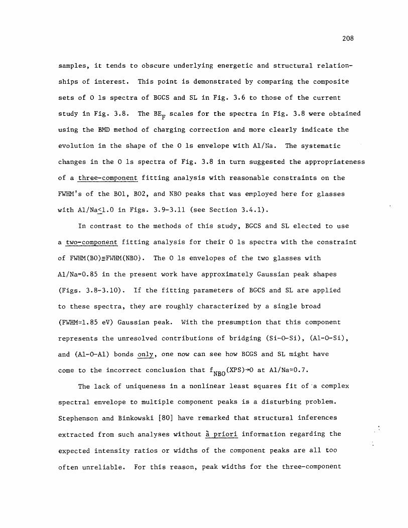
208
samples, it tends to obscure underlying energetic and structural relation-
ships of interest. This point is demonstrated by comparing the composite
sets of 0 ls spectra of BGCS and SL in Fig. 3.6 to those of the current
study in Fig. 3.8. The BEF scales for the spectra in Fig. 3.8 were obtained
using the BMD method of charging correction and more clearly indicate the
evolution in the shape of the 0 ls envelope with Al/Na. The systematic
changes in the 0 ls spectra of Fig. 3.8 in turn suggested the appropriateness
of a three-component fitting analysis with reasonable constraints on the
FWII's of the B01, B02, and NBO peaks that was employed here for glasses
with Al/Na<1.0 in Figs. 3.9-3.11 (see Section 3.4.1).
In contrast to the methods of this study, BGCS and SL elected to use
a two-component fitting analysis for their 0 ls spectra with the constraint
of FWHM(BO)-FWHM(NBO). The 0 ls envelopes of the two glasses with
Al/Na=0.85 in the present work have approximately Gaussian peak shapes
(Figs. 3.8-3.10). If the fitting parameters of BGCS and SL are applied
to these spectra, they are roughly characterized by a single broad
(FWHM=1.85 eV) Gaussian peak. With the presumption that this component
represents the unresolved contributions of bridging (Si-O-Si), (Al-O-Si),
and (Al-0-Al) bonds only, one now can see how BCGS and SL might have
come to the incorrect conclusion that fNBO(XPS)*O at Al/Na=0.7.
The lack of uniqueness in a nonlinear least squares fit of a complex
spectral envelope to multiple component peaks is a disturbing problem.
Stephenson and Binkowski [80] have remarked that structural inferences
extracted from such analyses without ' priori information regarding the
expected intensity ratios or widths of the component peaks are all too
often unreliable. For this reason, peak widths for the three-component
209
fitting analysis used in the current study of NAS glasses with Al/Na<1.0
were constrained in accordance with insights gained from experience with
simpler NS glasses in Chapter 2. As indicated above, this analysis of NAS
glasses provides a logical explanation for the markedly changing shape of
the 0 ls envelope with Al/Na up to the equivalence point and strong
evidence for the classical model which compliments and reinforces the
findings of structural studies by other techniques. Moreover, an analysis
of the 0 ls spectrum obtained in a preliminary study [81] of a vacuum-
fractured natural Ab crystal [82] yielded r(XPS)=1.03 vs. r(theo)=l.00.
This result agrees well with that obtained for Ab glass (Fig. 3.15) and
thus offers additional support for the appropriateness of the fitting
analysis employed here.
In principle, a residual analysis of 0 ls spectra for NAS glasses
like the one of LPV [19] should produce conclusions compatible with those
from a curve fitting analysis about glass structure. In practice, however,
the relative energetic alignment of each pair of NAS and NS 0 ls spectra
prior to subtraction is critically important. LPV decided to scale the
position of their 0 ls envelopes relative to that of a nearby sodium
Auger line. Significantly, SL [20] reported the energetic separation
between a sodium Auger peak and the NBO component of the 0 ls spectrum
increases even more rapidly with Al/Na below the equivalence point than
the shift of the Na Is peak relative to the BO2 component of the 0 ls
envelope in the present study (Fig. 3.12). From the standpoint of the
current structural interpretation, the positions of BO1, B02, and NBO
bonding states of an NAS glass would become increasingly misaligned in
relation to the BO and NBO states of the NS glass as Al/Na increases up

210
to unity by following the scaling procedure of LPV. Serious questions
can therefore be raised regarding the propriety of the difference spectra
generated by LPV. Moreover, there is a possibility that structural arti-
facts are represented in their analysis since the photoemission spectra
were acquired from vacuum-milled surfaces (see Section 1.2.2). These
objections thus undermine the basis for LPV's anomalous conclusion that
fNBo(XPS) is virtually independent of Al/Na.
3.5.2. Glasses with 1.0<Al/Na<l.5
The analyses of 0 ls spectra for NAS glasses with 1.0<Al/Na<1.5 in
this study are consistent with Lacy's mechanism [44] for the incorporation
of aluminum atoms in excess of sodium atoms. The parameter r'(XPS) is
generally found to describe 0 ls spectral envelopes for glasses of the
series A=0.57 and XSi=0.67 above the equivalence point. Although the
range of r'(XPS) for these compositions is rather small in Fig. 3.16,
this parameter corresponds well to theoretical estimates based on the
tricluster model for NAS glasses with Al/Na>1.0 (see Section 3.4.2). In
contrast, the analyses of the 0 ls spectra in Figs. 3.9 and 3.10 disagree
with theoretical predictions derived for the interstitial model of
Taylor and Rindone [34] for the accommodation of excess aluminums in
NAS glasses with Al/Na>1.0.
Opposed to the sharp increase in the binding energy of the Na ls
level relative to the BEF of the BO2 component of the 0 ls envelope
as Al/Na increases below the equivalence point in Fig. 3.12, the energetic
separation between the Na ls level and the BO2+B04 component is constant
for Al/Na>l.0 in the series of glasses with A=0.57 and XSi=0.67. The
inflection at Al/Na=1.0 indicates that sodium ions are not greatly

211
affected by the incorporation of excess aluminum atoms in compositions
above the equivalence point. A similar conclusion was drawn from optical
luminescence results [23,24] for thallium-doped NAS glasses with A=0.57
at Al/Na>1.0. These observations are not inconsistent with the formation
of tricluster configurations in glasses with Al/Na>1.0.
Whereas the energetic separation between the Si 2p and Al 2p levels
is invariant up to the equivalence point, there is some suggestion in
Fig. 3.12 that the binding energy of the Al 2p level increases relative
to the BEF of the Si 2p level at Al/Na>1.0 in the glasses with A=0.57
IVand X Si=0.67. In the banding arrangement of a tricluster, an Al group
contains three oxygen atoms in the "underbonded" B03 chemical state and
one oxygen in the "overbonded" BO4 state (see Section 3.2.1 and 3.4.1).
A relaxation in the valence charge distribution from these aluminums
towards the "underbonded" oxygens in their coordination shell conceivably
results in a higher binding energy for the Al 2p level with increasing
Al/Na above the equivalence point.
The invariance in the FWHM of the Si 2p pnotopeak for all NAS glasses
with Al/Na>l.0 is understood to mean NBO/Si=0 for all silicon atoms in
each of these compositions. The small increase in the FWH! of the Al 2p
photopeak for NAS glasses with Al/Na>1.0 may indicate the occurrence of
two energetically distinct chemical states for aluminum atoms: the first
in AlIV groups for which sodiums ions are available as charge compensators,
and the second in AlIV groups within tricluster configurations.
The conclusions for the atomic arrangement of NAS glasses with
1.0<A1/Na<l.5 from this XPS study are in accord with structural details
provided by other techniques including XES, XANES, EXAFS, and optical

212
luminescence spectroscopy as described in Section 3.2.1. There is little
compelling structural evidence to support the conventional idea that a
significant fraction of the aluminum atoms in RAS glasses with 1.0<Al/R<2.0
exist in Al groups either as network modifiers [26,38,41,42], inter-
stitial charge compensators [34], or as network formers (though weaker
than AlIV groups) [39,40]. On the contrary, most structural investigations
have given results which are better explained by the tricluster model for
these compositions.
One reservation about the existence of tricluster configurations in
RAS glasses with Al/R>1.0 comes from the observation that such a bonding
arrangement violates Zachariasen's rules for the bonding topology of
multicomponent oxide glasses [83]--i.e., one oxygen atom is shared
among three tetrahedral centers (two SiIV groups and an Al V group).
However, analogous bonding arrangements are not unknown in aluminosilicate
crystals [5-7,44]--e.g., sillimanite exhibits an oxygen atom shared by
Si IV , Al V , and AlV I groups. In addition, triclusters are structurally
stable with regard to the extended electrostatic valence rule (see
Sections 2.2.2 and 3.2.1). It follows that tricluster configurations in
RAS glasses with Al/R>1.0 allow for the formation of continuous single-
phase glasses (within limits to be explored in the next subsection)
having fully connected aluminosilicate networks by relaxing the rigid
structural constraints of the 4-2 coordination network found in RAS
glasses with Al/R=1.0.
More serious objections can be raised about an underlying assumption
for the parameter r'(XPS) that was introduced here in the interpretation
of 0 ls photoemission spectra for NAS glasses with 1.0<Al/Na<1.5.

213
While the ranking of valence charges thought to exist on oxygen anions
in the BO1, BO2, BO3, and BO4 chemical states is plausible (see Section
3.4.1), the assumption that the binding energies for these states exhibit
a two-fold degeneracy is somewhat tenuous. An empirical determination by
diffraction methods of the distribution in electron density among oxygen
anions of an aluminosilicate crystal containing bonding arrangements
similar to tricluster configurations (e.g., sillimanite) could provide
some means for assessing the validity of this simplifying approximation.
Also, a first-principles calculation of the effective Mulliken atomic
charges on oxygen atoms in B01 and B03 vs. BO2 and BO4 bonding states
for an NAS glass with triclusters at Al/Na>l.0 would be useful in this
regard.
Although the tricluster model provides a reasonable explanation for
the slowly evolving shape of the 0 ls envelope with Al/Na for NAS com-
positions with Al/Na>1.0 in Figs. 3.9 and 3.10, some concern about the
uniqueness of this interpretation is warranted (especially in view of the
above comments). Peak widths for the two-component fitting analysis
of these 0 ls spectra were constrained in a way, however, that is natural
extension of the three-component fitting analysis applied to 0 ls spectra
for NAS compositions with Al/Na<l.0. Despite the limitations of the
two-component fitting analysis for glasses with Al/Na>l.0, permissive
evidence for the tricluster model has been demonstrated which is consistent
with results from structural studies by other techniques.
3.5.3. Additional Implications for Glass Structure
For glasses in the NAS ternary system with XSi >0.50 it has beenshown that the integrated intensities of various BO and NBO components%
shown that the integrated intensities of various BO and NBO components

214
of the 0 is spectral envelope are directly related to glass composition
in a predictable and rather general manner within two adjacent composition-
al regions: O<Al/Na<1.0 and 1.0<Al/Na<l.5. If one accepts the classical
model and tricluster models as satisfactory descriptions for the atomic
arrangement of glasses in these respective regions, then lines of constant
fNBO(theo) in Fig. 3.17 and constant r(theo) and r'(theo) in Fig. 3.18 can
be mapped for the SiO 2-rich corner of the NAS system. Areas of phase
separation (L+L) [70-73] and the series of compositions with A=0.57 are
indicated in these ternary diagrams. Inspection of Fig. 3.18 makes clear
the nature of the pronounced variation in r(XPS) for Al/Na<1.0 versus
the slow change in r'(XPS) for Al/Na>l.0 in Fig. 3.9 for this line of
glass compositions.
In Fig. 3.18, a compositional line is drawn which shows where BO1+ O
for the ratio of chemical states
8XBO2+BO3+BO4 8AIr"(theo) = B02+BO3+B4 X Al (3.15)
B01 2X Si-5X A +(.5
under the assumptions of the tricluster model for NAS glasses with Al/Na>1.0
(see Section 3.4.2). The line r"=o thus defines the extent of the region
above the equivalence point within which the aluminum avoidance principle
is valid in theory. It extends from the Ne composition to a binary
SiO 2-Al 203 composition with XAl=0.286. Interestingly, the latter com-
position coincides with the center of a broad metastable miscibility gap
between X A=0.12 and 0.43 as determined by Jantzen et al. [73]. In aAl
modification of their structural rationale for phase separation, it is
suggested here that substantial violation of the aluminum avoidance rule

Xsi + X AI + XN0 = 1.0
No20 /50Z
Fig. 3.17:
Si02
\At20350
40 30 20 10mol%
Lines of constant fNBO(theo)=NBO/BO1+BO2+NBO for NAS
glasses with X .>0.50.Si.-
215

216
Si02Xsi+ XA + XNa 1.0
r (theo)= 8X I fo .x S2XSi-3XAI-XNo
N020/50Z
Fig. 3.18:
r' (theo)= XAI +3XNao A I.0
BAIr'(theo)= x5_XAI,
2XS-5X+XNo
40 30 20 10mol%
Lines of constant r(theo)=B02/BO1, constant r' (theo)=
BO2+BO4/BO1+B03, and r" (theo)=BO2+BO3+BO4/BOl== for
NAS glasses with X S>0.50.S i-

217
provides more of an impetus for this process rather than the mere presence
of oxygen atoms in the "overbonded" BO4 chemical state. More generally,
for NAS glasses with Al 203 contents greater than those demarked by r"=-
in Fig. 3.18, the incorporation of oxygens in the B05 state conceivably
IV IVdestabilizes the aluminosilicate network based on Al and Si groups
and thus leads to the incorporation of A1V I groups as seen in the XES
results of Hanada et al. [51].
Along the series of NAS glasses with Al/Na=1.0, the XPS results of
this study indicate that a small fraction of oxygen atoms exist in the
BO5 bonding state for glasses with XS i=0.50 and 0.54 near the compositional
line r"=-. It is shown in Fig. 3.18 that the extent of the series of
glasses with A=0.57 above the equivalence point falls well short of the
compositional line r"=-. It therefore seems reasonable to expect that
the aluminum avoidance principle holds for even the most aluminous
compositions (Al/Na=1.5) in the two series of glasses with A=0.57 and
XSi=0.67 as was assumed for the spectral analysis in Section 3.4.1.
According to the crystallite hypothesis [84,85] and the more recent
crystalline cluster models of Phillips [86] and Goodman [87] for glass
structure (see Sections 2.2.1 and 2.2.2), vitreous silicates are viewed
as aggregates of discrete, randomly-oriented, crystalline particles of
1-10 nm in size. For a ternary glass, these particles supposedly have
morphologies of the stoichiometric compounds forming the compatibility triangle
associated with the glass composition as determined from an equilibrium
phase diagram. While certain similarities exist between the short-range
structure of glasses and crystalline compounds of the NAS ternary system

218
(e.g., the commonality of SiI V and Al groups), the preponderance of
structural evidence indicates that AlV I groups do not exist in jadeite-
like bonding arrangements for glasses with Al/Na<1.0 (see Section 3.5.1)
or in jadeite-, mullite-, or corundum-like bonding configurations for
glasses with 1.0<Al/Na<2.0 (see Section 3.5.2). This observation thus
argues against the crystallite or crystalline cluster models as valid
structural descriptions for NAS glasses. Instead, the systematic variation
of the structural parameters fNBO r, and r' with composition in
Figs. 3.17 and 3.18, as projected from the photoemission results of the
present study, are better viewed in the context of the random network
model for glass structure (see Sections 2.2.1 and 2.2.2).
While a full rationalization of structure-property relationships
for RAS glasses is beyond the scope of this Thesis, the transition from
the classical model for O<AlI/R<l.0 to the tricluster model for Al/R>1.0 is
thought to provide a reasonable basis for future work attempting to
understand in detail the variation of physical properties at Al/R=1.0-1.2
(Table 3.1). In particular, the compositional dependences of fNBO' r,
and r' in Figs. 3.17 and 3.18 conceivably hold important keys to explaining
why some properties mostly exhibit extrema or discontinuities at Al/R=1.O
(e.g., density in Fig. 3.1(b))while others tend to display such features
at Al/R=1.1-1.2 (e.g., viscosity in Figs. 3.1(c) and (d)) in series of
RAS glasses with O<Al/R<2.0.
Aside from resolving controversies generated by earlier XPS studies
of NAS glasses, the experimental methods and analytical approach described
in this Chapter also offer a firm foundation for future structural
investigations of other RAS glasses (R=Li,K, etc.), MO-Al203-Si0 2 glasses
(M=Mg, Ca, etc.), and R20-A203-Si0 2 glasses (A=B, Ga, In). Finally, one

219
can readily appreciate how the structural and chemical information from
this photoemission work on NAS glasses would be highly relevant to the
understanding of heterogeneous catalysis at aluminosilicate surfaces.

220
3.6. SUMMARY:
Photoemission spectra of the 0 is, Si 2p, Al 2p, and Na is core levels
for three series of NAS glasses (A =0.57, XSi=0.67, and Al/Na=1.0) have
been acquired from vacuum-fractured surfaces and corrected for electro-
static charging effects by the BMD method. The integrated intensities of
the various BO and NBO components of the O is spectra are shown to be
directly related to glass composition in a predictable and rather general
way within two adjacent compositional regions: O<Al/Na<1.0 and
1.0<Al/Na<l.5. In marked contrast to several previous XPS studies of NAS
glasses, a curve fitting analysis of the 0 is spectra for glasses with
O<AI/Na<1.0 gives strong support to tne classical structural model for
the incorporation of aluminum atoms. Along the series of compositions
with Al/Na=l.0, this analysis also indicates the aluminum avoidance
principle begins to break down in the range of 0.57<Al/Si<0.85. A curve
fitting analysis for 0 is spectra of glasses with 1.0<Al/Na<l.5 is consistent
with Lacy's tricluster model for the accommodation of aluminum atoms in
excess of sodium atoms. Finally, trends in the binding energies and
widths of 0 is, Si 2p, Al 2p, and Na is photopeaks provide collateral
support for the above structural conclusions about NAS glasses.
This work demonstrates the ability of XPS to supply quantitative
information about the structural chemistry of complex (i.e., ternary)
silicate glass systems. Such information not only compliments and
reinforces the findings of structural studies by other techniques, but also
has potential for yielding uniquely clarifying insights into how the
variation of many physical properties of glasses with composition can be
correlated to structural changes.

221
REFERENCES
[1] N.J. Kreidl, "Inorganic Glass-Forming Systems"; pp. 105-299 inGlass: Science and Technology (Glass-Forming Systems, Vol. 1).
Edited by D.R. Uhlmann and N.J. Kreidl. Academic, New York, 1983.
[2] G.E. Harlow and G.E. Brown, Jr., "Low Albite: An X-ray and Neutron
Diffraction Study", Am. Mineral., 65[9,10] 986-95 (1980).
[3] G. Donnay, J.F. Schairer, and J.D.H. Donnay, "Nepheline Solid
Solutions", Mineral. Mag., 32 [245] 93-109 (1959).
[4] M. Cameron, S. Sueno, C.T. Prewitt, and J.J. Papike, "High-
Temperature Crystal Chemistry of Acmite, Diopside, Hedenbergite,
Jadeite, Spodumene, and Ureyite", Am. Mineral., 58 [7,8] 594-618
(1973).
[5] C.W. Burnham, "Refinement of the Crystal Structure of Kyanite",Z. Kristallogr., 118 [5,6] 337-60 (1963).
[6] R. Sadanaga, M. Tokonami, and Y. Takeuchi, "Tne Structure of Mullite,2A1 20 3 SiO, and Relationship with the Structures of Sillimanite
and Adalusite", Acta. Cryst., 15 65-8 (1962).
[7] C.W. Burnham, "Refinement of the Crystal Structure of Sillimanite",
Z. Kristallogr., 118 [1,2] 127-48 (1963).
[8] R.D. Shannon and C.T. Prewitt, "Effective Ionic Radii in Oxides and
Fluorides", Acta Cryst., B25 [8] 925-46 (1969).
[9] L. Pauling, The Nature of the Chemical Bond; Chapter 13. Cornell
University Press, Ithaca, New York, 1960.
[10] C.W. Burnham and M.J. Buerger, "Refinement of the Crystal Structure
of Andalusite", Z. Kristallogr., 115 [3,4] 269-90 (1961).
[11] P.R. Anderson, "X-ray Photoelectron Spectroscopy of Sodium-Silicate
Glasses", Am. Chem. Soc. Meeting, New York, 1973.
[12] R. Bruckner, H.-U. Chun, and H. Goretzki, "Photoelectron Spectroscopy
(ESCA) on Alkali Silicate and Soda Aluminosilicate Glasses",
Glastechn. Ber. 51[l] 1-7 (1978).
[13] B.W. Veal and D.J. Lam, "XPS Study of Sodium Oxide in Amorphous
SiO "; pp. 299-303 in The Physics of SiO 2 and Its Interfaces.
Edited by S.T. Pantelides. Pergamon, New York, 1978.
[14] R. Bruckner, H.-U. Chun, H. Goretzki, and M. Sammet, "XPS Measurements
and Structural Aspects of Silicate and Phosphate Glasses", J. Non-Cryst.
Solids, 42 [1-3] 49-60 (1980).
[15] J.S. Jen and M.R. Kalinowski, "An ESCA Study of the Bridging to
Non-Bridging Oxygen Ratio in Sodium Silicate Glass and the Correlations
to Glass Density and Refractive Index", J. Non-Cryst. Solids,
38-39 21-6 (1980).

222
[1b] B.M.J. Smets and T.P.A. Lommen, "The Structure of GermanosilicateGlasses, Studied by X-ray Photoelectron Spectroscopy", J. Non-Cryst.Solids, 46 [1] 21-32 (1981).
[17] T. Sasaki, M. Kawaguchi, M. Yamane, an Y. Suginohara, "On theQuantitative Analysis of 00, 0 , and 0 Ions in Binary Silicate byX-ray Photoelectron Spectroscopy", J. Japan Inst. Metals, 45[8] 790-6 (1981).
[18] B.W. Veal, D.J. Lam, A.P. Paulikas, and W.Y. Ching, "XPS Study ofCaO in Sodium Silicate Glasses", J. Non-Cryst. Solids, 49 [1-3]309-20 (1982).
[19] D.J. Lam, A.P. Paulikas, and B.W. Veal, "X-ray PhotoemissionSpectroscopy Studies of Soda Aluminosilicate Glasses", J. Non-Cryst.Solids, 42 [1-3] 41-8 (1980).
[20] B.M.J. Smets and T.P.A. Lommen, "The Incorporation of Aluminum Oxideand Boron Oxide in Sodium Silicate Glasses, Studied by X-rayPhotoelectron Spectroscopy," Phys. Chem. Glasses, 22 [6] 158-62(1981).
[21] Y. Kaneko, H. Nakamura, M. Yamane, K. Mizoguchi, and Y. Suginohara,"Photoelectron Spectra of Silicate Glasses Containing Trivalent
Cations", Yogyo-Kyokai-Shi, 91 [7] 321-4 (1983).
[22] H. Wakabayashi, R. Terai, and H. Yamanaka, "Effects of TrivalentOxides on Electrical Conductivity in Alkali Silicate Glasses",Yogyo-Kyokai-Shi, 93 [4] 209-16 (1985).
[23] M.N. Alexander, P.I.K. Onorato, C.W. Struck, J.R. Rozen, G.W. Tasker,
and D.R. Uhlmann, "Structure of Alkali (Alumino) Silicate Glasses.I. Tl+ Luminescence and the Nonbridging Oxygen Issue", J. Non-Cryst.
Solids, 79 [1,2] 137-54 (1986).
[24] M.N. Alexander, P.I.K. Onorato, C.W. Struck, G.W. Tasker, andD.R. Uhlmann, "Structure of Alkali (Alumino) Silicate Glasses.II. Luminescence of Tl+-doped Sodium Aluminosilicates",J. Non-Cryst. Solids, in press.
[25] J.F. Schairer and N.L. Bowen, "The System Na20-Al 2 03-Si0 2 ", Am. J.Sci., 254 [3] 129-95 (1956).
[26] J.O. Isard, "Electrical Conduction in Aluminosilicate Glasses",J. Soc. Glass Technol., 43 [211] 113-23T (1959).
[27] E.I. Galant, "Refractive Index and Coordination Transformation inAluninoborosilicate Glasses"; pp. 451-3 in The Structure of Glass
(Vol. 2). Edited by E.A. Porai-Koshits. Consultants Bureau,New York, 1960.

223
[28] D.E. Day and G.E. Rindone, "Properties of Soda AluminosilicateGlasses: I,", J. Am. Ceram. Soc., 45 [10] 489-96 (1962);"II", ibid., 496-504; "III", ibid., [12] 579-81.
[29] P.W.L. Grahm and G.E. Rindone, "Properties of Soda AluminosilicateGlasses: IV,", J. Am. Ceram. Soc., 47 [1] 19-24 (1964).
[30] E.L. Williams and R.W. Heckman, "Sodium Diffusion in Soda-Lime-Aluminosilicate Glasses", Phys. Chem. Glasses, 5 [6] 166-71 (1964).
[31] E.F. Riebling, "Structure of Sodium Aluminosilicate Melts Containingat Least 50 mole % SiO at 15000C", J. Chem. Phys., 44 [8] 2857-65(1966).
[32] R.W. Heckman, J.A. Ringlien, and E.L. Williams, "Sodium Diffusionand Electrical Conductivity in Sodium-Aluminosilicate and Sodium-Calcium-Aluminosilicate Glasses", Phys. Chem. Glasses, 8 [4] 145-50(1967).
[33] R. Terai, "Self-diffusion of Sodium Ions and Electrical Conductivityin Sodium Aluminosilicate Glasses", Phys. Chem. Glasses, 10 [4]
146-52 (1969).
[34] T.D. Taylor and G.E. Rindone, "Properties of Soda AluminosilicateGlasses: V,", J. Am. Ceram. Soc., 53 [12] 692-95 (1970).
[35] W.C. La Course, "Structural Influences on Diffusion in Glass --The Mixed Site Effect", J. Non-Cryst. Solids, 21 [3] 431-4 (1976).
[36] J.E. Shelby and R.J. Eagan, "Helium Migration in Sodium Aluminosilicate
Glasses", J. Am. Ceram. Soc., 59 [9-10] 420-5 (1976).
[37] J.E. Shelby, "Helium Migration in Lithium Aluminosilicate Glasses",J. Appl. Phys., 48 [4] 1497-1502 (1977).
[38] J.E. Shelby,"'Viscosity and Thermal Expansion of Lithium Alumino-
silicate Glasses", J. Appl. Phys., 49 [12] 5685-91 (1978).
[39] K. Hunold and R. Bruckner, "Physical Properties and StructuralDetails of Sodium Aluminosilicate Glasses and Melts", Glastechn.Ber., 53 [6] 149-61 (1980).
[40] K. Hunold and R. Bruckner, "Chemical Diffusion of Sodium and Aluminum
Ions in Sodium Aluminosilicate Melts", Glastechn. Ber., 53 [8]207-19 (1980).
[41] A. Klonkowski, "The Structure of Sodium Aluminosilicate Glass",
Phys. and Chem. Glasses, 24 [6] 166-71 (1983).
[42] J.L. Piguet and J.E. Shelby, "Electrical Conductivity of LithiumAluminosilicate, Galliosilicate, and Alumino/Galliosilicate Glasses",Adv. Ceram. Mater., 1 [2] 192-7 (1986).

224
[43] B.E. Yoldas, "The Nature of the Coexistence of Four- and Six-co-ordinatedAl3+ in Glass", Phys. Chem. Glasses, 12 [1] 28-32 (1971).
[44] E.D. Lacy, "Aluminum in Glasses and Melts", Phys. Chem. Glasses, 4 [6]234-8 (1963).
[45] M. Taylor and G.E. Brown, "Structure of Mineral Glasses - I. TheFeldspar Glasses NaAlSi 0 8 , KAISi 0 CaAl Si 0 8", Geochimica etCosmochimica Acta, 43 [1]]61-75 (17 ); "If. he SiO -NaAISi0 Join"ibid., [9] 1467-73.
[46] R.W. Wardle and G.W. Brindley, "The Dependence of the Wavelength ofAlKa Radiation from Alumino-Silicates on the Al-0 Distance",Am. Mineral., 56 [11,12] 2123-8 (1971).
[47] H. Tagai, T. Iwai, and Y. Mori, The Seventh Meeting on Basic Scienceof Ceramics, Tokyo, 1969.
[48] R. Terai, S. Kuroda, and T. Ueno, Daikoshi-Kiho, 20 172 (1969).
[49] S. Sakka, "Study on the Coordination Number of Aluminum Ions inAlumino-silicate Glasses by Means of AlK Fluorescence X-ray Spectra",Yogyo-Kyokai-Shi, 85 [4] 168-73 (1977).
[50] S. Sakka, Science of Glass and Amorphous Materials; Chapter 6.Uchida-Rokakuho-Shinsha, Tokyo, ±983.(in Japanese).
[51] T. Hanada, T. Aikawa, and N. Soga, "Coordination of Aluminum inAmorphous Sodium Aluminosilicate Films", J. Non-Cryst. Solids, 50
[3] 397-405 (1982).
[52] S.A. Brawer and W.B. White, "Raman Spectroscopic Investigation ofthe Structure of Silicate Glasses (II). Soda-Alkaline Earth-AluminaTernary and Quartenary Glasses", J. Non-Cryst. Solids, 23 [2]261-78 (1977).
[53] B.O. Mysen, D. Virgo, and I. Kushiro, "The Structural Role ofAluminum in Silicate Melts - a Raman Spectrographic Study at 1Atmosphere", Am. Mineral., 66 [7,8] 678-701 (1981).
[54] P. McMillan, B. Piriou, and A. Navrotsky, "A Raman SpectroscopicStudy of Glasses along the Joins Silica-Calcium Aluminate, Silica-Sodium Aluminate, and Silica-Potassium Aluminate", Geochim.Cosmochim. Acta, 46 [11] 2021-37 (1982).
[55] F. Seifert, B.0. Mysen, and D. Virgo, "Three-dimensional NetworkStructure of Quenched Melts (Glasses) in the Systems SiO -NaAlO 210Si0 2-CaAl 204 , and SiO 2-MgAl 204 ", Am. Mineral., 67 [7,8] 996-717 (1982).
[56] P. McMillan and B. Piriou, "Raman Spectroscopic Studies of Silicateand Related Glass Structure - A Review", Bull. Mineral., 106 [1,2]
57-75 (1983).

225
[57] W.B. White and D.G. Minser, "Raman Spectra and Structure of NaturalGlasses", J. Non-Cryst. Solids, 67 [1-3] 45-59 (1984).
[58] D.A. McKeown, F.L. Galeener, and G.E. Brown, Jr., "Raman Studies
of Al Coordination in Silica-rich Sodium Aluminosilicate Glasses
and Some Related Minerals", J. Non-Cryst. Solids, 68 [2,3]
361-78 (1984).
[59] B.O. Mysen, D. Virgo, and F.A. Seifert, "Relationships between
Properties and Structure of Aluminosilicate Melts", Am. Mineral.,
70 [1,2] 88-105 (1985).
[60] D. Muller, W. Gessner, H.J. Behrens, and G. Scheler, "Determination
of the Aluminum Coordination in Aluminum-Oxygen Compounds by Solid-
State High-Resolution 2 7A1 NMR", Chem. Phys. Lett., 79 [1] 59-62
(1981).
[61] B.H.W.S. de Jong, C.M. Schramm, and V.E. Parziale, "Polymerization
of Silicate and Aluminate Tetrahedra in Glasses, Melts, and Aqueous
Solutions - IV.", Geochim. Cosmochim. Acta, 47 [7] 1223-36 (1983).
[62] E. Hallas, U. Haubenreiber, M. Hahnert, and D. Muller, "NMR
Investiga ons of Na O-Al2 0 -Si0 2 Glasses by Using the Chemical
Shift of Al Nuclei , Glas echn. Ber., 56 [3] 63-70 (1983).
[63] J.M. Thomas, J. Klinowski, P.A. Wright, and R. Roy, "Probing the
Environment of Al Atoms in Noncrystalline Solids: Al 20 -SiO 2 Gels,Soda Glass, and Mullite Precursors", Angew. Chem. Int. d. Engl.,
22 [8] 614-6 (1983).
[64] D.A. McKeown, G.A. Waychunas, and G.E. Brown, Jr., "EXAFS and XANES
Study of The Local Coordination Environment of Sodium in a Series
of Silica-Rich Glasses", J. Non-Cryst. Solids, 74 [2,3] 325-48
(1985).
[65] D.A. McKeown, G.A. Waychunas, and G.E. Brown, Jr., "EXAFS Study of the
Coordination Environment of Aluminum in a Series of Silica-Rich
Glasses", J. Non-Cryst. Solids, 74 [2,3] 349-71 (1985).
[66] T.F. Soules, "Molecular Dynamic Calculations of Glass Structure
and Diffusion in Glass" J. Non-Cryst. Solids, 49 [1-3] 29-52 (1982).
[67] W. Loewenstein, "The Distribution of Aluminum in the Tetrahedra of
Silicates and Aluminates", Am. Mineral., 39 [1,2] 92-6 (1954).
[68] A. Navrotsky, G. Peraudeau, P. McMillan, and J.-P. Coutures, "A
Thermochemical Study of Glasses and Crystals along the Joins
Silica-Calcium Aluminate and Silica-Sodium Aluminate", Geochim.
Cosmochim. Acta, 46 [11] 2039-47 (1982).

226
[69] J.B. Murdoch, J.F. Stebbins, and I.S.E. Carmichael, "High-Resolution2 9Si NMRStudy of Silicate and Aluminosilicate Glasses: The Effect
of Network-Modifying Cations", Am. Mineral., 70 [3,4] 332-43 (1985).
[70] R.J. Charles, "Metastable Liquid Immiscibility in Alkali Metal Oxide-
Silica Systems", J. Am. Ceram. Soc., 49 [2] 55-62 (1966).
[71] R.J. Charles, "Activities in Li2O-, Na 0-, and K20-SiO 2 Solutions",J. Am. Ceram. Soc., 50 [12] 631-41 (1997).
[72] B.G. Varshal, "Liquation Phenomena and the Structure of Glasses in
Three-Component Aluminosilicate Systems", Fizika i Khimiya Stekla,
1 [1] 47-51 (1975).
[73] C.M. Jantzen, D. Schwahn, J. Schetten, and H. Herman, "The Si02-Al203System. Part 1.", Phys. Chem. Glasses, 22 [5] 122-37 (1981);
"Part 2." ibid., 138-44.
[74] Perkin-Elmer, Physical Electronics Division, Eden Prairie, MN.
[75] M.F. Koenig and J.T. Grant, "Signal-to-Noise Measurements in X-ray
Photoelectron Spectroscopy", Surf. Interface Anal., 7 [5] 217-22 (1985).
[76] Compare Fig. 3.7 in Chapter 3 with Fig. 2.6(b) in Chapter 2.
[77] See Fig. 2.9 in Chapter 2.
[78] Compare Fig. 3.13 in Chapter 3 vs. Fig. 2.12 in Chapter 2.
[79] See Table 2.1 in Chapter 2.
[80] D.A. Stephenson and N.J. Binkowski, "X-ray Photoelectron Spectroscopy
of Silica in Theory and Experiment", J. Non-Cryst. Solids, 22 [2]
399-421 (1976).
[81] G.W. Tasker, unpublished data.
[82] Amelia County, Virginia.
[83] W.H. Zachariasen, "The Atomic Arrangement in Glass", J. Am. Chem. Soc.,
54 [10] 3841-51 (1932).
[84] J.T. Randell, H.P. Rooksby, and B.S. Cooper, "The Structure of Glasses;
The Evidence of X-ray Diffraction", J. Soc. Glass Technol., 14 219-29
(1930).
[85] N. Valenkov and E. Poray-Koshits, "X-ray Investigation of the Glassy State",
Z. Kristallogr. 95 [3,4] 195-229 (1936).

227
[86] J.C. Phillips, "Spectroscopic and Morphological Structure ofTetrahedral Oxide Glasses"; pp. 93-171 in Solid State Physics(Advances in Research and Applications, Vol. 37). Edited byH. Ehernreich, D. Turnbull, and F. Seitz. Academic, New York, 1982.
[87] C.H.L. Goodman, "The Structure and Properties of Glass and the
Strained Mixed Cluster Model", Phys. Chem. Glasses, 26 [1] 1-10(1985).

228
CHAPTER 4
CONCLUSIONS
1. Adaptation of the biased metal-dot (BMD) method to the analysis of
vacuum-fractured surfaces of insulators in XPS yields the following
benefits:
a. Experimental difficulties associated with static and
differential charging of insulators are largely eliminated.
b. The binding energy scale for an insulating sample is
referred to its surface Fermi level, consistent with Schottky
barrier formation at metal-insulator interfaces.
c. Information can be obtained about the bulk structure and
chemistry of an insulator. Artifacts associated with surface
preparation by ion-sputtering or vacuum-milling techniques
are avoided.
2. The similarity of O is and Si 2p core level spectra acquired from
vacuum-fractured and fire-polished surfaces of Si0 2 glass lends support
to a view that extensive surface reconstruction of siloxane bonds (Si-O-Si)
occurs at fracture surfaces. Nonbridging oxygens (e.g., Si-0* or Si=0)
are not observed in detectable concentrations >0.5 atomic % (> 3% of a
monolayer) near vacuum-fractured surfaces.
3. The 0 ls and Si 2p lines for several types of Si0 2 glass (T18,
TO8, and T19) are not significantly different in binding energy or width
from the 0 ls and Si 2p lines of a-quartz. Since the range of the Si-O-Si
bridging angle is considerably larger in vitreous SiO 2 than in crystalline

229
SiO 2 , this result suggests the covalent-ionic nature of the Si-O bond
(i.e., in regard to the transfer of valence charge between silicon and
oxygen atoms) does not change appreciably with bridging angle.
4. The 0 ls spectral envelopes for a-quartz and SiO 2 glass are
characterized by single bridging oxygen components of equal width. Si=O
bonds or strained siloxane bonds are not observed in detectable concentrations
for SiO 2 glass. These XPS results call into question the assertions of
recent crystalline cluster models for glass structure. A lower limit on
the cluster radius required to obtain consistency with fNBO(XPS) + 0 is
estimated and found unreasonably large (>30 nm). One concludes the random
network model remains the most appropriate structural description for
vitreous SiO 2.
5. In accord with earlier work, an analysis of 0 ls spectra for
R20-SiO 2 glasses has shown fNBO(XPS) agrees with conventional theory for
the accommodation of alkali atoms in a vitreous silicon-oxygen network
for XR<0.30, but departs negatively from this theory for XR>0.30. Hydration
of these glasses or the possibility of a less efficient mechanism for the
incorporation of alkali are discounted as being significant factors behind
this discrepancy.
6. Trends in the binding energies and widths of the 0 ls, Si 2p,
and Na ls photopeaks for a series of Na20-SiO2 glasses support a
contention that while single-phase R20-SiO2 glasses do not exhibit any
long-range order, chemical details of the short-range atomic structure
bear a close resemblance to those in R20-Si0 2 crystals. In the absence
of any empirical evidence to the contrary, the photoemission results
for R20-Si0 2 glasses are best understood in the context of the random

230
network model for glass structure.
7. The proportions of various bridging and nonbridging oxygen
components of 0 is spectra for three series of Na20-Al 203-SiO 2 glasses are
demonstrated to be directly related to glass composition in a predictable
and rather general way within two adjacent compositional regions:
O<Al/Na<l.0 and 1.0<Al/Na<1.5.
8. In contrast to prior XPS studies, an analysis of 0 is spectra
for Na20-Al 203-SiO 2 glasses with O<Al/Na<1.0 gives strong support to the
classical model for incorporation of aluminum atoms in tetrahedral sites
as network formers. The present work clearly shows that fNBO(XPS)-*O at
Al/Na=1.0 as the equivalence point is approached from below along two series
of compositions with A=0.57 and XSi=0.67. This analysis also suggests the
aluminum avoidance principle begins to break down in the range of
0.57<Al/Si<0.85 as Al/Si goes from 0.33 to 1.0 in the series of compositions
with Al/Na=1.0.
9. An analysis of 0 is spectra for Na20-Al 203-SiO 2 glasses with2 2 3 2 0
1.0<Al/Na<1.5 is consistent with Lacy's tricluster model for the incorporation
of aluminum atoms in excess of sodium atoms in tetrahedral sites as
network formers.
10. Trends in the binding energies and widths of the 0 is, Si 2p,
Al 2p, and Na is photopeaks for Na20-Al 203-SiO 2 glasses lend collateral
support to the classical model for Al/Na<1.0 and the tricluster model
for Al/Na>1.0.

231
CHAPTER 5
SUGGESTIONS FOR FUTURE WORK
1. Employ angle-resolved photoemission spectroscopy to further
characterize vacuum-fractured versus fire-polished surfaces of vitreous SiO 2.
The enhanced surface sensitivity that is achievable with this method would
allow surface reconstruction and relaxation accompanying fracture and
thermal activation to be studied in greater detail than was possible in
this Thesis. Extension of this experimental approach to the study of
R2 0-SiO 2 (RS) glasses (R = Li, Na, K, etc.) would likely give important
insight into the discrepancy between the fraction of nonbridging oxygens that
is expected from conventional theory and the significantly smaller value
that has often been reported in XPS studies of RS glasses with high alkali
content.
2. Provisions for hydrating a sample under well-controlled conditions
in an auxiliary chamber connected by a feedthrough to the XPS chamber and
for heating the sample during photoemission measurements would enable the
investigation of hydration and dehydration phenomena at glass surfaces.
A better understanding of weathering and aqueous corrosion of glasses
would be a practical benefit of such work.
3. An XPS study of K 20-SiO 2 (KS) glasses using the experimental
procedures described in this Thesis is desirable. Important information
about the structural chemistry of RS glasses with low alkali content
(i.e., below the range of 0.2<X Na<0.4 for Na2 O-SiO2(NS ) glasses examined-- Na- 2 2(N)gasseaie
in the present study) could thus be obtained without the complication of
phase separation. Furthermore, the photoemission results for KS glasses
would provide a means for corroborating general structural conclusions
for RS glasses advanced here.

232
4. On the basis structural information about Na20-Al203-SiO2(NAS)
glasses obtained in the present photoemission work and the results of prior
structural studies by other techniques, a detailed rationalization of
structure-property relationships for R20-Al203-SiO 2 (RAS) glasses now
appears feasible. Such efforts might eventually yield models which are
capable of quantitatively predicting the variation of physical properties
for RAS glasses as a function of composition.
5. The experimental methods and interpretational approach of the
present study of NAS glasses have provided a firm foundation for future
XPS studies of other RAS glasses (R = Li, K, etc.), MO-Al203-SiO 2 glasses
(M = Mg, Ca, etc.), and R20-A203-SiO 2 glasses (A = B, Ga, In). Photo-
emission data for RAS glasses other than NAS glasses would be especially
useful for the modeling of structure-property relationships proposed
above.

233
APPENDIX 1
XPS of Polycrystalline Al 203
Photoemission spectra have also been acquired from vacuum-fractured
surfaces of Lucalox brand polycrystalline Al 20 3 [1]. The experimental
details and procedures for this work are similar to those described in
Sections 1.3.1, 1.3.2, and 1.3.4. Spectra for the 0 Is and Al 2p core
levels were collected from six samples; the spectrum of the valence band
(40-0 eV) was recorded for two of these samples. All spectra were obtained
at an apparent surface potential Q = -10 V and corrected to Au 4f7/2=84.00
eV by the BMD method.
Typical spectra of the 0 is and Al 2p core levels are presented in
Fig. Al.l. In contrast to the symmetric and nearly Gaussian shape of
the Al 2p line in Fig. Al.l(b), the 0 is line in Fig. Al.l(a) is distinctly
asymmetric with a tail skewed to higher binding energies. Binding energies
for the main component of the 0 is spectrum and the Al 2p spectrum are
531.61 eV (standard deviation cy =0.15 eV) and 75.12 eV (y = 0.15 eV),
respectively.
The fracture mode for these polycrystalline samples was intergranular.
In survey spectra (1100-0 eV), the presence of some calcium impurity was
indicated by the detection of weak Ca 2p doublet lines. Calcium is a
common impurity in polycrystalline Al 203 and is known to segregate at
grain boundaries. While the asymmetry of the 0 is line in Fig. Al.l(a)
may be related to the calcium impurity, it might also arise from surface
reconstruction during fracture. Additional work is needed to clarify
this issue.

234
Valence band spectra from two vacuum-fractured surfaces which have
been overlayed to emphasize the reproducibility of these measurements
are shown in Fig. A1.2. The upper valence band (UVB) exhibits two
peaks at 6.70 and 10.70eV and has a baseline width of 10.0 eV. The lower
valence band (LVB) consists of one peak at 23.50 eV with a distinct
shoulder at higher binding energies.
A more detailed discussion of these results and a critical comparison
with earlier work will be given elsewhere [2].
[1] General Electric Co., Schenectady, NY.
[2] R.H. French and G.W. Tasker, "X-ray Photoelectron Spectroscopy
of A1203", to be published.

235
=Li
537 535 533 531 529 527LiI-
(b) A l 2p
CMI.
88 78 76 74 72 70
BINDING ENERGY (eV)Fig. Al.l: Vacuum-fractured Lucalox brand polycrystalline Al 203
(a) 0 ls and (b) Al 2p core levels.

ILLN
I-
0-4,i
IL
I-0-4 wId
Id
40 36 32 28 24 20 16 12 8 4 0
BINDING ENERGY (eV)Fig. A1.2: Valence band spectrum for vacuum-fractured Lucalox brand
polycrystalline Al203.

237
APPENDIX 2
Hydration of Fire-polished Surfaces of Fused Quartz
Data are reported here for a study of the kinetics of hydration for
fire-polished surfaces of T08 fused quartz [1] by XPS. Fire-polished
surfaces of fused quartz plates, prepared as described in Section 1.3.3,
were subsequently hydrated under two conditions of differing severity:
immersion in 18 M H20 at 250 C versus 1000 C. The variation of the binding
energies for the 0 ls, Si 2p, and C ls photopeaks from their initial
values for a fire-polished surface as a function of time at the two
temperatures of hydration are presented in Fig. A2.1. Sets of spectra for
each hydration time were acquired at an apparent surface potential
Q =-10 V and corrected to Au 4f7/2 = 84.00 eV by the BMD method (see
Section 1.3.3). These experiments were conducted using one sample for
the study at 250 C and two samples for the study at 1000 C. In contrast,
fire-polished samples which were exposed to the prevailing humidity of
the laboratory atmosphere showed little if any change in the binding
energies of the core levels over time intervals comparable to those in
Fig. A2.1 (i.e., %1000 min).
Surfaces of SiO which have been heated to 800 0 C are known to be2 A
virtually dehydroxylated and therefore hydrophobic [2]. Consequently,
the fire-polished surfaces examined before hydration treatments in H20
were remarkably free of contamination due to adsorption of CO2 or
hydrocarbons from the ambient laboratory atmosphere. Such surfaces are
only efficiently rehydrated by immersion in boiling H20, as indicated
in Fig. A2.1. The rehydration of fire-polished fused quartz surfaces is

238
seen to be associated with a shift in the binding energies of the 0 is and
Si 2p core levels to lower energies with time of immersion in H20. This
process proceeds at a much more rapid rate in H20 at 1000 C than at 250 C
with an ultimate decrease in binding energy for each level of '1 eV.
While the C is photopeak from surface contamination does not exhibit
a significant shift in binding energy in Fig. A2.1, its intensity (relative
to the 0 is and Si 2p lines) increases markedly with time of hydration.
With increasing time of hydration, the fire-polished Si02 surface apparently
undergoes a transformation from one that is hydrophobic without many sites
for adsorption of carbonaceous species to one that is fully hydroxylated with
abundant sites for adsorption of such contaminants.
The widths of the 0 is and Si 2p photopeaks were both observed to
broaden with the duration and severity of the hydration treatment--e.g.,
the FWHM of the 0 is line increased from 1.29 eV for the fire-polished
surface to 1.46 eV and 1.65 eV after 5 min and 750 min, respectively, in
boiling H20. However, a nonbridging oxygen component associated with the
formation of hydroxyl bonds (Si-0H) could not be clearly distinguished from
the bridging oxygen component for siloxane bonds (Si-0-Si) in the 0 is
spectral envelope of any hydrated sample. A curve fitting analysis of the
0 is spectrum for samples hydrated under the conditions of these
experiments is likely complicated by the adsorption of oxygen-
containing carbonaceous species from the laboratory atmosphere [3]. Studies
of the hydration kinetics under better-controlled conditions (e.g., in a
reaction chamber connected to the XPS test chamber by a vacuum feed-
through) are thus required for better understanding of the interaction
of H20 with SiO 2 surfaces.

239
[1] Heraeus-Amersil, Inc., Sayreville, NJ.
[2] R.K. Iler, The Chemistry of Si0 2 ; Chapter 6. Wiley-Interscience,
New York, 1979.
[3] 0. Puglisi, A. Torrisi, and G. Marletta, "XPS Investigation of
the Effects Induced by Silanization on Real Glass Surfaces",
J. Non-Cryst. Solids, 68 [2,3] 219-30 (1984).

100 200 300Cumulative
400 500hydration time
600 700(minutes)
800 900
Fig. A2.1: BEF for O is, Si 2p, and C is core levels of fire-polished
TO8 fused quartz vs. time of hydration.
U)
ci~C
C-oC
O L0

241
BIOGRAPHICAL NOTE
The author was born in Washington, D.C. on 8 November 1950. He attended
Richard Montomery High School in Rockville, MD. In September 1968, he
entered the Pennsylvania State University and graduated from that institution
in June 1972 with a B.S. in Ceramic Science. From 1972 to 1976, he was
employed by Bell Laboratories in Murray Hill, NJ where he contributed to
the development of the Modified Chemical Vapor Deposition (MCVD) process
for the production of glass fiber optical waveguides. In September 1976,
he entered M.I.T. to pursue a Ph.D. in Ceramics with Professor Donald R.
Uhlmann as his advisor. In October 1986, he began work at Galileo Electro-
Optics in Sturbridge, MA. Including anticipated publications from his
doctoral thesis, he has authored or co-authored 20 scientific papers. He is
also the co-inventor of two U.S. patents.