Journal Pre-proof Lara-Pezzi, PhD, Pablo Garcia-Pavia, MD, PhD
43
Journal Pre-proof Clinical characteristics and determinants of phenotype in TMEM43 Arrhythmogenic right ventricular cardiomyopathy type 5. Fernando Dominguez, MD, PhD, Esther Zorio, MD, PhD, Juan Jimenez-Jaimez, MD, PhD, Rafael Salguero-Bodes, MD, Robert Zwart, PhD, Esther Gonzalez-Lopez, MD, PhD, Pilar Molina, MD, PhD, Francisco Bermúdez-Jiménez, MD, PhD, Juan F. Delgado, MD, PhD, Aitana Braza-Boïls, PhD, Belen Bornstein, MD PhD, Jorge Toquero, MD PhD, Javier Segovia, MD, PhD, J Peter Van Tintelen, MD, PhD, Enrique Lara-Pezzi, PhD, Pablo Garcia-Pavia, MD, PhD PII: S1547-5271(20)30094-1 DOI: https://doi.org/10.1016/j.hrthm.2020.01.035 Reference: HRTHM 8275 To appear in: Heart Rhythm Received Date: 17 November 2019 Accepted Date: 3 January 2020 Please cite this article as: Dominguez F, Zorio E, Jimenez-Jaimez J, Salguero-Bodes R, Zwart R, Gonzalez-Lopez E, Molina P, Bermúdez-Jiménez F, Delgado JF, Braza-Boïls A, Bornstein B, Toquero J, Segovia J, Van Tintelen JP, Lara-Pezzi E, Garcia-Pavia P, Clinical characteristics and determinants of phenotype in TMEM43 Arrhythmogenic right ventricular cardiomyopathy type 5., Heart Rhythm (2020), doi: https://doi.org/10.1016/j.hrthm.2020.01.035. This is a PDF file of an article that has undergone enhancements after acceptance, such as the addition of a cover page and metadata, and formatting for readability, but it is not yet the definitive version of record. This version will undergo additional copyediting, typesetting and review before it is published in its final form, but we are providing this version to give early visibility of the article. Please note that, during the production process, errors may be discovered which could affect the content, and all legal disclaimers that apply to the journal pertain. © 2020 Published by Elsevier Inc. on behalf of Heart Rhythm Society.
Transcript of Journal Pre-proof Lara-Pezzi, PhD, Pablo Garcia-Pavia, MD, PhD

Journal Pre-proof
Clinical characteristics and determinants of phenotype in TMEM43 Arrhythmogenicright ventricular cardiomyopathy type 5.
Fernando Dominguez, MD, PhD, Esther Zorio, MD, PhD, Juan Jimenez-Jaimez,MD, PhD, Rafael Salguero-Bodes, MD, Robert Zwart, PhD, Esther Gonzalez-Lopez,MD, PhD, Pilar Molina, MD, PhD, Francisco Bermúdez-Jiménez, MD, PhD, JuanF. Delgado, MD, PhD, Aitana Braza-Boïls, PhD, Belen Bornstein, MD PhD, JorgeToquero, MD PhD, Javier Segovia, MD, PhD, J Peter Van Tintelen, MD, PhD, EnriqueLara-Pezzi, PhD, Pablo Garcia-Pavia, MD, PhD
PII: S1547-5271(20)30094-1
DOI: https://doi.org/10.1016/j.hrthm.2020.01.035
Reference: HRTHM 8275
To appear in: Heart Rhythm
Received Date: 17 November 2019
Accepted Date: 3 January 2020
Please cite this article as: Dominguez F, Zorio E, Jimenez-Jaimez J, Salguero-Bodes R, Zwart R,Gonzalez-Lopez E, Molina P, Bermúdez-Jiménez F, Delgado JF, Braza-Boïls A, Bornstein B, Toquero J,Segovia J, Van Tintelen JP, Lara-Pezzi E, Garcia-Pavia P, Clinical characteristics and determinants ofphenotype in TMEM43 Arrhythmogenic right ventricular cardiomyopathy type 5., Heart Rhythm (2020),doi: https://doi.org/10.1016/j.hrthm.2020.01.035.
This is a PDF file of an article that has undergone enhancements after acceptance, such as the additionof a cover page and metadata, and formatting for readability, but it is not yet the definitive version ofrecord. This version will undergo additional copyediting, typesetting and review before it is publishedin its final form, but we are providing this version to give early visibility of the article. Please note that,during the production process, errors may be discovered which could affect the content, and all legaldisclaimers that apply to the journal pertain.
© 2020 Published by Elsevier Inc. on behalf of Heart Rhythm Society.

Clinical characteristics and determinants of phenotype in TMEM43 1 Arrhythmogenic right ventricular cardiomyopathy type 5. 2 3 4 Short title: Phenotype of TMEM43 ARVC Type 5 5 6 7 Authors: 8
Fernando Dominguez, MD, PhD1,2, Esther Zorio, MD, PhD2,3,4, Juan Jimenez-9
Jaimez, MD, PhD5, Rafael Salguero-Bodes, MD2,6, Robert Zwart, PhD7, Esther 10
Gonzalez-Lopez, MD, PhD1,2, Pilar Molina, MD, PhD4,8, Francisco Bermúdez-11
Jiménez MD, PhD5, Juan F. Delgado MD, PhD2,6, Aitana Braza-Boïls, PhD3,4, 12
Belen Bornstein, MD PhD9, Jorge Toquero, MD PhD1, Javier Segovia, MD, 13
PhD1,2, J Peter Van Tintelen, MD, PhD10, Enrique Lara-Pezzi, PhD2,11,12, Pablo 14
Garcia-Pavia, MD, PhD1,2,13 15
16
Affiliation: 17
1. Department of Cardiology. Hospital Universitario Puerta de Hierro, 18
Madrid, Spain. 19
2. CIBERCV, Madrid, Spain. 20
3. Department of Cardiology. Hospital Universitario La Fe, Valencia, 21
Spain. 22
4. CAFAMUSME Research group, IIS La Fe, Valencia, Spain 23
5. Department of Cardiology. Hospital Universitario Virgen de las Nieves, 24
Granada, Spain. 25
6. Department of Cardiology. Hospital Universitario 12 de Octubre, i+12. 26
Facultad de Medicina UCM, Madrid, Spain. 27
7. Department of Genome Analysis, Academic Medical Centre, University 28
of Amsterdam, Amsterdam, The Netherlands. 29
8. Department of Pathology, Instituto de Medicina Legal y Ciencias 30
Forenses and Histology Unit, Universitat de València, Valencia, Spain. 31
9. Department of Biochemistry, Hospital Universitario Puerta de Hierro, 32
Madrid, Spain. 33
10. Department of Genetics, University Medical Centre Utrecht, University 34
Utrecht, Utrecht, The Netherlands. 35
11. Myocardial Biology Programme, Centro Nacional de Investigaciones 1
Cardiovasculares (CNIC), Madrid, Spain. 2
12. National Heart and Lung Institute, Imperial College London, UK. 3
13. Universidad Francisco de Vitoria (UFV), Pozuelo de Alarcón, Spain. 4
5
Funding: This work was supported by grants from the Instituto de Salud Carlos 6
III [PI14/0967 and PI17/1941, CPII14/00027, PI14/01477, PI18/0158 and La 7
Fe Biobank PT17/0015/0043], the Isabel Gemio Foundation, the Spanish 8
Society of Cardiology [2014 Basic Research Grant], the European Union 9
[CardioNeT-ITN-289600 and CardioNext-608027], and from the Spanish 10
Ministry of Economy and Competitiveness [RTI2018-096961-B-I00, SAF2015-11
65722-R and SAF2012-31451]. This work was also supported by the Plan 12
Estatal de I+D+I 2013-2016 – European Regional Development Fund 13
(FEDER) “A way of making Europe”, Spain. The CNIC is supported by the 14
Instituto de Salud Carlos III (ISCIII), the Ministerio de Ciencia, Innovación y 15
Universidades (MCNU) and the ProCNIC Foundation, and is a Severo Ochoa 16
Center of Excellence (SEV-2015-0505). 17
18
Disclosures: None. 19
20
Word count: 5181 21
Correspondence: 1
Pablo Garcia-Pavia, MD, PhD, Department of Cardiology. Hospital 2
Universitario Puerta de Hierro, Manuel de Falla, 2. Majadahonda, Madrid, 3
28222, Spain. Email: [email protected] 4
Enrique Lara-Pezzi, PhD, Centro Nacional de Investigaciones 5
Cardiovasculares Carlos III, Melchor Fernandez Almagro, 3, 28029 Madrid, 6
Spain. Email: [email protected] 7

ABSTRACT: 1
2
Background 3
Arrhythmogenic right ventricular cardiomyopathy type V (ARVC-5) is the 4
most aggressive heterozygous form of ARVC. It is predominantly caused by 5
a fully penetrant mutation (p.S358L) in the non-desmosomal gene TMEM43 6
endemic to Newfoundland, Canada. To date, all familial cases reported 7
worldwide share a common ancestral haplotype. It is unknown whether the 8
p.S358L mutation by itself causes ARVC-5 or if the disease is influenced by 9
genetic or environmental factors. 10
11
Objective 12
To examine the phenotype, clinical course and the impact of exercise on 13
patients with p.S358L ARVC-5 without the Newfoundland genetic 14
background. 15
16
Methods 17
We studied 62 affected individuals and 73 non-carriers from 3 TMEM43-18
p.S358L Spanish families. Impact of physical activity on phenotype was also 19
evaluated. 20
21
Results 22

Haplotype analysis revealed that the 3 Spanish families were unrelated to 1
ARVC-5 patients with the Newfoundland genetic background. Two families 2
shared 10 microsatellite markers in a 4.9 cM region surrounding TMEM43; 3
the third family had a distinct haplotype. Affected individuals presented a 4
38.7% incidence of SCD, higher in males. LV involvement was common with 5
40% of mutation carriers showing LVEF<50%. Compared with non-carriers, 6
R wave in V3 was lower (3.2±2.8 vs 7.5±3.6 mV; P<0.001) and QRS in right 7
precordial leads wider (104.7±24.0 vs 88.2±7.7 ms;P=0.001). History of 8
vigorous exercise showed a trend towards more ventricular arrhythmias 9
only in women (P=0.053). 10
11
12
13
Conclusions 14
ARVC-5 is associated with high risk of SCD and characteristic clinical and 15
ECG features irrespective of geographical origin and genetic background. 16
Our data suggest that, as in desmosomal ARVC, vigorous physical activity 17
could aggravate the phenotype of TMEM43 mutation carriers. 18
19
Word count: 256 20
21

Keywords: TMEM43, arrhythmogenic right ventricular cardiomyopathy, 1
exercise, genetics, arrhythmia. 2
Introduction 1
Arrhythmogenic right ventricular cardiomyopathy (ARVC) is a common 2
cause of sudden cardiac death (SCD) in young adults. It is considered 3
primarily a disease of the desmosome, as mutations in desmosomal genes 4
have been identified in ~50% of patients who fulfil diagnostic criteria (1). 5
The most aggressive heterozygous form of ARVC is ARVC type V 6
(ARVC-5). A missense mutation on chromosome 3p25, within the non-7
desmosomal gene encoding transmembrane protein 43 (TMEM43 8
c.1073C>T, p.S358L), was reported in 2008 as a cause of ARVC-5 in 15 9
families from the island of Newfoundland, Canada (2,3). These families 10
share a common ancestral haplotype, and the affected subjects present an 11
autosomal dominant, fully penetrant, sex-influenced and high-risk form of 12
ARVC. Later, other mutations in the same gene have been associated with 13
ARVC-5, but p.S358L is the predominant one (4). 14
The p.S358L mutation leading to ARVC-5 was also identified in 15
families from Germany, Denmark and North America, and haplotype 16

analysis revealed that those families shared a common ancestor with the 1
Newfoundland affected patients (5). 2
To date, the TMEM43-p.S358L missense mutation responsible for 3
ARVC-5 has only been reported once in a patient with a non-4
Newfoundland origin (4). This single case was described in Toronto 5
(Canada) and corresponds to a 43 year-old male from New Zealand with a 6
confirmed de novo mutation, suggesting that a hot spot for this sequence 7
alteration might exist at this point in TMEM43. 8
No descriptions of additional non-Newfoundland patients have 9
appeared and data are lacking regarding patients with ARVC-5 without the 10
Newfoundland genetic background. It is also currently unknown whether 11
the p.S358L mutation itself causes ARVC-5 or if the disease expression is 12
influenced by the genetic background or environmental factors. 13
In the present study, we sought to describe the phenotype and 14
clinical expression of non-Newfoundland-related ARVC-5. 15
16
Methods 17

Study population 1
Three apparently unrelated families with a high incidence of sudden cardiac 2
death (SCD) across multiple generations were evaluated at four centres in 3
Madrid, Valencia and Granada, Spain. This study was approved by the 4
ethics committee of the participant centers and complies with the 5
Declaration of Helsinki. All participants provided written informed consent. 6
We investigated the phenotype and natural history of ARVC-5 disease 7
caused by the p.S358L mutation in these families by comparing clinical 8
events and test results in affected versus unaffected family members (family 9
controls) born at a priori 50% risk, an ascertainment strategy unrelated to 10
clinical presentation. Haplotypes from the three families were compared 11
with those present in Newfoundland-related individuals from Denmark, 12
Germany, North America and Newfoundland. 13
Subjects were considered affected if they were genetically confirmed, 14
obligate carriers of the p.S358L mutation or had SCD ≤ 50 years (3). 15
Subjects were considered unaffected if they did not carry the p.S358L 16

mutation. The remaining subjects were considered unknown and were not 1
studied further. 2
To minimise the bias present in recognising cases with SCD and 3
missing cases with minimal disease, we limited the analysis to subjects from 4
sibships where the disease status (affected or unaffected) of ≥50% of 5
siblings was known, as previously described (6). Clinical evaluation included 6
a detailed medical history, physical examination, ECG and transthoracic 7
echocardiogram. 8
The burden of physical activity was assessed by means of a 9
structured telephone interview to available individuals. Participants were 10
asked about the intensity and duration of regularly performed exercise, 11
including leisure-related, transportation and work activities since 10 years of 12
age. Intensity was rated according to the Multi-Ethnic Study of 13
Atherosclerosis Typical Week Physical Activity Survey as previously 14
described (7). Exercise duration was determined after asking the subjects 15
between which ages and how many hours per day had they participated in 16
each physical activity. Physical activity was considered vigorous when a high 17

intensity exercise (>70% of maximum oxygen uptake) was performed more 1
than 50 hours per year (8). 2
3
Haplotype analysis 4
A total of 16 microsatellite markers spanning TMEM43 on chromosome 5
3p25 were selected for haplotype analysis, which was performed in selected 6
members from the three Spanish families and in three additional families 7
from Newfoundland, Denmark and North America. None of the families 8
were known to be related. The genetic distances (cM) were inferred from 9
the deCODE genetic map. 10
11
Statistical analysis 12
Results are presented as mean (standard deviation) for continuous variables 13
with normal distribution, as median (interquartile range) for continuous 14
variables without normal distribution, and as number (percentage) for 15
categorical data. For statistical analysis, Student s t-test and the Mann-16
Whitney non-parametric test were used in two-group comparisons. The chi-17

square test or Fisher's exact test were used for categorical variables. A two-1
tailed P-value of <0.05 was considered to be statistically significant. All 2
statistical analyses were performed using the SPSS package, version 20.0 3
(SPSS Inc., Chicago, IL, USA). 4
Results 1
The three families comprised a total of 180 subjects (Figure 1). The family 2
evaluated in Madrid (Family 1) had a total of 131 members across 7 3
generations. The 5-generation family from Valencia had 24 members 4
(Family 2) and the remaining 25 subjects belonged to a 5-generation family 5
from Granada (Family 3). Considering the three families, a total of 62 6
patients (59.7% males; mean age at last evaluation or death 39.1±17.6 7
years) were classified as affected (30 genetically-confirmed carriers, 14 were 8
obligate carriers and 18 untested individuals who had SCD ≤50 years of 9
age and belonged to sibships where the disease status of ≥50% of siblings 10
were known). The remaining subjects were either non-carriers (n=73, 56.2% 11
males; mean age at last evaluation or death 46.1±17.6 years) or their 12
clinical status was considered as unknown (n=45, 42.2% males; mean age at 13
last evaluation or death 50.0±41.5 years). Subjects in the unknown group 14
were not further analysed. 15
The p.S358L (c.1073C>T) TMEM43 missense mutation was initially 16
identified using next-generation sequencing cardiomyopathy panels 17

(including >50 genes) or by exome sequencing in the probands from the 3 1
families. No other pathogenic variants were identified in desmosomal or 2
other cardiomyopathy-related genes. The presence of the p.S358L TMEM43 3
mutation in probands was confirmed by PCR amplification and Sanger 4
sequencing. Family members who agreed to undergo genetic testing were 5
tested for the presence of the p.S358L TMEM43 mutation also by Sanger 6
sequencing. 7
8
Haplotype analysis 9
Haplotype analysis revealed that index patients from Family 1 and 2 shared 10
10 microsatellite markers in a 4.9 cM region surrounding TMEM43. 11
Comparison of the haplotypes from Family 1 and 2 with those of patients 12
with p.S358L ARVC-5 from Newfoundland, United States and Denmark 13
showed that only 5 common markers were shared (Figure 2). Thus, the 14
p.S358L mutation leading to ARVC-5 in Family 1 and 2 was not inherited 15
from the same ancestor as the patients in the other regions. The haplotype 16
from Family 3 differed from the other two Spanish families and from the 17
Newfoundland-origin patients, and this family was considered to have a 1
different genetic origin (Figure 2). 2
3
Genotype and phenotype analysis 4
Pedigree analysis and clinical follow-up of Family 1 revealed 36 affected 5
subjects. A total of 15 subjects tested positive for the TMEM43-p.S358L 6
mutation, while 11 individuals were obligate carriers and 10 had 7
experienced SCD ≤50 years of age and belonged to sibships where the 8
disease status of ≥50% of siblings was known. Family 2 had 12 affected 9
subjects (5 confirmed carriers, 2 obligate carriers and 5 subjects with SCD ≤ 10
50 years). In Family 3, there were 14 affected subjects (10 confirmed 11
carriers, 1 obligate carrier and 3 SCD ≤ 50 years). Clinical, ECG and 12
echocardiographic findings of the three families are summarised in Table 1 13
and pedigrees are depicted in Figure 1. 14
TMEM43-affected individuals showed a 31% incidence of SCD ≤50 15
years (n=19) and the youngest individual who suffered SCD was 22-year-16
old male. As in the Newfounland ARVC-5 population, SCD was significantly 17

more prevalent among male subjects compared to female subjects (45.9% 1
vs 8.0%; P=0.002). QRS duration was also longer in male mutation carriers 2
compared to non-carriers (115.6±27.2 vs 94.5±15.6 ms;P=0.02). A total of 24 3
carriers presented SCD (mean age 44.6±14.3 years). Heart failure was the 4
cause of death in 2 mutation carriers (ages 47 and 70) and stroke in 5
another 2 (ages 64 and 69). 6
Considering echocardiographic parameters, mean left ventricular 7
ejection fraction (LVEF) was significantly lower in mutation carriers than in 8
non-carriers at first evaluation (56.2±10.9% vs 62.1±7.0%;P<0.001), while 9
left ventricular end diastolic volume (LVEDV) was significantly greater 10
(106.3±40.4 vs 84.1±18.3 ml;P=0.03). LVEF was found to be <50% in 37.5% 11
of the mutation carriers and almost 20% of affected patients presented 12
clinical heart failure (Table 1). 13
Regarding right ventricular echocardiographic parameters, systolic function 14
assessed by TAPSE was found to be lower in carriers (19.1±8.4 vs 23.5±3.5 15
mm;P=0.03) despite right ventricle (RV) size was not significantly different 16
between carriers and non-carriers (Table 1). 17

Regarding ECG findings, mean QRS duration in right precordial leads 1
was significantly wider in mutation carriers than in non-carriers (104.7±24.0 2
vs 88.2±7.7 ms;P=0.002), and mean R in V3 was 3.2±2.8 as compared with 3
7.5±3.6 mV in non-carriers (P<0.001) (Table 1 and Figure 3). 4
Shown in Figure 3 is a representative three-dimensional 5
endomyocardial voltage map of an ARVC-5 patient who had undergone 6
ventricular tachycardia ablation, showing extensive areas of scar in the right 7
ventricle. Also shown are cardiac MRI from two affected patients. 8
Additionally, Figure 4 shows the postmortem study of the heart from an 9
ARVC-5 patient who experienced SCD, with biventricular fatty infiltration 10
and evidence of fibrosis in the histological analysis. 11
12
Physical activity in patients with ARVC-5 13
Among the 27 mutation carriers available for exercise telephone interviews, 14
13 (76.9% male) were considered to have a vigorous physical activity 15
history since the age of 10, and the remaining 14 (35.7% male) were 16
classified as non-vigorous exercisers. In the first group, 9 of the 13 patients 17

had performed daily farming and agriculture activities, including 1
heavy carrying and lifting, for more than 10 years. One patient worked 2
transporting heavy furniture for 7 years, another worked 15 years as a 3
baker lifting >20 kg bags, and the remaining 2 practised sports with high 4
dynamic demand at vigorous intensity for a mean of >50 hours per year 5
(7). 6
The prevalence of ventricular tachycardia and/or ventricular 7
fibrillation (VT/VF) was 61.5% in the vigorous physical activity group 8
compared with 28.6% in the group with a history of less physical activity, 9
and the difference showed a statistical trend but did not reach significance 10
(P=0.08). As male ARVC-5 subjects are known to have a very poor 11
prognosis with high incidence of arrhythmias, we undertook a sub-analysis 12
by sex. We found no differences regarding VT/VF incidence in men who 13
performed high intensity exercise (60% in males with both vigorous and 14
non-vigorous exercise history; P=1). By contrast, vigorous exercise 15
presented a statistical trend towards more ventricular arrhythmias in women 16

(66.7% vs 11.1%;P=0.054). However, exercise did not have a negative 1
impact on echocardiographic and ECG findings (Table 2). 2
3
4
Discussion 5
The present study provides the first clinical description of ARVC-5 in 6
families with a non-Newfoundland-related genetic background. Our study 7
confirms that ARVC-5 caused by the p.S358L mutation in TMEM43 is a fully 8
penetrant arrhythmic cardiomyopathy associated with a high risk of SCD 9
irrespective of the patients geographical origin and genetic background. 10
Moreover, it confirms the worse prognosis of male mutation carriers, that 11
left ventricle (LV) structural and functional abnormalities are frequent in 12
ARVC-5, and that ECG signs such as lower voltages in V3 (which depicts 13
poor R wave progression [PRWP]) and prolonged QRS in right precordial 14
leads are hallmarks of the disease. Finally, our study shows for the first time 15
that vigorous exercise is likely to be associated with arrhythmias in ARVC-5, 16
particularly among female patients. 17
18
Phenotype of ARVC-5 in non-Newfoundland patients 1
The observed phenotype and clinical course of the affected Spanish 2
individuals was in concordance with previous reports of ARVC-5 patients from 3
Newfoundland (2,3). ECG characteristics of the affected patients were 4
prolonged QRS duration in right precordial leads, as well as PRWP, which 5
was identified by a lower R wave voltage in V3. Interestingly we observed 6
that a cut-off value of R amplitude <4.5mV had a sensitivity of 80.6% and a 7
specificity of 74.1% to predict mutation carriers in our cohort. 8
We also observed that the LV was enlarged and dysfunctional in 9
affected individuals as compared with non-carriers, with almost 40% of the 10
affected subjects with a LVEF under 50% (and ventricular arrhythmia in 89% 11
of these). In contrast, at the right ventricle (RV) only systolic function was 12
statistically different between mutation carriers and non-carriers. Actually, in 13
our patients, the 2010 modified Task Force criteria (9) would have established 14
a definite diagnosis in only 37% of affected patients. 15
These findings support a biventricular involvement with a LV predominance 16
in ARVC-5 and reflect that the newly proposed term of “arrhythmogenic 17

cardiomyopathy” is probably more appropriate for individuals harbouring a 1
mutation in TMEM43 (10). 2
3
Regarding the influence of sex on the disease, we observed a more severe 4
phenotype among male carriers (Figure 5), as previously described in the 5
Newfoundland cohort (2,3). This finding along with the high SCD rate 6
observed, supports the adoption of the Newfoundland protocol to prevent 7
SCD outside its endemic area. In that protocol, tailored management based 8
on genetic findings is recommended including the implantation of an ICD 9
in male mutation carriers by the age of 18 years, even in the absence of 10
any cardiac abnormality. In female carriers, ICD is recommended only in the 11
presence of any abnormal cardiac clinical test and, in particular, when there 12
is an excess of premature ventricular ectopics present in 24-hour Holter 13
ECG. The therapeutic strategy adopted in Newfoundland takes into account 14
the gender differences observed in their ARVC-5 population and that the 15
youngest SCD case in their area was a 19-year-old male (3). Similarly, in our 16
cohort we observed a worst clinical course among male mutation carriers 1
and the youngest individual who suffered SCD was a 22 years-old male. 2
The Newfoundland ICD protocol has proven to be highly effective in 3
their population and is a major factor in prolonging survival among these 4
patients (6). Indeed, as a result of its adoption in Newfoundland, the 5-year 5
survival in males who has risen from 65% to 95%, and from 85% to 97% in 6
females (6). 7
We have adopted the Newfoundland s ICD implantation protocol in 8
our ARVC-5 Spanish families and have been using during the last 7 years. 9
Since the adoption of the protocol, we have not had any additional cases 10
of SCD whereas 10 VT/VF events have been aborted during this period. 11
Overall, our results show that the TMEM43-p.S358L mutation causes 12
the aggressive ARVC-5 in non-Newfoundland-related families and in 13
different geographical regions, suggesting that the mutation effect is not 14
influenced by additional genetic factors. Our findings should enable 15
clinicians worldwide encountering individuals with same genetic defect to 16
recognise the disease and adopt appropriate management, following the 1
pioneering work of the Newfoundland group. 2
3
Impact of physical activity in ARVC-5 4
A history of vigorous physical activity presented a trend towards 5
increased risk of VT/VF in our cohort. However, in the subanalysis by gender 6
this trend was restricted to females (66.7% of VT/VF in vigorous exercise vs 7
11.1% in non vigororous; P=0.054), and males presented a high prevalence 8
of VT/VF episodes irrespective of the exercise burden (60% in both groups). 9
Previous studies in desmosomal ARVC have shown a clearly increased 10
arrhythmic risk associated with endurance exercise irrespective of sex 11
(11,12). 12
While our results could be influenced by the limited number of 13
individuals evaluated, the absence of worsening by exercise in males (who 14
are the more severely affected) together with the finding that ECG and 15
echocardiographic parameters are comparable in exercised and unexercised 16
male genetic carriers suggest that genotype is a very strong contributor to a 17
severe phenotype in ARVC-5 in males and that other factors do not seem to 18
play a primary role. Nevertheless, exercise might play a role in the disease 19

phenotype in female mutation carriers in whom the genotype effect on the 1
clinical course is known to be weaker. Another factor that could be involved in 2
these findings is hormonal. It has been described that low levels of estradiol 3
could enhance cardiovascular events in females with ARVC (13). As regular 4
exercise lowers estradiol levels (14), this could partly explain the mechanism 5
of how vigorous exercise affects female ARVC5 patients. 6
Although, it has been previously observed that physical activity modifies 7
cardiac structure among ARVC patients without desmosomal mutations (15), 8
our study is the first to provide data about exercise impact on TMEM43 9
mutation carriers. 10
A recent study from our group using an ARVC-5 transgenic mouse 11
model has shown that TMEM43 protein is predominantly located at nuclear 12
membrane where it interacts with emerin and β-actin. In this model, 13
TMEM43-S358L shows partial delocalization to the cytoplasm, reduced 14
interaction with emerin and β-actin, and activation of GSK3β (16). As ARVC-5 15
is a very rare disease, animal models might also be helpful in the future to 16
elucidate potential epigenetic and environmental factors that could impact 17
on ARVC-5 phenotype, as has been previously described for other ARVC 18
subtypes (17,18). 19
20
Limitations 21
The number of patients included in this study is limited, even taking 1
into account that ARVC-5 is a very rare disease. Particularly, results 2
regarding impact of physical activity on ARVC-5 phenotype should be taken 3
with caution as they were derived from only 27 mutation carriers. 4
Moreover, determination of physical activity performed over a large time 5
period by telephonic interviews using questionnaires is subject to several 6
potential bias. 7
Lastly, there was a non-negligible number of subjects in which the clinical 8
status was unknown. Although information from non-genotyped individuals 9
with premature SCD were only considered when those subjects belonged 10
to sibships where the disease status of ≥50% of siblings was known, and 11
this methodology has already been used to characterize natural history in 12
ARVC-5 in Newfoundland (3), we admit that this approach could also have 13
caused an important selection bias. 14
15
Conclusions 16
ARVC-5 is a fully penetrant arrhythmic cardiomyopathy associated with a 17

high risk of SCD irrespective of the patients geographical origin and 1
genetic background. Our data confirm that the disease is sex-influenced, 2
with a more severe expression in male patients, and that involvement of 3
left ventricle is common. As in other subtypes of ARVC, vigorous physical 4
activity seems to aggravate the phenotype of TMEM43 mutation carriers, 5
particularly among female carriers in whom the genotype effect is weaker. 6

References 1
1. Fressart V, Duthoit G, Donal E, et al. Desmosomal gene analysis in 2
arrhythmogenic right ventricular dysplasia/cardiomyopathy: spectrum of 3
mutations and clinical impact in practice. Europace 2010;12:861-8. 4
2. Merner ND, Hodgkinson KA, Haywood AF et al. Arrhythmogenic Right 5
Ventricular Cardiomyopathy Type 5 Is a Fully Penetrant, Lethal 6
Arrhythmic Disorder Caused by a Missense Mutation in the TMEM43 7
Gene. Am J HumGenet. 2008;82 :809–21. 8
3. Hodgkinson KA, Connors SP, Merner N, et al. The natural history of a 9
genetic subtype of arrhythmogenic right ventricular cardiomyopathy 10
caused by a p.S358L mutation in TMEM43. Clin Genet. 2013;83:321–11
31. 12
4. Baskin B, Skinner JR, Sanatani S, et al. TMEM43 mutations associated 13
with arrhythmogenic right ventricular cardiomyopathy in non-14
Newfoundland populations. Hum Gen. 2013;132:1245-52. 15
5. Milting H, Klauke B, Christensen AH, et al. The TMEM43 16
Newfoundland mutation p.S358L causing ARVC-5 was imported from 17
Europe and increases the stiffness of the cell nucleus. Eur Heart J 18
2015;36:872-81. 19
6. Hodgkinson KA, Howes AJ, Boland P, et al. Long-Term Clinical 20
Outcome of Arrhythmogenic Right Ventricular Cardiomyopathy in 21
Individuals with a p.S358L Mutation in TMEM43 Following Implantable 22
Cardioverter Defibrillator Therapy. Circ Arrhythm Electrophysiol. 23
2016;9: e003589 24
7. Turkbey EB, Jorgensen NW, Johnson WC et al. Physical activity and 1
physiological cardiac remodelling in a community setting: the Multi-2
Ethnic Study of Atherosclerosis (MESA). Heart. 2010;96:42-8. 3
8. Mitchell JH, Haskell W, Snell P, Van Camp SP. Task Force 8: 4
classification of sports. J Am Coll Cardiol 2005;45:1364–7 5
9. Marcus FI, McKenna WJ, Sherrill D, et al. Diagnosis of arrhythmogenic 6
right ventricular cardiomyopathy/Dysplasia: Proposed modification of 7
the task force criteria. Circulation 2010;121:1533–41. 8
10. Spezzacatene A, Sinagra G, Merlo M, et al. Arrhythmogenic Phenotype 9
in Dilated Cardiomyopathy: Natural History and Predictors of Life-10
Threatening Arrhythmias. J Am Heart Assoc. 2015; 4: e002149. 11
11. James CA, Bhonsale A, Tichnell C, et al. Exercise increases age-12
related penetrance and arrhythmic risk in arrhythmogenic right 13
ventricular dysplasia/cardiomyopathy-associated desmosomal mutation 14
carriers. JACC. 2013;62:1290-7. 15
12. Ruwald AC, Marcus F, Estes NA, et al. Association of competitive and 16
recreational sport participation with cardiac events in patients with 17
arrhythmogenic right ventricular cardiomyopathy: results from the North 18
American multidisciplinary study of arrhythmogenic right ventricular 19
cardiomyopathy. Eur Heart J 2015;36:1735-43. 20
13. Akdis D, Saguner AM, Shah K, et al. Sex hormones affect outcome in 21
arrhythmogenic right ventricular cardiomyopathy/dysplasia: Froma 22
stemcell derived cardiomyocyte-based model to clinical biomarkers of 23
disease outcome. Eur. Heart J 2017;38:1498-1508. 24
14. Smith AJ, Phipps WR, Thomas W, Schmitz KH, Kurzer MS. The effects 1
of aerobic exercise on estrogen metabolism in healthy premenopausal 2
women. Cancer Epidemiol. Biomarkers Prev 2013;22;756–64. 3
15. Sawant AC, Bhonsale A, te Riele AS, et al. Exercise has a 4
disproportionate role in the pathogenesis of arrhythmogenic right 5
ventricular dysplasia/cardiomyopathy in patients without desmosomal 6
mutations. JAHA 2014;3:e001471. 7
16. Padrón-Barthe L, Villalba-Orero M, Gómez-Salinero JM, et al. Severe 8
cardiac dysfunction and death caused by ARVC type 5 is improved by 9
inhibition of GSK3. Circulation 2019;140:1188-1204. 10
17. Kirchhof P, Fabritz L, Zwiener M, et al. Age and training-dependent 11
development of arrhythmogenic right ventricular cardiomyopathy in 12
heterozygous plakoglobin-deficient mice. Circulation 2006;114:1799-13
806. 14
18. Padrón-Barthe L, Domínguez F, Garcia-Pavia P, Lara-Pezzi E. Animal 15
models of arrhythmogenic right ventricular cardiomyopathy: what have 16
we learned and where do we go? Insight for therapeutics. Basic Res. 17
Cardiol. 2017;112. 18

Figures 1
2
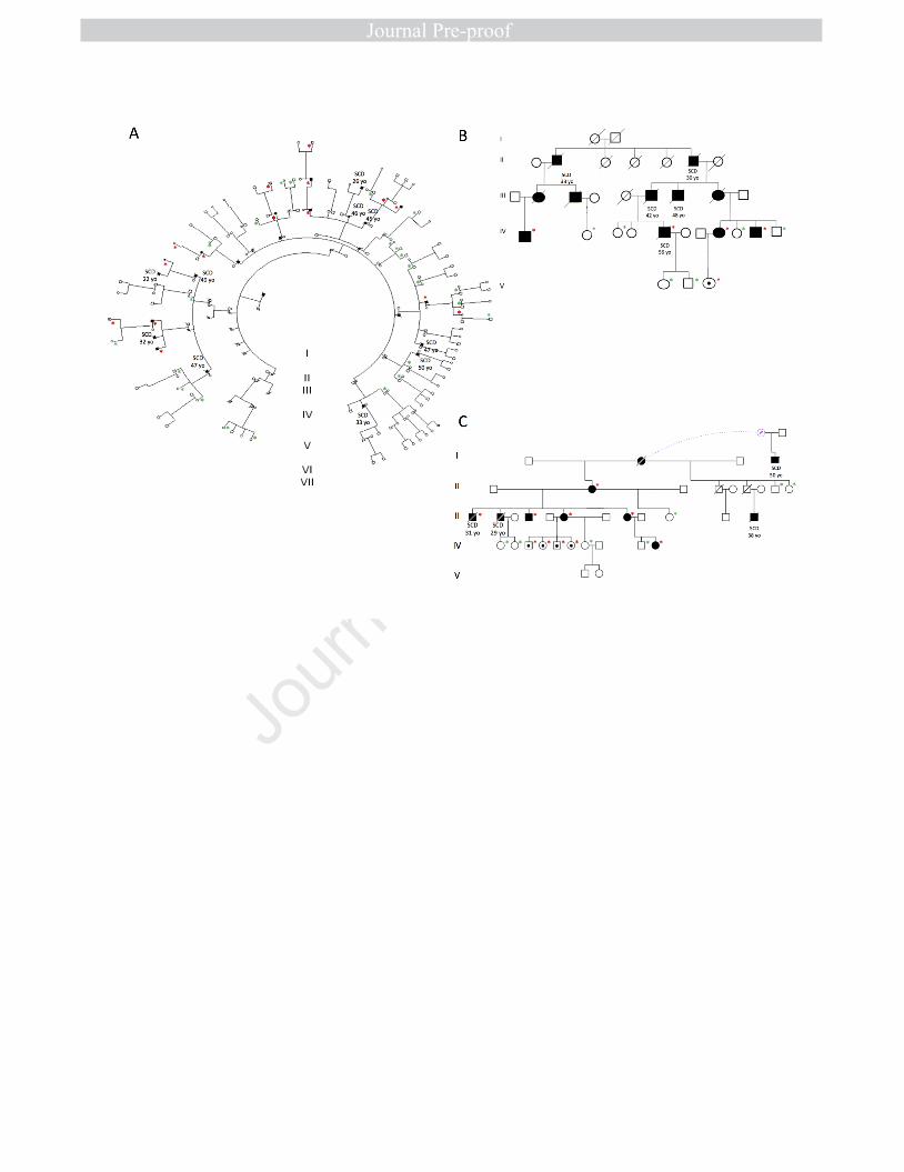
Figure 1. Pedigrees of the three Spanish families with the p.S358L mutation 3
in TMEM43. A: Family 1. Madrid, B: Family 2. Valencia, C: Family 3. Granada. 4
Red asterisk: confirmed genetic carriers, green asterisk: confirmed non-5
carriers. 6
SCD: Sudden cardiac death 7
8
Figure 2. Haplotypes surrounding TMEM43 in p.S358L mutation carriers of 9
three Spanish families compared with those from Newfoundland, Denmark 10
and North America. Markers from the family from Madrid are depicted in yellow 11
and those from the family from Granada are in green. The Newfoundland 12
markers appear framed and those shared with the other families are 13
underlined. Families from Madrid and Valencia only share 5 markers with 14
the Newfoundland family whereas the family from Granada shares 6 genetic 15
markers with the Newfoundland family. In contrast, the Danish and North 16
American families share with the Newfoundland family 10 and 8 markers, 17
respectively. Moreover, the latter share the 4 markers surrounding the 18
mutation with the Newfoundland family, confirming the presence of a 19
common ancestor. 20
21
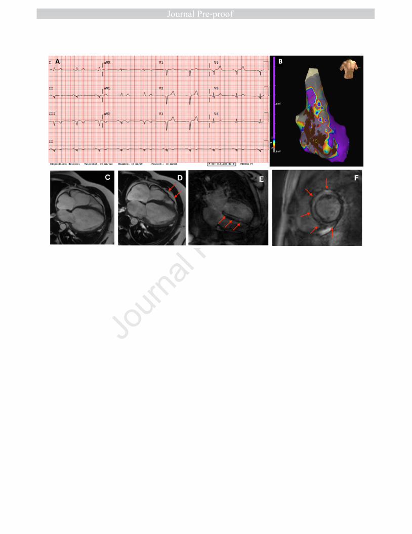
Figure 3. (A) Representative 12-lead ECG of a 55 year-old female ARVC-5 22
patient. Poor R wave progression with 1 mV R wave in V3 and widened QRS 23
(110 ms). (B) Three-dimensional voltage endomyocardial map of a 47 year-1
old ARVC-5 male with VT episodes who underwent substrate ablation. Grey 2
areas represent low voltage tissue (scar) along the interventricular septum 3
and inferior wall. Brown dots represent radiofrequency applications. (C,D) 4
CMR images of a 48 year-old ARVC5 male subject with biventricular dilatation, 5
wall motion abnormalities and dysynchronic contraction (red arrows) LVEF 6
35%, RVEF 46%. (E,F) CMR images of the same subject depicting severe and 7
almost concentric intramyocardial late gadolinium enhancement (red arrows). 8
9
Figure 4. Post-mortem study of the heart of an ARVC-5 TMEM43 p.S358L 10
heterozygous carrier and victim of SCD. (A,B) Macroscopic study showing 11
biventricular fibro-fatty infiltration, including transmural (RV), subendocardial 12
(RV and LV) and intramyocardial (LV) localizations. (C,D) Histologic view with 13
Masson´s trichrome staining depicting fatty infiltration (asterisks) and fibrosis 14
(arrowheads), together with cardiomyocyte degeneration (arrows). 15
16
17
Figure 5. Survival curve in 62 ARVC-5 affected patients according to sex. 18
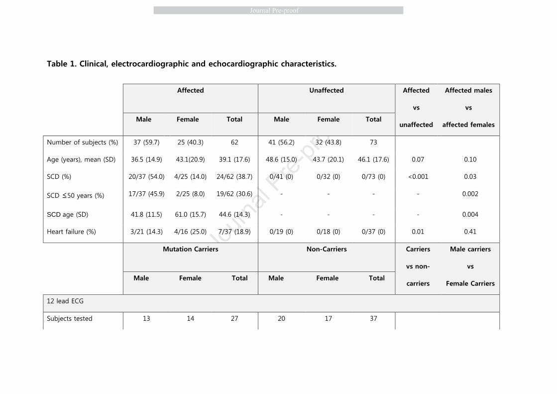
Table 1. Clinical, electrocardiographic and echocardiographic characteristics.
Affected
Unaffected Affected
vs
unaffected
Affected males
vs
affected females Male Female Total Male Female Total
Number of subjects (%) 37 (59.7) 25 (40.3) 62 41 (56.2) 32 (43.8) 73
Age (years), mean (SD) 36.5 (14.9) 43.1(20.9) 39.1 (17.6) 48.6 (15.0) 43.7 (20.1) 46.1 (17.6) 0.07 0.10
SCD (%) 20/37 (54.0) 4/25 (14.0) 24/62 (38.7) 0/41 (0) 0/32 (0) 0/73 (0) <0.001 0.03
SCD ≤50 years (%) 17/37 (45.9) 2/25 (8.0) 19/62 (30.6) - - - - 0.002
SCD age (SD) 41.8 (11.5) 61.0 (15.7) 44.6 (14.3) - - - - 0.004
Heart failure (%) 3/21 (14.3) 4/16 (25.0) 7/37 (18.9) 0/19 (0) 0/18 (0) 0/37 (0) 0.01 0.41
Mutation Carriers
Non-Carriers Carriers
vs non-
carriers
Male carriers
vs
Female Carriers Male Female Total Male Female Total
12 lead ECG
Subjects tested 13 14 27 20 17 37

QRS duration, ms (SD) 115.6 (27.1) 94.5 (15.5) 104.7 (24.0) 90.3 (9.9) 85.8 (7.1) 88.2 (8.9) <0.001 0.02
V3 R voltage, mV (SD) 2.6 (2.3) 3.6 (3.2) 3.2 (2.8) 8.6 (4.1) 6.2 (2.4) 7.5 (3.6) <0.001 0.35
Echocardiogram
Subjects tested 14 15 29 19 14 41
LVEF, % (SD) 53.6 (9.0) 58.7 (12.2) 56.2 (10.9) 63.3 (7.3) 60.8 (6.6) 62.1 (7.0) <0.001 0.21
LVEDD HF, % (SD) 113.0 (18.8) 98.7 (12.6) 106.9 (17.5) 97.8 (9.5) 94.4 (10.4) 96.6 (9.6) 0.07 0.13
LVEDV, ml (SD) 125.0 (43.6) 80.2 (12.9) 106.3 (40.4) 90.6 (15.64) 70.3 (16.62) 84.1 (18.3) 0.03 0.04
TAPSE, mm (SD) 17.5 (8.9) 22.6 (4.6) 19.9 (7.5) 23.5 (3.0) 23.6 (4.1) 23.5 (3.5) 0.03 0.10
RVBD 4CH, mm (SD) 40.8 (8.3) 32.3 (2.7) 35.6 (7.6) 35.9 (9.1) 32.50 (2.7) 34.8 (7.3) 0.60 0.053
4CH, four-chamber view; SCD, sudden cardiac death; SD, standard deviation; RVBD, right ventricle basal diameter; LVEF, left ventricular
ejection fraction; LVEDD, left ventricular end-diastolic diameter; LVEDV, left ventricular end-diastolic volume; TAPSE, tricuspid annular
plane systolic excursion.
Table 2. Impact of physical activity on clinical, electrocardiographic and echocardiographic characteristics in TMEM43-
p.S358L mutation carriers

Vigorous physical activity
Non-vigorous physical activity Vigorous
vs non-
vigorous
Vigorous PA in
males
vs
vigorous PA in
females
Male Female Total Male Female Total
Number of subjects, n
(%) 10/13 (76.9) 3/13 (23.1) 13 5/14 (35.7) 9/14 (64.3) 14
Age (years), mean (SD) 38.1 (18.6) 47.3 (9.6) 40.2 (17.1) 33.8 (14.7) 38.0 (16.9) 36.5 (15.7) 0.56 0.44
ICD implanted, n (%) 7/10 (70.0) 3/3 (100) 10/13 (76.9) 4/5 (90.0) 3/9 (33.3) 7/14 (50.0) 0.24 0.53
SVT/VF, n (%) 6/10 (60.0) 2/3 (66.7) 8/13 (61.5) 3/5 (60.0) 1/9 (11.1) 4/14 (28.6) 0.08 0.85
Heart failure, n (%) 1/10 (10.0) 1/3 (33.3) 2/13 (15.4) 1/5 (20.0) 1/9 (11.1) 2/14 (14.3) 1 0.42
12 lead ECG
Subjects tested, n 8 3 11 4 9 13
QRS width in ms, mean
(SD) 114.0 (31.7) 91.0 (12.7) 107.7 (29.2) 125.3 (7.81) 97.1 (18.1) 105.8 (21.5)
0.85 0.27
V3 R voltage in ms, 3.1 (2.8) 2.0 (2.0) 2.8 (2.6) 1.5 (1.3) 3.1 (2.4) 2.6 (2.2) 0.84 0.55

4CH, four-chamber view; PA: Physical activity, RVBD, right ventricle basal diameter; SVT, sustained ventricular tachycardia; VF,
ventricular fibrillation; SD, standard deviation; LVEF, left ventricular ejection fraction; LVEDD, left ventricular end-diastolic
diameter; LVEDV, left ventricular end-diastolic volume; TAPSE, tricuspid annular plane systolic excursion.
*: Only 1 subject available
mean (SD)
Echocardiogram
Subjects tested, n 7 3 10 3 9 12
LVEF, % (SD) 54.4 (9.5) 63.3 (6.4) 56.8 (9.4) 51.0 (9.3) 58.4 (12.7) 55.8 (11.8) 0.81 0.17
LVEDD, mm (SD) 53.4 (11.1) 48.0 (10.6) 47.5 (7.6) 52.4 (6.3) 44.5 (7.1) 51.8 (10.7) 0.86 0.49
LVEDV, ml (SD) 119.0 (49.3) 87.0 (10.5) 107.0 (41.2) 140.0 (32.5) 70.0 (9.9) 105.0 (44.9) 0.94 0.23
TAPSE, mm (SD) 16.6 (10.4) 17.5 (0.7) 16.7 (9.2) 19.5 (0.7) 21.0 (9.6) 20.7 (8.5) 0.32 0.90
RVBD 4CH, mm (SD) 42.2 (8.5) 30.0 (1.7) 37.6 (9.1) 34.0 * 32.0 (3.2) 32.4 (2.9) 0.17 0.06