Journal of Cell Science Accepted...
41
1 Epiprofin orchestrates epidermal keratinocyte proliferation and differentiation Takashi [Nakamura] 1, 2, 3 ‡, Yasuo [Yoshitomi] 3 , Kiyoshi [Sakai] 3 , Vyomesh [Patel] 4 , Satoshi [Fukumoto] 1 , and Yoshihiko [Yamada] 3 ‡ 1 Division of Pediatric Dentistry, Department of Oral Health and Development Sciences, and 2 Liaison Center for Innovative Dentistry, Tohoku University Graduate School of Dentistry, Sendai, Japan 3 Laboratory of Cell and Developmental Biology and 4 Oral and Pharyngeal Cancer Branch, National Institute of Dental and Craniofacial Research, National Institutes of Health, Bethesda, MD Running title: Epiprofin function in skin development Keywords: Sp transcription factor; Development of skin; Proliferation; Differentiation; Keratinocytes; Transit amplifying cells; Stem cells; p63/Notch Word numbers: 7,821 ‡Address correspondence to: Yoshihiko Yamada, Bldg. 30, Rm. 407, National Institute of Dental and Craniofacial Research, NIH, Bethesda, MD 20892. Tel: 301-496-2111, Fax: 301-402-0897, E-mail: [email protected] © 2014. Published by The Company of Biologists Ltd. Journal of Cell Science Accepted manuscript JCS Advance Online Article. Posted on 24 October 2014
Transcript of Journal of Cell Science Accepted...
1
Epiprofin orchestrates epidermal keratinocyte proliferation and differentiation
Takashi [Nakamura]1, 2, 3
‡, Yasuo [Yoshitomi]3, Kiyoshi [Sakai]
3, Vyomesh [Patel]
4,
Satoshi [Fukumoto]1, and Yoshihiko [Yamada]
3‡
1Division of Pediatric Dentistry, Department of Oral Health and Development Sciences,
and 2Liaison Center for Innovative Dentistry, Tohoku University Graduate School of
Dentistry, Sendai, Japan
3Laboratory of Cell and Developmental Biology and
4Oral and Pharyngeal Cancer
Branch, National Institute of Dental and Craniofacial Research, National Institutes of
Health, Bethesda, MD
Running title: Epiprofin function in skin development
Keywords: Sp transcription factor; Development of skin; Proliferation; Differentiation;
Keratinocytes; Transit amplifying cells; Stem cells; p63/Notch
Word numbers: 7,821
‡Address correspondence to: Yoshihiko Yamada, Bldg. 30, Rm. 407, National Institute of
Dental and Craniofacial Research, NIH, Bethesda, MD 20892. Tel: 301-496-2111, Fax:
301-402-0897, E-mail: [email protected]
© 2014. Published by The Company of Biologists Ltd.Jo
urna
l of C
ell S
cien
ceA
ccep
ted
man
uscr
ipt
JCS Advance Online Article. Posted on 24 October 2014
2
Abstract
The basal layer of the epidermis contains stem cells and transit amplifying (TA) cells
that rapidly proliferate and differentiate further into the upper layers of the
epidermis. A number of molecules have been identified as regulators for this process
including p63 and Notch1. However, little is known about the mechanisms that
regulate the transitions from stem cells to proliferating or differentiating TA cells.
Here we demonstrate that Epiprofin (Epfn) plays critical distinct roles in these
transition stages as a cell cycle regulator and a transcription factor. Epfn knockout
mice have a thickened epidermis, in which p63-expressing basal cells formed
multiple layers due to accumulation of premature TA cells with reduced
proliferation, and a reduction in differentiating keratinocytes expressing Notch1. We
found that low levels of Epfn expression increased proliferation of human
immortalized keratinocyte (HaCaT) cells by increasing EGF-responsiveness and
superphosphorylation of Rb. In contrast, high levels of Epfn expression promoted
cell cycle exit and differentiation, by reducing E2F transactivation and inducing
Notch1 expression. Our findings identify multiple novel functions of Epiprofin in
epidermal development.
Jour
nal o
f Cel
l Sci
ence
Acc
epte
d m
anus
crip
t
3
Introduction
Skin self-renews throughout life by constantly replenishing keratinocytes in the outer-
most layer of the epidermis by balancing the production of stem cells and transit
amplifying (TA) cells, which are the committed keratinocyte precursors (Jones and Watt,
1993). The epidermis arises from a single ectodermal layer and renews by highly
coordinated regulatory programs controlling proliferation and differentiation. The
epidermis consists of four distinct layers that are mostly composed of keratinocytes in
different stages of maturation. The basal layer of the epidermis contains stem cells and
TA cells. These TA cells divide a limited number of times (3−5) before commitment to
terminal differentiation. Basal keratinocytes attach to the underlying basement membrane
through integrins, such as 31 and 64. Other integrins, such as 21 and 51 for
collagen/laminin and fibronectin binding, respectively, are also involved in basal
keratinocyte interaction with the basement membrane (Burgeson and Christiano, 1997).
When TA cells eventually stop proliferating and begin to differentiate, expression of these
integrins is reduced, resulting in detachment of keratinocytes from the basement
membrane and cell movement toward the upper layers of the epidermis (Fuchs, 2008).
Proliferation of TA cells is stimulated by the mitogenic epidermal growth factor
(EGF) via the EGF receptor (EGFR)/ERK/MAPK signaling pathway. Although the
EGFR is also expressed by stem cells, these cells generally respond poorly to EGF
(Jensen and Watt, 2006; Powell et al., 2012; Wong et al., 2012). The difference in EGF
responsiveness is due in part to the reduced levels of the Lrig proteins in TA cells, which
negatively regulate EGF-induced signaling by promoting the ubiquitination and
degradation of EGFR (Jensen and Watt, 2006; Laederich et al., 2004; Watt and Jensen,
2009). Expression of Lrig1 is essential for epidermal homeostasis; Lrig1 knockout mice
show epidermal hyperplasia, and knockdown of Lrig1 expression in cultured human
keratinocytes induces stem cell expansion (Jensen and Watt, 2006; Suzuki et al., 2002).
Several cell cycle regulators have been implicated in skin morphogenesis. For example,
retinoblastoma protein (Rb) and the associated pocket proteins (p107, p130) regulate
epidermal differentiation and maintenance (Ruiz et al., 2004).
Jour
nal o
f Cel
l Sci
ence
Acc
epte
d m
anus
crip
t
4
Coordinated epidermal differentiation programs are regulated by a series of genes.
Among them, Notch1 and p63 (deltaN) are critical regulators of epidermal keratinocyte
development (Blanpain et al., 2006). The transcription factor p63 is necessary for the
proper development of skin, teeth, limbs, and craniofacial tissues. In the epidermis, p63 is
required to maintain the population of stem cells and TA cells and to ensure TA cell
commitment to the epidermal cell lineage (Koster et al., 2004; Mills et al., 1999;
Pellegrini et al., 2001; Romano et al., 2012; Truong et al., 2006; Yang et al., 1999). A
deficiency of p63 results in the absence of keratinocyte stratification and differentiation
(Mills et al., 1999; Yang et al., 1999). Loss of Notch signaling also disrupts epidermal
differentiation (Lowell et al., 2000; Rangarajan et al., 2001) as evidenced by
hyperkeratosis, while overexpression of activated Notch1 in basal keratinocytes induces
the loss of proliferative capacity and induction of premature differentiation (Blanpain et
al., 2006; Lefort et al., 2007; Nguyen et al., 2006). The expression of Notch1 and p63 in
the epidermis is mutually exclusive: Notch1 is primarily expressed by differentiating
keratinocytes, while p63 expression occurs in the basal layer, which mainly contains stem
cells and maturing TA cells (Nguyen et al., 2006). A negative regulatory loop involving
p63 and Notch signaling controls TA cell commitment to keratinocyte differentiation.
Thus, the balance between p63 and Notch1 expression and signaling is crucial for the
development and controlled renewal of the epidermis. However, it is not clear how TA
cell cycle exit is promoted and Notch1 transcription is induced during the transition stage
from proliferation to differentiation.
We previously identified Epiprofin (Epfn/Sp6) based on mRNA analysis of tooth
germs and demonstrated its expression in certain ectodermal tissues such as teeth, skin,
hair follicles, and limb buds (Nakamura et al., 2004). Epfn is a member of the Sp family
of transcription factors, which contains a transactivation/suppressor domain at the N-
terminus and three C2H2 zinc finger motifs at the C-terminus for DNA binding (Suske et
al., 2005). Epfn knockout (Epfn–/–) mice display striking phenotypic features, including
defects in tooth morphology, supernumerary tooth formation, digit fusion, skin
abnormalities, and hairlessness (Hertveldt et al., 2008; Ibarretxe et al., 2012; Nakamura et
al., 2008; Talamillo et al., 2010). In addition to excess teeth, Epfn–/– mice display severe
enamel hypoplasia due to defects in dental epithelial differentiation into enamel matrix-
Jour
nal o
f Cel
l Sci
ence
Acc
epte
d m
anus
crip
t
5
secreting ameloblasts (Nakamura et al., 2008). In the absence of Epfn, the inner dental
epithelium loses its characteristic ability for rapid proliferation, leading to severe enamel
hypoplasia (Nakamura et al., 2008). Therefore, Epfn may be a multifunctional regulator
of both proliferation and differentiation, depending on the developmental stage.
In this study, we demonstrated that Epfn−/− mice develop hyperplastic epidermis
and hyperkeratosis of the skin. In the Epfn−/− epidermis, p63-expressing basal
keratinocytes form multiple cell layers, and Notch1 expression is reduced. We found that
in the absence of Epfn, premature TA (pre-TA) cells with reduced proliferation activity
accumulate and show partial commitment to differentiation. In cell culture, Epfn
promotes proliferation of keratinocyte progenitors at low levels of Epfn expression, while
high Epfn expression promotes cell cycle exit and induces differentiation through
inducing Notch1. Our findings reveal that Epfn is a novel regulator of epidermal
development.
Jour
nal o
f Cel
l Sci
ence
Acc
epte
d m
anus
crip
t
6
Results
Defects in skin development and in morphogenesis in Epfn−/− mice
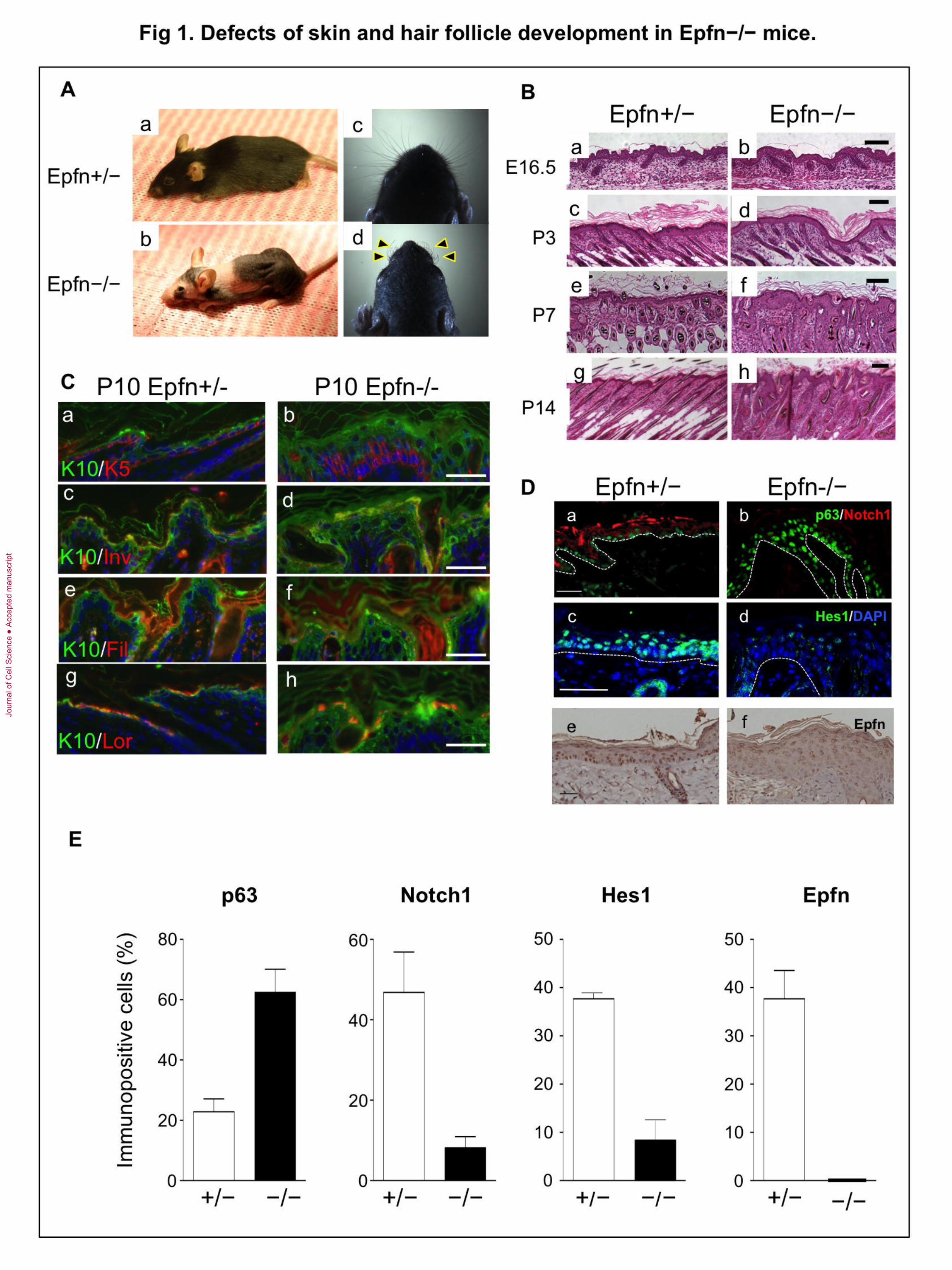
Homozygous Epiprofin knockout (Epfn−/−) mice are smaller than normal and display
defects in both skin development and hair formation (Fig. 1Ab), as well as the tooth
dysmorphogenesis as previously described (Hertveldt et al., 2008; Nakamura et al.,
2008). In contrast, heterozygous littermates (Epfn+/−) show no obvious phenotype (Fig.
1Aa). Knockout mice are hairless in most areas and have short, thin, wrinkled whiskers
(Fig. 1Ac, d). There are no apparent histological abnormalities in the epidermal and
pelage hair follicles of Epfn−/− mice until approximately embryonic day 16.5 (Fig. 1Ba,
b). After this stage, however, abnormalities in the epidermis and hair follicles become
progressively more severe. After postnatal day 7 (P7), Epfn−/− epidermal layers are
significantly thicker than those of the heterozygotes, due to hyperkeratosis and increased
hypercellularity (Fig. 1Bc–h).
We subsequently examined possible defects in epidermal differentiation at P10 by
double-immunofluorescence staining using antibodies for the differentiation markers
(keratins) K5, K10, involucrin, filaggrin, and loricrin (Fig. 1C). In control Epfn+/−
littermates, K5 was expressed in the basal epidermal layer (Fig. 1Ca), whereas K10 was
expressed primary in the suprabasal layer of the epidermis (Fig. 1Ca, c, e, g). In Epfn−/−
mice, the K5- and K10-positive cells formed multiple cell layers where K5-positive cells
were located on the bottom part of the thickened epidermis as compared to the K10-
positive cells (Fig. 1Cb). Involucrin, filaggrin, and loricrin were expressed in
differentiating keratinocyte layers in the control epidermis (Fig. 1Cc, e, g), while the
expression of these genes was reduced in the Epfn-/- epidermis (Fig. 1Cd, f, h).
To address the molecular basis of the abnormal formation of multiple keratinocyte
layers in the Epfn−/− epidermis, we analyzed the expression of p63 and Notch1, which
regulate cell fate and keratinocyte differentiation in the epidermis (Fig. 1Da, b). In the
control P13 epidermis of heterozygous mice, p63 was expressed in the basal layer, and
Notch1 expression was observed in differentiating keratinocytes, in agreement with
previous reports (Fig. 1Da, b) (Kurata et al., 2004; Mills et al., 1999; Rangarajan et al.,
2001). In the Epfn−/− epidermis, the p63-positive cells formed multiple layers (Fig. 1Db)
Jour
nal o
f Cel
l Sci
ence
Acc
epte
d m
anus
crip
t

7
and the number of p63-expressing cells was increased (Fig. 1E), while Notch1 expression
was virtually undetectable in the epidermis (Fig. 1Db, E). Expression of Hes1, a
transcription factor and downstream target of Notch, was also considerably lower in the
Epfn−/− epidermis compared with the heterozygotes (Fig. 1Dc, d, E), indicating
perturbed Notch signaling in the Epfn−/− epidermis. Finally, Epfn was expressed in basal
layer keratinocytes and in differentiating keratinocytes in the epidermis during embryonic
stages in the control epidermis, but not in the Epfn−/− epidermis (Fig. 1De, f, E). The
schematic diagram of the expression pattern for p63, Notch1, and Epfn in the epidermis is
shown in Supplemental Fig. 1.
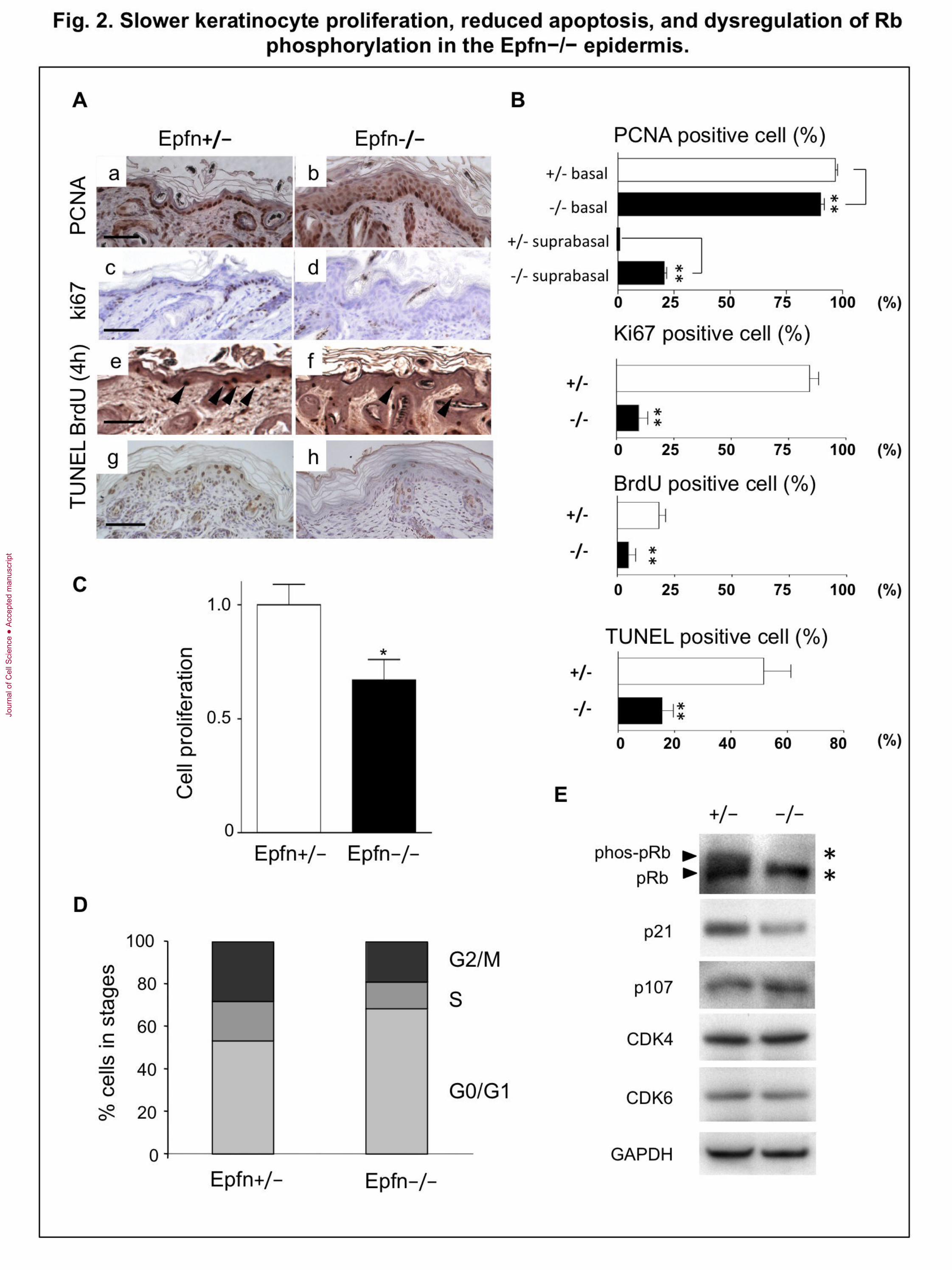
Reduced keratinocyte proliferation and apoptosis in the Epfn−/− epidermis
The epidermis of Epfn−/− mice exhibited multiple layers of K5- and p63-expressing
basal cells (Fig. 1C), suggesting dysregulation of both cell proliferation and apoptosis.
We examined proliferation in the Epfn−/− epidermis by immunostaining for the
proliferating cell nuclear antigen PCNA (a marker for late G1 and S phases), Ki67, and
by BrdU incorporation, while apoptosis was analyzed by TUNEL staining (Fig. 2A, B).
In the P7 Epfn+/− epidermis, the majority of the basal epidermal keratinocytes formed a
single cell layer, and most of the cells were PCNA-positive (Fig. 2Aa, 2B). The number
of PCNA-positive cells in the basal layer was significantly lower in the Epfn−/−
epidermis, but the total number of cells exhibiting some PCNA immunoreactivity was
higher in the Epfn−/− epidermis because of the long half-life of PCNA and the
hypercellularity (Fig. 2Aa, b; Fig. 2B). Many Ki67-positive cells were detected in the
basal layer of the P7 Epfn+/− epidermis, while the number of Ki67-positive cells were
reduced in the Epfn−/− epidermis (Fig. 2Ac, d; Fig. 2B). Similarly, short-term
incorporation of BrdU for 4 h to detect TA cells revealed that a significantly greater
number of basal cells were proliferating in the control P7 Epfn+/− epidermis, compared
to the Epfn−/− epidermis (Fig. 2Ae, f; Fig. 2B). These results suggest that TA cell
proliferation is inhibited in the Epfn−/− epidermis. However, these cells accumulate,
resulting in hypercellularity. In addition, TUNEL staining analysis revealed that the
number of apoptotic cells in P3 Epfn−/− epidermis was significantly reduced compared
with that in the Epfn+/− epidermis (Fig. 2Ag, h; Fig. 2B). The lower rate of programmed
Jour
nal o
f Cel
l Sci
ence
Acc
epte
d m
anus
crip
t

8
cell death in the early postnatal mutant epidermis may have contributed to the observed
increase in epidermal layer thickness and hypercellularity in Epfn−/− mice.
Results thus far indicate that loss of Epfn disrupts the normal balance of TA cell
proliferation and differentiation that is necessary for proper skin morphogenesis. To
examine the effects of Epfn on cell proliferation under controlled conditions, we used
primary keratinocytes isolated from the epidermis of newborn Epfn+/− and Epfn−/−
mice. There were significantly fewer cells in cultures derived from Epfn−/− mice
compared with wild-type cultures after 4 days in serum-free and low-Ca2+
(differentiation-restricted) media (KGM), suggesting that Epfn normally promotes
keratinocyte proliferation (Fig. 2C). FACS analysis revealed that approximately half of
the keratinocytes from the Epfn+/− epidermis were in the proliferating phases (G2/M and
S), while the majority (approximately 70%) of the keratinocytes from the Epfn−/−
epidermis were in the quiescent G0/G1 phase (Fig. 2D). Taken together, these results
suggest that Epfn is essential for TA cell proliferation.
To determine the molecular pathways leading to cell cycle disruption in
keratinocytes from Epfn−/− mice, we examined the expression and phosphorylation
status of the cell cycle regulators Rb, p21 (cyclin-dependent kinase inhibitor 1), and p107
(Rbl1), and the cyclin-dependent kinases CDK4 and CDK6 (Fig. 2E). Cultured
keratinocytes from the control Epfn+/− epidermis expressed both the phosphorylated
form of Rb (phos-Rb) and the unphosphorylated form of Rb. The expression of Phos-Rb
and p21 was reduced in Epfn−/− keratinocytes but the p107 expression was not. The
cyclin-dependent kinases CDK4 and CDK6 were expressed at similar levels in both cell
types. These results suggest that Epfn promotes keratinocyte proliferation by regulating
Rb phosphorylation and p21 expression (Fig. 2E).
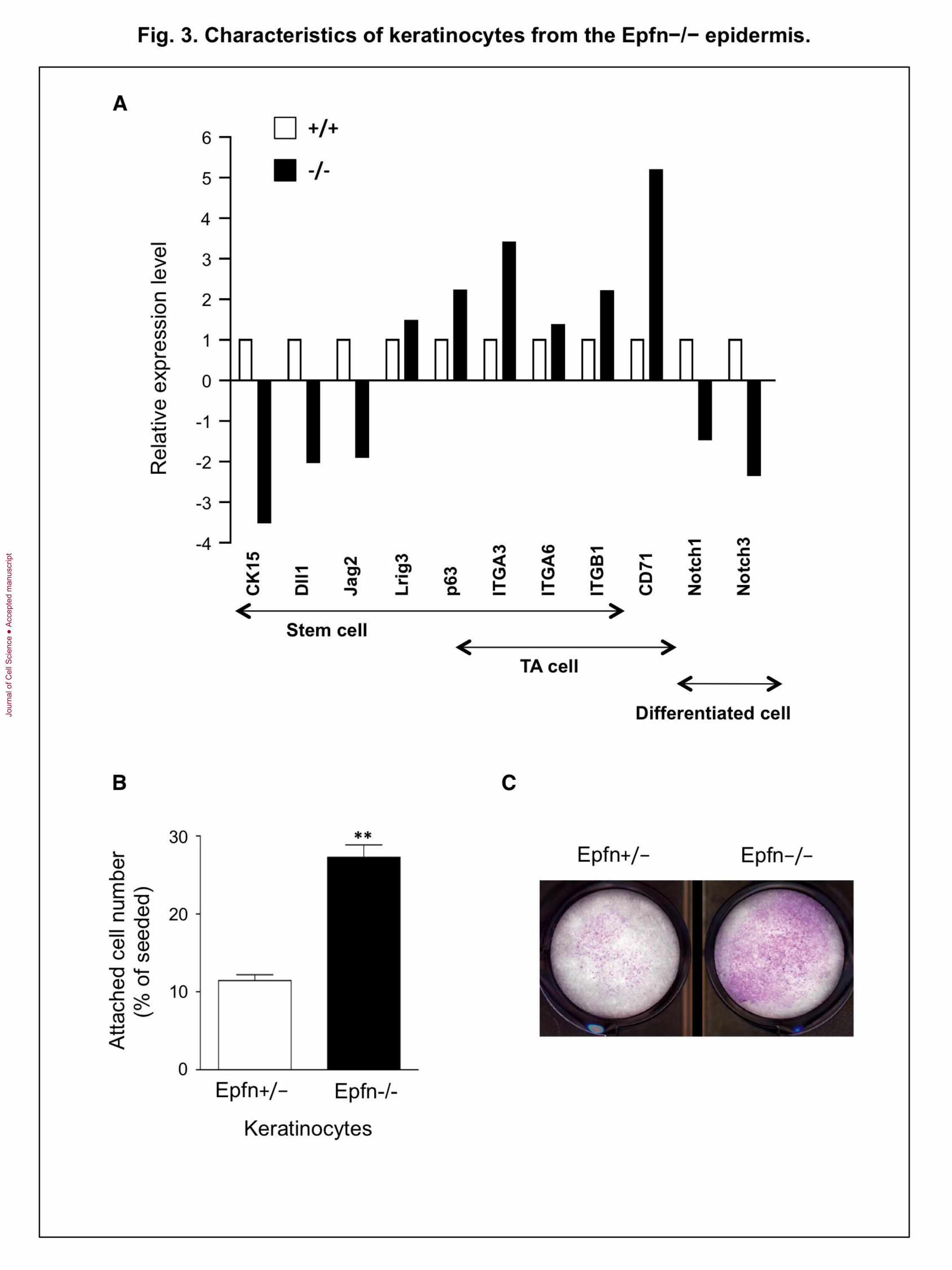
Accumulation of premature TA cell-like keratinocytes in the Epfn−/− epidermis
The basal epidermis of Epfn−/− mice exhibited ectopic expression of keratins, and basal
keratinocyte-like cells expressing K5 and p63 formed multiple cell layers (Fig. 1).
Moreover, isolated keratinocytes from the Epfn−/− epidermis proliferated more slowly
compared with keratinocytes derived from the Epfn+/− epidermis (Fig. 2C). To identify
additional genes regulated by Epfn, we compared the expression of genes characteristic
Jour
nal o
f Cel
l Sci
ence
Acc
epte
d m
anus
crip
t
9
of normal stem cells and TA cells in Epfn−/− and Epfn+/+ primary keratinocytes using
microarray analysis (Fig. 3A). In Epfn−/− keratinocytes, stem cell markers such as CK15
(cytokeratin 15) and the Notch ligands Dll1 and Jag2 were significantly downregulated
compared with wild-type cells, while other markers, such as Lrig3 and p63, were
upregulated. The integrins 3 (ITGA3), 6 (ITGA6), and 1 (ITGB1), which are markers
for both stem cells and TA cells, and CD71 (transferrin receptor), a marker of TA cells,
were upregulated in Epfn−/− keratinocytes. However, Notch1 and Notch3, markers of
differentiated keratinocytes, were downregulated in Epfn−/− keratinocytes, consistent
with immunohistochemical observations using the antibodies to Notch1 and Hes1 (Fig.
1D, E). These differences in gene expression between Epfn +/+ and Epfn−/−
keratinocytes were confirmed by quantitative PCR analysis using individual gene primers
(data not shown). Therefore, the premature TA-like (pre-TA) cells that accumulated in the
Epfn−/− epidermis have an immature phenotype, retaining certain stem cell marker genes
while expressing only some TA cell-specific genes. Moreover, accumulated pre-TA cells
in the Epfn−/− epidermis are not capable of rapid proliferation, which is a key
characteristic of normal TA cells.
Basal keratinocytes express integrins such as 31, 64, and 51 at the basal
cell surface that act to anchor immature cells into the underlying basement membrane
(Burgeson and Christiano, 1997). When TA cells differentiate, expression of these
integrins is reduced and the cells detach and migrate toward the surface layers (Fuchs,
2008). However, immunostaining of the basal Epfn−/− epidermis revealed integrin 6
expression over the entire peripheral cell surface (data not shown), consistent with an
immature phenotype. In vitro, stem cells rapidly attach to fibronectin-coated wells within
15 min of incubation because of strong integrin expression (Jones and Watt, 1993). To
characterize the functional effects of integrin expression by pre-TA cells in the Epfn−/−
epidermis, we analyzed the attachment activity of keratinocytes from the Epfn−/−
epidermis to fibronectin (Fig. 3B). Approximately 30% of the keratinocytes from the
Epfn−/− epidermis attached to fibronectin-coated wells within 15 min of incubation,
compared with only 10% of the keratinocytes from Epfn+/− mice. Colony-forming
assays confirmed that the Epfn−/− keratinocytes formed more cell colonies than the
Jour
nal o
f Cel
l Sci
ence
Acc
epte
d m
anus
crip
t
10
Epfn+/− keratinocytes (Fig. 3C), indicating that these Epfn−/− keratinocytes retain
certain stem cell-like and immature cell properties.
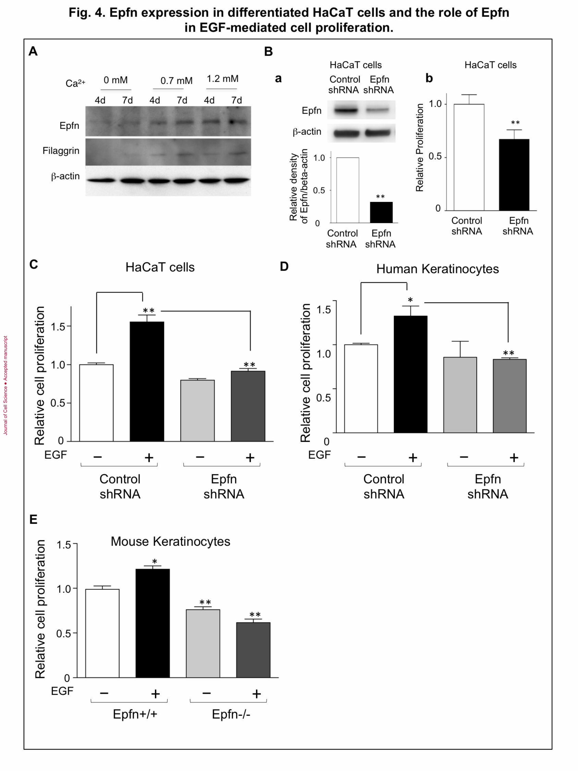
Roles of Epfn in proliferation of HaCaT cells and keratinocytes
To address the mechanism of Epfn actions in the proliferation, we used the human
epidermal keratinocyte cell line HaCaT, which proliferates in a serum-free and low-Ca2+
keratinocyte growth medium (KGM) but can be induced to differentiate in KGM
containing a high Ca2+
concentration (Boukamp et al., 1988). HaCaT cells express Epfn at
a low level under the proliferation medium (Fig. 4A left two lanes, no exogenous Ca2+
addition). Elevating the Ca2+
levels in the medium by the addition of 0.7 mM or 1.2 mM
Ca2+
increased Epfn expression as well as markers of keratinocyte differentiation such as
filaggrin (Fig. 4A, right four lanes). Transfection of a targeted Epfn shRNA into HaCaT
cells reduced the endogenous Epfn RNA and protein levels (Fig. 2Ba). Epfn shRNA
significantly reduced HaCaT cell proliferation (Fig. 4Bb) in the low-Ca2+
medium,
consistent with the results in the primary keratinocyte cultures from the Epfn−/− mice
(Fig. 2C). We observed similar results using a different Epfn shRNA (data not shown).
One possible explanation for the reduced proliferation of Epfn−/− keratinocytes
may be the reduction in EGF-mediated proliferation. The Lrig family of proteins
promotes EGFR degradation and negatively regulates EGF-mediated stem cell
proliferation, thereby maintaining a slow proliferation (Cai et al., 2009; Laederich et al.,
2004; Muller et al., 2013; Rondahl et al., 2013). The microarray analysis showing
upregulated Lrig3 expression in Epfn−/− keratinocytes (Fig. 3A) suggests that Epfn
normally inhibits Lrig expression and that Epfn deficiency leads to a loss of the EGF-
mediated proliferation of keratinocytes. We explored these possibilities by testing the
EGF response in the control shRNA- and in the Epfn shRNA-transfected HaCaT cells
(Fig. 4C). The HaCaT cells were incubated in the absence or the presence of EGF for
three days and the cell proliferation was analyzed by colorimetric assays using the WST-
8 Kit. EGF increased proliferation of the control shRNA-transfected HaCaT cells, while
the Epfn shRNA-transfected HaCaT cells did not respond to EGF (Fig. 4C). The
expression level of Lrig1 was increased in the Epfn knockdown HaCaT cells
(Supplemental Fig. 2). We confirmed the involvement of Epfn in EGF-mediated
Jour
nal o
f Cel
l Sci
ence
Acc
epte
d m
anus
crip
t
11
proliferation in primary human keratinocytes transfected with the same control shRNA-
and Epfn shRNA used for HaCaT cells (Fig. 4D). Two different Epfn shRNAs reduced
the expression of endogenous Epfn in human keratinocytes (Supplemental Fig. 3). EGF
treatment increased proliferation of primary human keratinocytes, but it did not increase
proliferation of the Epfn shRNA-transfected cells (Fig. 4D). Similarly, the primary mouse
Epfn−/− keratinocytes from the Epfn−/− epidermis did not respond to EGF (Fig. 4E).
These results suggest that Epfn promotes EGF-mediated TA cell proliferation in part by
suppressing Lrig expression.
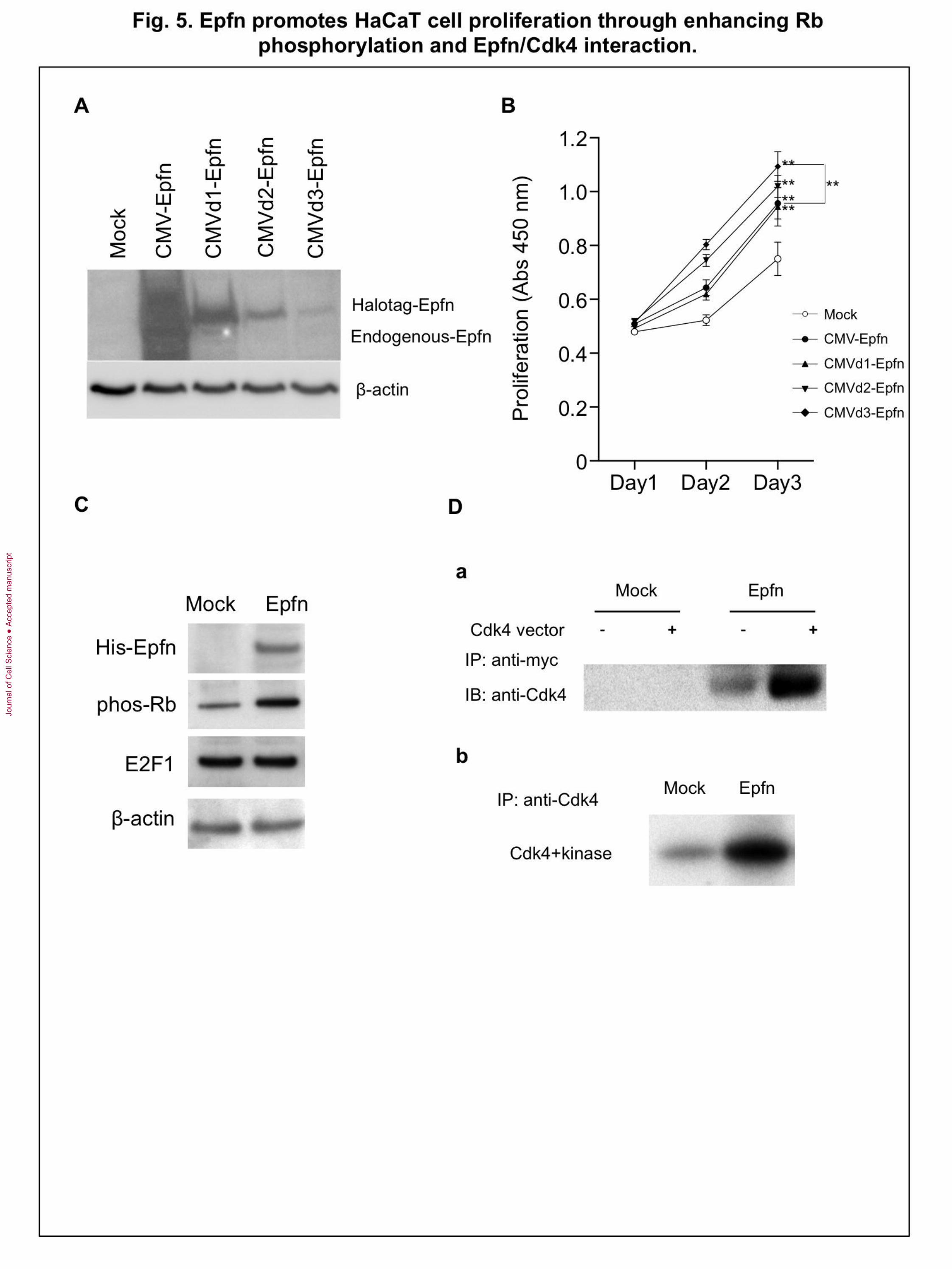
Epfn promotes HaCaT cell proliferation by forming a complex with CDK4 and
enhancing Rb phosphorylation
Next we examined whether Epfn promotes proliferation of keratinocytes through
interacting with cell cycle signaling molecules using HaCaT cells. We analyzed the dose-
dependent effect of Epfn expression on HaCaT cell proliferation two days after
transfection of HaCaT cells with a Halo-tagged Epfn expression vector under the control
of various deletions of CMV promoters (Fig. 5A). Western blotting using an anti-Epfn
antibody revealed a gradual decrease in the expression of recombinant Halo-tagged Epfn
with sequential deletions in the CMV promoter region (CMV, CMVd1-d3) (Fig. 5A). We
found that low levels of Epfn expression (CMVd2-d3) promoted proliferation of HaCaT
cells compared with high levels of Epfn expression (CMV, CMVd1) (Fig. 5B). These
results suggest that low levels of Epfn expression are involved in promoting proliferation.
To identify the molecular mechanism for Epfn-mediated cell proliferation, we
examined the expression of cell cycle regulators in HaCaT cells transfected with a His-
tagged Epfn expression vector (Fig. 5C). Epfn expression increased phosphorylation of
Rb (phos-Rb) (Fig. 5C) but had no effect on Rb protein levels (data not shown), and did
not affect the expression of E2F1, a transcription factor that binds to Rb in a cell cycle-
dependent manner and is a critical regulator of cell cycle control. Epfn-induced Rb
phosphorylation is consistent with decreased phos-Rb in Epfn−/− keratinocytes (Fig.
2E). Rb phosphorylation by the complex of cyclin D/CDK from the G1 to S phase of the
cell cycle results in the release of active E2F from the E2F/Rb complex. Consequently,
E2F activates the transcription of genes that drive cell cycle progression. Because Epfn
Jour
nal o
f Cel
l Sci
ence
Acc
epte
d m
anus
crip
t

12
promoted Rb phosphorylation in HaCaT cells (Fig. 5C), we speculated that Epfn might
also interact with the cyclin D/CDK complex. As CDK4 is a critical regulator of Rb
phosphorylation, we assessed possible interactions between Epfn and CDK4 by co-
immunoprecipitation assays (Fig. 5Da). HEK293T cells were cotransfected with Myc-
tagged Epfn and CDK4 expression vectors, and the extracts were immunoprecipitated
with an anti-Myc antibody and analyzed by Western blotting using an anti-CDK4
antibody. The CDK4 antibody recognized the protein complex pulled down by anti-Myc,
strongly suggesting that Epfn directly or indirectly complexes with CDK4 (Fig. 5Da).
Moreover, the isolated Epfn/CDK4 complex strongly enhanced Rb phosphorylation in
vitro (Fig. 5Db). These results suggest that Epfn complexes with CDK4 and that this
Epfn/CDK4 complex promotes cell proliferation by enhancing phosphorylation of Rb
(Scheme in Fig. 7B).
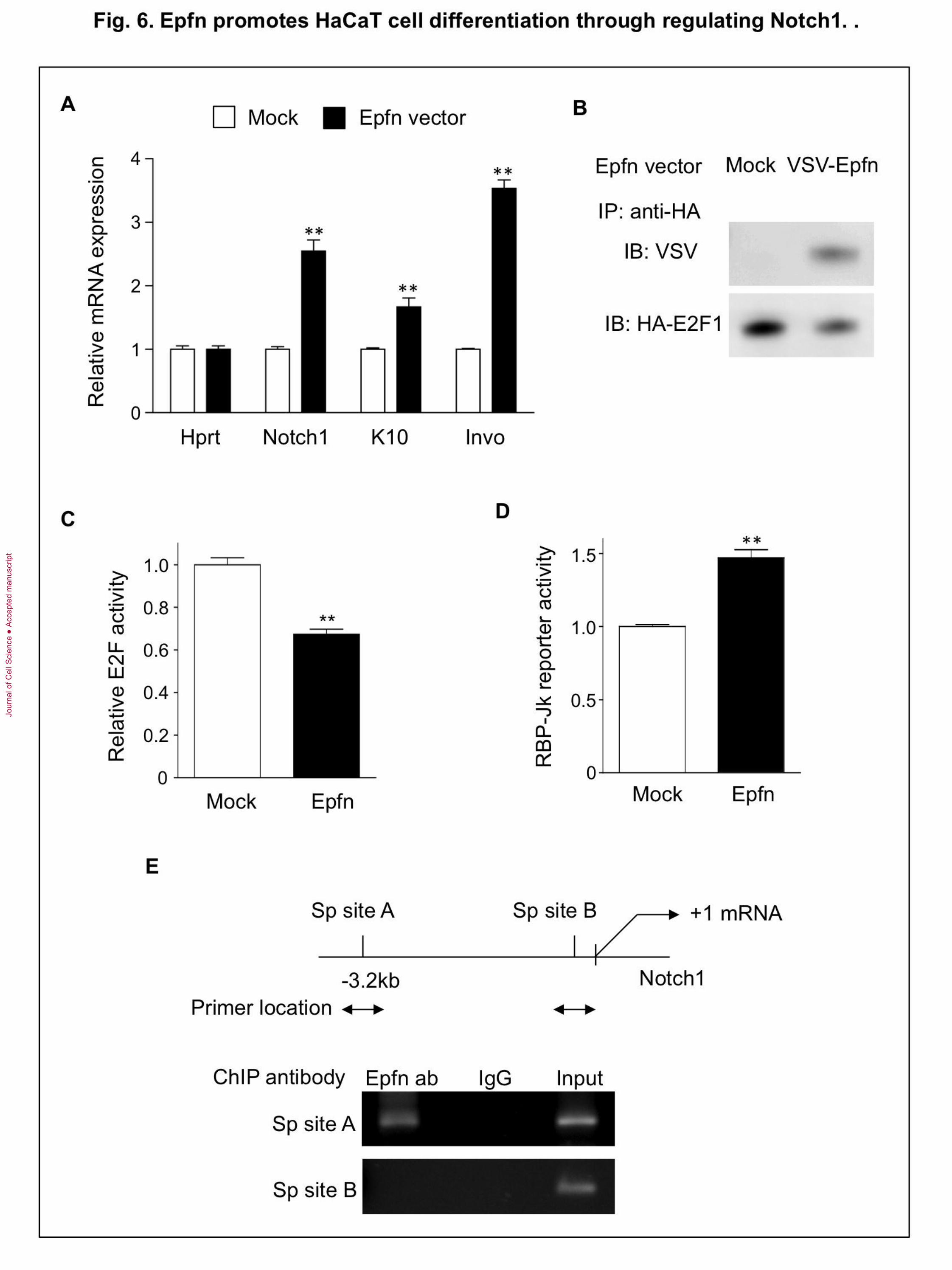
Epfn promotes cell cycle exit and differentiation
TA cells rapidly proliferate, but only for a limited number of divisions before cell cycle
exit for terminal differentiation. The factor involved in “switching” this transition is not
clear. Epfn is continuously expressed from basal and differentiating keratinocytes in the
epidermis, while in the Epfn−/− epidermis, keratinocyte differentiation is reduced. In cell
culture, Epfn expression is induced during HaCaT cell differentiation in the high-Ca2+
media (Fig. 4A). Therefore, Epfn may play a pivotal role in promoting cell cycle exit and
triggering differentiation processes. To address this hypothesis, we first examined the
ability of Epfn to promote differentiation of HaCaT cells in the low-Ca2+
medium. We
found that Epfn overexpression in HaCaT cells induced mRNA expression for Notch1,
K10, and involucrin (Invo) compared with that of HaCaT cells transfected with a control
vector (Fig. 6A). These results suggest that overexpression of Epfn promotes the
expression of keratinocyte differentiation markers.
E2F transcription factors activate genes that promote cell cycle progression, and
therefore, they can induce cell cycle re-entry. In addition, E2F1 forms a heterocomplex
with Sp family transcription factors (Lin et al., 1996; Rotheneder et al., 1999). Therefore,
we examined the interactions between Epfn and E2F1 by cotransfection of HaCaT cells
with the VSV-G-tagged Epfn and HA-tagged E2F1 expression vectors. The extracts were
Jour
nal o
f Cel
l Sci
ence
Acc
epte
d m
anus
crip
t

13
immunoprecipitated with an anti-HA antibody and Western blotted using anti-E2F1 and
ant-VSV antibodies. We found that Epfn coprecipitated with E2F1 (Fig. 6B), suggesting
that Epfn forms a protein complex with E2F1. We subsequently examined E2F
transactivation activity using an E2F reporter construct in HaCaT cells transfected with
the E2F1 and Epfn expression vectors (Fig. 6C). Epfn and E2F1 expression reduced E2F-
mediated transcriptional activity, suggesting that Epfn inhibited E2F activity and cell
cycle progression by sequestering the protein within an Epfn/E2F complex. Complex
formation between Epfn and E2F when Epfn expression is elevated may explain why
maturing TA cells exit the cell cycle after only a limited number of cell divisions (Fig. 7).
Epfn promotes Notch1 transcription in HaCaT cell differentiation
Notch1 promotes keratinocyte differentiation (Rangarajan et al., 2001). It is therefore
likely that Epfn overexpression promotes HaCaT cell differentiation through Notch1
activation. To examine whether or not Epfn induces Notch signaling, we used an RBP-Jk
reporter construct since RBP-Jk is a DNA binding factor and interacts with Notch as a
canonical Notch target protein (de la Pompa et al., 1997). We found that Epfn transfection
significantly increased RBP-Jk reporter activity (Fig. 6D), suggesting that the Epfn-
mediated increase in Notch1 mRNA led to the activation of Notch1 signaling. We further
examined Epfn-induced Notch1 transcription by Epfn binding to the Notch1 promoter by
ChIP assays. The human Notch1 promoter contains two distinct putative Sp binding
regions (Sp site A at −3.0 kb and Sp site B at −50 bp), both of which have multiple
potential Sp binding motifs upstream of the Notch1 gene transcriptional start site (Fig.
6E, upper panel). ChIP analysis revealed that Epfn bound to Sp site A, but not to Sp site
B (site B) (Fig. 6E, bottom panel), or to other promoter regions (data not shown).
Immunoprecipitation of nuclear extracts using a rabbit IgG antibody as a negative control
resulted in no genomic amplification. These results suggest that Epfn overexpression
promotes Notch1 transcription and concomitant activation of Notch-dependent gene
transcription cascades that are necessary for TA differentiation into keratinocytes (Fig. 7).
Jour
nal o
f Cel
l Sci
ence
Acc
epte
d m
anus
crip
t

14
Discussion
Our in vivo and cell culture results revealed that Epfn is essential for epidermal
keratinocyte development by promoting both TA cell proliferation and differentiation
through distinct mechanisms (Fig. 7). In the Epfn−/− epidermis, p63 was expressed in
multiple cell layers (Fig. 1Da, b) rather than in just a single basal layer of the normal
epidermis. These p63 expressing basal layer-like cells lost rapid proliferating activity,
which is a characteristic of TA cells. In addition, the expression of Notch1 and other
differentiation marker genes was severely inhibited (Fig. 1Db, E, Supplemental Fig.1).
Significant reductions of BrdU-positive cells were observed, and weak PCNA-
positive cells were found in the multi cell layers, likely corresponding to p63-positive
cells in the multi layer in the Epfn−/− epidermis (Fig. 2A, B). In addition, primary
keratinocytes from Epfn−/− epidermis reduced proliferation activity (Fig. 2C), and
accumulated in the G1/G0 phase (Fig. 2D). Endogenous Epfn-knockdown reduced
proliferation of HaCaT cells (Fig. 4B). These results suggest that Epfn is necessary for
rapid TA cell proliferation. The proliferation of HaCaT cells by forced expression of Epfn
at a low level further supports the positive regulation by Epfn in keratinocyte
proliferation.
Rb binds to and inactivates members of the E2F transcription factor family that
normally serve to promote cell cycle progression to the S phase. Phosphorylation of Rb
by cyclin-dependent kinases (CDKs) releases E2F from the inactive E2F/Rb complex,
allowing E2Fs to activate the genes that are necessary for cell cycle progression
(Hanahan, 2000; Harbour et al., 1999). Epfn deficiency reduced Rb phosphorylation in
keratinocytes (Fig. 2E) and dental epithelial cells (Nakamura et al., 2008), whereas Epfn
enhanced Rb phosphorylation by forming a complex with CDK4, leading to proliferation
of HaCaT cells (Fig. 5C, D). Our data demonstrate the novel mechanism by which Epfn
regulates the G1/S transition and is therefore involved in the rapid phase of TA cell
proliferation. The Rb proteins also participate in cell lineage specification during
adipogenesis, cardiogenesis, hematopoiesis, and myogenesis (Bergh et al., 1999; Classon
et al., 2000; Li et al., 2000; Papadimou et al., 2005). In myogenesis, Rb and Sp1 (the
most ubiquitously expressed Sp family proteins) interact with tissue-specific transcription
Jour
nal o
f Cel
l Sci
ence
Acc
epte
d m
anus
crip
t
15
factors to regulate cell fate and muscle cell differentiation, suggesting that
phosphorylation of Rb family proteins by the Sp family proteins may be a common
mechanism for controlling progenitor proliferation (Guo et al., 2003). However, it is not
clear whether Epfn interacts with either Rb or Sp1 in interfollicular epidermal formation.
Lrig is a marker of epidermal stem cells and maintains stem cell quiescence by
suppressing EGF/EGFR signaling in the basal epidermis (Jensen et al., 2009). Stem cells
and TA cells express similar levels of EGFR and are equally exposed to EGF. Lrig1/3
interacts with EGFR and leads to EGFR degradation through ubiquitylation (Gur et al.,
2004; Laederich et al., 2004). The Epfn−/− keratinocytes and Epfn-knockdown HaCaT
cells showed increased Lrig expression and reduced EGF-mediated proliferation (Fig.
3A; Supplemental Fig. 2; Fig. 4C-E). These results revealed the critical roles of Epfn in
TA cell proliferation by acting as both a positive regulator through Rb phosphorylation
and a negative regulator by suppressing Lrig expression.
Epfn is expressed in the basal layer keratinocytes and proliferating HaCaT cells,
and its expression levels are increased during differentiation of keratinocytes and HaCaT
cell differentiation. These expression patterns suggest that Epfn is involved in
keratinocyte differentiation processes. Indeed, we found that Epfn transfection into
HaCaT cells induced the expression of keratinocyte differentiation markers such as
Notch1 and K10, even when cultured in a low-Ca2+
media. Therefore, in addition to
proliferation, Epfn is required for differentiation of epidermal keratinocytes. This result is
in agreement with our previous report on tooth development, in which Epfn is critical for
both proliferation of the inner dental epithelium, an ameloblast progenitor, similar to the
TA cell of the basal layer epidermis, and its differentiation into the enamel-secreting
ameloblast during amelogenesis (Nakamura et al., 2008).
The Notch signaling pathway regulates cell fate determination during the
development of many tissues (Artavanis-Tsakonas et al., 1999; Blanpain et al., 2006;
Boulter et al., 2012; Fuchs, 2008; Moriyama et al., 2008; Xiong et al., 2013). In the
epidermis, Notch1 signaling is involved in cell cycle exit and commitment to
differentiation (Breunig et al., 2007; Georgia et al., 2006; Rangarajan et al., 2001). A
conditional Notch1 deficiency in the basal epidermal layer of newborn mice resulted in
the formation of multiple p63-expressing cell layers, delayed cell cycle exit, and
Jour
nal o
f Cel
l Sci
ence
Acc
epte
d m
anus
crip
t
16
premature keratinocyte differentiation (Devgan et al., 2005; Rangarajan et al., 2001). In
the Epfn−/− epidermis, Notch1 expression was significantly reduced, and multiple p63-
expressing cell layers were formed (Fig. 1Db, E), similar to the Notch1-deficient
epidermis. In addition, expression of the cell cycle inhibitor p21, one of the targets of
Notch1 in keratinocytes (Devgan et al., 2005; Dotto, 2008; Nicolas et al., 2003;
Rangarajan et al., 2001), was reduced in the Epfn−/− epidermis and gave similar
phenotypes to that of the Notch-deficient epidermis (Fig. 2E). Epfn induced Notch1
expression and promoted its transactivation activity as well as HaCaT cell differentiation
(Fig. 6A, D). These results strongly suggest that Epfn is upstream of Notch1 and
regulates Notch1 expression. Epfn bound to the Notch1 promoter, suggesting the
involvement of the transcriptional activation of the Notch1 gene (Fig. 6E). The
expression of p63 and Notch is mutually exclusive because of a negative regulatory loop
(Nguyen et al., 2006). Therefore, Epfn-induced Notch1 expression probably also
suppresses p63 expression. Our results revealed that Epfn promotes the commitment to
keratinocyte differentiation through the induction of Notch1 transcription and promotion
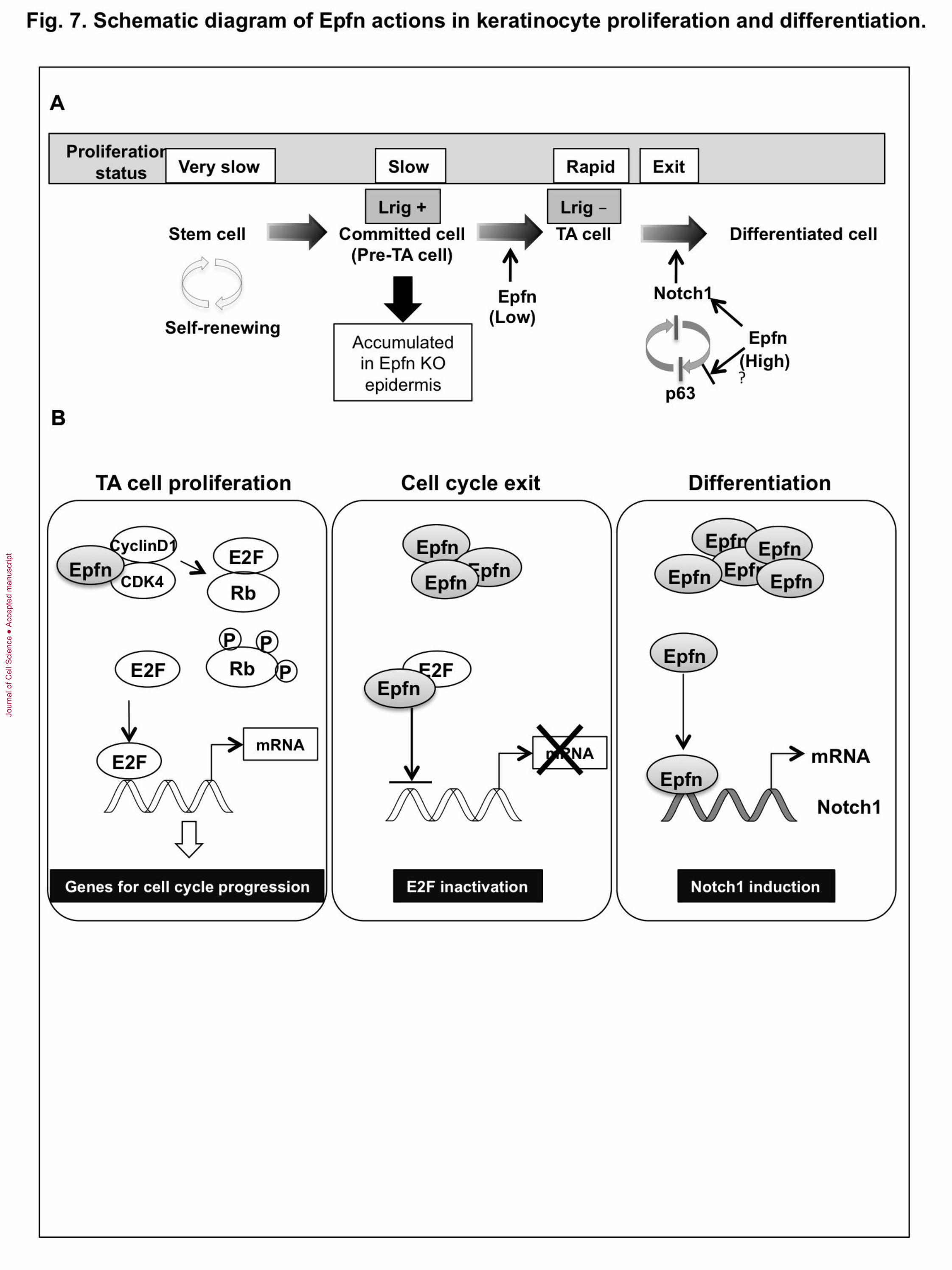
of the Notch1/p63 negative regulatory loop (Fig. 7).
In normal organogenesis, cell cycle exit is a prerequisite for differentiation. How
does Epfn act as a positive regulator of both cell proliferation and differentiation? One
possible explanation for this apparent contradiction is that Epfn expression levels may
regulate the switch from proliferation to differentiation. Low levels of Epfn expression
promoted proliferation of HaCaT cells, while high levels did not (Fig. 5B). Epfn
interacted with E2F and reduced E2F transactivation activity in HaCaT cells (Fig. 6B, C),
suggesting that Epfn inhibits the DNA binding of E2F by sequestration in a multiprotein
complex. The inhibition of E2F activity reduced the expression of genes required for cell
cycle progression and promoted cell cycle exit. We propose that low Epfn levels increase
free active E2F through Rb phosphorylation and concomitant release of E2F from
E2F/Rb complexes (which results in proliferation), whereas high Epfn expression
increases the formation of inactive E2F/Epfn complexes, resulting in cell cycle exit
(Fig.7B). In addition, post-translational modifications of Epfn, such as phosphorylation
and sumoylation, may also be involved in this functional switch from proliferation to
differentiation in Epfn activity.
Jour
nal o
f Cel
l Sci
ence
Acc
epte
d m
anus
crip
t
17
In summary, our data provide evidence for a novel mechanism that sequentially
regulates TA cell proliferation and differentiation through multiple distinct functions of
Epfn as a cell cycle regulator and a transcription factor (Fig. 7B). The switch from
proliferation to differentiation is regulated in part by the level of Epfn expression. Our in
vitro and in vivo studies suggest that Epfn promotes rapid TA cell proliferation by
enhancing Rb phosphorylation and suppressing EGFR antagonist Lrig1 expression, and
promotes keratinocyte differentiation by inducing Notch1 transcription. Our findings
suggest that Epfn regulates the p63/Notch axis in interfollicular epidermal formation.
Thus, Epfn plays multiple roles in orchestrating keratinocyte proliferation and
differentiation crucial to epidermal homeostasis and morphogenesis (Fig. 7).
Jour
nal o
f Cel
l Sci
ence
Acc
epte
d m
anus
crip
t
18
Materials and methods
Mice and cells
Epfn−/− mice were generated as previously described (Nakamura et al., 2008). The
animal protocol was approved by the National Institute of Dental and Craniofacial
Research (NIDCR) ACU Committee. All mice were housed in an animal facility
approved by the American Association for the Accreditation of Laboratory Animal Care.
Primary keratinocytes were prepared from newborn Epfn+/+, Epfn+/−, and
Epfn−/− mice, as described (Hennings et al., 1980), with minor modifications. In brief,
the mouse skin was floated on a layer of dispase (5 mg/ml) in Ca+2
and Mg2+
-free PBS at
4°C overnight. The dermis and epidermis were separated and keratinocytes were
collected by mincing the epidermis, placing it in 0.25% trypsin/0.5 mM EDTA and
incubating for 15 min at 37°C. After resuspended with Ca+2
and Mg2+
-free PBS, the
keratinocytes were filtered through a 100µm cell strainer (BD Dickinson, Franklin Lakes,
NJ USA) and centrifuged at 800 rpm for 10 min. The keratinocytes were cultured in a
serum-free and low-Ca2+
keratinocyte growth medium (KGM) (Invitrogen, Carlsbad, CA,
USA).
HaCaT cells, a spontaneously transformed human epithelial cell line from adult
skin (Boukamp et al., 1988), were obtained from Dr. Silvio Gutkind (NIDCR, NIH).
Primary human keratinocytes and HEK293T cells were obtained from Life Technologies
(Invitrogen, Carlsbad, CA, USA). Primary human keratinocytes were cultured in
EpilifeTM
media (Invitrogen, Carlsbad, CA, USA) and HEK293T cells were cultured in
DMEM (Invitrogen, Carlsbad, CA, USA).
Transfection, and proliferation assay
For transfection, exponentially growing HaCaT cells were electroporated with the Epfn
expression vector (Epfn-pcDNA3.1/myc-His vector (Nakamura et al., 2004) or Halo-
tagged Epfn vectors (Promega, Madison, WI, USA) using Amaxa Nucleofector Solution
V (Lonza, Walkersville, MD, USA). For the gene knockdown studies, HaCaT cells or
primary human keratinocytes were transfected with either the Epfn shRNA vector (ID:
TRCN0000017806, Open Biosystems, Inc., Huntsville, AL USA) or control pLKO
Jour
nal o
f Cel
l Sci
ence
Acc
epte
d m
anus
crip
t
19
vector, and the resulting puromycin-resistant colonies were pooled and used for
proliferation analysis. We also used another Epfn shRNA (ID: TRCN0000017807, Open
Biosystems) and obtained similar results.
Cell proliferation was assessed using the Cell Counting Kit-8 containing WST-8,
according to the manufacturer’s manual (Dojindo Laboratories, Kumamoto, Japan). EGF
(Sigma, St. Louis, MO, USA) was added to the cell cultures as a mitogenic cytokine (10
ng/mL). The cells were cultured in the absence or presence of EGF for three days and
then the proliferation activities were analyzed. To test HaCaT cell proliferation by Epfn at
various Epfn expression levels, we prepared Halo-tag Epfn expression vectors driven by
full-length and various deletions of the CMV promoter (CMV, CMV-d1, -d2, and-d3)
(Promega, Madison, WI, USA). In these experiments, we used the reverse transfection
method with Lipofectamine LTX reagent (Invitrogen, Carlsbad, CA, USA) according to
the manufacture protocol. The transfection mixtures for individual vectors and HaCaT
cells (10,000 cells/well) were incubated with KGM. Cell proliferation assay was
performed on days 0, 1, and 2 after the medium change to KGM. The protein expression
of Halo-tagged Epfn in HaCaT cells was examined on day 1 by Western blot analysis
with anti-Epfn and anti--actin antibodies. For transfection of primary human
keratinocytes, ViaFect TM
(Promega, Madison, WI, USA) was employed with the reverse-
transfection method.
Tissue sections, immunohistochemistry, and antibodies
Tissues were fixed with 4% paraformaldehyde in PBS at 4°C overnight. For histological
analysis, sections were stained with Harris hematoxylin and eosin Y. For
immunohistochemistry, sections were boiled with a targeted retrieval solution (Dako,
Carpinteria, CA, USA) and incubated in 1% bovine serum albumin/PBS (blocking
reagent) for 1 h before incubation with the primary antibody. We used antibodies against
cytokeratin 10 (K10) (Sigma, St. Louis, MO, USA), cytokeratin 5 (K5), involucrin,
filaggrin, and loricrin (all from Covance, Berkeley, CA, USA), p63 (BD Pharmingen, San
Jose, CA, USA), Notch1 (Cell Signaling Technology, Danvers, MA, USA and BD
Pharmingen, San Jose, CA, USA), Hes1 (Millipore, Billerica, MA, USA), PCNA
(Zymed, Carlsbad, CA, USA), Ki67 (Abcam, Cambridge, MA, USA), and BrdU (Roche,
Jour
nal o
f Cel
l Sci
ence
Acc
epte
d m
anus
crip
t
20
Indianapolis, IN, USA). The affinity purified rabbit polyclonal antibody to Epfn was
described previously (Nakamura et al., 2008). Primary antibodies were detected with Cy-
3-conjugated and Cy-5-conjugated secondary antibodies (Jackson ImmunoResearch,
West Grove, PA, USA). Nuclear staining was performed with Hoechst dye (Sigma, St.
Louis, MO, USA). All the immunopositive cells within the epidermis grid (600 m X
600 m) were counted and the positive cell ratio was calculated by dividing the total
number of cells in the grid. To caliculate the ratio, at least four independent regions were
counted.
Western blot, immunoprecipitation assay, and kinase assay
Total cellular proteins were prepared from exponentially growing HaCaT cells cultured in
KGM with either 0 mM, 0.7 mM, or 1.2 mMCa2+
using NP-40 lysis buffer with a
protease inhibitor cocktail (Roche, Indianapolis, IN, USA). Protein extracts were
separated on 4-12% NuPAGE gels (Invitrogen, Carlsbad, CA, USA), followed by
Western blotting. Kinase assays were performed using subconfluent 293T cells
transfected with both HA-tagged CDK4 and Epfn expression vectors. Cells were washed
in cold PBS and incubated on ice for 10 min with 500 µl of IP buffer (20 mM HEPES,
pH 7.5, containing 10 mM EGTA, 40 mM -glycerophosphate, 1 mM DTT, and 1% NP-
40), supplemented with a protease inhibitor cocktail (Roche, Indianapolis, IN, USA) and
2 mM orthovanadate. Protein extracts were incubated with an anti-HA-antibody (Thermo
Scientific, Rockford, IL, USA) at 4°C for an additional 1 h. Immune complexes were pull
downed with G-protein Sepharose beads and washed with IP buffer twice. Immune
complexes on the beads were equivalent with Rb kinase buffer (50 mM HEPES, pH 7.5,
containing 2.5 mM EGTA, 10 mM -glycerophosphate, 10 mM MgCl2, 5 mM MnCl2,
1 mM DTT, 0.1 mM sodium orthovanadate, 1 mM NaF, and 2 μCi of [γ-32P]ATP. The
beads were resuspended with Rb kinase buffer containing 2 µg of GST-Rb (Millipore,
Billerica, MA, USA) and were subsequently incubated at 30°C for 30 min. Five
millimolars of cold ATP were added to each reaction mixture to reduce the background
signal. The reaction was stopped by adding SDS sample buffer. The samples were boiled
for 5 min and resolved by SDS-10% PAGE. The gels were dried and autoradiographed on
BioMAX MR film for visualization. The co-immunoprecipitation assay was performed
Jour
nal o
f Cel
l Sci
ence
Acc
epte
d m
anus
crip
t
21
with an HA-Tag Co-IP kit (Thermo Scientific, Rockford, IL, USA) using subconfluent
HaCaT cells transfected with both the HA-tagged E2F1 expression vector, which was
kindly gifted from Dr. William Kaelin, and the VSV-tagged Epfn expression vectors. The
immune complexes on the beads were resuspended with lysis buffer and the samples
were boiled for 5 min and resolved using NuPage 4-12% gradient gel (Invitrogen,
Carlsbad, CA, USA). Immunoblot was performed with anti-phosphorylated Rb, anti-Rb,
anti-CDK4 ( Cell Signaling Technology, Danvers, MA, USA), anti-VSV (Sigma-Aldrich,
St. Louis, MO, USA), anti-E2F1 (Santa Cruz Biotechnology, Dallas, TX, USA), anti-
GAPDH (Santa Cruz Biotechnology, Dallas, TX, USA), and anti--actin antibodies (Cell
Signaling Technology, Danvers, MA, USA).
.
BrdU and TUNEL assays
BrdU (100 mg/kg body weight) was intraperitoneally injected into P13 mice. The mice
were euthanized 4 h later, and the skin was dissected and fixed with 4%
paraformaldehyde. BrdU incorporation was visualized with diaminobenzidine (DAB)
using the BrdU detection kit (Roche, Indianapolis, IN, USA). TUNEL assays (Roche,
Indianapolis, IN, USA) were performed on paraffin-embedded skin sections from P3 and
P7 mice. The sections were counterstained with hematoxylin. The ratios of the BrdU and
TUNEL positive cells within the grid in the epidermis (600 m X 600 m) were
calculated by dividing the total number of cells in the grid.
FACS analysis
Primary keratinocytes (2.0 × 10⁶ cells) from either Epfn+/− or Epfn−/− mice were
cultured for three days in type I collagen-coated dishes (BD Bioscience, Franklin Lakes,
NJ, USA) and washed three times in PBS, followed by the fixation with ice-cold 70%
ethanol for 30 min. After fixative removal, cells were centrifuged (900 rpm for 10 min),
and the pellet was resuspended in 500 µl of propidium iodide solution (50 µg/ml in PBS)
together with RNase A (10 mg/ml, Invitrogen, Carlsbad, CA, USA) and incubated on ice
for 30 min before cell cycle analysis using the FACSCalibur (BD Biosciences, Franklin
Lakes, NJ, USA).
Jour
nal o
f Cel
l Sci
ence
Acc
epte
d m
anus
crip
t
22
Real-time qPCR and microarray analysis
Primary keratinocytes or HaCaT cells were transiently transfected using Epfn vectors.
After 48 h, total RNA was extracted using the TRIzol reagent (Invitrogen, Carlsbad, CA,
USA). After DNase I (Sigma) treatment, 2 µg of total RNA was used for reverse
transcription to generate cDNA, which was subsequently used as a template for PCR
reactions with gene-specific primers (Table SI). Real-time qPCR was performed for
expression analysis using the 2XSYBR green supermixture (BioRad, Hercules, CA,
USA) and the Chromo4 thermocycler (MJ Research, Waltham, MA, USA). For
microarray analysis, total RNA was extracted from the primary keratinocytes of newborn
Epfn+/+ and Epfn−/− mice. Affymetrix mouse 430 2.0 array chips were used to analyze
gene expression profiles. Data analysis was performed using Ingenuity Pathway Analysis
software.
Luciferase assay
The E2F and RBP-Jk activities were analyzed using the Cignal E2F and RBP-Jk Reporter
(luc) Kit (Qiagen, Valencia, CA, USA). The E2F and RBP-Jk reporter plasmids
containing the control Renilla luciferase construct were transfected into HaCaT cells in a
48-well plate using lipofectamine LTX with reagent. The activities of the firefly and
Renilla luciferases were determined 48 h after transfection with the dual luciferase
reporter assay system (Promega, Madison, WI, USA) using a luminometer (Promega,
Madison, WI, USA). The luciferase activities were normalized for the Renilla luciferase
activity of the internal control.
Notch1 promoter analysis and ChIP assay
A 4.0-kb human Notch1 promoter sequence was analyzed with Genomatix software
(Genomatix, Germany) to predict potential factor binding sites. Multiple Sp or KLF
binding elements were predicted 3.2kb upstream of the transcription start site and at the
proximal promoter of the human Notch1 gene. Chromatin DNA and nuclear protein
complexes were extracted from the HaCaT cells transfected with His-tagged Epfn or
empty expression vectors, and ChIP assays were conducted with an anti-His antibody
Jour
nal o
f Cel
l Sci
ence
Acc
epte
d m
anus
crip
t
23
(Novagen, Madison, WI, USA) and EZ ChIP kit (Millipore, Billerica, MA, USA). ChIP
primer sequences were shown in Table S2 (Lefort et al., 2007).
Statistical analysis
The data were analyzed with Prism 4 software. Differences between groups were
analyzed for statistical significance using Student’s t-tests.
Acknowledgements
We wish to thank Kenneth Yamada, Thomas Bugge, Karin List, Roman Szabo, and
Maurizio Pacifici for their valuable suggestions.
Competing interests
The authors declare no competing interest.
Author contributions
T.N., S.F., and Y.Y. conceived the study. T.N. performed the most experiments
and analyzed the data, with some contributions from Ya.Y. and V.P. T.N., Ya.Y., V.P., and
Y.Y. interpreted the data. T.N. and Y.Y. wrote the manuscript.
Funding
This work was supported in part by the Intramural Research Program of the National
Institutes of Health, National Institute of Dental and Craniofacial Research, USA (Y.Y.)
and grants-in-aid (24390441, 24659908 to T.N.) from the Ministry of Education, Science,
and Culture of Japan, and the NEXT program (LS010 to S.F.), as well as a fellowship
from the Japan Society for the Promotion of Science (T.N., YaY).
Jour
nal o
f Cel
l Sci
ence
Acc
epte
d m
anus
crip
t

24
References
Artavanis-Tsakonas, S., Rand, M. D. and Lake, R. J. (1999). Notch signaling:
cell fate control and signal integration in development. Science 284, 770-6.
Bergh, G., Ehinger, M., Olsson, I., Jacobsen, S. E. and Gullberg, U. (1999).
Involvement of the retinoblastoma protein in monocytic and neutrophilic lineage
commitment of human bone marrow progenitor cells. Blood 94, 1971-8.
Blanpain, C., Lowry, W. E., Pasolli, H. A. and Fuchs, E. (2006). Canonical
notch signaling functions as a commitment switch in the epidermal lineage. Genes Dev
20, 3022-35.
Boukamp, P., Petrussevska, R. T., Breitkreutz, D., Hornung, J., Markham, A.
and Fusenig, N. E. (1988). Normal keratinization in a spontaneously immortalized
aneuploid human keratinocyte cell line. J Cell Biol 106, 761-71.
Boulter, L., Govaere, O., Bird, T. G., Radulescu, S., Ramachandran, P.,
Pellicoro, A., Ridgway, R. A., Seo, S. S., Spee, B., Van Rooijen, N. et al. (2012).
Macrophage-derived Wnt opposes Notch signaling to specify hepatic progenitor cell fate
in chronic liver disease. Nat Med 18, 572-9.
Breunig, J. J., Silbereis, J., Vaccarino, F. M., Sestan, N. and Rakic, P. (2007).
Notch regulates cell fate and dendrite morphology of newborn neurons in the postnatal
dentate gyrus. Proc Natl Acad Sci U S A 104, 20558-63.
Burgeson, R. E. and Christiano, A. M. (1997). The dermal-epidermal junction.
Current opinion in cell biology 9, 651-8.
Cai, M., Han, L., Chen, R., Ye, F., Wang, B., Han, F., Lei, T. and Guo, D.
(2009). Inhibition of LRIG3 gene expression via RNA interference modulates the
proliferation, cell cycle, cell apoptosis, adhesion and invasion of glioblastoma cell
(GL15). Cancer Lett 278, 104-12.
Jour
nal o
f Cel
l Sci
ence
Acc
epte
d m
anus
crip
t
25
Classon, M., Kennedy, B. K., Mulloy, R. and Harlow, E. (2000). Opposing
roles of pRB and p107 in adipocyte differentiation. Proc Natl Acad Sci U S A 97, 10826-
31.
de la Pompa, J. L., Wakeham, A., Correia, K. M., Samper, E., Brown, S.,
Aguilera, R. J., Nakano, T., Honjo, T., Mak, T. W., Rossant, J. et al. (1997).
Conservation of the Notch signalling pathway in mammalian neurogenesis. Development
124, 1139-48.
Devgan, V., Mammucari, C., Millar, S. E., Brisken, C. and Dotto, G. P. (2005).
p21WAF1/Cip1 is a negative transcriptional regulator of Wnt4 expression downstream of
Notch1 activation. Genes Dev 19, 1485-95.
Dotto, G. P. (2008). Notch tumor suppressor function. Oncogene 27, 5115-23.
Fuchs, E. (2008). Skin stem cells: rising to the surface. J Cell Biol 180, 273-84.
Georgia, S., Soliz, R., Li, M., Zhang, P. and Bhushan, A. (2006). p57 and Hes1
coordinate cell cycle exit with self-renewal of pancreatic progenitors. Dev Biol 298, 22-
31.
Guo, C. S., Degnin, C., Fiddler, T. A., Stauffer, D. and Thayer, M. J. (2003).
Regulation of MyoD activity and muscle cell differentiation by MDM2, pRb, and Sp1. J
Biol Chem 278, 22615-22.
Gur, G., Rubin, C., Katz, M., Amit, I., Citri, A., Nilsson, J., Amariglio, N.,
Henriksson, R., Rechavi, G., Hedman, H. et al. (2004). LRIG1 restricts growth factor
signaling by enhancing receptor ubiquitylation and degradation. EMBO J 23, 3270-81.
Hanahan, D. (2000). Benefits of bad telomeres. Nature 406, 573-4.
Harbour, J. W., Luo, R. X., Dei Santi, A., Postigo, A. A. and Dean, D. C.
(1999). Cdk phosphorylation triggers sequential intramolecular interactions that
progressively block Rb functions as cells move through G1. Cell 98, 859-69.
Jour
nal o
f Cel
l Sci
ence
Acc
epte
d m
anus
crip
t
26
Hennings, H., Michael, D., Cheng, C., Steinert, P., Holbrook, K. and Yuspa,
S. H. (1980). Calcium regulation of growth and differentiation of mouse epidermal cells
in culture. Cell 19, 245-54.
Hertveldt, V., Louryan, S., van Reeth, T., Dreze, P., van Vooren, P., Szpirer, J.
and Szpirer, C. (2008). The development of several organs and appendages is impaired
in mice lacking Sp6. Dev Dyn 237, 883-92.
Ibarretxe, G., Aurrekoetxea, M., Crende, O., Badiola, I., Jimenez-Rojo, L.,
Nakamura, T., Yamada, Y. and Unda, F. (2012). Epiprofin/Sp6 regulates Wnt-BMP
signaling and the establishment of cellular junctions during the bell stage of tooth
development. Cell and tissue research 350, 95-107.
Jensen, K. and Watt, F. (2006). Single-cell expression profiling of human
epidermal stem and transit-amplifying cells: Lrig1 is a regulator of stem cell quiescence.
Proc. Natl. Acad. Sci. U.S.A. 103, 11958-63.
Jensen, K. B., Collins, C. A., Nascimento, E., Tan, D. W., Frye, M., Itami, S.
and Watt, F. M. (2009). Lrig1 expression defines a distinct multipotent stem cell
population in mammalian epidermis. Cell Stem Cell 4, 427-39.
Jones, P. H. and Watt, F. M. (1993). Separation of human epidermal stem cells
from transit amplifying cells on the basis of differences in integrin function and
expression. Cell 73, 713-24.
Koster, M. I., Kim, S., Mills, A. A., DeMayo, F. J. and Roop, D. R. (2004). p63
is the molecular switch for initiation of an epithelial stratification program. Genes Dev
18, 126-31.
Kurata, S., Okuyama, T., Osada, M., Watanabe, T., Tomimori, Y., Sato, S.,
Iwai, A., Tsuji, T., Ikawa, Y. and Katoh, I. (2004). p51/p63 Controls subunit alpha3 of
the major epidermis integrin anchoring the stem cells to the niche. J Biol Chem 279,
50069-77.
Jour
nal o
f Cel
l Sci
ence
Acc
epte
d m
anus
crip
t
27
Laederich, M. B., Funes-Duran, M., Yen, L., Ingalla, E., Wu, X., Carraway,
K. L., 3rd and Sweeney, C. (2004). The leucine-rich repeat protein LRIG1 is a negative
regulator of ErbB family receptor tyrosine kinases. J Biol Chem 279, 47050-6.
Lefort, K., Mandinova, A., Ostano, P., Kolev, V., Calpini, V., Kolfschoten, I.,
Devgan, V., Lieb, J., Raffoul, W., Hohl, D. et al. (2007). Notch1 is a p53 target gene
involved in human keratinocyte tumor suppression through negative regulation of
ROCK1/2 and MRCKalpha kinases. Genes Dev 21, 562-77.
Li, F. Q., Coonrod, A. and Horwitz, M. (2000). Selection of a dominant
negative retinoblastoma protein (RB) inhibiting satellite myoblast differentiation implies
an indirect interaction between MyoD and RB. Mol Cell Biol 20, 5129-39.
Lin, S. Y., Black, A. R., Kostic, D., Pajovic, S., Hoover, C. N. and Azizkhan, J.
C. (1996). Cell cycle-regulated association of E2F1 and Sp1 is related to their functional
interaction. Mol Cell Biol 16, 1668-75.
Lowell, S., Jones, P., Le Roux, I., Dunne, J. and Watt, F. M. (2000).
Stimulation of human epidermal differentiation by delta-notch signalling at the
boundaries of stem-cell clusters. Curr Biol 10, 491-500.
Mills, A. A., Zheng, B., Wang, X. J., Vogel, H., Roop, D. R. and Bradley, A.
(1999). p63 is a p53 homologue required for limb and epidermal morphogenesis. Nature
398, 708-13.
Moriyama, M., Durham, A. D., Moriyama, H., Hasegawa, K., Nishikawa, S.,
Radtke, F. and Osawa, M. (2008). Multiple roles of Notch signaling in the regulation of
epidermal development. Developmental cell 14, 594-604.
Muller, S., Lindquist, D., Kanter, L., Flores-Staino, C., Henriksson, R.,
Hedman, H. and Andersson, S. (2013). Expression of LRIG1 and LRIG3 correlates
with human papillomavirus status and patient survival in cervical adenocarcinoma. Int J
Oncol 42, 247-52.
Jour
nal o
f Cel
l Sci
ence
Acc
epte
d m
anus
crip
t
28
Nakamura, T., de Vega, S., Fukumoto, S., Jimenez, L., Unda, F. and Yamada,
Y. (2008). Transcription factor epiprofin is essential for tooth morphogenesis by
regulating epithelial cell fate and tooth number. J Biol Chem 283, 4825-33.
Nakamura, T., Unda, F., de-Vega, S., Vilaxa, A., Fukumoto, S., Yamada, K.
M. and Yamada, Y. (2004). The Kruppel-like factor epiprofin is expressed by epithelium
of developing teeth, hair follicles, and limb buds and promotes cell proliferation. J Biol
Chem 279, 626-34.
Nguyen, B. C., Lefort, K., Mandinova, A., Antonini, D., Devgan, V., Della
Gatta, G., Koster, M. I., Zhang, Z., Wang, J., Tommasi di Vignano, A. et al. (2006).
Cross-regulation between Notch and p63 in keratinocyte commitment to differentiation.
Genes Dev 20, 1028-42.
Nicolas, M., Wolfer, A., Raj, K., Kummer, J. A., Mill, P., van Noort, M., Hui,
C. C., Clevers, H., Dotto, G. P. and Radtke, F. (2003). Notch1 functions as a tumor
suppressor in mouse skin. Nat Genet 33, 416-21.
Papadimou, E., Menard, C., Grey, C. and Puceat, M. (2005). Interplay
between the retinoblastoma protein and LEK1 specifies stem cells toward the cardiac
lineage. EMBO J 24, 1750-61.
Pellegrini, G., Dellambra, E., Golisano, O., Martinelli, E., Fantozzi, I.,
Bondanza, S., Ponzin, D., McKeon, F. and De Luca, M. (2001). p63 identifies
keratinocyte stem cells. Proc Natl Acad Sci U S A 98, 3156-61.
Powell, A. E., Wang, Y., Li, Y., Poulin, E. J., Means, A. L., Washington, M. K.,
Higginbotham, J. N., Juchheim, A., Prasad, N., Levy, S. E. et al. (2012). The pan-
ErbB negative regulator Lrig1 is an intestinal stem cell marker that functions as a tumor
suppressor. Cell 149, 146-58.
Rangarajan, A., Talora, C., Okuyama, R., Nicolas, M., Mammucari, C., Oh,
H., Aster, J. C., Krishna, S., Metzger, D., Chambon, P. et al. (2001). Notch signaling is
Jour
nal o
f Cel
l Sci
ence
Acc
epte
d m
anus
crip
t
29
a direct determinant of keratinocyte growth arrest and entry into differentiation. EMBO J
20, 3427-36.
Romano, R. A., Smalley, K., Magraw, C., Serna, V. A., Kurita, T., Raghavan,
S. and Sinha, S. (2012). DeltaNp63 knockout mice reveal its indispensable role as a
master regulator of epithelial development and differentiation. Development 139, 772-82.
Rondahl, V., Holmlund, C., Karlsson, T., Wang, B., Faraz, M., Henriksson, R.
and Hedman, H. (2013). Lrig2-deficient mice are protected against PDGFB-induced
glioma. PLoS One 8, e73635.
Rotheneder, H., Geymayer, S. and Haidweger, E. (1999). Transcription factors
of the Sp1 family: interaction with E2F and regulation of the murine thymidine kinase
promoter. Journal of molecular biology 293, 1005-15.
Ruiz, S., Santos, M., Segrelles, C., Leis, H., Jorcano, J. L., Berns, A.,
Paramio, J. M. and Vooijs, M. (2004). Unique and overlapping functions of pRb and
p107 in the control of proliferation and differentiation in epidermis. Development 131,
2737-48.
Suske, G., Bruford, E. and Philipsen, S. (2005). Mammalian SP/KLF
transcription factors: bring in the family. Genomics 85, 551-6.
Suzuki, Y., Miura, H., Tanemura, A., Kobayashi, K., Kondoh, G., Sano, S.,
Ozawa, K., Inui, S., Nakata, A., Takagi, T. et al. (2002). Targeted disruption of LIG-1
gene results in psoriasiform epidermal hyperplasia. FEBS Lett 521, 67-71.
Talamillo, A., Delgado, I., Nakamura, T., de-Vega, S., Yoshitomi, Y., Unda, F.,
Birchmeier, W., Yamada, Y. and Ros, M. A. (2010). Role of Epiprofin, a zinc-finger
transcription factor, in limb development. Dev Biol 337, 363-74.
Truong, A., Kretz, M., Ridky, T., Kimmel, R. and Khavari, P. (2006). p63
regulates proliferation and differentiation of developmentally mature keratinocytes.
Genes Dev. 20, 3185-97.
Jour
nal o
f Cel
l Sci
ence
Acc
epte
d m
anus
crip
t
30
Watt, F. M. and Jensen, K. B. (2009). Epidermal stem cell diversity and
quiescence. EMBO Mol Med 1, 260-7.
Wong, V. W., Stange, D. E., Page, M. E., Buczacki, S., Wabik, A., Itami, S.,
van de Wetering, M., Poulsom, R., Wright, N. A., Trotter, M. W. et al. (2012). Lrig1
controls intestinal stem-cell homeostasis by negative regulation of ErbB signalling.
Nature cell biology 14, 401-8.
Xiong, W., Morillo, S. A. and Rebay, I. (2013). The Abelson tyrosine kinase
regulates Notch endocytosis and signaling to maintain neuronal cell fate in Drosophila
photoreceptors. Development 140, 176-84.
Yang, A., Schweitzer, R., Sun, D., Kaghad, M., Walker, N., Bronson, R. T.,
Tabin, C., Sharpe, A., Caput, D., Crum, C. et al. (1999). p63 is essential for
regenerative proliferation in limb, craniofacial and epithelial development. Nature 398,
714-8.
Jour
nal o
f Cel
l Sci
ence
Acc
epte
d m
anus
crip
t

31
Figure legends
Fig. 1. Defects of skin and hair follicle development in Epfn−/− mice. (A) Defective
skin, hair follicle, and whisker formation (arrowheads) in 3-month-old Epfn+/− (a, c) and
Epfn−/− (b, d) mice. (B) Defective skin morphogenesis and hair follicle formation.
Histological analysis of developing skin and skin morphogenesis at E16.5 (a, b), P3 (c,
d), P7 (e, f), and P14 (g, h) of Epfn+/− (a, c, e, g) and Epfn−/− (b, d, f, h) mice. Bars: 100
μm. Bottom panel: A schematic diagram of the epidermis and expression patterns of
marker proteins for keratinocytes. (C) Expression of epidermal keratinocyte
differentiation marker proteins in P10 Epfn+/− (a, c, e, g) and Epfn−/− (b, d, f, h) mice.
K10 (green); K5 (red) (a, b); K10 (green), involucrin (red) (c, d); K10 (green); filaggrin
(red) (e, f); K10 (green); and loricrin (red) (g, h). Hoechst (blue) (a−h). (D) The
expression of p63 (green) and Notch1 (red) (a, b) in P13 mice, and Hes1 (green) and
Hoechst (blue) (c, d) in P10 mice. The white dotted-lines indicate the basement
membrane of the epidermis. Epfn (DAB) (e, f) in 3-month-old mice. Bars: 100 m. (E)
Ratios of immuno-positive cells for p63, Notch1, Hes1, and Epfn in the epidermis of
Epfn+/−and Epfn−/−.
Fig. 2. Slower keratinocyte proliferation, reduced apoptosis, and dysregulation of
Rb phosphorylation in the Epfn−/− epidermis. (A) PCNA (a, b), Ki67 (c, d) and BrdU
(e, f) staining in the P7 epidermis and TUNEL staining (g, h) in the P3 epidermis. Bar:
100 m. (B) Relative numbers of PCNA-, Ki67-, BrdU-, and TUNEL-positive cells in
Epfn+/− and Epfn−/− skin. (C) Proliferation of primary keratinocytes from newborn
Epfn+/− and Epfn−/− mice. (D) FACS analysis of cell cycle progression in primary
keratinocytes from newborn Epfn+/− and Epfn−/− mice. *P < 0.05 (E) Expression of cell
cycle regulators as revealed by Western blotting. Reduced Rb phosphorylation (phos-Rb)
and expression of cell cycle regulators in primary keratinocytes from newborn Epfn+/−
and Epfn−/− mice. Upper arrowhead and asterisk, phos-Rb; lower arrowhead and
asterisk, Rb protein.
Jour
nal o
f Cel
l Sci
ence
Acc
epte
d m
anus
crip
t
32
Fig. 3. Characteristics of keratinocytes from the Epfn−/− epidermis. (A) Expression
of various genes characteristic of stem cells, TA cells, and mature keratinocytes in
primary cultures derived from newborn Epfn+/+ and Epfn−/− mouse epidermis as
measured by microarray analysis. (B) Cell attachment and (C) colony formation assay
using primary keratinocytes from newborn Epfn+/− and Epfn−/− mice.
Fig. 4. Epfn expression in differentiated HaCaT cells and the role of Epfn in EGF-
mediated cell proliferation. (A) Immunoblot analysis of Epfn and filaggrin expression
in differentiating HaCaT cells cultured in a low-Ca2+
media (KGM) with the addition of 0
mM, 0.7 mM, or 1.2 mM Ca2+
for 4 days (4d) or 7 days (7d). (B) HaCaT cell
proliferation. (a) Upper panel: Western blot analysis of endogenous Epfn expression in
HaCaT cells transfected with control shRNA and Epfn shRNA. Lower panel:
densitometric analysis of the Western blot assays using NIH-ImageJ. (b) Proliferation of
HaCaT cells transfected with control shRNA and Epfn shRNA. (C) EGF-mediated
proliferation of HaCaT cells transfected with control shRNA and Epfn shRNA. (D) EGF-
mediated proliferation of human epidermal keratinocytes transfected with control shRNA
and Epfn shRNA. (E) EGF-mediated proliferation of keratinocytes from the wild-type
(Epfn +/+) and Epfn−/− epidermis. Primary keratinocytes were cultured in KGM for 4
days with or without recombinant EGF (10 ng/ml). **P<0.01
Fig. 5. Epfn promotes HaCaT cell proliferation through enhancing Rb
phosphorylation and Epfn/Cdk4 interaction. (A) Immunoblot analysis of Epfn
expression in HaCaT cells transfected with various Halo-tagged Epfn expression vectors
driven by either the full length CMV promoter (CMV-Epfn) or deletions in the CMV
promoter (CMVd1-Epfn, CMVd2-Epfn, CMVd3-Epfn). CMV–Epfn (full-length CMV
promoter). Bottom panel: -actin, control. (B) The relationship between Epfn expression
levels and HaCaT cell proliferation. HaCaT cells were transfected with one of four Halo-
tagged Epfn expression vectors. The proliferation was analyzed at various time points.
**P < 0.01 (C) Immunoblot analysis of cell cycle proteins. HaCaT cells were transfected
with a His-tagged Epfn expression vector, and lysates were analyzed by Western blotting
using antibodies against His-tag, phos-Rb, E2F1, and -actin (the gel loading control).
Jour
nal o
f Cel
l Sci
ence
Acc
epte
d m
anus
crip
t
33
(D) Co-immunoprecipitation assay of Epfn and Cdk4 (a) and Rb phosphorylation assay
(b) in HEK293 cells transfected with Mock or Epfn (myc-tag) expression vectors.
Fig. 6. Epfn promotes HaCaT cell differentiation through regulating Notch1. (A)
Expression of keratinocyte marker genes as revealed by qPCR in HaCaT cells transfected
with either Mock or Epfn expression vectors. (B) Co-immunoprecipitation assay of Epfn
and E2F1 in HaCaT cells transfected with E2F1 (HA-tag) and Epfn (VSV-tag) expression
vectors. (C) E2F activity assay in HaCaT cells transfected with Mock and Epfn
expression vectors. (D) RBP-Jk reporter activity in HaCaT cells transfected with either
Mock or Epfn expression vectors. (E) ChIP analysis of the Notch1 promoter using the
anti-VSV-tag antibody in HaCaT cells transfected with either Mock or Epfn (VSV-tag)
expression vectors. Upper panel: In silico prediction of potential Sp transcription factor
binding sites (Sp sites A and B) in the human Notch1 promoter. Bottom: Nuclear extracts
from either VSV-tag-Epfn- or Mock-transfected HaCaT cells were used for ChIP analysis
with primer sets for Sp sites A and B.
Fig. 7. Schematic diagram of Epfn actions in keratinocyte proliferation and
differentiation. (A) Epfn promotes rapid TA cell proliferation. In the absence of Epfn,
premature TA (pre-TA) cells accumulate in the epidermis. Pre-TA cells are committed to
differentiation but retain some stem cell characteristics, such as slow proliferation in part
due to elevated Lrig expression. As Epfn expression increases, Epfn promotes cell cycle
exit and activates Notch1 expression, triggering differentiation. Epfn-induced Notch
expression suppresses p63 expression. In addition, Epfn may directly inhibit p63
expression. Epfn regulates the p63 and Notch signaling pathways that are essential for
epidermal development, maintenance, and renewal. (B) Multiple functions of Epfn during
keratinocyte proliferation and differentiation. In TA cells, Epfn enhances Rb
phosphorylation by interacting with CDK4 to promote proliferation. As Epfn expression
increases, Epfn binds to E2F, which inhibits cell progression and promotes cell cycle exit
by inhibiting E2F transactivation activity. Epfn also promotes keratinocyte differentiation
by inducing Notch1 transcription.
Jour
nal o
f Cel
l Sci
ence
Acc
epte
d m
anus
crip
t
34
Supplementary Fig. S1. Scheme of Epfn, p63, and Notch 1 expression in epidermis.
A schematic diagram of the epidermis and expression patterns of Epfn, p63, and Noch1.
Stem cells and TA cells are localized in the basal layer and attach to the basement
membrane. Basal layer keratinocytes express p63 and Epfn. Differentiating keratinocytes
in the upper layers are not proliferative and express Notch1 and Epfn.
Supplementary Fig. S2. Expression of Lrig1 mRNA in Epfn-knockdown HaCaT
cells.
Expression of Lrig1 mRNA expression in HaCaT cells transfected with control shRNA
and Epfn shRNA.The expression of Lrig1 was enhanced in Epfn knock-down HaCaT
cells.
Supplementary Fig. S3. Expression of endogenous Epfn protein in Epfn-knockdown
primary human keratinocytes.
Upper panel: Western blot analysis of the endogenous Epfn expression in human primary
keratinocytes transfected with control shRNA and Epfn shRNA (806 and 807). Lower
panel: densitometric analysis of the Western blot assays using NIH ImageJ.
Supplementary Table S1.
Primers used in qPCR analysis.
Supplementary Table S2.
Primers used in ChIP analysis.
Jour
nal o
f Cel
l Sci
ence
Acc
epte
d m
anus
crip
t
Jour
nal o
f Cel
l Sci
ence
Acc
epte
d m
anus
crip
t
Jour
nal o
f Cel
l Sci
ence
Acc
epte
d m
anus
crip
t
Jour
nal o
f Cel
l Sci
ence
Acc
epte
d m
anus
crip
t
Jour
nal o
f Cel
l Sci
ence
Acc
epte
d m
anus
crip
t
Jour
nal o
f Cel
l Sci
ence
Acc
epte
d m
anus
crip
t
Jour
nal o
f Cel
l Sci
ence
Acc
epte
d m
anus
crip
t
Jour
nal o
f Cel
l Sci
ence
Acc
epte
d m
anus
crip
t