
JNC Highlights November 2020
16
JNC Highlights November 2020 Latest Research and Reviews The following articles are part of Volume 155, Issue 1 – Pages 1-110 Amyloid‐beta 1–42 induced glutamatergic receptor and transporter expression changes in the mouse hippocampus Jason H.Y. Yeung, Beatriz Calvo‐Flores Guzmán, Thulani H. Palpagama, Jayarjun Ethiraj, Ying Zhai, Warren P. Tate, Katie Peppercorn, Henry J. Waldvogel, Richard L.M. Faull, Andrea Kwakowsky Read the full article ‘Amyloid‐beta1–42 induced glutamatergic receptor and transporter expression changes in the mouse hippocampus’ by J. H. Y. Yeung, B. C.‐F. Guzmán, T. H. Palpagama, J. Ethiraj, Y. Zhai, W. P. Tate, K. Peppercorn, H. J. Waldvogel, R. L. M. Faull, A. Kwakowsky, (J. Neurochem. 2020, vol. 155 (1), pp. 19–62) on doi:10.1111/jnc.15099 Cover Image Front cover: Alzheimer's Disease (AD) is the leading type of dementia worldwide. Glutamate is the main excitatory neurotransmitter in the brain and plays an essential role in the function and health of neurons and neuronal excitability. This is the first comprehensive anatomical study to characterize the subregion‐ and cell layer‐specific effect of beta‐amyloid (Aβ1‐42) on the expression of specific glutamate receptors and transporters in the mouse hippocampus, using immunohistochemistry with confocal microscopy. Our data demonstrate heterogeneous expression changes in glutamatergic signaling components in an in vivo Aβ1‐42‐injected mouse model of AD. These changes have the ability to alter normal glutamatergic signaling and could contribute to AD pathology. Due to the pivotal role of the glutamatergic system in regulating synaptic activity, any disruption to its composition could have consequences for normal neuronal function. These findings also indicate that Aβ1‐42 induces brain region and layer specific expression changes of the glutamatergic receptors and transporters, suggesting complex and spatial vulnerability of this pathway during AD neuropathology. Image content: The image shows glutamate receptor GluA1 subunit (green) and glutamate transporter vGluT1 (red) immunoreactivity in CA3 region of the mouse hippocampus. Nuclei were counterstained with Hoechst dye (blue).
Transcript of JNC Highlights November 2020

JNC Highlights November 2020
Latest Research and Reviews
The following articles are part of Volume 155, Issue 1 – Pages 1-110
Amyloid‐beta 1–42 induced glutamatergic receptor and transporter expression changes in the
mouse hippocampus
Jason H.Y. Yeung, Beatriz Calvo‐Flores Guzmán, Thulani H. Palpagama, Jayarjun Ethiraj, Ying Zhai, Warren
P. Tate, Katie Peppercorn, Henry J. Waldvogel, Richard L.M. Faull, Andrea Kwakowsky
Read the full article ‘Amyloid‐beta1–42 induced glutamatergic receptor and transporter expression
changes in the mouse hippocampus’ by J. H. Y. Yeung, B. C.‐F. Guzmán, T. H. Palpagama, J.
Ethiraj, Y. Zhai, W. P. Tate, K. Peppercorn, H. J. Waldvogel, R. L. M. Faull, A. Kwakowsky, (J.
Neurochem. 2020, vol. 155 (1), pp. 19–62) on doi:10.1111/jnc.15099
Cover Image
Front cover: Alzheimer's Disease (AD) is the leading type of dementia worldwide. Glutamate is the main
excitatory neurotransmitter in the brain and plays an essential role in the function and health of neurons
and neuronal excitability. This is the first comprehensive anatomical study to characterize the subregion‐
and cell layer‐specific effect of beta‐amyloid (Aβ1‐42) on the expression of specific glutamate receptors
and transporters in the mouse hippocampus, using immunohistochemistry with confocal microscopy. Our
data demonstrate heterogeneous expression changes in glutamatergic signaling components in an in vivo
Aβ1‐42‐injected mouse model of AD. These changes have the ability to alter normal glutamatergic
signaling and could contribute to AD pathology. Due to the pivotal role of the glutamatergic system in
regulating synaptic activity, any disruption to its composition could have consequences for normal
neuronal function. These findings also indicate that Aβ1‐42 induces brain region and layer specific
expression changes of the glutamatergic receptors and transporters, suggesting complex and spatial
vulnerability of this pathway during AD neuropathology.
Image content: The image shows glutamate receptor GluA1 subunit (green) and glutamate transporter
vGluT1 (red) immunoreactivity in CA3 region of the mouse hippocampus. Nuclei were counterstained
with Hoechst dye (blue).

Toward a dissection of β‐Amyloid localized effects on glutamatergic hippocampal repertoire
J. Alfredo Méndez Arturo Ortega
Hippocampal excitatory glutamatergic transmission is critically involved in cognitive functions such as
learning and memory. A severe impairment of spatial memory is associated with the Alzheimer's disease
characteristic augmentation of soluble Amyloid‐beta1‐42 which in turn leads to glutamatergic
neurotransmission dysfunction. As the molecular basis of such correlations has not been completely
understood, this Editorial highlights a study in the current issue of the Journal of Neurochemistry in which
Yeung and coworkers provide an elegant anatomical study that sheds light into this problematic. Through
a rigorous immunohistochemical approach, a sub‐regional expression pattern of ionotropic glutamate
receptors and vesicular transporters was determined in control and beta amyloid‐injected mouse
hippocampus. The selected areas participate in information processing and thus, in memory formation.
Furthermore, the authors discuss their findings in the context of cognitive deficits present in Alzheimer's
disease patients delivering an easy to follow analysis of plausible molecular events that disturb proper
glutamate signaling. This study takes an important step towards a better understanding of the complexity
of Amyloid‐beta1‐42 and glutamatergic neurotransmission interactions.
Editorial Highlight

Role of dynein–dynactin complex, kinesins, motor adaptors, and their phosphorylation in
dendritogenesis
Aleksandra Tempes, Jan Weslawski, Agnieszka Brzozowska, Jacek Jaworski
One of the characteristic features of different classes of neurons is the shape of their dendritic arbors. To
properly develop dendritic trees, neurons need to accurately control the intracellular transport of various
cellular cargo which relays on motor proteins. This review focuses on the contribution of the dynein–
dynactin complex, kinesins, their adaptors, and the phosphorylation of these proteins in dendritogenesis.
We review not only the effects of the motor activity of these proteins in dendrites on dendritogenesis but
also lesser‐known aspects of their contribution, for example, dynein‐driven axonal transport or regulation
of MT dynamics, polarity, and other motors speed.
Review articles

Involvement of the Hsp70/TLR4/IL‐6 and TNF‐α pathways in delayed‐onset muscle soreness
Rafaela Silva dos Santos, Flávio Protasio Veras, David Wilson Ferreira, Morena Brazil Sant'Anna, Pablo
Christiano Barboza Lollo, Thiago Mattar Cunha, Giovane Galdino
Few studies have investigated delayed‐onset muscle soreness (DOMS), and most have found
proinflammatory cytokine involvement. This study was the first to further demonstrate that exercise
increases Hsp70 in muscle and spinal cord (1), which activates TLR4 in the spinal microglia (2) and therefore
releases pronociceptive cytokines as TNF‐α and IL‐6 (3), that through the blood veins (4) activate muscle
nociceptors (5), or released by microglia contribute to spinal sensitization of DOMS (6).
Original articles
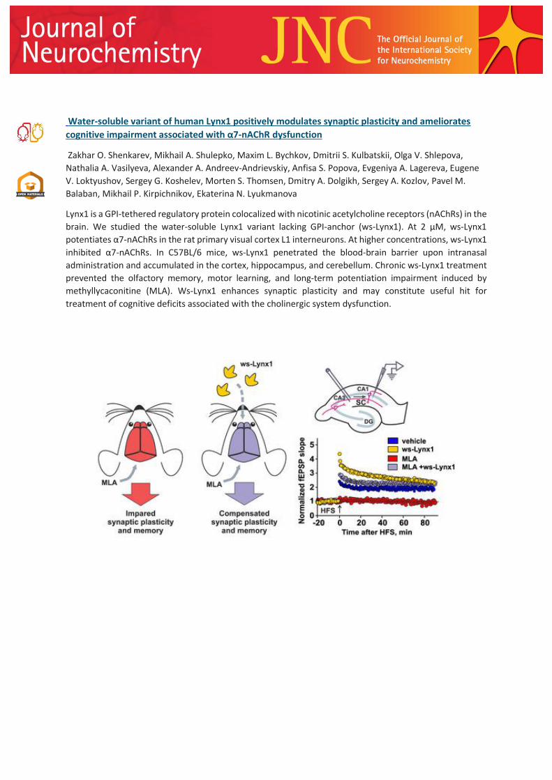
Water‐soluble variant of human Lynx1 positively modulates synaptic plasticity and ameliorates
cognitive impairment associated with α7‐nAChR dysfunction
Zakhar O. Shenkarev, Mikhail A. Shulepko, Maxim L. Bychkov, Dmitrii S. Kulbatskii, Olga V. Shlepova,
Nathalia A. Vasilyeva, Alexander A. Andreev-Andrievskiy, Anfisa S. Popova, Evgeniya A. Lagereva, Eugene
V. Loktyushov, Sergey G. Koshelev, Morten S. Thomsen, Dmitry A. Dolgikh, Sergey A. Kozlov, Pavel M.
Balaban, Mikhail P. Kirpichnikov, Ekaterina N. Lyukmanova
Lynx1 is a GPI‐tethered regulatory protein colocalized with nicotinic acetylcholine receptors (nAChRs) in the
brain. We studied the water‐soluble Lynx1 variant lacking GPI‐anchor (ws‐Lynx1). At 2 µM, ws‐Lynx1
potentiates α7‐nAChRs in the rat primary visual cortex L1 interneurons. At higher concentrations, ws‐Lynx1
inhibited α7‐nAChRs. In C57BL/6 mice, ws‐Lynx1 penetrated the blood‐brain barrier upon intranasal
administration and accumulated in the cortex, hippocampus, and cerebellum. Chronic ws‐Lynx1 treatment
prevented the olfactory memory, motor learning, and long‐term potentiation impairment induced by
methyllycaconitine (MLA). Ws‐Lynx1 enhances synaptic plasticity and may constitute useful hit for
treatment of cognitive deficits associated with the cholinergic system dysfunction.

Amyloid‐beta1–42 induced glutamatergic receptor and transporter expression changes in the
mouse hippocampus
Jason H.Y. Yeung, Beatriz Calvo‐Flores Guzmán, Thulani H. Palpagama, Jayarjun Ethiraj, Ying Zhai, Warren
P. Tate, Katie Peppercorn, Henry J. Waldvogel, Richard L.M. Faull, Andrea Kwakowsky
These findings indicate that Aβ1–42 induces region‐ and layer‐specific expression changes in the
hippocampus of the glutamatergic receptors and transporters, suggesting complex and spatial
vulnerability of this pathway during development of AD neuropathology. These changes have the ability
to alter normal glutamatergic signaling and could contribute to AD pathology. A better understanding of
glutamatergic receptor and transporter changes in AD could lead to new pharmacological approaches to
target specific components of the signaling pathway.

BRUP‐1, an intracellular bilirubin modulator, exerts neuroprotective activity in a cellular
Parkinson’s disease model
Tetsushi Kataura, Shinji Saiki Kei‐ichi Ishikawa, Wado Akamatsu, Yukiko Sasazawa, Nobutaka Hattori,
Masaya Imoto
Since bilirubin levels were decreased in patients with Parkinson's disease (PD), we searched for
compounds that restore bilirubin levels by using a ratiometric bilirubin probe, Flag‐UnaG‐2A‐mCherry, and
identified BRUP‐1. We also found that BRUP‐1 inhibited the binding of Keap1 to Nrf2 leading to the
activation of Nrf2‐HO‐1 pathway, thereby increasing bilirubin level. BRUP‐1 showed potent
neuroprotective activity through the activation of Nrf2‐HO‐1‐bilirubin pathway in a PD model using
neuronal PC12D cells. Neuroprotective activity of BRUP‐1 was also shown in iPSC‐derived neurons, which
suggests that chemical modulation of heme metabolism using BRUP‐1 may be beneficial for treatment of
PD.
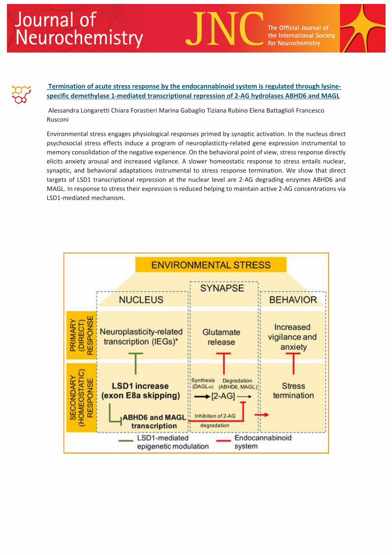
Termination of acute stress response by the endocannabinoid system is regulated through lysine‐
specific demethylase 1‐mediated transcriptional repression of 2‐AG hydrolases ABHD6 and MAGL
Alessandra Longaretti Chiara Forastieri Marina Gabaglio Tiziana Rubino Elena Battaglioli Francesco
Rusconi
Environmental stress engages physiological responses primed by synaptic activation. In the nucleus direct
psychosocial stress effects induce a program of neuroplasticity‐related gene expression instrumental to
memory consolidation of the negative experience. On the behavioral point of view, stress response directly
elicits anxiety arousal and increased vigilance. A slower homeostatic response to stress entails nuclear,
synaptic, and behavioral adaptations instrumental to stress response termination. We show that direct
targets of LSD1 transcriptional repression at the nuclear level are 2‐AG degrading enzymes ABHD6 and
MAGL. In response to stress their expression is reduced helping to maintain active 2‐AG concentrations via
LSD1‐mediated mechanism.

The following articles are part of Volume 155, Issue 2 – Pages 111-224
Respiratory syncytial virus tropism for olfactory sensory neurons in mice
Bertrand Bryche, Maxence Frétaud, Audrey Saint‐Albin Deliot, Marie Galloux, Laura Sedano, Christelle
Langevin, Delphyne Descamps, Marie‐Anne Rameix‐Welti, Jean‐François Eléouët, Ronan Le Goffic, Nicolas
Meunier
Read the full article ‘Respiratory syncytial virus tropism for olfactory sensory neurons in mice’ by B.
Bryche, M. Frétaud, A. Saint‐Albin Deliot, M. Galloux, L. Sedano, C. Langevin, D. Descamps, M.‐
A. Rameix‐Welti, J.‐F, Eléouët, R. Le Goffic, N. Meunier, (J. Neurochem. 2020, vol. 155 (2), pp.
137–153) on doi:10.1111/jnc.14936
Cover Image
Front cover: While the respiratory syncytium virus (RSV) is mainly known for the damage it causes in the
respiratory tract, it could also impact the central nervous system. We explored globally the impact of
RSV in the mouse nasal cavity and showed that RSV can infect olfactory sensory neurons, giving the virus
access to the central nervous system by entering the olfactory bulb. Our study also yielded a 3D atlas of
the nasal cavity in mice, providing tools to improve our understanding of virus tropism in the upper
respiratory tract.
Image content: The present image is a periodic acid–Schiff reaction (PAS)‐stained coronal section of the
mice nasal cavity.
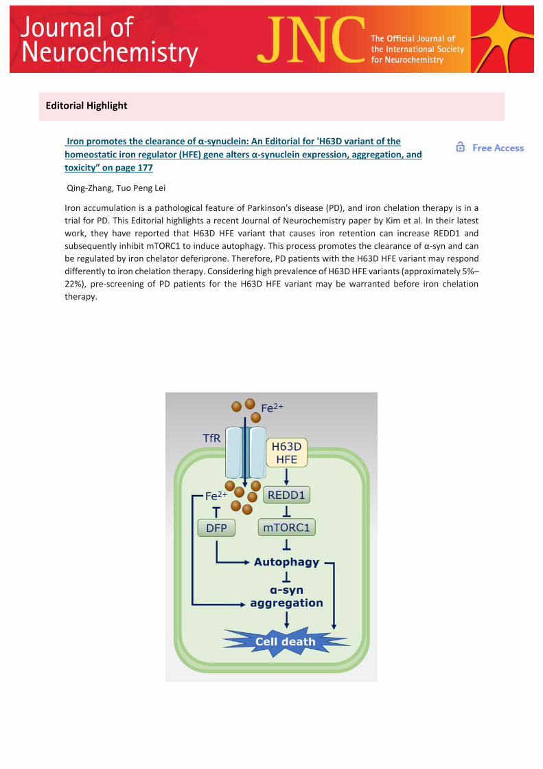
Iron promotes the clearance of α‐synuclein: An Editorial for 'H63D variant of the
homeostatic iron regulator (HFE) gene alters α‐synuclein expression, aggregation, and
toxicity” on page 177
Qing‐Zhang, Tuo Peng Lei
Iron accumulation is a pathological feature of Parkinson's disease (PD), and iron chelation therapy is in a
trial for PD. This Editorial highlights a recent Journal of Neurochemistry paper by Kim et al. In their latest
work, they have reported that H63D HFE variant that causes iron retention can increase REDD1 and
subsequently inhibit mTORC1 to induce autophagy. This process promotes the clearance of α‐syn and can
be regulated by iron chelator deferiprone. Therefore, PD patients with the H63D HFE variant may respond
differently to iron chelation therapy. Considering high prevalence of H63D HFE variants (approximately 5%–
22%), pre‐screening of PD patients for the H63D HFE variant may be warranted before iron chelation
therapy.
Editorial Highlight

What works and what does not work in Alzheimer’s disease? From interventions on risk
factors to anti‐amyloid trials
Szofia Bullain, Rachelle Doody
In this review, we summarize past approaches to finding an anti‐amyloid‐β monoclonal antibody capable of
altering the disease course in Alzheimer's disease (AD). Key learnings from the field have helped shape the
current AD clinical development programs, including translating epidemiological observations into potential
treatment options, evaluating therapies across the AD continuum, using biomarkers to assess target
engagement and as a surrogate of efficacy, and enriching study populations. Because of the underlying
complex pathophysiology of AD, disease‐modifying therapies targeting both amyloid‐dependent and ‐
independent mechanisms may be needed to provide clinical benefit across the disease continuum.
Review articles

Respiratory syncytial virus tropism for olfactory sensory neurons in mice
Bertrand Bryche, Maxence Frétaud, Audrey Saint‐Albin Deliot, Marie Galloux, Laura Sedano, Christelle
Langevin, Delphyne Descamps, Marie‐Anne Rameix‐Welti, Jean‐François Eléouët, Ronan Le Goffic, Nicolas
Meunier
While the respiratory syncytium virus (RSV) is mainly known for its damaged in the respiratory tract, it could
also impact the central nervous system. We explored globally the impact of RSV in the mouse nasal cavity
and showed that RSV can infect olfactory sensory neurons giving access to the central nervous system by
entering the olfactory bulb. Our study also provides a 3D atlas of the nasal cavity in mice giving tools to
improve our understanding of virus tropism in the upper respiratory tract.
Original articles
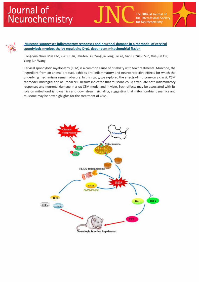
Muscone suppresses inflammatory responses and neuronal damage in a rat model of cervical
spondylotic myelopathy by regulating Drp1‐dependent mitochondrial fission
Long‐yun Zhou, Min Yao, Zi‐rui Tian, Shu‐fen Liu, Yong‐jia Song, Jie Ye, Gan Li, Yue‐li Sun, Xue‐jun Cui,
Yong‐jun Wang
Cervical spondylotic myelopathy (CSM) is a common cause of disability with few treatments. Muscone, the
ingredient from an animal product, exhibits anti‐inflammatory and neuroprotective effects for which the
underlying mechanisms remain obscure. In this study, we explored the effects of muscone on a classic CSM
rat model, microglial and neuronal cell. Results indicated that muscone could attenuate both inflammatory
responses and neuronal damage in a rat CSM model and in vitro. Such effects may be associated with its
role on mitochondrial dynamics and downstream signaling, suggesting that mitochondrial dynamics and
muscone may be new highlights for the treatment of CSM.
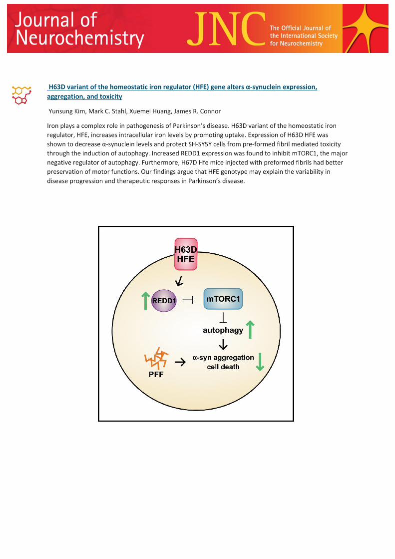
H63D variant of the homeostatic iron regulator (HFE) gene alters α‐synuclein expression,
aggregation, and toxicity
Yunsung Kim, Mark C. Stahl, Xuemei Huang, James R. Connor
Iron plays a complex role in pathogenesis of Parkinson’s disease. H63D variant of the homeostatic iron
regulator, HFE, increases intracellular iron levels by promoting uptake. Expression of H63D HFE was
shown to decrease α‐synuclein levels and protect SH‐SY5Y cells from pre‐formed fibril mediated toxicity
through the induction of autophagy. Increased REDD1 expression was found to inhibit mTORC1, the major
negative regulator of autophagy. Furthermore, H67D Hfe mice injected with preformed fibrils had better
preservation of motor functions. Our findings argue that HFE genotype may explain the variability in
disease progression and therapeutic responses in Parkinson’s disease.

Neuroprotective actions of leptin facilitated through balancing mitochondrial
morphology and improving mitochondrial function
Ying Cheng, Matthew Buchan, Karina Vitanova, Laura Aitken, Frank J. Gunn‐Moore, Rona R. Ramsay,
Gayle Doherty
The amelioration of mitochondrial dysfunction in neurodegeneration such as Alzheimer’s disease, is an
established strategy in the search for treatments for these disorders. In this study, we demonstrate that
the anti‐obesity hormone, leptin, can benefit mitochondrial dynamics and function through supporting
mitochondrial membrane potential and balancing the expression of fission and fusion proteins, while
boosting the expression and activity of mitochondrial antioxidant enzymes MAOB. Thus leptin’s
neuroprotection may be facilitated through the regulation of mitochondrial function and dynamics.
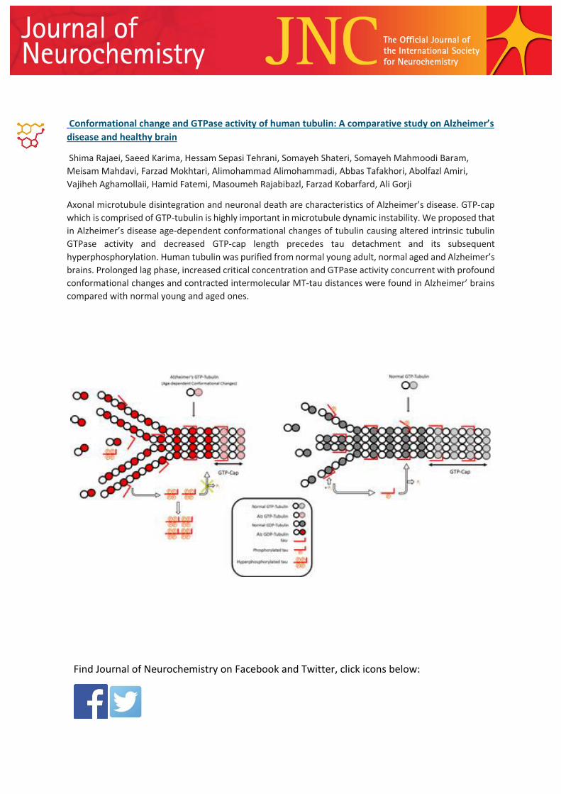
Conformational change and GTPase activity of human tubulin: A comparative study on Alzheimer’s
disease and healthy brain
Shima Rajaei, Saeed Karima, Hessam Sepasi Tehrani, Somayeh Shateri, Somayeh Mahmoodi Baram,
Meisam Mahdavi, Farzad Mokhtari, Alimohammad Alimohammadi, Abbas Tafakhori, Abolfazl Amiri,
Vajiheh Aghamollaii, Hamid Fatemi, Masoumeh Rajabibazl, Farzad Kobarfard, Ali Gorji
Axonal microtubule disintegration and neuronal death are characteristics of Alzheimer’s disease. GTP‐cap
which is comprised of GTP‐tubulin is highly important in microtubule dynamic instability. We proposed that
in Alzheimer’s disease age‐dependent conformational changes of tubulin causing altered intrinsic tubulin
GTPase activity and decreased GTP‐cap length precedes tau detachment and its subsequent
hyperphosphorylation. Human tubulin was purified from normal young adult, normal aged and Alzheimer’s
brains. Prolonged lag phase, increased critical concentration and GTPase activity concurrent with profound
conformational changes and contracted intermolecular MT‐tau distances were found in Alzheimer’ brains
compared with normal young and aged ones.
Find Journal of Neurochemistry on Facebook and Twitter, click icons below:


