IV & Infusion Therapy Certificate
103
IV & Infusion Therapy Certificate 2021
Transcript of IV & Infusion Therapy Certificate

IV & Infusion Therapy Certificate
2021

Dear Student/Participants,
Please find the York University copyright statement. This is an annual reminder to all students about the copyright policy of the University.
Access to and use of the course materials is restricted to students enrolled in the IV & Infusion Therapy Certificate course offered by Health Leadership & Learning Network. All materials for this course are provided with the permission of the rights holder, under the terms of a license or other agreement, or under the application of statutory exceptions of the Copyright Act. Copyright and all rights are maintained by the author(s) or by other copyright holder(s). Copying this material for distribution (e.g. uploading material to a commercial third-party website) can lead to a violation of Copyright law. Find out more about copyright here: www.yorku.ca/copyright
If you have any questions, please contact us here in HLLN at 416 736 2100 X22170 or [email protected]. Thank you, Tania Xerri
____________________________________ Tania Xerri, Director, Health Leadership and Learning Network A Leader in Health Continuing Professional Education Faculty of Health York University 4700 Keele St. HNES 019, Toronto, ON M3J 1P3
Information, Privacy and Copyright | Office of the Counsel 1050 Kaneff Tower | York University | 4700 Keele St., Toronto ON M3J 1P3 Canada

IV & Infusion Therapy Week 1
Karen Laforet MClSc, RN, CHNC (C), VA-BCTM, CVAA (c)

For their generous donations and support
WWW.CVAA.INFO
https://cvaa.info/en/certification/information
Join CVAA before end of year and receive 10% discount on certification
exam.

Introductions
Work Setting

Objectives:• By the conclusion of this session, participants will be able to:• Review rationale for infusion therapy• Define common universal terms used for infusion therapy and vascular
access• Discuss core safe infusion practices• List six function of the integumentary system• Identify risk to skin integrity for a person receiving IV therapy• Compare the sympathetic and the parasympathetic nervous systems.
© Copyright Klaforet
Vascular Access gone wrong…

Core Practice Principles• Foundational elements to all practice:• Infection Prevention and Control• Ethics• Evidence-Informed Practice• Informed Consent• Hand Hygiene• Safe Handling and Disposal of Hazardous Materials & Sharps• Product use (following Manufacturer’s instructions)• Patient Education and Competency• Documentation.
Infusion Therapy: History• 1656 Sir Christopher Wren—used quill and bladder to inject dog with opium • 1667: 1st human transfusion: 15 yr boy France (Jean Baptiste Denis, King’s physician)• 1834: Dr. James Blundell transfused women hemorrhaging post-partum• 1900 Karel Landsteiner : 3 out of 4 blood groups• 1925: Dextrose used as infusate• 1940: disposable administration sets developed• 1950’s: parenteral fluids introduced and used safely• 1957: McGawa Labs introduced winged infusion needle (Butterfly)• 1960: PICC lines introduced into ICUs• 1963-65: first successful TPN• 1964: First disposable IV catheter (Angiocath by Deseret)• 1970: CDC IV therapy guidelines published• 1972: Implanted ports access introduced• 1975: Canadian Intravenous Nurses Association (CINA) founded• 1980: First Standards of Practice published by National Intravenous Therapy
Association (NITA—now known as INS)• 1983: Home infusions started and in US, first blood transfusion initiated• 2006: CINA changed name to Canadian Vascular Access Association (CVAA)
© Copyright Klaforet
1964 First disposable cannula introduced—one of the greatest
modern inventions

Infusion Therapy
• It is the most common invasive procedure in health care.• 90% of all patients who experience healthcare will have an IV at some
point in time (ISMP 2015, CDC 2012, SHN)
© Copyright Klaforet
What…is infusion therapy
• Introduction of fluids,blood, and drugs directly intothe vascular system: i.e.arteries, bone marrow andveins.
Google image

Why Infusion Therapy?
• To administer fluids and medications• To provide parenteral nutrition• To provide avenue for dialysis/apheresis• To transfuse blood products• To provide avenue for hemodynamic monitoring• To provide avenue for diagnostic testing
Goal of Infusion Therapy
• Positive outcome • First stick Success
• Painless (#6 worry for patients is painful IV start)
• Effective treatment• Complete recovery
(the why)
© Copyright Klaforet
Google image

Healthcare professionals need to be proficient in the assessment, selection,
insertion and care & maintenance of vascular access devices and the infusions/medications
provided
CRITICAL for patient safetyEarly recognition and prevention of complications. Knowing anatomy, physiology and pathophysiology will minimize patient risk
Google image

Google images
Common Terms• Infusion therapy: replaces IV therapy—it’s more
than ‘pick and stick’• VAD: Vascular access device (VAD) in place of IV
cathetero PVAD in place of PIVo CVAD in place of central line or CVC or CIC
• Health Professional (HP or HCP) in place of nurse, doctor, phlebotomist…etc.
• DIVA: Difficult IV access © Copyright Klaforet

Infusion related clinical competenciesCore content areas: • Anatomy & Physiology• Fluids & Electrolytes• Vascular Access—peripheral & central: assessment, selection,
care & maintenance• Infection control and safe infusion practice• Complications• Pharmacology (including chemotherapy)• Pediatric Population Implications• Geriatric Population Implications• Transfusion Medicine• Parenteral Nutrition
© Copyright Klaforet
Documentation• Use standardized medical terminology and approved abbreviations. • Ensure all documentation includes date, HCP signature, and credentials.• Use standardized templates to document VAD insertion, care and maintenance, and infusion therapy (e.g., flow
sheets) • Document procedural compliance and task completion for CVADs (e.g., insertion checklists) • Consider use of structured checklists for peripheral vascular access device (PVAD) insertion.• Include the following in all procedural documentation
a) Procedural details, including any deviationsb) Barriers to care when present c) Patient participation in, understanding of, and responses to therapy and interventiond) Evaluation of expected outcomes, side effects and adverse events, including intervention and patientresponsee) Relevant lab results.

Identify 10 things wrong in this video
YouTube
Common Sources of PVAD Contamination
Hand Hygiene VAD Location Skin Surface ContaminatedEquipment
Idle Catheters Asepsis breach

Hand Hygiene 7,8
Sax H, et al. J Hosp Infect 2007; 67(1): 9–21
Hand hygiene is the simplest, most effective measure for helping to prevent healthcare association infections.
Compliance ranges 40-60% among healthcare workers.
Research shows that there are even lower rates of hand hygiene when gloves are worn.
E. Hand Hygiene
What is the preferred method of hand hygiene?

Moments of Hand Hygiene
(PHAC, 2012; RCN, 2016; SHN, 2012)
1. Perform hand hygiene at the standard moments of hand hygiene,including, but not limited to (PHAC, 2012; RCN, 2016; SHN,2012): [IA]
Hand Hygiene: VA & Infusion Therapy (CVAA 2019)
1. Before and afterpalpating insertion
2. Before handling ormanipulating infusion
system
3. Before putting onand after removing
gloves
4. Before and after inserting, orreplacing, or accessing, or repairing
or dressing a VAD

Provide information to patients on
WHEN and HOW to perform hand
hygiene.
• Clinical implications for infusion therapy:• Physical & functional characteristics
o Integumentatryo Nervouso Circulatoryo Vascularo Skeletal
• Know location of veins, arteries, nerves for vascularaccess
© Copyright Klaforet

The skin of terrestrial animals is essential for their survival.…the principle role of skin is to keep harmful agents out and water in. In order to accomplish this seemingly trivial task, nature has developed an elaborate structure, termed the stratum corneum, which provides to the skin its barrier function.
Schaefer and Redelmeier
© Copyright Klaforet

© Copyright Klaforet Google images
Google images

Skin Function
ProtectionCommunication
Thermoregulation
Immunity
Metabolism
SensationGoogle images
Protection
• Stratum corneum (uppermost layer)oThickness= .01 mmoComposed of • Corneocytes• Water• Lipids
oSemi-permeable
© Copyright Klaforet
Google images

Protection
• FibroelasticityoStress against
mechanical forces• Transepidermal
Water loss (TEWL)oEvaluates barrier
function© Copyright Klaforet Google images
Thermoregulation
• Maintenance of body temperature: oVasoregulationoSweatingoEccrine Glands oApocrine Glands
© Copyright Klaforet

Synthesis of Vitamin D
© Copyright Klaforet
Google images
Immunity
• Microflora• Langerhans's cells • Dermal lymphatics• Mast Cells

Resident vs Transient Microbiota• Resident• Acquired rapidly during &
after birth• Always present• Unable to remove • In health human internal
tissues are free from microorganismso Bloodo Braino Muscleo CSF
• Transient• Live in or on the body for a
period of time (hours, days, weeks, or months) then move on or die off
• Cannot live on d/t competition, elimination by body’s defenses or chemical/physical changes in the body
• Able to remove through washing
Symbiotic Relationship
• Mutualistico Both organisms benefit (e.g. E. Coli synthesizes Vit K & B)
• Commensalistico None with Bacteriao If on/in the skin either helping or harming
• Opportunistico Pathogenic outside normal environment (e.g. E. Coli, Staph aureus

Communication & Sensation
Google Image
Threats to Skin IntegrityMoisture Associated Skin Damage (MASD)
General term for inflammation or skin erosion caused by prolonged exposure to a source of moisture such as urine, stool, sweat, wound drainage, saliva, or mucus
© Copyright Klaforet

Medical Adhesive-related skin injury (MARSI):
• Prevalent, under-recognised and preventable complication specifically related to skin damage caused by medical adhesive
• Can occur in any patient group or setting
© Copyright Klaforet
3M Image
Chemicals
• Antiseptics• Skin barriers• Soap• Tackifiers
© Copyright Klaforet

Neurologic System
Nervous System• Information travels from one neuron
to the next via synapses:– Electrical—thru open fluid channels
between nerve cells– Chemical
• Small molecule rapid acting (acetylcholine, epinephrine, norepinephrine, histamine)
• Neuropeptides –slow-acting (vasopressin, insulin, angiotensin II, oxytocin, bradykinin)
Google Image

Anatomic Divisions
Google Image
Autonomic Nervous System
Google Image

Sensory Receptors r/t IV therapyReceptor Sensation Effect on IV therapyMechanoreceptor Skin tactile senses
Deep tissue. Arterial pressure controlled thru baroreceptors (largearteries)
Palpating veins or arteries;Application antiseptic sol’n;DressingsPuncture; tourniquet; excessive infusion or circulating blood volume;
Thermoreceptors Cold, warm Heat, cold applications; infiltration, extravasation, phlebitis
Nociceptor Pain Puncture; dressingremoval; infusion irritating sol’n; heat or cold app
Chemoreceptor ¯ Arterial BP stimulates aortic & carotid arteries to respond to O2 & CO2 levels;
Blood osmotic changes
¯ Volume of solution = ¯ blood volume.
Infusion of hypertonic or hypotonic sol’n
Neurovascular bundle

References• Unless stated otherwise all images are from Karen’s personal files
• Alexander, M., Corrigan, A., Gorski, L., Hankins, J., & Perucca, R. (Eds.). (2010). Infusion nursing: An evidence-based approach (3rd ed.). St. Louis, MO: Saunders/Elsevier.
• CVAA Infusion therapy and vascular access guidelines. 2019. • O’Grady, N.P., Alexander, M., Burns, L.A., Dellinger, E.P., Garland, J.,
Heard, S.O., & Healthcare Infection Control Practices Advisory Committee. (2011). Guidelines for the prevention of intravascular catheter-related infections. American Journal of Infection Control, 39(4 Supplement 1), S1-S34. Doi: 10.1016/j.ajic.2011.01.003.
• Weinstein, S., & Hagle, M. (2014). Plumer’s principles and practice of infusion therapy (9th ed.). Philadelphia, PA: Wolters Kluwer Health, Lippincott Williams & Wilkins.
© Copyright Klaforet

IV & Infusion TherapyWeek 2
Karen Laforet MClSc, RN, CCHN (c), VA-BCTM, CVAA (c)
Objectives
• Identify key difference between artery and vein anatomy• Name two veins commonly used for peripheral vascular access• List components of the Cardiovascular System pertinent for Infusion
therapy• Outline the vascular access device process• Differentiate peripheral devices and central devices• Summarize the key components of vascular access device selection

Name the vein
Cardiovascular System

Heart, Mediastinum
Google Image
Circulatory System• Divided into 2 main
systems: Pulmonary & systemic
• Systemic veins are classified into 3 classes:1. Superficial—venipuncture
o Where are superficial veins located? (superficial fascia)
2. Deep3. Venous sinuses
• Superficial and deep veins unite—especially in lower extremities
© Copyright Klaforet
Google Image
Mechanisms of venous return• Pressure gradients
• 7 -13 mmHg venous pressure towards the heart• Venules (12 -18 mmHg) to CVP (~5mmHg)
• This is why calf pump is so important!)
• Gravity drains blood from head and neck• Muscle pumps• Thoracic pump
• Inhalation: thoracic cavity expands (¯pressure), abdominal pressure = forces blood upward
• Blood flows faster with inhalation (consider consequence with COPD patients)
• Cardiac suction of expanding atrial space© Copyright Klaforet
Google Image
Anatomy of Artery & Vein
Google Image

Vein Anatomy and Physiology
© Copyright Fresenius Kabi AG
Google Image
Tunica Adventitiathe outer layer of the vessel• Connective tissue• Contains the arteries and veins
supplying blood to vessel wall (Vasa vasorum) [may penetrate to the tunica media], lymph channels
• Afferent and sympathetic nerves
© Copyright Fresenius Kabi AG
Google Image
© Copyright Fresenius Kabi AG
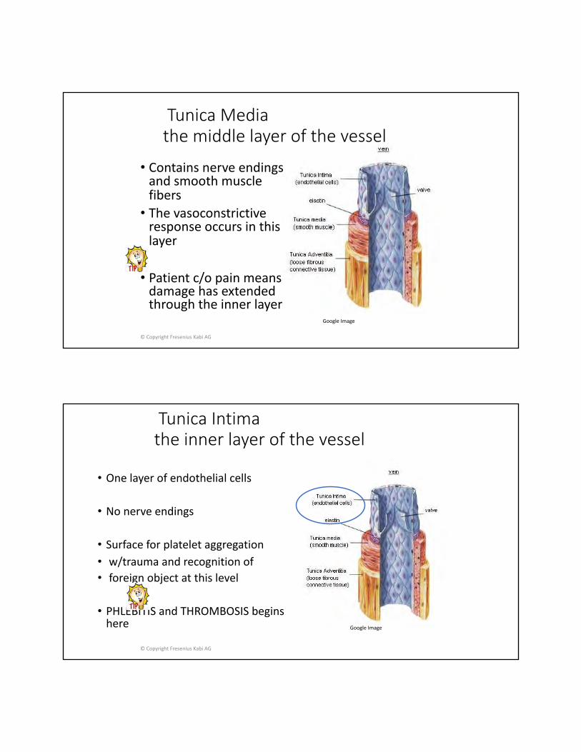
Tunica Mediathe middle layer of the vessel
• Contains nerve endings and smooth muscle fibers• The vasoconstrictive
response occurs in this layer
• Patient c/o pain means damage has extended through the inner layer
Google Image
Tunica Intimathe inner layer of the vessel
• One layer of endothelial cells
• No nerve endings
• Surface for platelet aggregation • w/trauma and recognition of • foreign object at this level
• PHLEBITIS and THROMBOSIS begins here
© Copyright Fresenius Kabi AG
Google Image

• Prevent backflow and blood pooling
• More in lower extremities and the longer vessels
• Vein dilates at valve attachment
© Copyright Fresenius Kabi AG
Valvespresent in MOST veins
Google Image
• Occur at branching• May cause a noticeable
bulge in the vein• If IV started too close to
the valve may have false reading of no blood return).
Courtesy of BardTM
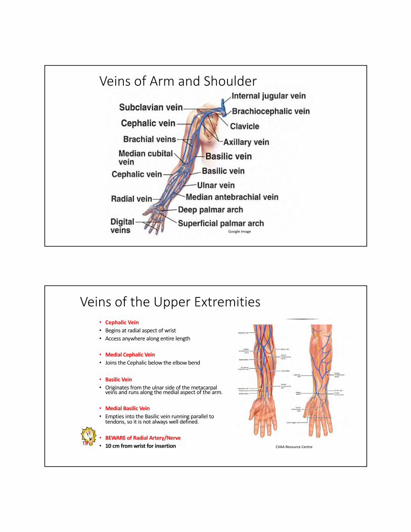
Veins of Arm and Shoulder
Google Image
Veins of the Upper Extremities• Cephalic Vein• Begins at radial aspect of wrist• Access anywhere along entire length
• Medial Cephalic Vein• Joins the Cephalic below the elbow bend
• Basilic Vein• Originates from the ulnar side of the metacarpal
veins and runs along the medial aspect of the arm.
• Medial Basilic Vein• Empties into the Basilic vein running parallel to
tendons, so it is not always well defined.
• BEWARE of Radial Artery/Nerve• 10 cm from wrist for insertion CVAA Resource Centre

Peripheral vascular access devices areubiquitous in healthcare and risk of harmis significantFollowing evidence-based practices will helpprotect patients from infection.
Celum
VADs
Peripheral Central Special
Over-the-Needle Short & Long length
Non-tunneled Dialysis
Butterfly Tunneled Apheresis
Extended Dwell Implanted Umbilical
Midline PICC Intraosseous
Arterial Hypodermoclysis

PVADS• Approximately 75 % of all Intravenous Access devices inserted
are peripheral
• Catheter, less than 3 inches (7.5 cm) in length• Over-the-needle catheter is most common
• Gauge sizes 14- 27• Winged/ non-
winged• Single or double
lumen• Over the needle
catheter• Terminates in a
peripheral vein

PVAD
PVAD • Tip terminates in a peripheral vein• Any catheter whose tip is not in the
bottom 1/3 of the superior vena cava (or is considered a peripheral VAD
Example- Tip position in the Subclavian vein
Courtesy K. Laforet
PVAD: Midline Catheters PVAD’s with the tip terminating in the Basilic, cephalic or brachial vein distal to the shoulder—level with axilla
• Single or double lumen• 1.9 Fr - 5 Fr (adults); 22-24 g for pediatric
catheters
• Polyurethane or silicone material
Initiated above or below the Antecubital Fossa in one of the following
• Basilic
• Cephalic
• Median Cephalic
• Brachial VeinsGoogle Image

CVAD
Google Image
Google Image

CVAD Tip Location• Ensure optimal tip location for CVADs [IC]• In distal superior vena cava (SVC) or cavoatrial junction
(CAJ); if using CXR, measure from carina, trachea-bronchial angle, or thoracic vertebral bodies [IB]
• Femoral VAD should have tip location within the inferior vena cava and above the level of the diaphragm.
Google Image

Non Tunneled Catheters• Recommended for short-term access to the central
circulation in critical situations, or when peripheral access is inadequate or inappropriate.
• Used mainly for ICU/CCU patients. Not safe for alternate care settings
• Percutaneous insertion of catheter into the IJ, subclavian or femoral site
• Associated with high risk of CRI d/t skin exit pint of catheter in close proximal to the entry point of vein used
• Available in single, double, triple, quad or 5 lumen• Generally open-ended.
Google Image

Implanted Devices (aka “Port”)
• An implanted reservoir generally placed in the chest or arm, attached to a catheter with tip position in the central vasculature.
• Infusate is delivered to the reservoir via an external non-coring needle and extension tubing
Shutterstock
Implanted Devices• Requires a minor surgical procedure for placement and removal.• When not in use, requires less maintenance than other VADs.• Medication delivery requires injection through skin using a non-coring needle• Intended for long term therapy up to 5 years• Q 6 -12 week flush with 0.9%NaCl for maintenance (Heparin may be ordered)• Implanted devices with or without valve
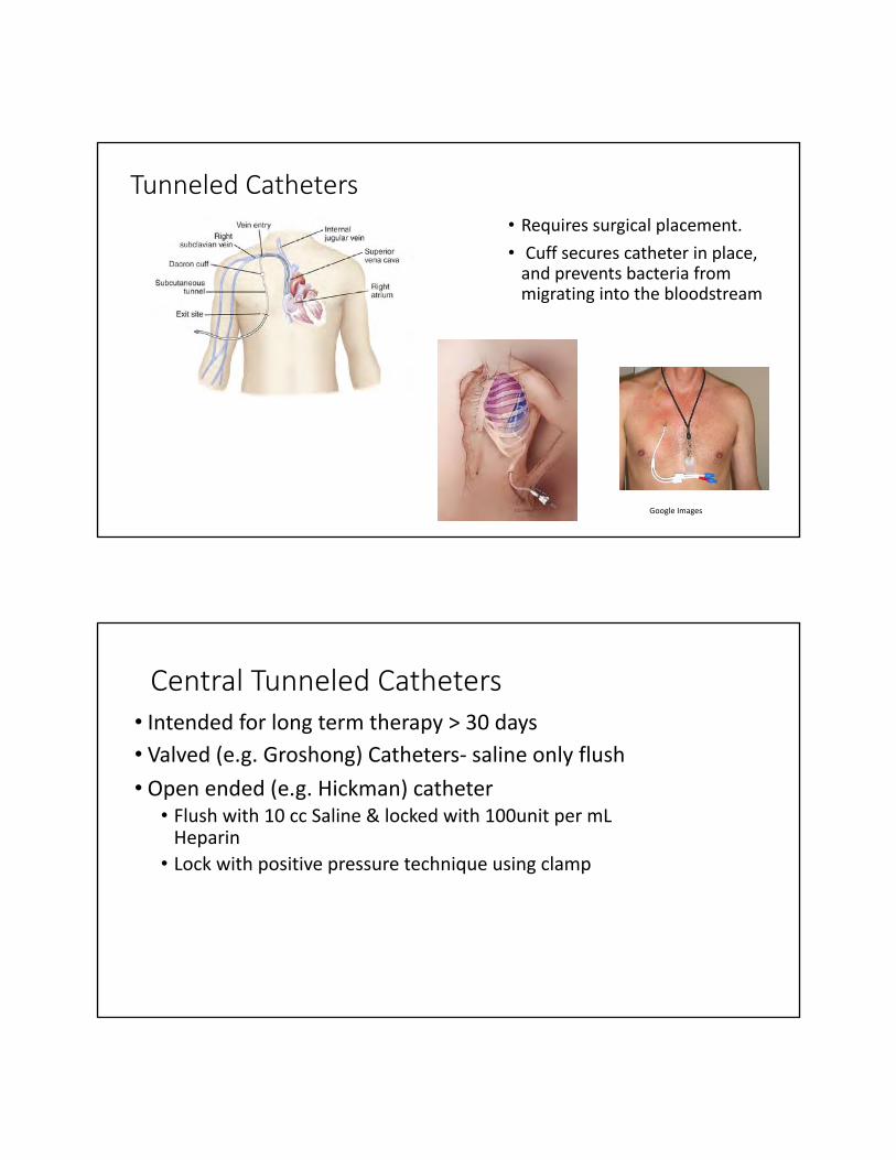
Tunneled Catheters• Requires surgical placement.• Cuff secures catheter in place,
and prevents bacteria from migrating into the bloodstream
Google Images
Central Tunneled Catheters• Intended for long term therapy > 30 days• Valved (e.g. Groshong) Catheters- saline only flush• Open ended (e.g. Hickman) catheter
• Flush with 10 cc Saline & locked with 100unit per mL Heparin
• Lock with positive pressure technique using clamp

Valved vs Non-valved catheters
• Non-valved VAD: open-ended tip; lumen hub has clamps on external portion of catheter to stop backflow• For IVAD: refers to device not having an integrated valve
• Valved VAD: integrated valve• Valve located at catheter tip (distal end) or in the catheter hub (proximal end)• Valve opens with infusion or flushing and when pressure is exerted for
aspiration or blood sampling• Valve is neutral or remains closed when no pressure is applied• Prevents blood coming into the catheter
Valved vs non-valved

Hemodialysis VAD• Tunneled or implanted VAD; AV fistula, or insertion of arteriovenous
graft:• All considered surgical procedures
• Administering meds, solutions through any hemodialysis device requires specific MD order
• Can a nurse remove one of these devices? Yes: if validated competency
• Venipuncture is not to be performed on the extremity containing AV fistula or graft
• Vein preservation KEY for patients who are likely to need vascular access for hemodialysis
• Any access is considered sterile: sterile gloves and mask required • Can one use PI ointment or Polysporin at exit site? Yes: only if
ointment doesn’t interact with catheter material as per manufacturer’s IFU. ONLY CVAD where ointment is used
Apheresis & Ultrafiltration• Large bore central catheter, percutaneously
or surgically placed to maintain high flow rates and accommodate large bld volumes.
• Apheresis: removal of blood plasma from the body by withdrawal of blood
• Separates into plasma and cells• Cells reintroduced• Used to remove antibodies in treating
autoimmune diseases• Tip of catheter resides at junction of SVC and
R atrium• Ultrafiltration: removes excess salt and water
in patients in fld overload (e.g. CHF)

Umbilical Catheter• Arterial or venous access for newborns • Arterial umbilical catheter tip is located in the
descending aorta above the level of diaphragm and below the L subclavian artery• Venous umbilical catheter tip: located in inferior vena
cava above the level of diaphragm• Removal: performed aseptically, slowly over several
minutes; manual compression with sterile gauze until hemostasis • Monitor site X12 hrs then daily• Complications: bleeding/hemorrhage, air embolism,
infection, thrombosis, vascular perforation, peripheral vascular constriction.
Interosseous
• Inject directly into the marrow of a bone.• Provides fluids and medication when intravenous access is not
available or not feasible• Emergency access:• i.e. codes, in the field, pediatrics
• SOP: pediatrics: after 2 attempts use I.O. access and replace within 24 – 48 hours

Assessment Pre-insertion (Prep) Insertion Care &
Maintenance Removal
Life Cycle of a Vascular Access Device (VAD)
J. LeDonne, 2018
10 % 90 %
VAD Selection (CVAA, 2019)
• Determine the appropriate type of VAD:a) Use device with minimum number of lumens b) Always select the smallest gauge catheter that will accommodate the
prescribed therapy c) Consider use of a 22-gauge PVAD for most infusion therapies. d) Consider using longer length PVAD for insertion with ultrasound

VAD Access Site Preparation
College of Nurses Standards
• MD order required to place a device
• Nurse shall be competent in:• Insertion technique• Infection prevention measures• Identifying potential complications• Implementing nursing interventions
Primary GoalOne patientOne stickOne device
Google Image

VAD Planning (CVAA 2019)
1. Use a systematic process to develop a patient-centric vascular access plan prior to or at onset of therapy that optimizes vessel preservation and guides device selection.
2. 2. Ensure VAD planning is an ongoing process throughout treatment.
3. 3. Determine: is vascular access is necessary or if an alternate route is appropriate (e.g., oral, sublingual, inhaled, nasal, transdermal, topical, subcutaneous)
4. Select the device that is the least invasive for the duration and type of therapy and promotes vessel preservation.
Principles of site selection• Catheter/Vein Ratio • Hemodilation and hemodilution (Dilution of infusate)• Vessel preservation• Location
• When would you use the hand vs the forearm? (there are exceptions)
• A TRUE emergency (person is to die in the next 90 sec)• Just because the vein is large doesn’t mean you put in a
large cannula “If you want an IV to fail, put it in the elbow or the hand”(J LeDonne)

Device Selection (CVAA 2019)
Determine the appropriate type of VAD:• Always select the smallest gauge catheter that will accommodate
the prescribed therapy• Consider use of a 22-gauge PVAD for most infusion therapies. • Consider using longer length PVAD for insertion with ultrasound
Determine vascular access needs according to:a) Intended frequency and duration of therapyb) Prescribed therapy (e.g., osmolarity, pH, vesicant, and irritant properties)c) History of vascular access and comorbidities (e.g., renal status)d) Age and developmental stagee) Anatomyf) Activitiesg) Skin integrityh) Patient’s preferences and lifestylei) Available resources for VAD care and maintenance.
• Identify risks and benefits associated with each type of VAD • Determine the minimum number of lumens required for the plan of care• Determine if a VAD designed with power injection capabilities is needed
(CVAA 2019)

pH Scale• Measures concentration of hydrogen ions (H+) in a solution. • 0 to 6 being acidic, • 7 neutral • 8 to14 being alkaline (base)
What is critical to understand is that a small change in pH results in a large change in H+ ion concentration.
• pH - Blood pH = 7.35 – 7.45• pH of 5 – 9 minimizes disruption of venous endothelium
• Medications & IV fluids with a pH of 5-9 can be safely administered via peripheral IV
Google Image

Acid pH Scale for common Medications
Acid Drug/Fluid• Stomach Acid pH 1 Vancomycin pH 2.4• Lemon Juice pH 2 Ciproflaxin pH 3.3-4.6• Vinegar pH 3 Tobramycin pH 3-6.5
Neutral• Tap Water pH 7 Erythromycin pH 6.5-7.7
Ceftriaxone pH: 6.5 - 7
CVAA Guidelines 2019

Site Selection• Veins on Dorsal & Ventral
surfaces of Upper Extremities• Metacarpal• Cephalic• Basilic• Median Veins• Distal Areas – of arms
• Avoid use of radial veins -Potential risk for nerve damage if fluid goes interstitial• Painful Insertion
D0 Not Use:i. Areas of flexion (e.g., wrist, ACF), except in trauma or emergency situation (to avoid nerve damage and depletion of antecubital veins)ii. Chest wall, digits, or breastiii. Lower legs, except in non-walking childreniv. Insertion areas that are painful on palpation or with veins that are obviously compromised (e.g., thrombosis, redness, cording, bruising, infiltration, phlebitis, engorgement)v. Extremity with arteriovenous (AV) fistula/graft sitevi. Affected extremity after extravasation for subsequent VAD insertion until symptoms are resolved (RCN, 2016).

1. Cephalic2. Median Cubital Vein3. Accessory Cephalic Vein4. Basilic Vein5. Cephalic Vein6. Median Ante Brachial Vein

IV & Infusion TherapyWeek 3
Karen Laforet MClSc, RN, CCHN (c), VA-BCTM, CVAA (c)
Objectives
• List potential complications that may occur when a VAD is in situ (PVAD or CVAD)• Identify prevention strategies to reduce risk of complications• Discuss the need for regularly or routine VAD observation• Differentiate infiltration from extravasation• Explain the role fibrin places in VAD occlusion• Describe steps to assess and manage complications

Case Study
89 yrs old male admitted from ER to Orthopedic Unit with a fractured L hipfollowing a fall.
#20 gauge IV catheter—inserted into L forearm(L arm edematous--etiology unknown)
Film dressing with border intact. Smallamount of sanguineous drainage noted underdressing
Several pieces of tape secure the IV tubing onunderside of arm
Skin tear post tape removal Courtesy K. Laforet
VAD Assessment O.P.A.L. 2018
•O: Observe fluid container, infusion system, insertion site•P: Palpate the insertion site for changes in
temperature, erythema, tenderness or firmness along the vein•A: Aspirate for free-flowing blood return that looks
like whole blood (robust blood flow)• L: Listen to the patient concerns and symptoms.

Look
Feel
Ask
VAD Site Assessment (CVAA 2019
Reasons why VADs Fail
Dislodgement Patient Interference Occlusion Infiltration
Phlebitis Infection Catheter Migration

Possible VAD complications
• Phlebitis• Infiltration• Extravasation• Embolism• Blood vessel damage• Thrombosis• Nerve injury• Infection• Catheter migration
7
Phlebitis• Definition: inflammation of one or all three layers of the vein wall.• Causes:
• Mechanical• Chemical• Bacterial
• Signs and symptoms• Erythema• Swelling• Pain• Tenderness• Induration• Warmth• Cording• Red streak

Phlebitis Scale
Grade Clinical Criteria0 No symptoms1 Erythema at access site with or without pain2 Pain at access site with erythema and/or edema3 Pain at access site with erythema; Streak formation; Palpable
venous cord4 Pain at access site with erythema; Streak formation; Palpable
venous cord >1 inch in length; Purulent drainage
Visual Infusion Phlebitis ScoreVIP Scale
NHS

Prevention
• Medication• Venous access• Securement/Movement• Choosing appropriate devices• Monitoring VADs closely
Intervention
• Stop infusion at the first sign of phlebitis• Determine the etiology of the phlebitis
• Discontinue PIV catheter and restart at a new site or consider alternate mode of delivery• Apply warm packs• If discontinuing treatment, ensure patient is educated on after-care

Infiltration
• Definition: Inadvertent administration of IV fluids (non-vesicant) into the surrounding tissue• Causes:• Catheter placed in area of flexion• Dislodgement of catheter
Nursing 2002,
Infiltration Scale5
Grade Clinical Criteria
0 No Symptoms1 Skin blanched, edema <1 inch, cool to touch, with or without pain
2 Skin blanched, edema >6 inches, cool to touch, with or without pain
3 Skin blanched/translucent; gross edema>6 inches; cold to touch; mild to moderate pain; possible numbness
4 Skin blanched/translucent; skin tight, leaking, discoloured, bruised, swollen; gross edema > 6inches, deep pitting tissue edema, circulatory impairment, moderate to severe pain; infiltration of any amount of blood product, irritant or vesicant

Prevention & Management
• Selection of appropriate VAD –size & catheter• Technique• Securement• Monitoring
• Stop infusion and remove PVAD• Warm compress
Extravasation
• Definition: Inadvertent administration of a vesicant medication or solution into the surrounding tissues.• Causes:
• Infusion of hyperosmolar or infusate that is acidic, alkaline, vasoconstrictive, or cytotoxic• Inadequate securement of VAD• Traumatic insertion• Multiple venipuncture attempts• Small or frail veins• PIV site in area of flexion• Use of an infusion pump or power injection• Presence of a fibrin sheath• Dislodged or non-coring needle for ports.

Extravasation
Prevention• Recognizing the type of medication and its effect as an irritant
• Example: Acyclovir (pH 10.5-11.6)
• Instruct patient to inform staff of pain and burning at insertion site
• Recognize institutional policies and procedures for administering vesicant medication
• Avoid using the dorsal surface of the hand and areas of flexion where tendon and nerve damage is likely to occur
• Give vesicants last when multiple drugs are ordered

Interventions
• Elevate affected extremity
• Contact physician
• Local thermal application (Cold or heat)
• Documentation
• Antidotes
NON-VESICANT Infusions PVAD1
Acute care continuous infusionsEvery 4 hours, alert/orientedEvery 1 - 2 hours for sedated patients, who are cognitively or sensory impaired or critically ill.
Alternate care settingsOnce a shift or visitInstruct patient/caregiver how to assess PVAD every 4 hours while awake and to report any changes immediately
Electronic Infusion Devices
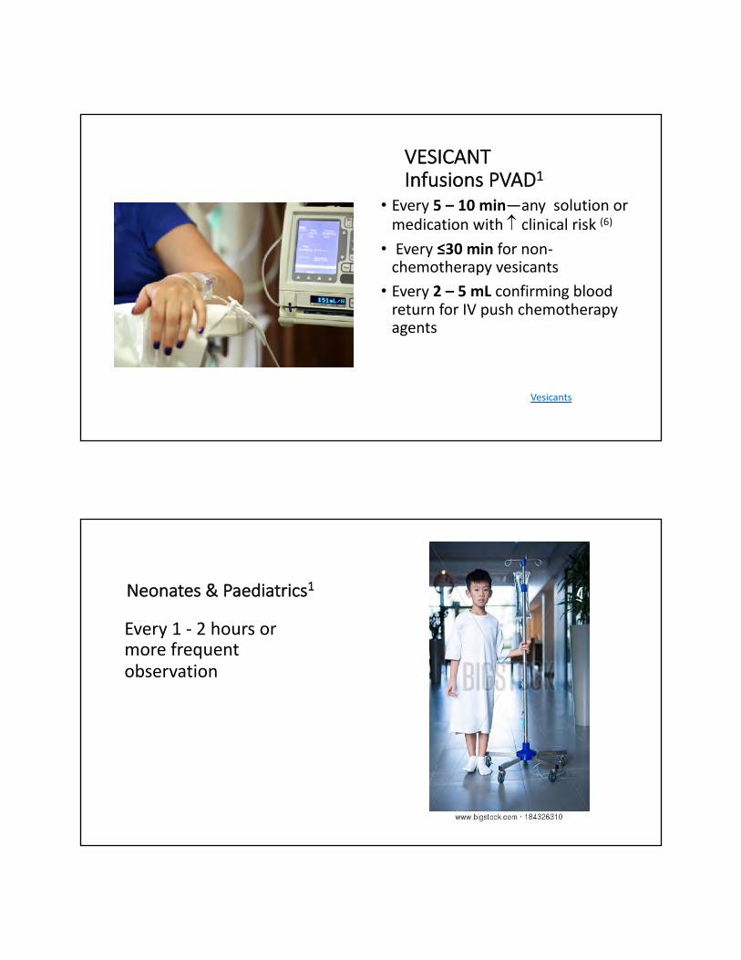
VESICANT Infusions PVAD1
• Every 5 – 10 min—any solution or medication with clinical risk (6)
• Every ≤30 min for non-chemotherapy vesicants • Every 2 – 5 mL confirming blood
return for IV push chemotherapy agents
Vesicants
Neonates & Paediatrics1
Every 1 - 2 hours or more frequent observation

Common Sources of VAD Contamination
Hand Hygiene VAD Location Skin Surface ContaminatedEquipment
Idle Catheters Asepsis breach
YouTube
Equipment contamination 13, 14
• Touch points include:• Catheter hub, • Injection ports• Administration set•Medication/infusi
on administration
KLaforet

Idle catheters• Catheters are a nidus
for infection due to fibrin build-up
• Increased risk of contamination each time the device is accessed:
• Flushing frequency—ask why so often?
• Contamination at hub or with flush syringe.
Asepsis Breach
Google image
Google image

PVAD Securement & Stabilization 2,3
Use 22 ga wheneverpossible
VAD location Use suturelesssecurement to limit VAD
movement
Visually inspect VAD atregular intervals
Patency, Flushing & Locking
• Flush VAD and confirm patency at established intervals:• IVAD (non-accessed/not in use): no more frequently than monthly:
consider extending frequency to three months
• Lock all VADs with the sterile preservative-free 0.9% sodium chloride flush using the appropriate technique to maintain VAD patency.
• Aspirate all alternative lock solutions prior to use of the VAD or according to MRP/manufacturer’s instructions for use • Exception: low-dose heparin (e.g., 10 or 100 units/mL) may be flushed through • If unable to aspirate, alternative lock solutions may have to be flushed through
(except high-dose heparin, e.g., if therapeutic dose must be flushed through, MRP should be notified).

Aseptic technique 14, 16
• Asepsis is defined as a process for keeping away disease-producing organisms in sufficient quantity to cause infection
• Whether the procedure is “simple” or complex, the goal for healthcare professionals is to prevent the transfer of pathogens
• Aseptic non-touch technique (ANTT®)
Catheter Related Blood Stream Infection (CRBSI)• Interventions
• Assess for signs and symptoms of CRBSI
• Examine the patient and carefully rule out other sources of infection
• Cultures• Consider time-to-positivity blood cultures
• When a sample for blood culture is drawn, the needless connector should be changed prior
• Draw out blood cultures prior to the initiation of antimicrobial therapy
• Antimicrobial therapy is based on identified or presumed microorganism

Signs and Symptoms of CVAD Occlusion
• Upon Infusion or Flushing• Resistance• Sluggish flow• Inability to infuse fluids• Frequent occlusion alarms on infusion pump• Infiltration or extravasation or swelling or leaking at the insertion site.
• Upon aspiration of blood• Inability to withdraw blood• Sluggish blood return
Thrombosis
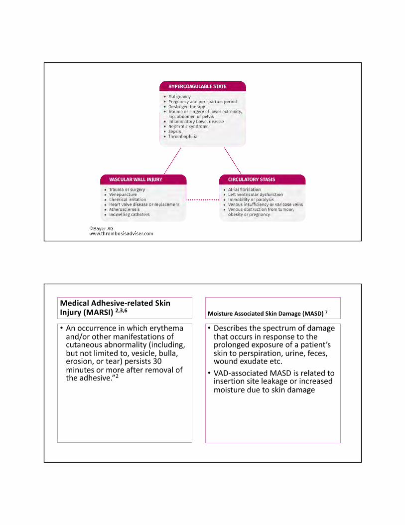
Medical Adhesive-related Skin Injury (MARSI) 2,3,6
• An occurrence in which erythema and/or other manifestations of cutaneous abnormality (including, but not limited to, vesicle, bulla, erosion, or tear) persists 30 minutes or more after removal of the adhesive.”2
Moisture Associated Skin Damage (MASD) 7
• Describes the spectrum of damage that occurs in response to the prolonged exposure of a patient’s skin to perspiration, urine, feces, wound exudate etc.• VAD-associated MASD is related to
insertion site leakage or increased moisture due to skin damage

Tension Injury
Allergic Contact Dermatitis
Protect skin 2, 3, 7, 8
• Antiseptics and adhesives are chemicals that may irritate the skin• Skin MUST be clean and dry before applying any product• For patients at-risk of skin breakdown, or for patients receiving long-
term therapy, apply skin protectant prophylactically• Alcohol-free product to reduce xerosis• VAD application requires sterile product• Avoid water soluble products if exudate or denuded skin• Products must be completely dry before applying the dressing
Air Embolism• Definition: when air enters the vascular system leading to complications• Causes:
• Catheter fracture• Disconnection of IV sets• Deep inspiration during catheter removal/access device change
• Signs and symptoms• Hypoxia• Hypotension• Pallor• Palpitations and arrhythmias• Chest and shoulder pain• LOC
• Prevention• All air purged from syringes, administration sets, needless connectors• Clamp open-ended catheters
• Management• Position the patient in left lateral Trendelenburg • Administer Oxygen

IV & Infusion TherapyWeek 4
Karen Laforet MClSc, RN, CCHN (c), VA-BCTM, CVAA (c)
Objectives
• Differentiate fluid balance in adult, older adult, paediatric populations
• Describe infusion therapy and name three types seen in your care setting
• Identify a critical safety step when infusing blood or blood products
• Explain the difference between primary and secondary infusion and safety considerations for both
• Name five ways pediatric patients differ from adults specific to vascular access insertion
• Discuss medication safety considerations for older adults

Agenda
• Infusion solutions• Paediatric Considerations• Older Adults• Aseptic Technique• Infusion Complications• Transfusion• TPN• Documentation guidelines

Definition of IV infusion
• Regarded as an amount of fluid in excess of 100mL designated for parenteral infusion because the volume must be administered over a long period of time• Solution is defined by the USP as a liquid preparation that contains
one or more soluble chemical substances usually dissolved in water.• Solutions are not intended for admin by infusion or injection
Characteristics and Types of IV Fluids
• Fits into three main categories• Isotonic• Hypertonic• Hypotonic

Infusion Therapy for Dummies 2017
Isotonic Solutions
• Examples:• Lactated Ringers (275 mOsm/L• 0.9%NaCl• 2.5% dextrose/0.45% sodium
chloride• 5% dextrose and water• Normosol® 3• Plasmalyte® A• Plasmalyte® R• lsolyte® • E · Ringer's
• ECF fluid replacement--dehydration• Treating metabolic acidosis• Sodium depletion• Initiating and terminating blood
infusions• Closely monitor for fluid overload• Liver converts lactate to HCO3 so
do not infuse in someone with pH>7.5• 5%DW after admin, dextrose is
quickly metabolized leaving only water (hypotonic). Monitor for fluid overload

Hypertonic Solutions• Osmolarity is >300 mOsm/L (higher than solute concentration in serum)• Exerts more osmotic pressure than ECF. • When used fluid is pulled from the cells and interstitial compartment into the blood
vessel.• E.g. Blood cells placed in a hypertonic solution will lose water to the solution (to
balance solute concentration) therefore cells will shrink.• Patients receiving hypertonic solutions need to be monitored closely for fluid
overload. • Used post-op to ¯ risk of edema, stabilize BP, regulate urine output• Examples:
• 3 – 5% NaCl• 10 – 20% Dextrose in Water• 5% Dextrose in Lactated Ringers; 5% Dextrose in 0.45% NaCl• 10% Dextrose and 0.45% NaCl• 5% NaBicarb injection; 10 -15% Mannitol
Hypertonic Solutions
• Administer with great caution to prevent pulmonary edema• Infuse hypertonic NaCl solutions
slowly—e.g 200 mL over a minimum of 4 hours• Careful to prevent infiltration
and trauma to the tissues
• Should only be used in critical situations (Na: <110 mEq/L) with neurological symptoms• Administer in controlled setting
like ICU• Only small volumes needed for
correction• Use pump to control infusion

Hypotonic Solutions
• Osmolarity < 280 mOsm/L (lower concentration than serum)• Exert less osmotic pressure that fluid in the ECF therefore water is
drawn from the ECF.• Fluid shifts out of blood vessels into the cells and interstitial spaces• Blood cells placed in a hypotonic solution will draw the solution into
the cells (causing swelling and bursting).
Hypotonic Solutions
Examples:• 0.45%NaCl (154 mOsm/L)• 0.33%NaCl (103 mOsm/L)• 2.5%DW (126 mOsm/L)
• Administer cautiously• Causes a fluid shift from the
intravascular (Bld vessel) into the ICF• Can cause cardiovascular
collapse from IVF depletion and fluid in brain cells• Do not give to patients at risk
of 3rd spacing: burns, trauma, low serum protein, liver disease or malnutrition

Paediatrics
Physical and developmental considerations
• Body circumference changes 3X increase in length and approx. 20-fold increase in weight between birth and adolescence• Stress level and basal metabolic rates are exceedingly higher than
adults

Common reasons for IV therapy
• Maintenance of fld therapy:• Younger the child greater risk of fluid & electrolyte imbalance
• Antibiotic therapy• Most common reason in Paeds is sepsis• Choice of device based on length of treatment and vein availability
• Medication therapy• Anticancer drugs• Nutritional support• Transfusion therapy
Anatomy and Physiology 1. Vessel size – smaller- locations the same
2. Circulating blood volume-greater per unit of body wt
• Neonate: 85-90 mg/ml
• Infant Child: 75-80 mg/ml
• Child : 70-75 mg/ml
• Adolescent: 65-70 mg/ml
3. Fluid and electrolyte metabolism-
• ↑ amount free water in extracellular space
• ↑ Na and Cl ------ ↓ K, Mg and phosphate

Anatomy Physiology (2)
4. Thermoregulation • Thin subcutaneous leads to ↑ loss of heat- cold stress• Loss of heat → hypothermia → ↑oxygen and caloric requirement• Keeping baby warm is a priority
5. Renal function• May have difficulty dealing with fluid /electrolyte balance• Watch for fluid overload d/t hyperosmolarity; happens very quickly

Special Considerations
§Neonates: Birth to 4 wks. Can loose fluids quickly§ Infants: 1 to 12 months. Growing quickly§Toddlers: 12-36 months. Aware of surroundings §Early childhood: 36 mths-6 yrs. Understands, questions
reacts§School age : 6-12 yrs. growth spurts §Adolescent : 11-18 years: hormones, emotional
development
Considerations
• Neonates and Infants : Involves family in the process • Take time to explain using simple terminologies• Scalp IVs can be distressing to parents• People can think medication is going to the brain
• Toddlers and up: Being in the correct environment • Involve parents • Room is their safe area consider using designated treatment area• Consider sedation if child is too restless or stressed• Addressing their curiosity for a better experience, doll with IV in • Requires patience and creativity • For prolonged therapy consider PICC lines to reduce stress of venipunctures

Considerations (2)• Detailed attention needed in dosage calculations/infusion rates
• Smaller vessels require slower infusion to prevent complications like extravasation or infiltration• Check infusion site frequently • Smaller catheters require more frequent flushing • Monitor output
• Body height and weight very important in calculation of dosages
• Choose the correct equipment • Correct gauge and sized needles • Chlorhexidine is not considered appropriate for use in children under 2 months • Immobilizing the limb • Topical anesthetic• Warm packs
Types of Vascular Access
• PIV: short term and easily dislodged• Over the needle catheters 27-19 gauge < 2”• Midlines
• Intraosseous • If 2 unsuccessful attempts or circulatory collapse• Must be changed within 24 – 48 hrs.

Advantages Disadvantages VeinsNon-Tunnelled CVC For short term emergency Tx in critical care.
No needle sticks involved. Lower rates of infections in Paeds than adults
High risk of infection if access is proximal to diaper area. May be difficult to access in infants due to significant superior arch of the subclavian vein
Internal JugularInternal SubclavianFemoral veins
Tunnelled CVC Lumens larger than PICCS=more fluid volume. Less risk of infection than Non-tunnelled CVC. Easily accessed for care by patient and family
At risk of accidental removal, if child is very active. Insertion procedure is invasive and requires anesthesia
Tunneled via internal jugular veins and subclavian, exits in the chest
Peripherally Implanted Central Catheter
No needle stick involved in insertion. Less invasive, more economical. After care is easier
Higher infection rate in neonates than in children, contraindicated in patients with chronic renal failure
Inserted in the basilic, internal jugular, lower extremities (saphenous or popliteal), scalp vein
Implanted Ports Surgically placed and tunnelled. Lowest risk of infection compared to other CVCs. No external connection to pullAllows for swimming.
Painful during accessing. Ports may be displaced with vigorous activities. Surgically removed
Internal and external jugular as well as cephalic veins
Hemodialysis Catheter
Catheters are large-bore and double lumen for haemodialysis, peritoneal dialysis and renal transplant patients
2single lumen catheters Recommended over 1 double lumen catheter for improved performance
Tunneled via jugular vein, ends in the Rtatrium. Femoral vein feasible as well
Sites—Infants…

SitesSites Pros Cons Precautions
Scalp Veins are: Readily visible, easy to access, no valves. Commonly used in infants and toddlers.Not easily tampered with
Infusion can infiltrate easilyMay lead to distorted appearance of Infants heads with infiltrationCannula may not be easy to secure
Aim needle downward towards venous return flow. Be aware of family’s cultural orientation towards hair shaving and appearance
Foot More visible in chubby infants, veins readily dilate, hands remain free. Beneficial with children presenting with neuropathy eg spina bifida
Prevents walkingHigh risk of phlebitisLimited sizes of cannula
Though child may not be walking, but may kick often
Upper Extremities
Easily accessible and palpable in older children, use of larger catheters sizes enabled, access to larger veins for increased haem dilution
Increased nerve endings hence more painful Challenging access in chubby children
Childs activities requiring use of hands may be restricted

SitesSite Pros Cons Precautions
Femoral Bigger veins Does not have too much bacterial growth
Difficult to maintain due to increased activity of lower limbs in children.Needs limb immobilization
Restraining required
Umbilical Accessible in neonates and 1 week old in critical situations
Need special trainingHeavily colonized area
Used only in emergency situations
Intraosseous Provides immediate access Need special training Used only in emergency situations
Complications
• Occlusion• Thrombosis- congenital heart disease, oncology and GI issues• Malposition: can lead to endothelial wall injury• Air embolism• CLABSI- more common in immunocompromised children• DVT• Phlebitis occurs usually after age 10

Risks of infusion therapy in Children
• Acidosis related to poor thermoregulation• Dehydration or over-hydration• Immature liver results in electrolyte imbalance
Maintenance
• Keep access sites warm• Keep infants warm• Avoid use of kling/any wrap over sites for quick and frequent
visual assessment• Use appropriate stabilizers to prevent catheter dislodgement (arm
pads, IV house)
Use asepsis for prevention of any and all infections!!!! NOT just for kiddies!

Older Adults

• Increased dryness and wrinkles• Decreased number of sweat glands, subcutaneous fat & vascularity • Decreased epidermal turnover• Vitamin D production is decreased• Elastin fibres calcify• Dec vascular network hair follicles and glands• Shorter capillary loops• Dec nerve endings• Decreased skin tugor (lost of mucopolysaccharides—sebum prod)• Decreased sweat glands and sweat production• Dec melanocytes = ¯ protection from UV • Dec mast cells = ¯ histamine release (delayed reaction response)
Factors Affecting Skin Characteristics over time
Where’s the water?
• Adult body is approximately 60% water• Older adults are closer to 40 -
50% water• Most of body’s water is
intracellular—especially muscle• Aging ¯ muscle mass = bodies
contain < water
Total Body Weight (TBW) 60% water
ICF = 40% Body
weight
ECF = 20% Body
weight

Dehydration Problem for Older Adults
• Older adults have:• Less muscle = less water• ¯ thirst• Chronic dehydration
• Dehydration is difficultTo correctChronic dehydration = losesymptoms that tell you are…
Need to identify impending dehydration:• Lab tests: BUN: Creatinine
ratio >25• Na (); Glucose (), Bicarb
(¯) • Dry mucous membranes—
mouth & nose• Tongue furrows• Dry axilla• Sunken eyes• Poor skin turgor
What’s the risk? • Less TBW means less drugs are distributed in water (water soluble
meds) therefore lower dose is needed in an older adult• Less water means risk of dose toxicity
Older adult
Younger adult

Specific considerations: Vein selection
• Thin skin and loss of subcutaneous fat à mechanical inflammation and infiltration • If possible select sites with
sufficient tissue and skeletal support
Things to remember
• Older adults may not be aware of his or her surroundings; slower to adapt to environmental changes• They might get tangled, tripped
or fall• Make sure the tubing is not
dangling or dragging on the floor

VAD insertion for Older Adults
• Pre-hydration is often necessary before insertion• This is important specially with significant loss of fluid or induction of high risk
medications that have the potential for irritation • This will help prevent intravascular depletion• Insertion of the larger VAD will be much easier.
Vein selection
• Assess entire surface of both arms• Skin condition • When possible, sites should not hamper older adult performance of
ADL• Site should have sufficient tissue and skeletal support• Distal sites (first) to preserve future access sites• Avoid previously used insertion sites; bruised areas• No larger than 22ga. 24 Ga preferred
Things to remember
• Skin is fragile and delicate à too vigorous scrubbing or pulling action may damage surface skin tissue • Older skin has lost some natural moisture à excessive use of
alcohol may add skin dryness and cracking• Allow the antiseptic to dry completed (at least 30 to 60
seconds) • Shaving not recommended à remove hair by clipping
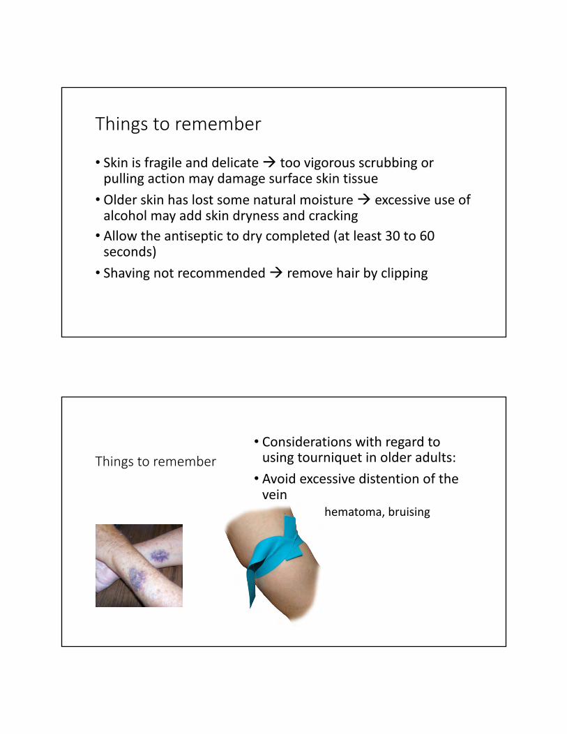
Things to remember• Considerations with regard to
using tourniquet in older adults: • Avoid excessive distention of the
vein • Trauma, hematoma, bruising
Things to remember
• Amount of tape applied to the skin should be minimized in patient with delicate skin• Skin barrier can be use to protect skin from the effect of adhesive/drying
nature of the antiseptic/ repetitive tape removal and dressing changes
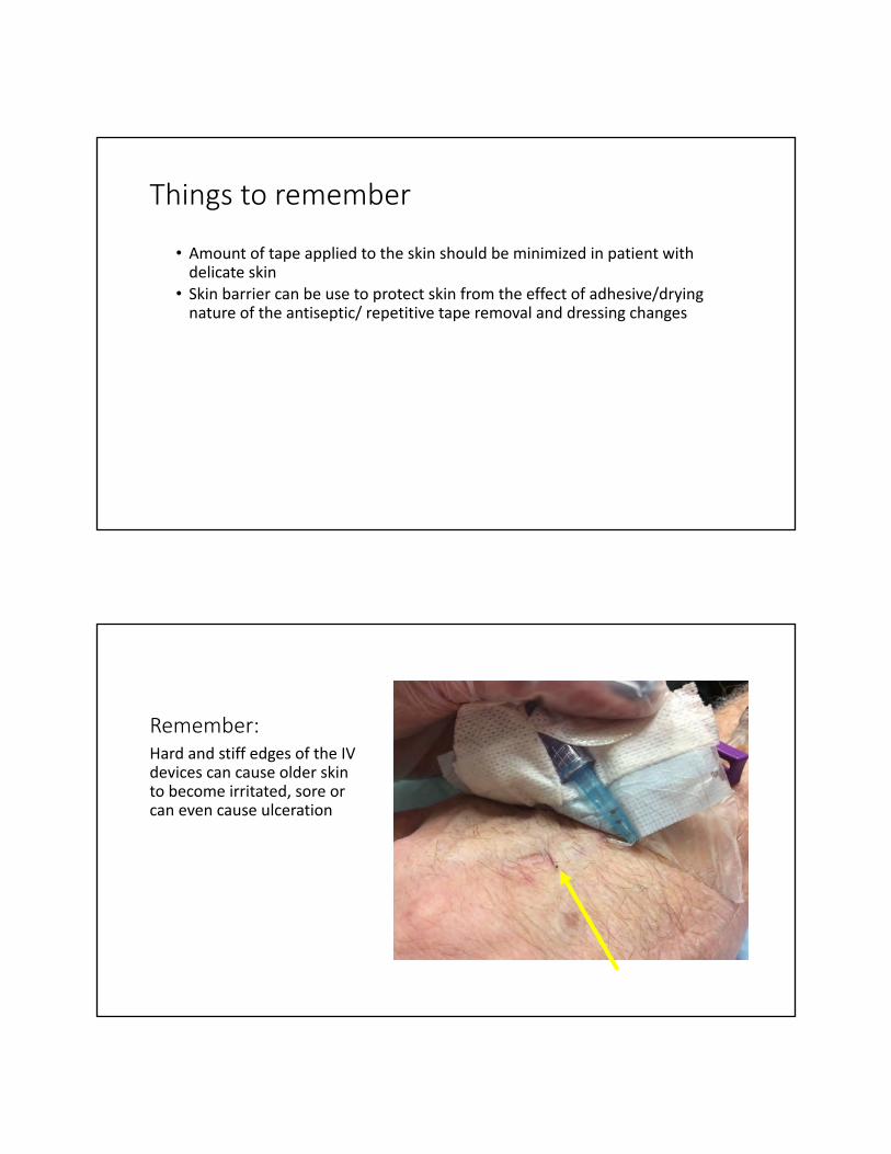
Remember:Hard and stiff edges of the IV devices can cause older skin to become irritated, sore or can even cause ulceration

VAD Protection & Stabilization
• Site protection (e.g., waterproof sleeve, dry garment, plastic dome/protector, mesh sleeve• Site protection methods are in addition to primary dressing
&securement device.• b) Joint stabilization (ONLY if absolutely necessary—restricts
movement of already stiff joints) • c) Restraint/physical immobilization device (e.g., soft
device, tie or mitt used to immobilize arm or hand)
Types of blood transfusion● Allogenic blood transfusion ( someone else's blood )● Autologous blood transfusion ( own blood )● Exchange blood transfusion

IV Access• Transfusing rapidly and under pressure through too small an IV access can cause
destruction of red blood cells. What is considered “too small”?
• Ensure that the IV access is dedicated to the transfusion.• Medications and solutions other than normal saline can cause hemolysis or clotting of
the blood component
• When transfusing through a CVAD with multiple lumens, medications/solutions can be infused through other lumens without damaging the blood component/product.
• IV pumps, blood warmers, and rapid infusers must be suitable for transfusion and not damage the blood component/product.
Practice Guide
• Follow your provincial standards for blood transfusion• REMEMBER: • A blood transfusion is a human tissue transplant.• Anemia tolerance is based on the assessment of signs and symptoms.• Provide clinical information related to anemia tolerance when reporting
lab values.• Verify blood products at the patient’s bedside according to facility
policy and procedure.• Transfuse one unit of RBCs at a time, then reassess the patient.• Limit phlebotomy and blood loss from lab testing.

IV AccessPRODUCT/COMPONENT CATEGORY IV ACCESS
RED BLOOD CELLS RAPID TRANSFUSION IN ADULTS 20 - 22 Ga
RED BLOOD CELLS ROUTINE TRANSFUSIONS IN ADULTS 22 – 24 Ga
OTHER BLOOD COMPONENTS ANY SIZE ADEQUATE
All Blood Components/Products Pediatrics and Adults 22-25G
All Blood Components/Products Adults and Pediatrics Central Venous Access Devices (CVAD)
Acute Reactions - Risk and Description
Acute Transfusion Reaction Risk of Event Description
Minor Allergic Reaction 1 in 100 Mild allergic reaction to an allergen in the blood component/product.
Anaphylaxis 1 in 40,000 Potentially fatal reaction caused by an allergen that the patient has been sensitized to.STOP transfusion
Febrile Non-Hemolytic 1 in 300 Mild usually self-limiting reaction associated with donor white blood cells or cytokines in the blood component/product. Usually presents with fever and/or rigors (shaking).

Acute Reactions - Risk and Description
Acute Transfusion Reaction
Risk of Event
Description
Acute Hemolytic Transfusion Reaction
1 in 40,000 Potentially fatal reaction caused by blood group incompatibility. Can also be caused by chemical hemolysis (e.g. incompatible solutions) or mechanical hemolysis (e.g. improper storage). Can result in renal failure, shock and coagulopathy.
Transfusion Related Acute Lung Injury (TRALI)
1 in 12,000 Acute hypoxemia with evidence of new bilateral lung infiltrates on X-Ray and no evidence of circulatory overload. Patients often require ventilatory support. Usually occurs within 1-2 hours of start of transfusion and rarely after 6 hours. Usually resolves within 24-72 hours with death occurring in 5-10%. Cause not fully understood.Postulated to be related to donor or recipient antibodies acquired through pregnancy or transfusion.
Acute Reactions - Risk and Description
Acute Transfusion Reaction
Risk of Event
Description
Transfusion Associated Circulatory Overload (TACO)
1 in 100 Circulatory overload from excessively rapid transfusion and/or in patients at greater risk for overload (e.g. very young, elderly, impaired cardiac function). Preventative measures include slower transfusion rates and pre-emptive diuretics for patients at risk.
Hypotensive Reaction Very Rare Bradykinin mediated hypotension. Characterized by profound drop in blood pressure usually seen in patients on ACE Inhibitors unable to degrade bradykinin in blood component/product.

Parenteral Nutrition
• Define terminology related to parenteral nutrition (PN) and implications for vascular access. • Discuss potential complications of PN and associated interventions • Identify clinical implications for peripheral PN. • Types and composition of PN. • Indications for PN. • Supplies and procedures for PN administration.
What is Parenteral Nutrition?§ Provides essential nutrients and calories intravenously, when the person
cannot meet these needs through oral diet or enteral feeding§ PN composed of carbohydrates, fats, protein, amino acids, minerals and
vitamins § Amount of each component is individualised, based on patient
assessment§ 3 types:
§ PPN: Peripheral Parenteral Nutrition§ TPN: Total Parenteral Nutrition§ TNA: Total nutrient admixture

Indications
§Bowel disorders such as short bowel syndrome§ Inflammatory bowel disease § Fistulas§Bowel Obstruction§Malabsorption disorders (pancreatitis, cystic fibrosis)§Motility Disorders§Cancer
AdministrationContinuous or Cyclic § Continuous
§ Infusion over a 24 hour period§ Patient is usually in a hospital setting§ May be receiving PN, TPN or TNA
§ Cyclical§ 8-12 hr infusions, typically overnight§ Preferred method as it allows patient more freedom during awake hours§ More closely mimic normal oral intake (period of fasting between meals)§ Decreases the risks associated with long term TPN

Complications§Most common complication is VAD infection§Prevention:
§ Aseptic Technique§ Change dressing as per institution protocol§ Change IV lines every 24 hrs or at the start of every new infusion
§ Line occlusion § Fibrin sheath§ Intraluminal occlusion from infusate
Complications
§ Long term PN may lead to Fatty Liver disease§ Cholecystitis
§ Caused by lack of use of the Gut = Bile build up = inflammation
§ Feelings of Hunger § Never actually feel full. Long term TPN without oral intake can lead to gut
atrophy
§ Refeeding Syndrome§ Can occur when restarting solids