Isolation and Identification of Protease Enzyme Producing ...
12
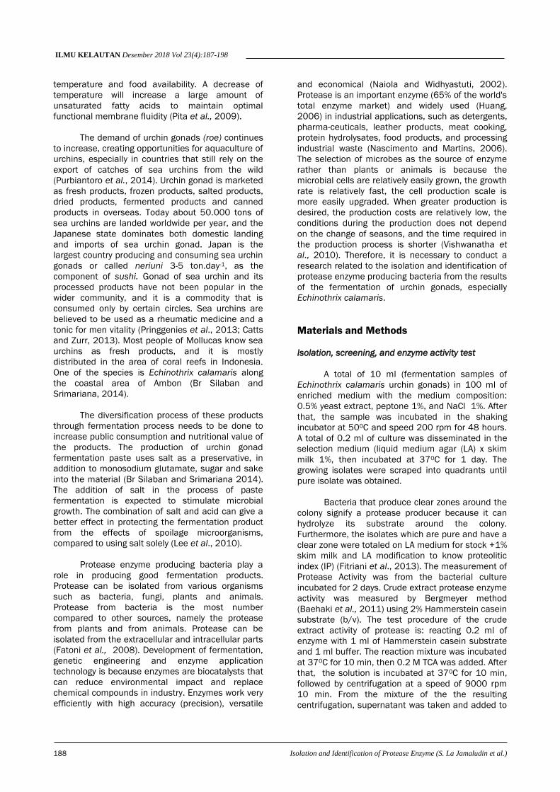
ILMU KELAUTAN Desember 2018 Vol 23(4):187-198 ISSN 0853-7291 *) Corresponding author © Ilmu Kelautan, UNDIP ijms.undip.ac.id DOI: 10.14710/ik.ijms.23.4.187-198 Received : 23-06-2017 Accepted : 24-08-2017 Isolation and Identification of Protease Enzyme Producing Bacteria from Fermentation of Gonad Sea Urchin ( Echinothrix calamaris ) Siani La Jamaludin 1* , Johanis Fritzgal Rehena 2 , Cecilia Anna Seumahu 3 and Dominggus Rumahlatu 2 1 Postgraduate Biology Education Study Program, Pattimura University Jl. Dr. Tamaela, Ambon 97116 Indonesia 2 Biology Education Study Program, Faculty of Teacher Training and Education Science, Pattimura University Jl. Ir. M. Putuhena, Ambon 97233 Indonesia 3 Biology Department, Faculty Mathematics and Science, Pattimura University Jl. Ir. M. Putuhena, Ambon 97233 Indonesia Email: [email protected] Abstract Bekasang of gonad sea urchin is one of the traditional fermentation products which generally involves microorganisms spontaneously. Fermented paste products have a long shelf life and are processed quite easily using protease enzymes. Good exploration of producing protease from bakasam is needed to obtain the protease enzyme-producing microorganism with different characters. The method used in this research is screening with clear zone, measuring the activity of crude extract of protease enzyme characterization of bacteria through gram staining. Identification of PCR microorganisms with 63F primer and 1387r primer and sequencing gen target. The results showed that there were eight isolates of protease enzyme-producing bacteria (G1, G2, G3, G4, G5, G6, G7, and G8) indicated by clear zones around single-colonic bacterial streaks. Only five bacterial isolates (G1, G4, G6, G7, and G8) were tested for the enzyme activity. These isolates have characteristics of positive gram bacteria. The interpretation of the results of molecular analysis using PCR obtained that the target gene shown by the band at a distance of 1300 bp. The results were then sequenced and BLASTN sequences of 16S rRNA gene from five bacterial isolates, namely: G1 was Staphylococcus piscifermentans strain CIP103958 with 99% similarity; Isolate G4 was Staphylococcus saprophyticus strain ATCC 15305 with 99% similarity; Isolate G6 was Staphylococcus condimenti F-2 strain with 99% similarity; Isolate G7 was Bacillus amyloliquefaciens subsp. plantarum strain FZB42 with 99% similarity; And G8 isolates was Lactobacillus plantarum strain JCM 1149 with 99% similarity. Keywords: bekasang, protease, gonad sea urchins Introduction Sea urchins (phylum Echinodermata) is an international mainstay commodity as well as foodstuffs which have important economic value. There are about 84 species of sea urchins found in Indonesian waters, and five of them, namely, Diadema setosum, Arbacia punctulata, Salmacis bicolor, Echinometra mathaei, Tripneustes gratilla and Echinothrix calamaris can be consumed and have economic value. The habitat of sea urchins is in seagrass beds to corals. Diadema setosum also becomes an indicator of the health of coral reefs. One cause of coral bleaching is the population of setosum Diadema that eats algae which is symbiotic zooxanthelae (Qiu et al., 2014) The gonads of Sea urchin have long been used as raw food as well as cooked food, daily known by the name of sea urchin eggs. Analysis of gonad protein of several species of sea urchins show that the gonad of sea urchins contains around 28 species /about 32.1% of the total amino acids, which are dominated by phenylalanine, lysine and valine (Amarowicz et al., 2012). In addition, the sea urchin gonads also contains vitamin A, B complex, and minerals that are essential for growth (Kalogeropoulos et al., 2012). Complete nutritional composition and supported by delicious taste makes the sea urchin gonads as the favoured dishes of communities in many countries. The maturity time of urchin gonad is influenced by hydrodynamic conditions of location. Maturation period of urchins between April and June for Paracentrotus lividus population in Tunisia, although the gonad index also seems to vary in different locations (Navarro and Guirado, 2011). Two aquatic ecosystem factors that may affect the quality of fatty acids of gonadal tissue are water
Transcript of Isolation and Identification of Protease Enzyme Producing ...
ILMU KELAUTAN Desember 2018 Vol 23(4):187-198 ISSN 0853-7291
*) Corresponding author
© Ilmu Kelautan, UNDIP
ijms.undip.ac.id
DOI: 10.14710/ik.ijms.23.4.187-198
Received : 23-06-2017
Accepted : 24-08-2017
Isolation and Identification of Protease Enzyme Producing Bacteria
from Fermentation of Gonad Sea Urchin (Echinothrix calamaris)
Siani La Jamaludin1*, Johanis Fritzgal Rehena2, Cecilia Anna Seumahu3 and
Dominggus Rumahlatu2
1Postgraduate Biology Education Study Program, Pattimura University
Jl. Dr. Tamaela, Ambon 97116 Indonesia 2Biology Education Study Program, Faculty of Teacher Training and Education Science, Pattimura University
Jl. Ir. M. Putuhena, Ambon 97233 Indonesia 3Biology Department, Faculty Mathematics and Science, Pattimura University
Jl. Ir. M. Putuhena, Ambon 97233 Indonesia
Email: [email protected]
Abstract
Bekasang of gonad sea urchin is one of the traditional fermentation products which generally involves
microorganisms spontaneously. Fermented paste products have a long shelf life and are processed quite easily
using protease enzymes. Good exploration of producing protease from bakasam is needed to obtain the protease
enzyme-producing microorganism with different characters. The method used in this research is screening with
clear zone, measuring the activity of crude extract of protease enzyme characterization of bacteria through gram
staining. Identification of PCR microorganisms with 63F primer and 1387r primer and sequencing gen target. The
results showed that there were eight isolates of protease enzyme-producing bacteria (G1, G2, G3, G4, G5, G6, G7,
and G8) indicated by clear zones around single-colonic bacterial streaks. Only five bacterial isolates (G1, G4, G6,
G7, and G8) were tested for the enzyme activity. These isolates have characteristics of positive gram bacteria. The
interpretation of the results of molecular analysis using PCR obtained that the target gene shown by the band at a
distance of 1300 bp. The results were then sequenced and BLASTN sequences of 16S rRNA gene from five
bacterial isolates, namely: G1 was Staphylococcus piscifermentans strain CIP103958 with 99% similarity; Isolate
G4 was Staphylococcus saprophyticus strain ATCC 15305 with 99% similarity; Isolate G6 was Staphylococcus
condimenti F-2 strain with 99% similarity; Isolate G7 was Bacillus amyloliquefaciens subsp. plantarum strain
FZB42 with 99% similarity; And G8 isolates was Lactobacillus plantarum strain JCM 1149 with 99% similarity.
Keywords: bekasang, protease, gonad sea urchins
Introduction
Sea urchins (phylum Echinodermata) is an
international mainstay commodity as well as
foodstuffs which have important economic value.
There are about 84 species of sea urchins found in
Indonesian waters, and five of them, namely,
Diadema setosum, Arbacia punctulata, Salmacis
bicolor, Echinometra mathaei, Tripneustes gratilla
and Echinothrix calamaris can be consumed and
have economic value. The habitat of sea urchins is
in seagrass beds to corals. Diadema setosum also
becomes an indicator of the health of coral reefs.
One cause of coral bleaching is the population of
setosum Diadema that eats algae which is symbiotic
zooxanthelae (Qiu et al., 2014)
The gonads of Sea urchin have long been
used as raw food as well as cooked food, daily
known by the name of sea urchin eggs. Analysis of
gonad protein of several species of sea urchins
show that the gonad of sea urchins contains around
28 species /about 32.1% of the total amino acids,
which are dominated by phenylalanine, lysine and
valine (Amarowicz et al., 2012). In addition, the sea
urchin gonads also contains vitamin A, B complex,
and minerals that are essential for growth
(Kalogeropoulos et al., 2012). Complete nutritional
composition and supported by delicious taste makes
the sea urchin gonads as the favoured dishes of
communities in many countries.
The maturity time of urchin gonad is
influenced by hydrodynamic conditions of location.
Maturation period of urchins between April and June
for Paracentrotus lividus population in Tunisia,
although the gonad index also seems to vary in
different locations (Navarro and Guirado, 2011). Two
aquatic ecosystem factors that may affect the
quality of fatty acids of gonadal tissue are water
ILMU KELAUTAN Desember 2018 Vol 23(4):187-198
188 Isolation and Identification of Protease Enzyme (S. La Jamaludin et al.)
temperature and food availability. A decrease of
temperature will increase a large amount of
unsaturated fatty acids to maintain optimal
functional membrane fluidity (Pita et al., 2009).
The demand of urchin gonads (roe) continues
to increase, creating opportunities for aquaculture of
urchins, especially in countries that still rely on the
export of catches of sea urchins from the wild
(Purbiantoro et al., 2014). Urchin gonad is marketed
as fresh products, frozen products, salted products,
dried products, fermented products and canned
products in overseas. Today about 50.000 tons of
sea urchins are landed worldwide per year, and the
Japanese state dominates both domestic landing
and imports of sea urchin gonad. Japan is the
largest country producing and consuming sea urchin
gonads or called neriuni 3-5 ton.day-1, as the
component of sushi. Gonad of sea urchin and its
processed products have not been popular in the
wider community, and it is a commodity that is
consumed only by certain circles. Sea urchins are
believed to be used as a rheumatic medicine and a
tonic for men vitality (Pringgenies et al., 2013; Catts
and Zurr, 2013). Most people of Mollucas know sea
urchins as fresh products, and it is mostly
distributed in the area of coral reefs in Indonesia.
One of the species is Echinothrix calamaris along
the coastal area of Ambon (Br Silaban and
Srimariana, 2014).
The diversification process of these products
through fermentation process needs to be done to
increase public consumption and nutritional value of
the products. The production of urchin gonad
fermentation paste uses salt as a preservative, in
addition to monosodium glutamate, sugar and sake
into the material (Br Silaban and Srimariana 2014).
The addition of salt in the process of paste
fermentation is expected to stimulate microbial
growth. The combination of salt and acid can give a
better effect in protecting the fermentation product
from the effects of spoilage microorganisms,
compared to using salt solely (Lee et al., 2010).
Protease enzyme producing bacteria play a
role in producing good fermentation products.
Protease can be isolated from various organisms
such as bacteria, fungi, plants and animals.
Protease from bacteria is the most number
compared to other sources, namely the protease
from plants and from animals. Protease can be
isolated from the extracellular and intracellular parts
(Fatoni et al., 2008). Development of fermentation,
genetic engineering and enzyme application
technology is because enzymes are biocatalysts that
can reduce environmental impact and replace
chemical compounds in industry. Enzymes work very
efficiently with high accuracy (precision), versatile
and economical (Naiola and Widhyastuti, 2002).
Protease is an important enzyme (65% of the world's
total enzyme market) and widely used (Huang,
2006) in industrial applications, such as detergents,
pharma-ceuticals, leather products, meat cooking,
protein hydrolysates, food products, and processing
industrial waste (Nascimento and Martins, 2006).
The selection of microbes as the source of enzyme
rather than plants or animals is because the
microbial cells are relatively easily grown, the growth
rate is relatively fast, the cell production scale is
more easily upgraded. When greater production is
desired, the production costs are relatively low, the
conditions during the production does not depend
on the change of seasons, and the time required in
the production process is shorter (Vishwanatha et
al., 2010). Therefore, it is necessary to conduct a
research related to the isolation and identification of
protease enzyme producing bacteria from the results
of the fermentation of urchin gonads, especially
Echinothrix calamaris.
Materials and Methods
Isolation, screening, and enzyme activity test
A total of 10 ml (fermentation samples of
Echinothrix calamaris urchin gonads) in 100 ml of
enriched medium with the medium composition:
0.5% yeast extract, peptone 1%, and NaCl 1%. After
that, the sample was incubated in the shaking
incubator at 500C and speed 200 rpm for 48 hours.
A total of 0.2 ml of culture was disseminated in the
selection medium (liquid medium agar (LA) x skim
milk 1%, then incubated at 370C for 1 day. The
growing isolates were scraped into quadrants until
pure isolate was obtained.
Bacteria that produce clear zones around the
colony signify a protease producer because it can
hydrolyze its substrate around the colony.
Furthermore, the isolates which are pure and have a
clear zone were totaled on LA medium for stock +1%
skim milk and LA modification to know proteolitic
index (IP) (Fitriani et al., 2013). The measurement of
Protease Activity was from the bacterial culture
incubated for 2 days. Crude extract protease enzyme
activity was measured by Bergmeyer method
(Baehaki et al., 2011) using 2% Hammerstein casein
substrate (b/v). The test procedure of the crude
extract activity of protease is: reacting 0.2 ml of
enzyme with 1 ml of Hammerstein casein substrate
and 1 ml buffer. The reaction mixture was incubated
at 370C for 10 min, then 0.2 M TCA was added. After
that, the solution is incubated at 370C for 10 min,
followed by centrifugation at a speed of 9000 rpm
10 min. From the mixture of the the resulting
centrifugation, supernatant was taken and added to
ILMU KELAUTAN Desember 2018 Vol 23(4):187-198
Isolation and Identification of Protease Enzyme (S. La Jamaludin et al.) 189
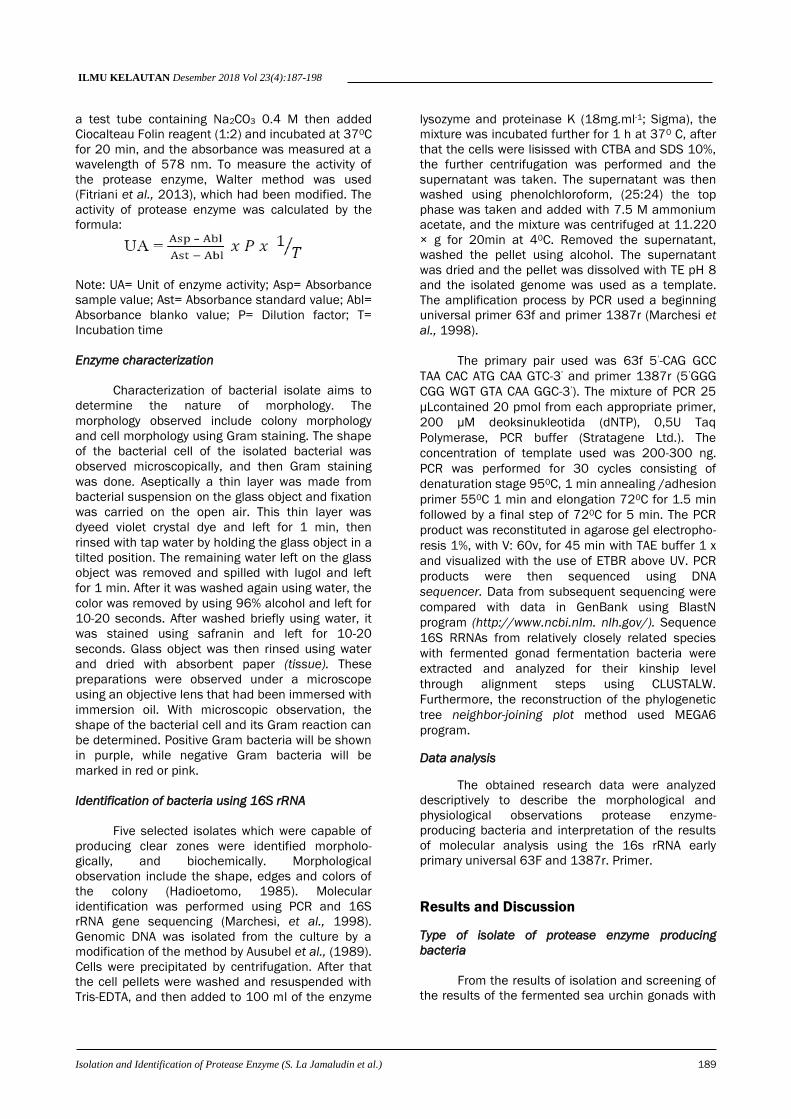
a test tube containing Na2CO3 0.4 M then added
Ciocalteau Folin reagent (1:2) and incubated at 370C
for 20 min, and the absorbance was measured at a
wavelength of 578 nm. To measure the activity of
the protease enzyme, Walter method was used
(Fitriani et al., 2013), which had been modified. The
activity of protease enzyme was calculated by the
formula:
Note: UA= Unit of enzyme activity; Asp= Absorbance
sample value; Ast= Absorbance standard value; Abl=
Absorbance blanko value; P= Dilution factor; T=
Incubation time
Enzyme characterization
Characterization of bacterial isolate aims to
determine the nature of morphology. The
morphology observed include colony morphology
and cell morphology using Gram staining. The shape
of the bacterial cell of the isolated bacterial was
observed microscopically, and then Gram staining
was done. Aseptically a thin layer was made from
bacterial suspension on the glass object and fixation
was carried on the open air. This thin layer was
dyeed violet crystal dye and left for 1 min, then
rinsed with tap water by holding the glass object in a
tilted position. The remaining water left on the glass
object was removed and spilled with lugol and left
for 1 min. After it was washed again using water, the
color was removed by using 96% alcohol and left for
10-20 seconds. After washed briefly using water, it
was stained using safranin and left for 10-20
seconds. Glass object was then rinsed using water
and dried with absorbent paper (tissue). These
preparations were observed under a microscope
using an objective lens that had been immersed with
immersion oil. With microscopic observation, the
shape of the bacterial cell and its Gram reaction can
be determined. Positive Gram bacteria will be shown
in purple, while negative Gram bacteria will be
marked in red or pink.
Identification of bacteria using 16S rRNA
Five selected isolates which were capable of
producing clear zones were identified morpholo-
gically, and biochemically. Morphological
observation include the shape, edges and colors of
the colony (Hadioetomo, 1985). Molecular
identification was performed using PCR and 16S
rRNA gene sequencing (Marchesi, et al., 1998).
Genomic DNA was isolated from the culture by a
modification of the method by Ausubel et al., (1989).
Cells were precipitated by centrifugation. After that
the cell pellets were washed and resuspended with
Tris-EDTA, and then added to 100 ml of the enzyme
lysozyme and proteinase K (18mg.ml-1; Sigma), the
mixture was incubated further for 1 h at 370 C, after
that the cells were lisissed with CTBA and SDS 10%,
the further centrifugation was performed and the
supernatant was taken. The supernatant was then
washed using phenolchloroform, (25:24) the top
phase was taken and added with 7.5 M ammonium
acetate, and the mixture was centrifuged at 11.220
× g for 20min at 40C. Removed the supernatant,
washed the pellet using alcohol. The supernatant
was dried and the pellet was dissolved with TE pH 8
and the isolated genome was used as a template.
The amplification process by PCR used a beginning
universal primer 63f and primer 1387r (Marchesi et
al., 1998).
The primary pair used was 63f 5’-CAG GCC
TAA CAC ATG CAA GTC-3’ and primer 1387r (5’GGG
CGG WGT GTA CAA GGC-3’). The mixture of PCR 25
μLcontained 20 pmol from each appropriate primer,
200 μM deoksinukleotida (dNTP), 0,5U Taq
Polymerase, PCR buffer (Stratagene Ltd.). The
concentration of template used was 200-300 ng.
PCR was performed for 30 cycles consisting of
denaturation stage 950C, 1 min annealing /adhesion
primer 550C 1 min and elongation 720C for 1.5 min
followed by a final step of 720C for 5 min. The PCR
product was reconstituted in agarose gel electropho-
resis 1%, with V: 60v, for 45 min with TAE buffer 1 x
and visualized with the use of ETBR above UV. PCR
products were then sequenced using DNA
sequencer. Data from subsequent sequencing were
compared with data in GenBank using BlastN
program (http://www.ncbi.nlm. nlh.gov/). Sequence
16S RRNAs from relatively closely related species
with fermented gonad fermentation bacteria were
extracted and analyzed for their kinship level
through alignment steps using CLUSTALW.
Furthermore, the reconstruction of the phylogenetic
tree neighbor-joining plot method used MEGA6
program.
Data analysis
The obtained research data were analyzed
descriptively to describe the morphological and
physiological observations protease enzyme-
producing bacteria and interpretation of the results
of molecular analysis using the 16s rRNA early
primary universal 63F and 1387r. Primer.
Results and Discussion
Type of isolate of protease enzyme producing
bacteria
From the results of isolation and screening of
the results of the fermented sea urchin gonads with
ILMU KELAUTAN Desember 2018 Vol 23(4):187-198
190 Isolation and Identification of Protease Enzyme (S. La Jamaludin et al.)
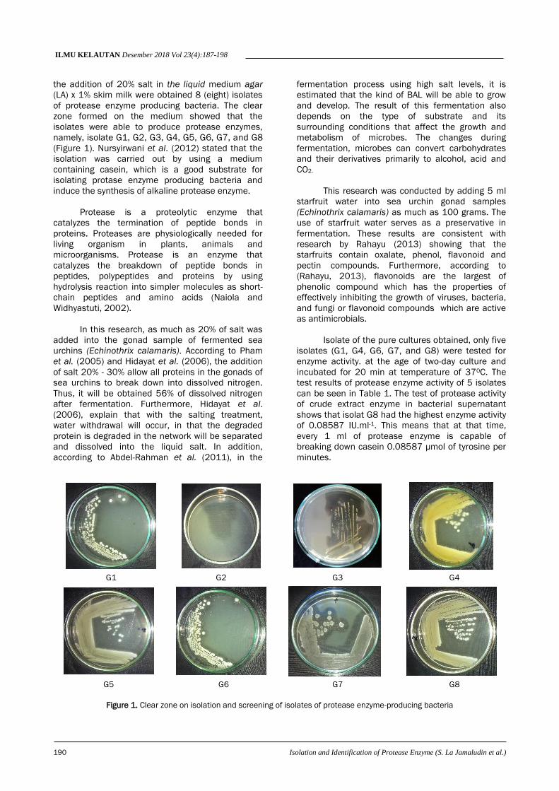
the addition of 20% salt in the liquid medium agar
(LA) x 1% skim milk were obtained 8 (eight) isolates
of protease enzyme producing bacteria. The clear
zone formed on the medium showed that the
isolates were able to produce protease enzymes,
namely, isolate G1, G2, G3, G4, G5, G6, G7, and G8
(Figure 1). Nursyirwani et al. (2012) stated that the
isolation was carried out by using a medium
containing casein, which is a good substrate for
isolating protase enzyme producing bacteria and
induce the synthesis of alkaline protease enzyme.
Protease is a proteolytic enzyme that
catalyzes the termination of peptide bonds in
proteins. Proteases are physiologically needed for
living organism in plants, animals and
microorganisms. Protease is an enzyme that
catalyzes the breakdown of peptide bonds in
peptides, polypeptides and proteins by using
hydrolysis reaction into simpler molecules as short-
chain peptides and amino acids (Naiola and
Widhyastuti, 2002).
In this research, as much as 20% of salt was
added into the gonad sample of fermented sea
urchins (Echinothrix calamaris). According to Pham
et al. (2005) and Hidayat et al. (2006), the addition
of salt 20% - 30% allow all proteins in the gonads of
sea urchins to break down into dissolved nitrogen.
Thus, it will be obtained 56% of dissolved nitrogen
after fermentation. Furthermore, Hidayat et al.
(2006), explain that with the salting treatment,
water withdrawal will occur, in that the degraded
protein is degraded in the network will be separated
and dissolved into the liquid salt. In addition,
according to Abdel-Rahman et al. (2011), in the
fermentation process using high salt levels, it is
estimated that the kind of BAL will be able to grow
and develop. The result of this fermentation also
depends on the type of substrate and its
surrounding conditions that affect the growth and
metabolism of microbes. The changes during
fermentation, microbes can convert carbohydrates
and their derivatives primarily to alcohol, acid and
CO2.
This research was conducted by adding 5 ml
starfruit water into sea urchin gonad samples
(Echinothrix calamaris) as much as 100 grams. The
use of starfruit water serves as a preservative in
fermentation. These results are consistent with
research by Rahayu (2013) showing that the
starfruits contain oxalate, phenol, flavonoid and
pectin compounds. Furthermore, according to
(Rahayu, 2013), flavonoids are the largest of
phenolic compound which has the properties of
effectively inhibiting the growth of viruses, bacteria,
and fungi or flavonoid compounds which are active
as antimicrobials.
Isolate of the pure cultures obtained, only five
isolates (G1, G4, G6, G7, and G8) were tested for
enzyme activity. at the age of two-day culture and
incubated for 20 min at temperature of 370C. The
test results of protease enzyme activity of 5 isolates
can be seen in Table 1. The test of protease activity
of crude extract enzyme in bacterial supernatant
shows that isolat G8 had the highest enzyme activity
of 0.08587 IU.ml-1. This means that at that time,
every 1 ml of protease enzyme is capable of
breaking down casein 0.08587 μmol of tyrosine per
minutes.
G1 G2 G3 G4
G5 G6 G7 G8
Figure 1. Clear zone on isolation and screening of isolates of protease enzyme-producing bacteria
ILMU KELAUTAN Desember 2018 Vol 23(4):187-198
Isolation and Identification of Protease Enzyme (S. La Jamaludin et al.) 191
According to Fadda et al. (2010), protease
activity occurs because of an increase in the kinetic
energy of the enzyme molecule and the increased
movement of substrate molecules that cause
collisions between enzyme molecules and the the
increasing substrate. The activity of the protease
enzyme will decrease at a temperature above the
optimum temperature. This is because high
temperatures break up secondary bonds such as
hydrogen bonds that keep the enzyme in its natural
structure, so that the secondary, tertiary structure of
the enzyme is partially damaged followed by a
decrease in activity. Temperature is one of the
factors that influence enzyme activity. The higher the
temperature, the more increased the enzyme activity
until the optimum temperature occurs. The activity
of protease enzyme reaches optimum activity at
50ºC. This is in line with what is proposed by
Baehaki et al. (2011) that at a lower temperature
than the optimum temperature, the enzyme activity
is also low, due to the low activation energies
available. The energy is needed to create an active
complex level condition, both from enzyme
molecules and from substrate molecules. Increased
temperatures also have an effect on the substrate
conformation change, so that the substrate active
side experiences barriers to enter the enzyme's
active side, and causes the decrease in enzyme
activity. Secondly, the increase in thermal energy of
molecules that make up the structure of the enzyme
protein itself causes the breakdown of non-covalent
interactions (hydrogen bonds, van der walls bonds,
hydrophobic bonds and electrostatic interactions)
that keep the enzyme 3D structures together, so
that enzymes are denatured. Denaturation causes
the enzyme-folding structure to open up on its
surface, so that the active side of the enzyme
changes and results in a decrease in enzyme
activity.
Characterization of protease enzyme producing
bacteria
The appearance of colony morphology is
generally round (circular) and the gram color is
purple. Isolate G1 in colony morphology is large,
cream pigmentation, rounded edges (round), slightly
convex elevation (convex). Isolates G4, morphology
of medium colonies, yellow pigmentation, spherical
(Circular), rounded edges (Entire), slightly convex
elevation (convex), Isolate G6, medium colony
morphology, Cream pigmentation, rounded edges
(Entire), slightly convex elevation (convex). Isolate
G7, large colony morphology, cream pigmentation,
rounded (Circular), slightly dull edge (Undulate), and
thin flat elevation (Flat) and Isolate G8, small colony
morphology, White pigmentation, optical appearance
features transparent, circular, circular edge (entire),
flat elevation (Flat). The morphological observation
of cells through gram staining, all bacterial isolates
showed gram positive and the forms of bacterial
cells were in the form of coccus and bacil (Table 2.).
The five bacterial isolates are then
characterized by the gram staining method. The
research results prove that Aal isolates have
morphological characteristics of the cell colony, that
is, positive gram bacterial cells shown in purple. The
gram staining method is based on differences in the
composition of the cell wall they have. Gram-positive
cells have walls with thicker peptidoglycan layers
than gram-negative cells. Gram-negative bacteria
contain lipids and fats in a higher percentage that
that on gram-positive bacteria.
Abel et al. (2014) explains that the cell wall of
Gram-negative bacteria consists of 5-20%
peptidoglycan, the rest is polysaccharide, whereas
gram-positive bacteria contains of 90%
peptidoglycan, the rest is teikoat acid. The cells of
gram positive bacteria appear purple, because it can
form complex ties with the first color, purple complex
of iodine crystals. In gram-negative bacteria, 95%
alcohol can increase the porosity of the cell wall by
dissolving the lipid in the outer membrane, so that
the purple crystals of iodine will be released and the
cell becomes colorless. Furthermore, the cell will be
red because it is colored by a the comparison color,
which is safranin.
Identification of protease enzymes producing
bacteria amplification of 16S rRNA
Amplification of the 16S rRNA gene of five
bacteria isolates used PCR with primers 63F and
1387r. The use of primer is a universal primer
capable of amplifying the 16S rRNA gene in protease
enzyme-producing bacteria which can be seen in the
following Figure 2.
Table 1. Culture protease activity 48 hours of five isolates at 370C
Sample code Absorbance U1 (average) Absorption U2 (average) Protease Enzyme Activity (IU.ml-1)
G1 0.729±0.109 0.369±0.017 0.00174
G4 0.772±0.151 0.358±0.008 0.00079
G6 0.665±0.025 0.372±0.026 0.00493
G7 0.813±0.060 0.400±0.009 0.00517
G8 0.674±0.029 0.819±0.020 0.08587
ILMU KELAUTAN Desember 2018 Vol 23(4):187-198
192 Isolation and Identification of Protease Enzyme (S. La Jamaludin et al.)
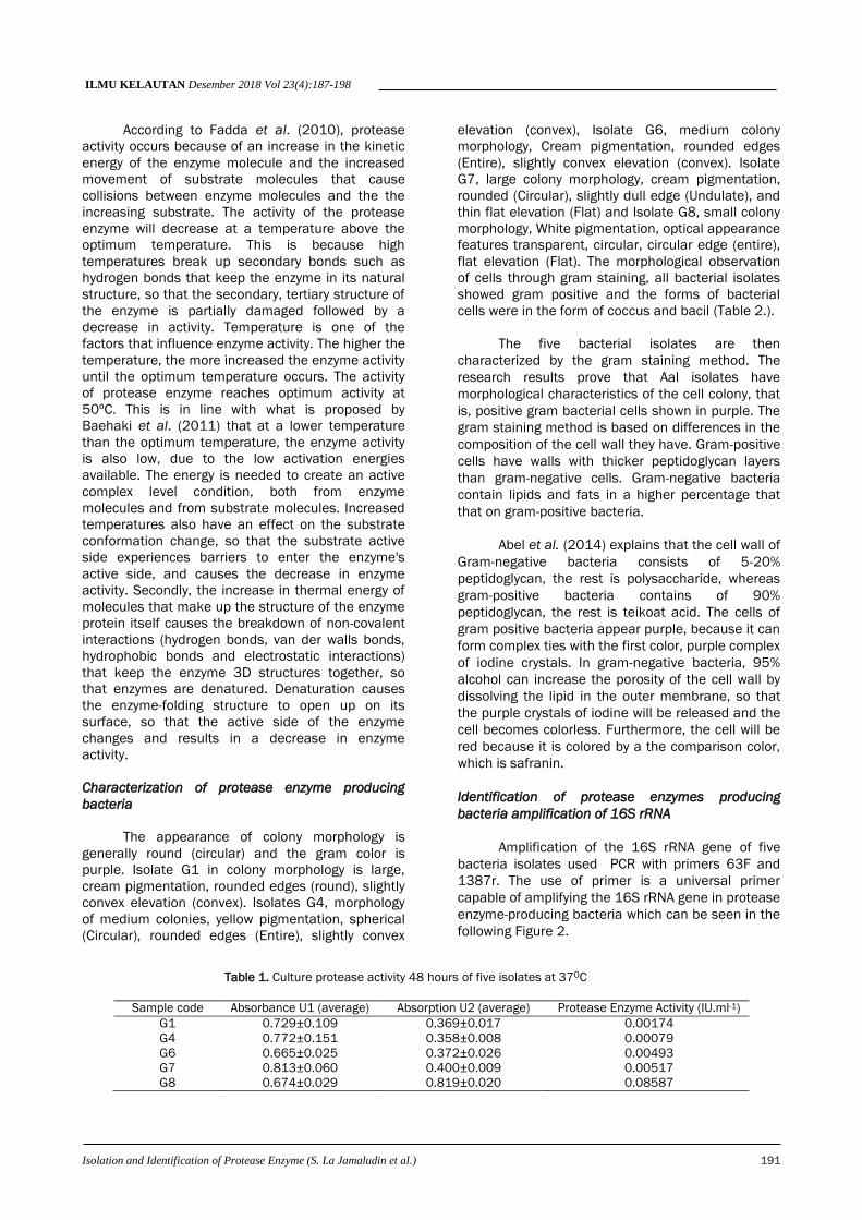
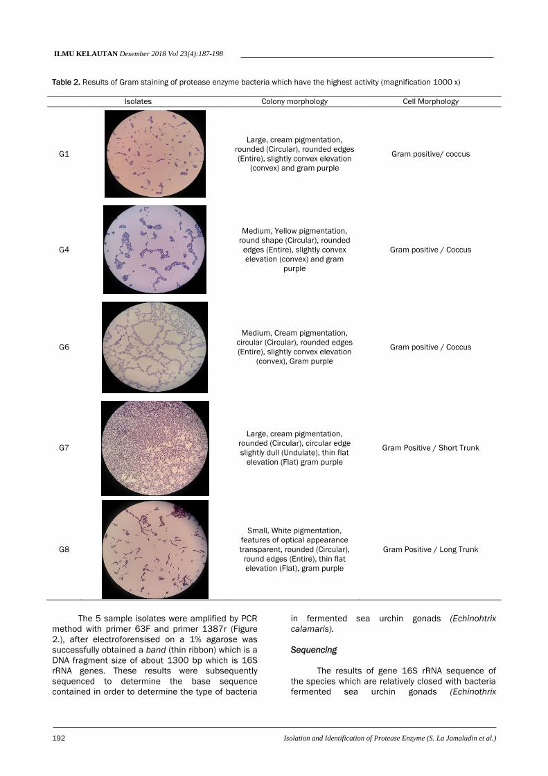
Table 2. Results of Gram staining of protease enzyme bacteria which have the highest activity (magnification 1000 x)
Isolates Colony morphology Cell Morphology
G1
Large, cream pigmentation,
rounded (Circular), rounded edges
(Entire), slightly convex elevation
(convex) and gram purple
Gram positive/ coccus
G4
Medium, Yellow pigmentation,
round shape (Circular), rounded
edges (Entire), slightly convex
elevation (convex) and gram
purple
Gram positive / Coccus
G6
Medium, Cream pigmentation,
circular (Circular), rounded edges
(Entire), slightly convex elevation
(convex), Gram purple
Gram positive / Coccus
G7
Large, cream pigmentation,
rounded (Circular), circular edge
slightly dull (Undulate), thin flat
elevation (Flat) gram purple
Gram Positive / Short Trunk
G8
Small, White pigmentation,
features of optical appearance
transparent, rounded (Circular),
round edges (Entire), thin flat
elevation (Flat), gram purple
Gram Positive / Long Trunk
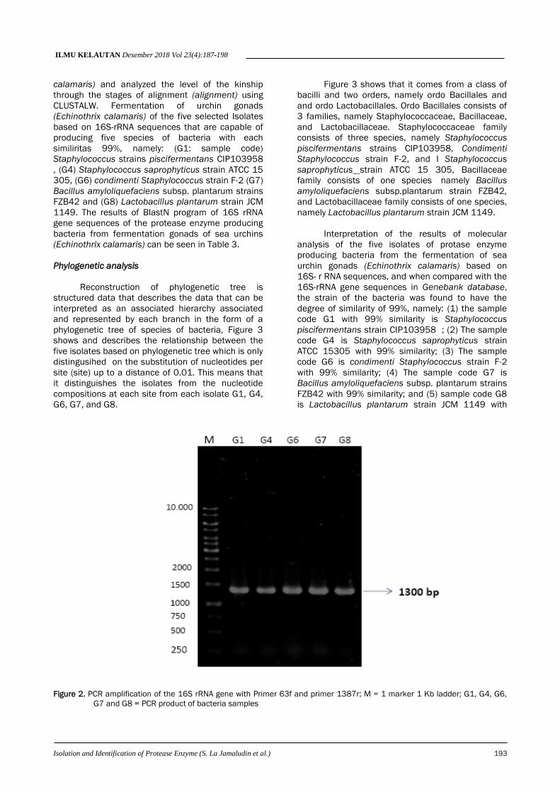
The 5 sample isolates were amplified by PCR
method with primer 63F and primer 1387r (Figure
2.), after electroforensised on a 1% agarose was
successfully obtained a band (thin ribbon) which is a
DNA fragment size of about 1300 bp which is 16S
rRNA genes. These results were subsequently
sequenced to determine the base sequence
contained in order to determine the type of bacteria
in fermented sea urchin gonads (Echinohtrix
calamaris).
Sequencing
The results of gene 16S rRNA sequence of
the species which are relatively closed with bacteria
fermented sea urchin gonads (Echinothrix
ILMU KELAUTAN Desember 2018 Vol 23(4):187-198
Isolation and Identification of Protease Enzyme (S. La Jamaludin et al.) 193
calamaris) and analyzed the level of the kinship
through the stages of alignment (alignment) using
CLUSTALW. Fermentation of urchin gonads
(Echinothrix calamaris) of the five selected Isolates
based on 16S-rRNA sequences that are capable of
producing five species of bacteria with each
similiritas 99%, namely: (G1: sample code)
Staphylococcus strains piscifermentans CIP103958
, (G4) Staphylococcus saprophyticus strain ATCC 15
305, (G6) condimenti Staphylococcus strain F-2 (G7)
Bacillus amyloliquefaciens subsp. plantarum strains
FZB42 and (G8) Lactobacillus plantarum strain JCM
1149. The results of BlastN program of 16S rRNA
gene sequences of the protease enzyme producing
bacteria from fermentation gonads of sea urchins
(Echinothrix calamaris) can be seen in Table 3.
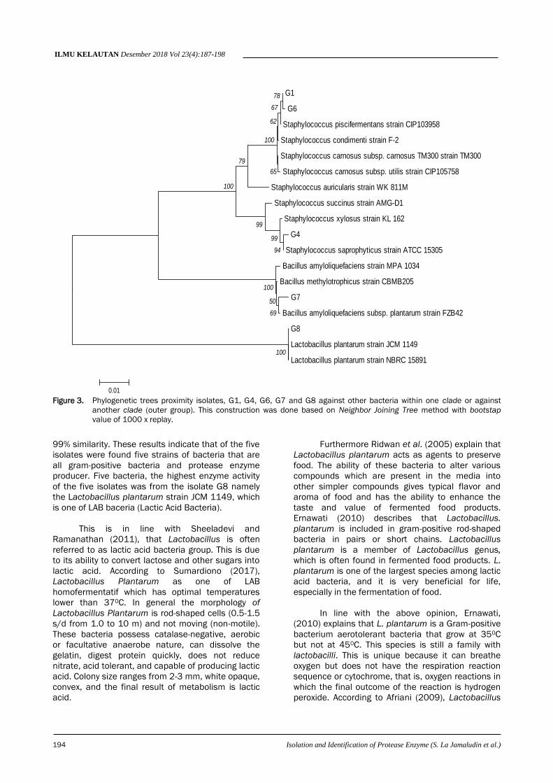
Phylogenetic analysis
Reconstruction of phylogenetic tree is
structured data that describes the data that can be
interpreted as an associated hierarchy associated
and represented by each branch in the form of a
phylogenetic tree of species of bacteria, Figure 3
shows and describes the relationship between the
five isolates based on phylogenetic tree which is only
distingusihed on the substitution of nucleotides per
site (site) up to a distance of 0.01. This means that
it distinguishes the isolates from the nucleotide
compositions at each site from each isolate G1, G4,
G6, G7, and G8.
Figure 3 shows that it comes from a class of
bacilli and two orders, namely ordo Bacillales and
and ordo Lactobacillales. Ordo Bacillales consists of
3 families, namely Staphylococcaceae, Bacillaceae,
and Lactobacillaceae. Staphylococcaceae family
consists of three species, namely Staphylococcus
piscifermentans strains CIP103958, Condimenti
Staphylococcus strain F-2, and I Staphylococcus
saprophyticus strain ATCC 15 305, Bacillaceae
family consists of one species namely Bacillus
amyloliquefaciens subsp.plantarum strain FZB42,
and Lactobacillaceae family consists of one species,
namely Lactobacillus plantarum strain JCM 1149.
Interpretation of the results of molecular
analysis of the five isolates of protase enzyme
producing bacteria from the fermentation of sea
urchin gonads (Echinothrix calamaris) based on
16S- r RNA sequences, and when compared with the
16S-rRNA gene sequences in Genebank database,
the strain of the bacteria was found to have the
degree of similarity of 99%, namely: (1) the sample
code G1 with 99% similarity is Staphylococcus
piscifermentans strain CIP103958 ; (2) The sample
code G4 is Staphylococcus saprophyticus strain
ATCC 15305 with 99% similarity; (3) The sample
code G6 is condimenti Staphylococcus strain F-2
with 99% similarity; (4) The sample code G7 is
Bacillus amyloliquefaciens subsp. plantarum strains
FZB42 with 99% similarity; and (5) sample code G8
is Lactobacillus plantarum strain JCM 1149 with
Figure 2. PCR amplification of the 16S rRNA gene with Primer 63f and primer 1387r; M = 1 marker 1 Kb ladder; G1, G4, G6,
G7 and G8 = PCR product of bacteria samples
ILMU KELAUTAN Desember 2018 Vol 23(4):187-198
194 Isolation and Identification of Protease Enzyme (S. La Jamaludin et al.)
Figure 3. Phylogenetic trees proximity isolates, G1, G4, G6, G7 and G8 against other bacteria within one clade or against
another clade (outer group). This construction was done based on Neighbor Joining Tree method with bootstap
value of 1000 x replay.
99% similarity. These results indicate that of the five
isolates were found five strains of bacteria that are
all gram-positive bacteria and protease enzyme
producer. Five bacteria, the highest enzyme activity
of the five isolates was from the isolate G8 namely
the Lactobacillus plantarum strain JCM 1149, which
is one of LAB baceria (Lactic Acid Bacteria).
This is in line with Sheeladevi and
Ramanathan (2011), that Lactobacillus is often
referred to as lactic acid bacteria group. This is due
to its ability to convert lactose and other sugars into
lactic acid. According to Sumardiono (2017),
Lactobacillus Plantarum as one of LAB
homofermentatif which has optimal temperatures
lower than 370C. In general the morphology of
Lactobacillus Plantarum is rod-shaped cells (0.5-1.5
s/d from 1.0 to 10 m) and not moving (non-motile).
These bacteria possess catalase-negative, aerobic
or facultative anaerobe nature, can dissolve the
gelatin, digest protein quickly, does not reduce
nitrate, acid tolerant, and capable of producing lactic
acid. Colony size ranges from 2-3 mm, white opaque,
convex, and the final result of metabolism is lactic
acid.
Furthermore Ridwan et al. (2005) explain that
Lactobacillus plantarum acts as agents to preserve
food. The ability of these bacteria to alter various
compounds which are present in the media into
other simpler compounds gives typical flavor and
aroma of food and has the ability to enhance the
taste and value of fermented food products.
Ernawati (2010) describes that Lactobacillus.
plantarum is included in gram-positive rod-shaped
bacteria in pairs or short chains. Lactobacillus
plantarum is a member of Lactobacillus genus,
which is often found in fermented food products. L.
plantarum is one of the largest species among lactic
acid bacteria, and it is very beneficial for life,
especially in the fermentation of food.
In line with the above opinion, Ernawati,
(2010) explains that L. plantarum is a Gram-positive
bacterium aerotolerant bacteria that grow at 350C
but not at 450C. This species is still a family with
lactobacilli. This is unique because it can breathe
oxygen but does not have the respiration reaction
sequence or cytochrome, that is, oxygen reactions in
which the final outcome of the reaction is hydrogen
peroxide. According to Afriani (2009), Lactobacillus
G1
G6
Staphylococcus piscifermentans strain CIP103958
Staphylococcus condimenti strain F-2
Staphylococcus carnosus subsp. carnosus TM300 strain TM300
Staphylococcus carnosus subsp. utilis strain CIP105758
Staphylococcus auricularis strain WK 811M
Staphylococcus succinus strain AMG-D1
Staphylococcus xylosus strain KL 162
G4
Staphylococcus saprophyticus strain ATCC 15305
Bacillus amyloliquefaciens strain MPA 1034
Bacillus methylotrophicus strain CBMB205
G7
Bacillus amyloliquefaciens subsp. plantarum strain FZB42
G8
Lactobacillus plantarum strain JCM 1149
Lactobacillus plantarum strain NBRC 15891100
69
50
100
94
99
99
100
79
78
67
62
65
100
0.01
ILMU KELAUTAN Desember 2018 Vol 23(4):187-198
Isolation and Identification of Protease Enzyme (S. La Jamaludin et al.) 195
plantarum has the ability to be able to live in low
acid conditions. It is also able to produce
antimicrobial bacteriocins that play a role in
suppressing the growth of microbial pathogens.
Meanwhile, according to Wagih et al. (2012),
Lactobacillus plantarum is capable of producing
plantaricin bakteriocin that can act as an
antibacterial and antifungal.
The activity of the highest protease enzyme
producing bacteria from the results of fermentation
of sea urchin gonads (Echinothrix calamaris) is
Bacillus amyloliquefaciens subsp with a value of
0.00517 IU/ml. This research is in line with the
research conducted by Novita et al. (2006) on the
partial characterization of protease enzyme crude
extract of Bacillus amyloliquefaciens NRRLB-14
396. The research results show that the protease
was isolated from the bacterium Bacillus
amyloliquefaciens NRRL B-14 396 to an average
value of protease enzyme activity of 0.431 units.ml-1
at 400C and pH 7.0. Furthermore, according to Choi
and Kim (2013), the production of serine proteases
uses Bacillus amyloliquefaciens DJ-4 by adding NaCl
and heat. In addition, Novita et al (2006 ) also
conducted research using the bacterium Bacillus
amyloliquefaciens S-94 using PMSF inhibitor, it was
known that the produced protase was included as
serine protease. Asokan and Jayanthi (2010) also
explain that the bacterium Bacillus
amyloliquefaciens is widely used because it has
good characteristics to produce alkaline protease.
Bacillus amyloliquefaciens can produce several
other enzymes that have commercial potential such
as α-amylase, β-glucanase, hemicellulase and
neutral protease.
The bacteria which are identified in this
research as a producer of protease enzyme of the
results of the fermentation sea urchins gonads
(Echinothrix calamaris) (next is from the
Staphylococcus genus comprising three strains. The
strains of bacteria code G1 is the bacterium
Staphylococcus piscifermentans strain CIP103958 ;
code G4 is Staphylococcus saprophyticus strain
ATCC 15 305; and code G6 is Staphylococcus
condimenti strain F-2. the results of the research are
consistent with research conducted by Fatoni et al.
(2008), that one of the microorganisms from liquid
waste of tofu which can produce protease
extracellular is thought to be Staphyllococcus sp.
The highest specific activity of protease purification
is obtained in 60% ammonium sulfate fraction (FS-
60%) amounted to 68.22 U.mg-1 protein with a purity
level of 19.24 times of crude enzyme extract. The
produced protease has an optimum pH of 8.0 and
an optimum temperature of 400C. According to
Milicevic et al. (2014), the bacteria of the genus
Staphyllococcus spp can be isolated from the
fermentation of sauce with low frequency (low salt
content) as follows: Staphylococcus xylosus, S.
eqourum, S. saprophyticus, S. carnosus, S.
equorum, S. succinuss, S.warneri, S. vitulinus, S.
pasteuri, S. epidermidis, S. lentus, S. haemoliticus S.
intermedius, S. saprophyticus, S. hominis,
S.auricularis .
According to Zell et al. (2008), strains of
Staphylococcus such as Staphylococcus condimenti,
Staphylococcus piscifermentans, Staphylococcus
equorum and Staphylococcus succinus are usually
obtained from the isolation of fermented foods
which is traditionally done. Furthermore, Zell et al.
(2008) explain that a large number of bacteria such
as S. piscifermentans, S. condimenti, S. equorum,
and S. succinus subsp. casei, are consistently found
in large amounts in fermented foods, and also acts
as a starter in the manufacture of bread and food.
Based on the characterization of gram
staining which is done on protease enzyme
producing bacteria in this research, the
morphological features of the colonies are generally
spherical (circular) and gram-purple on the genus
Staphylococcus. The bacteria Staphylococcus
piscifermentans strain CIP103958 on G1 isolates
with large-sized colony morphology, cream
pigmentation, rounded edges (entire), elevation
slightly convex (convex). The bacteria
Staphylococcus saprophyticus strain ATCC 15 305
in isolates G4, with a medium colony morphology,
yellow pigmentation, globular (Circular), rounded
edges, slightly convex elevation. The bacteria
Staphylococcus condimenti strain F-2 shows the G6
isolates, moderate colony morphology, cream
pigmentation, rounded edges (Entire), elevation
slightly convex (convex). The oservations of cell
morphology through Gram staining, three species of
protease enzyme producing bacteria showed gram-
positive and form cocci-shaped bacteria cells. The
culture age 2 days of incubation for 20 min at 370C.
This is in line with what is stated by Mathema et al.,
(2009) that Staphylococcus is gram-positive cocci-
shaped bacteria, with a diameter of about 0.5-1.5
μm, occur in single, in pairs, tetrads, short chains, or
like a cluster of grapes. These bacteria are
nonmotile, not forming spores, and not having a
capsule, although in some strains such as S. Aureus
has a shape like a capsule which is unusual and
capable of producing a biofilm on the prosthetic
group,. Most of these bacteria are able to grow on a
medium containing 10-15% NaCl. Staphylococci can
grow at temperatures as low as 15° C and as high
as of 450C, especially in some bacteria, including S.
aureus. At the macromolecular level, staphylococci
has a genome size of 2-3 Mb, with a GC composition
in general is 30-39%, some containing few and
moderate plasmids. The structure of the cell walls is
ILMU KELAUTAN Desember 2018 Vol 23(4):187-198
196 Isolation and Identification of Protease Enzyme (S. La Jamaludin et al.)
resistant to the action of lysozyme, but it is
susceptible to lysostaphin, where there are
metalloendopeptidase glycylglycine which is
specifically shaped cross between pentaglisin and
residual peptidoglycan.
Furthermore, Zell et al. (2008), explains that
in general the genus Staphylococcus is a Gram
positive cocci shaped/spherical (rounded) bacteria,
the formation is irregular like grapes. It grows up in a
variety of media, fermentation of carbohydrates and
produce white pigment to the old yellow (golden).
Staphylococcus which is pathogens is capable of
hemolyzing blood, coagulazing plasma, and
producing various enzymes and toxins. Genus heat-
stable staphylococcal enterotoxin can cause food
poisoning (food poisoning). This genus quickly forms
resistant strains of various antimicrobial and
becomes difficult to treat.
The research results which were conducted
subsequent alignment analysis were then performed
phylogenetic tree reconstruction using the neighbor-
joining plot on MEGA6 program. It can be seen the
relationship between the bacteria found. It shows
that the degree of the relationship between isolates
G1, G6, that is with bacteria Staphylococcus
piscifermentans strain CIP103958 and
Staphylococcus condimenti strain F-2. Isolates G4
has the relationship with the bacteria
Staphylococcus saprophyticus strain ATCC 15 305,
G7 is with the bacterial isolates of Bacillus
amyloliquefaciens subsp.plantarum FZB42 strains
and isolates G8 is with the bacteria Lactobacillus
plantarum strain JCM 1149. This is consistent with
what has been stated by Crossley and Kent ( 2009)
based on the phylogenetic tree.
Conclusions
The isolation of bacteria fermentation urchin
gonads (Echinothrix calamaris) resulted in 8 (eight)
bacteria that are protease producers. Protease
enzyme activity of 5 isolates (G1, G4, G6, G7 and
G8) ranged from 0.00079 to 0.08587 IU.ml-1, in
which isolates G8 has the highest enzyme activity.
Observations of cell morphology by gram staining, all
isolates showed gram-positive bacteria and the
shape of the bacteria is cocci and bacil.
Identification of the protease enzyme producing
bacteria based on the results of molecular analysis
using PCR technique with universal primer 63F and
primer 1387r in which the results are further
sequenced and performed BlastN sequences of 16S
rRNA genes of five isolates namely: G1 is
Staphylococcus piscifermentans strain CIP103958
with the similarity of 99%; G4 isolates namely
Staphylococcus saprophyticus strain ATCC 15 305
with the similarity of 99%; Isolates G6 namely
Staphylococcus condimenti strain F-2 with the
similarity of 99%; Isolates G7 namely Bacillus
amyloliquefaciens subsp. plantarum strains FZB42
with the similarity of 99%; and isolates the G8,
namely Lactobacillus plantarum strain JCM 1149
with the similarity of 99%.
References
Abdel-Rahman, M.A., Tashiro, Y. & Sonomoto, K.
2011. Lactic acid production from
lignocellulose-derived sugars using lactic acid
bacteria: overview and limits. J. Biotechnol.
156(4): 286-301. doi: 10.1016/j.jbiotec.2011.
06.017
Abel, E.E., Poonga, P.R.J. & Panicker, S.G., 2014.
Effects of Different Solvent Extracts of Cassia
tora Leaves against Gram Positive Bacteria. Int.
J. Pharm. Life Sci. 5(4): 3436-3439.
Afriani. 2009. The effect of using lactat acid bacteria
starter Lactobacillus plantarum and
Lactobacillus fermentum on the total of lactat
acid bacteria. J. Ilmiah Ilmu-Ilmu Peternakan,
8(6):279-285.
Amarowicz, R., Synowiecki, J. & Shahidi, F. 2012.
Chemical composition of shells from red
(Strongylocentrotus franciscanus) and green
(Strongylocentrotus droebachiensis) sea
urchin. Food Chem. 133(3): 822-826. doi:
10.1016/j.foodchem.2012.01.099
Asokan, S. & Jayanthi, C. 2010. Alkaline protease
production by Bacillus licheniformis and
Bacillus coagulans. J. Cell Tiss. Res. 10(1):
2119.
Baehaki, A. & Budiman, A., 2011. Isolasi dan
karakterisasi protease dari bakteri tanah rawa
Indralaya, Sumatera Selatan. J. Teknol. dan
Industri Pangan, 22(1): 37-42
Br Silaban, B. & Srimariana, E.S. 2014. Kandungan
nutrisi dan pemanfaatan gonad bulu babi
(Echinothrixs calamaris) dalam pembuatan kue
bluder. J. Pengolah. Hasil Perikan. Ind. 16(2):
108-118.
Catts, O. & Zurr, I. 2013. The vitality of matter and
the instrumentalisation of life. Architectural
Design. 83(1): 70-75.
Choi, Y.H., Lee, J.S., Bae, S.Y., Yang, K.J., Yeom,
K.W., Jo, D.H., Kang, O.H. & Baik, H.S. 2013.
Isolation of bacteria with protease activity from
ILMU KELAUTAN Desember 2018 Vol 23(4):187-198
Isolation and Identification of Protease Enzyme (S. La Jamaludin et al.) 197
Cheonggukjang and purification of fibrinolytic
enzyme. J. Life Sci. 23(2):259-266. doi: 10.53
52/JLS.2013.23.2.259.
Crossley, K.B., Jefferson, K.K., Archer, G.L. & Fowler,
V.G. 2009. Staphylococci in human disease.
John Wiley & Sons.
Ernawati. 2010. Isolation and Identification of Lactic
Acid Bacteria on Fresh Goat Milk. Thesis.
Fakultas Sains dan Teknologi Universitas Islam
Negeri (UIN) Maulana Malik Ibrahim Malang.
Fadda, S., López, C. & Vignolo, G., 2010. Role of
lactic acid bacteria during meat conditioning
and fermentation: peptides generated as
sensorial and hygienic biomarkers. Meat
Science. 86(1): 66-79.
Fatoni, A. & Lestari, P. 2012. Isolasi dan
Karakterisasi Protease Ekstraseluler dari
Bakteri dalam Limbah Cair Tahu. J. Natur
Indonesia, 10(02): 83-88.
Fitriani, Natsir, H. & Salama, D. 2013. Exploration of
Protease Enzyme Producing Microba From Hot
Lejja South Sulawesi Soppeng. Jurusan Kimia
FMIPA Universitas Hasanuddin.
Huang, G., Ying, T., Huo, P. & Jiang J. 2006.
Purification and Characterization oF a
Protease From Thermophilic Bacillus Strain
HS08. African. Biotechnol. 5: 2433-2438.
Kalogeropoulos, N., Mikellidi, A., Nomikos, T. &
Chiou, A. 2012. Screening of macro-and
bioactive microconstituents of commercial
finfish and sea urchin eggs. Food Sci. Technol.
46(2): 525-531. doi: 10.1016/j.lwt.2011.11.
014
Lee, S.J., Ha, W.H., Choi, H.J., Cho, S.Y. & Choi, J.W.,
2010. Hepatic detoxification and antioxidant
activity in sea-urchin roe and ethanol extract of
roe. Korean J. Fish. Aquatic Sci. 43(5):428-436.
doi: 10.5657/KFAS.2010.43.5.428.
Mathema, B., José R. Mediavilla, Liang Chen. & Barry
N. 2009. Kreiswirth Evolution and Taxonomy Of
Staphylococci. Public Health Research Institute
Tuberculosis Center University of Medicine and
Dentistry of New Jersey 225 Warren Street
Newark, NJ 07103, USA.From Food and Starter
Cultures. Institute of Microbial Genetics,
University of Tubingen, Waldhäuser Str.,
Tubingen, Germany.
Milicevic, B., Danilovic, B., Zdolec, N., Kozachinski,
L., Dobranic, V. & Savic, D. 2014. Microbiota of
The Fermented Sausages: Influence To Product
Quality and Safety. University of Nis, Faculty of
Technology, Leskovac, Serbia.
Naiola, E. & Widhyastuti, N., 2002. Isolation,
Selection and Optimalization of Protease
Production of Some Bacterial Isolates. Berita
Biologi, 6(3): 467-473, doi: 10.14203/berita
biologi.v6i3.1219
Nascimento, W.C.A.D. & Martins, M.L.L., 2006.
Studies on the stability of protease from
Bacillus sp. and its compatibility with
commercial detergent. Brazilia. J. Microbiol.
37(3): 307-311. doi: 10.1590/S1517-838220
06000300020
Navarro R.M. & Guirado D.J. 2011. Relationships
Between Algal Food and Gut and Gonad
Conditionsin The Mediterranean Sea Urchin
Paracentrotus lividus (Lam.) Mediter. Mar. Sci.
13(2): 227-238. doi: 10.12681/mms.302
Novita, W., Arif, K., Nisa, F.C. & Murdiyatmo, U.
2006. Partial characterization of crude
protease extract from Bacillus
amyloliquefaciens NRRL B-14 396. J. Teknologi
Pertanian, 7(2): 96-105.
Nursyirwani, N., Asmara, W., Wahyuni, A.E.T.H. &
Triyanto, T. 2012. Isolasi Bakteri Asam Laktat
dari Usus Ikan Kerapu Macan (Epinephelus
fuscoguttatus) dan Potensinya Sebagai
Antivibrio. Ilmu Kelautan. 16(2): 70-77. doi:
10.14710/ik.ijms.16.2. 70-77
Pita, I.M., García, F.J. & Pita, M.L. 2009. Males and
females gonad fatty acids of the sea urchins
Paracentrotus lividus and Arbacia lixula
(Echinodermata). Helgol. Mar. Res. 64: 135–
142.
Pringgenies, D., Yoram, W. & Ridho, A. 2013. Sexual
Behavior and Blood Testosterone Levels of Rats
(Rattus norvegicus) Wistar Strain Due to sea
urchin gonad feeding (Diadema setosum).
Prosiding Seminar Nasional Bioteknologi
Kelautan dan Perikanan Tahun ke 1, pp. 81-
89.
Purbiantoro, W., Utomo, N.B.P. & Sudrajad, O.A.
2014. The Addition of Ulva reticulata AS a
Stimulan into artificial feed on the gonad
condition of the collector sea urchin
(Tripneustes gratilla Linnaeus 1758). J. Ilmu
Teknol. Kel. Trop. 6(1): 63-79.
Qiu, J.W., Lau, D.C., Cheang, C.C. & Chow, W.K.
2014. Community-level destruction of hard
ILMU KELAUTAN Desember 2018 Vol 23(4):187-198
198 Isolation and Identification of Protease Enzyme (S. La Jamaludin et al.)
corals by the sea urchin Diadema
setosum. Mar. Poll. Bull. 85(2): 783-788.
Ridwan, R., Ratnakomala, S., Kartina, G. &
Widyastuti, Y., 2005. The effect of the addition
of rice bran and Lactobacillus plantarum 1BL-2
in the manufacture of elephant grass silage
(Pennisetum purpureum). Media Peternakan,
28(3): 117-123.
Sumardiono, S., Pudjihastuti, I., Jos, B., Taufani, M.
& Yahya, F., 2017, May. Modification of
cassava starch using combination process
lactic acid hydrolysis and micro wave heating to
increase coated peanut expansion quality. AIP
Conference Proceedings, 1840(1): 060005
Vishwanatha, K.S., Rao, A.A. & Singh, S.A., 2010.
Acid protease production by solid-state
fermentation using Aspergillus oryzae MTCC
5341: optimization of process parameters. J.
Ind. Microbiol. & Biotechnol. 37(2): 129-138.
Wagih., El-Shouny, W., Abo-Kamar, A., El-
Shanshoury, A.E. & Ragy, S. 2012. Production
of Plantaricin by Lactobacillus plantarum. J
Microbiol. Biotechnol. Food Sci. 1(6): 1488-
1504.
Zell,C., Resch, M., Rosenstein, R., Albrecht, T.,
Hertel, C. & Götz, F. 2008. Characterization of
toxin production of coagulase-negative
staphylococci isolated from food and starter
cultures. Int. J. Food Microbiol. 127: 246–251.