ISOLATION AND CHARACTERIZATIO OF N MACRONUCLEI OF INFECTED
10
J. Cell Sci. 73, 389-398 (1985) 389 Printed in Great Britain © The Company of Biologists Limited 1985 ISOLATION AND CHARACTERIZATION OF MACRONUCLEI OF PARAMECIUM CAUDATUM INFECTED WITH THE MACRONUCLEUS-SPECIFIC BACTERUM HOLOSPORA OBTUSA MANFRED FREIBURG Zoologisches Institut der Universitat Miinster, Badestr. 9, D-4400 Munsler, FRG SUMMARY Macronuclei from Paramecium caudalum infected with Holospora oblusa may be isolated on sucrose step gradients. Macronuclei containing primarily infectious forms can be separated from those bearing predominantly reproductive forms. RNA polymerase activity in infected macro- nuclei is greater by a factor of 5 than that in uninfected macronuclei. Proteinase activity is also significantly higher. INTRODUCTION Endosymbiotic associations between bacteria and eukaryotes are commonly reported (Taylor, 1983). The prokaryotic cell (the cytobiont (Taylor, 1979) or endocytobiont (Schwemmler, 1979)) may live non-specifically within the intra- cellular habitat provided by the host, but some endosymbiotic bacteria are special- ized to invade specific cellular organelles. Three species of the bacterial genus Holospora invade exclusively the nuclei of the ciliate Paramecium caudatum. Like other ciliates P. caudatum contains two different nuclei, the generative, transcriptionally inactive micronucleus and the vegetative, transcriptionally active macronucleus (for a review see Nanney, 1980). Both types of nuclei can be infected by some species of the genus Holospora. Whereas Holospora elegans and H. undulata can only be found in the micronucleus of P. caudatum, H. obtusa is restricted to the macronucleus (for a review see Gortz, 1983). In all Holospora species two morphologically different forms can be distinguished. The long infec- tious form is taken up from the surrounding medium into a food vacuole and then transported into the nucleus that is specific to the species. Having reached the nucleus the infectious form differentiates by multiple fissions into the smaller reproductive form, which multiplies by binary fission. From the reproductive form the infectious form can then be developed and released into the medium (Ossipov & Podlipaev, 1977; Gortz & Dieckmann, 1980). Normally, the endonuclear bacteria do not destroy the P. caudatum cell. They Key words: Paramecium, macronucleus isolation, endonuclear bacteria, RNA synthesis, proteinase activity.
Transcript of ISOLATION AND CHARACTERIZATIO OF N MACRONUCLEI OF INFECTED

J. Cell Sci. 73, 389-398 (1985) 389Printed in Great Britain © The Company of Biologists Limited 1985
ISOLATION AND CHARACTERIZATION OFMACRONUCLEI OF PARAMECIUM CAUDATUM
INFECTED WITH THE MACRONUCLEUS-SPECIFICBACTERUM HOLOSPORA OBTUSA
MANFRED FREIBURGZoologisches Institut der Universitat Miinster, Badestr. 9, D-4400 Munsler, FRG
SUMMARY
Macronuclei from Paramecium caudalum infected with Holospora oblusa may be isolated onsucrose step gradients. Macronuclei containing primarily infectious forms can be separated fromthose bearing predominantly reproductive forms. RNA polymerase activity in infected macro-nuclei is greater by a factor of 5 than that in uninfected macronuclei. Proteinase activity is alsosignificantly higher.
INTRODUCTION
Endosymbiotic associations between bacteria and eukaryotes are commonlyreported (Taylor, 1983). The prokaryotic cell (the cytobiont (Taylor, 1979) orendocytobiont (Schwemmler, 1979)) may live non-specifically within the intra-cellular habitat provided by the host, but some endosymbiotic bacteria are special-ized to invade specific cellular organelles. Three species of the bacterial genusHolospora invade exclusively the nuclei of the ciliate Paramecium caudatum.
Like other ciliates P. caudatum contains two different nuclei, the generative,transcriptionally inactive micronucleus and the vegetative, transcriptionally activemacronucleus (for a review see Nanney, 1980). Both types of nuclei can be infectedby some species of the genus Holospora. Whereas Holospora elegans and H.undulata can only be found in the micronucleus of P. caudatum, H. obtusa isrestricted to the macronucleus (for a review see Gortz, 1983). In all Holosporaspecies two morphologically different forms can be distinguished. The long infec-tious form is taken up from the surrounding medium into a food vacuole and thentransported into the nucleus that is specific to the species. Having reached thenucleus the infectious form differentiates by multiple fissions into the smallerreproductive form, which multiplies by binary fission. From the reproductive formthe infectious form can then be developed and released into the medium (Ossipov &Podlipaev, 1977; Gortz & Dieckmann, 1980).
Normally, the endonuclear bacteria do not destroy the P. caudatum cell. They
Key words: Paramecium, macronucleus isolation, endonuclear bacteria, RNA synthesis,proteinase activity.

390 M. Freiburg
live in the nuclear microenvironment in a more or less permanent state, evenproviding the host with a certain resistance against a second infection by anotherHolospora species (Ossipov, Skoblo & Rautian, 1975; Gortz & Dieckmann, 1977).The persistent association indicates that a well-balanced equilibrium exists betweenthe cytobiont and its host. We have isolated the bacteria within their habitat, theinfected nucleus, in order to study more closely the physiological interactions.
Isolation of macronuclei by centrifugation through continuous or step gradients ofsucrose is a well-established procedure for many ciliates (for a review seeCummings, 1977). In the present investigation these methods are modified for themass isolation of macronuclei from P. caudatum infected with H. obtusa. Experi-ments on RNA synthesis and proteinase activity of the isolated infected macronucleiindicate that these nuclei are well suited for in vitro studies and provide a system thatis not as complex as the intact cell, but has retained functional characteristicsconnected with the infection.
MATERIALS AND METHODS
Cells and culture conditions
Paramecium caudatum strain 27aG3 , mating type V (syngen 3) obtained from Dr KoichiHiwatashi, Sendai University, Japan was grown at 25°C in 2-1 Fernbach flasks in 11 mediumprepared according to Gortz & Dieckmann (1980). Infection of the cultures with the macro-nucleus-specific bacterium//, obtusa was performed as described by Gortz &Fujishima (1983) forH. elegans. To obtain macronuclei bearing predominantly the long, infectious form of//, obtusa,cultures were infected 8-10 days before isolation, whereas cultures infected 7 days before isolationyield macronuclei bearing predominantly the short reproductive form.
Isolation of macronuclei and endonuclear bacteria
Paramecium cells from 41 (cell density 700-1000 cells/ml) were filtered through eight layers ofgauze, renewed each time after 11 of fluid had passed through, and subsequently concentrated bygentle centrifugation in an oil-test centrifuge (Hettich Roto, Silenta, 1 min at 200 #).
The following steps were performed at 4°C. The packed cells were resuspended in 0-25 M-TSCM buffer (lOmin-Tris, pH7-9, 0-25 M-sucrose, 3 mM-CaCl2, 8mM-MgCl2), sedimented oncemore in the oil-test centrifuge and the pellet was resuspended in 10 ml TSCM buffer containing0-2% Nonidet P40 (Shell Company, FRG) 0-1 mM-phenylmethanesulphonyl fluoride (PMSF)and 0-1 % (w/v) spermidine. The cell suspension was stirred in an ice-bath with a slowly movingmagnetic bar for 5 min. Subsequently, cell lysis was performed by passing the suspension five toten times (depending on the grade of infection) through a 20 ml pipette. When cell lysis wascomplete as checked in the phase-contrast microscope, the lysate was diluted by addition of 025 M-TSCM buffer containing 0-1 % spermidine and 0-1 miu-PMSF and layered over a cushion of 20 ml1 6 M - T S C M (TSCM containing l-6M-sucrose, 0-1% spermidine and 0-lmM-PMSF). Macro-nuclei were sedimented to the bottom of the tube by centrifugation for 10 min at 700g in theswing-out rotor of a Christ Junior II KS centrifuge. The macronuclear sediment was resuspendedin 0-5 ml of 1-6M-TSCM and 9-5 ml 2 - 2 M - T S C M (TSCM containing 2-2 M-sucrose, 0-1%spermidine and 0 1 mM-PMSF), layered over a cushion of 2 - 2 M - T S C M and centrifuged for 20 minat 16 300 £ in the HB4 rotor of a Sorvall centrifuge. Macronuclei were either sedimented to thebottom of the tube (non-infected or bearing predominantly the short, reproductive form of H.obtusa) or formed a band at the top of the 2 - 2 M - T S C M cushion (bearing predominantly the long,infectious form of//, obtusa). Isolated macronuclei were resuspended in TGMED buffer (50 mM-TrisHCl, pH 7-9, 30 % glycerol, 5 mM-MgCl2, 0-1 miu-EDTA, 0-1 mM-PMSF and 0-5 niM-dithio-threitol) and either used immediately or frozen in liquid N2 and stored at — 70°C. H. obtusa cells
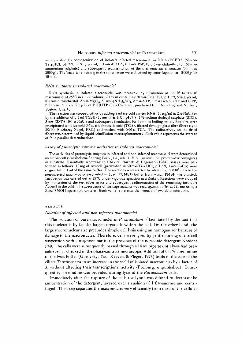
Holospora-infected macronuclei in Paramecium 391
were purified by homogenization of isolated infected macronuclei in 0 0 5 M - T G E D A (50mM-Tris_;HCl, pH7-9, 30% glycerol, 0-lmM-EDTA, 0-1 miu-PMSF, 0-5 mM-dithiothreitol, 50mM-ammonium sulphate) and subsequent sedimentation of the macronuclear chromatin (5 min at2000 #). The bacteria remaining in the supernatant were obtained by centrifugation at lOOOOg'forlOmin.
RNA synthesis in isolated macronuclei
RNA synthesis in isolated macronuclei was measured by incubation of 1X10⁵ to 4xlO5
macronuclei at 25°C in a total volume of 155 /xl containing 50mM-TrisHCl, pH7-9, 5% glycerol,0-5 min-dithiothreitol, 2 mM-MgCl2, 50 mM-(NH4)2SO4, 2 miu-ATP, 1 min each of CTP and GTP,001 mM-UTP and 2-5 /i.Ci of pH]UTP (35-7 Ci/mmol, purchased from New England Nuclear,Boston, U.S.A.).
The reaction was stopped either by adding 2 ml ice-cold carrier RNA (50 /ig/ml in 2 M-NaCl) orby the addition of 0-5 ml TSSE (50mM-TrisHCl, pH7-9, 1% sodium dodecyl sulphate (SDS),5mM-EDTA, 0 1 M-NaCl) and subsequent incubation for 1 min in boiling water. Samples wereprecipitated with ice-cold 05M-trichloracetic acid (TCA), filtered through glass-fibre filters (type85/90, Macherey-Nagel, FRG) and washed with 001 M - T C A . The radioactivity on the driedfilters was determined by liquid scintillation spectrophotometry. Each value represents the averageof four parallel determinations.
Assay of proteolytic enzyme activities in isolated macronuclei
The activities of proteolytic enzymes in infected and non-infected macronuclei were determinedusing Azocoll (Calbiochem-Behring Corp., La Jolla, U.S.A.; an insoluble protein—dye conjugate)as substrate. Essentially according to Chavira, Burnett & Hageman (1984), assays were per-formed as follows: 10mg of Azocoll (prewashed in 50mM-Tris-HCl, pH7-9, 1 mM-CaCy weresuspended in 1 ml of the same buffer. The reactions were started by addition of 2X 10s infected ornon-infected macronuclei suspended in 50ft\ TGMED buffer from which PMSF was omitted.Incubation was carried out at 25°C under vigorous agitation in a shaker. Reactions were stoppedby immersion of the test tubes in ice and subsequent sedimentation of the remaining insolubleAzocoll in the cold. The absorbance of the supernatants was read against buffer at 520 nm using aZeiss PMQII spectrophotometer. Each value represents the average of two determinations.
RESULTS
Isolation of infected and non-infected macronuclei
The isolation of pure macronuclei in P. caudatum is facilitated by the fact that
this nucleus is by far the largest organelle within the cell. On the other hand, the
large macronuclear size precludes simple cell lysis using an homogenizer because of
damage to the macronuclei. Therefore, cells were lysed by gentle stirring of the cell
suspension with a magnetic bar in the presence of the non-ionic detergent Nonidet
P40. The cells were subsequently passed through a 10 ml pipette until lysis had been
achieved as checked in the phase-contrast microscope. Addition of 0-1 % spermidine
to the lysis buffer (Gorovsky, Yao, Keevert & Pleger, 1975) leads in the case of the
ciliate Tetrahymena to an increase in the yield of isolated macronuclei by a factor of
3, without affecting their transcriptional activity (Freiburg, unpublished). Conse-
quently, spermidine was provided during lysis of the Paramecium cells.
Immediately after the rupture of the cells the lysate was diluted to decrease the
concentration of the detergent, layered over a cushion of l-6M-sucrose and centri-
fuged. This step separates the macronuclei very efficiently from most of the cellular

392 M. Freiburg
debris - discharged trichocysts, the food bacteria and the micronuclei. The lattercan be further purified to homogeneity by metrizamide isopycnic centrifugationaccording to Shiomi, Higashinakagawa, Saiga & Mita (1980; data not shown).
Infection of macronuclei with the macronucleus-specific bacterium H. obtusa
Fig. 1. P. caudatum; unfixed, slightly pressed, phase-contrast. X430. A. Non-infected;B, macronucleus infected with H. obtusa. Note that the infected macronucleus (mac)shows a significant increase in size compared to the non-infected cell.
Fig. 2. Photomicrographs of isolated macronuclei and endonuclear bacteria, A. Centri-fuge tube after centrifugation at 16 300 g for 20 nun. Macronuclei bearing predominantlythe infectious form of H. obtusa band at the top of the 2-2M-sucrose cushion (arrow).B. Isolated macronuclei derived from the 16 300£ sediment bearing predominantly thereproductive form of//, obtusa; bacteria are visible only after lysis of the macronuclei;differential interference contrast. X400. c. Isolated macronucleus derived from the topof the 2-2M-sucrose cushion bearing predominantly the infectious form of H. obtusa;differential interference contrast. X770. D. Long infectious and short reproductiveforms of H. obtusa purified from isolated macronuclei as indicated in Materials andMethods; differential interference contrast. X1600.

Holospora-infected macronuclei in Paramecium 393
leads, depending on the extent of infection, to a more or less drastic swelling of thenuclei (Fig. 1B). Consequently, the macronuclei bearing predominantly the infec-tious form of H. obtusa band at the top of the 2-2M-sucrose cushion whencentrifuged as indicated in Materials and Methods (Fig. 2A,c). Macronuclei bearing

394 M. Freiburg
predominantly the reproductive form sediment at the bottom. As can be seen fromFig. 2B at this stage of purification macronuclei are completely free from cytoplasmiccontaminants and food bacteria, and show a shape similar to that found within theintact cell. In some preparations the macronuclear sediment contains water-insoluble crystals released from the Paramecium cells during homogenization. Theoverall yield of isolated macronuclei at this last stage of purification is 90—100% inthe case of the non-infected nuclei and, due to their higher fragility lower forinfected nuclei, ranging from 50 to 80%.
RNA synthesis in infected and non-infected macronucleiSince the macronucleus off. caudatum is the organellar target for the infection,
we are interested in detecting any organelle-specific in vttro response of the host to
Incubation time (min)
Fig. 3. Incorporation of [3H]UMP into RNA in isolated macronuclei as a function oftime. Tests were carried out as described in Materials and Methods with 1-5X10⁵ non-infected (O O) or infected ( • A) macronuclei per test.

Holospora-infected macronuclei in Paramecium 395
Table 1. RNA synthesis in infected macronuclei in vitro and distribution of[3H] UMP incorporation among macronuclear chromatin and endonuclear bacteria
[3H]UMP incorporation (c.p.m.)
Infected macronuclei 87 5362000^ sediment 76361
(macronuclear chromatin)10000^ sediment 392
(H.obtusa; 8-6x10⁷ cells)10 000# supernatant 2130
Isolated macronuclei (bearing predominantly the reproductive form of H. obtusa) wereincubated with nucleoside triphosphates as indicated in Materials and Methods. After 30min thereaction was stopped by chilling the test tubes in an ice-bath and increasing the concentration ofthe non-labelled UTP to 1 mM. The [3H]UMP incorporation into RNA was either determineddirectly from the total incubation cocktail containing 7-5X 10⁵ macronuclei, or an equal number ofmacronuclei were homogenized by vigorous pipetting, and macronuclear chromatin and endo-nuclear bacteria were separated by centrifugation at 2000 # for 5 min. Subsequently, bacteria wereharvested from the supernatant by centrifugation at 10 000 # for 10 min. Each probe was lysed byaddition of TSSE buffer and processed as described in Materials and Methods.
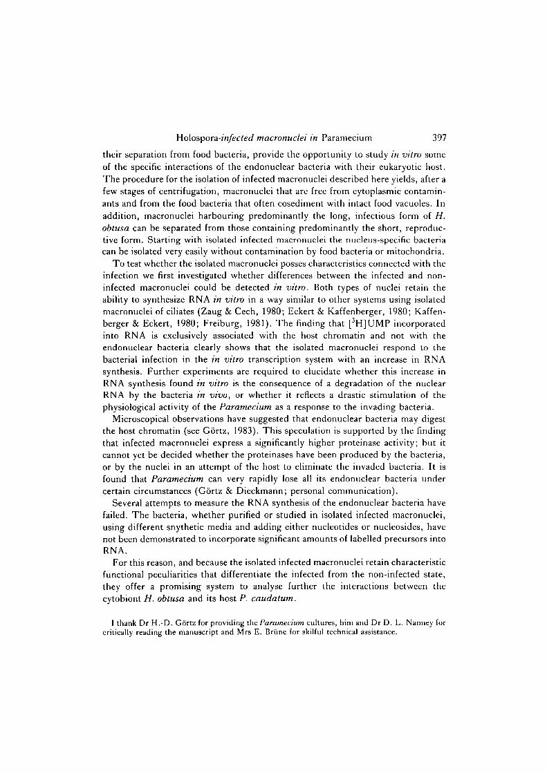
H. obtusa. Therefore, an in vitro transcription system has been used to studywhether RNA synthesis in the macronucleus is affected by the presence of bacteria.As can be seen from Fig. 3, RNA synthesis is stimulated very drastically, by a factorof approximately 5, in the macronuclei harbouring bacteria compared to the non-infected nucleus. Each of the infected macronuclei contained approximately100-250 bacteria of the reproductive form and approximately 20-30 of the infec-tious form.
To test whether the increase in the [3H]UMP accumulation in the infectedmacronuclei is due to RNA synthesis by the bacteria, the host nucleus or both,macronuclei were homogenized after 30 min incubation in an in vitro transcriptioncocktail containing nucleoside triphosphates. Macronuclear fragments including thechromatin were then separated from the bacteria by centrifugation. As one can seefrom Table 1, radioactivity was associated solely with the macronuclear chromatin,whereas the endonuclear bacteria were not labelled. This result indicates that theincrease in RNA synthesis in the isolated infected macronuclei is due to an increasein the RNA polymerase activity of Paramecium.
Proteinase activity in infected and non-infected macronuclei
Endonuclear symbionts infecting the micro- and macronucleus of ciliates cannotaffect only the shape and size of the host nucleus, but in addition can cause changesin the fine structure of the chromatin (see Gortz, 1983). This finding leads to thepossibility that the bacteria may digest the host chromatin, or structural elements ofthe macronucleus like microtubules or microfilaments. For that reason it seemedpromising to compare the proteinase activities associated with infected and non-infected isolated macronuclei.
To measure proteinase activities equal numbers of macronuclei were incubated

396 M. Freiburg
Incubation time (min)
Fig. 4. Time-dependent hydrolysis of Azocoli after addition of T5X105 non-infected(O O) or infected (A A) macronuclei per test. Tests were carried out asdescribed in Materials and Methods. Each value represents the average of two indepen-dent experiments.
different times with Azocoll (insoluble cowhide to which an azo-dye is attached).When the peptide bonds of the collagen are hydrolysed by proteinases present in themacronuclear preparations, the dye is released into the suspending buffer and itsabsorbance at 520 nm can be measured. As can be seen from Fig. 4, the amount of thedye released into the suspending buffer during the incubation is linear as a functionof time for both types of macronuclei, but is significantly higher in the case ofsymbiont-bearing macronuclei. This observation indicates that the infection of themacronucleus is correlated with a drastic increase in the proteinase activity assoc-iated with this isolated organelle.
DISCUSSION
The macronucleus of/3, caudatum can be regarded as a microenvironment for thecytobiotic bacterium H. obtusa. The mass isolation of infected macronuclei, and
Ho\ospora-infected macronuclei in Paramecium 397
their separation from food bacteria, provide the opportunity to study in vitro someof the specific interactions of the endonuclear bacteria with their eukaryotic host.The procedure for the isolation of infected macronuclei described here yields, after afew stages of centrifugation, macronuclei that are free from cytoplasmic contamin-ants and from the food bacteria that often cosediment with intact food vacuoles. Inaddition, macronuclei harbouring predominantly the long, infectious form of H.obtusa can be separated from those containing predominantly the short, reproduc-tive form. Starting with isolated infected macronuclei the nucleus-specific bacteriacan be isolated very easily without contamination by food bacteria or mitochondria.
To test whether the isolated macronuclei posses characteristics connected with theinfection we first investigated whether differences between the infected and non-infected macronuclei could be detected in vitro. Both types of nuclei retain theability to synthesize RNA in vitro in a way similar to other systems using isolatedmacronuclei of ciliates (Zaug & Cech, 1980; Eckert & Kaffenberger, 1980; Kaffen-berger & Eckert, 1980; Freiburg, 1981). The finding that [3H]UMP incorporatedinto RNA is exclusively associated with the host chromatin and not with theendonuclear bacteria clearly shows that the isolated macronuclei respond to thebacterial infection in the in vitro transcription system with an increase in RNAsynthesis. Further experiments are required to elucidate whether this increase inRNA synthesis found in vitro is the consequence of a degradation of the nuclearRNA by the bacteria in vivo, or whether it reflects a drastic stimulation of thephysiological activity of the Paramecium as a response to the invading bacteria.
Microscopical observations have suggested that endonuclear bacteria may digestthe host chromatin (see Gortz, 1983). This speculation is supported by the findingthat infected macronuclei express a significantly higher proteinase activity; but itcannot yet be decided whether the proteinases have been produced by the bacteria,or by the nuclei in an attempt of the host to eliminate the invaded bacteria. It isfound that Paramecium can very rapidly lose all its endonuclear bacteria undercertain circumstances (Gortz & Dieckmann; personal communication).
Several attempts to measure the RNA synthesis of the endonuclear bacteria havefailed. The bacteria, whether purified or studied in isolated infected macronuclei,using different snythetic media and adding either nucleotides or nucleosides, havenot been demonstrated to incorporate significant amounts of labelled precursors intoRNA.
For this reason, and because the isolated infected macronuclei retain characteristicfunctional peculiarities that differentiate the infected from the non-infected state,they offer a promising system to analyse further the interactions between thecytobiont H. obtusa and its host P. caudatum.
I thank Dr H.-D. Gortz for providing the Paramecium cultures, him and Dr D. L. Nanney forcritically reading the manuscript and Mrs E. Briine for skilful technical assistance.

398 M. Freiburg
REFERENCES
CHAVIRA, R. JR, BURNETT, T H . J. & HAGEMAN, J. H. (1984). Assaying proteinases with azocoll.Analyt. Biochem. 136, 446-450.
CUMMINGS, D. J. (1977). Methods for the isolation of nuclei from ciliated protozoans. In Meth.Cell Biol., vol. 16 (ed. D. M. Prescott), pp. 97-112. New York, London: Academic Press.
ECKERT, W. A. & KAFFENBERGER, W. (1980). Regulation of rRNA metabolism in Tetrahymena.I. Nutritional shift-down. Eur.J. Cell Biol. 21, 53-62.
FREIBURG, M. (1981). Functional states of RNA polymerase in the macronucleus of Telraliymenapyriformis and their dependence on culture growth. J . Cell Sci. 47, 267-275.
GORTZ, H.-D. (1983). Endonuclear symbionts in ciliates. Int. Rev. Cytol., Suppl. 14, 145-176.GORTZ, H.-D. & DIECKMANN, J. (1977). Infektiose, auf die Kerne von Paramecium caudatum
spezialisierte Partikel. Verh. dt. Zool. Ges. 1977, 267.GORTZ, H.-D. & DIECKMANN, J. (1980). Life cycle and infectivity of Holospora elegans (Hafkine),
a micronucleus-specific symbiont of Paramecium caudatum (Ehrenberg). Protistologica 16,591-603.
GORTZ, H.-D. & FUJISHIMA, M. (1983). Conjugation and meiosis of Paramecium caudatuminfected with the micronucleus-specific bacterium Holospora elegans. Eur. J. Cell Biol. 32,86-91.
GOROVSKY, M. A., YAO, M.-C, KEEVERT, J. B. & PLEGER, G. L. (1975). Isolation of micro- andmacronuclei of Tetrahymena pyriformis. In Meth. Cell Biol., vol. 9d (ed. D. M. Prescott), pp.311-327. New York, London: Academic Press.
KAFFENBERGER, W. & ECKERT, W. A. (1980). Regulation of rRNA metabolism in Tetrahymenapyriformis. II. Nutritional shift-up. Eur.J. Cell Biol. 21, 200-207.
NANNEY, D. L. (1980). Experimental Ciliatology. New York: Wiley.OSSIPOV, D. V. & PODLIPAEV, S. A. (1977). Electron microscope examinations of early stages of
infection of Paramecium caudatum by bacterial symbionts of the macronucleus (Iota-bacteria).Ada Protozool. 16, 289-308 (in Russian with English summary).
OSSIPOV, D. V., SKOBLO, J. J. & RAUTIAN, M. S. (1975). Iota-particles, macronuclear symbioticbacteria of ciliate Paramecium caudatum clone M-115. Ada Protozool. 14, 263—280.
SCHWEMMLER, W. (1979). Mechanismen der Zellevolution. Berlin, New York: de Gruyter.SHIOMI, Y., HIGASHINAKAGAWA, T., SAIGA, H. & MITA, T. (1980). Metrizamide isopycnic
centrifugation for the isolation of macro- and micronuclei from Paramecium. J. UOEH 2 (3),323-330.
TAYLOR, F. J. R. (1979). Symbionticism revisited: a discussion of the evolutionary impact ofintracellular symbiosis. Proc. R. Soc. bond. B, 204, 267-286.
TAYLOR, F. J. R. (1983). Some eco-evolutionary aspects of intracellular symbiosis. Int. Rev.Cytol., Suppl. 14, 1-28.
ZAUG, A. J. & CECH, Th. R. (1980). In vitm splicing of the ribosomal RNA precursor in nuclei ofTetrahvmena. Cell 19, 331-338.
(Received 6 June 1984 -Accepted 7 September 1984)