IRDL Cloning: A One-Tube, Zero-Background, Easy-to-Use ...
9
IRDL Cloning: A One-Tube, Zero-Background, Easy-to-Use, Directional Cloning Method Improves Throughput in Recombinant DNA Preparation Jiancai Wang 1,2 , Ronghua Xu 2 *, Aizhong Liu 2,3 * 1 School of Life Sciences, University of Science and Technology of China, Hefei, People’s Republic of China, 2 Key Laboratory of Tropical Plant Resources and Sustainable Use, Xishuangbanna Tropical Botanical Garden, Chinese Academy of Sciences, Kunming, People’s Republic of China, 3 Kunming Institute of Botany, Chinese Academy of Sciences, Kunming, Kunming, People’s Republic of China Abstract Rapid and efficient construction of expression vectors and subsequent transformation are basic recombinant methods for the investigation of gene functionality. Although novel cloning methods have recently been developed, many laboratories worldwide continue to use traditional restriction digestion-ligation methods to construct expression vectors owing to financial constraints and the unavailability of appropriate vectors. We describe an improved restriction digestion-ligation (IRDL) cloning method that combines the advantage of directional cloning from double digestion-ligation with that of a low background observed by using a positive selection marker gene ccdB to facilitate digestion and ligation in a single tube. The IRDL cloning overcomes the time-consuming and laborious limits of traditional methods, thereby providing an easy-to-use, low-cost, and one-step strategy for directional cloning of target DNA fragments into an expression vector. As a proof-of- concept example, we developed two yeast vectors to demonstrate the feasibility and the flexibility of the IRDL cloning method. This method would provide an effective and easy-to-use system for gene cloning and functional genomics studies. Citation: Wang J, Xu R, Liu A (2014) IRDL Cloning: A One-Tube, Zero-Background, Easy-to-Use, Directional Cloning Method Improves Throughput in Recombinant DNA Preparation. PLoS ONE 9(9): e107907. doi:10.1371/journal.pone.0107907 Editor: Andre van Wijnen, University of Massachusetts Medical, United States of America Received May 18, 2014; Accepted August 20, 2014; Published September 22, 2014 Copyright: ß 2014 Wang et al. This is an open-access article distributed under the terms of the Creative Commons Attribution License, which permits unrestricted use, distribution, and reproduction in any medium, provided the original author and source are credited. Data Availability: The authors confirm that all data underlying the findings are fully available without restriction. All relevant data are within the paper and its Supporting Information files. Funding: This work was supported by the National NSFC (Grant no. 31300569 to RX) and by the ‘‘Hundreds of Talents’’ program of the Chinese Academy of Sciences (AL). The funders had no role in study design, data collection and analysis, decision to publish, or preparation of the manuscript. Competing Interests: The authors have declared that no competing interests exist. * Email: [email protected] (RX); [email protected] (AL) Introduction Molecular cloning is an essential tool in modern molecular biology and biotechnology. With the rapid development of genomics in rapidly dissecting the functionality of specific genes, the requirement for constructing various vectors for gene transformation has become more acute than ever before. Therefore, developing a simple, efficient, universal, and cost- effective method for directional cloning of PCR products is particularly timely. Traditionally, the cloning of an insert into an expression vector involves the use of Type II restriction enzymes to generate the appropriate DNA fragments encompassing the target sequence, followed by modification of DNA ends to generate blunt or sticky ends, and ligation of the DNA fragments to generate plasmids or other types of DNA vectors [1]. This procedure is extremely laborious and time consuming applied to a high-throughput format and is often limited by the relatively few restriction sites available [2]. Additionally, incomplete digestion of the vector reduces the efficiency of the screening process in identifying the single successfully cloned construct. Over the past two decades, a wide variety of novel cloning technologies, such as the Gateway system (Invitrogen), the Creator system and Infusion cloning (Clontech), LIC [3], TA cloning [4], MAGIC [5], SLIC [6], and Golden Gate cloning [7] have been developed, thereby improving cloning efficiency substantially. Gateway and Creator cloning systems are both site-specific recombination systems typically requiring a manual two-step process: i.e., inserting a target gene into a donor vector and subcloning the target gene from the donor vector into a recipient plasmid for subsequent expression. Like traditional cloning, the two-step cloning process is laborious and time consuming in a high-throughput setting, and the commercial recombinase is usually expensive. The MAGIC system, which uses in vivo recombination to transfer the gene from donor to recipient plasmid, decreased experimental costs substantially, but the experimental process remains laborious and time consuming. The LIC and SLIC methods use the exonuclease activity of T4 DNA polymerase to generate long cohesive ends between the vector and the target DNA fragments for subsequent annealing and in vivo repair. These methods require multiple experimental steps and long primers, which usually increase the cost and difficulty of PCR amplification. Infusion cloning enables easy construction of the expression vector in vitro, but also requires long primers. The TA cloning method, a one-step cloning process widely used for cloning PCR fragments into vectors, is rather limited in its construction of expression vectors since the insertion of fragments is non- directional, thereby increasing the labor in screening the correct clone. Golden Gate cloning is considerably faster and more accurate, with the potential of assembling multiple fragments in PLOS ONE | www.plosone.org 1 September 2014 | Volume 9 | Issue 9 | e107907
Transcript of IRDL Cloning: A One-Tube, Zero-Background, Easy-to-Use ...

IRDL Cloning: A One-Tube, Zero-Background,Easy-to-Use, Directional Cloning Method ImprovesThroughput in Recombinant DNA PreparationJiancai Wang1,2, Ronghua Xu2*, Aizhong Liu2,3*
1 School of Life Sciences, University of Science and Technology of China, Hefei, People’s Republic of China, 2 Key Laboratory of Tropical Plant Resources and Sustainable
Use, Xishuangbanna Tropical Botanical Garden, Chinese Academy of Sciences, Kunming, People’s Republic of China, 3 Kunming Institute of Botany, Chinese Academy of
Sciences, Kunming, Kunming, People’s Republic of China
Abstract
Rapid and efficient construction of expression vectors and subsequent transformation are basic recombinant methods forthe investigation of gene functionality. Although novel cloning methods have recently been developed, many laboratoriesworldwide continue to use traditional restriction digestion-ligation methods to construct expression vectors owing tofinancial constraints and the unavailability of appropriate vectors. We describe an improved restriction digestion-ligation(IRDL) cloning method that combines the advantage of directional cloning from double digestion-ligation with that of a lowbackground observed by using a positive selection marker gene ccdB to facilitate digestion and ligation in a single tube. TheIRDL cloning overcomes the time-consuming and laborious limits of traditional methods, thereby providing an easy-to-use,low-cost, and one-step strategy for directional cloning of target DNA fragments into an expression vector. As a proof-of-concept example, we developed two yeast vectors to demonstrate the feasibility and the flexibility of the IRDL cloningmethod. This method would provide an effective and easy-to-use system for gene cloning and functional genomics studies.
Citation: Wang J, Xu R, Liu A (2014) IRDL Cloning: A One-Tube, Zero-Background, Easy-to-Use, Directional Cloning Method Improves Throughput in RecombinantDNA Preparation. PLoS ONE 9(9): e107907. doi:10.1371/journal.pone.0107907
Editor: Andre van Wijnen, University of Massachusetts Medical, United States of America
Received May 18, 2014; Accepted August 20, 2014; Published September 22, 2014
Copyright: � 2014 Wang et al. This is an open-access article distributed under the terms of the Creative Commons Attribution License, which permitsunrestricted use, distribution, and reproduction in any medium, provided the original author and source are credited.
Data Availability: The authors confirm that all data underlying the findings are fully available without restriction. All relevant data are within the paper and itsSupporting Information files.
Funding: This work was supported by the National NSFC (Grant no. 31300569 to RX) and by the ‘‘Hundreds of Talents’’ program of the Chinese Academy ofSciences (AL). The funders had no role in study design, data collection and analysis, decision to publish, or preparation of the manuscript.
Competing Interests: The authors have declared that no competing interests exist.
* Email: [email protected] (RX); [email protected] (AL)
Introduction
Molecular cloning is an essential tool in modern molecular
biology and biotechnology. With the rapid development of
genomics in rapidly dissecting the functionality of specific genes,
the requirement for constructing various vectors for gene
transformation has become more acute than ever before.
Therefore, developing a simple, efficient, universal, and cost-
effective method for directional cloning of PCR products is
particularly timely.
Traditionally, the cloning of an insert into an expression vector
involves the use of Type II restriction enzymes to generate the
appropriate DNA fragments encompassing the target sequence,
followed by modification of DNA ends to generate blunt or sticky
ends, and ligation of the DNA fragments to generate plasmids or
other types of DNA vectors [1]. This procedure is extremely
laborious and time consuming applied to a high-throughput format
and is often limited by the relatively few restriction sites available
[2]. Additionally, incomplete digestion of the vector reduces the
efficiency of the screening process in identifying the single
successfully cloned construct. Over the past two decades, a wide
variety of novel cloning technologies, such as the Gateway system
(Invitrogen), the Creator system and Infusion cloning (Clontech),
LIC [3], TA cloning [4], MAGIC [5], SLIC [6], and Golden Gate
cloning [7] have been developed, thereby improving cloning
efficiency substantially. Gateway and Creator cloning systems are
both site-specific recombination systems typically requiring a
manual two-step process: i.e., inserting a target gene into a donor
vector and subcloning the target gene from the donor vector into a
recipient plasmid for subsequent expression. Like traditional
cloning, the two-step cloning process is laborious and time
consuming in a high-throughput setting, and the commercial
recombinase is usually expensive. The MAGIC system, which uses
in vivo recombination to transfer the gene from donor to recipient
plasmid, decreased experimental costs substantially, but the
experimental process remains laborious and time consuming. The
LIC and SLIC methods use the exonuclease activity of T4 DNA
polymerase to generate long cohesive ends between the vector and
the target DNA fragments for subsequent annealing and in vivorepair. These methods require multiple experimental steps and long
primers, which usually increase the cost and difficulty of PCR
amplification. Infusion cloning enables easy construction of the
expression vector in vitro, but also requires long primers. The TA
cloning method, a one-step cloning process widely used for cloning
PCR fragments into vectors, is rather limited in its construction of
expression vectors since the insertion of fragments is non-
directional, thereby increasing the labor in screening the correct
clone. Golden Gate cloning is considerably faster and more
accurate, with the potential of assembling multiple fragments in
PLOS ONE | www.plosone.org 1 September 2014 | Volume 9 | Issue 9 | e107907

one pot and replacing the two steps of digestion and ligation with a
single restriction-ligation step. Since this system is based on the
action of the type II restriction enzyme BsaI, the backbone
expression vectors require removal of internal BsaI sites [7].
Although the aforementioned cloning methods facilitate more
efficient cloning, most laboratories worldwide continue to use
traditional restriction digestion and ligation methods to construct
expression vectors because of insufficient financial support and
unavailability of appropriate vectors. Improvement of the restric-
tion digestion-ligation methodology will increase the speed of the
cloning process and save time, money, and labor. Here, we report
an improved restriction digestion-ligation (IRDL) cloning method.
This system combines the advantages of directional cloning of
double digestion-ligation with that of a low background observed
by using a positive selection marker gene to facilitate digestion and
ligation in a single tube. Numerous cloning tests performed in our
laboratory have shown that the IRDL cloning has a nearly 100%
cloning efficiency. This system may be widely used for the
insertion of PCR products into expression vectors in a one-step,
single-tube digestion-ligation reaction.
Materials and Methods
Reagents and Genetic techniquesAll basic DNA manipulation procedures were performed
according to protocols described in Sambrook et al. [8]. The
FastDigest restriction enzyme was purchased from Fermentas
(Vilnius, Lithuania), the QuickCut restriction enzyme was purchased
from TaKaRa (Kyoto, Japan), and the restriction enzyme with
CutSmart buffer was purchased from New England BioLabs
(Ipswich, MA, USA). T4 DNA ligase and ATP were purchased
from Fermentas. High fidelity DNA Polymerase TransStart FastPfu
(TransGen, Beijing, PR China) was used for DNA amplification.
Plasmids were prepared using High-purity Plasmid DNA Mini-
preparation Kit (Generay, Shanghai, PR China). The DNA
concentration was measured with a NanoDrop Spectrophotometer
ND-2000 (Thermo Scientific; Waltham, MA, USA). All plasmids
used for IRDL cloning carry the gene ccdB, which is lethal to most
host Escherichia coli strains; expression of the ccdB protein interferes
with DNA gyrase and blocks the passage of DNA and RNA
polymerases and leads to double-strand DNA breakage and cell
death [9]. Plasmids containing the ccdB gene were propagated in the
E. coli DB3.1 strain and plasmids without the ccdB gene were
propagated in the E. coli Trans1-T1 strain (TransGen). All plasmids
derived from PCR products were verified by sequencing. The strains
and plasmids used in this study are listed in Table S1. Primers used in
this study, synthesized by Generay, are listed in Table S2.
Plasmids, IRDL cloning reaction, and transformationThe plasmid pWXY1.0 was constructed by replacement of the
URA3 autotrophic marker of p426-GAL with the TRP1 marker
by recombination in S. cerevisiae W303-1A and insertion of a 732-
bp PCR-amplified ccdB cassette containing multiple cloning sites
(MCS) from pCXSN by conventional cloning. The plasmid
pWXY3.0 was constructed by replacement of the TRP1 marker of
pWXY1.0 with the URA3 marker by recombination in yeast. To
generate the constructs pWXY1.0-EGFP, pWXY1.0-JcDGAT1,
pWXY1.0-JcPDAT1, and pWXY1.0-LacZ, EGFP, JcDGAT1,
JcPDAT1, and LacZ flanked by the appropriate restriction sites
were amplified from pXDG [9], p426-JcDGAT1 [10], pYES2.1-
JcPDAT1 [11], and pYES2.1-LacZ (Invitrogen, Carlsbad, CA,
USA) and subsequently inserted into pWXY1.0 by IRDL cloning.
Details of these constructs were presented in Materials and
Methods S1.
The standard IRDL cloning reaction mixture contained the
following ingredients: 50 ng vector, the appropriate quantity of
insert DNA in a 3:1 molar ratio of insert to vector, 1 mL 106FastDigest buffer, 0.5 mL 10 mM ATP, 0.25 mL FastDigest
restriction enzyme 1, 0.25 mL FastDigest restriction enzyme 2,
0.25 mL T4 DNA Ligase, and distilled H2O up to a total volume of
10 mL. The IRDL cloning reaction mixture was incubated at 37uCor at 20uC for 30 min. Subsequently, 5 mL of the IRDL cloning
reaction was transformed into 50 mL Trans1-T1 Phage resistant
chemically competent cells (TransGen) according to the manu-
facturer’s instructions. The transformation efficiency of Trans1-T1
Phage resistant chemically competent cells was 109 cfu/mg of
pUC19 DNA. The transformed cells were plated on LB plates
containing 100 mg/mL ampicillin (Amp+).
To generate the ScDGA1 fusion protein, the ScDGA1 gene
flanked by SpeI and AscI sites was amplified from S. cerevisiaegenomic DNA with the primers DGA1-1 and DGA1-2 (Table S2).
To generate the EGFP fusion protein, the EGFP gene flanked by
AscI and XhoI sites was amplified from the plasmid pCambia35s-
EGFP with the primers GFP-2 and GFP-7 (Table S2). After gel
purification, approximately 50 ng of each PCR product was added
to IRDL cloning digestion and ligation mixture contained 0.25 mL
SpeI, 0.25 mL AscI, 0.25 mL XhoI, and 0.25 mL T4 DNA ligase.
The reaction mixture was incubated at 37uC or 20uC for 30 min
before E. coli transformation.
To generate the pWXY1.0-JcDGAT2 construct, the JcDGAT2gene was amplified from p426-JcDGAT2 [10] by two parallel
PCRs with the primers pairs, P1–P2 and P3–P4 (Table S2),
respectively. The PCR products were purified from agarose gels,
and subsequently mixed together. Next, the PCR products were
denatured by heating at 95uC for 10 min, followed by reannealing
by cooling to room temperature. Concomitantly, a restriction
digest was performed with 100 ng pWXY1.0, 1 mL 106FastDigest buffer, 0.25 mL EcoRI, and 0.25 mL XhoI up to a
10 mL volume with distilled H2O, and incubated at 37uC for 5–
10 min, followed by 80uC for 5 min. Next, 2 mL reannealed PCR
products (100 ng/mL), 0.25 mL T4 DNA ligase, 0.25 mL 10 mM
ATP, and 0.5 mL 106 FastDigest buffer were added to the
digestion mixture up to a final volume of 15 mL with distilled H2O,
and the mixture was then incubated at 25uC for 30 min before E.coli transformation.
Yeast expression, TLC analysis, microscopy, and westernblot
pWXY3.0-ScDGA1-EGFP was transformed into yeast quadru-
ple mutant H1246a with the LiAc/SS Carrier DNA/PEG method
[12]. Transformants were selected by growth on synthetic
complete medium with the appropriate amino acid omitted and
supplemented with 2% (w/v) glucose. Positive colonies were
transferred into liquid SC media with the appropriate amino acid
omitted and supplemented with 2% (w/v) glucose, followed by
incubation at 28uC overnight. Overnight cultures were diluted up
to an optical density (OD) of 0.4 in induction medium (SC-ura +2% galactose +1% raffinose), and induced for 24–36 h at 28uC.
Cells were collected and lysed using glass beads and stored at
280uC for subsequent lipid analysis and protein extraction. Yeast
lipid extraction, TLC analysis, microscopy and western blotting
were performed as described previously [10,13].
Results
Since most projects in our laboratory require construction of a
single expression vector, we often use a double-digestion and
ligation method to generate expression vectors, but often
IRDL Cloning
PLOS ONE | www.plosone.org 2 September 2014 | Volume 9 | Issue 9 | e107907

encounter background problems because of incomplete digestion
of the vector. To develop an easy-to-use, fast, efficient, directional,
zero-background method, we inserted a lethal gene cassette
flanked by two MCS (Fig. 1A). The expression vector also
contained a selection marker, allowing for antibiotic selection,
and an origin of replication in E. coli. The gene of interest was
amplified by a high fidelity DNA polymerase with primers
consisting of one available restriction site at each 59 end. Since
the two restriction enzymes used for cleavage of PCR products and
expression vector were distinct, the overhangs after cleavage were
incompatible. After gel-purification, the PCR products were
mixed with the expression vector to perform a double-digestion
and ligation procedure in a single tube. Once the digestion and
ligation are in balance, two possible plasmids are present: the
original expression vector and the desired recombinant plasmid
(Fig. 1A). Because of the positive selection of the lethal gene, only
E. coli harboring the desired construct could grow after
transformation on selective media containing the appropriate
antibiotics.
One-step directional cloning of a single gene into anexpression vector by the IRDL cloning
To test the IRDL cloning system, we constructed a yeast
expression vector pWXY1.0 (Fig. 2A). The pWXY1.0 contains a
ccdB cassette for positive selection in commonly used E. colistrains (such as DH5a, TOP10), a TRP1 autotrophic marker for
selection in S. cerevisiae known as W303-1A, a pBR322 origin of
replication for E. coli, a 2 morigin of replication for S. cerevisiae, a
glucose-depressed GAL1 promoter for inducing expression, and a
CYC1 terminator. Two MCS are located at both ends of the ccdBcassette, each with four distinct restriction sites. To test the
efficiency of the IRDL cloning system, we cloned the gene EGFP(0.7 kb) with the vector pWXY1.0. The gene EGFP was initially
amplified by TransStart FastPfu DNA polymerase with primers
GFP1 (containing the restriction KpnI site at 59 end) and GFP2
(containing the restriction XhoI site at 59 end) (Table S2). The
PCR products of EGFP were gel-purified and were inserted into
pWXY1.0 through the standard IRDL cloning reaction as
described in materials and methods, and the reaction mixture
were transformed to E. coli strain Trans1-T1. The results showed
that the IRDL cloning system generated a large number of
colonies, whereas both reaction mixtures (without T4 DNA ligase
and restriction enzymes, respectively) as controls did not generate
any colony on selective plate (Fig. 2B). Further, 48 colonies on LB
plates were randomly sorted out for PCR analysis, resulting in the
PCR products of all 48 colonies presented expected size bands (see
Fig. 2C). Among these positive transformants, 23 of 48 colonies
were subjected to restriction enzymatic analysis, leading to that the
all colonies tested were true recombinants (Fig. 2D). The ten of
these positive colonies was randomly selected and further
confirmed by subsequent sequencing. These results showed that
the IRDL cloning method was effective.
Apart from FastDigest restriction enzyme and digestion buffer
(ingredients not disclosed by supplier), QuickCut restriction
enzyme and digestion buffer (ingredients not disclosed by supplier)
and restriction enzyme from New England BioLabs and CutSmart
digestion buffer (50 mM potassium acetate, 20 mM Tris-acetate,
10 mM magnesium acetate, 100 mg/mL BSA, pH 7.9, 25uC) were
also tested (Table 1). All the reaction mixtures tested were
complemented with 0.5 mM ATP and 2.5 U T4 DNA ligase
(Fermentas), incubated at 37uC for 5–30 min, and immediately
transformed into competent E. coli cells, with transformants
subsequently selected on LB Amp+ plates. For all of the reaction
mixtures tested, more colonies appeared with longer incubation
times. Restriction enzyme from different companies did display
significant differences in colony number (ranging from 0.7–
26103). Although the reaction mixture was incubated at room
temperature (20uC), the results also displayed high cloning
efficiency. Although cloning efficiency was enhanced by heat
inactivation of the restriction enzyme after incubation prior to
transformation and increased insert/vector molar ratios (data not
shown), these actions were unnecessary for routine cloning. The
results also demonstrated that as little as 5 min of the restriction-
ligation step was sufficient to generate the desired construct for
routine cloning into expression vector.
To examine the cloning efficiency of IRDL cloning with
different insert fragments in length and incubation time, aside
from EGFP, the genes JcDGAT1 (1.5 kb), JcPDAT1 (2 kb), and
LacZ (3 kb) were further tested. Similarly, these fragments were
amplified by high fidelity DNA polymerase with specific primers
containing the appropriate restriction sites (Table S2), and then
the PCR products were purified and cloned into pWXY1.0
following the standard IRDL cloning steps with FastDigest
restriction enzyme and digestion buffer. As shown in Table 1,
the generated colony number increased with the elongation of
incubation times in all cases, whereas with the increasing of the
insert fragments in length the colony number decreased. When the
insert fragment was 3 kb (LacZ) the colony number varied from
109 to 731. When the insert of combined EGFP and ScDGA1 was
used the IRDL cloning produced 75 colonies after 5 min
incubated. These colonies were sufficient to satisfy the general
requirement in most laboratories. Further, the PCR inspection
with gene specific primers confirmed the positive clones, resulting
in a high cloning efficiency from 92%, 96% to 98% for LacZ,
JcPDAT1 and JcDGAT1, respectively.
Generation of fusion protein by IRDL cloningExpression of a fusion protein is a widely used methodology for
rapid and effective characterization of gene product function. To
test the ability of the IRDL cloning system in cloning two
fragments concomitantly, we generated a ScDGA1 and EGFPfusion plasmid (Fig. 3A) for overexpression in yeast. Two methods
exist to construct a fusion protein (Fig. 1B and 1C). The first
requires the use of restriction enzymes and ligases, and the second
employs overlap extension PCR. As the cloning of overlapping
PCR products was expected to be similar to one-insert cloning, we
tested the efficiency of assembly of two inserts, ScDGA1 and
EGFP in pWXY1.0, by digestion with three restriction enzymes.
The restriction-ligation mixtures were prepared as previously
described as one-insert cloning by using FastDigest restriction
enzymes. After gel-purification, the two PCR products ScDGA1(containing a 59 SpeI site and a 39 AscI site) and EGFP (containing
a 59 AscI site and a 39 XhoI site) were added to the mixture and
incubated at 37uC for 5–30 min. The transformation and
screening steps were performed as described above. Although
the number of colonies was significantly lower than that during the
cloning of a single insert, the 75 colonies obtained after a 5-min
reaction period were sufficient to satisfy the requirement of fusion
construction. PCR analysis of 24 randomly selected colonies from
LB Amp+ plates indicated that all colonies tested contained the
expected construct (Fig. 3B), with 10 of those colonies confirmed
to contain the insert by subsequent sequencing. One colony was
selected for plasmid purification and introduced into the S.cerevisiae mutant H1246a for GAL1-inducing expression. As a
dga1Dlro1Dare1Dare2D quadruple mutant (QM) strain, H1246alacks all four enzymes for neutral lipid synthesis, and triacylgly-
cerol (TAG) was not observed in this strain [14,15]. Therefore,
H1246a is extensively used in characterization of the gene
IRDL Cloning
PLOS ONE | www.plosone.org 3 September 2014 | Volume 9 | Issue 9 | e107907
encoding of diacylglycerol acyltransferase (DGAT) enzyme
[10,15]. As shown in Fig 3C, overexpression of ScDGA1-EGFP
fusion protein restored TAG synthesis similar to overexpression of
the ScDGA1 alone, indicating that the fusion protein has
comparable functionality to DGAT enzyme. Expression of
ScDGA1-EGFP was also confirmed by GFP fluorescence
(Fig. 3D) and western blotting (Fig. 3E).
Creating expression clones free of restriction sitelimitations
One limitation of the IRDL cloning method may stem from the
occasional presence of one or more internal restriction site(s) in the
gene of interest, thereby precluding the use of this strategy. A
general solution to this challenge is the use of two pairs of primers
to generate PCR products with sticky ends through enzyme-free
cloning [16,17], which could be complementary with a double-
digested quick-clone plasmid. Such cloning requires performance
of the double digestion first, followed by inactivation of the double
restriction enzyme by heat inactivation, addition of the PCR
products with sticky ends, and finally performance of ligation. To
test this strategy, we cloned a Jatropha curcas DGAT2 gene
(GenBank accession number: HQ827795), which contains a single
internal EcoRI site, into plasmid pWXY1.0. To generate PCR
products with EcoRI and XhoI overhangs, two parallel PCR
amplifications of JcDGAT2 were performed following the strategy
illustrated in Figure 4A. Primers P1, P2, P3, and P4 were
appended with a tail containing the following sequences: C,
TCGAG, AATTC, and G, respectively. The two PCR products
were gel-purified, mixed in equal volume, and denatured by
heating to 95uC, followed by cooling to room temperature,
thereby causing the double-stranded DNA molecules to denature
and reanneal. This process results in 25% of the DNA molecules
containing EcoRI and XhoI overhangs (Fig. 4A). To avoid EcoRI
cleavage of the JcDGAT2, the plasmid pWXY1.0 was double-
digested with EcoRI and XhoI, followed by 85uC heating for
5 min to inactivate the restriction enzyme. The reannealed
JcDGAT2 PCR products, T4 DNA ligase, and ATP were added
to the restriction mixture and incubated at room temperature for
20 min, followed by E. coli transformation. Colony PCR analysis
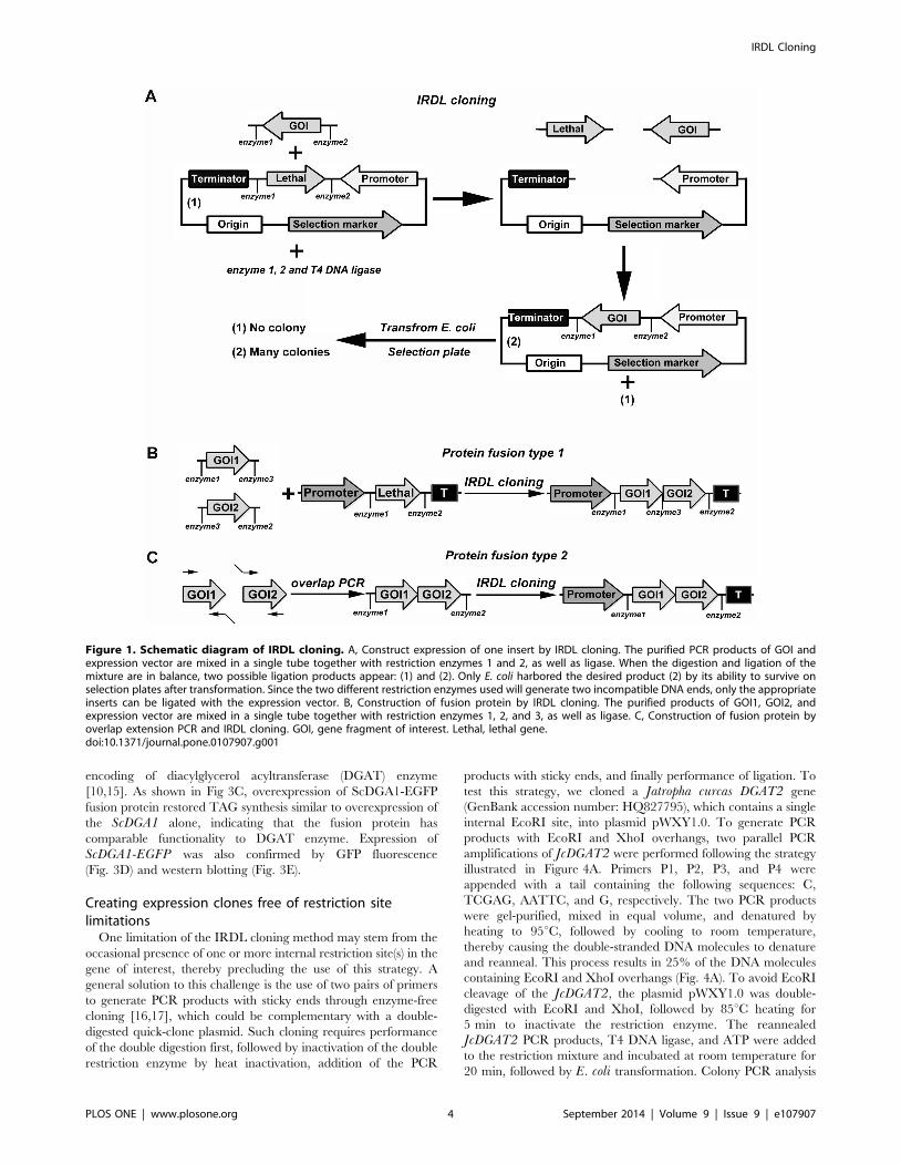
Figure 1. Schematic diagram of IRDL cloning. A, Construct expression of one insert by IRDL cloning. The purified PCR products of GOI andexpression vector are mixed in a single tube together with restriction enzymes 1 and 2, as well as ligase. When the digestion and ligation of themixture are in balance, two possible ligation products appear: (1) and (2). Only E. coli harbored the desired product (2) by its ability to survive onselection plates after transformation. Since the two different restriction enzymes used will generate two incompatible DNA ends, only the appropriateinserts can be ligated with the expression vector. B, Construction of fusion protein by IRDL cloning. The purified products of GOI1, GOI2, andexpression vector are mixed in a single tube together with restriction enzymes 1, 2, and 3, as well as ligase. C, Construction of fusion protein byoverlap extension PCR and IRDL cloning. GOI, gene fragment of interest. Lethal, lethal gene.doi:10.1371/journal.pone.0107907.g001
IRDL Cloning
PLOS ONE | www.plosone.org 4 September 2014 | Volume 9 | Issue 9 | e107907

Figure 2. Subcloning of one insert into a yeast expression vector pWXY1.0 by using IRDL cloning method. A, Schematic representationof one-step directional cloning of EGFP into a yeast expression vector pWXY1.0. B, Test of the IRDL cloning system. (1): Cloning of EGFP into pWXY1.0by standard IRDL cloning step (the purified PCR products of GFP and yeast expression vector pWXY1.0 are mixed in a single tube together withFastDigest buffer, restriction enzymes XhoI and KpnI, ATP and T4 DNA ligase, and incubated at 37uC for 30 min), followed by transformation into E.coli Trans1-T1 as described in materials and methods. (2): A control without T4 DNA ligase. (3): A control without restriction enzymes XhoI and KpnI. C,Colony PCR results from 48 recombinant colonies were run on 1% agarose gels. All of the colonies except the second clone tested contained thecorrect inserts. M, DNA ruler DL2501 from Generay. D, Plasmids DNA from 23 recombinant colonies and vector pWXY1.0 were digested with XhoI andKpnI and run on 1% agarose gels. DNA from all 23 recombinant colonies displayed the expected restriction pattern of pWXY1.0-EGFP. M, DNA rulerDL2502 (Generay). V, pWXY1.0.doi:10.1371/journal.pone.0107907.g002
IRDL Cloning
PLOS ONE | www.plosone.org 5 September 2014 | Volume 9 | Issue 9 | e107907

and restriction analysis of 10 randomly selected colonies indicated
that all colonies contained the expected construct (Fig. 4B),
confirmed by subsequent sequencing.
Discussion
Conventional cloning methods based on restriction digestion
and ligation have played a critical role in the construction of
recombinant DNA molecules. However, because of its laborious
and time-consuming nature, as well as limitations in restriction
sites in the MCS, a wide variety of novel cloning technologies have
been developed as alternate methodologies to restriction digestion
and ligation. Among these technologies, the Gateway system has
been the most popular choice for vector construction in the
research community for its convenience for DNA transfer into
multiple expression vectors [18]. Additionally, many Gateway-
compatible entry clone collections and expression vectors have
been generated for a multitude of species [19,20,21,22,23]. For
thousands of individual research groups worldwide, the Gateway
system is not practical, since most projects only require the
construction of a single expression vector. Moreover, the Gateway
system has limited utility for investigators working on non-model
organisms, since no entry clones and expression vectors are
available. To overcome these limitations in both traditional
methods and the Gateway system, we developed a substantially
improved technique over the traditional restriction digestion-
ligation method, referred to as IRDL cloning. A lethal marker
gene (ccdB) flanked by multiple distinct restriction sites was
introduced into the MCS so that PCR inserts and their backbone
vectors could be subjected to restriction digestion and ligation in a
single tube. With the positive selection of the ccdB gene and the
incompatible ends of the vectors, any undigested backbone vector
and self-ligation transformants were eliminated, and only correct
and directional cloning results were able to survive on the selection
plates. Here, we developed two yeast expression vectors to validate
our system. The results showed that a gene or DNA fragment of
interest can be easily cloned into an expression vector by a one-
step procedure, confirming that our system is an easy-to-use,
efficient, rapid, zero-background, directional cloning method.
With minor modifications (e.g., combined with infusion PCR or an
enzyme-free cloning method), our system can also be used to clone
DNA fragments with occasional internal restriction site.
Compared to Gateway and other systems, our system possesses
several advantages: (a) our system required no entry clones, and
only one-step incubation, followed by standard transformation was
required. It frees investigators from laborious cloning processes. By
contrast, conventional restriction digestion-ligation, Gateway, and
LIC all require multiple cloning procedures. Although the TA
cloning method could rapidly clone PCR products, preparation of
the TA vector involves multiple steps and the final TA-generated
construct typically possesses no directionality, resulting in difficul-
ties in selection [24]. TOPO cloning methods also perform rapid
cloning, but lack directionality. Although available from commer-
cial sources (Invitrogen and TransGen), no reports are currently
published on the development of high-quality expression vectors
based on TOPO cloning in individual laboratories, probable
related to technology difficulties. (b) Our system does not require
long primers for PCR amplification. By contrast, the Gateway,
Infusion, and LIC all require long primers, which commonly
increase cloning difficulties and cause errors in PCR amplification
[25], in addition to increased costs as the number of constructs
multiply. (c) Our system does not depend on expensive
recombinases, thereby saving on cost. Although a variety of tested
Gateway expression vectors are available from Invitrogen and
Ta
ble
1.
Effi
cie
ncy
of
clo
nin
gin
pW
XY
1.0
un
de
rd
iffe
ren
tco
nd
itio
ns.
Bu
ffe
rE
nz
ym
eg
rou
pIn
sert
Co
lon
yn
um
be
rS
ucc
ess
rate
5m
in1
0m
in2
0m
in3
0m
in
16Fa
stD
ige
stX
ho
I+K
pn
I0
.7k/
EGFP
26
43
82
58
71
33
11
00
%
16Q
uic
kCu
tX
ho
I+K
pn
I0
.7k/
EGFP
14
03
21
59
41
00
51
00
%
16C
utS
mar
tX
ho
I+K
pn
I0
.7k/
EGFP
41
07
52
11
32
18
57
10
0%
16Fa
stD
ige
stN
coI+
Spe
I0
.7k/
EGFP
20
93
60
63
59
85
10
0%
16Fa
stD
ige
stB
amH
I+Ec
oR
I0
.7k/
EGFP
25
74
71
74
21
43
71
00
%
16Fa
stD
ige
stX
ho
I+K
pn
I1
.5k/
JcD
GA
T12
10
41
75
78
89
09
8%
16Fa
stD
ige
stX
ho
I+K
pn
I2
k/Jc
PD
AT1
17
33
12
59
89
32
96
%
16Fa
stD
ige
stX
ho
I+K
pn
I3
k/La
cZ1
09
23
04
20
73
19
2%
16Fa
stD
ige
stX
ho
I+K
pn
I0
.7k/
EGFP
*1
21
27
63
54
78
61
00
%
16Fa
stD
ige
stX
ho
I+K
pn
I+A
scI
2k/
ScD
GA
1+E
GFP
75
18
03
60
72
01
00
%
All
reac
tio
nm
ixtu
res
con
tain
ed
1m
MA
TP
and
5W
eis
su
nit
T4
DN
Alig
ase
(Fe
rme
nta
s)an
din
cub
ate
dat
37uC
.Exp
eri
me
nts
we
rep
erf
orm
ed
usi
ng
50
ng
vect
or,
and
the
corr
esp
on
din
gam
ou
nt
of
inse
rtD
NA
ata
3:1
mo
lar
rati
oo
fin
sert
:ve
cto
r.N
on
eo
fth
ere
acti
on
mix
ture
sh
ere
was
inac
tiva
ted
wit
hh
eat
pri
or
toE.
coli
tran
sfo
rmat
ion
.*
ind
icat
es
the
reac
tio
nm
ixtu
rew
asin
cub
ate
dat
roo
mte
mp
era
ture
20uC
.Clo
nin
ge
ffic
ien
cie
sw
ere
giv
en
asco
lon
ies
nu
mb
er
on
LBp
late
sco
nta
inin
g1
00
mg/m
Lam
pic
illin
.Clo
nin
gsu
cce
ssra
tew
ere
evu
late
db
yco
lon
ies
PC
Rte
sto
f5
0ra
do
mse
lete
dco
lon
ies.
Spe
cifi
cp
rim
ers
of
dif
fere
nt
inse
rts
use
das
forw
ard
pri
me
r,an
dC
YC
1p
rim
er
inC
YC
1te
rmin
ato
ru
sed
asa
reve
rse
pri
me
r.d
oi:1
0.1
37
1/j
ou
rnal
.po
ne
.01
07
90
7.t
00
1
IRDL Cloning
PLOS ONE | www.plosone.org 6 September 2014 | Volume 9 | Issue 9 | e107907

from the research community at large, many small laboratories are
unwilling to use this technology, since the Gateway recombinase is
expensive. Even laboratories that can afford the cost of this system
may elect to scale down their reactions to a 5-mL volume. The cost
of recombinase is also often prohibitive in the Infusion kit and the
like, such as CloneEZ (Genescript, Nanjing, PR China), Cold
Fusion Cloning (SBI, Mountain View, CA, USA), Fast Seamless
Cloning (DoGene, Shanghai, PR China), and GENEART
Seamless cloning (Invitrogen), pEASY-Uni Seamless Cloning
(TransGen, Beijing, PR China), especially when used for a large-
scale protein expression and purification studies. Zhang et al. [1]
used crude bacterial extract containing recombinase to perform
seamless cloning by in vitro recombination, but the preparation of
crude recombinase extract is a multistep process requiring some
skill. By contrast, we used conventional restriction enzymes to
perform IRDL cloning. Considering that the cloning process
requires only small volume of enzymes, the cost is affordable for
the standard academic laboratory. (d) Compared to the more
complex development of plasmids based on Gateway, LIC, and
TA systems, the plasmids used in the IRDL cloning strategy for
different research purposes are easy to develop. The insertion of a
lethal gene cassette flanking multiple single restriction sites into the
backbone plasmid may complete the construct. (e) The expression
plasmid used in IRDL cloning does not require linearization or
any modification. By contrast, the plasmid used for Infusion
cloning requires linearization and purification [2] and the vector
used in the TA system requires prior preparation. Unfortunately,
the 39-dT overhang of the TA system is unstable and usually
absent during storage (see PCR cloning protocol, [26]). Since the
plasmids are unimpaired before digestion, the expression vectors
used in IRDL cloning are very stable, even during long-term
storage. (f) The Flexi Cloning system (Promega) used lethal gene
marker barnase, two rare restriction enzyme SgfI and PmeI, and
antibiotic resistance change to facilitate the shuttling of gene of
interest between the different Flexi Vectors [27]. Like the traditional
restriction digestion and ligation methods, cloning of PCR products
into the entry Flexi Vectors also involved multiple steps (See
Technical mannual of Flexi Vector System, http://www.promega.
com.cn/techserv/tbs/TM001-310/tm254.pdf), such as restriction
digest of PCR products and acceptor Flexi vector, cleanup of the
PCR products, ligation of PCR product and acceptor Flexi vector.
However, our IRDL cloning method simplified these experimental
steps into one step and one tube.
The ccdB gene encodes a lethal protein, which impairs
topoisomerase II (DNA gyrase) activity, resulting in irreparable
DNA damage and death of the host E. coli. Since cells containing
the gyrA462 mutation and cells containing the F9 episome carried
a ccdA gene encoding for an antidote to the ccdB-expressing toxic
protein, they are not sensitive to its ccdB-killing activity [28]. The
ccdB and ccdA of F9 episome expressed a toxin-antitoxin construct
that has been extensively used in the construction of various
plasmids with positive selection [9,24,25,29,30], including com-
mercial Gateway plasmids. Besides the ccdB lethal marker, other
lethal genes can be used as positive selection marker, e.g., the cell
lysis-related genes from phages, Mu, X174, and Q [28]; the
plasmid-encoding toxin, e.g., RelE, PemK/Kid, Doc, ShoB, Zor,
Figure 3. Fusion of two inserts, ScDGA1 and EGFP, into a yeast expression vector pWXY3.0 using IRDL cloning. A, The plasmidpWXY3.0-ScDGA1-EGFP generated by using IRDL cloning method. B, Colony PCR results from 24 recombinant colonies were run on 1% agarose gels.All of the colonies tested contained the corrected inserts. M, DNA ruler DL2501 (Generay). C, TLC separation of neutral lipids from different yeaststrains. The transgenic H1246a with the ScDGA1 and ScDGA1-EGFP restore triacylglycerol (TAG) synthesis compared with negative control (H1246awith pWXY3.0). D, Fluorescence microscopy of ScDGA1-EGFP fusion protein expression in transgenic yeast cells. E, Detection of ScDGA1-EGFP fusionprotein by western blotting.doi:10.1371/journal.pone.0107907.g003
IRDL Cloning
PLOS ONE | www.plosone.org 7 September 2014 | Volume 9 | Issue 9 | e107907

and MazF [31,32,33,34]; and other lethal genes, e.g., Hlg1 [35],
RCSB [28] and barnase [27]. Although the precise mechanisms
for their action are not well understood, these lethal genes should
theoretically function properly in the IRDL cloning system.
Recently, Mok and Li [36] developed a similar cloning method,
referred to as the detox system, but they did not combine the
digestion and ligation protocols into a single step. Therefore, the
resulting procedure is 20 times as long as the IRDL cloning
procedure (30 min digestion and 70 min ligation for detox cloning
versus 5 min digestion and ligation for IRDL cloning). Further-
more, we presented a remedy for cloning a gene of interest
containing an internal restriction site.
The IRDL cloning system is a powerful tool that is unrestricted
to cloning a functional gene. Any genetic elements with decoding
sequence information—including promoters, terminators, attenu-
ators, enhancers, and origins of replication—can also be cloned
using this method. Moreover, the IRDL cloning strategy is easy-to-
use in constructing plasmids for different research purposes, such
as constitutive expression, transient expression, gene silencing,
protein tagging, promoter activity, and protein subcellular
localization in bacteria, fungi, plants, insects, and mammals. The
cloned fragments can also be cleaved and transferred to another
vector based on the change of antibiotic marker. However, the
IRDL cloning method has a drawback when restriction sites
flanking the lethal marker occurred in the target gene of interest,
which would preclude its application. To overcome this problem
we proposed eight different restriction sites on both sides of the
lethal marker gene, which could provide with 16 possible
combinations for restriction digestion, might be usually sufficient
to clone a given gene. In addition, IRDL cloning is developed
based on the double restriction digestion for cloning a given gene.
In particular, when one constructs an expression vector for
analyzing the function of a given gene, IRDL cloning is quite
helpful.
Recently, several seamless cloning methods such as QC (Quick
and Clean) cloning, One-step SLIC (Sequence- and Ligation-
Independent Cloning) and FastCloning have been developed
[37,38,39]. These methods substantially improve traditional
cloning techniques, greatly facilitate cloning work because their
easy-to-use, fast and simple features. However, the three methods
Figure 4. Schematic diagram of cloning the gene of interest containing internal restriction sites into expression vector. A, generationof sticky-end fragments and cloning into pWXY1.0 by IRDL cloning. The JcDGAT2 was amplified by two pairs of primers, P1–P2, and P3–P4, whichwere appended with short sequence tails: C, TCGAG, AATTC, G, respectively. After gel-purification, the two PCR products were mixed together,denatured, and reannealed, resulting in 25% of the DNA fragments with EcoRI and XhoI overhangs. Concomitantly, the vector was double-digestedwith EcoRI and XhoI for 5–10 min at 37uC. After heat inactivation of the restriction enzyme at 95uC for 5 min, the vector was mixed with thereannealed JcDGAT2 containing EcoRI and XhoI overhangs, T4 DNA ligase and ATP were added and incubated at room temperature for 20 min, andfinally transformed into E. coli. B, Restriction digestion (EcoRI and XhoI) of minipreps of pWXY1.0 (V) and pWXY1.0-JcDGAT2 (lane 1 to lane 10). Therestriction pattern of pWXY1.0-JcDGAT2 generated by EcoRI and XhoI digestion was as predicted: 861 bp and 200 bp, respectively. M, DNA rulerDL2502 (Generay).doi:10.1371/journal.pone.0107907.g004
IRDL Cloning
PLOS ONE | www.plosone.org 8 September 2014 | Volume 9 | Issue 9 | e107907

require long primers for PCR amplification compared to IRDL
cloning method, which may increase the cost and cause errors in
PCR amplification as described above. In addition, both QC
cloning and one-step SLIC need linearize vectors before cloning.
The FastCloning is often confined when constructing a large
vector (such as a plant expression vector, larger than 12 kb often)
in length because this method requires to amplify the vector. In
contrast, QC cloning seems be more suitable for cloning specific
PCR products from unspecific mixes; one-step SLIC may be more
helpful for assembly multiple fragments; FastCloning may be more
usable for constructing expression vector, chimeras and producing
multiple mutations. IRDL cloning can be more helpful for
constructing a given expression vector. For cloning an uncertain
PCR fragment using some known sequences, such as PCR
products from TAIL PCR, adaptor PCR and RACE PCR, those
methods independent of restriction enzyme such as Gateway,
Infusion, TA, and LIC would be more helpful.
In summary, the low cost, convenience, and high efficiency of
the IRDL cloning system will have a potential for wide application
and facilitate plasmid construction and rapid speed for functional
genomics studies. Even laboratories with modest budgets can use
this system to perform high-throughput cloning or preparation of
expression constructs for functional genomics research. This
method is a useful alternate methodology to existing cloning
methods based on recombination.
Supporting Information
Table S1 Strains and plasmids used in this study.
(PDF)
Table S2 Primers in the study.
(PDF)
Materials and Methods S1
(PDF)
Acknowledgments
We would like to thank Dr. Sten Stymne (Swedish University of
Agricultural Sciences, Uppsala) for kindly providing the H1246a mutant.
Author Contributions
Conceived and designed the experiments: RX AL. Performed the
experiments: JW RX. Analyzed the data: JW RX. Contributed reagents/
materials/analysis tools: JW RX. Wrote the paper: RX AL.
References
1. Zhang YW, Werling U, Edelmann W (2012) SLiCE: a novel bacterial cell
extract-based DNA cloning method. Nucleic Acids Res 40: e55.2. Hartley JL (2006) Cloning technologies for protein expression and purification.
Curr Opin Biotechnol 17: 359–366.3. Aslanidis C, Dejong PJ (1990) Ligation-Independent Cloning of PCR Products
(LIC-PCR). Nucleic Acids Res 18: 6069–6074.4. Zhou MY, Clark SE, Gomezsanchez CE (1995) Universal cloning method by
TA strategy. Biotechniques 19: 34–35.
5. Li MZ, Elledge SJ (2005) MAGIC, an in vivo genetic method for the rapidconstruction of recombinant DNA molecules. Nat Genet 37: 311–319.
6. Li MZ, Elledge SJ (2007) Harnessing homologous recombination in vitro togenerate recombinant DNA via SLIC. Nat Methods 4: 251–256.
7. Engler C, Kandzia R, Marillonnet S (2008) A one pot, one step, precision
cloning method with high throughput capability. PloS One 3: e3647.8. Sambrook J, Russell DW (2001) Molecular Cloning: A Laboratory Manual:
CSHL press.9. Chen SB, Songkumarn P, Liu JL, Wang GL (2009) A versatile zero background
T-vector system for gene cloning and functional genomics. Plant Physiol 150:1111–1121.
10. Xu RH, Yang TQ, Wang RL, Liu AZ (2014) Characterisation of DGAT1 and
DGAT2 from Jatropha curcas and their functions in storage lipid biosynthesis.Func Plant Bio 41: 321–329.
11. Xu RH, Qiu LJ, Yang TQ, Wang RL, Tian B, Liu AZ (2013) Cloning andfunctional of phospholipids:diacylglycerol acyltransferase (JcPDAT1) cDNA
from Jatropha curcas. Chinese J Oil Crop Sci 35: 123–130.
12. Gietz RD, Schiestl RH, Willems AR, Woods RA (1995) Studies on thetransformation of intact yeast cells by the LiAc/SSDNA/PEG procedure. Yeast
11: 355–360.13. Chandrasekaran U, Wang XJ, Liu AZ (2013) Characterization, expression
profiling and heterologous function analysis of two oleosin genes PvOle1 andPvOle2 from sacha inchi (Plukenetia volubilis). Int J Agric Biol 15: 435–442.
14. Sandager L, Gustavsson MH, Stahl U, Dahlqvist A, Wiberg E, et al. (2002)
Storage lipid synthesis is non-essential in yeast. J Biol Chem 277: 6478–6482.15. Siloto RMP, Truksa M, He XH, McKeon T, Weselake RJ (2009) Simple
methods to detect triacylglycerol biosynthesis in a yeast-based recombinantsystem. Lipids 44: 963–973.
16. Tillett D, Neilan BA (1999) Enzyme-free cloning: a rapid method to clone PCR
products independent of vector restriction enzyme sites. Nucleic Acids Res 27:e26.
17. Walker A, Taylor J, Rowe D, Summers D (2008) A method for generatingsticky-end PCR products which facilitates unidirectional cloning and the one-
step assembly of complex DNA constructs. Plasmid 59: 155–162.18. Walhout AJM, Temple GF, Brasch MA, Hartley JL, Lorson MA, et al. (2000)
GATEWAY recombinational cloning: Application to the cloning of large
numbers of open reading frames or ORFeomes. Method Enzymol 328: 575–592.
19. Karimi M, Inze D, Depicker A (2002) GATEWAY vectors for Agrobacterium-mediated plant transformation. Trends Plant Sci 7: 193–195.
20. Curtis MD, Grossniklaus U (2003) A gateway cloning vector set for high-
throughput functional analysis of genes in planta. Plant Physiol 133: 462–469.
21. Earley KW, Haag JR, Pontes O, Opper K, Juehne T, et al. (2006) Gateway-
compatible vectors for plant functional genomics and proteomics. Plant J 45:
616–629.
22. Nakagawa T, Kurose T, Hino T, Tanaka K, Kawamukai M, et al. (2007)
Development of series of gateway binary vectors, pGWBs, for realizing efficient
construction of fusion genes for plant transformation. J Biosci Bioeng 104:
34–41.
23. Yashiroda Y, Matsuyama A, Yoshida M (2008) New insights into chemical
biology from ORFeome libraries. Curr Opin Chem Biol 12: 55–59.
24. Oh SK, Kim SB, Yeom SI, Lee HA, Choi D (2010) Positive-selection and
ligation-independent cloning vectors for large scale in planta expression for plant
functional genomics. Mol Cells 30: 557–562.
25. Wang C, Yin X, Kong X, Li W, Ma L, et al. (2013) A series of TA-based and
zero-background vectors for plant functional genomics. PloS One 8: e59576.
26. Guo B, Bi Y (2002) Cloning PCR products. In Chen B. and Janes H. (ed.), PCR
cloning protocols, Humana Press, Totowa, New Jersey. p.115.
27. Blommel PG, Martin PA, Seder KD, Wrobel RL, Fox BG (2009) Flexi vector
cloning. Methods Mol Biol 498: 55–73.
28. Choi YJ, Wang TT, Lee BH (2002) Positive selection vectors. Crit Rev
Biotechnol 22: 225–244.
29. Mondon P, Chang YC, Varma A, Kwon-Chung KJ (2000) A novel episomal
shuttle vector for transformation of Cryptococcus neoformans with the ccdB geneas a positive selection marker in bacteria. FEMS Microbiol Lett 187: 41–45.
30. Xu GY, Sui N, Tang Y, Xie K, Lai YZ, et al. (2010) One-step, zero-background
ligation-independent cloning intron-containing hairpin RNA constructs for
RNAi in plants. New Phytol 187: 240–250.
31. Gerdes K (2000) Toxin-antitoxin modules may regulate synthesis of macromol-ecules during nutritional stress. J Bacteriol 182: 561–572.
32. Yamaguchi Y, Park JH, Inouye M (2011) Toxin-antitoxin systems in bacteria
and archaea. Ann Rev Genet 45: 61–79.
33. Kamphuis MB, Monti MC, van den Heuvel RHH, Lopez-Villarejo J, Diaz-
Orejas R, et al. (2007) Structure and function of bacterial kid-kis and related
toxin-antitoxin systems. Protein Peptide Lett 14: 113–124.
34. Fozo EM (2012) New type I toxin-antitoxin families from ‘‘wild’’ and laboratory
strains of E. coli: Ibs-Sib, ShoB-OhsC and Zor-Orz. RNA Biol 9: 1504–1512.
35. Chattoraj P, Ganguly T, Nandy RK, Sau S (2008) Overexpression of a delayed
early gene hlg1 of temperate mycobacteriophage L1 is lethal to both M.smegmatis and E. coli. BMB Rep 41: 363–368.
36. Mok WWK, Li YF (2013) A highly efficient molecular cloning platform that
utilises a small bacterial toxin gene. Chembiochem 14: 733–738.
37. Thieme F, Engler C, Kandzia R, Marillonnet S (2011) Quick and clean cloning:
a ligation-independent cloning strategy for selective cloning of specific PCR
products from non-specific mixes. PLoS One 6: e20556.
38. Jeong JY, Yim HS, Ryu JY, Lee HS, Lee JH, et al. (2012) One-step sequence-
and ligation-independent cloning as a rapid and versatile cloning method for
functional genomics studies. Appl Environ Microbiol 78: 5440–5443.
39. Li CK, Wen AY, Shen BC, Lu J, Huang Y, et al. (2011) FastCloning: a highly
simplified, purification-free, sequence- and ligation-independent PCR cloning
method. Bmc Biotechnol 11: 92.
IRDL Cloning
PLOS ONE | www.plosone.org 9 September 2014 | Volume 9 | Issue 9 | e107907