IRAQI JOURNAL OF MEDICAL SCIENCES JMS... · Iraqi Journal of Medical Sciences Aims and Scope Iraqi...
96
Volume 8 (2) 2010 ISSN 1681-6579 IRAQI JOURNAL OF MEDICAL SCIENCES CHAIRMAN OF THE EDITORIAL BOARD Professor Adnan A. Anoze MRCP. Chief Editor Professor Fakhir S. Al-Ani PhD. EXECUTIVE EDITORIAL BOARD Editor Ghassan A. Al-Shamma PhD Prof. Editor Alaa G. Hussien FICMS Prof. Editor Nidhal Abdul-Muhymen PhD Prof. Editor Samir M. Jasim PhD Asst. Prof. Editor Muataz A. Al-Qazzaz FICMS Asst. Prof. Editor Hussam A. Ahmed FRCS Asst. Prof. Editor Enas T. Abdul-Karim DCH, PhD Asst. Prof. Editor Atheer J. Al-Saffar FICMS Asst. Prof. Editor Hasan A. AL-Hamadani FICMS Asst. Prof. Editor Hala S. Aref CABP Asst. Prof. Editor Waseem F. Mohammed FICMS Lecturer Editor Ali F. Hadi PhD Lecturer Editor Suhad M. Salih FICOG Lecturer Journal Secretary Esraa' S. NAJI Technical Editor Aliaa' N. Hatam
Transcript of IRAQI JOURNAL OF MEDICAL SCIENCES JMS... · Iraqi Journal of Medical Sciences Aims and Scope Iraqi...

Volume 8 (2) 2010 ISSN 1681-6579
IRAQI JOURNAL
OF MEDICAL SCIENCES
CHAIRMAN OF THE EDITORIAL BOARD
Professor Adnan A. Anoze MRCP.
Chief Editor
Professor Fakhir S. Al-Ani PhD.
EXECUTIVE EDITORIAL BOARD
Editor Ghassan A. Al-Shamma PhD Prof. Editor Alaa G. Hussien FICMS Prof. Editor Nidhal Abdul-Muhymen PhD Prof. Editor Samir M. Jasim PhD Asst. Prof.Editor Muataz A. Al-Qazzaz FICMS Asst. Prof.Editor Hussam A. Ahmed FRCSAsst. Prof.Editor Enas T. Abdul-Karim DCH, PhDAsst. Prof.Editor Atheer J. Al-Saffar FICMS Asst. Prof.Editor Hasan A. AL-Hamadani FICMS Asst. Prof.Editor Hala S. Aref CABP Asst. Prof.Editor Waseem F. Mohammed FICMS Lecturer Editor Ali F. Hadi PhD Lecturer Editor Suhad M. Salih FICOG Lecturer
Journal Secretary
Esraa' S. NAJI
Technical Editor Aliaa' N. Hatam

Iraqi Journal of Medical Sciences
All articles published represent the opinions of the authors and do not reflect the
policy of Iraqi Journal of Medical Sciences. All rights are reserved to
Iraqi Journal of Medical Sciences. No part of the journal may be
reproduced or transmitted in any form or by any means, electronic or mechanical,
including photocopying, recording, or via any storage or retrieval system, without
written permission from the journal.
All correspondence and subscription information requests should be addressed to:
The Editor of Iraqi Journal of Medical Sciences
P. O. Box 14222, Baghdad, Iraq.
College of Medicine
Baghdad, Iraq
Tel and Fax: 964-1-5224368
E-mail: [email protected]
© Copyright 2000

Advisory Committee
Deans of the Iraqi Colleges of Medicine
Prof. Adnan A. AnozeMedical College \ Al-Nahrain UniversityProf. Fadhil A. Al-KafajiMedical College \ Baghdad UniversityProf. Faris Abdul kareem Al-Kindi Medical College \ Baghdad UniversityProf. Mohammed H. Al-Alwan Medical College \ Al-Mustansiriya UniversityProf. Muzahim K.Al-Khyatt Medical College \ Al-Mosul UniversityProf. Faris Bekir Al-Sawaf Medical College \ Nainewa UniversityAsst. Prof. Ali Khiralla Al-Shaeli Medical College \ Babil University Asst. Prof.Muaied N. Majeed Medical College \ Thiqar University Prof. Abdullah Salih Hasan Medical College \ Al-Anbar UniversityProf. Abid Ahmed Selman Medical College \ Tikreet University Prof. Khudier K. Ibrahim Medical College \ Diyala University Prof. Zuhair U. Eissa Medical College \ Karbala University Prof. Mohammed S. Abdul-Zahra Medical College \ Al-Kufa University Prof. Thamir A. Hamdan Medical College \ Al-Basra UniversityProf. Rahi K. AL-Yasiri Medical College \ AL-Qadisiah UniversityProf. Fakhraddin N. Nassir Medical College \ Kirkuk University Asst. Prof.Ferhad Suliffan Medical College \ Duhok University LecturerAtta Gitti Allawi Medical College \ Wassit University LecturerAli Abdul-Aziz Al-Shawi Medical College \ Al-Emara UniversityLecturerMusaed Al-Dehan Medical College \ Al-Muthana University

Al-Nahrain College of Medicine committee members
Prof. Adnan A. Anoze Dean Medical College Prof. Hussam H. Ali Vice Dean Administrative affairs Asst. Prof.Abdul-Razak H. Ahmed Vice Dean Scientific & Students affairs Prof. Nidhal Abdul-Muhymen Chief Microbiology department Prof. Hashim M. AL-kadimy Chief Medicine department Prof. Maha M. AL-Bayati Chief Gynecology & Obstetrics department Prof. Alaa G. Hussien Chief Pathology&Forensic Medicine department Asst. Prof.Samir M. Jasim Chief Chemistry and Biochemistry department Asst. Prof.Hassan A. Hassan Chief Surgery department Asst. Prof.Farqad Badir Hamdan Chief Physiology department Asst. Prof.Haider J. Mobarak Chief Humman Anatomy department Prof. Lamia A.K. AL-Saady Chief Pediatric department Prof. Atheer J. Al-Saffar Chief Community department LecturerAbdul-Kareem H. Abid Chief Pharmacology&Theraputies department Asst. Prof.Mohammed A.Kadhim Representative of teaching staff members
Scientific Advisory Board
Prof. Mahmood Hayawi Hamash (Jordan\ Retierd) Prof. Rafi M. Al-Rawi (U.A.E\ Retierd) Prof. Anam Rasheed AL-Salihi (Irf Institute of Embryo Research & Infertility
Treatment / Al-Nahrain University) Prof. Amjad Dawood Niazi (Iraqi Board for Medical Specialization \ Retierd) Prof. Nazar Al-Hasani (Iraqi Board for Medical Specialization \ Retierd) Prof. Usama N. Rifat (U.A.E\ Retierd) Prof. Usama S. Al-Nasiri (Al-Nahrain University) Prof. Akrem J. Abod (U.A.E) Prof. Sarmad Khunda (Baghdad University \ Retierd ) Prof. Hikmat A.R. Hatam (Al-Nahrain University) Prof. Ryadh Abdul-Satar (U.A.E\ Retierd) Prof. Faroq H. Al-Jawad (Al-Nahrain University\ Retierd) Prof. Sami E. Matlob (Al-Nahrain University\ Retierd) Prof. Sawsan S. Al-Haidari (Al-Nahrain University) Prof. Yarub I. Khattab (Al-Nahrain University)

Iraqi Journal of Medical Sciences
Aims and Scope
Iraqi Journal of Medical Sciences is published by College of Medicine,
Al-Nahrain University. It is a quarterly multidisciplinary medical journal. High quality
papers written in English, dealing with aspects of clinical, academic or investigative
medicine or research will be welcomed. Emphasis is placed on matters relating to
medicine in Iraq in particular and the Middle East in general, though articles are
welcomed from anywhere in the world.
Iraqi Journal of Medical Sciences publishes original articles, case
reports, and letters to the editor, editorials, investigative medicine, and review
articles. They include forensic medicine, history of medicine, medical ethics, and
religious aspects of medicine, and other selected topics.

Iraqi JMS FORMAT INSTRUCTION TO AUTHORS
Iraqi Journal of Medical Sciences (Iraqi JMS) is a periodic, peer-reviewed journal published quarterly by College of Medicine, Al-Nahrain University. Iraqi JMS publishes manuscripts in all fields of health and medicine written in English. Types of Contributions: Original articles, review articles, case studies, editorials, medical education, history of medicine, ethics, practical points, medical quiz, conferences, meetings and letters to the Editor. Manuscripts: • Submission of a manuscript implies that is not being considered for publication anywhere. • The autor should provide the following: A. The author should provide a document officially state that the current work was carried out at the site which provides the certification. The document should be signed by the highest authorized member at that location. B. Document stated clearly that his current work is in agreement with the medical ethics provided either from the local ethical committee in the place where he did his work or from the Ministry of Health\Depart. Of Training & Improving skill\Research & Educational facilities, the approval has to be stated separetly in the method section C. Publication fees is 50,000 Iraqi dinars and an extra fees will be taken for extended paper (6 for each additional paper and up to 25). • Manuscripts submitted to IJMS are subject to editorial evaluation and revision by three referees. • The format of IJMS complies with the uniform requirements for manuscripts submitted to Biomedical Journals, published by the International Committee of Medical Journals Editors (ICMJE) (Vancouver, British Colombia, 1979) and its last update in October 2001, available on the web site www.icmje.org. • Manuscript should be typewritten double spaced on size A4 (29.5x21 cm) paper with wide margins. Page should be numbered consecutively. One original and three photocopies including figures, tables, and photographs should be submitted. Begin each of following sections on separate page in the following sequence: Title page, abstract and keywords, text, acknowledgments, references, tables, and legends for illustration. • Manuscript and figures will not be returned to the authors whether the editorial decision is to accept, revise or reject. • Manuscripts must be accompanied by a covering paper signed by all authors that the paper has not been published in and will not be submitted to any other journal if accepted in IJMS. • The page should contain (a) title of the manuscript, (b) names of each author (first name, middle initial and family name) including highest academic degree, (c) official academic and/or clinical title and affiliation (d) name and address of the institution where the work was done (e) name and address (E-mail if available) of the author to whom correspondence should be sent. Abstract: manuscript should include an abstract of not more than 150 words. Structured abstract typed on a separate sheet and consist of background, objective, method, results, and conclusion. Translation in Arabic to be included:
). هدف الدراسة، طريقة العمل، النتائج و االستنتاج الدراسة، ةخلفي(

• Keywords: three to ten keywords should be provided on the same page as the abstract in Arabic and English. As far as possible, be selected from the National Library of Medicine Medical Subject Headings. • The Arabic abstract should follow the United Medical Dictionary (Council of Arab Ministers of Health/WHO/ Arab Medical Union/ALESCO, 3rd edition. • Manuscript format: It should be divided into the following parts: introduction, materials and methods, results and discussion. • References: All references should be listed in consecutive numerical order by English numerical, in the order of citation in the text. Once a reference is cited all subsequent citations should be to the original number. Examples 1. Standard Journal Article: use et al when the number of authors exceeds 6.
Halliwell B, Gutteridge JMC. Oxygen toxicity, Oxygen radicals, transition metals and disease. Biochem J. 1984; 219: 1-14. 2. Books: Mann JI, Pyorala K, and Teuscher A. Diabetes in epidemiological perspective. London: Churchill Livingstone. 1983. 3. Chapter in book: Phillips SJ, and Whisnant JP. Hypertension and strock. In: Laragh JH, and Brenner BM. editors. Hypertension: Pathophysiology, diagnosis, and management. 2nd ed. NewYork: Raven Press; 1995. p. 465-78. • Tables: Each table should be typed on a separate page double-spaced, including all headings, number all tables with English numerals and include a short title. Vertical lines between columns are to be avoided. • Figures: All figures must be suitable for reproduction without being retouched or redrawn. Figure number, name of senior author, and title of the work should be written lightly on the back with red pencil. Photographs must be supplied as glossy black and white prints. The top of the figures should be indicated clearly. • Legends: Captions for figures must be typed; double spaced, and must not appear on the figure. Proof Reading will be done by the secretarial office of the journal. The principal author will receive a copy of the journal. The authors are responsible for accuracy of all statements, data, and references included in the manuscript. • After the manuscript has been accepted for publication, authors are required to supply the final version of the manuscript on CD in MS word version 6 or later. • All corresponding to be addressed to the Chief Editor on the address below: Chief Editor: Iraqi Journal of Medical Sciences College of Medicine, Al-Nahrain University, P.O. Box 14222, Tel. 5231521, Al-Kadhiymia, Baghdad, IRAQ.

Iraqi Journal of Medical Sciences A Medical Journal Encompassing All Medical Specializations
Issued Quarterly CONTENTS Editorial
TOWARD MORE OBJECTIVE TEACHING,SMALL GRUOP TEACHING Hikmat Abdul Rasuol FRCS.……………….…………………………… 1-3 ARTICLES
POSSIBLE ROLE OF TH-2 CELL-RELATED CYTOKINES (IL-6 AND IL-10) IN BREAST CANCER
Ahmed A .Al-Hassan, Nidhal Abdul Muhymen, Ala'a Ghany Hussien, Nahla G. Al-Khayli ……………………………………………………………………………… 4- 9
RELATIONSHIP OF PERIPHERAL BLOOD LYMPHOCYTES IMMUNE ALTERATION PHENOTYPE TO DISEASE ACTIVITY IN RHEUMATOID ARTHRITIS PATIENTS
Hayder F. Ghazi, Abdul-Razak H. Ahmad………................................................ 10-17
DETERMINATION SOME OF COMPLEMENT COMPONENTS IN INFERTILITY WOMEN WITH ANTISPERM ANTIBODIES
Batool Mutar Mahdi , Wafaa Hazim Salih, Bassma Maki , Annie Edmond Caitano , Dina Sami Ibrahim…………………………………………………….………………. 18 -23
CD14 AND BLADDER CANCER: IS THERE ANY CORRELATION Nidhal Abdulmohymen, Zainab Ashoor, Amera Khodher ………………….… 24 -30
OXIDATIVE AND ANTIOXIDANT STATUS IN SMOKING MEN Shaymaa Zahraw Al-Saedi ………………………………………………………….. 31- 37
THE ROLE OF ELECTROCONVULSIVE THERAPY IN GENERAL HOSPITAL PSYCHIATRIC INPATIENTS TREATMENT IN BAGHDAD.
Muhammad A.H.S. Al-Samarrai , Uday A.J. Khalid ………………….………… 38- 44
EVALUATION OF SERUM SOLUBLE INTERLEUKIN -2 RECEPTOR LEVEL IN DIAGNOSIS OF RHEUMATOID ARTHRITIS.
Eham Amir Ali ………………………….…………………………………………….. 45 -50
HEPATITIS A VIRUS INFECTION IN CHILDREN. Sawsan Ibrahim Al-Azzawi ,Munib Ahmad Al- Zubaidi, Haider Amin Aziz ….51- 56
MORPHOMETRIC ANALYSIS OF ODONTOID PROCESS. Hayder hammadi abdulameer …………………..…………………………………..57- 65
Case Report
MULTIPLE MYELOMA WITH BREAST MASSES AS EXTRA MEDULLARY PLASMOCYTOMAS.
Waseem F. Al Tememi, Alaa G. Hussein, Sinan W. Jasim, Ahmed H. Jasim, Abd AlMahdi A. Fattah ……………………………………………………………. 66- 71

Iraqi Journal of Medical Sciences 1
Editorial:
Toward more Objective Teaching Small Gruop Teaching
Hikmat Abdul Rasuol FRCS.
Small Group Teaching:
Small group teaching is one of variety of education method for promoting student learning and can be more wording experience.
This learning modality is indicative of the movement from a teacher- centred approach to a more student- centred approach.
Its needs to be planed carefully and to develop skills in group management.
The organizer of a course or program has to be clear about the rationales for using small group work and the outcome expected of this method.
The use of 50min lectures and small group may be complementally to the learning process.
Small group teaching is characterized by student participation and interaction.
Ideally effective small group work occurs when there are a small number of students.
It is usually difficult to ensure the participation of large number of students, the number of student in each group depend on experience of tutor.
Numbers in small groups are, however frequently fixed by curriculum demands. Advantages: There are advantages of small group teaching over large class:- • It familiarizes the students with an adult approach to learning. • It encourages students to take responsibility for their own learning. Head of National Accreditation Committee of Medical Colleges in Iraq
• It promotes deeper understanding of material. • It encourage problem – solving skills. • Encourage participation. • It develops:- 1. Interpersonal skills. 2. Communication skill. 3. Social team working skills. 4. Presentation skills. • It encourages an awareness of different views on issues and has the potential to encourage an attitude of tolerance. Notwithstanding these advantages, small group work should only be adopted when it is the most efficient approach to achieve these benefits /objective.
There are disadvantages of small group including
The tutor expertise. The role and the tutor can be
crucial of to the success of any small group work.
Staff may be more familiar with tradition mode of teaching and may need training with specific role of small group tutor, preparation need more time and expertise. Types • Student – centered discussion /dialog group. • Structured teacher – centered tutorial group usually focusing on an identified task. • Between these tow tasks there are a lot of creativity courses. • Seminars. • Workshops. • Clinical skills sessions. • Communication skills sessions.

Iraqi Journal of Medical Sciences 2
• Problem- based learning tutorials. • Clinical teaching sessions - Ward - based. - Ambulatory care, outpatient- based. - Community- based.
These sessions should be integral components of the course content and relate appropriately to the tutor learning offer
For example the week's work may be framed around a patient problem; the lectures and small group work, both theoretical and practical contribute to an understanding of patient problem.
These small group activities must complement the institutions overall curricular strategy, address specific course objectives and enhance the educational program.
The sessions should be seen to be an integral component of the course content. Preparation of tutor and requirement:
Tutors will wish to confirm the details of their role are perceived by the course organizers and ensure that they are properly briefed on the specific objective of the small group session.
The tutor should be the first to appear.
The success of small group learning may be judged by the extent to which trust is created. The session: • The tutor will set the scene, state the objective and suggest some basic ground rules and the session at this and subsequent stages of the session. • The tutor should visualize the student learning needs specifically from their point of view. • The tutor may merge with the group. • During discussion of session these issues should be discussed :- • Participation of all students. • Encourage critical thinking. • Articulation of thoughts.
• Encourage team work. • Review objectives. • Summary of achievement. • Feedback to learners is important and is widely regarded as one of strong things of small group teaching. • In interconnected sessions (e.g. problem-based model) there will be a need to agree on the topics for discussion at the next session. Student Role:- • They are the focus and key figure in any learning events. • To achieve the benefit , there should be : • Prior reading. a. Contributing actively to the conduct of the session and Contributing effectively on the issued raised. b. Have some rules in assessment and evolution. Evaluation and Assessment:
This needs careful consideration. The student should be informed on
nature of evaluation whether formative, summative or both. Performance:
To achieve it, it needs:- 1. Students self reporting. 2. Tutor observation and individual development of each student and contribution to discussion and problem solving. 3. External observation assesses the group process which should be in depth analysis of interaction and frequency and contribution by both students and staff. The assessment should include: 1. Attendance. 2. Contribution and ideas. 3. Research, analysis and preparation of materials. 4. Support and encouragement of team members and cooperation. 5. Practical contribution and end product.

Iraqi Journal of Medical Sciences 3
Course Follow-up: These courses need to be
evaluated as any modulation of teaching and this evaluation will be achieved by:- a. Evaluation of outcome as far as objective is concern by (OSCE- communication skill). b. Did the entire group share in conducting the task? c. Did anyone of group dominate? Conclusion:
This type of teaching is a powerful education tool and method and its benefit achieved when it is conducted carefully and skillfully and obviously the advantages includes:- I. Encouragement of independent self learning and critical thinking. II. To make this modality of teaching successful, it needs a trained staff. III. Development is an important part of the process. Preferences: 1. Bright, problem-based, small group learning-British Medical Journal, 1995; 311:342-343. 2. Collins, Brown JS, Newman SE, cognitive apprenticeship, teaching the craft of heading, writing and mathematics in (leswick and bled) knowing learning and in structure essay in honor and Robert Glaser. Lawrence Erlbaum, hillsdale, 1989; NJ, pp.453-494. 3. Healths field in 1990 how to assess group work, the time higher education supplement march 26:40-41. 4. Walton HJ, ASME medical education booklet no.1 small group methods in medical teaching medical education, 1997; 31:437-464.

Iraqi Journal of Medical Sciences 4
Possible Role of Th-2 Cell-Related Cytokines (IL-6 and IL-10) in Breast Cancer.
Ahmed A .Al-Hassan1 PhD, Nidhal Abdul Muhymen1 PhD, Ala'a Ghany
Hussien2 FICMS, Nahla G Al-Khayli3 FICMS.
Abstract Background: Breast cancer is a complex disease, many etiological agents are proposed to play a role in its pathogenecity, one of these factors is cytokines. Objectives:In the present study we measured the concentration of IL-6 and IL-10 in serum of breast cancer patients and examined their association with clinicopathological variables including stages of the disease and estrogen/progesterone receptor (ER, PR) expression on tumor cells, to determine whether it associate with the disease progression. Subjects and Methods: The study included 80 subjects, it comprised of 45 Breast cancer patients, 12 patients with benign breast lesions and 23 apparently healthy controls. ELISA method has been used for estimation the level of IL-6 and IL-10 in serum of three studied groups. Results: There was an elevation of IL-6 and IL-10 level in the sera of BC patients with
significant differences between BC and controls (p<0.001), also, this elevation was associated with progression of the tumor. In addition, IL-6 level was found to be inversely related to ER and PR expression (P< 0.05) while in regard IL-10 there was no significant differences in the median of IL-10 level between the patients who express positive and negative ER and PR. Conclusions: These data indicated that elevated IL-6 and IL-10 serum levels are associated with BC and associate with advanced stage of disease. It was feasible that assays for serum levels of IL-6 and IL-10 can be used as predictive tests for tumor progression in BC patients. Keywords: Breast cancer, IL-6, IL-10. IRAQI J MED SCI, 2010; VOL.8 (2):4-9
Introduction
Both the innate and acquired arms of the immune system are believed to play crucial roles in the anti-tumor response, it is well known that the interactions of tumor cells with their microenvironment may affect tumor growth and metastasis formation, among these, cytokines were suggested to play role in breast carcinoma (1,2).
Interleukin-6 (IL-6) is a multifunctional protein with multiple biologic activities on a variety of cells.
1Dept. Medical Microbiology, College of Medicine, Al-Nahrain University, 2 Dept. Pathology, College of Medicine, Al-Nahrain University, 3 Dept. Immunology, Teaching Laboratories/ Medical City. Address Correspondence to: Dr. Nidhal AbdulMohymen. E- mail: [email protected] Received: 4th June 2008, Accepted: 3rd June 2009.
It is produced by macrophages, T
cells (Th2), B cells, endothelial cells and tumor cells, IL-6 levels have been found to be elevated in several cancers including renal carcinoma (3), ovarian and other gynecological tumors (4), lung cancer (5), and breast cancer (6), it is able to promote tumor growth by upregulating anti apoptotic and angiogenic proteins in tumor cells. IL-6 plays a key role in regulating estrogen synthesis in normal and malignant breast tissues. The activities of estradiol 17 Beta-hydroxysteroid dehydrogenase and estrone sulfatase are all increased by IL-6.
Interleukin-10 is the most potent anti-inflammatory cytokine yet identified. It can also be produced by many types of tumor cells such as colon carcinoma, melanoma cells (7)

IL-6 and IL-10 in Breast Cancer….Ahmed A .Al-Hassan et al.
Iraqi Journal of Medical Sciences 5
and breast cancer (8). Thus, because of its potential ‘protective’ effects on tumor cells, particularly via inhibition of specific tumor-reactive cytotoxic T lymphocyte, IL-10 production and secretion may be reasonably supposed to be up-regulated in cancer patients. The current study is a trial to estimate IL-6 and IL-10 level in the patient’s sera in comparison with controls. This, however, might open a gate for entrance into the treatment of this disease.
Subjects and Methods
Subjects: Forty five breast cancer female
patients with age range from 28 to 73 years were eligible for this study. They included invasive ductal carcinoma, invasive lobular carcinoma, and in situ ductal carcinoma.The patients were admitted for surgery at Al-Kadhimia Teaching Hospital and nursing home hospital /medical city, for the period between March 2006 till March 2007. Data of estrogen and progesterone receptors status (immunohistochemically) were obtained from medical records of patients and validated by an experienced histopathologist. Controls were consisted of two groups: - A- Patient control group: - Twelve females with benign breast lesions (6 cases with fibrocystic disease and 6 with fibroadenoma) were involved in this study as a patient control group. B- Healthy control group: - A total of 23 healthy females’ volunteers who have no history or clinical evidence of any breast lesions and their sex matched with BC patients were selected as a healthy control group. Venous blood samples were collected preoperative. Methods:
IL-6 and IL-10 has been estimated by using a solid phase sandwich enzyme linked immuno sorbent assay
(ELISA) (BIOSOURCE, Europe S.A., Belgium, Lot No. 053804; 053303/A). Statistical analysis
All the data have been analyzed statistically using Kruskall-Wallis test and MannWhitney analysis for measuring the differences between the studying groups (9). Results: Estimation of serum level of IL-6
The level of IL-6 in sera of BC patients was significantly higher than the healthy control and patients control group (median = 19 pg /ml; 5.1 pg / ml; 6.6 pg / ml), respectively, (p<0.001) as shown in table 1.
The highest concentration of IL-6 was recorded in breast cancer patients with advanced stage -stage III- (median=39 pg/ml) in comparison to other stages (stage 0; stage I and stage II) (median=9.2 pg/ml), (P < 0.001), Table 2. Estimation of serum level of IL-10
The level of IL-10 in sera of BC patients was significantly higher than the healthy control and patients control group (median = 41 pg /ml; 11.1 pg / ml; 6.7 pg / ml)), respectively, (p<0.001), as shown in Table 3.
The highest concentration of IL-10 were recorded in breast cancer patients with advanced stage -stage III- (median=59 pg/ml) in comparison to other stages (stage 0; stage I and stage II) (median=15.5 pg/ml), (P < 0.001), Table 4. The association of IL-6 with estrogen and progesterone receptors
The results of association between serum IL-6 level and ER and PR expression in breast cancer samples were shown in tables- 5&6. IL-6 level was indeed found to be inversely correlated to ER and PR expression (p= <0.05). The association of IL-10 with estrogen and progesterone receptors

IL-6 and IL-10 in Breast Cancer….Ahmed A .Al-Hassan et al.
Iraqi Journal of Medical Sciences 6
In regard the correlation between serum IL-10 level and the expression of ER and PR, table- 5&6 revealed no significant differences in
the median of IL-10 level between the patients who express positive and negative ER and PR.
Table 1: The difference in median levels of serum IL-6 (pg/ml) concentration among the three studied groups.
Serum IL-6 BC cases BBL control Healthy control P (Kruskall-Wallis)Minimum 2.4 2 1.5 Maximum 196.3 75.2 65 Median 19 6.6 5.1 <0.001 NO. 45 12 23 P (Mann-Whitney) BC X Healthy control <0.001 BC X BBT <0.001
Table 2: The difference in median levels of serum IL-6 (pg/ml) according to the stage of disease.
Values Stage 0, I& II Stage III Mann-Whitney Minimum 2.4 3.8 Maximum 120 196.3 Median 9.2 39 <0.001 NO. 28 17
Table 3: The difference in median levels of serum IL-10 (pg/ml) concentration among the three studied groups.
Serum IL-10 BC cases BBL control Healthy control P (Kruskall-Wallis) Minimum 2.6 2.4 0 Maximum 113.4 54.6 44.1 Median 41 11.1 6.7 <0.001 NO. 45 12 23 P (Mann-Whitney) BC X Healthy control <0.001 BC X BBT <0.001
Table 4: The difference in median levels of serum IL-10 (pg/ml) according to the stage of disease.
Values Stage 0, I& II Stage III Mann-Whitney Minimum 2.6 3.2 Maximum 69 113.4 Median 15.5 59 <0.001 NO. 28 17

IL-6 and IL-10 in Breast Cancer….Ahmed A .Al-Hassan et al.
Iraqi Journal of Medical Sciences 7
Table 5: The difference in median levels of serum IL-6 and 10 (pg/ml) according to the estrogen receptors. Estrogen receptor
Positive (n=21) Negative (n=24) P Interleukin-6 conc. Range (2.4 – 61.8) (13 – 196.3) Median 7.9 25.2 <0.05 Interleukin-10 conc. Range (2.6 – 99.6) (1.6 – 113.4) Median 25.8 36.5 >0.05
Table 6: The difference in median levels of serum IL-6 and10 (pg/ml) according
to the progesterone receptors. Progesterone receptor
Positive (n=26) Negative (n=19) P Interleukin-6 conc. Range (2.4 – 74.8) (16 – 196.3) Median 8.4 26.2 <0.05 Interleukin-10 conc. Range (2.6 – 88.5) (4.4 – 113.4) Median 29.8 33.5 >0.05
Discussion
Regarding Th2- cells- related cytokines (IL-6 and IL-10), current results were in agreement with those of other authors who have demonstrated significantly higher levels of those cytokines in sera of patients with BC than those of control groups (10,11).
Moreover, in the present study there was a positive correlation between clinical stage and the serum levels of both cytokines (IL-6 and IL-I0), this result was inagreement with findings of Ordemann and associates, in (2002), and Kozlowski et al.,in 2003 who found that a high levels of IL-6 and IL-10 were frequently observed in stage III than in the other two tumor stages (I and II) (12,13).
Regarding IL-10, it is produced at high concentrations by a wide number of tumor cells, including breast carcinoma, it is a dominant cytokine found in the BC cells environment (14). Kucharzik et al., in (1997) have demonstrated that tumor cell derived
TGF-β1 and PGE2 are major factors for IL-10 stimulation (15). IL-10 may play an important role in tumorigenesis since it can suppress Th1 cells ability to secrete IL-2 and IFN-γ, both essential for an optimal cell-mediated anti-tumor activity (14, 16). IL-10 does not only affect effectors cells but can lead to diminished expression of MHC molecules by the tumor cells via down regulation of Transporter Associated with Antigen Presentation (TAP1) and (TAP2) proteins of the antigen-processing machinery (17).
Both PGE-2 and IL10 have been shown to suppress antigen presentation, to suppress cytotoxic T cell (CTL) responses, and to inhibit cytokine production by T cells and APC, perhaps most importantly IL12 that plays a central role in initiation and potentiation of cellular immune responses (18, 19).
Cytokines produced by Th2 lymphocytes have been proposed to

IL-6 and IL-10 in Breast Cancer….Ahmed A .Al-Hassan et al.
Iraqi Journal of Medical Sciences 8
promote cell survival by influencing the expression of proteins involved in the regulation of apoptosis. Tumor cells have been previously demonstrated to evade death signals generated by immune effectors or by therapeutic drugs through the development of effective antiapoptotic mechanism such as increased levels of caspase inhibitors or Bcl-2-family
members (20). Among the various prognostic
factors, lack of estrogen and progesterone receptors has consistently been associated with poorer prognosis (21). Of particular note, in present study we found an inverse correlation between expression of ER&PR and IL-6 serum levels, which is in agreement with the findings of other studies (22, 23). On the other hand, we observed that IL-10 was not correlated with ER and PR status, which is in disagreed with findings of some other studies (24, 25).
The inverse correlation between IL- 6 and ER&PR indicates that the high serum levels of this cytokine correlate with low ER&PR expression. Since low ER&PR expression is considered a prognosticator for poor disease outcome in BC, this suggests that the high IL-6 serum levels would predict poor outcome in BC.
This inverse correlation between IL-6 and ER status not only may reflect the greater aggressiveness of this subtype of breast tumors but it could also be the result of a direct regulation of cytokine expression by ER. Several reports have demonstrated a direct down regulation of cytokines by ER in different organs. This is not only the case for IL-6 (26) but also for IL-1 and TNF-α (26). Progesterone receptors are also known to down regulate the expression of a number of cytokines, including IL-1 (27) IL-6 (28)
IL-8 (29) and TNF-α (27). Purohit et al., in (2002) confirmed
these studies and claimed that IL-6
secretion is inhibited by estrogen synthesis in peripheral tissues, including normal and malignant breast tissues. Interestingly, they found that macrophages and lymphocytes which invade many breast tumors are important source of factors that can stimulate estrogen synthesis in malignant breast tissues which explains the high concentrations of estrogen present in breast tumors (30).
On the other hand, Chiu et al., in (2000), on their study on normal and transformed mammary epithelial cells reported that IL-6 secretion inhibited the growth of ER positive breast cancer cell lines. In contrast, ER negative breast cancer cell lines were resistant to IL-6 mediated growth of normal and transformed human mammary epithelial cells (31). References 1. Ben-Baruch A. Host microenvironment in breast cancer development: Inflammatory cells, cytokines and chemokines in breast cancer progression reciprocal tumor-microenvironment interactions. Breast Cancer Res 2003; 5:31-36. 2. Chavey C, Bibeau F, Gourgou-Bourgade S, Burlinchon S, Biorence B, Laune D, et al. Oestrogen receptor negative breast cancers exhibit high cytokine content. Breast Cancer Res 2007; 9(1):R15. 3. Blay JY, Negrier S, Combaret V. Serum levels of interleukin-6 as a prognosis factor in metastatic renal cell carcinoma. Cancer Res, 1992; 52: 3317-22. 4. Scambia G, Testa U, Benedetti P. Prognostic significance of IL-6 serum levels in patients with ovarian cancer. Br J Cancer, 1995; 71:354-6. 5. Yamaguchi T, Yamamoto Y, Yokota S. Involvement of interleukin-6 in the elevation of plasma fibrinogen levels in lung cancer patients Japanese. Journal of Clinical Oncology, 1998; pp: 740-744. 6. Kuang, Y, Zhang Z, Zhang X. Interleukin-6 and its soluble receptors in human breast cancer. Chung Hua Chung Liu Tsa Chill, 1998; 20(4): 305-7. 7. Gastl GA, Abrams JS, Nanus DM. Interleukin-l0 production by human carcinoma cell lines and its relationship to interleukin expression. Int J Cancer ,1993; 55: 96-101. 8. Venetsanakos E. High incidence of interleukin 10 mRNA but not interleukin 2

IL-6 and IL-10 in Breast Cancer….Ahmed A .Al-Hassan et al.
Iraqi Journal of Medical Sciences 9
mRNA detected in human breast tumours British. Journal of Cancer, 1997; 75:1826. 9. Sorlie DE. Medical biostatistics and epidemiology: Examination and Board review First ed, Norwalk, Connecticut, Appleton and Lange, 1995; 47-88. 10. Elsasser-Beile U, Kolble N, Grussenmeyer T, Schultze-Seemann W, Wetterauer U, Gallati H, Schulte Monting J, von Kleist S. Th1 and Th2 cytokine response patterns in leukocyte cultures of patients with urinary bladder, renal cell and prostate carcinomas. Tumour Biol (1998); 19:470. 11. Wise GJ, Marella VK, Talluri G, Shirazian D. Cytokine variations in patients with hormone treated prostate. Cancer J Urol, 2000; 164:722. 12. Ordemann J, Jacobic A, Braumann C, Schwenk W, Volk HD, Muller JM. “Immunomodulatory changes in patients with CRC. Int J Colorectal Dis, 2002; 17: 37-41. 13. Kozlowski L, Zakrzewska I, Tokajuk P, Wojtukiewicz M Z. Concentration of interleukin-6 (IL-6), interleukin-8 (IL-8) and interleukin-10 (IL-10) in blood serum of breast cancer patients. Rocz Akad Med Bialymst, 2003; 48:82-84. 14. Wong PY, Staren, ED, Tereshkova N, Braun DP. Functional analysis of tumor infiltrating leukocytes in breast cancer patients. J Surg Res, 1998; 76 1, pp 95–103. 15. Kucharzik T, Lugering N, Wide G, Domschke W, Stoll R. Colon carcinoma cell lines stimulate monocytes and lamina propria mononuclear cells to produce IL-10. Clin Exp Immunol, 1997; 110 (2) pp 296–302. 16. Moore KW, O'Garra A, de-Waal Malefyt R, Vieira P, Mosmann TR. Interleukin-10. Annu Rev Immunol, 1993; 11:165–90. 17. Zeidler R, Eissner G, Meissner P, Uebel, S, Tampe, R, Lazis S. Downregulation of TAPI in B lymphocytes by cellular and Epstein- Barr virus encoded interleukin- 10. Blood, 1997; 90, pp 2390-2397. 18. Van-der P, Kraan TC, Boeije LC, Smeenk RJ, Wijdenes J, Aarden LA. Prostaglandin-E2 is a potent inhibitor of human interleukin 12 production J Exp Med, 1995; 181: 775–9. 19. Beissert S, Hosoi J, Grabbe S, Asahina A, Granstein RD. IL-10 inhibits tumor antigen presentation by epidermal antigen-presenting cells. J Immunol, 1995; 154:1280. 20. Reed JC. Dysregulation of apoptosis in cancer. J Clin Oncol, 1999; 17:2941. 21. Skoog L, Humla S, Axelsson M, Frost M, Norman A, Nordenskjold B, Wallgren A. Estrogen receptor levels and survival of breast cancer patients a study on patients
participating in randomized trials of adjuvant therapy. Acta Oncol, 1987; 26:95-100. 22. Singer CF, Kronsteiner N, Hudelist G, Marton E, Walter I, Kubista M, et al. Interleukin 1 system and sex steroid receptor expression in human breast cancer: interleukin 1 alpha protein secretion is correlated with malignant phenotype. Clin Cancer Res; 2003; 9:4877-4883. 23. Chiu JJ, Sgagias MK, Cowan KH. Interleukin 6 acts as a paracrine growth factor in human mammary carcinoma cell lines. Clin Cancer Res, 1996; 2:215-221. 24. Carruba G, D'Agostino P, Miele M, Calabro M, Barbera C, Bella GD,et al. Estrogen regulates cytokine production and apoptosis in PMA-differentiated, macrophage-like U937 cells. J Cell Biochem, 2003; 90:187-196. 25. Curran EM, Judy BM, Newton LG, Lubahn DB, Rottinghaus GE, Macdonald R S, et al. Dietary soy phytoestrogens and ERalpha signalling modulate interferon gamma production in response to bacterial infection. Clin Exp Immunol, 2004; 135:219-225. 26. Pfeilschifter J, Koditz R, Pfohl M, Schatz H. Changes in proinflammatory cytokine activity after menopause. Endocr Rev, 2002; 23:90-119. 27. Davies S, Dai D, Wolf DM, Leslie KK. Immunomodulatory and transcriptional effects of progesterone through progesterone A and B receptors in Hec50co poorly differentiated endometrial cancer cells. J Soc Gynecol Investig ,2004; 11:494-499. 28. Kanda N, Watanabe S. 17Beta-estradiol inhibits MCP-1 production in human keratinocytes. J Invest Dermatol ,2003; 120:1058-1066. 29. Loudon JA, Elliott CL, Hills F, Bennett PR. Progesterone represses interleukin-8 and cyclo-oxygenase-2 in human lower segment fibroblast cells and amnion epithelial cells. Biol Reprod, 2003; 69:331-337. 30. Purohit A, Newman SP, Reed MJ. The role of Cytokines in regulating estrogen syntheses: implications for the etiology of breast cancer. Breast Cancer Res, 2002; 4(2): 65-69. 31. Chiu JJ, Sgagias MK, Cowan KH. Interleukin – 6 acts as a paracrine growth factor in human mammary carcinoma cell lines. Clin Cancer Res, 2000; 2 (1): 215-221.

Iraqi Journal of Medical Sciences 10
Relationship of Peripheral Blood Lymphocytes Immune Alteration Phenotype to Disease Activity in Rheumatoid Arthritis Patients.
Hayder F. Ghazi MSc; VMBChB, Abdul-Razak H. Ahmad PhD; MSc; MBChB.
Abstract Background: Rheumatoid arthritis (RA) is a systemic autoimmune disorder causing synovitis of diarthroidal joints in which activated T-lymphocytes has a prominent position in RA pathology and may be important in prediction of disease outcome. Objective: To evaluate cellular expression of certain activation marker on PBLs and their relevance to disease activity pattern of RA patients. Patients and methods: this study included forty six RA patients, seven patients with Osteo-arthritis (OA) and 10 apparently healthy individuals. The collection of our baseline data based on routine laboratory and clinical assessment of disease activity Score (DAS). Blood sample was taken from each subject in all groups, at the time of attendance. Lymphocytes were separated; slides were prepared fixed on charged slides, foiled, and kept at -20ºC until assayed. CD3 and CD54 expression was detected using Immunocytochemistry staining, while CD71was detected using direct immunoflouresence staining.
Results: The results of CD3 and CD54 revealed a statistically higher percentage of expression in rheumatoid arthritis patients when compared with that of the apparently healthy and OA control groups. The CD71 showed statistically significant higher expression in minimum disease activity group without any correlation with clinical and laboratory disease activity indices. Conclusions: we provide further evidence of a T-cell differentiation defect in RA, which could explain some of the well-characterized immunologic features of the disease but it is not related to the disease activity state. Keywords: Rheumatoid arthritis, disease activity, lymphocytes activation, immunocytochemistry, immunofluorescence and CD marker. IRAQI J MED SCI, 2010; VOL.8 (2): 10-17
Introduction
Rheumatoid arthritis (RA) is a quintessential autoimmune disease with a growing number of cells, mediators, and pathways implicated in this tissue-injurious inflammation (1). It’s strongly suggested that it was driven by specific T-cell-mediated cellular immunity against self-antigens and the T-cell-mediated cellular immunity was proposed to be involved in RA pathology (2, 3).
Intercellular adhesion molecule-1 (ICAM-1) is a cell surface molecule that
have been expressed upon lymphocyte Dept. Medical Microbiology, College of Medicine, Al-Nahrain University. Address Correspondence to: Dr. Hayder Faisal Ghazi E- mail: [email protected] Received: 6th April 2009, Accepted:17th February 2010.
activation, ICAM-1 gene expression on T cells regulated by Phosphotyrosyl Phosphatase Activity (4), and only mitogens or specific cytokines increase its expression on T lymphocytes (5-7).
The membrane glycoprotein transferrin receptor (TfR, CD71), In addition to being an iron transporter, it has been shown to play a role in T cell activation. Stimulation of the TfR with specific Abs results in T cell proliferation, IL-2 secretion, and protein kinase C activation. It appears to play as a costimulatory role in T cell activation (8,9).
Those markers were reported to play an important role not only in T cell recruitment, but also in T cell activation, proliferation, and in the development of specific immune responses. Furthermore, circulating

Immune alteration phenotype in rheumatoid arthritis patients…. Hayder F. Ghazi et al.
Iraqi Journal of Medical Sciences 11
leukocytes provide an important source for biomarker discovery for RA. In contrast to target tissue biopsy based approaches, which are often limited by restricted access to target tissues, profiling peripheral blood cells has emerged as an attractive biomarker discovery strategy (10, 11).
This study aimed to investigate the expression of immune alteration phenotype on PBL, like the expression of CD54 and CD71 and to correlate the results of PBL-alteration phenotype expression with different disease activity patterns. Materials and methods Subjects:
The present study groups consisted of 46 Iraqi patients with RA fulfilled the ACR classification criteria (12). They were recruited from the out-patient clinic at the Department of Rheumatology and Rehabilitation, Al-Kadhumyia Teaching Hospital in Baghdad. Also 7 age-and sex-matched osteo-arthritis patients and 10 apparently healthy controls were enrolled in the study. These controls were healthy blood donors.
The scoring system of the present disease activity was done according to modified DAS28-3. It combines of both clinical and laboratory parameters. The clinical examination of joint swelling and tenderness was performed for 28 joints (include the same joints: shoulders, elbows, wrists, metacarpophalangeal joints, proximal interphalangeal joints and the knees (13).
General laboratory and immunolaboratory assessments included erythrocyte sedimentation rate, C-reactive protein, and RF. Clinical and laboratory characteristics of the patient included in the study are summarized in Table 1. Blood samples:
A Blood sample (5 ml venous blood) was aspirated from a suitable
vein from all patients and unaffected controls. Blood was collected in pyrogen-free silicone-coated tubes with heparin. The blood samples were used for lymphocyte separation according to Isopaque-ficol technique (originally described by Boyum in 1968) (14).
Heparinised peripheral blood was diluted 1/1 with phosphate buffered saline (PBS), and mononuclear cells were isolated by ficoll density gradient centrifugation at 2000 rpm for 20 minutes. Mononuclear cells were washed three times with PBS for 5 minutes, resuspended at 2x10⁶cells/ml, and fixed on poly-L-lysine-coated glass slides, wrapped, and kept at -20°C until assayed. Immunocytochemistry staining method:
Briefly, the precoated charged slides were removed from freezer, allowed to reach room temperature, unwrapped and then dipping the slides into PBS-filled jar for about 5 minutes and slides were placed on a flat level surface, then endogenous peroxidase was quenched by initial incubation of the smears by enough drops of Peroxidase block for 5 minutes at room temperature then rinse with PBS from a washing bottle. The slides then placed in PBS wash bath for 2 minutes and excess buffer were taped and wiped around smears. Then, enough power block reagent (1/10 diluted in PBS) were applied for 5 minutes and excess blocking reagent was taped but not washed to ovoid non-specific binding of antibodies. Then, the coated lymphocytes were covered by 20 μl of 1/30 diluted mouse monoclonal Ab (primary Ab) specific human CD-marker (CD3 and CD54). Slides then incubated at 37°C for 1hr, and then unreacted monoclonal Ab was removed by three cycle for 2 minutes of washing with PBS. Then they were washed and wiped around the smear.
Immune alteration phenotype in rheumatoid arthritis patients…. Hayder F. Ghazi et al.
Iraqi Journal of Medical Sciences 12
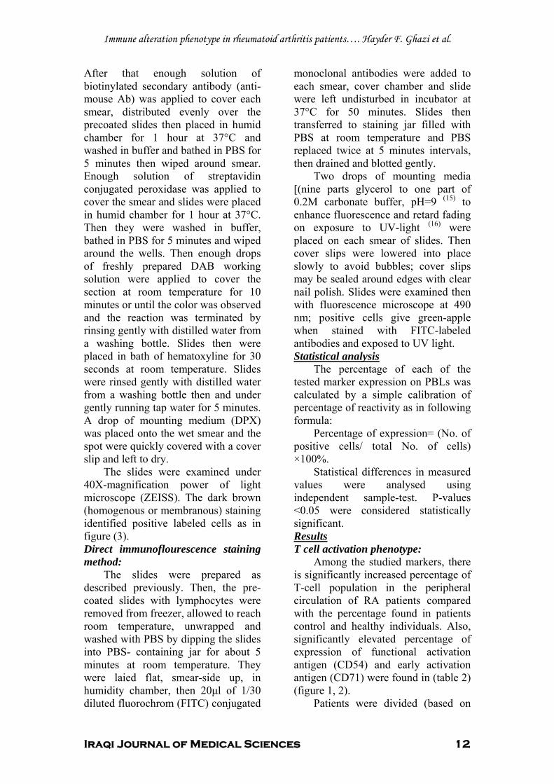
After that enough solution of biotinylated secondary antibody (anti-mouse Ab) was applied to cover each smear, distributed evenly over the precoated slides then placed in humid chamber for 1 hour at 37°C and washed in buffer and bathed in PBS for 5 minutes then wiped around smear. Enough solution of streptavidin conjugated peroxidase was applied to cover the smear and slides were placed in humid chamber for 1 hour at 37°C. Then they were washed in buffer, bathed in PBS for 5 minutes and wiped around the wells. Then enough drops of freshly prepared DAB working solution were applied to cover the section at room temperature for 10 minutes or until the color was observed and the reaction was terminated by rinsing gently with distilled water from a washing bottle. Slides then were placed in bath of hematoxyline for 30 seconds at room temperature. Slides were rinsed gently with distilled water from a washing bottle then and under gently running tap water for 5 minutes. A drop of mounting medium (DPX) was placed onto the wet smear and the spot were quickly covered with a cover slip and left to dry.
The slides were examined under 40X-magnification power of light microscope (ZEISS). The dark brown (homogenous or membranous) staining identified positive labeled cells as in figure (3). Direct immunoflourescence staining method:
The slides were prepared as described previously. Then, the pre-coated slides with lymphocytes were removed from freezer, allowed to reach room temperature, unwrapped and washed with PBS by dipping the slides into PBS- containing jar for about 5 minutes at room temperature. They were laied flat, smear-side up, in humidity chamber, then 20μl of 1/30 diluted fluorochrom (FITC) conjugated
monoclonal antibodies were added to each smear, cover chamber and slide were left undisturbed in incubator at 37°C for 50 minutes. Slides then transferred to staining jar filled with PBS at room temperature and PBS replaced twice at 5 minutes intervals, then drained and blotted gently.
Two drops of mounting media [(nine parts glycerol to one part of 0.2M carbonate buffer, pH=9 (15) to enhance fluorescence and retard fading on exposure to UV-light (16) were placed on each smear of slides. Then cover slips were lowered into place slowly to avoid bubbles; cover slips may be sealed around edges with clear nail polish. Slides were examined then with fluorescence microscope at 490 nm; positive cells give green-apple when stained with FITC-labeled antibodies and exposed to UV light. Statistical analysis
The percentage of each of the tested marker expression on PBLs was calculated by a simple calibration of percentage of reactivity as in following formula:
Percentage of expression= (No. of positive cells/ total No. of cells) ×100%.
Statistical differences in measured values were analysed using independent sample-test. P-values <0.05 were considered statistically significant. Results T cell activation phenotype:
Among the studied markers, there is significantly increased percentage of T-cell population in the peripheral circulation of RA patients compared with the percentage found in patients control and healthy individuals. Also, significantly elevated percentage of expression of functional activation antigen (CD54) and early activation antigen (CD71) were found in (table 2) (figure 1, 2).
Patients were divided (based on

Immune alteration phenotype in rheumatoid arthritis patients…. Hayder F. Ghazi et al.
Iraqi Journal of Medical Sciences 13
modified DAS28-3) in to two groups: high disease activity group (37 patients) and minimum disease activity group (9 patients). There was no statistical significant difference in the mean percentage of cells that express CD3. While, CD54 showed no statistical significant difference with lower percentage in active disease group and CD71 showed statistically
significant difference with higher expression in minimum disease activity group (table 3). Lack of influence of PBLs alteration phenotype with clinical and laboratory indices:
Our results showed no statistical correlation between studied markers and different clinical and laboratory parameters for disease activity (Table 4).
Figure 1: percentage of expression of PBLs markers in different study groups.
Figure 2: Percentage of expression of PBLs markers in RA patients subgroups.

Immune alteration phenotype in rheumatoid arthritis patients…. Hayder F. Ghazi et al.
Iraqi Journal of Medical Sciences 14
Table 1: patients and control characterstics. data are presented as means (SE).
Controls Osteo-arthritis group RA patients
RA patients High disease
activity group
Minimum disease
activity group Women/men 9/1 6/1 42/4 34/3 8/1
Age/years 48.6(10) 45.76 (7.6) 47.67(12.09) 48.06(11.96) 46.45(12.97) Disease duration
(months) ---- 88.61(72.88) 92.34(68.28) 76.73(92.67)
ESR (mm/1st h 12.50(3.31) 45.6 (12.5) 67.43(20.26) 70.94(19.54) 53(17.33) CRP (mg/l) 10.20(15.24) 22.4 (16.5) 43.956(55.078) 49.78(59.53) 20(17.75)
Tender joints ----- 10.58(5.42) 12.54(4.62) 4.77(3.19) Swollen joints ----- 7.35(4.52) 8.63(4.36) 3.66(1.80)
DAS-28(3) ----- 5.77(0.83) 6.11(0.63) 4.844(0.24) RF sero-positive
(No. (%)) 2(21.4%) 1 (16.6%) 34 (73.9%) 27(72.9%) 6(63.54%)
Duration of morning stiffness
(minutes) ----- 76.41(41.30) 84(41.72) 52.27(30.28)
ESR=erythrocytes sedimentation rate, CRP= C reactive protein, DAS= disease activity score, RF=rheumatoid factor.
Table 2: descriptive statistics (mean±S.E.) of PBLs markers in different study groups, comparison was done using ANOVA test.
healthy group OA group RA group ANOVA test CD3 72.042±1.52 74.3±0.99 79.213±1.4 <0.001 ** CD54 3.7±2 9.142±4.59 44.554±13 <0.001 ** CD71 2.3±1.82 4.571±1.98 25.534±9.59 <0.001 ** **: highly statistical significant difference at the level of p<0.001.
Table 3: Descriptive statistics (mean±S.E.) of PBLs markers in different RA subgroups, comparison was done using independent sample t-test.
Active Moderate Sig. 2 tailed CD3 79.242±1.39 79.061±1.66 0.761NS CD54 43.67±12.36 48.85±15.12 0.336 NS CD71 29.198±9.08 32.885±9.7 0.039* NS: no statistical significant difference. *: statistical significant difference at the level of p<0.05.
Immune alteration phenotype in rheumatoid arthritis patients…. Hayder F. Ghazi et al.
Iraqi Journal of Medical Sciences 15
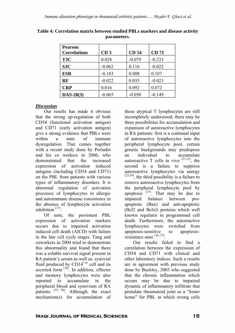
Table 4: Correlation matrix between studied PBLs markers and disease activity parameters.
Pearson Correlations CD 3 CD 54 CD 71 TJC 0.028 -0.079 -0.233 SJC -0.062 0.116 -0.022 ESR -0.183 0.008 0.107 RF -0.022 0.035 -0.023 CRP 0.016 0.092 0.072 DAS-28(3) -0.065 -0.050 -0.149
Discussion
Our results has made it obvious that the strong up-regulation of both CD54 (functional activation antigen) and CD71 (early activation antigen) give a strong evidence that PBLs were within a state of immune dysregulation. That comes together with a recent study done by Poriadin and his co workers in 2006; who demonstrated that the increased expression of activation induced antigens (including CD54 and CD71) on the PBL from patients with various types of inflammatory disorders. It is abnormal regulation of activation processes of lymphocytes in allergic and autoimmune disease consistence in the absence of lymphocyte activation inhibition (17).
Of note, the persistent PBL expression of activation markers occurs due to impaired activation induced cell death (AICD) with failure in the late cell cycle stages. Tang and coworkers in 2004 tried to demonstrate this abnormality and found that there was a soluble survival signal present in RA patient’s serum as well as, synovial fluid produced by CD14+ve cell and its secreted form (18). In addition, effecter and memory lymphocytes were also reported to accumulate in the peripheral blood and synovium of RA patients (19, 20). Although, the exact mechanism(s) for accumulation of
those atypical T lymphocytes are still incompletely understood; there may be three possibilities for accumulation and expansion of autoreactive lymphocytes in RA patients: first is a continual input of autoreactive lymphocytes into the peripheral lymphocyte pool, certain genetic backgrounds may predispose an individual to accumulate autoreactive T cells in vivo (21,22), the second is a failure to suppress autoreactive lymphocytes via anergy (23,24), the third possibility is a failure to remove autoreactive lymphocytes from the peripheral lymphocyte pool by apoptosis (25). That may be due to impaired balance between pro-apoptotic (Bax) and anti-apoptotic (Bcl2 and Bclxl) proteins which well known regulator in programmed cell death. Furthermore, the autoreactive lymphocytes were switched from apoptosis-sensitive to apoptosis-resistance state (26, 27).
Our results failed to find a correlation between the expression of CD54 and CD71 with clinical and other laboratory indices. Such a results are in agreement with previous study done by Buckley, 2003 who suggested that the chronic inflammation which occurs may be due to impaired dynamic of inflammatory infiltrate that postulate rheumatoid joint as a “foster home” for PBL in which wrong cells

Immune alteration phenotype in rheumatoid arthritis patients…. Hayder F. Ghazi et al.
Iraqi Journal of Medical Sciences 16
(PBL) accumulate in the wrong place (joints) at the wrong time (during the resolution of inflammation) leading to improper retention and survival (28).
In summary, we have confirmed and illuminated some of the T-cell differentiation defects in RA. Our data reinforce the importance of early and aggressive therapy for RA. Although T-cell abnormalities may predate clinical signs and symptoms, they appear to be perpetuated by inflammation. Therefore, the control of inflammation, particularly through the use of cytokine blockade, should minimize dysregulation of proliferation. References 1. Weyand CM and Goronzy J J Pathomechanisms in rheumatoid arthritis time for a string theory? J. Clin. Invest. 2006; 116:869–871. 2. Firestein GS and Zvaifler N J. How important are T cells in chronic rheumatoid synovitis? II. T cell-independent mechanisms from beginning to end. Arthritis Rheum. 2002; 46: 298-308. 3. Skapenko A, Lipesky P and Schulze-Koops H. T cell activation as starter and motor of rheumatic inflammation. Curr Top Microbiol Immunol. 2006; 305:195-211. 4. Roy J, Audette M and Tremblayi M J. Intercellular Adhesion Molecule-1 (ICAM-1) Gene Expression in Human T Cells Is Regulated by Phosphotyrosyl Phosphatase Activity. J of boil chem. 2001; 276: 14553–14561. 5. Dustin M L, Rothlein R, Bhan AK, Dinarello CA and Springer TA. Induction by IL 1 and interferon-gamma, tissue distribution, biochemistry, and function of a natural adherence molecule (ICAM-1). J. Immunol. 1986; 137:245-254. 6. Dustin M L, Singer KH, Tuck DT and Springer TA. Adhesion of T lymphoblasts to epidermal keratinocytes is regulated by interferon gamma and is mediated by intercellular adhesion molecule-1 (ICAM-1). J. Exp. Med. 1988; 167:1323-1340. 7. Buckle A M, and N Hogg. Human memory T cells express intercellular adhesion molecule-1 which can be increased by interleukin 2 and interferon-gamma. Eur. J. Immunol.1990; 20:337-341. 8. Cano E, Pizarro A, Redondo JM, Sa´nchez-Madrid F, Bernabeu C and Fresno M. Induction of T cell activation by monoclonal
antibodies specific for the transferrin receptor. Eur. J. Immunol. 1990; 20:765. 9. Salmeron A, Borroto A, Fresno M, Crumpton MJ, Ley SC and Alarco´n B. Transferrin receptor induces tyrosine phosphorylation in T cells and is physically associated with the TCR ζ-chain. J. Immunol. 1995; 154:1675. 10. Frank R and Hargreaves R. Clinical biomarker in drug discovery and development. Nat Rev Drug Discov. 2003; 2: 566-580. 11. Tsuang MT, Nossova N, Yager T, Tsuang, MM, Guo SC, Shyu KG and Liew CC. Assessing the validity of blood-based gene expression profiles for the classification of schizophrenia and bipolar disorder: A preliminary report. Am J Med Genet B Neuropsychiatr Genet. 2005; 133:1-5. 12. Arnett FC, Edworthy SM, Bloch DA, McShane DJ, Fries JF, Cooper NS,rt al. The American Rheumatism Association 1987 revised criteria for the classification of rheumatoid arthritis. Arthritis Rheum. 1988; 31: 315-24. 13. Fransen PL and van Riel CM. The Disease Activity Score and the EULAR response criteria Clin Exp Rheumatol. 2005; 23:93-99. 14. Boyum A. lymphocyte seperation, Scan J Clin Lab Invest.1968; 21(97). 15. Batty I. standardization of reagents and methodology in immunology. In Dorothy JF, cathrine Sheehan (eds): principles and laboratory diagnosis, clinical immunology, 1986; P 190-202, Lippincott. 16. Narin RC. Standardization in immunofluorescence. Clin Exp Immunol; 1968; 3:465. 17. Poridian GV, Salmasi ZM and Kasimizkii AN. Activation markers of lymphocytes as indicator of immune system dysregulation in inflammation. Patol Fiziol Eksp Ter. 2006; 1: 2-7. 18. Tang X, Yocum DE, Dejonghe D, Nordensson K, Lake DF and Richard J. Increase activation-induced cell death in peripheral lymphocytes of rheumatoid arthritis patients: the mechanism of action. J Immunol. 2004; 112: 496-505. 19. Thomas ML. The regulation of B- and T-lymphocyte activation by the transmembrane protein tyrosine phosphatase CD45. Curr Opin in Cell Biol. 1994; 6: 247 – 252. 20. Kohem CL, Brezinschek RI, Wisbey H, Tortorella C, Lipsky PE and Oppenheimer-Marks, N. Enrichment of differentiated CD45RBdim, CD27– memory T cells inthe peripheral blood, synovial fluid, and synovial tissue of patients with rheumatoid arthritis. Arthritis Rheum.1996;39:844–54.

Immune alteration phenotype in rheumatoid arthritis patients…. Hayder F. Ghazi et al.
Iraqi Journal of Medical Sciences 17
21. Kohsaka, H., Nanki, T., Ollier, W.E., Miyasaka, N. and Carson, D.A. (1996) Influence of the rheumatoid arthritis-associated shared epitope on T-cell receptor repertoire formation. Proc Assoc Am Phys. 108:323–8. 22. Griffiths, M.M., Wang, J. and Joe, B. (2000) Identification of four new quantitative trait loci regulating arthritis severity and one new quantitative trait locus regulating autoantibody production in rats with collagen-induced arthritis. Arthritis Rheum. 43:1278–89. 23. Taams, L.S. and Wauben, M.H. (2000) Anergic T cells as active regulators of the immune response. Hum Immunol. 61:633–9. 24. Snijders, A., Elferink, D.G. and Geluk, A. (2001) An HLA-DRB 1- derived peptide associated with protectionagain st rheumatoid arthritis is naturally processed by human APCs. J Immunol 166:4987–93. 25. Schrimer, M., Vallejo, A.N., Weyand, C.M. and Goronzy, J.J. (1998) Persistence apoptosis and elevated expression of Bcl2 in clonally expanded CD4+ CD28- T cells from rheumatoid arthritis patients. J. Immunol. 161: 1018-1025. 26. Wells, A.D., Li, X.C. Li, Y. (1999) Requirement for T-cell apoptosis in the induction of peripheral transplantation tolerance. Nat Med. 5:1303–7. 27. Ghazi, H.F., Abdulmohymen N., and A. H. Ahmad. (2008): Immunocytochemical detection of some apoptosis regulating proteins (P53 and Bcl-2) in Peripheral Blood Lymphocytes of Rheumatoid arthritis patients. Iraqi J of Med Sciences. 6 (1): 89-98. 28. Buckley, C.D. (2003). Why do leukocytes accumulate within chronically inflamed joints? Rheum. 42: 1433-1444.

Iraqi Journal of Medical Sciences 18
Determination some of complement components in infertility women with antisperm antibodies.
Batool Mutar Mahdi 1MBChB; MSc; FICM , Wafaa Hazim Salih1 BSc; MSc , Bassma
Maki 1 BSc , Annie Edmond Caitano 2MBChB, Dina Sami Ibrahim2BSc .
Abstract Background: Classical activation of complement by antigen and antibody complex leads to formation of membrane attack complex (MAC) that leads to formation holes on the spermatozoa ending in their destruction. Objective: To determine the complements levels and antisperm antibodies in the sera of infertile women of unknown etiology. Patients and methods: Study group consisted of 45 infertile women consulting Kammal El-Sammarei Hospital for Infertility and In Vitro Fertilization from Jun -2008 to June-2009. Twenty-four (53.3%) patients had primary infertility and the rest had secondary infertility. Control group: consisted of thirty fertile women. Blood samples were collected from them and anti sperm antibodies in the serum were detected by indirect immunofluorescence test (EURO IMMUNE –GERMENY). In addition to that serum were tested for complement levels (C3 and C4) using single
radial immune diffusions test (BINDARID) KIT BIRMINGHAM .UK. Results: Detections of antisperm antibodies in the serum of infertile women were (64.4%) which is significantly (p<0.05) higher from control group using indirect immunofluorescence test. There was a significant (p=0.000) difference in the complements levels among infertile women who had ASA positive and ASA negative and control group. Conclusions: These higher levels of complement components may be due to activation of classical pathway by ASA that directed against sperm antigens ending in defect in function and motility of the sperms. Key words: Infertility, antisperm antibody, complement. IRAQI J MED SCI, 2010; VOL.8 (2):18-23
Introduction
Complement (C) system is an enzymatic cascade of proteins that forms a vital part of the innate immune system and the end products of complement activation is pores formation by membrane attack complex (1). They present in low concentrations in the serum and once it was activated by any pathways (classical, alternative and Lectin), its levels were increased (2). The presence of antisperm antibodies in the reproductive tracts of some infertile individuals, and presence of complement in cervical and ovarian 1Dept. Microbiology, Al-Kindi College of Medicine, Baghdad University, 2 Central Public Health Laboratories Baghdad. Address Correspondence to: Dr. Batool Mutar Mahdi E- mail: [email protected] Received: 9th September 2009, Accepted: 21st February 2010.
follicular fluid, suggests that complement-mediated damage of spermatozoa is involved in some cases of infertility. Furthermore, deposition of maternal IgG and complement in the extra fetal tissues indicates that complement activation occurs within the fetoplacental unit (3). Complement and its regulation is important in reproduction, Donev etal 2008 reported CD59b was significantly expressed only in testis and played a role in sperm acrosome activation and motility (4). There was no evidence of antibody or complement fixation by viable spermatozoa. It had been found that antibodies present in the serum of women that bind to nonviable spermatozoa(ASA) belong to the IgG and IgM class then Complement fixation occurred via the classical (antibody-mediated) and alternative pathway. This indicated that viable

Complement component in the infertility women….. Batool Mutar Mahdi et al
Iraqi Journal of Medical Sciences 19
spermatozoa may possess antigenic properties different from nonviable spermatozoa. This leads to lack of immunological reaction of women to viable spermatozoa (5). Anti sperm antibodies could inactivate human sperm motility in the presence of complement, showing that complement-dependent inactivation of sperm motility might be the biological mechanism of female infertility(6), because incubation of motile sperm with complement-fixing immune sera resulted in a significant loss (43-87%) of motility, then activation of (C5b-9) induced alterations in sperm morphology leading to sperm lyses(7).
In this study, we tried to determine the presence of ASA in the sera of infertile women and measure the main complement components (C3 and C4) in the sera of same patients. Patients and methods
Patients group: consisted of 45 infertile women consulting Kammal El-Sammarei Hospital for Infertility and In Vitro Fertilization from Jun -2008 to June-2009. The exclusion criteria was women with congenital abnormalities in the uterus, tubes and ovaries , women who ages were more than 45 and less than 20 years , women with defect in ovulation, hormonal disturbances and tube occlusion were excluded. Thus, study group included only women with unknown cause of infertility.
Control group: consisted of thirty healthy fertile women.
Blood was collected from two groups and anti sperm antibodies (ASA) were detected in their serum by indirect immunofluorescence test using kit from EURO IMMUNE –GERMENY. The sites (head, neck and tail) where ASAs directed were also recorded.
Anti-nuclear antibodies were done by indirect immunofluorescence test using kit from EURO IMMUNE –
GERMENY for those who had positive antisperm antibodies to sperm head to get rid from cross-reactions.
Serum of both groups were tested for complement levels (C3 and C4) using single radial immune diffusion test (BINDARID) KIT BIRMINGHAM .UK. These tests were done in Immunological department- central public Health.
The study was approved by the Ethical Committee of the Al-Kindi College of Medicine- Baghdad University, Kammal El-Sammarei Hospital for Infertility and Central Public Health. All samples were obtained with informed consent in accordance with Kammal El-Sammarei Hospital for Infertility Declaration. This study was carried out with the approval of the Ministry of Health and District Health Authority Ethical Committee in Baghdad-Al-Resaffa. Statistical analysis Student's t-test and ANOVA test used in analysis data statistically by MiniTab statistical software program 13.20. A P- value ≤ 0.05 was considered to be significant. Results
The patients group consisted from forty-five female patients, their ages ranged from (22-45 years), (median=33). They were complaining from infertility, Twenty-four (53.3%) patients had primary infertility, their ages ranged from (22-40 years) (median =29.9) and the rest (No. =21, 46.7%) had secondary infertility, their ages ranged from (24-45 years) (median =31). The control group their ages were ranged between (17-39 years), median= 30.6.
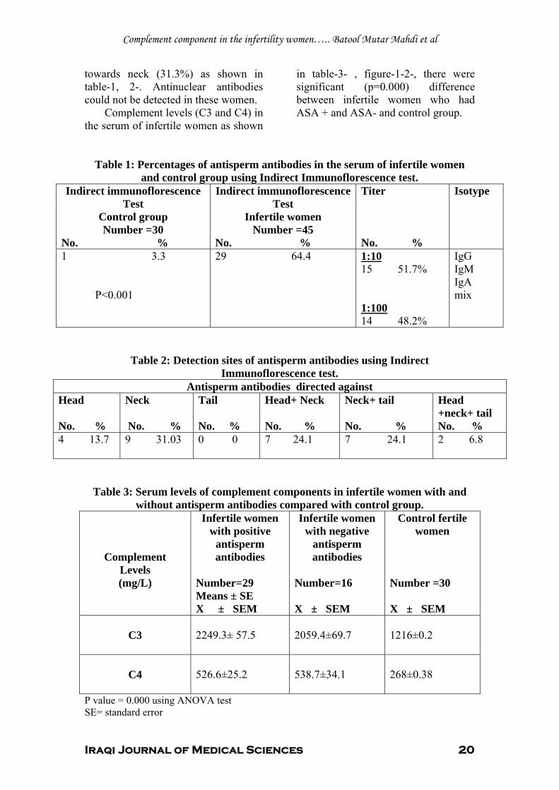
Detections of antisperm antibodies in the serum of infertile women were (64.4%) which is significantly (P<0.001) higher from control group (3.3%) using indirect immunofluorescence test. The highest percentage of antibodies was directed
Complement component in the infertility women….. Batool Mutar Mahdi et al
Iraqi Journal of Medical Sciences 20
towards neck (31.3%) as shown in table-1, 2-. Antinuclear antibodies could not be detected in these women.
Complement levels (C3 and C4) in the serum of infertile women as shown
in table-3- , figure-1-2-, there were significant (p=0.000) difference between infertile women who had ASA + and ASA- and control group.
Table 1: Percentages of antisperm antibodies in the serum of infertile women and control group using Indirect Immunoflorescence test.
Indirect immunoflorescence Test
Control group Number =30
No. %
Indirect immunoflorescence Test
Infertile women Number =45
No. %
Titer No. %
Isotype
1 3.3 P<0.001
29 64.4
1:10 15 51.7% 1:100 14 48.2%
IgG IgM IgA mix
Table 2: Detection sites of antisperm antibodies using Indirect Immunoflorescence test.
Antisperm antibodies directed against Head No. %
Neck No. %
Tail No. %
Head+ Neck No. %
Neck+ tail No. %
Head +neck+ tail No. %
4 13.7 9 31.03
0 0 7 24.1 7 24.1 2 6.8
Table 3: Serum levels of complement components in infertile women with and without antisperm antibodies compared with control group.
Complement Levels (mg/L)
Infertile women with positive
antisperm antibodies
Number=29 Means ± SE X ± SEM
Infertile women with negative
antisperm antibodies
Number=16 X ± SEM
Control fertile women
Number =30 X ± SEM
C3
2249.3± 57.5
2059.4±69.7
1216±0.2
C4
526.6±25.2
538.7±34.1
268±0.38
P value = 0.000 using ANOVA test SE= standard error

Complement component in the infertility women….. Batool Mutar Mahdi et al
Iraqi Journal of Medical Sciences 21
C3
AS
A p
C3A
SA
Ne
C3
cont
r
1200
1700
2200
2700
Boxplots of C3 ASA p - C3 contr(means are indicated by solid circles)
Figure 1: Serum levels of complement components C3 in infertile women with
and without antisperm antibodies compared with control group
C4
cont
o
C4A
SA
Ne
C4
AS
A p
750
650
550
450
350
250
Boxplots of C4 ASA p - C4 conto(means are indicated by solid circles)
Figure 2: Serum levels of complement components C4 in infertile women with
and without antisperm antibodies compared with control group
Discussion Spermatozoa are cells that must
survive transplantation into a foreign host in order to perform their physiological role to reach the oocyte and penetrate it. The biggest hurdle to overcome is innate immune defense that will target the invaders in the female genital tract.
The human immune system is trained during the early postnatal period. In women when become sexually active, their immune system will inevitably contact sperm antigens
after coitus. Therefore, once sperm, as an autoantigen, activates the human immune system, an autoimmune response against human sperm will occur and leads to formation of ASA against sperm antigens. This leads to complement activation and complement is a major player in innate immunity. Spermatozoa must therefore evade complement attack if they are wanted to reach their goal.
In order to complement activation needs antibodies and the antibodies in

Complement component in the infertility women….. Batool Mutar Mahdi et al
Iraqi Journal of Medical Sciences 22
this study were ASA and were detected in higher percentages in the infertile women 64.4% that is in agreement with other study (8). The highest percentage was directed against neck of the sperm. Those ASA that directed against head will affect penetration of ova and ASA directed against tail will affect movement of the sperms (9).These directed against neck when there were complement activation will lead to pores formation and damage the sperms especially when the isotype was IgG because IgM produced for only short period about two weeks (10). We found in this study a higher titer of ASA (1:100) in 48.2% of infertile women. IgG ASAs were capable of activating complement and depositing MC5b-9 on human sperm. Meanwhile, the concomitant detection of sperm-bound IgG and the initial (C3d) and terminal (C5b-9) complement components on the surface of human sperm could be confirmed using a flow cytometric assay (11). In addition to that, the deposition of activated C3 fragments, the assembly of terminal membrane attack complexes (C5b-9) and oxygen radicals could lead to C3-mediated sperm binding to neutrophils or C5b-9-mediated sperm-motility loss (12). This show the way to complement activation; we found significant higher levels of C3 and C4 (main components of complements) in the serum of infertile women with ASA. Complement evasion is achieved by the presence of complement regulators both in seminal plasma and on the spermatozoa (13). Women who have generated an anti-sperm antibody (ASA) response may be particularly at risk because C activation will be enhanced with subsequent spermatozoal damage and destruction and perhaps also inflammatory damage to the female reproductive tract (14,15)
and this was in agreement with our results. As a result, impairments of
complement components might predispose women to infections and autoimmune diseases that affect fertility (16, 17).
The message of this study is that C and C regulation play important though poorly defined roles in several components. Defects in C regulation may contribute to infertility and manipulation of C at this site may be of benefit either for improving fertility or for contraception. References 1. Kindt TJ, Goldsby RA and Osborne BA. Kuby Immunology. Sixth edition. WH Freeman and Company. New York .USA. 2007. Pp: 168-185. 2. Delves PJ, Martin SJ, Burton DR and Roitt IM. Essential immunology .11th edition. Blackwell publishing .UK. 2008. Pp: 21-36. 3. Rooney IA, Oglesby TJ and AtkinsonJP. Complement in human reproduction: activation and control.Immun Rese.1993; 12:267-294. 4. Donev RM, Sivasankar B, Mizuno M and Morgan BP.The mouse complement regulator CD59b is significantly expressed only in testis and plays roles in sperm acrosome activation and motility. Mol Immunol.2008; 45:534-542. 5. Vogelpoel FR, teVelde RE, Scheenjes E, Van Kooy R, Kremer J and Verhoef J. Antibody and complement-binding activity of viable and nonviable human spermatozoa. Arch Andro. 1987; 18:189-197. 6. D'Cruz OJ, Toth CA, Haas GG Jr. Recombinant soluble human complement receptor type 1 inhibits antisperm antibody- and neutrophil-mediated injury to human sperm. Biol. Reprod. 1996; 54, 1217-1228 7. D'Cruz OJ, Haas GG Jr, Wang BL, DeBault LE. Activation of human complement by IgG antisperm antibody and the demonstration of C3 and C5b-9-mediated immune injury to human sperm. J. Immunol. 1991; 146, 611-620. 8. Kapoor A, Talib VH and Verma SK.Immunological assessment of infertility by estimation of antisperm antibodies in infertile couples. Ind J Path Microbiol.1999; 42:37-43. 9. Jin-Chun Lu, Feng Huang Yu and Nian-Qing Lu. Antisperm Immunity and Infertility. Expert Rev Clin Immuno. 2008; 4:113-126. 10. Lu J-C, Huang Y_F and Lu N-Q. Antisperm immunity and infertility Exp Rev Clin Immuno.2008; 4:113-126.

Complement component in the infertility women….. Batool Mutar Mahdi et al
Iraqi Journal of Medical Sciences 23
11. D'Cruz OJ, Haas GG Jr. Lack of complement activation in the seminal plasma of men with antisperm antibodies associated in vivo on their sperm. Am. J. Reprod. Immunol. 1990; 24: 51-57. 12. D'Cruz OJ, Toth CA, Haas GG Jr. Recombinant soluble human complement receptor type 1 inhibits antisperm antibody- and neutrophil-mediated injury to human sperm. Biol. Reprod. .1996; 54: 1217-1228. 13. Harris CL, Mizuno M and Morgan BP. Complement and complement regulators in the male reproductive system. Molecular Immunology. 2006; 43: 57–67. 14. Troedsson M, Liu IK and Crabo B. Sperm transport and survival in the mare: a review. Theriogenology.1998; 50:807–818. 15. Troedsson MH, Loset K, Alghamdi AM, Dahms B and Crabo BG. Interaction between equine semen and the endometrium: the inflammatory response to semen. Anim. Reprod. Sci.2001; 68: 273–278. 16. Wen L, Atkinson JP and Giclas PC.Clinical and Laboratory evaluations of complement difficiency.J Allergy Clin Immunolo.2004; 51:336-340. 17. Boackle SA and Holers VM. Role of complement in the development of autoimmunity.Curr Dir Autoimmune.2003; 6:154-168.

Iraqi Journal of Medical Sciences 24
CD14 and Bladder Cancer: is there any correlation.
Nidhal Abdulmohymen1 PhD, Zainab Ashoor2 PhD, Amera Khodher 1PhD Abstract Background: Epithelial cells have evolved a variety of cell-and tissue-specific mechanisms for bacterial detection to enable cells to modulate the inflammatory response depending on the particular situation in a specific organ. These mechanisms provide a means of maintaining a proper balance between defense, tissue injury and their combined effects on organ function, and the moleculeCD14may have a role to play. Objective: to find any correlation between CD14 marker expression and bladder cancer. Material and methods: The immunoexpression of CD14 in paraffin sections from 96- bladder cancer tissues and 36-bladder tissues from patients with other bladder disease rather than cancer was investigated using immunohistochemical assay (IHC). The patients were divided into three groups: Group-1: Newly diagnosed bladder cancer patients, 69(43.9%), Group-2:Post-chemotherapy patients, 27(17.2%), Group-3: Other bladder disorders rather than bladder cancer 36(22.9%). The final diagnosis of patients with bladder cancer was established by clinical examination confirmed by cystoscopy and histopathological examination for bladder tissue specimens. Urinary tract infections were studied for all groups by culturing urine samples using specific culture media.
Results: The results showed that CD14 protein was over expressed in 68.57% of the patients with approximately equal frequent IHC score among patients(23.8%) for each of weak and intense immunoreactions, and (21.0%) for moderate one , and there was no significant difference in the scores of positive IHC CD14 expression in bladder tissue of the cancer patients when compared with non-cancer patients, but there was significant difference between cancerous patients in correlation to the tumor grades. Positive urine cultures were detected in 28(40.6%) of group-1, 13(48.1%) of group-2, and 12(33.3%) of group-3, while all healthy subjects were free of infection, and a significant difference between bacterial infected patients with and without bladder cancer, in which there was highly CD14 immunoexpression in bladder tissue in Gram-negative bacterial infected patients . Conclusion: CD14 expression correlated significantly with Gram-negative bacterial infection, but not with cancer. Key words: Immunohistochemistry, CD14, bladder cancer IRAQI J MED SCI, 2010; VOL.8 (2): 24-30
Introduction CD14, is an LPS receptor (1), and
plays an important role in the signal transduction causing endotoxic shock and is anchored to the monocyte / macrophage cell membrane via
1Dept. Medical Microbiology, College of Medicine, Al-Nahrain University, 2 Dept. Medical Microbiology, College of Medicine, Almustansiriya. Address Correspondence to: Dr. Nidhal Abdul-Mohymen. E- mail: [email protected] Received: 3th December 2008, Accepted: 12th October 2009.
glycosylophatidyl inositol (2, 3). Recent reports have described the
presence of mCD14 in epithelial cells, endothelial cells, and fibroblasts (4-6). Bladder epithelial cells express CD14 on their surfaces. This fact was evaluated by several investigators. The assessment of CD14 expression was based on using different bladder carcinoma cell lines that were analyzed by flow cytometry using the well-characterized anti-CD14 monoclonal My4 antibodies (7). They improved that

CD14 and Bladder Cancer…. Nidhal Abdulmohymen et al.
Iraqi Journal of Medical Sciences 25
the bladder carcinoma epithelial cells express mCD14
A soluble form of CD14 is secreted into the urine of superficial bladder cancer patients who receive intravesical BCG. The measurement of soluble urinary CD14 could be of prognostic significance for the response to immunotherapy (8).
In order to limit and control bacterial infections, the host relies on innate immune defense mechanisms, which initially engage epithelial cells of the mucosal surfaces. These cells are the first to encounter luminally localized bacteria and, they have evolved a variety of cell-and tissue-specific mechanisms for bacterial detection (9). The early recognition of pathogens by cells of the innate immune system is critical to the survival of the host. Attachment of bacteria to epithelial target cells as well as the action of a secreted toxin triggers mucosal chemokine responses in the urinary tract (10). However, the predominant mechanism prevents bacteria from gaining access to the sensitive upper urinary tract does not require the presence of bacteria per user; it rather recognizes the presence of bacterial LPS (11). For the above mentioned data, we intend to investigate, is there any correlation between bladder cancer and CD14 expression. Materials and methods
Subjects: patients groups which enrolled Tissue samples from cases of bladder cancer patients as group 1 (96 newly diagnosed cases and 16 cases who had received chemotherapy), and 10 cases of chronic non-specific cystitis, 10 cases of mild non-specific cystitis, 3 cases of chronic bilharizial cystitis, 7 cases of dysplastic urothelium
and 6 normal urothelium as group 2 which represent patients with diseases other than cancer and this group represent group 2.
Normal urothelium samples were taken from patients who did not suffer from bladder carcinoma in the past and had a macroscopically normal bladder mucosa during cystoscopy, and they were taken as control (group3). Tissue were fixed in 10 % buffered formalin and embedded in paraffin wax, were stained with hematoxylin-eosin.
Tumor grade was characterized by 2 independent pathologists
Voided urine samples were collected before cystoscopy or surgery for patients of groups 1-3 and for healthy subjects, as aseptically as possible, in sterile containers. The collected mid-stream specimens were transported to the laboratory within 30 minutes of the collection and cultured on a specific media for bacterial isolation. Immunohistochemical Detection of CD14 Proteins Expression in Paraffin Embedded Sections: According to procedure mentioned by Celis et al., 2005 (12).
The antibodies used were: Biotinylated Link: which is F (ab') rabbit anti-mouse IgG HRP-STAR13B (Serotec, UK), and Sheep anti-rabbit IgG: HRP-STAR54 (Serotec, UK), or secondary Anti-mouse Antibody conjugated with peroxidase enzyme. (Sigma).Mouse anti-human CD14 (Serotec,UK) (MCA596F): Batch No.0798.
Slides were examined and stained cells were counted with the assistance of an experienced histopathologist by light microscope at X400 magnification .Immunostaining was scored according to cut-off value. This cut-off for

CD14 and Bladder Cancer…. Nidhal Abdulmohymen et al.
Iraqi Journal of Medical Sciences 26
positivity was 10% positive cells for CD14 (7).Quantitative IHC scoring was evaluated by counting the number of positive and negative cell nuclei in several randomly selected fields in each section .Tumor reactivity was expressed as the marker percent (i.e., the number of stained tumor cells per 1000 cells in each section).More than 1000 cells evaluated under 40 X high power field and the percentage of positive cells was calculated as follows:
For CD14, the intensity of positivity scored as (7): a) 0: No reaction. b) 5-10 %: Weak reaction. c) 10-25 %: Moderate reaction. d) 50-80 %: Intense reaction. Statistical analysis
Student's t test, the chi-square (x2) test. Correlation coefficient was used. Probability values of p<0.05, and p<0.01 were considered statistically significant. Results
CD14 protein was overexpressed in 72 patients ( 68.6% ) ,with approximately equal frequent IHC score among patients( 23.8% ) for weak and intense immunoreaction ,and ( 21.0% ) for moderate one . Table 1shows the frequency distribution of CD14 overexpression scores in group subjects.
Positive immunoreaction of CD14 was found in bladder tissue samples of 41 out of 55 newly diagnosed patients with bladder carcinoma, 10 bladder tissue samples of previously diagnosed bladder carcinoma and had received chemotherapy ,and in 21 bladder tissue samples of patients without bladder cancer, including 3 normal urothelium biopsies ,with intense immunostaining reaction being the most frequent score among patients of group-1,and group-2.while weak immunostaining reaction
observed in group-3 as the most frequent score.(Table 2) . Frequency Of CD14 In Patients With & Without Bladder cancer
The CD14 IHC results tissue samples taking together all patients with bladder cancer( Group-1 and Group-2 ) ,and considering patients without bladder cancer and harboring other urological diseases,were correlated to each other and summarized in table 3, in which 51 out of 71( 48.57% ) bladder cancer patients showed positive results, while 21 of non-cancerous cases, showed positive results(Figure 1A).Chi-Square test showed that there was no statistical difference between bladder cancer and other urological disease for CD14 IHC Scores in tissue sample taken from each case . Frequency of CD14 IHC Scores In Bladder Cancer Patients In Correlation To Tumor Grade Of TCC:
CD14 protein immunostaining was evaluated in 105 paraffin-embedded bladder tumor specimens. CD14 identified by positive anti-CD14 reaction which is demonstrated at the lower part of the panel; and the luminal endothelial cells.
Regarding the tumor grade of transitional cell carcinoma, CD14 was detected in 15 out of 25of grade-1, 15 out of 19 of grade-2, and 12 out of 13 of grade-3. There was no CD14 detected in bladder tumor with grade-4.
Chi-square was used to compare the results of frequency distribution of CD14 scores among tumor grades of TCC and it showed a significant correlation between each score and tumor grade ( P <0.05 ) , Figure 1 . Frequency of CD14 Overexpression In Relation To the Bacterial Infection:
Forty out of 105 patients presented with Gram-negative bacterial infection,

CD14 and Bladder Cancer…. Nidhal Abdulmohymen et al.
Iraqi Journal of Medical Sciences 27
in which immunohistochemistry staining of CD14 protein, showed that 33 patients were positive with intense reaction being the most frequent score among them (19.0%) (Table 4).
The results showed highly significant correlation (P<0.01) in CD14 expression with bacterial infection criterion.
Figure 1: The percentage of CD14 overexpression in bladder carcinoma patients in
relation to the tumor grade.
Table 1: Frequency distribution of CD14 Overexpression
CD14 Overexpression Patients ( % )Positive 72 ( 68.6 ) _ Weak reaction 25 ( 23.8 ) _ Moderate reaction 22 ( 21.0) _ Intense reaction 25 ( 23.8 ) Negative 33 ( 31.4 )
Total 105(100)

CD14 and Bladder Cancer…. Nidhal Abdulmohymen et al.
Iraqi Journal of Medical Sciences 28
Table 2: Frequency table of CD14 IHC scores in study groups . CD14 IHC Scores
Groups
Total Group-1 Group-2 Group-3
0 No. %of Total
14 (25.5)
6 (37.5)
14 (40.0)
34 (31.4)
5-10 % No. %of Total
11 (20.0)
3 (18.8)
11 (31.4)
25 (23.8)
10-25% No. %of Total
12 (21.8)
3 (18.8)
7 (20.0)
22 (21.0)
50-80 % No. %of Total
18 (32.7)
4 (25.0)
3 (8.6)
25 (23.8)
Total No. %of Total
55 (52.4)
16 (15.2)
35 (33.3)
106 (100.0)
0: Negative .5-10 % : Weak reaction .10-25 %: Moderate reaction .50-80 %: Intense reaction.
Table 3: Frequency of CD14 IHC scores in patients with & without bladder cancer.
CD14 IHC Scores
Type-CA
Total
Chi-square
Sig.
Cancer Non-Cancer 0 No. %of Total
20 (28.2)
13 (38.2)
33 (31.4)
7.092
0.071
5-10% No. %of Total
14 (19.7)
11 (32.4)
25 (23.8)
10-25% No. %of Total
15 (21.1)
7 (20.6)
22 (21.0)
50-80% No. %of Total
22 (31.0)
3 (8.8)
25 (23.8)
Total No.(%ofTotal) 71(67.6) 34(32.4) 105(100%)

CD14 and Bladder Cancer…. Nidhal Abdulmohymen et al.
Iraqi Journal of Medical Sciences 29
Table 4: The percentage of CD14 expression in relation to the bacterial infection
Infection
Total No growth Gram-
negative Gram-Positive
0.0% No. %of Total
24 (22.9)
7 (6.7)
2 (1.9)
33 31.4
5-10 % No. %of Total
21 (20.0)
4 (3.8)
0 (0.0)
25 23.8
10-25% No. %of Total
13 (12.4)
9 (8.6)
0 (0.0)
22 21.0
50-80 % No. %of Total
5 (4.8)
20 (19.0)
0 (0.0)
25 23.8
Total No. %of Total
63 (60.0)
40 (38.1)
2 (1.9)
105 100.0
Chi-square 31.567 Sig. <0.01* *
** Highly significant correlation. Discussion
We have used the bladder tissue biopsies to study IHC expression of CD14 in patients with and without bladder cancer.
In the present investigation, we have characterized the immunoexpression of CD14 in bladder tissue of both cancer and non-cancer patients, without significant difference, but its expression in cancer tissue was with significant correlation to the tumor grade. Variation in CD14 expression levels among individuals should correlate with variation in the ability to mount an inflammatory reaction. The factors that contribute to the variable expression level of mCD14 in bladder epithelial cell line, has not been identified (13).Variation in mCD14 expression levels among individuals should correlate with variation in the ability to mount an inflammatory reaction.
High mCD14 expression levels were found in grade-1 and grade-2 then
in grade-3, while the small size of grade-4 sample showed no CD14 expression .A possible explanation for the down-regulation is that during chronic inflammation, inhibitory feedback mechanisms would decrease the expression of CD14. An alternative explanation is that the down-regulation is a consequence of death and shed of the urothelial cells.
In our study high CD14 IHC expression levels were found in Gram-negative bacterial infected bladder tissue of both cancerous and non-cancerous patients, and it's expression was significantly correlated with this type of bacterial infection (data are not shown) .This is compatible with the fact that CD14, is a cell surface protein involved in LPS binding (1), in which LPS is the major constituent of gram-negative bacterial cell wall. It is one member of a group of molecules, called pathogen-associated molecular pattern molecules that are recognized by host
CD14 and Bladder Cancer…. Nidhal Abdulmohymen et al.
Iraqi Journal of Medical Sciences 30
tissue that express pattern recognition receptors. Recognition of microorganisms by this mechanism forms part of the primitive form of defense called innate immunity (14). LPS stimulated an overall increase in mCD14 but specifically induced mCD14 in low mCD14 expression cells (5).It has been previously reported that tissue-specific CD14 expression is regulated at the level of transcription, and an 80-kb genomic fragment containing the critical regulatory elements that enhance the tissue-specific expression of CD14 (15).LPS specifically up-regulated several genes included CD14 genes (16), furthermore, LPS significantly induced CD14 mRNA expression (17). References 1. Wright SD, Ramos RA, Tobias PS, Uleritch RJ,and Mathison JC. CD14, a receptor for complexes of lipopolysaccharide (LPS) and LPS binding protein.Science 1990; 249: 1431-1433. 2. Goyert SM, Ferrero E, Retting WJ, Yenamandra AK, Obata F, and Le Beau MM.The CD14 monocyte differentiation antigen maps to a region encoding growth factors and receptors. Science, 1988; 239:497-500. 3. Hazoit A, Chen S, Ferrero E, Low MG, Silber R, and Goyert SM.The monocyte differentiatio n antigen, CD14 is anchored to the cell membrane by a phosphatidylinositol linkage. J.Immunol.1988; 141:547-552. 4. Jersmann H P A, Hii C S T, Hodge GL, and Ferrante A. Synthesis and surface expression of CD14 by human endothelial cells.Infect.Immunol . 2001; 69:479-485. 5. Putnins E E,Sanaie AR ,Wu Q ,and Firth JD. Induction of keratinocyte growth factor-1 expression by lipopolysaccharide is regulated by CD14 and Toll-Like receptors 2 and 4.Infect.Immunol. 2002; 70:6541-6548. 6. Tamai R ,Sakuta T, Matsushita K, Torii M, Takeuchi O ,Akira S, Akashi S ,and Tanaka H. Human gingival CD14 fibroblast primed with gamma interferon increase production of interleukin-8 in response to lipopolysaccharide through up-regulation of membrane CD14 and
MyD88 mRNA expression .Infe.Immunol. 2002;70:1272-1278 7. Schilling J D ,Martin SM ,Hunstad DA ,Patel KP, Mulvey MA, Justice SS,et al.CD14-and Toll-like receptor dependent activation of bladder epithelial cells by lipopolysaccharide and Type 1 piliated Escherichia coli. 2003; 71(3):1470-1480. 8. Jackson A M ,Lien E, Alexandroff AB ,Prescott S, Espevik T, James K, et al. Soluble urinary CD14 after intravesical bacilli Calmette Guerin immunotherapy for carcinoma in situ.Br.J.U, 1997; 80(5):766-71. 9. Bäckhed F and Richter-Dahlfors A.B: Bacteria-induced innate immune responses at epithelial linings. In Intracellular Pathoens: Membrane Interactions and Vacuole Biogenesis.Grovel, J. (ed) .2002; 3(3):153-158. 10. Uhlen P, Lae Stadius A, Jahnukainen T, Soderblom T, Backhed F, Celis G. Alpha-heamolysin of Uropathogenic Escherichia coli induces Ca+oscillations in renal epithelial cells.Nature, 2000; 405:694 -697 . 11. Bäckhed F, Meijer L, Normark S, and Richter-Dahlfors A. A: TLR4-dependent recognition of Lipopolysaccharide by epithelial cells requires sCD14 .Cell. Microbiol. 2004; 4:493-501. 12. Celis JE, Moreira JM A, Gromova T, Cabezon T, Ralfkaier U, Guldberg P, et al. Towards discovery-driven translational research in breast cancer.FEBS Journal, 2005; 272:2-15 . 13. Shimazu R ,Akashi S, Ogata H, Nagai Y, Fukudome K, Miyake K, and Kimoto M.MD-2,a molecule that confers lipopolysaccharide responsiveness on Toll-Like receptor 4.J.Exp.Med., 1999; 189:1777-1782 . 14. Zhang G and Chosh S. Toll-like receptor-mediated NF-kB activation: a phylogenetically conserved paradigm in innate immunity. J. Clin. Investig. 2001; 107:13-19. 15. Christopher JH, Paul DK, Francesco C, Pu Zhang, Michael SR, James P, and Dong-Er Z.Characterization of human endotoxin Lipopolysaccharide receptor CD14 expression in transgenic mice. The Journal of Immunology .1999; 162:503-509. 16. Marcia RS, Ngoe-Bich N, Timothy GH, and Ricardo S.Gene expression profiling of mouse bladder inflammatory responses to LPS, substance P,and Antigen-stimulation. American Journal of Pathology. 2002; 160:2095-2110. 17. Chomarat P, Banchereau J, Davoust J, and Palucka AK. IL-6 switches the differentiation of monocytes from dendritic cells to macrophages. Nat. Immunol., 2000; 1:510-514.

Iraqi Journal of Medical Sciences 31
Oxidative and antioxidant status in Smoking men.
Shaymaa Zahraw Al-Saedi MSc.
Abstract Background: Free radicals in cigarette smoke may cause oxidative damage to macromolecules, contributing to cardiovascular diseases and cancer. Decreased plasma antioxidant concentrations in smokers may indicate cigarette smoke–related oxidative stress. Objective: We compared the effects on serum antioxidant concentrations in confirmed active smokers with those in nonsmokers, independent of differences in dietary intakes and other covariates. Methods: Serum samples from 60 smokers, and 40 nonsmokers aged 15-60 years were analyzed for ascorbic acid (vitamin C), α-tocopherol (vitamin E), and retinol (vitamin A), by using high performance liquid chromatography (HPLC). The measurement of serum lipid profile, and total lipid peroxidation, oxidized HDL (Ox-HDL) was done as well. Results: Showed significantly lower serum antioxidant vitamins (A, C & E) concentrations
in smokers more than in nonsmokers. Smokers had significant elevation in serum malondialdehyde (MDA) (p<0.001) and the percentage of oxidized non high-density lipoprotein (Ox. non HDL %) with a significant reduction in the percentage of oxidized high-density lipoprotein (Ox. HDL %) as compared to the control (p<0.001). Conclusions: These results indicate that cigarette smokers have a significantly lower serum antioxidant status than do unexposed nonsmokers, independent of differences in dietary antioxidant intakes with an increased oxidative stress in smokers' sera. Key Words: Oxidized HDL, ascorbic acid, α-tocopherol , retinol , cigarette smokers. IRAQI J MED SCI, 2010; VOL.8 (2): 31-37
Introduction
Oxidative stress is a condition in which the cellular production of reactive oxygen species (ROS) exceeds the physiological capacity of the antioxidant defense system to render ROS inactive (1). Increased production of ROS involves the oxidation of lipids and lipoproteins, DNA, proteins and other molecules in ways that impair normal cellular function, possibly resulting in impaired health and disease (2). Normal cellular metabolism results in the production of ROS; however, both physical and environmental stressors can
Dept. Chemistry and Biochemistry College of Medicine, Al-Nahrain. Address Correspondence to: Shaymaa Zahraw Al-Saedi. E- mail: [email protected] Received: 11th November 2009, Accepted: 17th February 2010.
further increase ROS production. In this regard, two primary environmental stressors include cigarette smoking and high fat meals (1).
Cigarette smoking exacerbates ROS formation and poses a significant oxidant stress in vivo (3). In one puff of a cigarette, a smoker is exposed to more than 1015 free radicals in the gas phase alone (4), with additional exposure in the tar phase equal to more than 1017 free radicals per gram. It has been consistently reported that cigarette smokers have elevated biomarkers of oxidative stress compared with non-smokers and this represents a potential mechanistic link between regular cigarette smoking and cardio vascular disease (CVD).
The increased oxidative stress observed in smokers may be partly due to the lower blood antioxidant capacity

Oxidative and antioxidant in Smokers….Shaymaa Zahraw Al-Saedi
Iraqi Journal of Medical Sciences 32
routinely observed in smokers (5). It is possible that the addition of other ROS generators can further promote oxidative stress in cigarette smokers. To our knowledge, no investigation to date has studied the combined effects of cigarette smoking and oxidative stress biomarkers. Therefore, in the present investigation we compared blood antioxidant status and oxidative stress biomarkers in smoking men with those of age-matched control subjects. Subjects and Methods A- Subjects:
This study was conducted on 60 smoking men aged 15-60 years (the mean age 46.52�6.21 years) with at least one year of smoking, they were smoking ≥10 cigarettes per day. They were selected from Al- Kadhumia Teaching Hospital, for evaluation of serum lipid profile and antioxidant vitamins. Any smoker with any medicals illness that may affect the measured parameters such as cardiac, hepatic, endocrine, metabolic diseases, and alcoholism were excluded from the study. Details of clinical state were taken from each subject.
Depending on the years of smoking, the smokers were distributed into three groups: Group 1 (from 1-10 years of smoking): include 20 smokers, age range of 15-30 years (mean 22.52±7.43 years). Group 2 (from 11-20 years of smoking): include 20 smokers, with an age range of 30-45 years (mean age 36.34± 5.16 years). Group3 (from 21-30 years of smoking): were 20 smokers, of an age range of 46-60 years (mean age 56.41± 7.12 years).
Control group: Forty apparently age matched healthy non smoking men were considered as a control group (mean age 48.09±9.31 years). None of them was
alcoholic, or on any drug that may interfere with the results of the study. B-Blood specimens:
Ten milliliters of venous blood sample were taken from each smoker and control using plastic disposable syringes after 12 hours fast. The samples were transferred into clean plain test tube, left at room temperature for 15min for clotting, centrifuged, and then serum was separated into two portions: 1- For measurement of total cholesterol, triglycerides, HDL-C, total level of oxidized lipids (measured as total malondialdehyde, MDA) and specific levels of oxidized HDL (measured as HDL- MDA). 2- For measuring the concentration of antioxidant vitamins :- involve determination of serum levels of ascorbic acid (vitamin C), α-tocopherol (vitamin E),and retinol (vitamin A)
All assays were obtained by running duplicates for the test, control, & the standard. The tubes were stored at -20ºC until analysis, which was done within one month after collection. C-Methods:
High Performance Liquid Chromatography (HPLC), with Octa Decayl Silain (ODS) C-18 Column (250x4.6mm) packed with 5µm particle size (Fisher Company, USA) was used for measurement of the antioxidant vitamin concentration (A, C, &E). They were detected by SPD-10AVP ultraviolet- visible detector, at λ-max 290nm (6).
The thiobarbituric acid (TBA) method of Buege& Aust(1978) was used to measure serum MDA. It is based on the reaction with TBA to give a pink color that is read at 535 nm. The malondialdehyde concentrations were calculated using the molar extinction coefficient of 1.5*105 (7).

Oxidative and antioxidant in Smokers….Shaymaa Zahraw Al-Saedi
Iraqi Journal of Medical Sciences 33
The levels of oxidized HDL were obtained after HDL precipitation with Mg-phosphotungestic acid. Oxidized- non HDL (oxidized LDL-VLDL) was obtained by subtracting the value of oxidized HDL from the total oxidized lipids, i.e., oxidized non-HDL =total MDA- oxidized HDL (7). Results - Serum lipid profile: Serum triglyceride (TG), total cholesterol (TC), high density lipoprotein cholesterol (HDL-C), low density lipoprotein cholesterol (LDL-C), atherogenic index (expressed as LDL-C/ HDL-C) & LDL size index (expressed as TG/ HDL-C) are shown in Table 1. - Lipid peroxides profile: The results of total lipid peroxides, (expressed as s. MDA) and oxidized lipid fractions, which included Ox. HDL (expressed as HDL-MDA) and Ox.non-HDL are described as absolute values (for s. MDA) and as percentages from the total (for oxidized lipid fractions).
These results are shown in Table 2. Serum MDA was significantly increased in smokers group 1& 2 (i.e.) years of
smoking 1-10 and 11-20 when compared to the controls (P=0.03 &0.01 respectively) and it was highly significantly increased in group 3 (i.e.) years of smoking 21-30 when compared to the controls P=10-5). There was, also significant variation among smoker groups when compared with each other (ANOVA-P value was 10-3). There was a significant reduction of OX. HDL% fraction (P= 0.05, 10-3 & 10-4 respectively) when all groups compared with controls. Also, there was a significant variation between smokers groups when compared with each other. There was a significant elevation of Ox .non-HDL% when compared to the control (P=0.05, 10-3 &10-4 respectively), also there was a significant variation between smokers groups when compared with each other (ANOVA-P value was 10-3). -Antioxidant vitamins: The concentration of serum antioxidant vitamins (A, C, &E) are shown in table 1. They were significantly decreased in smokers when compared with controls in all groups.
Table 1: Serum lipid profile (mean ± SD) in different smokers and control groups.
Control ANOVA P-value*
Years of smoking Group 21-30 11-20 1-10
N=40 ~ N=20 N=20 N=20 1.17±0.53
0.01 2.12±0.36
7*10-4 1.82±0.52
10-4 1.75±0.65
0.05 Triglyceride (mmol/l)
t- test P-value* 4.32±0.93
0.7 5.3±1.1
10-3 5.3±0.69
10-3 4.82±1.11
0.05 total cholesterol
(mmol/l) t- test P-value*
1.16±0.3
0.3
1.03±0.32 0.3
1.04±0.23 0.5
1.08±0.24 0.04
HDL-C(mmol/l) t- test P-value*
2.65±0.92
0.9
3.1±1.23 0.05
3.06±0.55 0.05
3.01±1.05 0.07
LDL-C(mmol/l) t- test P-value*

Oxidative and antioxidant in Smokers….Shaymaa Zahraw Al-Saedi
Iraqi Journal of Medical Sciences 34
2.45±1.3
0.8
3.02±0.11 0.01
2.89±0.71 0.05
2.74±1.42 0.05
Atherogenic index (LDL-C/HDL-C) t- test P-value*
1.12±0.75 7*10-3
2.2±0.74 10-4
1.74±0.76 10-3
1.67±0.85 10-3
LDL size index (TG/HDL-C)
t- test P-value* *Student t- test was done between each smoker and control groups. *P-value considered significant at 0.05 or less.
Table 2: lipid peroxidation and its fractions (mean±SD) in different smokers group and control. t- test P-value*
OX. non-HDL %
t- test P-value*
OX.HDL %
t- test P-value*
S.MDA µ mol/l N Years of
Smoking 0.05 33.85±1.01 0.05 65.37±7.9 0.030.65±0.05 20 1-10 10-3 28.31±0.12 10-3 70.7±5.95 0.01 0.76±0.2 20 11-20 10-4 27.19±1.05 10-47 2.4±11.05 10-50.97±0.25 20 21-30
10-3 ~ 10-3 ~ 10-3 ~ ~ ANOVA p-value
~ 29±14.02 ~ 72±14.02 ~ 0.52±0.13 40 Control *Student t- test was done between each smokers group and control. *P-value was considered significant at 0.05 or less.
Table3: Antioxidant vitamins (mean ±SD) in different smokers group and control. t- test
P-value*Vit. E µmol/l
t- test P-value*
Vit. C µmol/l
t- test P-value*
Vit. A µmol/l n Years of
Smoking 0.05 10.85±1.01 0.01 24.84±1.76 0.04 1.13±0.12 20 1-10 10-3 8.31±0.12 10-3 18.31±3.19 0.01 0.81±0.09 20 11-20 10-3 7.19±1.05 10-3 17.14±3.08 10-3 0.63±0.11 20 21-30
10-3 ~ 10-3 ~ 10-3 ~ ~ ANOVA p-value
~ 18.39±2.08 ~ 39.20±1.45 ~ 1.56±0.23 40 Control *Student t- test was done between each smokers group and control. *P-value consider significant at 0.05 or less. Discussion
In this study oxidative stress (which is expressed as total lipid peroxide and oxidized lipid subfractions) had been measured to demonstrate the relation between smoking and oxidative stress.
The oxidation of LDL is a very complex process. The smoking state alters LDL size and composition. LDL in postprandial state appears to be more susceptible to oxidation than fasting LDL (8). Oxidation of LDL leads to
alteration of the apoplipoprotein B (apo B) recognition site and in the unregulated uptake of the LDL by the macrophages via the scavenger receptor, another important factor in LDL oxidation relates to ambient HDL concentrations. HDL carries important antioxidant enzymes, paroxanase and platelet activating factor acetyhydrolase, and also it serves to protect LDL from oxidation in order ways. HDL also

Oxidative and antioxidant in Smokers….Shaymaa Zahraw Al-Saedi
Iraqi Journal of Medical Sciences 35
appears to exchange undamaged phospholipids for oxidized phospholipids in LDL; HDL, from smoker subjects is less protective than the control subjects (9).
According to the present results, there was a significant elevation of the oxidized LDL% and reduction of the oxidized HDL% in all smoker groups with increase in the years of smoking. These results are in accordance with the results obtained from Sarafian, et al. (10) and Morrow, et al. (11). Serum lipid profiles were seen to be significantly elevated in all smoker groups and as the years of smoking increase, there are more pronounced lipid disturbances, except for the serum HDL-C which was reduced significantly when compared with control group as shown in table 1.
The changes in the serum lipids which were noticed in the smokers in the present study are in accord with previous report (12) while other report showed normal levels of serum LDL-C but of smaller and denser forms, which are more susceptible to oxidation (13).
However, the role of TG in cardio vascular disease (CVD) is a controversial subject. Many epidemiological trials do not identify hypertriglyceridaemia as an independent risk factor when the cholesterol and, in particular the HDL-C level, are taken into consideration. Nevertheless, these results must be interpreted with caution as hypertriglyceridaemia represent a very heterogeneous entity which is closely related to many factors that may affect coronary risk (tobacco consumption, hypertension, insulin resistance and sedantarity). Therefore, hypertriglyceridaemia and hypo-HDL-aemia may be the results of the same primary abnormality, as the HDL-C level is more stable, it is the parameter,
which will be identified as a protective factor in epidemiological trials (14). - Pro-oxidants and antioxidants
The results showed that, oxidative stress increased in smokers, this is clear from the highly significant elevation of serum MDA level and is agreement with the results of previous reports (15-17). This elevation in serum MDA may be due to the loss of balance between pro-oxidation and anti-oxidation, energy depletion, and accelerated aging in target organs, such as lungs, heart, kidney and brain. Evaluation of parameters for oxidative stress is a well-accepted technique to express the extent of cell damage (16). Previous studies have demonstrated that MDA levels increase and antioxidant capacity decreases in smokers(18), and this is in agreement with this study which indicate a highly significant increase in serum MDA levels in smokers compared to normal healthy control (p<0.001) as shown in table 2. This study demonstrated that current cigarette smokers have higher measures of lipid peroxidation than nonsmokersas shown in table 2. The finding of increased lipid peroxidation in
smokers supports the hypothesis that smoking increases free radical–mediated
oxidative damage of lipids, a putative risk factor for athero sclerosis cardio vascular disease.
Previous observational studies that assessed the extent of lipid peroxidation
in smokers and nonsmokers have yielded inconsistent results. Also in cross-sectional studies that enrolled healthy volunteers, patients with angina, diabetics, and young survivors of
myocardial infarction (19). There are several studies that showed an association between smoking and oxidative damage, including one cross-sectional study that demonstrated an
Oxidative and antioxidant in Smokers….Shaymaa Zahraw Al-Saedi
Iraqi Journal of Medical Sciences 36
association between cigarette smoking and autoantibody titer to oxidized LDL cholesterol (20- 22).
Serum levels of vitamin A, vitamin C, and vitamin E have been reported here to be lower in smokers than in nonsmokers as shown in table 3. In studies in which higher measures of lipid
peroxidation were found in smokers than in nonsmokers, smokers also had lower serum vitamin E levels, which could account for the reported difference (16). In other studies, antioxidant vitamin
supplements, including vitamin C, vitamin E, and vitamin A (15,17), decreased the extent of lipid peroxidation in smokers to baseline
levels of nonsmokers after only a few weeks of supplementation. In a study exclusively of smokers, a combined antioxidant supplement resulted in increased oxidative resistance to lipid peroxidation (23). Hence, the intake of antioxidants from diet or supplements
may have a major influence on the in vitro susceptibility of lipids to peroxidation and may account for the reported differences in lipid peroxidation between smokers and nonsmokers independent of the effects of cigarette smoke (19). Acknowledgment
I would like to express my deepest gratitude to prof. Ghassan A. Al- Shamma for his advice and his assistance to me in reading of this study, and I am indebted to the study participants who made this study possible. References 1. Bloomer RJ and Goldfarb AH. Anaerobic exercise and oxidativestress: a review. Can J Appl Physiol, 2004; 29, 245–263. 2. Chakravati B and Chakravati DN. Oxidative modification of proteins: Age-related changes. Gerontology, 2007; 53, 128–139. 3. Halliwell B Oxygen radicals: a commonsense look at their nature and medical importance. Med
Biol,1984; 62, 71–77. 4. Asmus K & Bonifacic M Free radical chemistry. In Handbook of Oxidants and Antioxidants in Exercise, pp.3–54 [CK Sen, L Packer and O Hanninen, editors]. Amsterdam,The Netherlands: Elsevier, 2000. 5. Alberg AJ .The influence of cigarette smoking on circulating concentrations of antioxidant micronutrients. Toxicology, 2002; 180, 121–137. 6. Bird I. High performance liquid chromatography principles and clinical applications. Bio.Med.J. 299, 23, P 783-787, 1989. 7. Buegge-J and Aust-SD. Microsomal lipid peroxidation. Meth Enzymol. 1978; 51: 302 -310. 8. Burke A, Fitzgerald GA. Oxidative stress and smoking-induced vascular injury. Prog. Cardiovasc. Dis. 2003, 46, 79-90. Int. J. Environ. Res. Public Health 2009, 6 9. Van Oostrom AJ, Sijmonsma TP, Verseyden C, Jansen EH, de Koning EJ, Rabelink T J, et al. Postprandial recruitment of neutrophils may contribute to endothelial dysfunction. J. Lipid Res. 2003, 44, 576-583. 10. Sarafian TA, Marques Magallanes JA, Hungyi Shau, Donald Tashkin, and Michael D. Roth. Oxidative Stress Produced by Marijuana Smoke. Am. J. Respir. Cell Mol. Biol., Volume 20, Number 6, June, 1999 1286-1293. 11. Morrow J D, Frei B, Longmire A W, Gaziano J M, Lynch S M, Shyr Y, and et al. Increase in Circulating Products of Lipid Peroxidation (F2-Isoprostanes) in Smokers-- Smoking as a Cause of Oxidative Damage. N. Engl. J. Med., May 4, 1995; 332(18): 1198 - 1203. 12. Bloomer RJ, Solis AD, Fisher-Wellman KH, Smith WA. Postprandial oxidative stress is exacerbated in cigarette smokers. Br. J. Nutr. 2008, 99, 1055-1060. 13. Chisolm GM, Steinberg D. The oxidative modification hypothesis of atherogenesis: an overview. Free Radic Biol Med. 2000; 28:1815–1826. doi: 10.1016/S08915849(00)00344-0. 14. Heinecke JW. Oxidants and antioxidants in the pathogenesis of atherosclerosis: implications for the oxidized low density lipoprotein hypothesis. Atherosclerosis. 1998; 141:1–15. 15. Ayaori M, Hisada T, Suzukawa M, Strauss W E, Oates J A, Roberts L J, and et al. Plasma levels and redox status of ascorbic acid and levels of lipid peroxidation products in active and passive smokers. Environ Health Perspect 2000; 108:105–8. 16. Schectman G, Byrd JC, Gruchow HW. The

Oxidative and antioxidant in Smokers….Shaymaa Zahraw Al-Saedi
Iraqi Journal of Medical Sciences 37
influence of smoking on vitamin C status in adults. Am J Public Health 1989; 79:158–62. 17. Romero-Alvira-D. Roche-E. High blood pressure, oxygen radicals, and antioxidants.Med-Hypothesis.1996; 46(4):414. 18. Salonen JT, Yla-Herttuala S, Yamamoto R, Butler S, Korpela H, Salonen, R,et al. Autoantibody against oxidised LDL and progression of carotid atherosclerosis. Lancet. 1992; 339:883–887. 19. Stocker R, Keaney JF. Role of oxidative modifications in atherosclerosis. Physiol Rev. 2004; 84:1381–1478. 20. Jalal I. Evolving lipoprotein risk factor: lipoprotein (a) and oxidized low-density lipoprotein. Clin Chem. 1998; 44:1827–1832. 21. Steinberg D. Low density lipoprotein oxidation and its pathobiological significance. J Biol Chem. 1997; 272:20963–20966. 22. Heinecke JW. Oxidants and antioxidants in the pathogenesis of atherosclerosis: implications for the oxidized low density lipoprotein hypothesis. Atherosclerosis. 1998; 141:1–15. 23. Van der Vaart H, Postma DS, Timens W, Ten Haccken NHT. Acute effects of cigarette smoke on inflammation and oxidative stress: a review. Thorax. 2004; 59:713–721.

Iraqi Journal of Medical Sciences 38
The Role of Electroconvulsive Therapy in General Hospital Psychiatric Inpatients Treatment in Baghdad.
Muhammad A.H.S. Al-Samarrai FRCP sych.; MRCPsych.; DPM , Uday A.J.
Khalid FABPsych.; IBPsych.; MBChB.
Abstract Background: Despite the controversy about the role of electroconvulsive therapy (ECT) it is still widely used in the treatment of some psychiatric disorders. Objectives: The aim of the study is to identify characteristics of the psychiatric inpatients and the role of ECT in the treatment of their disorders. Methods: Medical records of all inpatients admitted to Al Kadhymia teaching hospital; psychiatric unit were studied carefully. A special form was designed to collect the data. Diagnosis was made according to the International Classification of Diseases, the Tenth Revision (ICD-10).
All the patients were examined physically. Results: The 145 inpatients included were 76(52.4%) males and 69(47.8%) females with age distribution of 17-75 years. Males were younger than females and 64.7% of the total sample was under the age of 40 years. Average duration of admission was 4.6 weeks.
Schizophrenia made the most frequent diagnosis (41.4%) followed by depression (25.5%) and Mania (7.6%). All patients
received psychosocial and psychotropic treatment. Only 13 classical medicines were used.
ECT was received by 42% of the sample. There was higher numbers of males who received ECT than females and the difference was significant. There was no association between ECT and age. Only 5 patients aged between 60 and 66 years and 4 of them aged 17 years had ECT.
The primary usage was for Schizophrenia which represented 69% of ECT recipients followed by depression (23%). It was significant that males received ECT more than females. Conclusion: The findings suggest that majority of the inpatients can be treated with medications alone but still there is high rate of using ECT for many different disorders including schizophrenia. Key words: ECT, Psychiatry, inpatients IRAQI J MED SCI, 2010; VOL.8 (2):38-44
Introduction
ECT is a therapeutic procedure in which a tonic clonic seizer is induced by electrical stimulation of the brain. Electroconvulsive therapy (ECT) has a very important role in the treatment of many psychiatric disorders, especially depression (1). It has been employed in the treatment of schizophrenia in combination with antipsychotic which is more beneficial than either alone (2). Since being introduced in Italy in 1938, its mode of action has still not been clarified (1).
Dept. Medicine, College of Medicine, Al-Nahrain University. Address Correspondence to: Dr. Uday A.J. Khalid , E- mail: [email protected] Received: 12th October 2009, Accepted: 11th April 2010.
Treatment modalities have changed in many ways. Although modified ECT under general anesthesia is performed in most developed countries, still unmodified ECT is used in some places of the world (1, 3, 4). The rate of use of ECT among psychiatric inpatient ranged from 0.6%to 25% (5, 6). Among inpatients ECT was performed more to females than males (7). Most patients who were treated by ECT were 25-40 years old. Very few were above 60 years old (8). Diagnostic categories were variable. While affective disorders were the most common diagnostic category among inpateints treated by ECT in most centers, schizophrenia was the most common category in others (9, 10, 3) .ECT was

Electroconvulsive Therapy in Psychiatric Inpatients….Muhammad A. Al-Samarrai et al.
Iraqi Journal of Medical Sciences 39
especially used for treatment of depression disorder that is resistant to conventional treatments such as drugs or psychotherapy (11). The variability in modality of administration, rate of use and diagnostic categories was related to scope of availability of anesthetic service for psychiatric units, multiple including new drugs availability and level of development of the service i.e. presence of adequately trained staff and quality of units(4,6,8,11). Although the general trend in developed countries is to less use of ECT, the rate is increasing in some developed centers because of pressure of cost of psychopharmacueticals and other forms of treatment (12). In one study it was found that the attitudes of treating doctors in a developed center were very positive about using ECT but it was not used because of the negative attitudes of the staff and pressure of social and political stereotypy about use of ECT (11).
The aim of the study is to identify the demographic characteristics of psychiatric inpatients in Al Kadhymia teaching hospital and their treatments and the indications of ECT in that treatment. Patients and Methods
All the patients admitted consecutively to the psychiatric ward of the Teaching Hospital of Al-Nahrain College of Medicine in Al-Kadhymia, during three months period (from 7 February to 7 April 2002) were evaluated.
The hospital is located in the north west of Baghdad involves a psychiatric ward with 25 beds and outpatient clinic and run by two specialist psychiatrists.
Patients were referred to the psychiatric unit by psychiatrists from outside the hospital or by physicians of other different medical specialties. Some of the inpatients were either self referral or brought up by their relatives. During the time of the study
there was no compulsory admission and there was no mental health act. In general most of the admitted patients represented the severe cases which could not be treated as outpatients.
The study was retrospective. The medical records of all the patients admitted during the study period were reviewed carefully. Information related to sex, age, diagnosis and type of treatment were collected in a special form and diagnosis was made according to the International Classification of Diseases, Tenth revision (ICD -10)
Exclusion criteria was that patients who left the hospital early before completing their treatment by premature self discharge and those who had deficient information in the records.
All the patients were exposed to medical examination and investigations; including ECG, blood test and X-Ray, which were done routinely. Some patients who had physical problems were referred to specialist physicians for treatment. Fitness for electroconvulsive therapy (ECT) was approved for some patients. The type of ECT used was bilateral and unmodified. Number of ECT sessions was between 4 and 6 sessions. The policy of the psychiatrists working in the hospital that ECT was only given to inpatients. A written consent was taken from the patient or his close relatives.
Simple statistics was used for data analysis. Results
The evaluated sample included 145 patients; 76 (52.4 %) males and 69 (47.4 %) females. Age distribution was from 17 to 75 years with an average of 35.4 years (mode=37.5) for the total sample, 33.8 years (mode=28) for males and 37.2 years (mode=47.5) for females.
Electroconvulsive Therapy in Psychiatric Inpatients….Muhammad A. Al-Samarrai et al.
Iraqi Journal of Medical Sciences 40
Table 1, shows the distribution of the age of the sample. Patients under the age of 40 years represented 64.7% of the inpatients. Males were younger than females, as they formed 75% of the inpatients under the age of 40. (Table 1)
The duration of admission to hospital ranged between 3 to 16 weeks with average duration of 4.6 weeks.
The final diagnosis of patients was done according to the International Classification of Diseases 10th.Revision (ICD 10) (13) as illustrated in table 2. The most frequent diagnosis was schizophrenia (41.4%) followed by depression (25.5%) and mania (7.6%). (Table 2)
In relation to gender, there was statistical significant association between males and schizophrenia (X=6.5, d.f1, P< 0.05) while no such relationship was found between sex and depression despite the apparent increase of females (14.5%) over males (11%). (X=1.65, df1, P>0.05). Received Treatment:
In addition to the psychological, social, and other treatments there were two main physical therapies; psychotropic drugs and electroconvulsive therapy (ECT). i. Psychotropic Medications:
All the inpatients received psychotropic medications, which involved only 13 types of the classical drugs according to their availability during the time of the study due to the UN Sanction on Iraq. The most common drugs for males were chlorpromazine while diazepam was commonly used for females according to diagnosis. (TABLE 3)(Some patients had more than one diagnosis so the total here is 442) ii. Electroconvulsive therapy (ECT):
Out of the total sample, 61 patients (42%) received ECT while the rest of them were treated by medications and other therapies only.
There was higher percent of males who received ECT (64%) than females (36%). However halve of the females (56%) were never treated by ECT i.e. only 32% of the total female number and 51% of the total male number received ECT. That was statistically significant (X=5.6, df1, P<0.05). Table 4, shows the distribution of age and sex of patients with and without ECT.
For the sake of statistical comparison between age groups of the patients and its association with sex, the sample was divided into two groups i.e. those below and over 40 year old. Results showed that there was no significant association between age group and ECT use. (X=2.46, df1, P<0.05).
Only 5 inpatients (4 males and one female) aged 60- 66 received ECT. That treatment was given to only one male aged 17 and three females aged 18. Indications of ECT:
ECT was mainly used for schizophrenia; paranoid, catatonic and disorganized types. Schizophrenia represented 69% of ECT recipients while 23% of them were depressed and only 8% of them had mania and primary sever anorexia nervosa complicated with depressive stupor. Seventy percent of the schizophrenic patients received ECT. (TABLE 5)
Statistical analysis showed that ECT was used for patients with schizophrenia more than those with depressive disorders and the difference was statistically significant. (X=9.7, df1, P<0.05).
The same significant association was found among gender, schizophrenia and ECT; as ECT was given more to males (64.3%) than females with schizophrenia (X=6.6, df1, P<0.05). Such association was not significantly recognized in depression (X=1.8, df1, P>0.05).

Electroconvulsive Therapy in Psychiatric Inpatients….Muhammad A. Al-Samarrai et al.
Iraqi Journal of Medical Sciences 41
Table 1: Age distribution of the Sample Age Groups(years) Males
N % Females N %
Total N %
<19 7 5 3 2 10 7 20-29 32 2.2 19 13 51 35 30-39 18 12.4 15 10.3 33 22.7 40-49 6 4 17 12 23 16 50-59 8 5.5 12 8.3 20 13.8 60> 5 3.5 3 2 8 5.5 N= 145
Table 2: Psychiatric Disorders (ICD-10) of the Inpatients Psychiatric Disorders Males
N Females N
Total N %
Dementia 1 1 2 1.4 Alcoholism 5 - 5 3.4 Drug Abuse - 1 1 0.4 Schizophrenia 39 21 60 41.4 Delusional - 3 3 2 Schizoaffective - 1 1 0.7 Depressive episode 16 21 37 25.5 Mania 9 2 11 3.6 Generalized Anxiety 1 3 4 2.8 Panic Disorder 1 2 3 2 Obsessive Compulsive - 1 1 0.7 Dissociative- Conversional 1 6 7 4.8 Somatoform 2 2 4 2.8 Anorexia Nervosa - 2 2 1.4 Personality Disorder 2 1 3 2.1 Temporal Lobe Epilepsy 1 - 1 0.7 Total 76 69 145 100
N= 145
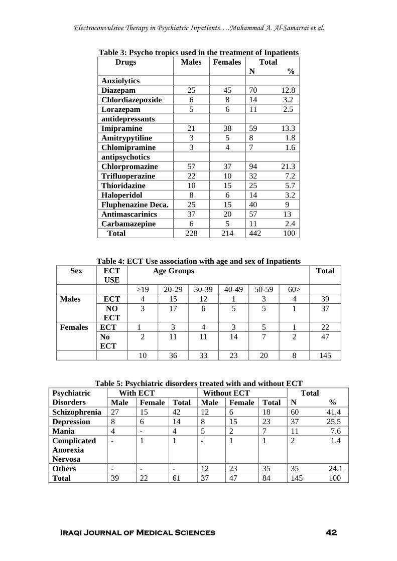
Electroconvulsive Therapy in Psychiatric Inpatients….Muhammad A. Al-Samarrai et al.
Iraqi Journal of Medical Sciences 42
Table 3: Psycho tropics used in the treatment of Inpatients Drugs Males Females Total
N % Anxiolytics Diazepam 25 45 70 12.8 Chlordiazepoxide 6 8 14 3.2 Lorazepam 5 6 11 2.5 antidepressants Imipramine 21 38 59 13.3 Amitrypytiline 3 5 8 1.8 Chlomipramine 3 4 7 1.6 antipsychotics Chlorpromazine 57 37 94 21.3 Trifluoperazine 22 10 32 7.2 Thioridazine 10 15 25 5.7 Haloperidol 8 6 14 3.2 Fluphenazine Deca. 25 15 40 9 Antimascarinics 37 20 57 13 Carbamazepine 6 5 11 2.4 Total 228 214 442 100
Table 4: ECT Use association with age and sex of Inpatients Sex ECT
USE Age Groups Total
>19 20-29 30-39 40-49 50-59 60> Males ECT 4 15 12 1 3 4 39
NO ECT
3 17 6 5 5 1 37
Females ECT 1 3 4 3 5 1 22 No ECT
2 11 11 14 7 2 47
10 36 33 23 20 8 145
Table 5: Psychiatric disorders treated with and without ECT Psychiatric Disorders
With ECT Without ECT Total N % Male Female Total Male Female Total
Schizophrenia 27 15 42 12 6 18 60 41.4 Depression 8 6 14 8 15 23 37 25.5 Mania 4 - 4 5 2 7 11 7.6 Complicated Anorexia Nervosa
- 1 1 - 1 1 2 1.4
Others - - - 12 23 35 35 24.1 Total 39 22 61 37 47 84 145 100
Electroconvulsive Therapy in Psychiatric Inpatients….Muhammad A. Al-Samarrai et al.
Iraqi Journal of Medical Sciences 43
Discussion The findings of this study have
some similarities and differences with other studies. First of all that although most inpatients can be treated by drugs alone, the rate using ECT among inpatients is higher than that which is reported in most literature(5,6). Explanation is that in the period during which the study was performed there was very gross deficiency of drugs in the country caused by the economic sanctions. Most drugs used are of conventional types .New drugs were hardly available. Even old drugs were sometimes not available. This made the treating psychiatrist in critical situation for treatment of sever cases with disturbed behavior that needed admission so ECT became an easy available mode of therapy. Other explanation is that families of patient who perform most nursing function and accompany patients in the wards do not tolerate long period of stay in hospital. In this case treating doctors will resort to using ECT in treatment to shorten period of recovery. The second point is the diagnostic categories. In this study schizophrenia was the most common category treated by ECT while affective disorders were more commonly treated by ECT according to many most literatures(9,10).The same problems of drugs and inability to keep patients for long periods in wards may be taken as explanation. Because cases of schizophrenia present more with agitation and disturbed behavior than affective disorders then ECT was performed more for schizophrenia to control behavior in short period while there was such deficiency of drugs. Most of cases of affective disorders in this study were those of depression that can be treated with drugs alone because such cases are not usually associated with disturbed behavior and there is no urge for using ECT for them except complicated cases or
when there is risk of suicide. In fact the most common rate of admission found in this study is for schizophrenia. While this indicate another explanation for the high rate of use of ECT found in this study ,it gives clue that admission to psychiatric unit is kept more to cases with disturbed behavior while other less sever cases such as those with depression can be treated and tolerated at home . Males were more treated by ECT than females while the reverse was found in most studies (7, 9, 10). In these studies affective disorders specifically resistant depression was the most common diagnostic category treated by ECT.Depression is more common in females and this explain the difference in gender for use of ECT between this study and other studies(9,10). Some studies had same results to this study and interestingly these studies are done in developing countries that may suffer of same problems of deficiency of drugs and lack of well trained and enough staff to control sever cases without use of ECT (3, 4, 8).This study need to be replicated again after improvement in drug availability nowadays
There is high rate of using ECT among psychiatric inpatients in general hospital in Baghdad.Schizophrenia was most common indication for ECT and this treatment is uses more for males than females. Deficiency of drugs and lack of trained staff is considered responsible for such high rate of use of ECT. References 1- Gelder M, Harrison P, Cowen Ph. Shorter text book of psychiatry.Oxford University Press.2006:565-572. 2- Abraham, Kulhara P. The efficacy of electroconvulsive therapy in the treatment of schizophrenia. A comparative study. Br J Psychiatry. 1987 August; 151:152-5. 3- Motohashi N, Awata S, Higuchi T.A questionnaire survey of ECT practice in university hospitals and national hospitals in

Electroconvulsive Therapy in Psychiatric Inpatients….Muhammad A. Al-Samarrai et al.
Iraqi Journal of Medical Sciences 44
Japan. Journal of ECT. 2004 March; 20(1):21-3. 4- Chanpattana W, Kramer BA Electroconvulsive therapy practice in Thailand. Journal of ECT. 2004 Junuary;20(2):94-8 5- Gazdag G, Kocsis N, Lipcsey A. Rates of electroconvulsive therapy use in Hungary in 2002. Journal of ECT. 2004 March; 20(1):42-44. 6- Little JD. ECT in the Asia Pacific region: what do we knowJournal of ECT? 2003 Junuary; 19(2):93-7. 7- Wood DA, Brgess PM. Epidemiological analysis of electroconvulsive therapy in Victoria, Austuralia. Aust N Z J Psychiatry. 2003 June;37(3):307-11 8- chung KF. Electroconvulsive therapy in Hong Kong: rates of use, indications, and outcome. J ECT. 2003 June; 19(2):98-102. 9- Stromgren LS. Electroconvulsive therapy in the Nordic countries, 1977-1987 Acta Psychiatr Scand. 1991 November; 84(5):428-34. 10- Thompson JW, Blaine JD. Use of ECT in the United States in 1975 and 1980. Am J Psychiatry. 1987 May;144(5):557-62 11- Muller U, Klimke A, Janner M, Gaebel W. Electroconvulsive therapy in psychiatric clinics in Germany in 1995. Nervenarzt. 1998 January; 69(1):15-26. 12- Doessel DP, Scheurer RW, Chant DC, Whiteford HA. Changes in private sector electroconvulsive treatment in Australia. Aust N Z J Psychiatry. 2006 April;40(4):362-7 13- WHO. International classification of disease.1991

Iraqi Journal of Medical Sciences 45
Evaluation of Serum Soluble Interleukin -2 Receptor level in Diagnosis of Rheumatoid Arthritis.
Eham Amir Ali PhD.
Abstract Background: soluble interleukin-2 receptor (sIL-2R) is secreted by lymphocytes upon activation and has been used as a marker of immune activation in several diseases. Objective: This study aimed to assess the potential clinical utility of serum level of serum soluble interleukin-2 receptor (sIL-2R) as a diagnostic tool in rheumatoid arthritis disease (RA). The study investigated also the association between serum sIL-2R levels with other parameters used for assessment of RA such as rheumatoid factor (RF), erythrocytes sedimentation rate (ESR), C-reactive protein (CRP), and uric acid. Methods: Serum sIL-2R levels, measured by ELISA, were evaluated in 25 RA patients who have positive rheumatoid factor (RF) and compared with those of 25 normal controls. The correlations with the other parameters were analyzed.
Results: Compared with the healthy control group, RA patients tended to have significantly higher serum sIL-2R and ESR concentrations (P<0.001). While no significant difference between both groups in serum uric acid was seen. Positive serum CRP (CRP level>6mg/dl) was found in 56% of patients. The sIL-2R level was positively correlated with RF and ESR, while a slight positive correlation with uric acid was noticed. Serum sIL-2R showed a high sensitivity and specificity for the patients with positive RF. Conclusions: A sIL-2R level is a sensitive and specific marker and can be useful for diagnosis of RA. Key words: C-reactive protein, Erythrocyte sedimentation rate, Rheumatoid arthritis, Soluble interleukine-2 receptors. IRAQI J MED SCI, 2010; VOL.8 (2):45-50
Introduction Rheumatoid arthritis (RA) is an
inflammatory autoimmune disease that primarily attacks the synovial membrane of the minor joints leading to joint stiffening, swelling, and loss of function in the joints. Its aetiology is unknown, and definitive diagnosis depends predominantly on characteristic clinical features, typical radiographic findings, the presence of auto-antibodies called rheumatoid factors (RF), elevated erythrocyte sedimentation rate (ESR) and C-reactive protein (CRP) (1, 2). Failure to meet these criteria does not therefore exclude the diagnosis, especially during the early stages of the disease. There is no single test for the disease Dept. Medical Biochemistry, College of Medicine Al-Mustaniriya University. Address Correspondence to: Dr. Eham Amir Ali. E- mail: [email protected] Received: 22nd October 2009, Accepted: 15th March 2010.
and only few symptoms may be present in the early stages. The common test is rheumatoid factor (RF) is present in 80% of adults who have RA (3), an antibody that is presented eventually in the blood of most people with RA. Rheumatoid factors can bind to normal circulating IgG, forming complexes that are deposited in the joints. These immune complexes can activate the complement cascade, resulting in a hypersensitive reaction, which leads to chronic inflammation of the joints (4).
Other common laboratory tests include complete blood picture, ESR, which measures inflammation in the body. C-reactive protein is another common test that measures disease activity (5 .Recent research has uncovered an important role of cytokines which promote inflammation, such as tumor necrosis factor-α (TNF-α) and interleukin-1ß (IL-1ß). Also, TNF-α and IL-1 are

Interleukin -2 Receptor in RA…. Eham Amir Ali
Iraqi Journal of Medical Sciences 46
considered to be the key cytokines in the development of RA (6). Soluble interleukin-2 receptor (sIL-2R; previously known as Tac) is a surrogate marker of T-lymphocyte activation and proliferation (7). A soluble fraction of the IL-2 receptor is released from the cell membrane. It is the released extracellular domain of the IL-2Rα by activated cells during a variety of autoimmune disorders including rheumatoid arthritis (RA) (8), and systemic lupus erythematosus (9). SIL-2R and CRP increases in RA (10). In RA, IL-2 protein and the IL-2 soluble receptor (sIL-2R) are preferentially expressed at disease onset, in comparison with later stages of the disease (11). Studies that have addressed this theme have shown discordant results since they have reported evidence pro and against an association between the current proposed markers of this disease (CRP and ESR) and sIL-2R (11).
The aim of the present study is (i) to determine whether there is a difference in sIL-2R levels between RA patients (who have positive RF) and healthy controls (ii) to estimate the sensitivity and specificity of sIL-2Rin diagnosis of RA, and (iii) to evaluate whether sIL-2R levels correlate with the other parameters used for assessment of RA. Materials and Methods 1-Subjects
The patients included in the study appeared free of the conditions that may cause raised serum RF including cancerous diseases, cirrhosis, and inflammatory lung diseases. Other connective tissue diseases that raise RF were excluded by serologic tests such as systemic lupus erythematosus.
The patients with RA were recruited from the private clinics and laboratory of outpatient department. Only patients with positive results for RF who fulfilled the American College
of Rheumatology diagnostic criteria for RA (12) were selected for inclusion in the study. Criteria for exclusion. Pregnant women, patients with cancer, diabetes mellitus, or autoimmune illnesses, patients with hepatitis, or patients under dialysis were excluded from the study.
The study includes 25 women with RA in addition to 25 healthy control women. The patients were assessed by a rheumatologist on presentation. Apparently healthy persons were asked to participate as controls and they had negative RF values and an absence of acute and chronic diseases. Sample collection: Ten milliliters of peripheral blood was withdrawn from each individual. Two milliliters of fresh blood poured in a tube containing 0.4ml of sodium citrate as anticoagulant in order to estimate ESR. Serum for serological tests was obtained by centrifugation at 3000rpm for 10 minutes and coded serum aliquots were stored at -20°C until it was analyzed. 2-Measurements: Estimation of RF:
The RF-latex kit supplied by Spinreact ® Company-Spain was used for diagnosis of RF in serum of the individuals The RF-latex is a slide agglutination test for the qualitative and semiquantitative detection of RF in human serum. Latex particles coated with human gammaglobulin are agglutinated when mixed with samples containing RF. RF (cut-off, <8IU/ml) Estimation of serum sIL-2R:
The concentrations of sIL-2Rs in serum samples were measured by the Invitrogen® Human sIL-2Rkit according to the solid phase sandwich Enzyme Linked-Immuno-Sorbent Assay (ELISA). Estimation of ESR:
Westergren's method used for estimation of ESR for patients. The

Interleukin -2 Receptor in RA…. Eham Amir Ali
Iraqi Journal of Medical Sciences 47
ESR cutoff values, measured by the Westergreen method, are: female< 20mm/h; male< 15mm/h Estimation of CRP:
The CRP-latex kit supplied by Spinreact ® Company-Spain was used for diagnosis of CRP in serum of the individuals. This kit is a slide agglutination test for the qualitative and semiquantitative detection of CRP in human serum. Latex particles coated with goat IgG anti-human CRP are agglutinated when mixed with samples containing CRP. CRP (cut-off value is 6 mg/l), Estimation of Uric acid:
Uric acid in serum is oxidized by uricase to allantoine and hydrogen peroxide, which under the influence of peroxidase enzyme, 4–aminophenazone, and 2-4 dichlorophenol sulfonate forms a red quinoneimine compound. The intensity of the red color formed is proportional to the uric acid concentration in the sample. Statistical Analysis
All statistics were carried out using Excell® program-Microsoft Corporation-USA. The results expressed as mean±standard deviation. Correlation coefficient values were estimated by regression analysis. Predictive value, sensitivity and specificity for measured parameters were estimated by the following formulas: Predictive value of positive result=TP/ (TP+FP)*100% Specificity=TN/ (TN+FP)*100% Sensitivity=TP/ (TP+FN)*100%
Where TP=true positive, TN=true negative, FP=false positive, FN=false negative.
Cutoff value for sIL-2Rwas expressed as (mean +2×standard
deviation) that equal 1957pg/ml. The difference between groups was estimated using Pooled Student t-test. The difference is said to be difference if p-value is less than 0.05. Results
The mean sIL-2R concentrations and ESR level were significantly higher (p<0.001) in patients with positive RF patients in comparison with healthy controls as shown in Table (1), while no significant difference between both groups in serum uric acid.
Positive serum CRP (CRP level>6mg/dL) were found in 56% of patients (14 / 25). Correlation coefficients (r) values for the patients group showed a positive correlation between the sIL-2R vs. RF (r=0.64) and sIL-2R vs. ESR (r=0.57). A slight positive correlation between sIL-2R vs. uric acid (r=0.34). There is no correlation between each pair of the compared parameters in the control group.
The sensitivity and specificity of the measured parameters are shown in Table (2). Because all patients were positive RF and all controls were negative RF, the sensitivity and specificity for RF=100%. Serum sIL-2R had 84% sensitivity and 96% specificity for the patients with positive RF which is higher sensitivity than other measured parameters. Every sample with values more than the cutoff value of healthy controls (mean + 2×standard deviation) was defined as positive for increased sIL-2Rconcentration. Predictive values showed that the cut off value for sIL-2R=1957pg/ml is an excellent medical decision limit for the prediction of RA.

Interleukin -2 Receptor in RA…. Eham Amir Ali
Iraqi Journal of Medical Sciences 48
Table 1: Serum concentration of sIL-2R, ESR, and uric acid of patients and control groups expressed as mean±standard deviation.
Parameter Patient Control p-value sIL-2R ( pg / ml) 2632±1274 935±511 P<0.001* ESR ( mm / hr ) 38.3±18.1 8.7±2.6 P<0.001* Uric acid ( mg / dl ) 6.1±1.9 4.6±0.9 p>0.05 (*): Significantly different.
Table 2: Sensitivity, Specificity, and Predictive values of the measured parameters.
Parameter Sensitivity Specificity Predictive value
RF 100% 100% 100%* sIL-2R 84% 96% 94 ESR 78% 96% 95 CRP 64% 92% 86 Uric acid 42% 73% 78 (*): Because all patients have positive RF value and healthy control have negative RF test intentially. Discussion
Elevated sIL-2Rconcentration in RA patients in comparison with healthy control group (Table 1) is in accordance with many other researches (10, 13). Iraqi RA patients showed high mean sIL-2Rlevel (2632 ± 1274 pg/ml), while the increase in sIL-2R concentration in other studies were 1532pg/ml (14) and 1855pg/ml (15). The reason for these differences may be due to the severity of disease or effects of medication on the sIL-2R level in serum as noted in various studies.
Suenaga et al (1998) (16) suggested that sIL-2R measurements to be helpful for the early diagnosis of RA in patients with joint pain, but without symptoms of bone or joint destruction. A high serum sIL-2R level at baseline is a predictor of remission in patients with acute RA (17). Suenaga et al (1998) (16) have demonstrated that an increased concentration of sIL-2R in the serum of patients with joint pain is a predictor for the future development of RA. Spadaro et al (1997) (18) observed that treatment of RA patients with methotrexate for 6 months was able to decrease the levels of sIL-2R. However, the results of sIL-2R in the present work disagreed with the results
of one research. Fro¨de et al (2002) (19) showed that the median levels of sIL-2R did not significantly differ in comparison with those of controls, whereas ESR levels but not CRP were significantly increased. Altogether, these inflammatory indices seem to independently reflect a final pathway of multifactorial events (19). The reason of the indifference in that study may be due to their low number of patients (n=21) and controls (n=7 only).
An increase of sIL-2R levels during RA has been noted, both in serum/plasma and in synovial fluid (20,
21). Detailed clinical trials showed that serum sIL-2R levels are related to disease duration and a decline in sIL-2Rconcentration may result from joint improvement (22).
Findings from clinical trials raise a question on whether sIL-2R concentration in serum provides a reliable immunological marker to assess disease activity in RA. Earlier studies reported the possible advantages of sIL-2R measurements for these purposes (23). Tebib et al (1995) (21) do, however, question the utility of sIL-2R as such a marker, since it is neither specific nor sensitive

Interleukin -2 Receptor in RA…. Eham Amir Ali
Iraqi Journal of Medical Sciences 49
to measure disease activity in an outpatient RA population.
The most commonly measured laboratory markers of disease activity in RA are the ESR and CRP. In recent studies, it was reported that CRP is more sensitive than ESR as a marker of disease activity because ESR is additionally affected by several factors, such as age, sex, anaemia, elevated fibrinogen and immunoglobulin levels, renal failure, pregnancy, and abnormal red blood cell morphology (1). Both CRP and ESR give similar information about non-specific inflammation. A high or increasing amount of serum CRP suggests an acute infection or inflammation. CRP appears and disappears more quickly than changes in ESR. Therefore, CRP level may drop to normal following successful treatment, whereas ESR may remain elevated for a longer period (24). As a blood test, CRP is not specific. A high result serves as a general indication of acute inflammation. In cases of inflammatory rheumatic diseases, such as rheumatoid arthritis and lupus, doctors can utilize the CRP test to assess the effectiveness of a specific arthritis treatment and monitor periods of disease flare-up. Its value is as a general indicator, not specific
Some reports indicate relationships between sIL-2R and laboratory markers of inflammation, erythrocyte sedimentation rate (ESR) and C-reactive protein (CRP) (21, 22, 25,
26, 27). In patients with RA, IL-2R correlated weakly with ESR (r = 0.24), and CRP (r = 0.24) (13). The finding of our research showed higher r-value for IL-2R with ESR (r=0.57) indicating the reliability of ESR as a diagnostic tool for RA.
The normal serum uric acid in most RA patients are in accordance with general knowledge about serum uric acid in RA(28) and may be useful
as first step for differentiation between gout and RA. References 1. Richardson C, Emery P. Laboratory markers of disease activity. J.Rheumatol 1996; 23: 23–30. 2. Tighe H, Carson DA. Rheumatoid factors. In: Kelley WN, Ruddy S, Harris Jr, Sledge CB, eds. Textbook of Rheumatology. Philadelphia, PA: WB Saunders, 1997: 241–249. 3. The Mayo Clinic.com Rheumatoid Factor: Definition. 4. Kuby, Kuby _ Immunology; Ch.20 Autoimmunity: p.467. 5. National institute of arthritis and musculoskeletal and skin diseases (NIAMS); http://www.niams.nih.gov/Health_Info/Rheumatic_Disease/default.asp. 6. Taylor PC. Anti-cytokines and cytokines in the treatment of rheumatoid arthritis. Curr Pharm Des 2003; 9:1095–106. 7. Castillo FM, Romero TA, Est´evez J, et al. Concentrations of cytokines, soluble interleukin-2 receptor, and soluble CD30 in sera of patients with hepatitis B virus infection during acute and convalescent phases. Clinical and Diagnostic Laboratory Immunology. 2002;9(6):1372–5. 8. Wolf RE, Brelsford WG, Hall VC, Adams SB. Cytokines and soluble interleukin-2 receptors in rheumatoid arthritis. J Rheumatol 1992; 19:524-8. 9. Sawada S, Hashimoto H, Iijma S, et al. Increased soluble IL-2 receptor in serum of patients with systemic lupus erythematosus. Clin Rheumatol 1993;12:204-9 10. Vincenzo Pasceri and Edward T. H. Yeh. A Tale of Two Diseases: Atherosclerosis and Rheumatoid Arthritis. Circulation 1999; 100; 2124-6. 11. Corrigan VM, Arastu M, Khan S, et al. Functional IL-2 receptor beta (CD122) and gamma (CD132) chains are expressed by fibroblast-like synoviocytes: activation by IL-2 stimulates monocyte chemoattractant protein-1 production. J Immunol 2001; 166: 4141–7. 12. American College of Rheumatology Subcommittee. Recommendations for the medical management of osteoarthritis of the hip and knee: 2000 update. American College of Rheumatology Subcommittee on Osteoarthritis Guidelines. Arthritis Rheum 2000; 43: 1905–1915. 13. Steiner G, Studnicka-Benke A, Witzmann G, et al. Soluble receptors for tumor necrosis factor and interleukin-2 in serum and synovial fluid of patients with rheumatoid arthritis, reactive arthritis and osteoarthritis. J Rheumatol. 1995; 22(3):406-12.

Interleukin -2 Receptor in RA…. Eham Amir Ali
Iraqi Journal of Medical Sciences 50
14. Lee GL, Chen MY, Chuang CY, Chen CY. Serum interleukin-2 receptor in systemic lupus erythematosus and rheumatoid arthritis. Zhonghua Min Guo Wei Sheng Wu Ji Mian Yi Xue Za Zhi. 1988; 21(1):16–22. (English abstract). 15. Pountain G, Hazleman B, Cawston TE. Circulating levels of IL-1 β, IL-6 and soluble IL-2 receptor in polymyalgia rheumatica and giant cell arteritis and rheumatoid arthritis. Br J Rheumatol. 1998; 37(7):797–8. 16. Suenaga Y, Yasuda M, Yamamoto M, et al. Serum interleukin-2 receptor for the early diagnosis of rheumatoid arthritis. Clin Rheumatol. 1998; 17(4):311–7. 17. Kuuliala A, Söderlin M, Kautiainen H, et al. circulating soluble interleukin-2 receptor level predicts remission in very early reactive arthritis. Scand J Rheumatol. 2005 Sep-Oct; 34(5):372-5. 18. Spadaro A, Taccari E, Riccieri V, et al. Relationship of soluble interleukin-2-receptor and interleukin-6 with class-specific rheumatoid factors during low-dose methotrexate treatment in rheumatoid arthritis. Rev Rheum Engl. 1997; 64: 89–94. 19. Fro¨ de TS, Tenconi P., Debiasi MR. et al. TNF-α and sIL-2R levels in rheumatoid arthritis. Mediators of Inflammation. (2002)11; 345–349. 20. Nassonov EL, Samsonov MY, Chichasova NV, et al. Soluble adhesion molecules in rheumatoid arthritis. Rheumatology (Oxford). 2000; 39(7):808–10. 21. Tebib JG, Letroublon MC, Noel E, et al. sIL-2Rlevels in rheumatoid arthritis: poor correlation with clinical activity is due in part to disease duration. Br J Rheumatol. 1995; 34(11):1037–40. 22. Rubin LA, Snow KM, Kurman CC, et al. Serial levels of soluble interleukin 2 receptor in the peripheral blood of patients with rheumatoid arthritis: correlations with disease activity. J Rheumatol. 1990; 17(5):597–602. 23. Wood NC, Symons JA, Duff GW. Serum interleukin-2-receptor in rheumatoid arthritis: a prognostic indicator of disease activity? J Autoimmun.1988; 1(4):353–61. 24. Lab Tests Online; American Association for Clinical Chemistry http://www.labtestsonline.org/understanding/analytes/crp/glance.html. 25. Symons JA, Wood NC, Di Giovine FS, DuffGW. Soluble IL-2 receptor in rheumatoid arthritis. Correlation with disease activity, IL-1 and IL-2 inhibition. JImmunol. 1988; 141(8):2612–8.
26. Itoh M, Goto Y, Ohta Y, et al. Relations between surface expression of the interleukin-2 receptor and release of the soluble form of the receptor in cultured mononuclear cells from patients with rheumatoid arthritis or systemic lupus erythematosus. Clin Rheumatol. 1998; 17(1):26–30. 27. Klimiuk PA, Sierakowski S, Latosiewicz R, et al. Interleukin-6, soluble interleukin-2 receptor and soluble interleukin-6 receptor in the sera of patients with different histological patterns of rheumatoid synovitis. Clin Exp Rheumatol. 2003; 21(1):63–9. 28. Richard Ravel, Clinical Laboratory Medicine: Clinical Applications of Laboratory Data. Ch.23 Bone, Joint, and Collagen-Vascular Disorders 6th edition (1995) by Mosby Co USA.

Iraqi Journal of Medical Sciences 51
Hepatitis A virus infection in children.
Sawsan Ibrahim Al-Azzawi 1MBChB; MRCP, Munib Ahmad Al- Zubaidi1 MBChB; DCH; FICMS- Paed, Haider Amin Aziz 2 MBChB; FICMS- Paed.
Abstract Background: most hepatitis A viral infections in children are asymptomatic or have mild non specific manifestations but some are complicated. Objective: to evaluate all cases of hepatitis A viral infection in children who were admitted over one year period and enlighten out the abnormal presentations and predict complications. Patients and methods: during the period from 1st of June 2005- 1st of June 2006, fifty cases of hepatitis A viral infection (who had hepatitis A virus IgM +ve) were studied and analyzed regarding the clinical presentations, coarse of the illness, complications and outcome. Results: fifty patients enrolled in the study, with male to female ratio of 1.8:1 and 32 (64%) of them presented in the age of 1-5 years. Fulminant hepatic failure was found in 7 (14%) cases, 5 (10%) had prolonged cholestasis , 5
(10%) had exacerbation of pre- existing chronic liver disease, 2 (4%) had recurrent or relapsing hepatitis A viral infection, and 5 (10%) cases had extra hepatic manifestations , 3 (6%) were G6PD deficient patients who had sever hemolysis. Conclusions: hepatitis A viral infection can present in different ways. The level of consciousness, presence of ascites and severely abnormal biochemical and hematological values were of great help in predicting complicated cases of hepatitis A viral infection. Key words: hepatitis A, abnormal presentations, children. IRAQI J MED SCI, 2010; VOL.8 (2):51-56
Introduction
Hepatitis A is the commonest viral hepatitis in the developing world (1). It commonly spreads from person to person by fecal – oral route (2). It often can pass from caregivers to children or adults in settings where there is close contact (3). Travelers to endemic areas are at high risk and may transmit hepatitis A virus (HAV) after infection (4). Most hepatitis A viral infections in children younger than 5 years of age are asymptomatic or have mild, non specific manifestations but some of HAV infections might have complications such as fulminant hepatic failure ( which is a clinical syndrome resulting from massive necrosis of
1Dept. pediatric, College of Medicine, Baghdad University, 2 Teaching Hospital, Medical City. Address Correspondence to: Dr. Munib Ahmad Al- Zubaidi, E- mail: [email protected] Received: 2nd November 2009, Accepted: 28th February 2010.
hepatocytes or from severe functional impairment of hepatocytes. The currently accepted definition in children includes: biochemical evidence of acute liver injury (usually <8 wk duration); no evidence of chronic liver disease; and hepatic-based coagulopathy in the presence of clinical hepatic encephalopathy) (2) and it occurs in less than 1 in 10000 case (5), prolonged cholestasis, recurrent hepatitis, and extrahepatic manifestations or may present on top of already pre- existing chronic liver disease (1).
One patient had multiorgan dysfunction including liver failure, hepatic encephalopathy, renal failure, pleural effusion, pericardial effusion and hematological dysfunction as a sequale of hepatitis A infection in otherwise healthy male (6).
The aim of this study was to evaluate all cases admitted with HAV infection over one year period and to enlighten the abnormal presentations

Hepatitis A virus infection in children….Sawsan Ibrahim Al-Azzawi et al
Iraqi Journal of Medical Sciences 52
and predict complicated cases of HAV infection. Patients and methods
This prospective study was done at Children Welfare Teaching Hospital, Medical City in Baghdad in the period from 1st of June 2005 till 1st of June 2006, including cases admitted with hepatitis. Sixty five patients with hepatitis like illness were admitted during the period of the study, only 50 cases with HAV IgM +ve were included in the study. Each case was studied regarding clinical presentations, course of the illness, complications and the outcome. Thorough physical examination and all the necessary investigations were done to the patients in the study. Results
Fifty patients were studied that had HAV IgM +ve, 32 (64%) males and 18 (36%) female with male to female ratio of 1.8:1.
Thirty-two (64%) patients presented in the age group between 1-5 years (table 1).
All patients included in the study presented with jaundice (100%), 35 (70%) patients had hepatomegaly, 6 (12%) patients had leg edema and 3 of them had additional ascites. Disturbed consciousness was found in 8 (16%) cases, 4 (8%) had typical encephalopathy, 3 of them developed convulsion (Table 2).
Twenty –four (48%) patients had complications: seven (14%) patients
presented with fulminant hepatic failure, two of them were died (4%), one after 24 hours of admission and the other after 3 days. five patients (10%) had prolonged cholestasis with mean days of illness of 45 days, one of them had sever hypoglycemia and developed convulsion during the course of the illness. Another five patients (10%) had pre- existing chronic liver disease (2 had Wilson disease and 3 had autoimmune hepatitis), one of the Wilsons patients was semiconscious and had positive family history of same disease. Two of the patients with autoimmune hepatitis presented with disturbed consciousness and generalized edema.
Relapsing hepatitis occurred in 2 patients (4%), both within 4 weeks of the initial infection. Extra hepatic manifestation was found in 5 cases, sever haemolysis was seen in 4(8%) patients, 3 of them were G6PD deficient patients and presented with very dark colour urine, hepatosplenomegaly and sever pallor. two of them required blood transfusion; one of those had impaired renal function. The fifth patient presented with history of convulsion (Table 3)
The mean biochemical and hematological values are important in predicting the outcome of the cases of HAV infection as the mean TSB, S.ALT, S.AST, S. Alkaline phosphatase, PT, and PTT were all higher in complicated cases (Table 4).
Table 1: distribution of patients according to age and sex
Age (years) Male N0. %
Female No. %
Total No. %
< 1 0 0 0 0 0 0 >1-5 20 62.5 12 37.5 32 64 >5-10 10 66.7 5 33.3 15 30 >10-15 2 66.7 1 33.3 3 6 Total 32 64 18 36 50 100

Hepatitis A virus infection in children….Sawsan Ibrahim Al-Azzawi et al
Iraqi Journal of Medical Sciences 53
Table 2: Signs and symptoms of the disease at presentation of the patients in order of frequency.
Signs and symptoms No. % Jaundice 50 100 Fever 40 80 Dark colour urine 36 72 Tender hepatomegaly 35 70 Vomiting 30 60 Clay colour stool 20 40 Abdominal distension 12 24 Splenomegaly 9 18 Disturbed consciousness (encephalopathy)
8 16
Leg edema 6 12 Pallor 4 8 Convulsion 4 8 Ascites 3 6
Table 3: Distribution of complicated cases according to clinical presentation.
% NO. Types of complication 14 7 Fulminant hepatic failure 10 5 Prolonged cholestasis
10 5 HAV on top of pre-existing chronic liver disease
4 2 relapsing hepatitis 8 2
4 1
Extra hepatic manifestations: haemolysis convulsion
48 24 Total
Table 4: Mean biochemical and hematological values of the patients
'''PTT mean
''PT mean
'ALK.phosphmean
*** S.AST mean
**S.ALTmean
*TSB mean
Test Presentation
38 ± 4 15 ± 2 65 ± 5 71 ± 4 69 ± 4 7.8 ± 1 Non complicated (NO: 26)
52 ± 5 42 ± 3 75 ± 6 108 ± 8 89 ± 5 16.6 ± 2 Complicated (NO: 24) *total serum bilirubin, **Serum alanin amino transferase, *** Serum aspartate amino transferase, 'Alkaline phosphatase, ''Prothrombin time, ''' Partial thromboplastin time

Hepatitis A virus infection in children….Sawsan Ibrahim Al-Azzawi et al
Iraqi Journal of Medical Sciences 54
Discussion Hepatitis A viral infection accounts
for about 50% of the clinically apparent hepatitis, not associated with chronic liver disease, persistent viraemia or intestinal carrier state (2).
Our study included hospitalized patients which were usually ill and jaundiced with hepatitis like illness. We actually isolated every case until proved or disproved to be HAV IgM+ve. There was male predominance which is consistent with Anand AC study (7), while no sex predilection was apparent in Amin J et al study (8).
The most common age group was between 1-5 years (64%) of cases. In the developing nations, the age of acquisitions is usually before 2 years of age while in western societies it is frequent in person's age 5-17 years (9). No case under one year was found in our series as it is usually uncommon in infants less than one year of age (10). Splenomegaly plus hepatic enlargement were found in all cases of G6PD deficient patients and all of them had severed anemia and very high total serum bilirubin.
The complicated cases in this study were 48% which is a high percent, it is probably because all cases admitted to the hospital were seriously ill and the mild, ambulant cases usually not admitted. It was reported that fulminant hepatic failure may varies between 0.1% of symptomatic infected children to10 % (1, 9). In our study the most common complication was the fulminant hepatic failure (14%), all those patients were semiconscious, three had ascites and leg edema and two of them died within 3 days of admission. Apart from hepatic insufficiency, the course of HAV infection may be characterized by a relapse following initial improvement (relapsing hepatitis A) and prolonged cholestasis (11). Although relapse occurs in 3-21% of patients with acute
hepatitis A (1, 8, 12), we had 2 relapsing cases (4%) within 4 weeks of the previous infection. Persistent or prolonged cholestasis may follow the acute infection and it may persist for more than 3 months (13). In this study 5(10%) patients had prolonged course which ranges from 30- 60 days (mean 45 days). One of those patients had protracted hypoglycemia and deep jaundice, he was managed with small doses of steroid and Ursodeoxycholic acid as recommended by one author (14), and he improved and was discharged well.
Although ascites and pleural effusion are possible benign and early complications of acute HAV infection that resolve spontaneously regardless of the illness outcome (2, 3), all 3 cases (6%) seen in this study with ascites proved to have chronic liver disease.
All types of chronic liver disease can present as acute hepatitis (14) but some can be uncovered during the superadded acute HAV infection. Data from literature indicate a high fatality rate during the HAV super infection in patients with chronic hepatitis B and C, particularly those with cirrhosis and in patients with alcoholic cirrhosis (15).
Five (10%) patients had previous chronic liver disease (Wilson's disease was evident in 2 cases (4%) and 3 (6%) had autoimmune hepatitis), the presentation of which was not a straight forward, the children had firm to hard liver texture with features of acute hepatitis like illness, 3 of all had ascites and 2 of them were semiconscious. Family history was the helping clue in the diagnosis of Wilson case.
Immunization with hepatitis A vaccine was recommended in all patients with chronic liver disease by many authors (3, 14,15), but not in India as there was a high prevalence of pre-existing antibodies in these patients (7).

Hepatitis A virus infection in children….Sawsan Ibrahim Al-Azzawi et al
Iraqi Journal of Medical Sciences 55
Although extra hepatic manifestations are rare in HAV infection, we had 5 cases (10%), four patients (8%) presented with evidence of hemolytic anemia and three (6%) of those were G6PD deficient, all of them had hepatosplenomegaly, sever anemia and very high serum bilirubin. One of those was a boy 6 years old on the verge of renal failure but fortunately recovered with supportive therapy.
Hemolytic anemia as a complication of acute hepatitis had been reported in up to 23% of patients. However the incidence may rise to 70-87% in patients with G6PD deficiency, massive intravascular haemolysis with renal failure, hepatic encephalopathy and even death have been reported (16, 18).
One patient (2%) had convulsion at the start of the icteric phase but without neck stiffness, and his CSF findings were normal. Looking in the literature, there was only one case report for a five years old child presented with convulsion and neck rigidity, and because HAV RNA was demonstrated in the CSF, it was thought that convulsion might be related to this viral infection (19).
Hepatitis A virus associated mortality world wide is 0.2- 0.4 % (1),but in this study it was 4% which is due to fulminant hepatic failure as our hospital is a tertiary center and we receive terminal cases.
In our study, all the mean hematological and biochemical levels in complicated cases were greater than that in uncomplicated patients (Table 4) which is similar to Sainokami S et al findings (20). Increased levels of serum transaminases and prolonged PT and PTT gave an idea of the unusual presentation and sometimes bad prognosis.
It was concluded that the Hepatitis A viral infection can present in different ways.
It occurs mostly in the age group (> 1-5 years).
Hepatitis A viral infection on the top of pre-existing chronic liver disease or in G6PD deficient cases could affect the presentation and the course of the disease. The level of consciousness, presence of ascites and severely abnormal biochemical and hematological values were of great help in predicting complicated cases of hepatitis A viral infection. References 1. Davidson S. Acute Hepatitis in Diseases of Liver and Biliary System in Children by Deirdre A. Kelly; Blackwell publications, second edition. 2004; Ch6:92-104. 2. Yazigi N., Balistreri W., Viral Hepatitis In Kliegman R.M., Behrman R.V., Jenson H.B, Stanton F. Nelson Textbook of Pediatric 18thed. Philadelphia W.B. Saunders 2007; 355: 1680 -.90. 3. Keefe EB, Iwarsons Mc, Mahon BJ, et al. Safety and immunogenicity of hepatitis A vaccine in patients with chronic liver disease . Hepatology 1998; 27:881-6. 4. Lemon SM,Thomas DL. Vaccines to prevent viral hepatitis. New Engl.J Med 1997; 336:196-204. 5. Koff RS. Hepatitis A. Lancet 1998; 341:1643-9. 6. Rasheed A, Saeed S. Acute hepatitis A virus infection presenting with multiorgan dysfunction: a case report.Cases J, 2009; July 30(2): 8124. 7. Anand AC, Nagpal AK. Should one vaccinate patients with chronic liver disease for HAV in India? J Assoc Physicians India 2004; 52:785-7. 8. Amin J, Gilbert GL, Escott RG, et al. Hepatitis A epidemiology in Australia. National section 2001; 174(7):338-41. 9. Gilroy R, Mukherjee S. Article on hepatitis A, last updated: August 11, 2004. (E-medicine, internet). 10. Kemmer NM, Miskovisky EP. Hepatitis A.Infect Dis Clin North Am 2000 Sep; 14 (3): 6-9. 11. Durand F, Clinical forms of hepatitis A. Rev Med Intern.2000 Jan; 21(1):50-7. 12. Arslan S, Caksen H, Oner AF,et al. Relapsing Hepatitis A in children. Acta Paediatr Taiwan 2002 Nov-Dec; 43(60):358-60. 13. Ginsber GM, Slater PE, and Shouva LD: cost benefits analysis of a nation wide infant immunity area of intermediate endemicity. J Hepatology 2001 Jan; 34 (1): 92-9.

Hepatitis A virus infection in children….Sawsan Ibrahim Al-Azzawi et al
Iraqi Journal of Medical Sciences 56
14. Shepherd R. Complicatios and management of chronic liver disease in Disease of Liver and Biliary system in children by Deirdre A.Kelly. Blackwell Publication, Second Edition 2004;Ch 14:259-79. 15. Lefilliare P, Villeneeuve JP.Fulminant hepatitis in patients with chronic liver diseases. Can. J Pub, Health 2000; 91(3): 168-70. 16. Chau TN, Lai JY. Haemolysis complicating acute viral hepatitis in patients with normal or deficient G6PD activity. Scand. Infec Diseases 1997;29(6):551-3. 17. Sharma D, Sibal A. Making a case for hepatitis A vaccination in G6PD deficient subjects. Indian J of Pediatrics 2005; 72: 640. 18. Gotsman and Muszkat M.G6PD deficiency is associated with increased initial clinical severity of acute viral hepatitis A.J of Gastroenterology and Hepatology 2001; V 16: Issue 11: 1239. 19. Cam S, Ertem D, Koroglu OA et al. Hepatitis A virus infection presenting as seizures. Pediatrics Infect. Dis 2005 July; 24(70): 652-3. 20. Sainokami S, Abe K, Ishikawa K, et al. Influence of load of hepatitis A virus on disease severity and its relationhipwith clinical manifestation in patients with hepatitis A. J Gastroenteral Hepatol.2005 Aug; 20(8): 1165-75.
Iraqi Journal of Medical Sciences 57
Morphometric analysis of odontoid process.
Hayder hammadi abdulameer MBChB; FICMS.
Abstract Background: The axis vertebra is the strongest cervical vertebrae; the characteristic feature of it is the presence of strong bony element, the dens (odontoid process). Several investigations have shown its relation with certain congenital anomalies. Objective: The aim of this study was to evaluate the value of some morphometric parameters measured from the odontoid process of the axis vertebra to judge the degree of retroflexion of the odontoid process in relation to the morphological architecture. Method: 30 randomly selected dry bones of adult second cervical vertebrae were used. These vertebrae were examined grossly and morphometrically, and the data obtained were analyzed by using global lab image/2 computer program. Results: Morphometric results of this study showed that the retroflexion of the dens should
be related to dorsal deviation, if the anteroposterior distance of the dens is fixed. The thicker anteroposterior dens is longer, wider, but less dorsally deviated. Conclusion: We may be able to consider the dorsally deviated dens and the anteroposterior thickness of the dens as indicator for the degree of retroflexion of the dens. The study may elaborate the possibility of using the above parameters as markers for the retroflexion of dens in cases of Chiari I malformation. Thus a preliminary diagnosis could be achieved. Key words: morphometry, dens, congenital anomaly. IRAQI J MED SCI, 2010; VOL.8 (1): 57-65
Introduction
Vertebra C2 is the strongest of the cervical vertebrae. C1, carrying the cranium, rotates on C2, as when a person turns the head. The axis has two large, flat bearing surfaces, the superior articular facets, on which the atlas rotates. The distinguishing feature of the axis is the blunt tooth-like dens (odontoid process), which projects superiorly from its body. Both the dens (G. tooth) and the spinal cord inside its coverings are encircled by the atlas. The dens lie anterior to the spinal cord and serves as the pivot about which the rotation occurs. The dens are held in position against the posterior aspect of the anterior arch of the atlas by the transverse ligament of the atlas (1).
Dept. human anatomy, college of medicine, Al-Nahrain University. Address Correspondence to: Dr. Hayder H. Abdulameer, E- mail: [email protected] Received: 3rd December 2009, Accepted: 28th April 2010.
The axis, acts as an axle for rotation of the atlas and head around the strong dens (odontoid process), which projects cranially from the superior surface of the body.
The dens are conical in shape. It may be tilted a little, up to 14°, posteriorly, or, less often, anteriorly on the body of the axis: it may also tilt laterally up to 10°. The apex is pointed, and from this point arises the apical ligament. The anterior surface bears an ovoid articular facet for the anterior arch of the atlas (2).
Anomalous development of the odontoid is uncommon, and its clinical significance lies in its potential for producing serious neurologic sequelae (3). These anomalous include either odontoid invagination, in which the odontoid process bulges upward into the foramen magnum and compresses the brainstem without deformity of the occipital bone (4), or Congenital and developmental osseous abnormalities and anomalies that affect the

Morphometric analysis of odontoid process….Hayder hammadi abdulameer
Iraqi Journal of Medical Sciences 58
craniocervical junction complex which can result in neural compression and vascular compromise and can manifest itself with abnormal cerebrospinal fluid dynamics (5).
Chiari I malformations are often associated with congenital craniocervical anomalies, such as platybasia, basilar invagination, and retroflexion of the odontoid process (6).
The aim of this study was to evaluate the values of some morphometric parameters measured from the odontoid process of the axis vertebra to judge the degree of retroflexion in relation to other morphological architectures. Material and methods
This study involves anatomical observation of 30 randomly selected dry bones of adult second cervical vertebrae obtain from anatomical museum of the department of human anatomy at Al- Nahrain College of medicine. The odontoid process of these vertebras was examined grossly. Certain morphometric measurements were done on the following parameters: • The line which connect the upper and lower point of the anterior surface of the body of the axis represented by L1 (figure1). • The mid-line of the anterior surface of the dens is represented by L2 (figure 2). • The angle between L1&L2 is called G1. This parameter could indicate the deviation of the anterior surface of dens from that of axis (figure 3). • The point of deviation of L1 across L2 called P1 (figure 3). • The most posterior point of the posterior surface of dens is P2(figure 3). • D1 represents the distance of the horizontal imaginary line between the most posterior point of dens P2 and the L2 (figure3&4).
• D2 represents the distance between P2 and the linear upward extension of L1 (figure3&4). • G2 represents the deduct value of D2 from D1 (Figure 4). • The length of dens represents the line between the P1 and its highest apical point (figure 5). • The width of the dens is widest anterior distance of the dens (figure 5). Measurements were done on the photographs of the odontoid process of the second cervical vertebra, The photograph of the axis vertebra were done according to the need of experimental analysis of this study regardless of the anatomical position of the second cervical vertebrae. The data obtained from these measurements were analyzed using the global lab-image2 computer program Global lab image2 software:
Is the keystone in this study by which all the results can be counted and analyzed manually or automatically (with less degree of user interference) (7). The software is a product from (Data Translation Inc. USA). As any other software, it needs special requirements to be installed on the PC. These requirements are: • MMX processor (Pentium III or higher). • Microsoft® Windows 98, ME, or XP. • At least 128 MB of random access memory (RAM). • At least 150 MB of available hard disk space. • CD-ROM drive. • SVGA monitor. • Display resolution set to 800x600 pixels or higher. • Color quality set to highest (32-bit). • Imaging device or frame grabber board (optional).

Morphometric analysis of odontoid process….Hayder hammadi abdulameer
Iraqi Journal of Medical Sciences 59
Results The statistical analyses of the dens
were as the following: The first group with significant relation ship these include: • The correlation between the measurements of the angle (G1) and the (G2) (figure 3, 4), the P value indicates highly significant relationship (table 1). • The correlation between angle (G1) and the distance between anterior and posterior points of dens (D1) (figure 3, 4), the P value indicates significant relationship (Table 1).
• The correlation between (D1) and length (figure 4, 5), the P value indicates highly significant relationship (table2). • The correlation between (D2) and the length (figure 4, 5), the P value indicates significant relationship (table2). • The correlation between (D1) and (D2) (figure 3); the P value indicates significant relationship (table2). • The correlation between (D2) and (G2) (figure 4); the P value indicates significant relationship (table2).
Table 1: statistical analysis of this study Type of relation
ship Correlation
coefficient (r2) Significance (P) value Notes
Angle (G1)with (G2)*
0.872 0.00001 Highly significant relation ship
Angle (G1) with length*
0.001 0.868 Non- significant relation ship
Angle (G1) with side to side *
0.015 0.524 Non- significant relation ship
Angle (G1) with D1#
0.136 0.046 Significant relation ship
Angle (G1) with D2*
0.313 0.058 Non- significant relation ship
The second group with non-significant relation ship these include: • The correlation between the measurements of the angle (G1) and the length (figure3, 5), the P value indicates non- significant relationship (Table 1). • The correlation between the measurements of angle (G1) with (D2) (figure 3, 4), the P value indicates non- significant relationship (Table 1). • The correlation between the measurements of length and G2 (figure5, 4), the P value indicates non- significant relationship (Table 2).
• The correlation between G2 and D1 (figure 4), the P value indicates non- significant relationship (Table 2). • The correlation between side to side measurements (figure 5) and the angle G1, G2, D1, D2, and the length of dens (figure3, 4), the P value indicates non- significant relationship (Table 2).

Morphometric analysis of odontoid process….Hayder hammadi abdulameer
Iraqi Journal of Medical Sciences 60
Table 2: statistical analysis of this study Type of relation ship Correlation
coefficient (r2) Significance (P)
value Notes
D1 and length of dens* 0.419 0.0001 Highly significant relation ship
D2 and length* 0.554 0.006 Significant relation ship Side to side and length# 0.006 0.697 Non- significant relation ship G2 and length * 0.192 0.154 Non- significant relation ship G2 and D1# 0.012 0.73 Non- significant relation ship Anteroposterior (D1)and D2 *
0.412 0.024 Significant relation ship
Side to side and D2* 0.01 0.757 Non- significant relation ship G2 and D2 * 0.477 0.013 Significant relation ship Side to side and (D1) # 0.000 0.961 Non- significant relation ship G2 and side to side* 0.005 0.832 Non- significant relation ship
# Negative correlation (-) *Positive correlation (+) Discussion
This study concerning with statistical analysis, and morphology with special emphasis on the degree of the inclination of the odontoid process, for functional and clinical manifestations, This inclination is important because The straight odontoid process is only subjected to stress of weight of the skull, but the dorsally inclined dens act to bending stress(8).
The orientations of the parameters and the statistical analysis showed in this study support the view that had been adapted previously in the anomalous dens as a clinical significance in producing serious neurological squeal(3). Although there are several recognized variations such as (aplasia, hypoplasia, and os odontoideum Congenital absence of the odontoid process, or Duplicated odontoid process),some anomalous may produces a serious clinical manifestations these may be associated with subluxation and neuraxial compression and atlantoaxial instability(9-11),headaches, pseudotumor-like episodes and hydrocephalus)(1), or compression of the upper cervical cord(13).
Chiari I malformations may be associated with changes that may be occurs in the cranio cervical region or herniation of the cerebellar tonsil below the foramen magnum (14). In addition to that, this malformation is often associated with retroflexion of the odontoid process. These signs confirm prior reports of an increased incidence of Chiari I malformation.
In this study we emphasized on the correlation between the degrees of inclination (retroflexion) in relations with other statistical analysis of the odontoid process.
The line suggested being the anterior vertebral line of the axis vertebra L1 which used for measurements of the degree of angulations (figure 1) is unchanging when the axis vertebra changes from normal anatomical position.
The angle between L1&L2 (G1), this parameter could indicate the deviation of the anterior surface of dens from that of axis (figure 3).G2 represents the deduct value of D2 from D1 this parameter could indicate the dorsal deviation of the anterior surface of dens (L2) excluding its anteroposterior thickness (Figure 4).

Morphometric analysis of odontoid process….Hayder hammadi abdulameer
Iraqi Journal of Medical Sciences 61
There is a significant relation ship with positive correlation between the degree of retroflexion (G1), and the dorsal deviation of the dens (G2), if the antero posterior distance of the dens is fixed so that the degree of retroflexion should be related to the dorsal deviation of the dens.
The significant relation ship with negative correlation between retroflexion (G1), and antero posterior distance of the dens (D1), means that whenever the degree of retroflexion increases, the antero posterior distance (thickness) decreases, however there is a significant relationship with positive correlation between this anteroposterior thickness(D1),thickness with dorsally deviated (D2), and the length of the dens, there is a non-significant relation ship between the length and the degree of retroflexion. This indicates that the longer dens is thicker anteroposterior, with more dorsally deviated. This positive correlation not related to the degree of retroflexion of the dens but it may be related to massive growth of the dens.
Although there is a non- significant relation ship between the retroflexion (G1), with the length, and
anteroposterior thickness with dorsally deviated (D2) dens, there is positive correlation between these parameters i.e. the more retroflexed dens, come with increases in the length, thickness with dorsally deviated, and side to side parameters of the odontoid process.
There is a non- significant relation ship with negative correlation between antero posterior thickness of the dens (D1), the dorsally deviated dens (G2), and side to side distance i.e. the thicker and wider dens is less dorsally deviated.
When the length of the dens increases, it may be bulges upward into the foramen magnum and compresses the brainstem and may be of a value in case of basilar invagination and brain stem compression (4).
The study may elaborate the possibility of using the length and width of dens demonstrated in anteroposterior X-ray as a marker for the retroflexion of dens in cases of Chiari I figure 6. Thus, a preliminary diagnosis could be reached, In addition to that evaluation of anterior dens provide essential anatomic data for safer surgical procedures (15).
Figure1: the line which connect the upper and lower point of the anterior surface of the body of the axis (L1).
1 cm
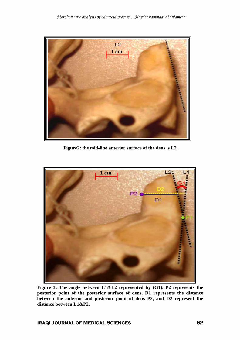
Morphometric analysis of odontoid process….Hayder hammadi abdulameer
Iraqi Journal of Medical Sciences 62
Figure2: the mid-line anterior surface of the dens is L2.
Figure 3: The angle between L1&L2 represented by (G1). P2 represents the posterior point of the posterior surface of dens, D1 represents the distance between the anterior and posterior point of dens P2, and D2 represent the distance between L1&P2.
1 cm
1 cm

Morphometric analysis of odontoid process….Hayder hammadi abdulameer
Iraqi Journal of Medical Sciences 63
Figure 4: the deduct value between (D2-D1) represented by G2.
Figure5: the length and the anterior width of the dens (Side to side).
Side to side
Length
P1
1 cm
1 cm

Morphometric analysis of odontoid process….Hayder hammadi abdulameer
Iraqi Journal of Medical Sciences 64
Figure 6: odontoid process in plain X-ray (lateral view). References 1. Moore Keith L, Dalley Arthur F. Clinically Oriented Anatomy, 5th EditionWilliams & Wilkins, 2006; 486. 2. Susan standring. Grays anatomy the anatomical basis in clinical practice,39th edition Churchill livingstone,2008; 745. 3. Hensinger RN, Fielding JW, Hawkins RJ. Congenital anomalies of the odontoid process Orthop Clin North Am. 1978;Oct;9(4):901-12 4. Kyoshima K, Kakizawa Y, Tokushige K, Akaishi K, Kanaji M, Kuroyanagi T , Odontoid compression of the brainstem without basilar impression-- "odontoid invagination". J Clin Neurosci. 2005; Jun;12(5):565-9 5. Menezes AH. Craniocervical developmental anatomy and its implications Childs Nerv Syst. 2008; Apr: 10.
6. Hwang SW, Heilman CB, Riesenburger RI, Kryzanski J. arthrodesis after transoral odontoidectomy and suboccipital craniectomy for ventral brain stem compression in Chiari I patients.Eur Spine,2008; J. Jul 16. 7. Maria SS, Vidal BC and Mello MLS .Image analysis of DNA fragmentation and loss in V79 cells under apoptosis. Genetic and Molecular Biology. 2000; 23:109-112. 8. Koebke J. Morphological and functional studies on the odontoid process of the human axis Anat Embryol (Berl). 1979; Jan 30; 155(2):197-208. 9. Stevens JM, Chong WK, Barber C, Kendall BE, Crockard HA.A new appraisal of abnormalities of the odontoid process associated with atlanto-axial subluxation and neurological disability. Brain Feb; 1994 ;117 ( Pt 1):133-48

Morphometric analysis of odontoid process….Hayder hammadi abdulameer
Iraqi Journal of Medical Sciences 65
10. Zadvornov IuN. Variants and developmental anomalies of the odontoid process of the axis Zh Vopr Neirokhir Im N N Burdenko, 1979 ;(1):30-8 11. Garant M, Oudjhane K, Sinsky A, O'Gorman AM. Duplicated odontoid process: plain radiographic and CT appearance of a rare congenital anomaly of the cervical spine. AJNR Am J Neuroradiol. 1997;Oct;18(9):1719-20 12. Milhorat Thomas H, Chou Mike W, Trinidad Elizabeth M M D, Kula Roger W M D, Mandell, Menachem MD, Wolpert, Chantelle MBA,et al. Chiari I Malformation Redefined: Clinical and Radiographic Findings for 364 Symptomatic Patients Neurosurgery. 1999; Volume 44 - Issue 5 - 1005-1017 13. Kremer P, Despaux J, Benmansour A, Wendling D. Spontaneous fracture of the odontoid process in a patient with ankylosing spondylitis. Nonunion responsible for compression of the upper cervical cord. Rev Rhum Engl Ed. Jun, 1995; 62(6):455-8. 14. Shane Tubbs, Matthew Bailey, William C. Barrow, Marios Loukas, Mohammadali M. Shoja4 and W. Jerry Oakes1, Morphometric analysis of the craniocervical juncture in children with Chiari I malformation and concomitant syringobulbia Neurosurgery vol.25 no.6, 2009; 689-692. 15. Tun K, Kaptanoglu E, Cemil B, Karahan ST, Esmer AF, Elhan A:. A neurosurgical view of anatomical evaluation of anterior C1-C2 for safer transoral odontoidectomy Eur Spine J. 2008 ; Jun;17(6):853-6.

Iraqi Journal of Medical Sciences 66
Multiple myeloma with breast masses as extra medullary plasmocytomas,A Case Report.
Waseem F. Al Tememi1 CABMS; FIBMS (hematology); FICMS, Alaa G. Hussein2
MSc (pathology); FICMS, Sinan W. Jasim3 MBChB, Ahmed H. Jasim3 MBChB, Abd AlMahdi A. Fattah3 MBChB
Abstract Background: Breast involvement by immunolymphoproliferative disorders is rare. Primary & secondary malignant lymphomas of the breast are much more common than multiple myeloma, of which only 18 cases were reported in previous literature, till the year 2000. Case presentation: We report a young female patient presented simultaneously with bilateral multiple breast masses & pathological fracture in right humorous that proved later on via histopathological examination to be a case of multiple myeloma associated with
extramedullary plasmocytoma involving breasts. Discussion and Conclusion: There were diagnostic difficulties caused by the lack of specific radiological & sonographic features that differentiate between primary or secondary breast tumor & breast infiltration by immunolymphoproliferative disorders. Key words: Plasma cell diseases, plasmocytoma, multiple myeloma, breast mass IRAQI J MED SCI, 2010; VOL.8 (1):66-71
Introduction Breast involvement in multiple
myeloma & solitary extramedullary plasmcytoma is very unusual. In most cases, the manifestation of this disease is usually systemic (1).
Multiple myeloma is a neoplastic plasma cell dyscrasia characterized by clonal proliferation of plasma cells & overproduction of paraprotein(2).
It is the most common plasma cell disorder representing 1% of all types of cancers (2).
Incidence is 4.5/100 000 /year in US. It increases progressively with age.
The mean age at diagnosis of multiple myeloma is 65-70 years, while the onset before 40 years is very rare (2). 1Dept. Medicine, College of Medicine, Al Nahrain University, 2Dept. Pathology College of Medicine, Al Nahrain University, 3Al Kadhimyia Teaching Hospital. Address Correspondence to: Dr. Waseem F. Al Tememi, Al Kadhimyia teaching hospital. E- mail: [email protected] Received:5th November 2009, Accepted: 11th April 2010.
Impaired hemopoeisis, renal insufficiency, osteolytic bone lesions, recurrent infections, hypercalcemia are the most common systemic manifestation of the disease.
Extra medullary myeloma (plasmocytoma) is defined as malignant proliferations of plasma cells with or without bone involvement (2). When occur in the soft tissue, it usually involves the upper respiratory tract & oral cavity. Plasmocytoma of breast is very rare (3).
This report describes a young female patient with bilateral breast masses with pathological fracture that proved to be part of manifestation of multiple myeloma with extra medullary plasmocytoma involving both breasts. Case presentation
Thirty four years old, an engineer, female patient presented with thirst lethargy & diffuse bone pain with difficulty in walking of 3 weeks duration. Two months ago she sustained right upper limb pain after a trivial trauma, which proved to be, later on, due to pathological fracture of

Breast involvement in multiple myeloma (Sep. 2009).… Waseem F. Al Tememi et al
Iraqi Journal of Medical Sciences 67
the upper end of right humerus by open biopsy ( Figure 1), (pieces of bone at that site were sent for histopathological examination & immune histochemical staining) . Blurred vision & headache were reported but no fainting attacks or other system complaint. Menstrual history is normal. She had negative past medical & surgical history.
There is negative family history for breast cancer or other chronic or malignant diseases. She is a mother of one boy, 3 years old, with no use of any contraception or other medications. There is no history of smoking.
On examination, a young female patient ill looking with limited mobility & a POP splint over right upper limb. She is pale dehydrated, afebrile & not jaundiced. Tachycardia 112 beats per minute regular normal volume, blood pressure 100/65 mmHg. There is generalized bone tenderness especially over the limbs. There were no palpable lymph nodes any where. Abdomen, chest & cardiovascular examination were normal.
Breasts examination revealed the presence of 2 masses in the left breast (at upper inner quadrant), largest one around 2.5 x2.5 cm, and another mass in the right breast with no palpable axillary lymph nodes.
After admission a thorough evaluation performed including the basic hematological & biochemical investigations as follows;
PCV 24%, hemoglobin 76 g/l, white blood cell 4.9 X 10^9/l (Neutrophil. 51%, Lymphocyte 44%, Monocyte 3%) ESR 130 mm/hr
Blood film revealed normochromic normocytic with excess roulaux, no abnormal leucocytes and adequate platelet
Blood sugar 110 mg/dl, urea. 36 mg /dl, creatinine 1.2 mg/dl, uric acid 6.5 mg /dl
Total serum protein 11g/dl, S. Ca (serum calcium) 11.5 mg/dl, S. ALP (alkaline phosphatase) 128.8 IU/L & S. LDH (lactate dehydrogenases) 200 IU/ L.
Normal prothrombin time & partial thromboplastin time &liver enzyme tests.
Normal general urine exam with no proteinuria. Urine for bence jones protein was negative
Serum protein electrophoresis: albumin 2.8 g/dl (3.5-5), α-1 0 .23 g/dl (0.1-0.3), α-2 0.72 g/dl(0.6-1.0), β 0.75 g/dl(0.7-1.1) & γ 4.79 g/dl(0.8-1.6) with monoclonal band in γ region &marked reduction at albumin fraction.
Bone marrow aspiration revealed cellular fragment with active normoblastic erythropoiesis, active myelopoiesis sequence maturation no excess in blast. Plasma cells 5%, active megakaryopoeisis.
Bone marrow biopsy showed sections with infiltration of bone marrow by numerous clusters of immature plasma cells with normoblasts in diffuse manner.
Breast ultrasound ; Multiple ill defined complex density non homogenous masses at medial quadrant of left breast largest size 3.5x 3cm , no calcification normal skin, subcutaneous tissue & axillary region, suggestive of malignancy ,another small similar mass in right breast 2.0 x1.5 cm
FNAC (fine needle aspiration cytology) from right breast mass; sheets of lymphocytes &immature plasma cells suggestive of hemopioeitic malignancy infiltrating the breast.
Skull X ray showed 3 ill defined lytic lesions largest is 3x 0.5cm (Figure 2)
Spine X rays & pelvis X rays were normal.
Right shoulder & humerus X ray showed multiple lytic lesion over head, neck &shaft humerus with

Breast involvement in multiple myeloma (Sep. 2009).… Waseem F. Al Tememi et al
Iraqi Journal of Medical Sciences 68
subperiosteal reaction & pathological fracture at upper third of humerous.
Bone biopsy from site of fracture showed diffuse infiltrations of bone trabculea by immature plasma cells ( plasmablasts) with binucleated forms. Picture consists with multiple myeloma.
Immune histochemical staining of the same specimen revealed sheets of cells showing eccentric nuclei with granular cytoplasm consistent with plasma cell differentiation with frequent mitosis &abnormal nuclear pleomorphism & binucleation. These cells are positive for CD79a, CD 38, and CD 138 with kappa chain restriction. Picture consistent with malignant plasma cell disorder.
The presence of paraproteinemia& monoclonal gammopathy with skeletal lytic changes in addition to plasma cells in bone marrow biopsy are fulfilling major criteria of a diagnosis of multiple myeloma as well as the finding of related organ tissue injury (ROTI) like anemia & hypercalcemia. Breast masses are suggestive of extramedullary plasmocytoma.
After full evaluation, the treatment strategy started in form of zolderonic acid for hypercalcemia, blood
transfusion for anemia & then a specific therapy started in form of combination of thalidiomide 100 mg /day orally, continuously, plus VAD protocoal (vincristin 0.5 mg i.v.infusion /day, adriamycin 9 mg/mm^2[16mg] i.v infusion over 24 hours & dexamethason 40 mg /day i.v. for 4 days) every 28 days.
After 4 cycles of VAD protocol with thalidomide over the whole period in addition to zolderonic acid monthly, patient improved concerning her bone pain, general condition, physical capability with healing of her fracture & removal of splint. Bilateral breast masses disappeared on clinical examination ,Her last review of investigations revealed PCV 42%, Hb. 132 g/l, WBC 5.2 x10^9/l with normal differential count & normal platelet &blood film . ESR 23 mm/hr.
Total serum protein is 7.5 g/dl, Albumin 5.0 g/dl with normal serum protein electrophoresis & no M- band. Normal renal function & normal serum calcium level. Breast U/S normal no mass was seen. Skeletal bone survey showed no evidence of any new lesions. Old lesions are smaller in size & less in number.
Figure 1: Right humerus X ray

Breast involvement in multiple myeloma (Sep. 2009).… Waseem F. Al Tememi et al
Iraqi Journal of Medical Sciences 69
Figure 2:Lateral skull X ray
Discussion The differential diagnoses for this case is 1. Primary breast carcinoma with bony metastasis (primary synchronous multiple breast cancer). 2. Multiple plasmocytoma with extramedullary plasmocytoma involving breast. 3. Multiple myeloma with extramedullary plasmocytoma 4. Breast non Hodgkin lymphoma with bone involvement. 5. Primary osteosarcoma with breast metastasis.
Final diagnosis settled to be multiple myeloma with extramedullary plasmocytoma
Multiple myeloma is B cell lineage disseminated malignancy, due to clonal proliferation of plasma cells (4).
Clinically apparent extraosseous manifestations are present in <5% of the cases & usually associated with more aggressive behavoiur, resistance to treatment & short survival period (4).
It is a localized growth of plasma cells. It can occur in association with bony structure (medullary) or in other area like in nasopharynx ( extra medullary) & it may occur as solitary plasmocytoma without other evidence of multiple myeloma(2).
Clinico-pathological studies shows involvement, in 2/3 of patients, of liver, spleen & lymph nodes while rare cases report breast plasmocytoma in literatures.
Non Hodgkin lymphoma & multiple myeloma are most frequent lymphoproliferative disorders but their localization in breast is quite rare (4).
Few cases of breast multiple myeloma are reported in literatures (4).
Ross et al described one case & review of 10 similar records (5).
Furthermore one case reported by Collins et al (6) & 2 cases by Moulopoulus et al (7). More recently Kim (8) & Ariad (9) described another 2 cases, in addition to report of 2 cases by Pasquini E. et al (4).

Breast involvement in multiple myeloma (Sep. 2009).… Waseem F. Al Tememi et al
Iraqi Journal of Medical Sciences 70
The appearance of breast nodules in patients with immunoproliferative disorders makes it mandatory to differentiate between primary breast cancer & hematological malignancy (10) as well as other benign conditions that may mimic these conditions like pyogenic breast abscess, fat necrosis, lymphocytic mastitis , fibroadenoma or even synchronous breast cancer(3,11).
Rarely multiple myeloma as a systemic disease can involve the breast as extramedullary site ( Pasquini E et al reviewed only 16 cases were published in literatures yet)five of them as solitary plasmocytoma, four were synchronous with diagnosis of multiple myeloma , while one case in which breast mass preceedes the diagnosis of systemic diseases. In four patients, the breast involvement followed an established diagnosis of multiple myeloma (4).
Among these cases, distribution of breast nodules was unilateral in 9 patients & bilateral in rest of cases (4).
Review of literatures revealed little help in the use of mammography or ultrasound examination in differentiation between myeloma involving breast & breast cancer, because multiple myeloma may give atypical appearance such as speculated mass that is difficult to be differentiated from primary carcinoma of breast by these investigations unless you have a guided core biopsy (3, 5, 11).
The latest review of diagnostic criteria of multiple myeloma according to the international classification scheme as it is clinico-pathological diagnosis depends on the presence of M protein in serum or urine plus one or more of the following that must be presented which are: 1. Marrow plasmocytoma >5% in absence of underlying reactive process. 2. Tissue biopsy demonstrating replacement & distortion of normal tissue by plasma cells.
3. More than 500 plasma cells /mm^2 in peripheral blood. 4. Osteolytic lesions unexplained by other causes.
In case where the M protein is absent in serum & urine, it must have radiological evidence of osteolytic lesions or palpable tumors & one or more of the following that must be presented: 1. Marrow plasmacytosis >20% from 2 sites in absence of reactive process. 2. Tissue biopsy demonstrates replacement & distrortion of normal tissue by plasma cells (2).
Extramedullary plasmocytoma may represent the initial manifestation of systemic multiple myeloma or otherwise remain solitary for long time (4).
The treatment of solitary plasmocytoma of breast should consisis of local excision followed by brachiotherapy whereas when breast involvement is secondary to disseminated multiple myeloma the treatement should be treatment of the basic disease employing the most widely used schedules like VAD or melphalan based chemotherapy[4] in addition to new immunomodulating agent (2). References 1. Aysenur M and Esin EU. Plsaocytoma of the breast. Eu. Radiol. 1994;4:500-101 2. Angela D, Martha Q L, and Philip R G. Multiple Myeloma in John P. G. ,John F. , George M. R. , Frixos P. ,Bertil G., Daniel A. A.& Robert T. M. Wintrobe Clinical Hematology, Lippincott Williams & Walkins,12ed.: 2009;99:2372-2375 3. Kaviani A, Djamali- Zavareie M, Neoparast M, Keyhani- Roagha S. Recurrence of primary extramedullary plasmocytoma in brest both simulating breast carcinoma, World Journal of surgical oncology. 2004; 2:29. 4. Pasquini E, Rinaldi P, Nicolini M, Papi M, Fabbri P, Bernardi L, et al. Breast involvement in immunolymphoproliferative disorders : report of 2 cases of multiple myeloma of the breast. Annals of Oncology 2000; 11:1353-1359. 5. Ross J S, King T M, Spector J I, et al. Plasocytoma of the breast. An unusual case of

Breast involvement in multiple myeloma (Sep. 2009).… Waseem F. Al Tememi et al
Iraqi Journal of Medical Sciences 71
recurrent myeloma. Arch Intern Med 1987; 147: 1838-1840. 6. Collin C D, Kedar R P, Cosgrove P O, Case Report: myeloma of the breast- appearance on ultrasound & color Doppler Br j Radiol 1994;67: 399-400. 7. Moulopoulus L A, Grandfielf C A, Dimopoulos M A, et al. Extraosseous multiple myeloma. Imaging features. Am J Roentgenol. 1993; 161:1083-1087. 8. Kim E E, Sawwaf Z V, Sneigej N. Multiple myeloma of the breast. Breast Disease 1996; 9:229-233. 9. Ariad S, Lewis D, Cohen R, Bezwoda W R. Breast Lymphoma: A clinical & pathological review & 10 years treatment results. South Africa Med. J. 1995; 85:85-90. 10. Chan N H L, Lam T P W, Yuen J H F, Leong L L Y, Conditions that mimic primary breast carcinoma on mammography & sonography. J H K Col Radiol. 2004; 7:49-55. 11. David S and Garrett T. J. Multiple myeloma masquerading as metastatic breast cancer. Cancer 1986; 57:923-924.

١ص ٢ ، العدد ٨ م، المجلد ٢٠١٠المجلة العراقية للعلوم الطبية
المجلة العراقية للعلوم الطبية قائمة المحتويات
مقاالت ال
البين ( ٢- الدور االحتمالي للمدورات الخلوية ذات العالقة بخاليا تي المساعدة في سرطان الثدي ) ١٠والبين بيضاضي ٦بيضاضي
١.....................نهلة الخيلي، عالء غني حسين ، نضال عبد المهيمن ، احمد عبد الحسن عباس الحسن
عالقة التغير المظهري المناعي لخاليا الدم اللمفية المحيطية مع مقياس فعالية المرض في المرضى المصابين بالتهاب المفاصل الرثوي
٢ ................................................................حمد أعبد الرزاق حردان ، حيدر فيصل غازي
وى بعض مكونات المتمم في النساء اللواتي يعانين من العقمتحديد مست ٣................. دينا سامي ابراهيم، عاني ادموند كتانو، بسمة مكي، وفاء حازم صالح، بتول مطر مهدي
؟هل هناك اى عالقه:و سرطان المثانه CD14 المعلم
٤............................................... .............عامره خضير ، زينب عاشور، نضال عبد المهيمن
حالة المؤكسدات ومضادات االكسدة عند الرجال المدخنين ٥.................................................................................... شيماء زهراو ندى الساعدي
لدى المرضى النفسيين الراقدين في دور العالج بالرجات االختالجية الكهربائية
مستشفى عام في بغداد ٦........................................................ عدي خالدعبد الجبار، محمد عبد الحميد السامرائي
تقييم مستويات مستقبل االنترلوكين الذائب في المصل في تشخيص التهاب المفاصل
الرثوي ٧............................................................................................... .. إيهام عامر علي
عند األطفال) أ ( التهاب الكبد الفيروسي نوع
٨............................................ حيدر أمين عزيز، منيب أحمد الزبيدي، سوسن إبراهيم العزاوي
س الشكلي للناتئ السنيتحليل القيا ٩....................................................................................... حيدر حمادي عبد االمير
٢ص ٢ ، العدد ٨م، المجلد ٢٠١٠الطبية المجلة العراقية للعلوم
:تقرير حالة
، ورم نقوي متعدد مع كتلة الثدي المكونة من ورم البالزموايات خارج نخاع العظم ."تقرير حالة "٢٠٠٩أيلول عبد المهدي عبد الرسول ، أحمد حسين جاسم، سنان وحيد جاسم، عالء غني حسين، فاضل التميمي وسيم ١٠............................................................................................................ فاتح

م٢٠١٠هـ، ١٤٣١، لثانيا، العدد ثامنالمجلد ال
المجلة العراقية للعلوم الطبية
رئيس هيئة التحرير@ @
شان عنوزعدنان عبد خاألستاذ الدآتور
التنفيذية تحريرالهيئة
ــس التحريــــــــرـرئيــــ ان شفيق ــــــفاخــــر سلم. د.أ ررــــــــــــــــمح شماع غسان عبد االمير ال. د.أ ررـــــــــــــــــمح ـــــــــينــي حسـعالء غن. د.أ ررةــــــــــــــــمح نـــــد المهيمــــال عبـنض. د.أ ـررـــــــمحـــــــــ سمير مـــحـمود جاســـم .د.م.أ ررـــــــــــــــــمح عبد المجيد القزاز معتز. د.م.أ ررـــــــــــــــــمح د ـحسام عبـد الآريـم أحم.د.م.أ ررةــــــــــــــــمح يمرالآد ـبإيناس طالب ع.د.م.أ رةرـــــــــــــــــمح د األمير ـــواد عبـأثير ج. د.م.أ ـــــــــررمحـــــــــ داني ــيز الحمـزن عحس.د.م.أ ررةــــــــــــــــــمح ــــارف ح عـــالة سامـه. د.م.أ ررــــــــــــــــــمح وسيــــم فاضــل مـحمــــد .د.م ررـــــــــــــــــمح علـــــــي فـــؤاد هــــادي . د.م ررةـــــــــــــــــمح د صـــــالح ـسهــــاد محم. د.م
سآرتارية المجلة
سامي ناجي راءـإس
المحرر الفني
اء نوري حاتمـعلي
).٩٦٤- ١- ٥٢٢٤٣٦٨( تلفون و فاآس. بغداد، العراق ١٤٢٢٢عنون المراسالت إلى المجلة العراقية للعلوم الطبية، صندوق بريد ت
٢٠٠٠لسنة ٧٠٩رقم اإليداع في دار الآتب و الوثائق ببغداد
ةـة األستشاريـالهيئ
عمـداء آليـات الطـب العراقية الحاليين
عدنان عبد خشان عنوز.د.أ النهرين/ آلية الطب
الخفاجيعباس فاضل . د.أ بغداد/آلية الطب
ريم خزعلفارس عبد الآ. د.أ الآندي/آلية الطب
محمد حسن علوان.د.أ المستنصرية/آلية الطب
الخياطقاسم مزاحم . د.أ الموصل/آلية الطب
فارس بآر الصواف.د.أ نينوى/آلية الطب
علي خيراهللا الشاعلي. د.م.أ بابل/آلية الطب
مؤيد ناجي مجيد. د.م.أ ذي قار /آلية الطب
عبد اهللا صالح حسن. د.أ األنبار/ آلية الطب
عبد أحمد سلمان. د.أ تآريت/ آلية الطب
خلف إبراهيم خضير.د.أ ديالى/ آلية الطب
عيسى زهير عمران.د.أ آربالء/ آلية الطب
محمد سعد عبد الزهرة. د.أ الآوفة/ آلية الطب
ثامر أحمد حمدان. د.أ البصرة/ آلية الطب
الياسريآلف راهي . د.أ القادسية/ آلية الطب
آرآوك/ آلية الطب
دهوك /آلية الطب
فخر الدين نجم ناصر. د.أ
فرهاد سوليفان.د.م.أ
آطي عالوي عطا. د.م واسط/ آلية الطب
علي عبد العزيز الشاوي. د.م العمارة/ آلية الطب
مساعد الدهان. د.م المثنى/ آلية الطب

السـادة أعضاء مجلس آليـة الطب/ جامعة النهريـن
د الآليةــعمي/ عدنان عبد خشان عنوز .د.أ
معاون العميد للشؤون األدارية /حسام حسون علي . د.أ
معاون العميد للشؤون العلمية والطلبة/ عبد الرزاق حردان أحمد. د.م.أ
رئيسة فرع الحياء المجهرية/ نضال عبد المهيمن. د.أ
رئيس فرع الباطنية/ هاشم مهدي هاشم . د.أ
رئيسة فرع النسائية والتوليد/ البياتي مها . د.أ
وآالة/رئيس فرع الباثولوجي والطب العدلي / عالء غني حسين. د.أ
رئيس فرع الآيمياء والآيمياء الحياتية/ سميرمحمود جاسم . د.م.أ
رئيس فرع الجراحة/ حسن أحمد حسن. د.م.أ
رئيس فرع الفسلجة/ فرقد بدر حمدان . د.م.أ
رئيس فرع التشريح البشري/ اظم حيدر جواد آ. د.م.أ
رئيسة فرع طب األطفال/ لمياء عبد الآريم حمودي. د.م.أ
رئيسة فرع طب المجتمع واألسرة وآالة/ أثير جواد عبد األمير.د.م.أ
رئيس فرع الفارماآولوجي وآالة/ عبد الآريم حميد عبد . م
في الجامعةممثل عن أعضاء مجلس الآلية / محمد عبد آاظم الجبوري . د.م.أ
اإلستشاريين المعتمين للمجلة العراقيـة للعلوم الطبية
)متقاعد/ األردن(محمود حياوي حماش . د. أ
)متقاعد/ األمارات العربية المتحدة(رافع الراوي . د.أ
)النهرين/معهد األجنة والعقم (أنعم رشيد الصالحي . د.أ
)متقاعد/ جلس العراقي لألختصاصات الطبيةالم(أمجد داود نيازي . د.أ
)متقاعد/ العراقي لألختصاصات الطبيةالمجلس (نزار الحسني . د. أ
)متقاعد/ األمارات العربية المتحدة(أسامة نهاد رفعت . د.أ
)نـالنهري( أسامة الناصري. د.أ
)األمارات العربية المتحدة(أآرم جعفر عبود . د.أ
)متقاعد/ دادـغجامعة ب( سرمد خوندة.د.أ
)نـجامعة النهري(حآمت عبد الرسول حاتم . د.أ
)متقاعد/ األمارات العربية المتحدة(رياض عبـد الستار . د.أ
)متقاعد/ نـجامعة النهري(فاروق حسن الجواد . د.أ
)متقاعد/ جامعة النهرين(سامي إسطيفان مطلوب . د.أ
)جامعة النهرين(سوسن ساطع عباس . د.أ
)جامعة النهرين(ب إدريس عبد القادر يعر. د.أ

الملخصات العربية
1 المجلة العراقية للعلوم الطبية
)انث 2-انذس االزتان نهذساخ انخهح راخ انؼاللح تخالا ت انساػذج
( ف سشؽا انثذ 11انث تؼاػ 6تؼاػ
ازذ ػثذ انسس ػثاط انسس1, ؼال ػثذ ان
1, ػالء غ زس
2هح انخه ,
3
انخالطح
ه ال.اضد اتخرتدج ندان لدا ف تد ضرطان الزدي د ضدرع ض. دي. ال زدر ضد خهفح انذساسح:
.اضراتخ, اعية ضىا المي اث الخلت
ت ضصد 01البه نضات 5ت الي اضت الغالت قطىا حركس البه نضات ذف انذساسح:
حددف تغددا ضصدداعبخا ضرتدد ضددرطان الزددي لخغيددي تمددا اعا كددان لدد مرقددت نخ ددي المددرع,
طددررت نضددمه علددل ضراعدد المددرع ضددماس ضطددخ برث ارضددخرصه للمخغددراث ارضراتددت ال
البرصطخرن مل الخرا ال ضت لخغيي اعا كاوج ضصاعبت لخط المرع.
نطرطان تضصان تضرض 34اشخملج مل عالت 80 حضمىج الي اضت :االشخاص ؽشائك انؼم
كمضممددت ت ظاردداضددلم راةاضدد 12 ت ندداضراع الزددي الغمددية ضصددان تضرضدد 01 ,الزددي
يم ELISAح ىت تغا ضخرانطت الخمرة نمافة ضاصدت المىامدت تغا . اضخخي تانط ي ق ت ل
ت ضص ضضاضع الي اضت الزررت. 01البه نضات 5ضطخ البه نضات
تد ضصد ضرتد 01البده نضاتد 6ىدا ا حاداف تد ضطدخ البده نضاتد انتائــح:
ي ضع اخدخر ض.ىد نده المرتد الطدطرة , كدالل داا ار حاداف كدان ضصداع ضرطان الز
كدان ضدرحبظ م طدا ضدع ضدماس 5لخط المرع. نارتاتت ال علل تان ضطخ البه نضات
رصددي تددر 01ضطددخابرث ارضددخرصه البرصطددخرن نىمددا نخصددض البدده نضاتدد
ظدددرن اضاندددا ا ضدددلبا للمطدددخ برث ارضدددخرصىت ض.ىددد تددد ضطدددخاي نددده المرتددد الددداه
البرصطخروت.
تد 01البده نضاتد 5الىخائش اشا ث ال ان ا حااف ضطخ البده نضاتد :االستتاخاخ
المص ح ن ضصاعبت لطدرطان الزدي لدا مرقدت نالمرعلدت المخ يضدت ضده المدرع. ضده المرئدف
خاث المصددلت للبدده نضاتدد ضم دده اضددخخياضا كاغصدداث ال ددب نددان دداي الاغصدداث للمطدد
حىبؤت لخط ال ت ضرت ضرطان الزي.
. 01, البه نضات 5ضرطان الزي ,البه نضات يفتاذ انكهاخ:
1
خايؼح انش[ ـفشع األزاء اندشح ] كهح انطة 2
خايؼح انش[ ـفشع ػهى األيشاع ] كهح انطة 3
انختثشاخ انتؼهح ـ يذح انطة
9 -4 ص 2انؼذد 8و اندهذ 2111اندهح انؼشالح نهؼهو انطثح

الملخصات العربية
2 ة للعلوم الطبية المجلة العراقي
ػاللح انتغش انظش اناػ نخالا انذو انهفح انسطح يغ يماط فؼانح
انشع ف انشػ انظات تانتاب انفاطم انشث
دا ازذ ػثذ انشصاق زش , زذس فظم غاص
انخالطح
لخدداا المااصد الرردد دد اعدي اضددراع عاحددت المىامدت طددب الخدداا الغ دداس إ خهفةح انذساسةةح:
السرلد للمااصدد رىائدت الخركدد , عدذ حل.دد الخرددا اللمادت المى ددطت ف اا ضمداا تدد اضراتددت
.الخاا المااص الرر لاا .خ ي نان لا ف ضف ت حىبؤ ضآب المرع
ح ير الخ.بر الخل لب.ض فاب الخى ظ ت خرا الي اللمات المغطت صلخا ذف انذساسح:
نىمظ و اط ضرع إلخاا المااص الرر.
صددممج دداي الي اضدد للخغددر مدده وطدد اظددا فاب الخغددر المظددر انةةاد ؽةةشق انؼةةم:
6صدداا نالخدداا المااصدد الرردد, ضددرض ض 35المىددام تدد خرددا الددي اللماددت المغطددت تدد
اشدخاض اصدغاس ظارداا. صم.دج الم.لضداث 01ضرت ضصانه نالخداا المااصد ال.ظمد
ارضاض امخمافاا ملد الخ دف المخخبدر الطدرر لا.الدت المدرع. اخداث مىداث الدي ضده كد
ا اللمات ميث ضىدا ارشخاض لضمع المضاضع ت قج اضخ ا ة الطب الم.الش, تصلج الخر
لغده قدج الخغدر. قطدج 11º-ضطغاث مل شرائظ زصاصت ض غو, عاظج ني صت عرا ي
قطج ناضخخيا طر ت الخصبغ المىام الخل ال مائ 43 2وطبت اظا الم.لماث المىامت
لخلدد المخددآل ت ددي قددص وطددبت اظددا ي ناضددخخيا الخصددبغ المىددام ا 60نىمددا المغدديف المىددام
المباشر.
قددي اظددرث وطددبت اظددا مالددت تدد ضرتدد الخدداا 43 2ان وخددائش المغدديف المىددام انتةةائح:
المااصدد الرردد نم.ىددت اعصددائ مالددت ض ا وددت نمضمددمخ الطددطري الصددغت المصددانت
ىطب اظا ت ي لعع تر اعصائ ض.ى ن 60نالخاا المااص ال.ظم. اضا المغيف المىام
املدد تدد المضممددت ضخيوددت شددية المددرع. مددي صددف ا ا حبدداط ض.ددا ضددع فاب المددرع
المخخبرت الطررت.
ضه وخائش اي الي اضت, قيضىا افلت اتداتت للخلد الخمداس الغاصد تد الخردا الخائد االستتاج:
ر ضىامدت اتدغت لداا ت ضرت الخاا المااصد الررد الخد ضده المم ده ان حتدظ ضظدا
المرع الخ لص لا مرقت نا.الت المرع
الخدداا المااصدد الررددي ت.الددت المددرعي حغددر المىددام المظددري حصددبغ يفتةةاذ انكهةةاخ:
ضىام كمائي حصبغ ضىام ضخال ضغيفاث ضىامت.
[خايؼح انش –كهح انطة ]االزاء اندشح فشع
17 - 11ص 2انؼذد 8و اندهذ 2111انؼشالح نهؼهو انطثح اندهح
الملخصات العربية
3 ة للعلوم الطبية المجلة العراقي
تسذذ يست تؼغ يكاخ انتى ف انساء انهات ؼا ي انؼمى
تتل يطش يذ1
, فاء زاصو طانر1
, تسح يك1
, ػا اديذ كتا2
, دا
ساي اتشاى2
انخالطحه اروسماث البرحىت, الا الضسس ارضاض المخمف مبا ة مه ضلطلت ض 9خهفح انذساسح
للضاز المىام.حغاس اا الضاز ؤف ت الىات ال ح ه ر ا مل صيا الخلت.
حغيي ضطخ المخمف ت ضص الىطاس اللاح .اوه ضه ال. ف ضع حاصي ارصطدا ذف انذساسح:
المىامت الااحت للغاضه .
ضرضت .اوه ضه ال. ف راص.ه ضطخ ا كماب الطاضرائ 34حخالف الي اضت ضه ؽشمح انؼم:
اضدرةة ضده 21ضضممدت اخدر حخد لف ضده 1118الد عسدران 1117-لل. ف للاخرة ضه عسران
الىطاس الطب.اث ال اف اث ملد اروضداا نصد ة طب.دت. حدف ضدغ الدي ضده داحه الا خده حدف
كالل حغيي ارصطا المىامت الااحت المصت تي الغاضه. C3,C4حغيي ضطخ المخمف
كان ىا تر ضف اعصائا نده حلدل الا خده نالىطدبت لرصطدا المىامدت الااحدت للغداضه ح:ـانتائ
.C3,C4المخمف
لغداضه اعخماب ا حااف وطبت المخمف وخضت ر حااف وطبت حاصي ارصطا المىامت الااحت ل االستتاج:
ضماف ال خل ت مم الغاضه.
م ف, ارصطا المىامت الااحت للغاضه, المخمف يفتاذ انكهاخ:
1
تغذاد[خايؼح –ؽة انكذكهح]االزاء اندشح فشع2
يختثش انظسح انشكض
23 - 18ص 2انؼذد 8و اندهذ 2111اندهح انؼشالح نهؼهو انطثح

الملخصات العربية
4 ة للعلوم الطبية المجلة العراقي
؟ سشؽا انثا:م ان ا ػالل CD14 انؼهى
ؼال ػثذ ان1صة ػاشس,
2,ػايش خؼش
2
حانخالط لداث ضخخلادت خاصدت نالخردا اروطدضت للخ.درآان الخرا الطرئت قي ط ث :حخهفح انذساس
ع الم.ده ودف ال.ضد . عياد ت.الداث الخاندت ح.خمدي ملد المقدمل الب خرا حم ه الخرا إل
اي ارلاث حتر ضلت للمازودت نده الديتاف اصدانت الىطدش الخد رر ملد ت.الدت ال.ضد ي ان
ل ف ت اعياد المازوت. CD14المطخضي
ت وطش المزاوت لمرت ضرطان المزاوت ت CD14ف اضت الخ.بر المىام للم.لف :حذف انذساس
(وطش ضزاوت ضه ضرت .اون ضده إتدطراناث 25ضرطان المزاوت ) ( وطش ضزاوت لمرت85)
غر ضرطاوت ت المزاوت .
قطف المرت إل ررد ضضاضع ضرتت شملج9 انؼم: حمؽش
عالددددددت 58المضممددددددت ارل9ضرتدددددد ضددددددرطان المزاوددددددت الم خصددددددن عدددددديزاا حضددددددمىج)
(ي43,9%ضرتت()
الم.الضن ندددال.رس ال مدددا المضممدددت الزاوت9ضرتددد ضدددرطان المزاودددت الم خ صدددن ضطدددب اا
(ي المضممدت الزالزدت9عارث ضرتدت ةخدر للمزاودت مدي 17,2%عالت ضرتت()16حضمىج )
(. قي ةخخرث مىداث إف ا ضده المرتد لخغيدي 22,9%عالت ضرتت()25الطرطان حضمىج)
خمش المطالل البلت .
ل الزاوت( إل ضضاضع وطبت إل وف الطرطان كما حف ح طف ضرت الطرطان)المضممخه ار
إلدد9 ضددرطان خراالمزاوددت اروخ ددال يضددرطان خرددا المزاوددت الطددطغ امخمددافا ملدد الاغددا
ت وطش CD 14الىطض لل.ىاثي اضخخيضج ح ىت الخ.بر المىام الىطض لي اضت الخ.بر مه
المزاوت.
% ضه المرت . لف ده ىدا تدر 5ي57لبرحه ضصف ت ةظرث الىخائش إن اا ا ح:ـانتائ
ض.ىدد تدد وخددائش الخ.بددر المىددام لدداا البددرحه ندده ضرتدد ضددرطان المزاوددت المرتدد غددر
المصانه ناا المرع .
ل ه ىا تر ض.ى نه ضرت الطرطان ض ا وت ضع ف صت حصىف ال نده المصدانه
تد وطدش المزاودت CD 14المرتدت, عدذ ىدا ح.بدر مدال للد نخمدش الضدرارف ل د المضداضع
للمرت المصانه نالضرارف الطالبت لصبغت غرا .
مل الخردا ضدع ارصدان ندالضرارف الطدالب لصدبغت CD14ا حباط الخ.بر مه الم.لف االستتاج:
كرا ل ه لص ضع ارصان نالطرطان.
,ضرطا المزاو.CD14خ.بر المىام الىطض,ح ىت ال يفتاذ انكهاخ:
1
خايؼح انش[ ـفشع األزاء اندشح ] كهح انطة 2
[نستظشحدايؼح اان ـفشع األزاء اندشح ] كهح انطة
31 - 24ص 2انؼذد 8و اندهذ 2111اندهح انؼشالح نهؼهو انطثح

الملخصات العربية
5 ة للعلوم الطبية المجلة العراقي
ذ انشخال انذخزانح انؤكسذاخ يؼاداخ االكسذج ػ
شاء صشا ذ انساػذ
انخالطحالضددا الغددرة تدد فخددان الطدد ائر قددي حطددب حدديضرا ح كطدديا للضس دداث ال بددرة 9خهفةةح انذساسةةح
)ال.مرقت( ضما ؤف ال ارصانت نارضراع ال لبت الطرطان. ان الى صان تد حركدس ضضدافاث
ي ح كطي ضخ.ل نيخان الط ائراركطية ت البرزضا قي ر ال ص
حمج ف اضت حد رر الخديخه ملد حراكدس ضضدافاث اركطدية تد اضصداب المديخىه ذف انذساسح:
ض ا وخا ضع ضضممت الططرة ضه غر الميخىه ضه غر ارمخماف مل وف الخغات الخديخرث
ارخر.
( ضدده غددر 31ه المدديخىه )( ضدد51ومدداعس ضدده المصدد ضزمددت ندده ) ؽةةشق انؼةةم: االشةةخاص
C(ضددىت اضدخ.ملج ل دداش حركددس كد ضدده تخدداضه 51-04المديخىه ض.دديب اممددا ف خدراط ضدده )
ن ضخخيا ح ىت كرضاحغراتا الطائ مال ارفاس A تخاضه E)عاضض ارض نل(,تخاضه
خىه ض ا وخدا ضدع قاش ضطخ ت اركاضي ل دغ الدي ومدظ شدغ الدي تد اضصداب المدي
ضضممت الططرة ضه غر الميخىه.
( تد A, C& Eنىج الي اضت اوخااتا ض.ىا ت حراكس الاخاضىاث المضافة لركطية ) ح:ـانتائ
ضص الميخىه مى ت غر الميخىه, زافة ض.ىدت تد حركدس المدالن رىدائ ارليادي وطدبت
زاتت ضع اوخااع ض.ى ت وطبت الين المؤكطية ال.الت ال زاتت الين الغر المؤكطية مالت ال
مىي ض ا وخا ضع ضضممت الططرة.
اتغج الىخائش ان لي الميخىه اوخااتا ض.ىا ت حراكس ضضافاث اركطدية مدىف االستتاج:
كطدي تد ت غر الميخىه ضه غر ارمخماف مل وف الخغات ضدع ضرعظدت زدافة تد الضدي الخ
ضص الميخىه.
, تخداضه Eالبرحه ال غم مال ال زاتت المؤكطي,عاضض ارض نل, تخاضه يفتاذ انكهاخ:
A.حيخه الط ائر ,
انش[ حخايؼ ـ انطة حفشع انكاء انكاء انساتح ]كه
37 - 31ص 2انؼذد 8و اندهذ 2111اندهح انؼشالح نهؼهو انطثح
الملخصات العربية
6 ة للعلوم الطبية المجلة العراقي
دس انؼالج تانشخاخ االختالخح انكشتائح نذ انشػ انفس انشالذ ف
يستشف ػاو ف تغذاد
ػذ خانذػثذ اندثاس ,يسذ ػثذ انسذ انسايشائ
انخالطحب ف ال.درس نالرصداث ال.رصدت ال رنائدت تد مل الرغف ضه الضيب اليائر ع :خهفح انذساسح
مرس ارتطراناث الىاطت تان اي الطر ت ت ال.رس ر زالج حطخ.م نص ة اض.ت ت مرس
اي ارتطراناث
ال حغيي ف ال.رس نالرصاث ارخخرصت ال رنائدت تد مدرس ي اضت ي الح :ذف انذساسح
مددت ضدده المرتدد الىاطدده الراقدديه ضددع حغيددي ضاصددااث ددؤرس ارتددطراناث الىاطددت لمضم
المرت
حمدج المراص.دت ن.ىادت للطدضرث الطبدت ل اتدت المرتد الراقديه تد فدت الطد انؼم: حمؽش
الىاط ت ضطخ ا ال اظمت الخ.لم ت نغياف لمية رررت اشر. حف اضخ.ماب ومعس خداض لضمدع
ا للمراص.ت ال.اشرة للخصىف اليل لرضدراع. حمدج ضخان.دت الغالدت الم.لضاث حف الخ خا حب.
الصغت ال.اضت للمرت نص ة فق ت
%( 36,7)58%( ضه الاك 51,3) 65ضرضا اقيا كان ضىف 034حضمىج الي اضت انتائح:
% 63,5ضىت. الاك كاوا اق ممرا ضده اروداد 64 -06ضه ارواد كان حزع ارمما نه
اضانع 4.6ضىت. ض.يب تخرة الرقف كان 31ضه المرت كاوا حغج ضه
% ضددددده ال.ىدددددت حدددددري 30,3كدددددان الاصدددددا ارتدددددطراا اركزدددددر ح دددددرا ا عدددددذ شددددد
%(.حضمه مرس المرت اضخخيا الطر الىاطت ارصخمامت 6,5%( رف الش)14,4اركخ اا)
وما ضه ارفت الخ ليت 02ارفت عاث الخارر الىاط عذ حف اضخخيا
% ضه ال.ىت كان الاك اكزدر ضده 31 حف اضخ.ماب ال.رس نالرصاث ارخخرصت ال رنائت ل
ارواد ضده عدذ اضدخ.ماب داي الطر دت تد مرصدف نصد ة ضمدت. لدف ح ده ىدا مرقدت عاث
دظ ن.مدر خدراط نده ضرتد ت 4امت نه ال.مر اضخ.ماب داي الطر دت ال.رصدت. كدان ىدا
ضىت حف اضخ.ماب اي الطر ت ت مرصف. 06ا ن.ت ن.مر 51-55
% نده المرتد 58ارضخطباا الرئطد لل.درس ارخخرصد ال رندائ كدان الاصدا عدذ كدان
%(.12الاه حضمه مرصف اي الطر ت .اون ضه اا ارتطراا حري اركخ اا نىطبت)
اي الي اضت الد ان ىدا وطدبت مالدت ضده المرتد خضدمه مرصدف الرصداث ح ر االستتاج:
فت ت ظ. ارخخرصت ال رنائت مل الرغف ضه ان ض.ظف المرت حف مرصف نار
.الرصاث ارخخرصت,ال رنائت,المرت الراقيه يفتاذ انكهاخ:
انش[ حخايؼ ـ انطة ح]كه انثاؽح فشع
44 - 38ص 2انؼذد 8و اندهذ 2111هح انؼشالح نهؼهو انطثح اند

الملخصات العربية
7 ة للعلوم الطبية المجلة العراقي
تمى يستاخ يستمثم االتشنك انزائة ف انظم ف تشخض انتاب
انفاطم انشث
إاو ػايش ػه
انخالطحالاائ ضده قبد الخردا اللماادت مىديضا ح دن ت.الدت 1-ارز ضطخ ب اروخرلكه: خهفح انذساسح
طخ.م كمؤشر لرضخضانت المىامت لب.ض ارضراع.
ةصرددج دداي الي اضددت لخ ددف الاائددية الطددررت المغخملددت تدد ضطددخاث ضطددخ ب ةةذف انذساسةةح:
كمؤشر ع عطاضت ت.الت لخ خا ضرع الخداا sIL-2R) اروخرلكه الاائ ت المص )
( تد ضصد الدي sIL-2Rنده ضطدخاث ) المااص الرري كالل للخ ص مده ضدي الخدرانظ ضدا
ن.ض المخغراث المطخخيض ت ح ير الخاا المااص الرر ضرمت حرضد كرداث الدي
( عاضض البلل.RF ال.اض الرضاحسض ) C (CRP)-الغمر البرحه الا.اب
Enzyme Linkedناضددددطت ح ىددددت sIL-2R) قطددددج ضطددددخاث ) انؼةةةةم: حمةةةةؽش
Immunosorbent Assay (ELISA) تد ضصد الدي لخمطدت م دره ضده ضرتد الخداا
( شخصددا ضددلما حددف حغلدد 14نخلددل ال.ائددية إلدد ) sIL-2R)المااصدد الرردد ق وددج قددف )
المخغراث ارخر.SIL-2R) ار حباط ضا نه )
-ESR sIL تدد حراكددس كدد ضدده p<0.001)اظددرث الىخددائش زددافة ض.ىددت ) ح:ـانتائةة
2R الم اضت ت ضص الي لمرت الخاا المااص الرر ض ا وت نمضممت الطدطرةي تد عده
لف ح ه ىا ات اخخرتاث ض.ىت ضا نه كر المضممخه لغاضض البلل ت ضص الي .
-sIL) % ا حبظ ضطدخ )56ت ضص ف المرت كان عا قم اضان نم يا CRPصي ان
2R بد الضديب ضدع كد ضده )نمدا ر RF (ESR نىمدا كدان ار حبداط ن مد طااد ضدع عداضض
خصصددت عطاضددت مددالخه ضددع المرتدد الدداه لدديف قمدد ةكددية sIL-2R)البلددل. اظددر )
لل.اض الرضاحسض.
ضؤشددراا ضاددياا فق دداا ت.ددارا لخ ددخا sIL-2R) م دده ان ددن ح ددير ضطددخاث ) االسةةتتاج:
خاا المااص الرر.ال
ي ضدددرمت حرضددد كرددداث الدددي الغمدددري ال.اضددد C (CRP)-البدددرحه الا.دددابيفتةةةاذ انكهةةةاخ:
(ي الخاا المااص الرر. RFالرضاحسض )
[اندايؼح انستظشح ـ انطة حفشع انكاء انكاء انساتح ]كه
51 - 45ص 2انؼذد 8هذ و اند 2111اندهح انؼشالح نهؼهو انطثح

الملخصات العربية
8 ة للعلوم الطبية المجلة العراقي
انتاب انكثذ انفشس ع) أ ( ػذ األؽفال
سس إتشاى انؼضا1, ية أزذ انضتذ
1ضض, زذس أي ػ
2
انخالطحةغل عارث الخاا ال بي الارض وف) ة ( ح دن ندين ةمدراع ة ند مراع :خهفح انذساسح
ضرر نططت ل ه ن.ضا ن ض. يا.
لخ دف كد عدارث الخداا ال بدي الارضد مىدي ارطاداب الداه افخلدا المطخ دا :ذف انذساسح
خرب ضية ضىت اعية إلظا ارمراع الطررت الغربت المضامااث.
لغادت ارب ضده عسدران 1114خرب الاخرة ضده ارب ضده عسدران ؽشق انؼم: االشخاص
مطن عالت الخاا ال بي الارضد ودف) ة ( ضده عدذ ارمدراع حمج ف اضت حغل خ 1115
الطررت ضطرة المرع ضضامااح وخائض.
( ارىددان 90 0,7حددف شدمب خمطدده ضرضددا نالي اضدت كاوددج وطددبت الداك ل ودداد ) :حـانتائة
بع عدارث ( ضدىت. صدي مضدس ال بدي الغداف تد ضد4 0%( ضرضا ضه ت ت ممرت نه )53رررن )
4%( حادداقف عدداف رلخدداا ال بددي المددسضه مىددي 01عددارث ) 4%( اضددخمرا ت الرقددان مىددي 03)
%( 3%( كاوددج ىددا عددالخه رلخدداا ال بددي الارضدد وددف ) ة ( المىخ طددت )01عددارث )
%( غملدن صدات 5%( قي كان ررردت ضدىف )01عارث ) 4ةمراع ضرر رحخ.ل نال بي مىي
ماوا ضه حغل الي ال يي. G6PDإوسف و ا
ضده المم ده ةن حظدر ةمدراع الخداا ال بدي الارضد نطدر ضخخلادت إن ضطدخ :االستتاج
الم صف اضخط اس البطه قف الخغلرث ال مائت اليضت الغر طب.ت ني صت كبدرة د
خاا ال بي الارض وف ) ة ( عاث قمت مظمت ت حقع الغارث الم. ية ضه ال
الخاا ال بي وف) ة (, ةمراع ضرر غربت , ارطااب يفتاذ انكهاخ:
1
[خايؼح تغذاد ـ انطة ح]كه ؽة االؽفالفشع 2
يستشف زاح االؽفال ـ يذح انطة
56 - 51ص 2انؼذد 8و اندهذ 2111اندهح انؼشالح نهؼهو انطثح

الملخصات العربية
9 ة للعلوم الطبية المجلة العراقي
سهم انماط انشكه نهاتئ انست
زذس زاد ػثذ االيش
انخالطحالا ددري ال.ى دد الزاودد ) المغدد ( دد ضدده ةقدد الا ددراث ال.ى دد ي ضدده الصددااث : خهفةةح انذساسةةح
الممددسي لدداي الا ددري دد صددف نددرز مضددم قدد ) الىدداحء الطددى( ي ارضخ صدداساث المخ.دديفي
الىاحء الطى الخ اث الخل . ةتغج مه صف مرق نه
ف اضت ح ف ال ف لب.ض ال اضاث ال ل ضه الىاحء الطى للا دري ال.ى د الزاود ذف انذساسح:
لم.رتت ف صت اروزىاس الخلا للىاحء الطى نال.رق ل ل الخ ى.
حدف ةصدراس الاغدا ال.داو ضه الا دراث ال.ى د الزاود ن د م دائ ي 21ةخخر ؽشمح انؼم:
( الخاصدد نخغلدد الصدد ة Global lab image/2ال دداش ال دد ل لددا ناضددخخيا نراضضددت )
ن ضخضا الغاضا.
وخائش ال اش ال ل اظرث اروزىاس الخلا ض ان ن ل مرقت ناروغر الظر انتائح:
ت للىاحء الطى رانخ ي زافة ضمل الىاحء الطى للىاحء الطى لا رة المغ اعا كاوج المطاتت ارضاض
ارضاض الخلا ضخات ضع طب الىاحء الطى ي الىاحء الطى الطدمل دن اطدب امدرع ل ده
قل اروغرا الظر
م ه ضه اي الي اضدت ان و.خبدر ان اروغدرا الظدر للىداحء الطدى للمغد ضدمل االستتاج:
خلات كيل لي صت اروزىداس الخلاد يكدالل م ده الخصد الد اض اودت اضدخخيا المطاتت ارضاضت ال
( Chairi Iال اضاث امري كيل لي صت اروزىاس الخلا لا رة المغ ت عالت الخ اث الخل ت )
لالل م ه الخ خا ارل لاا المرع
المغ ( ي الخ اث الخل ت. ال اش ال ل ي الا رة ال.ى ت الزاوت ) يفتاذ انكهاخ:
خايؼح انش[ ـ] كهح انطة انتششـرفشع
65 - 57ص 2انؼذد 8و اندهذ 2111اندهح انؼشالح نهؼهو انطثح
سو م يتؼذد يغ كتهح انثذ انكح ي سو انثالصيااخ خاسج خاع انؼظى
" تمشش زانح "
2119أهل

الملخصات العربية
11 ة للعلوم الطبية المجلة العراقي
سى فاػم انت1
ػالء غ زس, 2سا زذ خاسى,
3أزذ زس خاسى,
3,
ػثذ انذ ػثذ انشسل فاتر3
حانخالطاكخىا ا ةصانت الزي ن ا الخ ارر اللما المىام واف الغيد ضه خدرب الغدارث :انخهفح
الزداو د الغدارث اركزدر . ملد ال. دص تد ن عدارث ضدرطان الزدي ارلد ة المطضلت ملماا
حطضر" ضه عارث ةصانت الزي نال الى المخ.يف نالخصض ند البرزضاداث خدا س
وخاف ال.ظف
ضه خرب ضراص.ت الغارث المطضلت تد الم دارث ال.لمدت حبده ةن ضزد داي الغدارث الخد قدي حدف
1111عالت ت ظ لغات ال.ا 07حطضلا و را
ىا وطض عالت شبت ضه عارث ال الى المخ.يف ضع ةصانت الزي ارمه : تمذى انسانح
ضددىت, ن.ددي ةصددانخا 23ةرطددر ندد البرزضادداث خددا س وخدداف ال.ظددف تدد ضرضددت ممرددا
ن طرضرت ت مظف ال.ضدي ارمده ضدع ار تد الضطدف ت در ف ضخ در , حدف ح خصدا ةخدرا
ال ضرع و د ضخ.ديف قدي ةضدخلمج المرضدت ال.درط المىاضد حغطدىج الغالدت ن وا ح.ف
ةخخاج ال خ الزيت
ةن ىا ص.نت نالغت ت حغيي ال.رضداث ال د.امت ة مرضداث المصداث :انالشح األستتاج
تدد الصددحت ضدده ةصدد الخاردد ندده عددارث ضددرطان الزددي ارلدد ة الزدداو عددارث
البرزضااث خا س وخاف ال.ظف
ضددرع و د ضخ.دديف, الخ ددارر اللماد المىددام, البرزضدداث, يفتةاذ انكهةةاخ:
الزي
1
انش[ حخايؼ ـ انطة ح]كه انطة انثاؽ فشع2
انش[ حخايؼ ـ انطة ح]كه انطة انؼذن نثاثنخافشع 3
يستشف انكاظح انتؼه
71 - 66ص 2انؼذد 8و اندهذ 2111اندهح انؼشالح نهؼهو انطثح