InVitro BioassayofEndotoxinUsingFluoresceinasapH ...
7
Yonsei Medical Journal Vol. 46, No. 2, pp. 268 - 274, 2005 Yonsei Med J Vol. 46, No. 2, 2005 Based on the biological activity of endotoxin, we propose a possible new method for detecting endotoxin using a pH- indication system of macrophage culture media. After RAW 264.7 macrophage cells were treated with lipopolysaccharide (LPS), the addition of fluorescein to the LPS-treated media reproductively reduced its absorption and emission spectra (it was a dose-dependent reduction). The advantages of this LPS- detection method were compared with the Limulus Amebocyte Lysate (LAL) test by using purified bacterial LPS (Salmonella minnessota, Escherichia coli, and Pseudomonas aeruginosa). Additionally, the absorption and fluorescence intensity of fluorescein, following treatment of RAW 264.7 cells with a high concentration of Staphylococcus aureus (Gram-positive, lysed bacteria), could not generally be detected by the LAL test, but they were found to be reduced, in a dose-response relationship, with this new system. The macrophage culture system-method might be a good supplement to the LAL assay for detection of LPS, Gram-negative and Gram-positive bac- teria. Key Words: Lipopolysaccharide, macrophage, fluorescein, Limulus Amebocyte Lysate test, Staphylococcus aureus, pH INTRODUCTION The outer membrane glycolipid component of Gram-negative bacteria is known as lipopolysac- charide (LPS) or endotoxin. LPS acts as a potent stimulus to a variety of cells, and it results in the enhanced expression of cytokines, adhesive pro- teins and pro-inflammatory molecules. 1 The most widely accepted test for endotoxin is the pyrogen test performed in rabbits, but both ethical and economic considerations call for this test to be replaced by in vitro methods. The most widely used in vitro alternative is the Limulus amoebo- cyte lysate (LAL) test: however, this test does not exactly parallel in vivo pyrogenic activity. The LAL assay is the currently favored method for the detection of endotoxin because of its high sensi- tivity. 2 It measures small amounts of the major pyrogen, endotoxin (C pathway), and it also measures 1,3- -glucans (G pathway) of fungi, β which are much less pyrogenic, 3 but this test does not react to pyrogenic substances from Gram-posi- tive bacteria. There are several problems associ- ated with this method, and especially when it is applied to biological samples. The same as in other biological assays, the reaction of the LAL assay also differs depending on the chemical and physiochemical structure of the endotoxin. 4 Macrophages are regarded as functional analo- gues of Limulus amoebocytes, and macrophages are exquisitely sensitive to endotoxin. Therefore, the use of macrophages or other related cell lines as endotoxin indicators has been proposed. The suggested processes that can be used as indicators for endotoxin have included interleukin-1, 5 TNF or interleukin-6 generation, 6 NO production or In Vitro Bioassay of Endotoxin Using Fluorescein as a pH Indicator in a Macrophage Cell Culture System Dong Hee Lee 1 , Hak-Joon Sung 1 , Dong-Wook Han 1 , Min-Sub Lee 1 , Gyu Ha Ryu 3 , Maki Aihara 4 , Kosuke Takatori 4 , and Jong-Chul Park 1,2 1 Department of Medical Engineering, 2 Brain Korea 21 Project for Medical Science, Yonsei University College of Medicine, Seoul, Korea; 3 Department of Medical Devices & Radiation Health, Korea Food & Drug Administration, Seoul, Korea; 4 Division of Microbiology, National Institute of Health Sciences, Tokyo, Japan. Received March 6, 2004 Accepted December 4, 2004 This work was supported by the Yonsei Research Fund of 2000, Yonsei University, Korea (No. 2000-1-0212). Reprint address: requests to Dr. Jong-Chul Park, Department of Medical Engineering, Yonsei University College of Medicine, 134 Shinchon-dong, Seodaemun-gu, Seoul 120-752, Korea. Tel: 82-2- 2228-1917, Fax: 82-2-363-9923, E-mail: [email protected]
Transcript of InVitro BioassayofEndotoxinUsingFluoresceinasapH ...

Yonsei Medical Journal
Vol. 46, No. 2, pp. 268 - 274, 2005
Yonsei Med J Vol. 46, No. 2, 2005
Based on the biological activity of endotoxin, we propose
a possible new method for detecting endotoxin using a pH-
indication system of macrophage culture media. After RAW
264.7 macrophage cells were treated with lipopolysaccharide
(LPS), the addition of fluorescein to the LPS-treated media
reproductively reduced its absorption and emission spectra (it
was a dose-dependent reduction). The advantages of this LPS-
detection method were compared with the Limulus Amebocyte
Lysate (LAL) test by using purified bacterial LPS (Salmonella
minnessota, Escherichia coli, and Pseudomonas aeruginosa).
Additionally, the absorption and fluorescence intensity of
fluorescein, following treatment of RAW 264.7 cells with a
high concentration of Staphylococcus aureus (Gram-positive,
lysed bacteria), could not generally be detected by the LAL
test, but they were found to be reduced, in a dose-response
relationship, with this new system. The macrophage culture
system-method might be a good supplement to the LAL assay
for detection of LPS, Gram-negative and Gram-positive bac-
teria.
Key Words: Lipopolysaccharide, macrophage, fluorescein,Limulus Amebocyte Lysate test, Staphylococcus aureus, pH
INTRODUCTION
The outer membrane glycolipid component of
Gram-negative bacteria is known as lipopolysac-
charide (LPS) or endotoxin. LPS acts as a potent
stimulus to a variety of cells, and it results in the
enhanced expression of cytokines, adhesive pro-
teins and pro-inflammatory molecules.1 The most
widely accepted test for endotoxin is the pyrogen
test performed in rabbits, but both ethical and
economic considerations call for this test to be
replaced by in vitro methods. The most widely
used in vitro alternative is the Limulus amoebo-
cyte lysate (LAL) test: however, this test does not
exactly parallel in vivo pyrogenic activity. The
LAL assay is the currently favored method for the
detection of endotoxin because of its high sensi-
tivity.2 It measures small amounts of the major
pyrogen, endotoxin (C pathway), and it also
measures 1,3- -glucans (G pathway) of fungi,β
which are much less pyrogenic,3 but this test does
not react to pyrogenic substances from Gram-posi-
tive bacteria. There are several problems associ-
ated with this method, and especially when it is
applied to biological samples. The same as in
other biological assays, the reaction of the LAL
assay also differs depending on the chemical and
physiochemical structure of the endotoxin.4
Macrophages are regarded as functional analo-
gues of Limulus amoebocytes, and macrophages
are exquisitely sensitive to endotoxin. Therefore,
the use of macrophages or other related cell lines
as endotoxin indicators has been proposed. The
suggested processes that can be used as indicators
for endotoxin have included interleukin-1,5TNF
or interleukin-6 generation,6 NO production or
In Vitro Bioassay of Endotoxin Using Fluorescein as a pHIndicator in a Macrophage Cell Culture System
Dong Hee Lee1, Hak-Joon Sung1, Dong-Wook Han1, Min-Sub Lee1, Gyu Ha Ryu3, Maki Aihara4,
Kosuke Takatori4, and Jong-Chul Park1,2
1Department of Medical Engineering, 2Brain Korea 21 Project for Medical Science, Yonsei University College of Medicine, Seoul,
Korea;3Department of Medical Devices & Radiation Health, Korea Food & Drug Administration, Seoul, Korea;4Division of Microbiology, National Institute of Health Sciences, Tokyo, Japan.
Received March 6, 2004
Accepted December 4, 2004
This work was supported by the Yonsei Research Fund of 2000,
Yonsei University, Korea (No. 2000-1-0212).
Reprint address: requests to Dr. Jong-Chul Park, Department ofMedical Engineering, Yonsei University College of Medicine, 134
Shinchon-dong, Seodaemun-gu, Seoul 120-752, Korea. Tel: 82-2-
2228-1917, Fax: 82-2-363-9923, E-mail: [email protected]

In Vitro Bioassay of Endotoxin
Yonsei Med J Vol. 46, No. 2, 2005
pteridine formation7 and the enhancement of
procoagulatory activity.8 Such systems that utilize
the measurement of cytokine levels have suffered
from a high degree of variability (distinct LPS
sensitivity) over the range of cell lines. In an effort
to avoid these disadvantages, we proposed here a
method for detecting endotoxin using a pH-indi-
cation system of macrophage culture media that
is based on the biological activity of endotoxin.
Endotoxin stimulates macrophages and cause
their induction of vacuoles (endosomes, lyso-
somes, etc.), that are kept acidic (pH 6) via
ATP-driven H+ pumps that are driven by the im-
mune response of the macrophages. A similar or
identical vacuolar H+ ATPase is thought to acidify
all endocytic and exocytic organelles, including
phagosomes, lysosomes, selected compartments of
the Golgi apparatus and many transport and
secretary vesicles.9,10 Therefore, the falling pH of
the culture media isolated from endotoxin-stimu-
lated macrophages could dramatically reduce the
fluorescence intensity of fluorescein, which can be
used as a pH indicator. Fluorescein and many of
its derivatives exhibit multiple, pH-dependent,
ionic equilibrium. Both the phenol and carboxylic
acid functional groups of fluorescein are almost
totally ionized in aqueous solutions above pH 9.
Acidification of the fluorescein dianion first proto-
nates the phenol (pKa 6.4) to yield the fluo-
rescein monoanion, and then it induces the
carboxylic acid (pKa 5) to produce the 3 neutral
species of fluorescein (Fig. 1).11
In this study, we compared the sensitivity and
range of detecting endotoxin of Gram-positive
and Gram-negative bacteria in a macrophage
culture detection system that used fluorescein as
a pH-indicator with the standard LAL test.
MATERIALS AND METHODS
Preparation of test materials and treatment on
cells
All the purified LPSs that originated from
Escherichia coli (E. coli), Salmonella minnesota (S.
minnesota), Pseudomonas aeruginosa (P. aeruginosa)
and Staphylococcus aureus (S. aureus) respectively,
were purchased from Sigma (St. Louis, MO, USA).
The stock solution of LPS of each strain was 105
ng/ml (pH 7.2) of Dulbecco's Modified Eagle's
Medium (DMEM, without phenol red, Gibco,
Grand Island, NY, USA) with 10% fetal bovine
serum (FBS, Gibco). The stock solution of S. aureus
was obtained from the American Type Culture
Collection (ATCC, Rockville, MD, USA), and it
was 107 colony forming units (CFU) ml-1 scale in
DMEM (without phenol red, pH 7.2) with 10%
FBS. This bacterial suspension was lysed by ultra-
sonication (Misonix Inc., Farmingdale, NY, USA)
to obtain the crude bacterial extract, including the
bacterial wall component. The macrophage used
in this study was the RAW 264.7 macrophage
(mouse macrophage cell line). It was obtained
from ATCC and cultured at 37 in a humidified
atmosphere (5% CO2/95% air) in DMEM con-
taining 10% FBS. For reacting the prepared LPS or
sonicated bacterial extractions, the suspension of
RAW 264.7 in DMEM without phenol red and
with 10% FBS was plated at 4 × 10⁵cells per well
on a 24 well-plate, and the macrophages were
allowed to attach for 24 hrs. The stock solution of
Fig. 1. Fluorescein and many of its deriva-tives exhibit multiple, pH-dependent ionicequilibria. Lowering the pH of the culturemedia isolated from endotoxin- stimulatedmacrophages could dramatically reduce thefluorescence intensity of fluorescein as apH indicator.
Dong Hee Lee, et al.
Yonsei Med J Vol. 46, No. 2, 2005
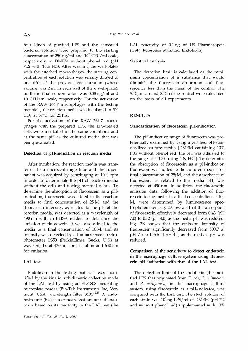
four kinds of purified LPS and the sonicated
bacterial solution were prepared to the starting
concentration of 250 ng/ml and 107 CFU/ml scale,
respectively, in DMEM without phenol red (pH
7.2) with 10% FBS. After washing the well-plates
with the attached macrophages, the starting con-
centration of each solution was serially diluted to
one fifth of the previous concentration (whose
volume was 2 ml in each well of the 6 well-plate),
until the final concentration was 0.08 ng/ml and
10 CFU/ml scale, respectively. For the activation
of the RAW 264.7 macrophages with the testing
materials, the reaction media was incubated in 5%
CO2 at 37°C for 25 hrs.
For the activation of the RAW 264.7 macro-
phages with the prepared LPS, the LPS-treated
cells were incubated in the same conditions and
at the same pH as the cultured media that was
being evaluated.
Detection of pH-indication in reaction media
After incubation, the reaction media was trans-
ferred to a microcentrifuge tube and the super-
natant was acquired by centrifuging at 1000 rpm
in order to determinate the pH of reaction media
without the cells and testing material debris. To
determine the absorption of fluorescein as a pH-
indication, fluorescein was added to the reaction
media to final concentration of 25 M, and the
fluorescein intensity, as related to the pH of the
reaction media, was detected at a wavelength of
490 nm with an ELISA reader. To determine the
emission of fluorescein, it was also added to the
media to a final concentration of 10 M, and its
intensity was detected by a luminescence spectro-
photometer LS50 (PerkinElmer, Bucks, U.K) at
wavelengths of 430 nm for excitation and 630 nm
for emission.
LAL test
Endotoxin in the testing materials was quan-
tified by the kinetic turbidimetric collection mode
of the LAL test by using an EL× 808 incubating
microplate reader (Bio-Tek Instruments Inc, Ver-
mont, USA; wavelength filter 340).12,13 A endo-
toxin unit (EU) is a standardized amount of endo-
toxin based on its reactivity in the LAL test (the
LAL reactivity of 0.1 ng of US Pharmacopeia
(USP) Reference Standard Endotoxin).
Statistical analysis
The detection limit is calculated as the mini-
mum concentration of a substance that would
diminish the fluorescein absorption and fluo-
rescence less than the mean of the control. The
S.D., mean and S.D. of the control were calculated
on the basis of all experiments.
RESULTS
Standardization of fluorescein pH-indication
The pH-indicative range of fluorescein was pre-
ferentially examined by using a certified pH-stan-
dardized culture media [DMEM containing 10%
FBS without phenol red; the pH was adjusted to
the range of 4.0-7.0 using 1 N HCl]. To determine
the absorption of fluorescein as a pH-indicator,
fluorescein was added to the cultured media to a
final concentration of 25μM, and the absorbance of
fluorescein, as related to the media pH, was
detected at 490 nm. In addition, the fluorescein
emission data, following the addition of fluo-
rescein to the media to a final concentration of 10μ
M, were determined by luminescence spec-
trophotometer. Fig. 2A reveals that the absorption
of fluorescein effectively decreased from 0.43 (pH
7.0) to 0.12 (pH 4.0) as the media pH was reduced.
Fig. 2B shows that the emission intensity of
fluorescein significantly decreased from 500.7 at
pH 7.5 to 145.6 at pH 4.0, as the media's pH was
reduced.
Comparison of the sensitivity to detect endotoxin
in the macrophage culture system using fluores-
cein pH indication with that of the LAL test
The detection limit of the endotoxin (the puri-
fied LPS that originated from E. coli, S. minnesota
and P. aeruginosa) in the macrophage culture
system, using fluorescein as a pH-indicator, was
compared with the LAL test. The stock solution of
each strain was 105 ng LPS/ml of DMEM (pH 7.2
and without phenol red) supplemented with 10%

In Vitro Bioassay of Endotoxin
Yonsei Med J Vol. 46, No. 2, 2005
FBS. The detection limit of this study was 1 ng/ml
LPS of E. coli, S. minnesota, and P. aeruginosa in the
case of both the fluorescein absorption and fluore-
scence assay (Fig. 5). The absorption and fluore-
scence intensity of fluorescein in the control media
without LPS was 0.44 ± 0.008 and 560 ± 20 (mean ±
S.E.M.), respectively. Fig. 3A establishes that the
absorption of fluorescein in the cultured media
was significantly reduced with the increasing LPS
concentration (on the whole scale from 1 to 500
ng/ml) of E. coli, S. minnesota, and P. aeruginosa.
At a 1 ng/ml LPS concentration of S. minnesota,
the absorption of fluorescein decreased from 0.44
of the non-treated control to 0.385, and at 500
ng/ml, it decreased to 0.2. The fluorescence data
also demonstrated the dose (LPS)-dependent
decrease of fluorescence intensity of fluorescein in
cultured media in all cases of E. coli, S. minnesota,
and P. aeruginosa (Fig. 3B). In the case of S.
minnesota, the emission of fluorescein was reduced
from 560 for the non-treated control to 481 at 1
ng/ml of LPS, and it was reduced to 285 at 500
ng/ml. The results in LAL test also had a similar
pattern to those results of our methods (Fig. 4).
Among the bacteria species examined, the
endotoxin unit (EU, the LAL reactivity of 0.1 ng
of Reference Standard Endotoxin) of S. minnesota
was detected from 0.49 of the control to 1.85 at 1
ng/ml of LPS and to 6982 at 500 ng/ml, which
was more sensitive than that of E. coli and P.
aeruginosa.
Fig. 2. The pH-indicative range of fluorescein wasexamined, in terms of both absorption and emission, bya certified pH-standardized culture media (DMEM con-taining 10% FBS without phenol red; pH 4.0 to 7.0). (A)The absorption of fluorescein decreased with the re-duced pH, from 0.43 at pH 7.0 to 0.12 at pH 4.0. (B) Theemission intensity of fluorescein decreased significantlyas the pH was reduced, from 500.7 at pH 7.5 to 145.6 atpH 4.0.
Fig. 3. Dose dependent diminution of fluorescein absorp-tion (A) and fluorescence (B) after LPS treatment [the LPSoriginated from three kinds of Gram-negative bacteria, S.minnesota ( ), E. coli ( ), and P. aeruginosa ( )]. Todetermine the absorption of fluorescein as a pH-indicator,fluorescein was added to the reaction media to a finalconcentration of 25 M, and the absorbance, as related tothe pH of the reaction media, was detected at 490 nm byan ELISA reader. To determine the fluorescein emission,fluorescein was also added to the media to a finalconcentration of 10 M, and its intensity was detected bya luminescence spectrophotometer at 430 nm of excitationand 630 nm of emission.
A
B
A
B

Dong Hee Lee, et al.
Yonsei Med J Vol. 46, No. 2, 2005
DISCUSSION
The Limulus lysate assay is currently being
widely used in most situations that are concerned
with the quantification of endotoxins because of
the assay's high sensitivity. This test has become
even more sensitive in recent years, and this is
due to a replacement of the classical clotting assay
by turbidimetric and colorimetric test formats and
to the determination of endpoints by kinetic
assays, as well as other additional improvements.
However, this assay is not specific for all kinds of
endotoxin, and it is drastically modified by the
presence of inhibitors or activators of the enzymes
included in the cascade reaction of the Limulus
assay.14,15
In addition, endotoxin undergoes chemi-
cal enzymatic degradation or modification in the
presence of certain host factors, such as lipopro-
tein.16,17 In most LAL formats, the highly pyro-
genic endotoxin and the much less pyrogenic 1,3-
β-D-glucans induce a similar LAL reaction.3
Previous reports have emphasized the variability
of the LAL test, particularly when using the test
to analyze complex biological specimens.
Urbaschek et al.18 reported that the LAL test with
FCS was hampered by factors that interfered with
the LAL reaction. For example, Eperon et al.19
tested various FCS lots with two distinct formats
of the LAL test (WAKO and Haemachem), and
with the cell culture test. Some FCS lots assayed
with two LAL test formats were positive on one,
but not on both types of tests. One single FCS lot
that was positive on both LAL assays induced
TNF generation in monocytoid cells. In light of
these previous findings, the determination of
endotoxin by the Limulus test, as in other bio-
logical assays, does not always reflect the true
amount of endotoxin.
In the current study, we have created an in vitro
alternative (the macrophage culture system using
fluorescein as the pH-indicator) to the LAL test,
and we compared the sensitivity and range for
detecting endotoxin of Gram-positive and Gram-
negative bacteria in a system of macrophage cul-
ture by using fluorescein as a pH-indicator with
the standard LAL test. The sensitivity to detect
endotoxin in the macrophage culture system
using fluorescein as pH-indicator for both the
absorption and emission wavelength was com-
pared with LAL test. Fig. 3 demonstrated that the
absorption and emission of fluorescein in the
reaction media were significantly reduced in
accordance with an increasing concentration of
LPS that originated from E. coli, S. minnesota, and
P. aeruginosa. The results of the LAL test also
demonstrated that the EU value was highly
dependent on the dose of LPS (Fig. 4). With a high
concentration of LPS, both the macrophage cul-
ture-method and LAL test showed similar pat-
terns for detecting the purified bacterial LPS, but
the correlation between the detection method and
LPS concentration was less significant than the
absorption at minute concentrations below the
unit ng/ml scale (Fig. 5). In the LAL test, there
was a good linear relationship between dose and
response with a small coefficient of intra-assay
variation at high concentrations. However, for
example, P. aeruginosa demonstrated a insensitive
dose-response relationship in the range from 0.04
to 1.0 ng LPS/ml (Fig. 5C). One of the most potent
LPSs on the LAL test was LPS from S. minnesota,
which was consistent with the results of the
macrophage culture system. It is quite within the
realms of possibility that for different endotoxins,
the LAL test is less sensitive than the macrophage
culture system for the detection of minute
concentrations of LPS that originate from different
bacteria.
In the current LAL study, the S. aureus bacteria
that were grown in pyrogen free media and had
no LPS contamination were not detected in a
Fig. 4. LAL test for detection of endotoxin unit (EU) inaccordance with LPS concentration. A serial dilution of 3different LPSs [S. minnesota ( ), E. coli ( ), and P.aeruginosa ( )] was assayed using the kinetic turbidime-
tric method.

In Vitro Bioassay of Endotoxin
Yonsei Med J Vol. 46, No. 2, 2005
suspension of 108 bacteria/ml (Fig. 6). For the
detection of S. aureus by the LAL test, the current
test exhibits control EU levels at all bacterial con-
centrations because Gram-positive bacteria have
no LPS in their cellular components. Thus, this
result supports the concept that the cell culture
test has a higher degree of specificity than the
LAL tests for the detection of unknown endo-
toxins such as bacterial particles that originate
from Gram-positive bacteria. Monocytes are
thought to be primary target cells for LPS, and
they are the main source of inflammatory cyto-
kines that are induced during bacterial infection.20Apart from LPS, other components of Gram-nega-
tive and Gram-positive bacteria, e.g. muramyl
peptide21 or lipoteichoic acid,22 are known to in-
teract with monocytes and macrophages and to
stimulate cytokine secretion.23
For example,
Moesby et al.24 reported that the production of
IL-6 by Mono Mac 6 (MM6) cells was a reliable
indicator for LPS stimulation. Although the MM6
assay showed a dose-response relationship
between LPS and IL-6 over a wide concentration
range (2.5-1000 pg/ml), in the case of Gram-
positive bacteria, a detection limit of approxi-
mately 3 × 10⁵ CFU/ml S. aureus was obtained,
which is comparable to the detection limit of the
macrophage-fluorescein bioassay in our study (103
CFU/ml). In the current study, S. aureus, a Gram-
positive bacterium, demonstrated no significant
reduction of absorption until the concentration
reached 102CFU/ml (lysed bacteria) scale. How-
ever, the absorption decreased significantly at
higher bacterial concentrations (> 103CFU/ml
Fig. 5. Comparison of the macrophage culture system-method and the LAL test at minute concentrations of LPSbelow the unit ng/ml scale [S. minnesota ( ), E. coli ( ),and P. aeruginosa ( )]. Absorption (A) and fluorescenceintensity (B) of fluorescein in the reaction media and theEU values (C) of 3 different LPSs were detected and com-pared with each other in the order of magnitude increasesfor concentration from 0.04 EU to 5 EU. The asterisk in-dicates statistical significance at p 0.05, as compared withuntreated controls. Data represent mean values (± S.D.) oftriplicate samples per condition for the experimental
method.
Fig. 6. Comparison of the macrophage culture system andthe LAL test, in terms of sensitivity and specificity tosonicated S. aureus (Gram-positive bacteria) extracts.Absorption ( ) of fluorescein in the reaction media andEU values ( ) of the lysed S. aureus extracts weredetected and compared with each other in the order ofmagnitude increases for concentrations from 10
2CFU/ml
scale to 107CFU/ml scale (n=4).
A
B
C

Dong Hee Lee, et al.
Yonsei Med J Vol. 46, No. 2, 2005
scale, Fig. 6). A simple in vitro assay that is less
susceptible to nonspecific interference and has
sensitivity to various pyrogens comparable to that
of the rabbit pyrogen test or even better would be
preferable. The current study might be a good
supplement to the LAL test for the detection of
LPS, Gram-negative and Gram-positive bacteria.
In conclusion, the results in the present study
indicated that the macrophage culture system-
method presented in this study demonstrates a
significant advantage in terms of its sensitivity
and its ability to detect unknown endotoxins such
as the bacterial particles that originate from Gram-
positive bacteria.
REFERENCES
1. Ulevitch RJ, Tobias PS. Receptor-dependent mecha-
nisms of cell stimulation by bacterial endotoxin. Annu
Rev Immunol 1995;13:437-57.
2. Iwanaga S, Morita T, Harada T, Nakamura S, Niwa M,
Takada K, et al. Chromogenic substrates for horseshoe
crab clotting enzyme. Its application for the assay of
bacterial endotoxins. Haemostasis 1978;7:183-8.
3. Iwanaga S. The limulus clotting reaction. Curr Opin
Immunol 1993;5:74-82.
4. Rietschel ET, Brade H, Brade L, Brandenburg K, Schade
U, Seydel U, et al. Lipid A, the endotoxic center of
bacterial lipopolysaccharides: relation of chemical struc-
ture to biological activity. Prog Clin Biol Res 1987;231:25-
53.
5. Laude-Sharp M, Haeffner-Cavaillon N, Caroff M,
Lantreibecq F, Pusineri C, Kazatchkine MD. Disso-
ciation between the interleukin 1-inducing capacity and
Limulus reactivity of lipopolysaccharides from Gram-
negative bacteria. Cytokine 1990;2:253-8.
6. Eperon S, De Groote D, Werner-Felmayer G, Jungi TW.
Human monocytoid cell lines as indicators of endoto-
xin: comparison with rabbit pyrogen and Limulus amoe-
bocyte lysate assay. J Immunol Methods 1997;207:135-45.
7. Werner-Felmayer G, Baier-Bitterlich G, Fuchs D, Hausen
A, Murr C, Reibnegger G, et al. Detection of bacterial
pyrogens on the basis of their effects on gamma inter-
feron-mediated formation of neopterin or nitrite in cul-
tured monocyte cell lines. Clin Diagn Lab Immunol
1995;2:307-13.
8. Jungi TW. A turbidimetric assay in an ELISA reader for
the determination of mononuclear phagocyte procoa-
gulant activity. J Immunol Methods 1990;133:21-9.
9. McCoy KL. Contribution of endosomal acidification to
antigen processing. Semin Immunol 1990;2:239-46.
10. Mellman I, Fuchs R, Helenius A. Acidification of the
endocytic and exocytic pathways. Annu Rev Biochem
1986;55:663-700.
11. Sjoback R, Nygren J, Kubista M. Absorption and Fluo-
rescence Properties of Fluorescein. Spectrochim Acta A
1995;51:7-12.
12. Cooper JF. Validation of bacterial endotoxin test
methods. LAL Times 1999;6:1-4.
13. FDA Interim Guidance (1991). U.S.FDA, Guideline on
validation of the Limulus amebocyte lysate test as an
in-process endotoxin test for human and animal paren-
teral drugs, biological products and medical devices,
DHHS, Dec. 1987 and Interim Guidance, 1991.
14. Levin J, Tomasulo PA, Oser RS. Detection of endotoxin
in human blood and demonstration of an inhibitor. J Lab
Clin Med 1970;75:903-11.
15. Takagi K, Moriya A, Tamura H, Nakahara C, Tanaka S,
Fujita V, et al. Quantitative measurement of endotoxin
in human blood using synthetic chromogenic substrate
for horseshoe crab clotting enzyme: a comparison of
methods of blood sampling and treatment. Thromb Res
1981;23:51-7.
16. Peterson AA, Munford RS. Dephosphorylation of the
lipid A moiety of Escherichia coli lipopolysaccharide by
mouse macrophages. Infect Immun 1987;55:974-8.
17. Ulevitch RJ. Interaction of bacterial lipopolysaccharide
and plasma high density lipoproteins. In: Berry LJ,
editor. Handbook of endotoxin. Amsterdam: Elsvier;
1985. p.372-88.
18. Urbaschek B, Becker KP, Ditter B, Urbaschek R. Quanti-
fication of endotoxin and sample-related interferences in
human plasma and cerebrospinal fluid by using a
kinetic Limulus amoebocyte lysate microtiter test. In:
Leive L, editor. Microbiology. Washington DC: Ameri-
can Society for Microbiology; 1985. p.39-42.
19. Eperon S, Jungi TW. The use of human monocytoid lines
as indicators of endotoxin. J Immunol Methods 1996;194:
121-9.
20. Kreutz M, Ackermann U, Hauschildt S, Krause SW,
Riedel D, Bessler W, et al. A comparative analysis of
cytokine production and tolerance induction by bacterial
lipopeptides, lipopolysaccharides and Staphylococcus
aureus in human monocytes. Immunology 1997;92:396-
401.
21. Johannsen L, Obal F Jr, Kapas L, Kovalzon V, Krueger
JM. Somnogenic activity of muramyl peptide-derived
immune adjuvants. Int J Immunopharmacol 1994;16:
109-16.
22. Himanen JP, Pyhala L, Olander RM, Merimskaya O,
Kuzina T, Lysyuk O, et al. Biological activities of lipotei-
choic acid and peptidoglycan-teichoic acid of Bacillus
subtilis 168 (Marburg). J Gen Microbiol 1993;139:2659-65.
23. Freudenberg MA, Galanos C. Tumor necrosis factor
alpha mediates lethal activity of killed Gram-negative
and Gram-positive bacteria in D-galactosamine-treated
mice. Infect Immun 1991;59:2110-5.
24. Moesby L, Jensen S, Hansen EW, Christensen JD. A
comparative study of Mono Mac 6 cells, isolated mono-
nuclear cells and Limulus amoebocyte lysate assay in
pyrogen testing. Int J Pharm 1999;191:141-9.