Invasive Pneumococcal Disease and Influenza …Invasive Pneumococcal Disease and Influenza Activity...
15
Invasive Pneumococcal Disease and Influenza Activity in a Pediatric Population: Impact of PCV13 Vaccination in Pandemic and Nonpandemic Influenza Periods Sergi Hernández, a Carmen Muñoz-Almagro, b,c,d,e Pilar Ciruela, a,b Núria Soldevila, b,f Conchita Izquierdo, a Maria Gemma Codina, g Alvaro Díaz, h Fernando Moraga-Llop, g Juan José García-García, b,c,d Ángela Domínguez, f,b Working Group of Projects PI11/02081 and PI11/2345 a Agència de Salut Pública de Catalunya, Generalitat de Catalunya, Barcelona, Spain b CIBER de Epidemiología y Salud Pública (CIBERESP), Instituto de Salud Carlos III, Madrid, Spain c Malalties Prevenibles amb Vacunes, Institut de Recerca Sant Joan de Déu, Esplugues de Llobregat, Spain d Hospital Sant Joan de Déu Barcelona, Universitat de Barcelona, Barcelona, Spain e Departament de Medicina, Universitat Internacional de Catalunya, Barcelona, Spain f Departament de Medicina, Universitat de Barcelona, Barcelona, Spain g Hospital Universitari Vall d'Hebron, Barcelona, Spain h Hospital de Nens, Barcelona, Spain ABSTRACT The objective of this study was to analyze the incidence, clinical presen- tation, and severity of invasive pneumococcal disease (IPD)-causing serotypes and the impact of the 13-valent pneumococcal conjugate vaccination during epidemic and nonepidemic influenza periods in Catalonia, Spain. This was a prospective study in persons aged 18 years diagnosed with IPD between 2012 and 2015 in three Catalan pediatric hospitals. IPD was defined as clinical infection together with isolation of Streptococcus pneumoniae by culture and/or detection by reverse transcription-PCR in a normally sterile sample. Incidence rate ratios (IRRs) and the fraction of IPD prevented associated with 13-valent pneumococcal conjugate vaccine (PCV13) were calculated. The bivariate analysis used the 2 test and the mul- tivariate analysis nonconditional logistic regression. A total of 229 cases of IPD were recorded. The incidence was higher during influenza epidemic periods (IRR, 2.7; 95% confidence interval [CI], 2.05 to 3.55; P 0.001), especially for pneumonia (IRR, 3.25; 95% CI, 2.36 to 4.47; P 0.001), with no differences in the distribution of pneumo- coccal serotypes. Complications during admission and sequel at discharge were greater during epidemic periods (adjusted odds ratio [aOR], 2.00; 95% CI, 1.06 to 3.77; P 0.03) than at nonepidemic periods (aOR, 3.38; 95% CI, 1.37 to 8.29; P 0.01). The prevented fraction for the population (PFp) of IPD in children aged 7 to 59 months was 48% to 49.4%. The PFp was higher in influenza epidemic than non- epidemic periods and increased when 2 doses of PCV13 or 1 after 24 months were administered. Influenza virus circulation increases the incidence of IPD in per- sons aged 18 years. In influenza epidemic periods, IPD cases were more severe. In- creased PCV13 coverage might increase the fraction of IPD prevented in epidemic and nonepidemic periods. KEYWORDS 13-valent pneumococcal conjugate vaccine, invasive pneumococcal disease, highly invasive serotypes, influenza virus, seasonality I nvasive pneumococcal disease (IPD) is a major cause of morbidity and mortality in adults and children and fluctuates seasonally during the winter months in temperate countries (1, 2). Seasonality affects both the disease incidence and the clinical presen- Citation Hernández S, Muñoz-Almagro C, Ciruela P, Soldevila N, Izquierdo C, Codina MG, Díaz A, Moraga-Llop F, García-García JJ, Domínguez Á, Working Group of Projects PI11/ 02081 and PI11/2345. 2019. Invasive pneumococcal disease and influenza activity in a pediatric population: impact of PCV13 vaccination in pandemic and nonpandemic influenza periods. J Clin Microbiol 57:e00363- 19. https://doi.org/10.1128/JCM.00363-19. Editor Daniel J. Diekema, University of Iowa College of Medicine Copyright © 2019 American Society for Microbiology. All Rights Reserved. Address correspondence to Sergi Hernández, [email protected]. Received 6 March 2019 Returned for modification 8 April 2019 Accepted 2 June 2019 Accepted manuscript posted online 12 June 2019 Published EPIDEMIOLOGY crossm August 2019 Volume 57 Issue 8 e00363-19 jcm.asm.org 1 Journal of Clinical Microbiology 26 July 2019 on April 26, 2020 by guest http://jcm.asm.org/ Downloaded from
Transcript of Invasive Pneumococcal Disease and Influenza …Invasive Pneumococcal Disease and Influenza Activity...
Invasive Pneumococcal Disease and Influenza Activity in aPediatric Population: Impact of PCV13 Vaccination inPandemic and Nonpandemic Influenza Periods
Sergi Hernández,a Carmen Muñoz-Almagro,b,c,d,e Pilar Ciruela,a,b Núria Soldevila,b,f Conchita Izquierdo,a
Maria Gemma Codina,g Alvaro Díaz,h Fernando Moraga-Llop,g Juan José García-García,b,c,d Ángela Domínguez,f,b WorkingGroup of Projects PI11/02081 and PI11/2345
aAgència de Salut Pública de Catalunya, Generalitat de Catalunya, Barcelona, SpainbCIBER de Epidemiología y Salud Pública (CIBERESP), Instituto de Salud Carlos III, Madrid, SpaincMalalties Prevenibles amb Vacunes, Institut de Recerca Sant Joan de Déu, Esplugues de Llobregat, SpaindHospital Sant Joan de Déu Barcelona, Universitat de Barcelona, Barcelona, SpaineDepartament de Medicina, Universitat Internacional de Catalunya, Barcelona, SpainfDepartament de Medicina, Universitat de Barcelona, Barcelona, SpaingHospital Universitari Vall d'Hebron, Barcelona, SpainhHospital de Nens, Barcelona, Spain
ABSTRACT The objective of this study was to analyze the incidence, clinical presen-tation, and severity of invasive pneumococcal disease (IPD)-causing serotypes andthe impact of the 13-valent pneumococcal conjugate vaccination during epidemicand nonepidemic influenza periods in Catalonia, Spain. This was a prospective studyin persons aged �18 years diagnosed with IPD between 2012 and 2015 in threeCatalan pediatric hospitals. IPD was defined as clinical infection together withisolation of Streptococcus pneumoniae by culture and/or detection by reversetranscription-PCR in a normally sterile sample. Incidence rate ratios (IRRs) andthe fraction of IPD prevented associated with 13-valent pneumococcal conjugatevaccine (PCV13) were calculated. The bivariate analysis used the �2 test and the mul-tivariate analysis nonconditional logistic regression. A total of 229 cases of IPD wererecorded. The incidence was higher during influenza epidemic periods (IRR, 2.7; 95%confidence interval [CI], 2.05 to 3.55; P � 0.001), especially for pneumonia (IRR, 3.25;95% CI, 2.36 to 4.47; P � 0.001), with no differences in the distribution of pneumo-coccal serotypes. Complications during admission and sequel at discharge weregreater during epidemic periods (adjusted odds ratio [aOR], 2.00; 95% CI, 1.06 to3.77; P � 0.03) than at nonepidemic periods (aOR, 3.38; 95% CI, 1.37 to 8.29; P �
0.01). The prevented fraction for the population (PFp) of IPD in children aged 7 to59 months was 48% to 49.4%. The PFp was higher in influenza epidemic than non-epidemic periods and increased when �2 doses of PCV13 or �1 after 24 monthswere administered. Influenza virus circulation increases the incidence of IPD in per-sons aged �18 years. In influenza epidemic periods, IPD cases were more severe. In-creased PCV13 coverage might increase the fraction of IPD prevented in epidemicand nonepidemic periods.
KEYWORDS 13-valent pneumococcal conjugate vaccine, invasive pneumococcaldisease, highly invasive serotypes, influenza virus, seasonality
Invasive pneumococcal disease (IPD) is a major cause of morbidity and mortality inadults and children and fluctuates seasonally during the winter months in temperate
countries (1, 2). Seasonality affects both the disease incidence and the clinical presen-
Citation Hernández S, Muñoz-Almagro C,Ciruela P, Soldevila N, Izquierdo C, Codina MG,Díaz A, Moraga-Llop F, García-García JJ,Domínguez Á, Working Group of Projects PI11/02081 and PI11/2345. 2019. Invasivepneumococcal disease and influenza activity ina pediatric population: impact of PCV13vaccination in pandemic and nonpandemicinfluenza periods. J Clin Microbiol 57:e00363-19. https://doi.org/10.1128/JCM.00363-19.
Editor Daniel J. Diekema, University of IowaCollege of Medicine
Copyright © 2019 American Society forMicrobiology. All Rights Reserved.
Address correspondence to Sergi Hernández,[email protected].
Received 6 March 2019Returned for modification 8 April 2019Accepted 2 June 2019
Accepted manuscript posted online 12June 2019Published
EPIDEMIOLOGY
crossm
August 2019 Volume 57 Issue 8 e00363-19 jcm.asm.org 1Journal of Clinical Microbiology
26 July 2019
on April 26, 2020 by guest
http://jcm.asm
.org/D
ownloaded from

tation. Reports have described an increased incidence of bacteremic pneumonia butnot of other clinical presentations during the winter months (3–5).
Environmental factors, such as temperature, humidity, pollution, and hours ofdaylight, help explain the seasonality (2, 6). Likewise, the circulation of respiratoryviruses during the winter months, especially the influenza virus, respiratory syncytialvirus (RSV), and metapneumovirus, have been associated with an increased incidenceof IPD, especially in children (2, 7). Although the mechanisms of interaction betweenrespiratory viruses and Streptococcus pneumoniae at the host level have been widelydescribed (8–11), the proportion of episodes of IPD attributable to the circulation ofrespiratory viruses is not clear (12, 13).
After the 2009 influenza pandemic, many studies showed that the influenza virusnot only increases the incidence of invasive pneumococcal pneumonia but also may bea factor influencing severity (4, 14–16).
Influenza virus infection favors the nasopharyngeal colonization of S. pneumoniae inchildren (3), which is a critical step in the subsequent development of IPD. However, therelationship between influenza virus and IPD-causing S. pneumoniae serotypes isunclear. Some reports have linked prior influenza virus infection with a subsequentepisode of IPD caused by highly invasive serotypes (7), while other authors link it withIPD produced by less invasive serotypes (17–19). Influenza epidemics have been shownto affect the distribution of IPD-causing serotypes (20).
The introduction of the pneumococcal conjugated heptavalent vaccine (PCV7) in2001 (21), the 10-valent conjugate vaccine (PCV10) in 2009 (22), and the 13-valentconjugate vaccine (PCV13) in 2010 (23) was associated with a significant reduction inIPD and a change in the distribution of the main disease-causing S. pneumoniaeserotypes (24). (25). In Catalonia, Spain, the PCV13 was not included in the childhoodvaccination schedule financed by the public health system until July 2016. In the 2012to 2015 study period, the estimated PCV13 coverage in children aged 7 to 59 monthsin Catalonia was 63% (26).
The aim of this study was to analyze variations in the incidence, clinical presentation,severity, and serotypes associated with IPD in Catalonia after the introduction of the13-valent conjugate vaccine and the impact of vaccination, measured as the fraction ofIPD prevented in the population, during epidemic and nonepidemic influenza periods.
MATERIALS AND METHODSData confidentiality and ethical aspects. No diagnostic tests were made or samples taken from any
participant in addition to those required by routine care. The study complies with the principles of theDeclaration of Helsinki and the legal structure in respect to international human rights and biomedicineand protection of personal data laws.
The Ethics Committee of Hospital Sant Joan de Déu approved the study. Informed consent signed byparents or legal guardians was given for all participants. All data were treated as confidential, and recordswere accessed anonymously.
Study design. A prospective study was conducted in persons aged �18 years diagnosed with IPDbetween 1 January 2012 and 31 December 2015 attended in three pediatric hospitals in Catalonia, Spain,namely, Hospital Sant Joan de Déu, Hospital Maternoinfantil Vall d’Hebrón, and Hospital de Nens deBarcelona. These hospitals are responsible for 20%, 8.5%, and 3%, respectively, of total hospitaldischarges in Catalonia of children aged �18 years, according to data from the Minimum Basic Data Setof Hospital Discharges (CMBDAH) (27), and the estimated reference population in this age group of thethree hospitals was from 422,666 in 2012 to 452,927 in 2015.
Selection of cases. Patients aged �18 years hospitalized due to IPD during the study period in theparticipating centers were included. IPD was defined as clinical infection together with the isolation byculture and/or detection of LytA gene DNA and an additional capsular gene of S. pneumoniae by reversetranscription-PCR (RT-PCR) in a normally sterile sample.
RT-PCR was carried out according to a standardized work protocol. Pediatricians requested RT-PCR incases of clinical suspicion in a patient with clinical and hospital admission criteria. RT-PCR was performedin the most appropriate sterile sample, namely, cerebrospinal fluid, pleural fluid, or plasma (never inwhole blood or in blood culture bottles), according to the clinical signs.
Identification, serotyping, and classification of S. pneumoniae. All strains isolated by culture wereserotyped using the Quellung reaction or dot blot by the National Centre for Microbiology, Majada-honda, Madrid, which allows 97 serotypes to be identified.
Capsular typing of all culture-negative and PCR-positive samples was performed using two methodsdepending on the amount of S. pneumoniae DNA available. If the amount was low (detection of LytAgene DNA and an additional capsular gene of S. pneumoniae by RT-PCR with the cycle threshold [CT] of
Hernández et al. Journal of Clinical Microbiology
August 2019 Volume 57 Issue 8 e00363-19 jcm.asm.org 2
on April 26, 2020 by guest
http://jcm.asm
.org/D
ownloaded from
�30 cycles), a previously described, real-time multiplex PCR technique that detects all pneumococcalcapsular types and differentiates serotypes 1, 3, 4, 5, 6A/C, 6B/D, 7F/A, 8, 9V/A/N/L, 14, 15B/C, 18C/B, 19A,19F/B/C, 23A, and 23F was used (28). If the amount of S. pneumoniae DNA was high (PCR-positivesamples with CT of �30 cycles), sequential multiplex PCR combined with fragment analysis andautomated fluorescent capillary electrophoresis to differentiate serotypes [1, 2, 3, 4, 5, 6A/6B, 6C,6,7C/(7B/40), 7F/7A, 9N/9L, 9V/9A, 10A, 10F/(10C/33C), 11A/11D, 12F/(12A/44/46), 13, 16F, 17F, 18/(18A/18B/18C/18F), 19A, 19F, 20(20A/20B), 21, 22F/22A), 23A, 23B, 24/(24A/24B/24F), 31, 34, 35A/(35C/42), 35B,35F/47F, 38/25F, and 39] was used (29).
Since PCR does not differentiate between serotypes 6A and 6C; 7F and 7A; 9V, 9A, and 9N; and 19F,19B, and 19C, these serotypes were considered vaccine serotypes 6A, 7F, 9V, and 19F, respectively.PCR-positive samples that were negative for the serotypes included in the sequential multiplex PCR(including all vaccine serotypes) were classified as other nonvaccine serotypes (ONVS).
The serotypes found were classified into two groups according to their invasiveness, namely, highlyinvasive serotypes (HIS; serotypes 1, 3, 4, 5, 7F, 8, 9N, 9V, 12F, 14, 18C, 19A, and 22F) and the remainingserotypes (non-HIS), as described by various authors (30–34).
Demographic, clinical, and epidemiological variables. The following demographic, clinical, andepidemiological variables were recorded for each case: age, sex, date of birth, date of onset of symptoms,date of hospitalization, clinical form of IPD (meningitis, septic shock, pneumonia, complicated pneumo-nia, musculoskeletal infection, occult bacteremia, and others), in-hospital complications, mechanicalventilation, intensive care unit (ICU) admission and length of stay, risk medical conditions (sickle cellanemia; congenital or acquired asplenia; human immunodeficiency virus; cochlear implant; congenitalimmunodeficiency; chronic heart disease; chronic lung diseases, including asthma if treated with a riskdose of oral corticosteroids; cerebrospinal fluid fistula; chronic renal failure, including nephrotic syn-drome; immunosuppressive treatment or radiotherapy; solid organ transplant; transplantation of hema-topoietic progenitors; and diabetes mellitus), date and evolution at discharge (discharge withoutsequelae, sequelae, and death), and the history of vaccination with any pneumococcal conjugate vaccine.
Definition of influenza epidemic periods. Influenza epidemic periods were established accordingto the data provided by the Pla d’Informació de les Infeccions Respiratòries Agudes a Catalunya (PIDIRAC)which, during the winter season (weeks 40 to 20) obtains daily information on morbidity due to acuterespiratory infections through the population registry, including data from sentinel doctors throughoutCatalonia (35). The epidemic threshold for influenza virus is established as �100 cases/105 inhabitants,and influenza epidemic periods were defined as weeks in which this incidence was reached and the twosubsequent weeks (7).
Reference population and estimated vaccination coverage. The reference population of the threehospitals used to measure weekly incidence rates (IRs) during the epidemic and nonepidemic influenzaperiods was calculated according to population data from the Statistical Institute of Catalonia anddetermined by calculating the percentage of discharges of each hospital and each age group of thestudy in relation to the total number of hospital discharges in Catalonia for these age groups (27) andextrapolating the data to the entire population. The estimated reference population aged �18 years ofthe three hospitals was stable during the study period and varied from 422,666 (31.5% of the Catalanpopulation aged �18 years) in 2012, to 442,032 (31.8% of the Catalan population aged �18 years) in2013, to 453,419 (32.6% of the Catalan population aged �18 years) in 2014, and 452,927 (32.5% of theCatalan population aged �18 years) in 2015. No other pediatric hospitals in the region were growing orcontracting during the study period.
The vaccination coverage of the reference population aged 7 to 59 months was estimated yearlyaccording to the vaccination data obtained in children aged 7 to 59 months treated for causes other thanIPD in the study hospitals, as described elsewere (26). The vaccinated population was defined as childrenwho had received �1 dose of PCV13 or as children who had received �2 doses of PCV13 or �1 doseafter 24 months in order to evaluate the possible differences related to the number of PCV13 dosesreceived.
Statistical analysis. The incidence rate ratios (IRRs) of IPD were calculated between epidemic andnonepidemic periods. For categorical variables, differences between periods were analyzed usingPearson’s chi-square test or Fisher’s exact test, and for continuous variables the Student’s t test was used.The 95% confidence intervals (CIs) were calculated, and P values of �0.05 were considered statisticallysignificant. A bilateral distribution was assumed for all P values.
Multivariate analysis was performed using nonconditional logistic regression to estimate the asso-ciation between the severity of cases in epidemic and nonepidemic periods. The following variables wereintroduced into the model: ICU admission, complications, mechanical ventilation, sequelae at discharge,death, and age. The lack of collinearity of the independent variables was verified using the varianceinflation factor (36).
To analyze the impact of PCV13 on the incidence of IPD in children aged 7 to 59 months duringinfluenza epidemic periods, the fraction of IPD prevented in the total period and in the epidemic andnonepidemic periods was calculated using the formula: prevented fraction in the population � (IR inunvaccinated � IR in total population)/IR in unvaccinated (37).
The IR in the unvaccinated population was calculated by dividing the number of unvaccinated IPDcases caused by PCV13 serotypes in children aged 7 to 59 months by the estimated unvaccinatedpopulation according to the estimated vaccine coverage published (27) and the reference estimatedpopulation in the same age group.
Invasive Pneumococcal Disease and Influenza Activity Journal of Clinical Microbiology
August 2019 Volume 57 Issue 8 e00363-19 jcm.asm.org 3
on April 26, 2020 by guest
http://jcm.asm
.org/D
ownloaded from
The IR in the total population was calculated by dividing the number of all IPD cases caused by PCV13serotypes in children aged 7 to 59 months by the estimated population according to the referenceestimated population in the same age group.
The analysis was performed using the SPSS v.24 statistical package.
RESULTS
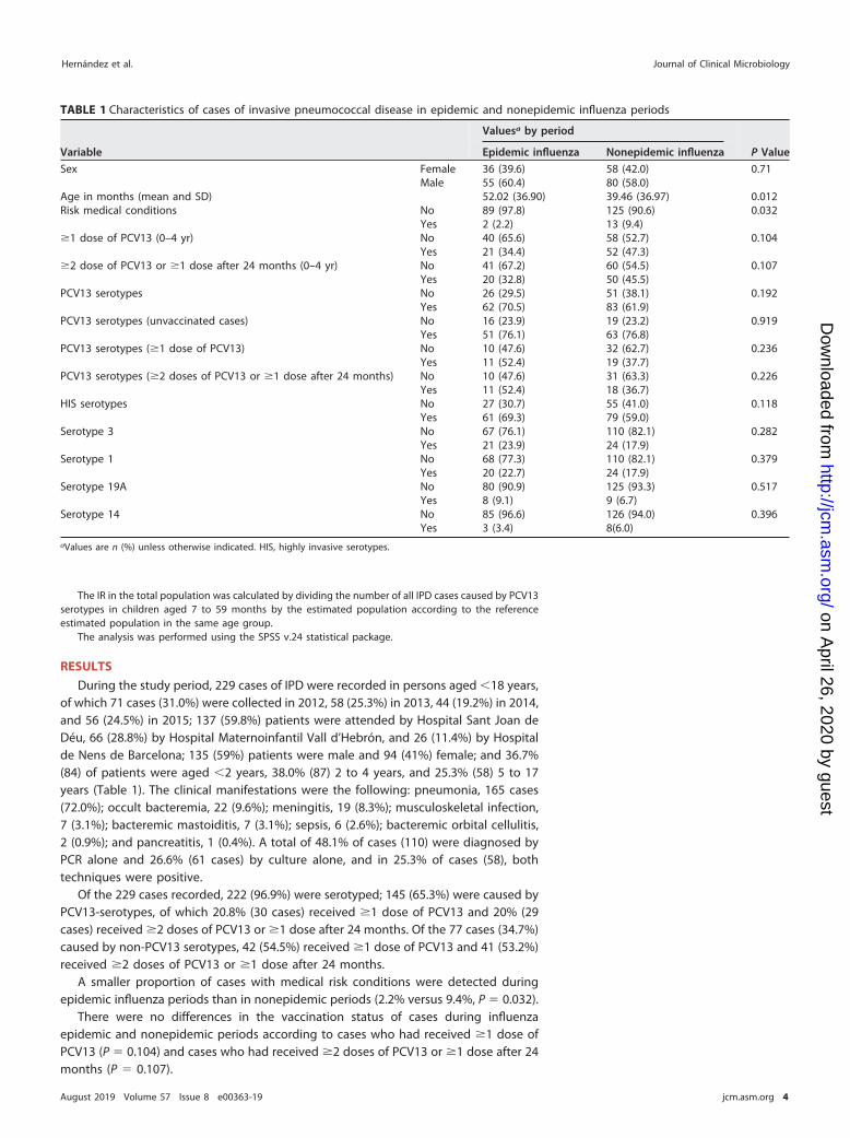
During the study period, 229 cases of IPD were recorded in persons aged �18 years,of which 71 cases (31.0%) were collected in 2012, 58 (25.3%) in 2013, 44 (19.2%) in 2014,and 56 (24.5%) in 2015; 137 (59.8%) patients were attended by Hospital Sant Joan deDéu, 66 (28.8%) by Hospital Maternoinfantil Vall d’Hebrón, and 26 (11.4%) by Hospitalde Nens de Barcelona; 135 (59%) patients were male and 94 (41%) female; and 36.7%(84) of patients were aged �2 years, 38.0% (87) 2 to 4 years, and 25.3% (58) 5 to 17years (Table 1). The clinical manifestations were the following: pneumonia, 165 cases(72.0%); occult bacteremia, 22 (9.6%); meningitis, 19 (8.3%); musculoskeletal infection,7 (3.1%); bacteremic mastoiditis, 7 (3.1%); sepsis, 6 (2.6%); bacteremic orbital cellulitis,2 (0.9%); and pancreatitis, 1 (0.4%). A total of 48.1% of cases (110) were diagnosed byPCR alone and 26.6% (61 cases) by culture alone, and in 25.3% of cases (58), bothtechniques were positive.
Of the 229 cases recorded, 222 (96.9%) were serotyped; 145 (65.3%) were caused byPCV13-serotypes, of which 20.8% (30 cases) received �1 dose of PCV13 and 20% (29cases) received �2 doses of PCV13 or �1 dose after 24 months. Of the 77 cases (34.7%)caused by non-PCV13 serotypes, 42 (54.5%) received �1 dose of PCV13 and 41 (53.2%)received �2 doses of PCV13 or �1 dose after 24 months.
A smaller proportion of cases with medical risk conditions were detected duringepidemic influenza periods than in nonepidemic periods (2.2% versus 9.4%, P � 0.032).
There were no differences in the vaccination status of cases during influenzaepidemic and nonepidemic periods according to cases who had received �1 dose ofPCV13 (P � 0.104) and cases who had received �2 doses of PCV13 or �1 dose after 24months (P � 0.107).
TABLE 1 Characteristics of cases of invasive pneumococcal disease in epidemic and nonepidemic influenza periods
Variable
Valuesa by period
P ValueEpidemic influenza Nonepidemic influenza
Sex Female 36 (39.6) 58 (42.0) 0.71Male 55 (60.4) 80 (58.0)
Age in months (mean and SD) 52.02 (36.90) 39.46 (36.97) 0.012Risk medical conditions No 89 (97.8) 125 (90.6) 0.032
Yes 2 (2.2) 13 (9.4)�1 dose of PCV13 (0–4 yr) No 40 (65.6) 58 (52.7) 0.104
Yes 21 (34.4) 52 (47.3)�2 dose of PCV13 or �1 dose after 24 months (0–4 yr) No 41 (67.2) 60 (54.5) 0.107
Yes 20 (32.8) 50 (45.5)PCV13 serotypes No 26 (29.5) 51 (38.1) 0.192
Yes 62 (70.5) 83 (61.9)PCV13 serotypes (unvaccinated cases) No 16 (23.9) 19 (23.2) 0.919
Yes 51 (76.1) 63 (76.8)PCV13 serotypes (�1 dose of PCV13) No 10 (47.6) 32 (62.7) 0.236
Yes 11 (52.4) 19 (37.7)PCV13 serotypes (�2 doses of PCV13 or �1 dose after 24 months) No 10 (47.6) 31 (63.3) 0.226
Yes 11 (52.4) 18 (36.7)HIS serotypes No 27 (30.7) 55 (41.0) 0.118
Yes 61 (69.3) 79 (59.0)Serotype 3 No 67 (76.1) 110 (82.1) 0.282
Yes 21 (23.9) 24 (17.9)Serotype 1 No 68 (77.3) 110 (82.1) 0.379
Yes 20 (22.7) 24 (17.9)Serotype 19A No 80 (90.9) 125 (93.3) 0.517
Yes 8 (9.1) 9 (6.7)Serotype 14 No 85 (96.6) 126 (94.0) 0.396
Yes 3 (3.4) 8(6.0)aValues are n (%) unless otherwise indicated. HIS, highly invasive serotypes.
Hernández et al. Journal of Clinical Microbiology
August 2019 Volume 57 Issue 8 e00363-19 jcm.asm.org 4
on April 26, 2020 by guest
http://jcm.asm
.org/D
ownloaded from
There were no differences in demographic or risk factors between vaccinated andunvaccinated cases in the same epidemic and nonepidemic influenza period, either incases who had received �1 dose of PCV13 or in cases who had received �2 doses ofPCV13 or �1 dose after 24 months (see Table S1 and S2 in the supplemental material).
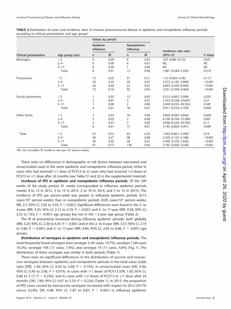
Incidence of IPD in epidemic and nonepidemic influenza periods. Of the 209weeks of the study period, 41 weeks corresponded to influenza epidemic periods(weeks 4 to 13 in 2012, 3 to 12 in 2013, 2 to 10 in 2014, and 3 to 14 in 2015). Theincidence of IPD per person-week was greater in influenza epidemic periods (0.13cases/105 person-weeks) than in nonepidemic periods (0.05 cases/105 person-weeks;IRR, 2.7; 95% CI, 2.05 to 3.55; P � 0.001). Significant differences were found in the 2- to4-year (IRR, 3.33; 95% CI, 2.12 to 5.19; P � 0.001) and 5- to 17-year (IRR, 4.39; 95% CI,2.53 to 7.63; P � 0.001) age groups but not in the �2-year age group (Table 2).
The IR of pneumonia increased during influenza epidemic periods, both globally(IRR, 3.25; 95% CI, 2.36 to 4.47; P � 0.001) and in the 2- to 4-year (IRR, 3.57; 95% CI, 2.19to 5.80; P � 0.001) and 5- to 17-year (IRR, 4.84; 95% CI, 2.64 to 8.86; P � 0.001) agegroups.
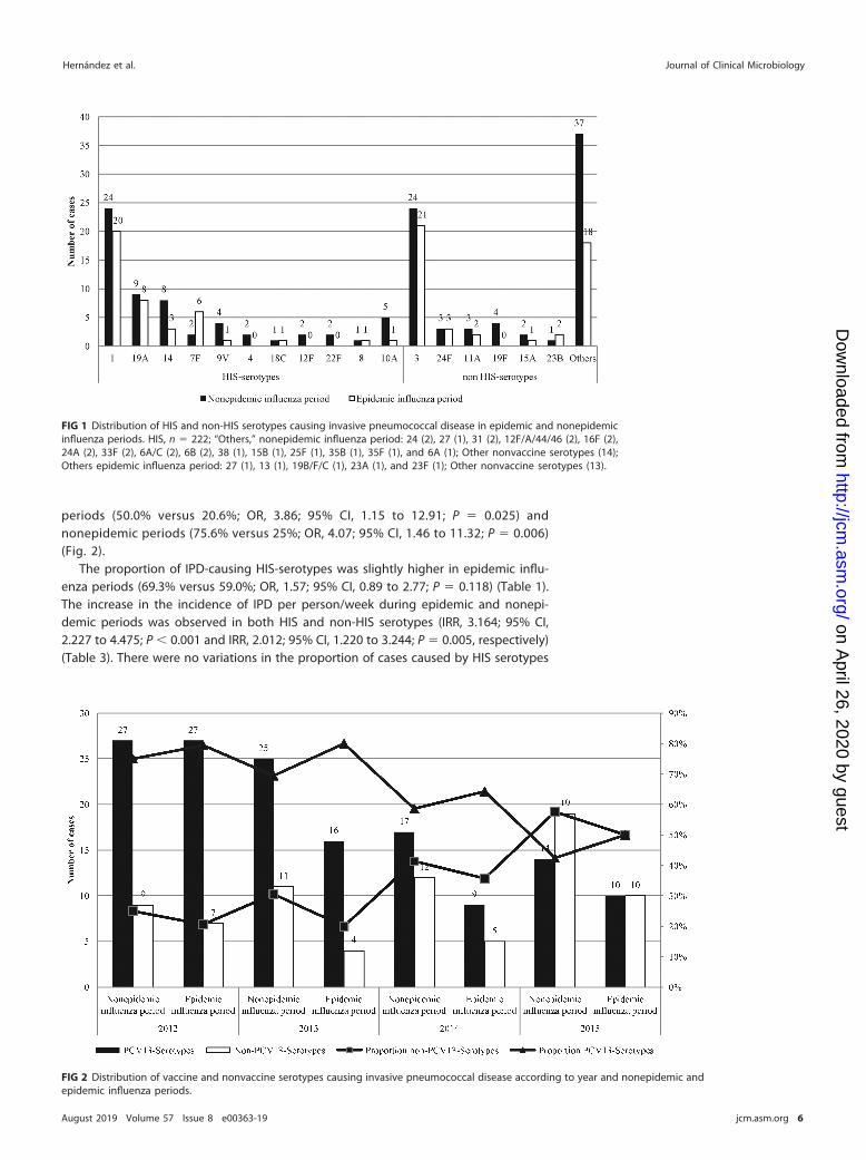
Distribution of serotypes in epidemic and nonepidemic influenza periods. Themost frequently found serotypes were serotype 3 (45 cases, 19.7%), serotype 1 (44 cases19.2%), serotype 19A (17 cases, 7.4%), and serotype 14 (11 cases, 4.8%) (Fig. 1). Thedistribution of these serotypes was similar in both periods (Table 1).
There were no significant differences in the distribution of vaccine and nonvac-cine serotypes between epidemic and nonepidemic periods in the total cases (oddsratio [OR], 1.46; 95% CI, 0.82 to 2.60; P � 0.192), in unvaccinated cases (OR, 0.96;95% CI, 0.45 to 2.06; P � 0.919), in cases with �1 doses of PCV13 (OR, 1.85; 95% CI,0.66 to 5.17; P � 0.236), and in cases with �2 doses of PCV13 or �1 dose after 24months (OR, 1.89; 95% CI, 0.67 to 5.33; P � 0.226) (Table 1). In 2015, the proportionof IPD cases caused by nonvaccine serotypes increased with respect to 2012 (54.7%versus 22.9%; OR, 4.08; 95% CI, 1.87 to 8.87; P � 0.001) in influenza epidemic
TABLE 2 Distribution of cases and incidence rates of invasive pneumococcal disease in epidemic and nonepidemic influenza periodsaccording to clinical presentation and age groupa
Clinical presentation Age group (yrs)
Values by period
Incidence rate ratio(95% CI) P value
Epidemicinfluenza
Nonepidemicinfluenza
n IR n IR
Meningitis �2 6 0.09 8 0.03 3.07 (0.88–10.10) 0.0512–4 0 0.00 4 0.01 NC NC5–17 0 0.00 1 0.00 NC NC
Total 6 0.01 13 0.00 1.891 (0.589–5.333) 0.214
Pneumonia �2 13 0.20 31 0.11 1.72 (0.825–3.38) 0.1132–4 34 0.24 39 0.07 3.572 (2.187–5.808) �0.0015–17 26 0.05 22 0.01 4.843 (2.639–8.964) �0.001
Total 73 0.10 92 0.03 3.251 (2.358–4.469) �0.001
Occult bacteremia �2 1 0.02 13 0.05 0.315 (0.007–2.099) 0.2552–4 2 0.01 3 0.01 2.732 (0.228–23.847) 0.315–17 1 0.00 2 0.00 2.049 (0.035–39.355) 0.581
Total 4 0.01 18 0.01 0.911 (0.224–2.764) 0.909
Other forms �2 2 0.03 10 0.04 0.820 (0.087–3.846) 0.8582–4 3 0.02 2 0.00 6.146 (0.704–73.589) 0.0615–17 3 0.01 3 0.00 4.098 (0.549–30.593) 0.11
Total 8 0.01 15 0.01 2.185 (0.802–5.491) 0.089
Total �2 22 0.33 62 0.23 1.454 (0.851–2.399) 0.142–4 39 0.27 48 0.08 3.329 (2.125–5.188) �0.0015–17 30 0.05 28 0.01 4.390 (2.535–7.626) �0.001
Total 91 0.13 138 0.05 2.702 (2.050–3.546) �0.001aNC, not calculable; IR, incidence rate (per 105 person-weeks).
Invasive Pneumococcal Disease and Influenza Activity Journal of Clinical Microbiology
August 2019 Volume 57 Issue 8 e00363-19 jcm.asm.org 5
on April 26, 2020 by guest
http://jcm.asm
.org/D
ownloaded from
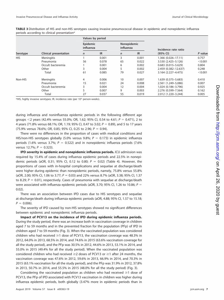
periods (50.0% versus 20.6%; OR, 3.86; 95% CI, 1.15 to 12.91; P � 0.025) andnonepidemic periods (75.6% versus 25%; OR, 4.07; 95% CI, 1.46 to 11.32; P � 0.006)(Fig. 2).
The proportion of IPD-causing HIS-serotypes was slightly higher in epidemic influ-enza periods (69.3% versus 59.0%; OR, 1.57; 95% CI, 0.89 to 2.77; P � 0.118) (Table 1).The increase in the incidence of IPD per person/week during epidemic and nonepi-demic periods was observed in both HIS and non-HIS serotypes (IRR, 3.164; 95% CI,2.227 to 4.475; P � 0.001 and IRR, 2.012; 95% CI, 1.220 to 3.244; P � 0.005, respectively)(Table 3). There were no variations in the proportion of cases caused by HIS serotypes
FIG 1 Distribution of HIS and non-HIS serotypes causing invasive pneumococcal disease in epidemic and nonepidemicinfluenza periods. HIS, n � 222; “Others,” nonepidemic influenza period: 24 (2), 27 (1), 31 (2), 12F/A/44/46 (2), 16F (2),24A (2), 33F (2), 6A/C (2), 6B (2), 38 (1), 15B (1), 25F (1), 35B (1), 35F (1), and 6A (1); Other nonvaccine serotypes (14);Others epidemic influenza period: 27 (1), 13 (1), 19B/F/C (1), 23A (1), and 23F (1); Other nonvaccine serotypes (13).
FIG 2 Distribution of vaccine and nonvaccine serotypes causing invasive pneumococcal disease according to year and nonepidemic andepidemic influenza periods.
Hernández et al. Journal of Clinical Microbiology
August 2019 Volume 57 Issue 8 e00363-19 jcm.asm.org 6
on April 26, 2020 by guest
http://jcm.asm
.org/D
ownloaded from
during influenza and noninfluenza epidemic periods in the following different agegroups �2 years (42.4% versus 55.0%; OR, 1.62; 95% CI, 0.54 to 4.61; P � 0.471), 2 to4 years (71.8% versus 68.1%; OR, 1.19; 95% CI, 0.47 to 3.02; P � 0.89), and 5 to 17 years(75.9% versus 78.6%; OR, 0.85; 95% CI, 0.25 to 2.96; P � 0.94).
There were no differences in the proportion of cases with medical conditions andHIS/non-HIS serotypes globally (5.0% versus 9.8%; P � 0.173) in epidemic influenzaperiods (1.6% versus 3.7%; P � 0.522) and in nonepidemic influenza periods (7.6%versus 12.7%; P � 0.323).
IPD severity in epidemic and nonepidemic influenza periods. ICU admission wasrequired by 15.4% of cases during influenza epidemic periods and 22.5% in nonepi-demic periods (aOR, 0.31; 95% CI, 0.12 to 0.80; P � 0.02) (Table 4). However, theproportions of cases with in-hospital complications and sequelae at discharge/deathwere higher during epidemic than nonepidemic periods, namely, 75.8% versus 55.8%(aOR, 2.00; 95% CI, 1.06 to 3.77; P � 0.03) and 22% versus 8.7% (aOR, 3.38; 95% CI, 1.37to 8.29; P � 0.01), respectively. Cases of pneumonia with sequelae at discharge/deathwere associated with influenza epidemic periods (aOR, 3.70; 95% CI, 1.26 to 10.86; P �
0.02).There was an association between IPD cases due to HIS serotypes and sequelae
at discharge/death during influenza epidemic periods (aOR, 4.88; 95% CI, 1.57 to 15.18;P � 0.006).
The severity of IPD caused by non-HIS serotypes showed no significant differencesbetween epidemic and nonepidemic influenza periods.
Impact of PCV13 on the incidence of IPD during epidemic influenza periods.During the study period, there was an increase both in vaccination coverage in childrenaged 7 to 59 months and in the prevented fraction for the population (PFp) of IPD inchildren aged 7 to 59 months (Fig. 3). When the vaccinated population was consideredchildren who had received �1 dose of PCV13, the vaccination coverage was 48.3% in2012, 64.0% in 2013, 68.5% in 2014, and 74.6% in 2015 (63.6% vaccination coverage forall the study period), and the PFp was 30.5% in 2012, 44.6% in 2013, 53.1% in 2014, and55.0% in 2015 (49.4% for all the study period). When the vaccinated population wasconsidered children who had received �2 doses of PCV13 or �1 after 24 months, thevaccination coverage was 47.6% in 2012, 59.6% in 2013, 66.9% in 2014, and 70.3% in2015 (63.1% vaccination for all the study period), and the PFp was 31.9% in 2012, 37.8%in 2013, 50.7% in 2014, and 55.5% in 2015 (48.0% for all the study period) (Fig. 3).
Considering the vaccinated population as children who had received �1 dose ofPCV13, the PFp of IPD associated with PCV13 vaccination in children was higher duringinfluenza epidemic periods, both globally (3.47% more in epidemic periods than in
TABLE 3 Distribution of HIS and non-HIS serotypes causing invasive pneumococcal disease in epidemic and nonepidemic influenzaperiods according to clinical presentationa
Serotype Clinical presentation
Values by period
Incidence rate ratio(95% CI) P value
Epidemicinfluenza
Nonepidemicinfluenza
n IR n IR
HIS Meningitis 1 0.001 3 0.001 1.366 (0.026–17.11) 0.757Pneumonia 56 0.078 65 0.022 3.530 (2.425–5.126) �0.001Occult bacteremia 1 0.001 6 0.002 0.683 (0.015–5.629) 0.804Other 3 0.004 5 0.002 2.459 (0.382–12.637) 0.248
Total 61 0.085 79 0.027 3.164 (2.227–4.475) �0.001
Non-HIS Meningitis 4 0.006 10 0.007 1.639 (0.375–5.683) 0.410Pneumonia 15 0.021 24 0.008 2.561 (1.249–5,086) 0.007Occult bacteremia 3 0.004 12 0.004 1,024 (0.186–3.796) 0.925Other 5 0.007 9 0.003 2.276 (0.599–7.564) 0.162
Total 27 0.037 55 0.019 2.012 (1.220–3.244) 0.005aHIS, highly invasive serotypes; IR, incidence rate (per 105 person-weeks).
Invasive Pneumococcal Disease and Influenza Activity Journal of Clinical Microbiology
August 2019 Volume 57 Issue 8 e00363-19 jcm.asm.org 7
on April 26, 2020 by guest
http://jcm.asm
.org/D
ownloaded from
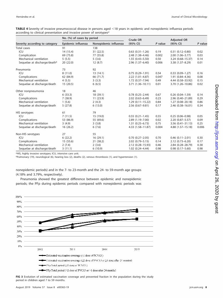
nonepidemic periods) and in the 7- to 23-month and the 24- to 59-month age groups(4.18% and 3.79%, respectively).
Pneumonia showed the greatest difference between epidemic and nonepidemicperiods; the PFp during epidemic periods compared with nonepidemic periods was
TABLE 4 Severity of invasive pneumococcal disease in persons aged �18 years in epidemic and nonepidemic influenza periodsaccording to clinical presentation and invasive power of serotypesa
Severity according to category
No. (%) of cases by periodCrude OR(95% CI) P value
Adjusted OR(95% CI) P valueEpidemic influenza Nonepidemic influenza
Total cases 91 138ICU 14 (15.4) 31 (22.5) 0.63 (0.31–1.26) 0.19 0.31 (0.12–0.80) 0.02Complication 69 (75.8) 77 (55.8) 2.48 (1.38–4.46) 0.002 2.00 (1.06–3.77) 0.03Mechanical ventilation 5 (5.5) 5 (3.6) 1.55 (0.43–5.50) 0.50 3.24 (0.68–15.37) 0.14Sequelae at discharge/deathb 20 (22.0) 12 (8.7) 2.96 (1.37–6.40) 0.006 3.38 (1.37–8.29) 0.01
Pneumonia 73 92ICU 8 (11.0) 13 (14.1) 0.75 (0.29–1.91) 0.54 0.33 (0.09–1.27) 0.16Complications 62 (84.9) 66 (71.7) 2.22 (1.01–4.87) 0.047 1.91 (0.84–4.36) 0.08Mechanical ventilation 4 (5.5) 3 (3.3) 1.72 (0.37–7.94) 0.49 4.44 (0.58–33.92) 0.15Sequelae at discharge/death 15 (20.5) 6 (6.5) 3.71 (1.36–10.11) 0.01 3.70 (1.26–10.86) 0.02
Other nonpneumonia 18 46ICU 6 (33.3) 18 (39.1) 0.78 (0.25–2.44) 0.67 0.26 (0.04–1.59) 0.14Complications 7 (38.9) 11 (23.9) 2.02 (0.63–6.49) 0.23 2.96 (0.40–21.89) 0.29Mechanical ventilation 1 (5.6) 2 (4.3) 1.29 (0.11–15.22) 0.84 1.27 (0.08–20.18) 0.86Sequelae at discharge/death 5 (27.8) 6 (13.0) 2.56 (0.67–9.81) 0.17 2.46 (0.38–16.01) 0.34
HIS serotypes 61 79ICU 7 (11.5) 15 (19.0) 0.55 (0.21–1.45) 0.55 0.25 (0.06–0.98) 0.05Complications 53 (86.9) 55 (69.6) 2.89 (1.19–7.00) 0.02 2.20 (0.87–5.57) 0.09Mechanical ventilation 3 (4.9) 3 (3.8) 1.31 (0.25–6.73) 0.75 3.56 (0.41–31.13) 0.25Sequelae at discharge/death 16 (26.2) 6 (7.6) 4.33 (1.58–11.87) 0.004 4.88 (1.57–15.18) 0.006
Non-HIS serotypes 27 55ICU 6 (22.2) 16 (29.1) 0.70 (0.27–2.05) 0.70 0.46 (0.11–2.01) 0.30Complications 15 (55.6) 21 (38.2) 2.02 (0.79–5.15) 0.14 2.12 (0.73–6.20) 0.17Mechanical ventilation 2 (7.4) 2 (3.6) 2.12 (0.28–15.93) 0.46 2.84 (0.28–28.79) 0.38Sequelae at discharge/death 3 (11.1) 6 (10.9) 1.02 (0.24–4.44) 0.98 0.98 (0.17–5.60) 0.98
aHIS, highly invasive serotypes; ICU, intensive care unit.bPulmonary (19), neurological (6), hearing loss (2), deaths (2), venous thrombosis (1), and hypertension (1).
FIG 3 Evolution of estimated vaccination coverage and prevented fraction in the population during the studyperiod in children aged 7 to 59 months.
Hernández et al. Journal of Clinical Microbiology
August 2019 Volume 57 Issue 8 e00363-19 jcm.asm.org 8
on April 26, 2020 by guest
http://jcm.asm
.org/D
ownloaded from
7.20% higher in children aged 7 to 59 months, 5.03% higher in children aged 7 to 23months, and 8.91% higher in children aged 24 to 59 months (Table 5).
Considering the vaccinated population as children who had received �2 dosesof PCV13 or �1 after 24 months, the PFp of IPD in children aged 7 to 59 monthsassociated with PCV13 vaccination was higher during influenza epidemic periods(9.17% more in epidemic periods than in nonepidemic periods) and in the7- to 23-month and the 24- to 59-month age groups (10.59% and 9.64%, respec-tively).
Meningitis only showed differences in the PFp between epidemic and nonepidemicperiods in the 7- to 59-month group (22.62% more in epidemic periods than innonepidemic periods).
Pneumonia showed differences between epidemic and nonepidemic periods; thePFp during epidemic periods compared with nonepidemic periods was 17% higherglobally, 10.17% higher in children aged 7 to 23 months, and 25.20% higher in childrenaged 24 to 59 months (Table 6).
The differences in the PFp of IPD in children aged 7 to 59 months associated withPCV13 vaccination between epidemic and nonepidemic periods were higher, bothglobally, in pneumonia and in all age groups, when the vaccinated population wasconsidered children who had received �2 doses of PCV13 or �1 after 24 months.
DISCUSSION
This study highlights the increase in the incidence of IPD in persons aged �18 yearsin influenza epidemic periods. Stratification by age showed the association was stron-gest in children aged 2 to 4 years and especially in the 5- to 17-year age group, butthere was no significant association in children aged �2 years. The annual PIDIRACreports show that during all the years studied, the highest cumulative incidence ofinfluenza infections was in the �4-year age group (35). Some reports have found anassociation between IPD and influenza (2, 13) but without having specifically analyzedchildren aged �2 years. Weinberger et al. (12) found a strong association between thecirculation of the respiratory syncytial virus (RSV) and episodes of IPD in children aged�2 years. During the period 2012 to 2015, the reports published by the MicrobiologicalNotification System of Catalonia (38) showed that 84.9% of cases of RSV infectionrecorded in Catalonia were in children aged �2 years and that the peak of RSV activitypreceded the influenza epidemic. The temporal coincidence between RSV and IPD inchildren aged �2 years could explain the high IR observed during nonepidemicinfluenza periods, which was much higher than the total IR in that period (0.23cases/105 person-weeks versus 0.05 cases/105 person-weeks), and why when consid-ering epidemic influenza periods and nonepidemic periods in this age group we foundno significant differences.
McCullers et al. (19) using animal models and Grijalva et al. (3) in a case-control studyshowed that the influenza virus increases the transmission of S. pneumoniae and,therefore, the risk of nasopharyngeal acquisition, a step prior to the development ofIPD. However, although we found a higher IR of IPD in epidemic periods globally, theincrease in the incidence during epidemic influenza periods compared with nonepi-demic periods was only statistically significant for the clinical presentation of pneumo-nia. Weinberger et al. (39) and Ben-Shimol et al. (40) postulated that viral respiratoryinfection increases susceptibility to pneumonia, an approach that would coincide withour results.
The great variability in IPD-causing serotypes found in our study, with none beingpredominant during the epidemic or nonepidemic influenza periods, suggests there isno relationship between the influenza virus and a specific S. pneumoniae serotype.Likewise, during the study period, the subtypes of viruses that cause influenza epidem-ics have varied (35), although a specific relationship with certain viral subtypes cannotbe ruled out. Launes et al. (20) found a significant decrease in the proportion of IPDcaused by serotype 1 during the 2009 pandemic; however, in the subsequent seasonalinfluenza season, caused by the same influenza A subtype virus, S. pneumoniae sero-
Invasive Pneumococcal Disease and Influenza Activity Journal of Clinical Microbiology
August 2019 Volume 57 Issue 8 e00363-19 jcm.asm.org 9
on April 26, 2020 by guest
http://jcm.asm
.org/D
ownloaded from
TAB
LE5
Prev
ente
dfr
actio
nof
inva
sive
pne
umoc
occa
ldi
seas
eca
used
by
sero
typ
esin
clud
edin
the
PCV1
3in
epid
emic
and
none
pid
emic
influ
enza
per
iods
acco
rdin
gto
clin
ical
pre
sent
atio
nan
dag
ein
child
ren
aged
7to
59m
onth
sa
Clin
ical
pre
sen
tati
onA
ge
gro
up(m
onth
s)
Val
ues
by
tota
lp
erio
dV
alue
sb
yep
idem
icin
fluen
zap
erio
ds
Val
ues
by
non
epid
emic
influ
enza
per
iod
sD
iffe
ren
cein
PFp
inep
idem
ican
dn
onep
idem
icp
erio
ds
(%)
IRin
unva
ccin
ated
pop
ulat
ion
IRin
tota
lp
opul
atio
n
Prev
ente
dfr
acti
onin
the
pop
ulat
ion
(%)
IRin
unva
ccin
ated
pop
ulat
ion
IRto
tal
pop
ulat
ion
Prev
ente
dfr
acti
onin
the
pop
ulat
ion
(%)
IRin
unva
ccin
ated
pop
ulat
ion
IRto
tal
pop
ulat
ion
Prev
ente
dfr
acti
onin
the
pop
ulat
ion
(%)
Men
ingi
tis7–
230.
031
0.00
971
.40.
053
0.01
571
.40.
026
0.00
771
.40.
0024
–59
0.00
00.
000
NC
0.00
00.
000
NC
0.00
00.
000
NC
NC
Tota
l0.
008
0.00
363
.60.
013
0.00
563
.60.
006
0.00
263
.60.
00
Pneu
mon
ia7–
230.
207
0.08
360
.00.
369
0.13
663
.20.
167
0.07
058
.25.
0324
–59
0.13
70.
078
43.1
0.35
80.
188
47.5
0.08
30.
051
38.6
8.91
Tota
l0.
152
0.08
047
.60.
354
0.17
251
.50.
102
0.05
744
.37.
20
Occ
ult
bac
tere
mia
7–23
0.03
10.
009
71.4
0.00
00.
000
NC
0.03
90.
011
71.4
NC
24–5
90.
014
0.00
561
.10.
018
0.00
761
.10.
013
0.00
561
.10.
00To
tal
0.01
80.
007
63.6
0.01
30.
005
63.6
0.01
90.
007
63.6
0.00
Oth
erfo
rms
7–23
0.02
10.
009
57.1
0.05
30.
015
71.4
0.01
30.
007
42.8
28.6
024
–59
0.00
40.
003
22.2
0.00
00.
007
NC
0.00
40.
002
61.1
NC
Tota
l0.
008
0.00
539
.30.
013
0.01
027
.20.
006
0.00
345
.4�
18.2
0
Tota
l7–
230.
290
0.11
062
.20.
475
0.16
665
.00.
245
0.09
660
.94.
1824
–59
0.15
50.
086
44.3
0.37
60.
202
46.3
0.10
10.
058
42.5
3.79
Tota
l0.
185
0.09
449
.40.
393
0.19
151
.50.
134
0.07
048
.03.
47aC
hild
ren
wer
eco
nsid
ered
vacc
inat
edif
they
had
rece
ived
�1
dose
ofPC
V13.
NC
,not
calc
ulab
le;I
R,in
cide
nce
rate
(per
105
per
son-
wee
ks);
PFp
,pre
vent
edfr
actio
nin
the
pop
ulat
ion.
Hernández et al. Journal of Clinical Microbiology
August 2019 Volume 57 Issue 8 e00363-19 jcm.asm.org 10
on April 26, 2020 by guest
http://jcm.asm
.org/D
ownloaded from
TAB
LE6
Prev
ente
dfr
actio
nof
inva
sive
pne
umoc
occa
ldi
seas
eca
used
by
sero
typ
esin
clud
edin
the
PCV1
3in
epid
emic
and
none
pid
emic
influ
enza
per
iods
acco
rdin
gto
clin
ical
pre
sent
atio
nan
dag
ein
child
ren
aged
7to
59m
onth
sa
Clin
ical
pre
sen
tati
onA
ge
gro
up(m
onth
s)
Val
ues
by
tota
lp
erio
dV
alue
sb
yep
idem
icin
fluen
zap
erio
ds
Val
ues
by
non
epid
emic
influ
enza
per
iod
s
Dif
fere
nce
inPF
pin
epid
emic
and
non
epid
emic
per
iod
s(%
)
IRin
unva
ccin
ated
pop
ulat
ion
IRin
tota
lp
opul
atio
n
Prev
ente
dfr
acti
onin
the
pop
ulat
ion
(%)
IRin
unva
ccin
ated
pop
ulat
ion
IRin
tota
lp
opul
atio
n
Prev
ente
dfr
acti
onin
the
pop
ulat
ion
(%)
IRin
unva
ccin
ated
pop
ulat
ion
IRin
tota
lp
opul
atio
n
Prev
ente
dfr
acti
onin
the
pop
ulat
ion
(%)
Men
ingi
tis7–
230.
009
0.00
367
.90.
000
0.00
0N
C0.
011
0.00
467
.9N
C24
–59
0.00
00.
000
NC
0.00
00.
000
NC
0.00
00.
000
NC
NC
Tota
l0.
008
0.00
187
.70.
013
0.00
010
0.0
0.00
60.
001
81.6
22.6
2
Pneu
mon
ia7–
230.
166
0.07
455
.40.
329
0.13
658
.70.
126
0.05
953
.310
.17
24–5
90.
133
0.07
841
.20.
347
0.18
845
.70.
080
0.05
136
.525
.20
Tota
l0.
144
0.07
746
.90.
349
0.17
250
.80.
095
0.05
443
.417
.00
Occ
ult
bac
tere
mia
7–23
0.01
80.
006
67.9
0.00
00.
000
NC
0.02
30.
007
67.9
NC
24–5
90.
014
0.00
559
.80.
017
0.00
759
.80.
013
0.00
559
.80.
00To
tal
0.01
50.
006
63.1
0.01
30.
005
63.1
0.01
60.
006
63.1
0.00
Oth
erfo
rms
7–23
0.00
90.
006
35.8
0.04
70.
015
67.9
0.00
00.
004
NC
NC
24–5
90.
003
0.00
319
.60.
000
0.00
7N
C0.
004
0.00
259
.8N
CTo
tal
0.00
50.
004
26.2
0.01
30.
010
26.2
0.00
30.
002
26.2
0.00
Tota
l7–
230.
203
0.08
956
.20.
376
0.15
159
.90.
161
0.07
454
.110
.59
24–5
90.
150
0.08
642
.40.
364
0.20
244
.50.
097
0.05
840
.69.
64To
tal
0.16
70.
087
48.0
0.37
50.
186
50.4
0.11
70.
063
46.1
9.17
aC
hild
ren
wer
eco
nsid
ered
vacc
inat
edif
they
had
rece
ived
�2
dose
sof
PCV1
3or
�1
dose
afte
r24
mon
ths.
NC
,not
calc
ulab
le;I
R,in
cide
nce
rate
(per
105
per
son-
wee
ks);
PFp
,pre
vent
edfr
actio
nin
the
pop
ulat
ion.
Invasive Pneumococcal Disease and Influenza Activity Journal of Clinical Microbiology
August 2019 Volume 57 Issue 8 e00363-19 jcm.asm.org 11
on April 26, 2020 by guest
http://jcm.asm
.org/D
ownloaded from
type 1 was detected in the same proportion as before. The results of our study supportthe idea that the circulation of influenza viruses is not related to the incidence ofvaccine or nonvaccine serotypes.
Some reports (17, 18) indicate that influenza virus infection has a greater effect onnon-HIS serotypes. This would mean that the influenza virus increases the susceptibilityof the host to bacterial infections. Thus, while HIS serotypes could cause IPD under anycircumstances, non-HIS serotypes would increase the ability to produce IPD in thepresence of the influenza virus. Weinberger et al. (18) found this same association onlyin adults without comorbidities. A Catalan study made before the introduction of the13-valent vaccine found an association between IPD caused by non-HIS serotypes andcoinfection with different respiratory viruses (17). Grijalva et al. (3) reported that theacquisition of a new S. pneumoniae serotype after influenza virus infection was ob-served in patients previously colonized by another serotype. In our study, although theproportion of IPD-causing HIS serotypes was higher in epidemic than in nonepidemicinfluenza periods, there was no statistically significant association and the increase inincidence during influenza epidemic periods was significant for both HIS and non-HISserotypes. The fact that our study did not permit an analysis of the previous state ofcolonization or accurate determination of the antecedents of infection by the influenzavirus or other respiratory viruses may have cushioned the specific effect of the influenzavirus on IPD cases caused by non-HIS-serotypes. Likewise, serotype 3 was considered anon-HIS serotype, whereas we considered it as an HIS-serotype, as indicated by authorswho evaluated the invasive capacity of the serotype, including episodes detected onlyby PCR with negative culture (33) and after the introduction of PCV13 (34). Otherauthors (14, 15) also found no differences between the serotypes causing IPD duringthe epidemic and nonepidemic influenza periods in children or adults, which suggeststhat the interaction between the influenza virus and the various pneumococcal sero-types is complex and depends on numerous factors and not only on the invasivecapacity of the serotype.
Four parameters were taken into account to assess the severity of IPD, namely, ICUstay, in-hospital complications, mechanical ventilation, and sequelae at discharge/death. ICU stay was associated with nonepidemic influenza periods due to the greaternumber of episodes of meningitis and sepsis recorded, as opposed to epidemic periods,in which most cases were pneumonia. Cases with in-hospital complications and se-quelae at discharge were much more frequent during epidemic periods. The lowprevalence of medical risk conditions in cases during influenza epidemic periods andthe fact that there were no significant differences between the serotypes causing IPDaccording to influenza activity suggest that the increase in severity in cases of IPDduring epidemic periods could be due to a synergistic effect between S. pneumoniaeand the influenza virus (41, 42). In the adjusted model, in cases of pneumonia, althoughan association between epidemic influenza periods and sequelae at discharge/deathwas found, no association was observed between complications and influenza epi-demic periods. This could be due to the large increase in serotypes causing pneumoniawith empyema or pleural effusion and necrotizing pneumonia recorded in recent years(7, 43), which have resulted in complicated pneumonia being the most frequent clinicalpresentation in both periods. The severity of cases of IPD caused by HIS serotypes wasassociated with influenza activity only in the proportion of cases with sequelae atdischarge/death.
Our results show that the PFp of IPD in children aged 7 to 59 months associated withthe PCV13 vaccine was higher during epidemic periods than in nonepidemic periods,with the greatest difference being in cases of pneumonia. These results seem logicalbecause influenza virus infection may increase the incidence of pneumococcal pneu-monia caused by both vaccine and nonvaccine serotypes. There are few studies on theimpact of PCV13 in the influenza season. McGarry et al. (5) calculated, in a predictivemodel, that PCV13 would prevent 63% to 67% of cases of invasive pneumococcalpneumonia, depending on the incidence of the influenza virus during the epidemicperiod. This percentage is higher than our results suggest, which may be because our
Hernández et al. Journal of Clinical Microbiology
August 2019 Volume 57 Issue 8 e00363-19 jcm.asm.org 12
on April 26, 2020 by guest
http://jcm.asm
.org/D
ownloaded from

vaccination coverage for the entire study period was only around 63% in children aged7 to 59 months since PCV13 was not administered systematically and was not financedby the Catalan health system until July 2016. The results shown in Fig. 3, although thenumber of cases analyzed separately each year was low, support this idea since theyshow that the PFp increases in tandem with the vaccination coverage. Another possibleexplanation would be the low effectiveness of PCV13 against serotype 3 (26), the mostfrequent serotype during influenza epidemic periods, together with the increase in theproportion of nonvaccine serotypes.
Domínguez et al. (26) found that the vaccination effectiveness of PCV13 in prevent-ing IPD was higher when �2 doses of PCV13 or �1 after 24 months were administeredthan when �1 dose of PCV13 was administered (90.0% versus 75.8%). In our study, thedifferences in the PFp found between epidemic and nonepidemic periods were higherwhen the vaccinated population was considered children who had received �2 dosesof PCV13 or �1 after 24 months compared with children who had received �1 dose ofPCV13. It is plausible to assume that in epidemic influenza periods, when the incidenceof IPD increases, the protective effect of PCV13 will be higher when administering avaccine schedule that offers greater effectiveness than in nonepidemic influenzaperiods, when this environment factor that favors the acquisition of IPD does not exist.
Although vaccination coverages have increased throughout the study period,Loughlin et al. (44) postulate that evidence of indirect protection in unimmunizedchildren was observed as vaccine uptake reached 75% in the target community. Theestimated vaccination coverage in our study was �75% during the 4 years; therefore,we can assume that the PFp found is the result of direct protection but will alsoincrease due to herd immunity after the introduction of the PCV13 vaccine into theCatalan vaccination schedule.
This study has some limitations. First, unlike other studies (3), the carrier status of S.pneumoniae before the development of IPD was not known. Likewise, there was nomicrobiological confirmation of prior infection by the influenza virus or other respira-tory viruses. However, in the periods considered epidemic, there was evidence of anincrease in the circulation of influenza viruses with respect to other respiratory viruses(33), so it seems plausible to assume that some IPD cases in the epidemic periodappeared after influenza infection.
In conclusion, our results show that during influenza epidemic periods the incidenceof all forms of IPD in persons aged �18 years increased, especially after the age of 2years and in cases of pneumonia. No association was observed between the increase inincidence rates of IPD in influenza epidemic periods and any specific pneumococcalserotype. In influenza epidemic periods, increases were observed in complications andin sequelae at discharge in cases of IPD.
The PFp of cases of IPD caused by PCV13 serotypes was higher in influenza epidemicperiods than in nonepidemic periods, and this difference increased when �2 doses ofPCV13 or �1 after 24 months were administered. The increase in vaccination coverageafter the addition of PCV13 to the vaccines financed by the Catalan health system willprobably increase the PFp in both epidemic and nonepidemic influenza periods,although the increase could be limited by the large proportion of cases of IPD causedby serotype 3 and, possibly, by an increase in other nonvaccine serotypes. Therefore, itremains essential to monitor IPD in order to detect possible serotype replacement afterPCV13 vaccination.
SUPPLEMENTAL MATERIALSupplemental material for this article may be found at https://doi.org/10.1128/JCM
.00363-19.SUPPLEMENTAL FILE 1, PDF file, 0.2 MB.
ACKNOWLEDGMENTSFernando Moraga-Llop reports participation in expert meetings and symposiums
organized by Pfizer and GSK. Carmen Muñoz-Almagro reports grants from Pfizer
Invasive Pneumococcal Disease and Influenza Activity Journal of Clinical Microbiology
August 2019 Volume 57 Issue 8 e00363-19 jcm.asm.org 13
on April 26, 2020 by guest
http://jcm.asm
.org/D
ownloaded from
laboratories and personal fees from GSK Laboratories, outside the submitted work.Magda Campins reports participation as an investigator in clinical trials from GSK andin expert meetings and symposiums organized by Pfizer and GSK. Juan José García-García reports personal fees from Pfizer. All other authors declare no competinginterests.
This work was supported by the National Plan of R�D�I 2008 to 2011 and ISCIIISub-Directorate General for Evaluation and Promotion of Research (projects PI11/02081and PI11/2345) and cofunded by European Regional Development Fund (ERDF) and theCatalan Agency for the Management of Grants for University Research (AGAUR grants2017/SGR 1342, 2014/SGR 505, and 2014/SGR 0742).
The members of the Working Group of Projects PI11/02081 and PI11/2345 areConchita Izquierdo, Pilar Ciruela, Sergi Hernández (Public Health Agency of Catalonia),Àngela Dominguez, Luis Salleras, Nuria Soldevila (University of Barcelona), Anna Solé-Ribalta, Carmen Muñoz-Almagro, Cristina Esteva, Johanna Martínez-Osorio, Juan JoséGarcía-García, Mariona F. de Sevilla, (Hospital Sant Joan de Déu Barcelona, University ofBarcelona, Barcelona), Ana María Planes, Fernando Moraga-Llop, Gemma Codina,Magda Campins, Sebastià González-Peris, Sonia Uriona, (Vall d’Hebron University Hos-pital, Barcelona), and Alvaro Díaz (Hospital de Nens, Barcelona).
REFERENCES1. World Health Organization. 2014. Pneumococcal disease. World Health
Organization, Geneva, Switzerland. https://www.who.int/immunization/diseases/pneumococcal/en/.
2. Ciruela P, Broner S, Izquierdo C, Hernández S, Muñoz-Almagro C, PallarésR, Jané M, Domínguez A. 2016. Invasive pneumococcal disease rateslinked to meteorological factors and respiratory virus circulation (Cata-lonia, 2006 –2012). BMC Public Health 16:400. https://doi.org/10.1186/s12889-016-3061-6.
3. Grijalva CG, Griffin MR, Edwards KM, Williams JV, Gil AI, Verastegui H,Hartinger SM, Vidal JE, Klugman KP, Lanata CF. 2014. The role of influ-enza and parainfluenza infections in nasopharyngeal pneumococcalacquisition among young children. Clin Infect Dis 58:1369 –1376. https://doi.org/10.1093/cid/ciu148.
4. Ampofo K, Herbener A, Blaschke AJ, Heyrend C, Poritz M, Korgenski K,Rolfs R, Jain S, Carvalho M, Pimenta FC, Daly J, Mason EO, Byington CL,Pavia AT. 2010. Association of 2009 pandemic influenza A (H1N1) infec-tion and increased hospitalization with parapneumonic empyema inchildren in Utah. Pediatr Infect Dis J 29:905–909. https://doi.org/10.1097/INF.0b013e3181df2c70.
5. McGarry LJ, Gilmore KE, Rubin JL, Klugman KP, Strutton DR, WeinsteinMC. 2013. Impact of 13-valent pneumococcal conjugate vaccine (PCV13)in a pandemic similar to the 2009 H1N1 in the United States. BMC InfectDis 13:229. https://doi.org/10.1186/1471-2334-13-229.
6. Kim PE, Musher DM, Glezen WP, Barradas MCR, Nahm WK, Wright CE.1996. Association of invasive pneumococcal disease with season, atmo-spheric conditions, air pollution, and the isolation of respiratory viruses.Clin Infect Dis 22:100 –106. https://doi.org/10.1093/clinids/22.1.100.
7. Ampofo K, Bender J, Sheng X, Korgenski K, Daly J, Pavia AT, Byington CL.2008. Seasonal invasive pneumococcal disease in children: Role of pre-ceding respiratory viral infection. Pediatrics 122:229 –237. https://doi.org/10.1542/peds.2007-3192.
8. Ellis GT, Davidson S, Crotta S, Branzk N, Papayannopoulos V, Wack A.2015. TRAIL� monocytes and monocyte-related cells cause lung dam-age and thereby increase susceptibility to influenza–Streptococcuspneumoniae coinfection. EMBO Rep 16:1203–1218. https://doi.org/10.15252/embr.201540473.
9. Cooper GE, Pounce ZC, Wallington JC, Bastidas-Legarda LY, Nicholas B,Chidomere C, Robinson EC, Martin K, Tocheva AS, Christodoulides M,Djukanovic R, Wilkinson TM, Staples KJ. 2016. Viral inhibition of bacterialphagocytosis by human macrophages: redundant role of CD36. PLoSOne 11:e0163889. https://doi.org/10.1371/journal.pone.0163889.
10. Hoffmann J, Machado D, Terrier O, Pouzol S, Messaoudi M, Basualdo W,Espínola EE, Guillen RM, Rosa-Calatrava M, Picot V, Bénet T, Endtz H,Russomando G, Paranhos-Baccalà G. 2016. Viral and bacterial co-infection in severe pneumonia triggers innate immune responses and
specifically enhances IP-10: a translational study. Sci Rep 6:38532.https://doi.org/10.1038/srep38532.
11. Avadhanula V, Rodriguez CA, Devincenzo JP, Wang Y, Webby RJ, UlettGC, Adderson EE. 2006. Respiratory viruses augment the adhesion ofbacterial pathogens to respiratory epithelium in a viral species- and celltype-dependent manner. J Virol 80:1629 –1636. https://doi.org/10.1128/JVI.80.4.1629-1636.2006.
12. Weinberger DM, Klugman KP, Steiner CA, Simonsen L, Viboud C. 2015.Association between respiratory syncytial virus activity and pneumococ-cal disease in infants: a time series analysis of US hospitalization data.PLoS Med 12:e1001776. https://doi.org/10.1371/journal.pmed.1001776.
13. Nicoli EJ, Trotter CL, Turner KM, Colijn C, Waight P, Miller E. 2013.Influenza and RSV make a modest contribution to invasive pneumococ-cal disease incidence in the UK. J Infect 66:512–520. https://doi.org/10.1016/j.jinf.2013.02.007.
14. Fleming-Dutra KE, Taylor T, Link-Gelles R, Garg S, Jhung MA, Finelli L, JainS, Shay D, Chaves SS, Baumbach J, Hancock EB, Beall B, Bennett N,Zansky S, Petit S, Yousey-Hindes K, Farley MM, Gershman K, Harrison LH,Ryan P, Lexau C, Lynfield R, Reingold A, Schaffner W, Thomas A, MooreMR. 2013. Effect of the 2009 influenza A (H1N1) pandemic on invasivepneumococcal pneumonia. J Infect Dis 207:1135–1143. https://doi.org/10.1093/infdis/jit008.
15. Nelson GE, Gershman KA, Swerdlow DL, Beall BW, Moore MR. 2012.Invasive pneumococcal disease and pandemic (H1N1) 2009, Denver,Colorado, USA. Emerg Infect Dis 18:208 –216. https://doi.org/10.3201/eid1802.110714.
16. Weinberger DM, Simonsen L, Jordan R, Steiner C, Miller M, Viboud C.2012. Impact of the 2009 influenza pandemic on pneumococcal pneu-monia hospitalizations in the United States. J Infect Dis 205:458 – 465.https://doi.org/10.1093/infdis/jir749.
17. Launes C, de-Sevilla MF, Selva L, Garcia-Garcia JJ, Pallares R, Muñoz-Almagro C. 2012. Viral coinfection in children less than five years oldwith invasive pneumococcal disease. Pediatr Infect Dis J 31:650 – 653.https://doi.org/10.1097/INF.0b013e31824f25b0.
18. Weinberger DM, Harboe ZB, Viboud C, Krause TG, Miller M, Mølbak K,Konradsen HB. 2013. Serotype-specific effect of influenza on adult inva-sive pneumococcal pneumonia. J Infect Dis 208:1274 –1280. https://doi.org/10.1093/infdis/jit375.
19. McCullers JA, McAuley JL, Browall S, Iverson AR, Boyd KL, HenriquesNormark B. 2010. Influenza enhances susceptibility to natural acquisitionof and disease due to Streptococcus pneumoniae in ferrets. J Infect Dis202:1287–1295. https://doi.org/10.1086/656333.
20. Launes C, García-García JJ, Triviño M, Peris N, Pallarés R, Muñoz-AlmagroC. 2014. Respiratory viruses, such as 2009 H1N1 influenza virus, couldtrigger temporal trends in serotypes causing pneumococcal disease. ClinMicrobiol Infect 20:O1088 –90. https://doi.org/10.1111/1469-0691.12744.
Hernández et al. Journal of Clinical Microbiology
August 2019 Volume 57 Issue 8 e00363-19 jcm.asm.org 14
on April 26, 2020 by guest
http://jcm.asm
.org/D
ownloaded from
21. European Medicines Agency. 2011. Summary of Prevenar 7 productcharacteristics. European Medicines Agency, London, United Kingdom.http://www.ema.europa.eu/docs/en_GB/document_library/EPAR_-_Summary_for_the_public/human/000323/WC500041558.pdf.
22. European Medicines Agency. 2009. Summary of Synflorix product char-acteristics. European Medicines Agency, London, United Kingdom.http://www.ema.europa.eu/docs/en_GB/document_library/EPAR_-_Pro-duct_Information/human/000973/WC500054346.pdf.
23. European Medicines Agency. 2014. Summary of Prevenar 13 productcharacteristics. European Medicines Agency, London, United Kingdom.http://www.ema.europa.eu/docs/en_GB/document_library/EPAR_-_Pro-duct_Information/human/001104/wc500057247.pdf.
24. Steens A, Bergsaker MA, Aaberge IS, Rønning K, Vestrheim DF. 2013.Prompt effect of replacing the 7-valent pneumococcal conjugate vac-cine with the 13-valent vaccine on the epidemiology of invasive pneu-mococcal disease in Norway. Vaccine 31:6232– 6238. https://doi.org/10.1016/j.vaccine.2013.10.032.
25. Ciruela P, Izquierdo C, Broner S, Hernádez S, Muñoz-Almagro C, PallarésR, Jane M. 2016. Epidemiology of invasive pneumococcal disease inCatalonia, 2012–2014 report. Agència de Salut Pública de Catalunya,Barcelona, Spain.
26. Domínguez Á, Ciruela P, Hernández S, García-García JJ, Soldevila N,Izquierdo C, Moraga-Llop F, Díaz A, F de Sevilla M, González-Peris S,Campins M, Uriona S, Martínez-Osorio J, Solé-Ribalta A, Codina G, EstevaC, Planes AM, Muñoz-Almagro C, Salleras L. 2017. Effectiveness of the13-valent pneumococcal conjugate vaccine in preventing invasive pneu-mococcal disease in children aged 7–59 months. A matched case-control study. PLoS One 12:e0183191. https://doi.org/10.1371/journal.pone.0183191.
27. Servei Català de la Salut. 2016. Registre del conjunt mínim bàsic dedades (CMBD) dels hospital d’aguts. Servei Català de la Salut, Barcelona,Spain. http://catsalut.gencat.cat/ca/proveidors-professionals/registres-catalegs/registres/cmbd/informes-anuals/.
28. Tarragó D, Fenoll A, Sánchez-Tatay D, Arroyo LA, Muñoz-Almagro C,Esteva C, Hausdorff WP, Casal J, Obando I. 2008. Identification of pneu-mococcal serotypes from culture-negative clinical specimens by novelreal-time PCR. Clin Microbiol Infect 14:828 – 834. https://doi.org/10.1111/j.1469-0691.2008.02028.x.
29. Selva L, del Amo E, Brotons P, Muñoz-Almagro C. 2012. Rapid and easyidentification of capsular serotypes of Streptococcus pneumoniae by useof fragment analysis by automated fluorescence-based capillary electro-phoresis. J Clin Microbiol 50:3451–3457. https://doi.org/10.1128/JCM.01368-12.
30. Brueggemann AB, Peto TE, Crook DW, Butler JC, Kristinsson KG, SprattBG. 2004. Temporal and geographic stability of the serogroup-specificinvasive disease potential of Streptococcus pneumoniae in children. JInfect Dis 190:1203–1211. https://doi.org/10.1086/423820.
31. Sleeman KL, Griffiths D, Shackley F, Diggle L, Gupta S, Maiden MC,Moxon ER, Crook DW, Peto TE. 2006. Capsular serotype-specific attackrates and duration of carriage of Streptococcus pneumoniae in a pop-ulation of children. J Infect Dis 194:682– 688. https://doi.org/10.1086/505710.
32. del Amo E, Brotons P, Monsonis M, Triviño M, Iñigo M, Selva L, Sa-LeãoR, Muñoz-Almagro C. 2014. High invasiveness of pneumococcal sero-types included in the new generation of conjugate vaccines. Clin Micro-biol Infect 20:684 – 689. https://doi.org/10.1111/1469-0691.12422.
33. del Amo E, Selva L, de Sevilla MF, Ciruela P, Brotons P, Triviño M,Hernandez S, Garcia-Garcia JJ, Dominguez Á, Muñoz-Almagro C. 2015.Estimation of the invasive disease potential of Streptococcus pneu-moniae in children by the use of direct capsular typing in clinicalspecimens. Eur J Clin Microbiol Infect Dis 34:705–711. https://doi.org/10.1007/s10096-014-2280-y.
34. Lindstrand A, Galanis I, Darenberg J, Morfeldt E, Naucler P, Blennow M,Alfvén T, Henriques-Normark B, Örtqvist Å. 2016. Unaltered pneumococ-cal carriage prevalence due to expansion of non-vaccine types of lowinvasive potential 8 years after vaccine introduction in Stockholm, Swe-den. Vaccine 34:4565– 4571. https://doi.org/10.1016/j.vaccine.2016.07.031.
35. Agència de Salut Pública de Catalunya. 2016. Pla d’informació de lesinfeccions respiratòries agudes a Catalunya (PIDIRAC). Agència de SalutPública de Catalunya, Barcelona, Spain. http://canalsalut.gencat.cat/ca/professionals/vigilancia-epidemiologica/pla-dinformacio-de-les-infeccions-respiratories-agudes-a-catalunya-pidirac/.
36. Katz MH. 2011. Multivariable analysis. A practical guide for clinicians andpublic health researches, 3rd ed, p 88 –92. Cambridge University Press,New York, NY.
37. Spasoff RA. 1999. Epidemiologic Methods for Health Policy. OxfordUniversity Press, New York, NY.
38. Agència de Salut Pública de Catalunya. 2017. Sistema de notificació micro-biològica de Catalunya (SNMC). Agència de Salut Pública de Catalunya,Barcelona, Spain. http://canalsalut.gencat.cat/ca/professionals/vigilancia-epidemiologica/sistema-de-notificacio-microbiologica-de-catalunya-snmc.
39. Weinberger DM, Grant LR, Steiner CA, Weatherholtz R, Santosham M,Viboud C, O’Brien KL. 2014. Seasonal drivers of pneumococcal diseaseincidence: impact of bacterial carriage and viral activity. Clin Infect Dis58:188 –194. https://doi.org/10.1093/cid/cit721.
40. Ben-Shimol S, Greenberg D, Hazan G, Shemer-Avni Y, Givon-Lavi N,Dagan R. 2015. Seasonality of both bacteremic and non-bacteremicpneumonia coincides with viral lower respiratory tract infections in earlychildhood, in contrast to non-pneumonia invasive pneumococcal dis-ease, in the pre- pneumococcal conjugate vaccine era. Clin Infect Dis60:1384 –1387. https://doi.org/10.1093/cid/civ023.
41. Weinberger DM, Harboe ZB, Viboud C, Krause TG, Miller M, Mølbak K,Konradsen HB. 2014. Pneumococcal disease seasonality: incidence, se-verity and the role of influenza activity. Eur Respir J 43:833– 841. https://doi.org/10.1183/09031936.00056813.
42. Palacios G, Hornig M, Cisterna D, Savji N, Bussetti AV, Kapoor V, Hui J,Tokarz R, Briese T, Baumeister E, Lipkin WI. 2009. Streptococcus pneu-moniae coinfection is correlated with the severity of H1N1 pandemicinfluenza. PLoS One 4:e8540. https://doi.org/10.1371/journal.pone.0008540.
43. de Sevilla MF, García-García JJ, Esteva C, Moraga F, Hernández S, Selva L,Coll F, Ciruela P, Planes AM, Codina G, Salleras L, Jordan I, Domínguez A,Muñoz-Almagro C. 2012. Clinical presentation of invasive pneumococcaldisease in Spain in the era of heptavalent conjugate vaccine. PediatrInfect Dis J 31:124 –128. https://doi.org/10.1097/INF.0b013e318241d09e.
44. Loughlin AM, Hsu K, Silverio AL, Marchant CD, Pelton SI. 2014. Direct andindirect effects of PCV13 on nasopharyngeal carriage of PCV13 uniquepneumococcal serotypes in Massachusetts’ children. Pediatr Infect Dis J33:504 –510. https://doi.org/10.1097/INF.0000000000000279.
Invasive Pneumococcal Disease and Influenza Activity Journal of Clinical Microbiology
August 2019 Volume 57 Issue 8 e00363-19 jcm.asm.org 15
on April 26, 2020 by guest
http://jcm.asm
.org/D
ownloaded from