Intraoperative Radiation Therapy for Soft Tissue Sarcomas
1
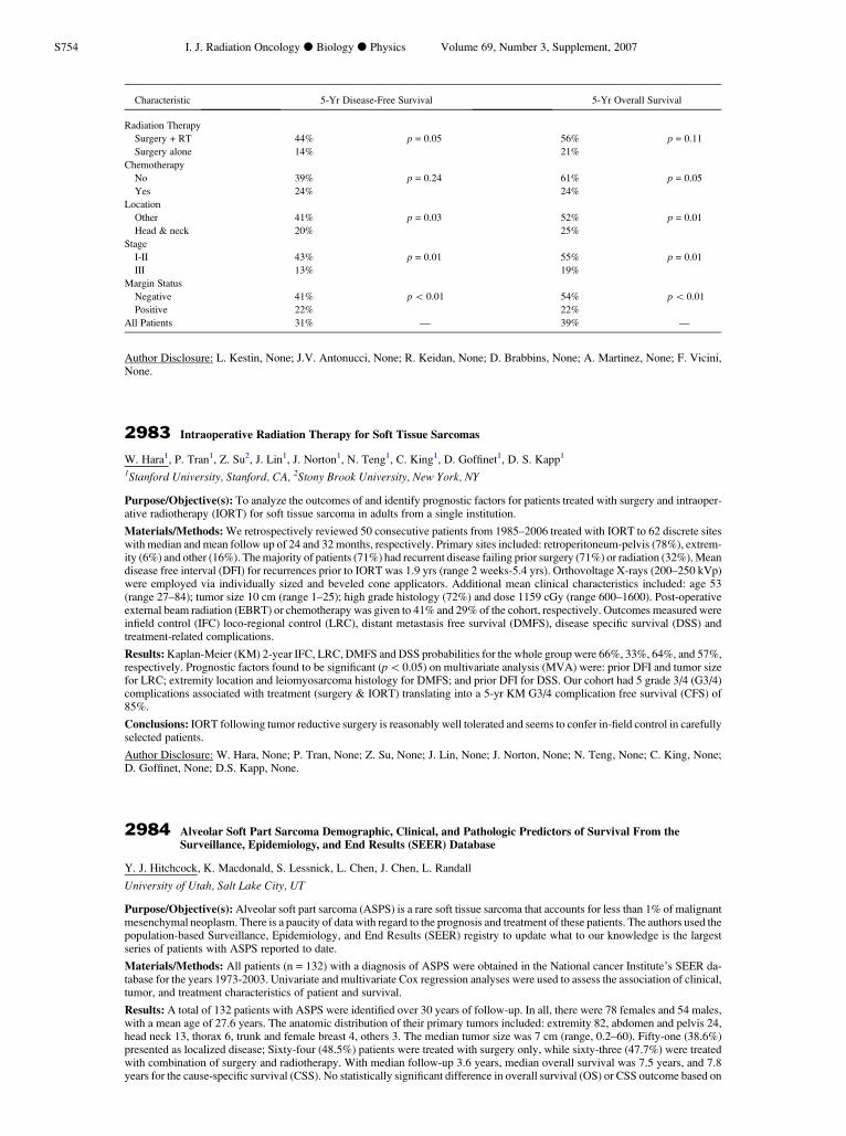
Characteristic 5-Yr Disease-Free Survival 5-Yr Overall Survival Radiation Therapy Surgery + RT 44% p = 0.05 56% p = 0.11 Surgery alone 14% 21% Chemotherapy No 39% p = 0.24 61% p = 0.05 Yes 24% 24% Location Other 41% p = 0.03 52% p = 0.01 Head & neck 20% 25% Stage I-II 43% p = 0.01 55% p = 0.01 III 13% 19% Margin Status Negative 41% p \ 0.01 54% p \ 0.01 Positive 22% 22% All Patients 31% — 39% — Author Disclosure: L. Kestin, None; J.V. Antonucci, None; R. Keidan, None; D. Brabbins, None; A. Martinez, None; F. Vicini, None. 2983 Intraoperative Radiation Therapy for Soft Tissue Sarcomas W. Hara 1 , P. Tran 1 , Z. Su 2 , J. Lin 1 , J. Norton 1 , N. Teng 1 , C. King 1 , D. Goffinet 1 , D. S. Kapp 1 1 Stanford University, Stanford, CA, 2 Stony Brook University, New York, NY Purpose/Objective(s): To analyze the outcomes of and identify prognostic factors for patients treated with surgery and intraoper- ative radiotherapy (IORT) for soft tissue sarcoma in adults from a single institution. Materials/Methods: We retrospectively reviewed 50 consecutive patients from 1985–2006 treated with IORT to 62 discrete sites with median and mean follow up of 24 and 32 months, respectively. Primary sites included: retroperitoneum-pelvis (78%), extrem- ity (6%) and other (16%). The majority of patients (71%) had recurrent disease failing prior surgery (71%) or radiation (32%). Mean disease free interval (DFI) for recurrences prior to IORT was 1.9 yrs (range 2 weeks-5.4 yrs). Orthovoltage X-rays (200–250 kVp) were employed via individually sized and beveled cone applicators. Additional mean clinical characteristics included: age 53 (range 27–84); tumor size 10 cm (range 1–25); high grade histology (72%) and dose 1159 cGy (range 600–1600). Post-operative external beam radiation (EBRT) or chemotherapy was given to 41% and 29% of the cohort, respectively. Outcomes measured were infield control (IFC) loco-regional control (LRC), distant metastasis free survival (DMFS), disease specific survival (DSS) and treatment-related complications. Results: Kaplan-Meier (KM) 2-year IFC, LRC, DMFS and DSS probabilities for the whole group were 66%, 33%, 64%, and 57%, respectively. Prognostic factors found to be significant (p \0.05) on multivariate analysis (MVA) were: prior DFI and tumor size for LRC; extremity location and leiomyosarcoma histology for DMFS; and prior DFI for DSS. Our cohort had 5 grade 3/4 (G3/4) complications associated with treatment (surgery & IORT) translating into a 5-yr KM G3/4 complication free survival (CFS) of 85%. Conclusions: IORT following tumor reductive surgery is reasonably well tolerated and seems to confer in-field control in carefully selected patients. Author Disclosure: W. Hara, None; P. Tran, None; Z. Su, None; J. Lin, None; J. Norton, None; N. Teng, None; C. King, None; D. Goffinet, None; D.S. Kapp, None. 2984 Alveolar Soft Part Sarcoma Demographic, Clinical, and Pathologic Predictors of Survival From the Surveillance, Epidemiology, and End Results (SEER) Database Y. J. Hitchcock, K. Macdonald, S. Lessnick, L. Chen, J. Chen, L. Randall University of Utah, Salt Lake City, UT Purpose/Objective(s): Alveolar soft part sarcoma (ASPS) is a rare soft tissue sarcoma that accounts for less than 1% of malignant mesenchymal neoplasm. There is a paucity of data with regard to the prognosis and treatment of these patients. The authors used the population-based Surveillance, Epidemiology, and End Results (SEER) registry to update what to our knowledge is the largest series of patients with ASPS reported to date. Materials/Methods: All patients (n = 132) with a diagnosis of ASPS were obtained in the National cancer Institute’s SEER da- tabase for the years 1973-2003. Univariate and multivariate Cox regression analyses were used to assess the association of clinical, tumor, and treatment characteristics of patient and survival. Results: A total of 132 patients with ASPS were identified over 30 years of follow-up. In all, there were 78 females and 54 males, with a mean age of 27.6 years. The anatomic distribution of their primary tumors included: extremity 82, abdomen and pelvis 24, head neck 13, thorax 6, trunk and female breast 4, others 3. The median tumor size was 7 cm (range, 0.2–60). Fifty-one (38.6%) presented as localized disease; Sixty-four (48.5%) patients were treated with surgery only, while sixty-three (47.7%) were treated with combination of surgery and radiotherapy. With median follow-up 3.6 years, median overall survival was 7.5 years, and 7.8 years for the cause-specific survival (CSS). No statistically significant difference in overall survival (OS) or CSS outcome based on S754 I. J. Radiation Oncology d Biology d Physics Volume 69, Number 3, Supplement, 2007
Transcript of Intraoperative Radiation Therapy for Soft Tissue Sarcomas
S754 I. J. Radiation Oncology d Biology d Physics Volume 69, Number 3, Supplement, 2007
Characteristic 5-Yr Disease-Free Survival 5-Yr Overall Survival
Radiation Therapy
Surgery + RT
44% p = 0.05 56% p = 0.11Surgery alone
14% 21%Chemotherapy
No
39% p = 0.24 61% p = 0.05Yes
24% 24%Location
Other
41% p = 0.03 52% p = 0.01Head & neck
20% 25%Stage
I-II
43% p = 0.01 55% p = 0.01III
13% 19%Margin Status
Negative
41% p \ 0.01 54% p \ 0.01Positive
22% 22%All Patients
31% — 39% —Author Disclosure: L. Kestin, None; J.V. Antonucci, None; R. Keidan, None; D. Brabbins, None; A. Martinez, None; F. Vicini,None.
2983 Intraoperative Radiation Therapy for Soft Tissue Sarcomas
W. Hara1, P. Tran1, Z. Su2, J. Lin1, J. Norton1, N. Teng1, C. King1, D. Goffinet1, D. S. Kapp1
1Stanford University, Stanford, CA, 2Stony Brook University, New York, NY
Purpose/Objective(s): To analyze the outcomes of and identify prognostic factors for patients treated with surgery and intraoper-ative radiotherapy (IORT) for soft tissue sarcoma in adults from a single institution.
Materials/Methods: We retrospectively reviewed 50 consecutive patients from 1985–2006 treated with IORT to 62 discrete siteswith median and mean follow up of 24 and 32 months, respectively. Primary sites included: retroperitoneum-pelvis (78%), extrem-ity (6%) and other (16%). The majority of patients (71%) had recurrent disease failing prior surgery (71%) or radiation (32%). Meandisease free interval (DFI) for recurrences prior to IORT was 1.9 yrs (range 2 weeks-5.4 yrs). Orthovoltage X-rays (200–250 kVp)were employed via individually sized and beveled cone applicators. Additional mean clinical characteristics included: age 53(range 27–84); tumor size 10 cm (range 1–25); high grade histology (72%) and dose 1159 cGy (range 600–1600). Post-operativeexternal beam radiation (EBRT) or chemotherapy was given to 41% and 29% of the cohort, respectively. Outcomes measured wereinfield control (IFC) loco-regional control (LRC), distant metastasis free survival (DMFS), disease specific survival (DSS) andtreatment-related complications.
Results: Kaplan-Meier (KM) 2-year IFC, LRC, DMFS and DSS probabilities for the whole group were 66%, 33%, 64%, and 57%,respectively. Prognostic factors found to be significant (p\0.05) on multivariate analysis (MVA) were: prior DFI and tumor sizefor LRC; extremity location and leiomyosarcoma histology for DMFS; and prior DFI for DSS. Our cohort had 5 grade 3/4 (G3/4)complications associated with treatment (surgery & IORT) translating into a 5-yr KM G3/4 complication free survival (CFS) of85%.
Conclusions: IORT following tumor reductive surgery is reasonably well tolerated and seems to confer in-field control in carefullyselected patients.
Author Disclosure: W. Hara, None; P. Tran, None; Z. Su, None; J. Lin, None; J. Norton, None; N. Teng, None; C. King, None;D. Goffinet, None; D.S. Kapp, None.
2984 Alveolar Soft Part Sarcoma Demographic, Clinical, and Pathologic Predictors of Survival From the
Surveillance, Epidemiology, and End Results (SEER) DatabaseY. J. Hitchcock, K. Macdonald, S. Lessnick, L. Chen, J. Chen, L. Randall
University of Utah, Salt Lake City, UT
Purpose/Objective(s): Alveolar soft part sarcoma (ASPS) is a rare soft tissue sarcoma that accounts for less than 1% of malignantmesenchymal neoplasm. There is a paucity of data with regard to the prognosis and treatment of these patients. The authors used thepopulation-based Surveillance, Epidemiology, and End Results (SEER) registry to update what to our knowledge is the largestseries of patients with ASPS reported to date.
Materials/Methods: All patients (n = 132) with a diagnosis of ASPS were obtained in the National cancer Institute’s SEER da-tabase for the years 1973-2003. Univariate and multivariate Cox regression analyses were used to assess the association of clinical,tumor, and treatment characteristics of patient and survival.
Results: A total of 132 patients with ASPS were identified over 30 years of follow-up. In all, there were 78 females and 54 males,with a mean age of 27.6 years. The anatomic distribution of their primary tumors included: extremity 82, abdomen and pelvis 24,head neck 13, thorax 6, trunk and female breast 4, others 3. The median tumor size was 7 cm (range, 0.2–60). Fifty-one (38.6%)presented as localized disease; Sixty-four (48.5%) patients were treated with surgery only, while sixty-three (47.7%) were treatedwith combination of surgery and radiotherapy. With median follow-up 3.6 years, median overall survival was 7.5 years, and 7.8years for the cause-specific survival (CSS). No statistically significant difference in overall survival (OS) or CSS outcome based on