International Journal of Current Microbiology and Applied ... D. Bemena, et al.pdf · Despite the...
26
Int.J.Curr.Microbiol.App.Sci (2014) 3(12): 924-949 924 Review Article Applications of bacteriocins in food, livestock health and medicine Léo D. Bemena 1 , Lulu A. Mohamed 1 , António Maximiano Fernandes 2 and Byong H. Lee1 3 * 1 Key laboratory of carbohydrate, School of Biotechnology, Jiangnan University, Wuxi, Jiangsu, China 214122 2 State Key Laboratory of Food Science and Technology, School of Food Science and Technology, Synergetic Innovation Center of Food Safety and Nutrition, Jiangnan University, Wuxi, Jiangsu 7214122, China 3 Department of Food Science /Agricultural Chemistry, McGill University, Montreal, Quebec, Canada H9X3V9 *Corresponding author ABSTRACT Introduction Antibiotics have been used as therapeutic and prophylactic treatments to control a variety of bacterial infections in livestock since their discovery in the half of 19 th century. Therefore, the continuous uses of these antibiotics led to the emergence of antibiotic-resistance in many bacteria relevant for animal and public health stresses (Diez-Gonzalez 2007; Mantovani et al. 2011). Several alternative approaches using probiotic bacteria were undertaken to control animal and foodborne pathogenic bacteria in livestock, but many of the beneficial effects of probiotics and the International Journal of Current Microbiology and Applied Sciences ISSN: 2319-7706 Volume 3 Number 12 (2014) pp. 924-949 http://www.ijcmas.com Increase of bacterial antibiotic resistance is a major cause for animal and public health stresses globally. Many bacteriocins and bacteriocins producing probiotic bacteria show potential for biotechnological, food and agro-industrial applications. The current global response to these useful bacteriocins needs to be improved by genetic or metabolic engineering. Due to the alarming rise in antibiotic resistance and adverse effects provoked by a number of antibiotics, bacteriocins have been applied in several fields: human health, food industry, animal health, and medicine, in particular as a substitution for the traditional growth promoters, antibiotics. The use of bacteriocins became an universal trend well over in 50 countries since their first discovery. Lactic Acid Bacteria (LAB) bacterocins are likely used because of their “safe” (GRAS) status, especially in food industry as bio-preservatives. Among these LAB bacteriocins, commercially marketed, nisin groups produced by Lactococcus lactis subsp. lactis and pediocins produced by Pediococcus sp. are the most characterized by their antilisterian property. Despite the widespread use in foods, their applications in livestock and medicine have been largely limited and further investigations are necessary. Keywords Bacteriocins, Nisin, Pediocin, Food, Livestock, Medicine, Quorum sensing, Metabolic engineering
Transcript of International Journal of Current Microbiology and Applied ... D. Bemena, et al.pdf · Despite the...

Int.J.Curr.Microbiol.App.Sci (2014) 3(12): 924-949
924
Review Article
Applications of bacteriocins in food, livestock health and medicine
Léo D. Bemena
1, Lulu A. Mohamed
1, António Maximiano Fernandes
2 and Byong H. Lee1
3*
1Key laboratory of carbohydrate, School of Biotechnology, Jiangnan University,
Wuxi, Jiangsu, China 214122 2State Key Laboratory of Food Science and Technology, School of Food Science and
Technology, Synergetic Innovation Center of Food Safety and Nutrition, Jiangnan University,
Wuxi, Jiangsu 7214122, China 3Department of Food Science /Agricultural Chemistry, McGill University,
Montreal, Quebec, Canada H9X3V9 *Corresponding author
A B S T R A C T
Introduction
Antibiotics have been used as therapeutic
and prophylactic treatments to control a
variety of bacterial infections in livestock
since their discovery in the half of 19th
century. Therefore, the continuous uses of
these antibiotics led to the emergence of
antibiotic-resistance in many bacteria
relevant for animal and public health
stresses (Diez-Gonzalez 2007; Mantovani et
al. 2011). Several alternative approaches
using probiotic bacteria were undertaken to
control animal and foodborne pathogenic
bacteria in livestock, but many of the
beneficial effects of probiotics and the
International Journal of Current Microbiology and Applied Sciences ISSN: 2319-7706 Volume 3 Number 12 (2014) pp. 924-949
http://www.ijcmas.com
Increase of bacterial antibiotic resistance is a major cause for animal and public
health stresses globally. Many bacteriocins and bacteriocins producing probiotic
bacteria show potential for biotechnological, food and agro-industrial applications.
The current global response to these useful bacteriocins needs to be improved by genetic or metabolic engineering. Due to the alarming rise in antibiotic resistance
and adverse effects provoked by a number of antibiotics, bacteriocins have been
applied in several fields: human health, food industry, animal health, and medicine, in particular as a substitution for the traditional growth promoters, antibiotics. The
use of bacteriocins became an universal trend well over in 50 countries since their
first discovery. Lactic Acid Bacteria (LAB) bacterocins are likely used because of their “safe” (GRAS) status, especially in food industry as bio-preservatives. Among
these LAB bacteriocins, commercially marketed, nisin groups produced by
Lactococcus lactis subsp. lactis and pediocins produced by Pediococcus sp. are the
most characterized by their antilisterian property. Despite the widespread use in foods, their applications in livestock and medicine have been largely limited and
further investigations are necessary.
K ey wo rd s
Bacteriocins,
Nisin,
Pediocin, Food,
Livestock,
Medicine, Quorum
sensing,
Metabolic
engineering

Int.J.Curr.Microbiol.App.Sci (2014) 3(12): 924-949
925
mechanisms are not fully elucidated (Fuller
and Tannock 1999). However, many
bacteriocins show potential for
biotechnological and agro-industrial
applications. Some bacteriocins show
desirable properties for in vivo application,
such as stability to low pH and heat, simple
production and extraction processes and
little, if any, inhibitory activity towards
eukaryotic cells. Therefore, bacteriocins
have been evaluated as the most promising
class of antimicrobial peptides to be used as
antibiotic substitutes in the field of animal
and human medicine or in designing and
production of new antimicrobials (Sahl and
Bierbaum 2008). Particularly on animal
trials, bacteriocin and bacteriocin-producing
bacteria may be useful to improve animal
nutrition and health through the
manipulation of ruminal fermentation, the
control of animal infections and the
inhibition of enteric pathogens (Patra 2012).
Although current applications of antibiotics
in the farm environment are apparently not
available, estimates are as high as 10.5
million pounds annually in the United States
for the poultry production (Mellon et al.
2001). The efficacy and cost-effectiveness
of many of these compounds are at the root
of their popularity, but looming or already
imposed restrictions or prohibitions on the
use of antibiotics as growth promoters have
drawn attention to possible alternatives
(Bedford 2000; Wierup 2000), Doyle 2001).
In contrast to the currently used antibiotics,
bacteriocins are often considered more
natural than common antibiotics because
they are thought to present in many of the
fermented foods eaten since ancient times
(Cleveland et al. 2001).
Bacteriocins have long attracted the interest
of food sector as potential natural food
preservatives against spoilage and
pathogenic bacteria (Kumar et al. 2011).
Nisin, pediocin and other bacteriocins
produced by lactic acid bacteria (LAB) have
received a great deal of attention because of
their beneficial effects to human health and
to food production as well as the
replacement of chemical preservatives that
are being continuously questioned with
regard of safety (Zendo 2013). Thus, this
potential offers a logical explanation for the
expanding trend of applications of LAB in
the food industry (Papagianni and
Anastasiadou 2009). In addition, no side
effects and no development of resistant
bacteria have been reported in the practical
use of LAB bacteriocins (Zendo 2013). The
administration of bacteriocin-producing
bacteria rather than the bacteriocins
themselves might be a more cost-effective
approach, but significant progress in
developing suitable producer strains will
have to be made before such an approach
will be feasible (Joerger 2003).
In this review, current trends and
perspectives on the applications of two most
known LAB bacteriocins: Nisin (lantibiotic)
and Pediocin (non-lantibiotic) in food
industry, livestock health, aquaculture as
well as medicine are discussed.
Bacteriocins
Bacteriocins are proteinaceous toxins
produced by bacteria and some archaea
members (Table 1) to inhibit the growth of
similar or closely related bacterial strain(s).
The inhibitory spectrum of bacteriocins can
be narrow and confined to closely related
species, or it can be relatively broad,
inhibiting a range of target organisms
(Mantovani et al. 2011). The bacteriocin
family is the most abundant and diverse
group of bacterial defenses (Riley 2009).
The application of bacteriocins in livestock
to control or/and to maintain intestinal
microflora of animals by feeding
bacteriocin-producing strains has been

Int.J.Curr.Microbiol.App.Sci (2014) 3(12): 924-949
926
largely achieved (Diez-Gonzalez 2007;
Papavassiliou 1961; Riley 2009). Novel
alternative strategies to reduce or eliminate
animal pathogens have also been tested by
different research groups. The alternatives
include bacteriocins, probiotic micro-
organisms and bacteriophages (Bedford
2000; Joerger 2003). Lactic acid bacteria
(LAB) can act antagonistically against a
wide range of food-borne pathogens and
spoilage organisms such as Salmonella (El-
Khatiband El-Rahman 1987; Guptaand
Savaliya 2012). Among LAB bacteriocins,
nisin is the most extensively characterized
and used (Mohanasirivasan et al. 2012).
Bacteriocins of Gram Positive Bacteria
Bacteriocins of Gram-positive bacteria are
as abundant and even more diverse as those
found in Gram-negative bacteria. The Gram-
positive bacteriocins resemble many of the
antimicrobial peptides produced by
eukaryotes; they are generally cationic,
amphiphilic, membrane-permeabilizing
peptides, approximately 2–6 kDa in size
(Riley 2009). Bacteriocins produced by
Gram-positive and Gram-negative bacteria
differ into several ecological and
evolutionary aspects. In Gram-positive
bacteria, the biosynthesis of bacteriocins is
self-regulated and bacteriocin production is
not a lethal event. In addition, the spectrum
of antimicrobial activity is broader than the
peptides from Gram-negative species and
bacteriocin release is controlled by specific
regulatory mechanisms (Mantovani et al.
2011). Additional roles have been proposed
for some bacteriocins produced by Gram-
positive bacteria, such as chemical
mediators in quorum sensing and
communication molecules in bacterial
consortia (Gillor 2007; Gobbetti et al. 2007).
Quorum sensing is one of well-studied
systems involved in bacteriocin gene
control. Quorum sensing is a cell-density
dependent regulatory system in which
autoinducing signal molecule mediates cell-
to-cell communication (Wang et al. 2013).
By using this system, each bacterial cell
senses the number of cells of same species
or same strain and adjusts the timing of
expression of certain genes. LAB often use
quorum sensing for the control of
bacteriocin expression in which LAB attack
the competitor only when the concentration
of the bacteriocin producers is high enough
to suppress the growth of competitive strain.
A landmark observation in the investigation
of bacteriocins of Gram-positive bacteria
was the documentation in 1947 that some of
the inhibitory activity of lactococci (group N
streptococci) toward other lactic acid
bacteria (LAB) are due to a molecule
characterized as a proteinaceous
antimicrobial called “group N inhibitory
substance”, or nisin (Heng et al. 2007). Also
this name has been suggested, partly on the
basis of their mode of action, that
bacteriocins of Gram-positive bacteria differ
from those of the Gram-negative bacteria
(Reeves 1965).
Nisin
Nisin was first discovered in the late 1920s
and early 1930s when it was described
as a toxic substance (Delves-Broughton et
al. 1996) produced by Lactococcus lactis,
subsp lactis. Approved by the US Food and
Drug Administration (FDA) for food
applications, nisin received GRAS status in
1988 (FDA 1988) and was authorized for
food preservation in the European Union by
Directive 95/2/EC in 1995 under the code
E234. The status as GRAS for use as an
anti-microbial agent on cooked meat and
poultry products was affirmed in 2001 by
the FDA (FDA 2011). Nisin is a small
peptide of 34 amino acids with a molecular
mass 3354 kDa ribosomally synthesized and
post-translationally modified peptide

Int.J.Curr.Microbiol.App.Sci (2014) 3(12): 924-949
927
contains five lanthionine rings, which can be
divided into two parts. The N-terminal half
including three rings (A, B, and C) is more
hydrophobic than the C-terminus containing
two rings (D and E) in Fig.1. The rigid ring
structures are separated by a flexible hinge
region (Diez-Gonzalez 2007; Takala 2005)
including one Dhb, two Dha one
lanthionine, and four methyllanthionine
residues (form the five lanthionine rings) in
its structure. The rigid ring structures are
separated by a flexible hinge region (Delves-
Broughton et al. 1996). The ring structures
give nisin a screw-like conformation that
possesses amphipathic characteristics. In
water its solubility and stability increases
with decreasing pH, showing maximum
solubility 57 mg/ml at pH 2 (Liu and Hansen
1990), and maximum stability at pH 3
(Davies et al. 1998). Nisin belongs to the
lantibiotic class of bacteriocins, cationic and
hydrophobic peptide. Nisin provides a
paradigm for studies of lantibiotic structure,
biosynthesis, and mode of action of
antimicrobial peptides, and is often referred
to as the “prototypical” lantibiotic
(Mantovani et al. 2011; Nolan and Walsh
2009).
Pediocin
Pediocins belong to the Class II of
unmodified bacteriocins which subdivided
into the groups of the pediocin-like
bacteriocins and the two-peptide
bacteriocins (Fig. 2). This class comprises
over 50 members with diverse origins. They
are generally small (<5 kDa) and are heat-
stable membrane-active and cationic
peptides with similar primary structures.
Their activity is retained at a wide pH range.
They are sensitive to most proteases. The
pediocin-like bacteriocins (36–48 residues)
are produced by many lactic acid bacteria
and share a 40–60% amino acid sequence
similarity (Heng et al. 2007; Papagianni
2003; Papagianni and Anastasiadou 2009;
Zacharof and Lovitt 2012). In general, class
IIa bacteriocins have a rather narrow
spectrum of activity (Drider et al. 2006).
The peptides of this group are known as
"antilisterian" or "Listeria-active" peptides
and they are characterized by a -YGNGV-N-
terminus (Papagianni and Anastasiadou
2009). The positively charged residues in
class IIa bacteriocins are located mostly in
the hydrophilic N-terminal region. It has
been shown for pediocin AcH/PA-1 that
electrostatic interactions and not the -
YGNGV- motif, govern the binding of the
pediocin and its fragments to phospholipids
vesicles (Chen et al. 1997). Lys11 and
His12: that are part of the cationic patch in
the N-terminal β-sheet-like region of
pediocin AcH/PA-1; are of special
importance for the electrostatic interactions
and subsequent mutagenesis studies, in
charged residues of pediocin AcH/PA-1, and
in sakacin P. Earlier research confirmed
these two amino acids were replaced by
neutral residues (Kazazic et al. 2002; Miller
et al. 1998b; Uteng et al. 2003). The C-
terminal region is important in determining
the target cell specificity for class IIa
bacteriocins (Drider et al. 2006).This has
been shown by combining N- and C-
terminal regions from different class IIa
bacteriocins (hybrid bacteriocins), which
displayed target cell specificities similar to
the bacteriocins from which the C-terminal
was derived (Fimland et al. 1996). Further
works carried out with pediocin AcH/PA-1
also showed the inhibition of the bactericidal
activity of the pediocin by cleaving the area
from residue 20 to residue 34. This is an
indication of a role for the C-terminal in
recognition of target cells (Fimland et al.
1998).
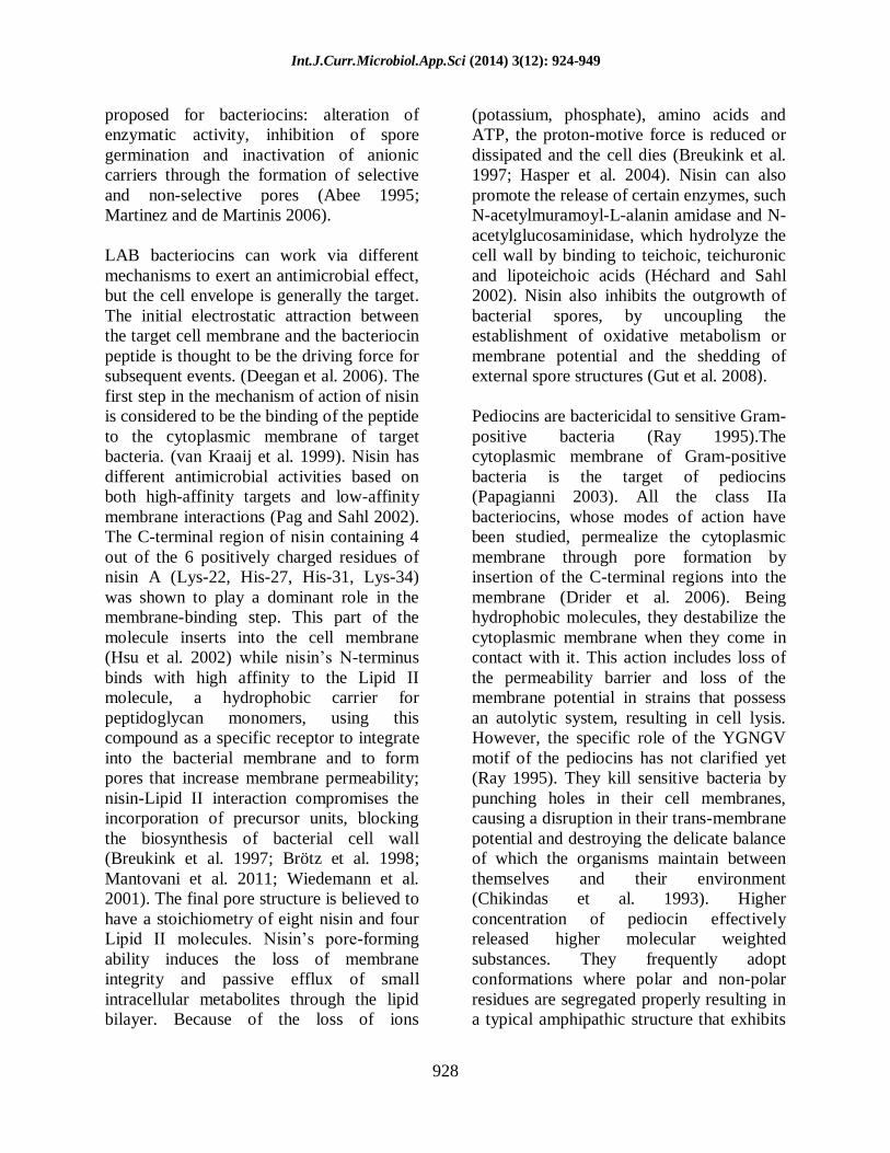
Mode of Action
Different mechanisms of action have been
Int.J.Curr.Microbiol.App.Sci (2014) 3(12): 924-949
928
proposed for bacteriocins: alteration of
enzymatic activity, inhibition of spore
germination and inactivation of anionic
carriers through the formation of selective
and non-selective pores (Abee 1995;
Martinez and de Martinis 2006).
LAB bacteriocins can work via different
mechanisms to exert an antimicrobial effect,
but the cell envelope is generally the target.
The initial electrostatic attraction between
the target cell membrane and the bacteriocin
peptide is thought to be the driving force for
subsequent events. (Deegan et al. 2006). The
first step in the mechanism of action of nisin
is considered to be the binding of the peptide
to the cytoplasmic membrane of target
bacteria. (van Kraaij et al. 1999). Nisin has
different antimicrobial activities based on
both high-affinity targets and low-affinity
membrane interactions (Pag and Sahl 2002).
The C-terminal region of nisin containing 4
out of the 6 positively charged residues of
nisin A (Lys-22, His-27, His-31, Lys-34)
was shown to play a dominant role in the
membrane-binding step. This part of the
molecule inserts into the cell membrane
(Hsu et al. 2002) while nisin‟s N-terminus
binds with high affinity to the Lipid II
molecule, a hydrophobic carrier for
peptidoglycan monomers, using this
compound as a specific receptor to integrate
into the bacterial membrane and to form
pores that increase membrane permeability;
nisin-Lipid II interaction compromises the
incorporation of precursor units, blocking
the biosynthesis of bacterial cell wall
(Breukink et al. 1997; Brötz et al. 1998;
Mantovani et al. 2011; Wiedemann et al.
2001). The final pore structure is believed to
have a stoichiometry of eight nisin and four
Lipid II molecules. Nisin‟s pore-forming
ability induces the loss of membrane
integrity and passive efflux of small
intracellular metabolites through the lipid
bilayer. Because of the loss of ions
(potassium, phosphate), amino acids and
ATP, the proton-motive force is reduced or
dissipated and the cell dies (Breukink et al.
1997; Hasper et al. 2004). Nisin can also
promote the release of certain enzymes, such
N-acetylmuramoyl-L-alanin amidase and N-
acetylglucosaminidase, which hydrolyze the
cell wall by binding to teichoic, teichuronic
and lipoteichoic acids (Héchard and Sahl
2002). Nisin also inhibits the outgrowth of
bacterial spores, by uncoupling the
establishment of oxidative metabolism or
membrane potential and the shedding of
external spore structures (Gut et al. 2008).
Pediocins are bactericidal to sensitive Gram-
positive bacteria (Ray 1995).The
cytoplasmic membrane of Gram-positive
bacteria is the target of pediocins
(Papagianni 2003). All the class IIa
bacteriocins, whose modes of action have
been studied, permealize the cytoplasmic
membrane through pore formation by
insertion of the C-terminal regions into the
membrane (Drider et al. 2006). Being
hydrophobic molecules, they destabilize the
cytoplasmic membrane when they come in
contact with it. This action includes loss of
the permeability barrier and loss of the
membrane potential in strains that possess
an autolytic system, resulting in cell lysis.
However, the specific role of the YGNGV
motif of the pediocins has not clarified yet
(Ray 1995). They kill sensitive bacteria by
punching holes in their cell membranes,
causing a disruption in their trans-membrane
potential and destroying the delicate balance
of which the organisms maintain between
themselves and their environment
(Chikindas et al. 1993). Higher
concentration of pediocin effectively
released higher molecular weighted
substances. They frequently adopt
conformations where polar and non-polar
residues are segregated properly resulting in
a typical amphipathic structure that exhibits

Int.J.Curr.Microbiol.App.Sci (2014) 3(12): 924-949
929
more peptide internalization and membrane
perturbation. Trans-membrane potential
(negative inside) in bacteria acts as a
potential driving force for insertion and
internalization of the antimicrobial peptides
promoting AMP interaction (Melo et al.
2009). For example, Pediocin PA-1 exerts
bactericidal or bacteriolytic effect depending
on the species of the sensitive cells (Bhunia
et al. 1991). Pediocins also act on some
sensitive bacterial strains in bacteriostatic
manner and thus retard further proliferation
of the sensitive cells (e.g. Pediocin ST18,
Pediocin CP2) (Mashal 2007).
Bioengineering of LAB Bacteriocins
In last two decades, there have been
significant advances in functional genomic
analysis of LAB and their biochemical
characterization of bacteriocins.
Considerable efforts have been made to
functionally characterize bacteriocin
operons and to express them in heterologous
systems (Coderre and Somkuti 1999; Miller
et al. 1998a; Osmanağaoğlu et al. 2000;
Tominaga and Hatakeyama 2007).
The genes responsible for bacteriocin
production are frequently associated with
mobilisable elements, or in the chromosome
in association with transposons or plasmids
(Belkum et al. 1998). The low-molecular-
weight bacteriocins of Gram-positive
bacteria generally appear to be translated as
pre-peptides that are subsequently modified
to form the mature biologically active
(bactericidal) molecules (Buchman et al.
1998). Specific auxiliary functions required
by bacteriocin-producing cells include
mechanisms for extracellular translocation
of the bacteriocin and for self-immunity to
the bactericidal activity of the molecule
(Jack et al. 1995). As is the case for most
bacteriocins, the lantibiotics are initially
synthesized with an N-terminal leader
peptide. In general, the pre-peptide is
modified by the action of other proteins
encoded by the bacteriocin gene cluster
before export (Deegan et al. 2006).
Biosynthesis of nisin: The genes involved
in biosynthesis of the model lantibiotic nisin
are located on a 70 kb conjugative
transposon (Rauch et al. 1991). Biosynthesis
of nisin is encoded by a cluster of 11 genes
(Fig. 3) of which the first gene, nisA,
encodes the precursor of nisin (Mierau and
Kleerebezem 2005). The first gene of the
nisin gene cluster, nisA, encodes the 57
amino acid nisin precursor, consisting of a
N-terminal leader, sequence followed by the
propeptide, from which nisin A is matured.
The structural gene is followed by ten other
genes i.e. nisB, nisT, nisC, nisI, nisP, nisR,
nisK, nisF, nisE, nisG, encoding regulatory
proteins, proteases, transport proteins and
immunity proteins (van Kraaij et al. 1999).
The proteins that are encoded by these genes
have been found to be homologous to gene
products of the gene clusters of other
lantibiotics, such as those of subtilin,
epidermin and Pep5 (van Kraaij et al. 1999).
Thus, as a result of their gene encoded
nature, lantibiotics have been the focus of
bioengineering with a view in elucidating
structure function relationships (Cortés et al.
2009; Cotter et al. 2005; Field et al. 2010;
Lubelski et al. 2008). The majority of works
that lead to enhanced peptides have resulted
as a consequence of manipulation of the
hinge region (Rouse et al. 2012). The hinge
comprises residues 20 (Asn), 21 (Met) and
22 (Lys) (Fig. 1), which are thought to
permit the movement of the N- and C-
termini relative to one another during pore
formation. The first success in this regard
related to the creation of nisin derivatives,
N20K and M21K, with enhanced anti-
microbial activity against Gram-negatives
(Yuan et al. 2004). Subsequent
investigations have further highlighted the

Int.J.Curr.Microbiol.App.Sci (2014) 3(12): 924-949
930
benefits of manipulating the hinge and
finally resulted in the identification of nisin
derivatives, such as nisin N20P, M21V,
K22T and K22S, which possess enhanced
specific activity against Gram-positive
pathogens (Field et al. 2008). Such activity
has been highlighted with the enhanced
specific activity of nisin M21V (or nisin V)
in foodborne and clinical purpose (Field et
al. 2010).
Biosynthesis of pediocin
Characteristically, class IIa bacteriocins like
other low-molecular-mass bacteriocins are
first formed as ribosomally synthesized
precursors or pre-peptides, which appear not
to be biologically active and contain a N-
terminal extension or leader sequence.
Subsequent cleavage of the pre-peptide at a
specific processing site removes the leader
sequence from the antimicrobial molecule
concomitantly with its export to the outside
of the cell (Håvarstein et al. 1994). The
leader peptide's removal during trans-
membrane translocation is accomplished by
the same protein that is associated with the
bacteriocin transport (Håvarstein et al. 1994;
Nes et al. 1996).
The amino acid sequence of a number of
class-IIa-bacteriocin leader peptides, which
vary in length from 18 up to 27 residues.
One important feature of the majority of
these leaders is the presence of two glycine
residues in the C-terminus at positions 32
and 31 relative to the processing site, though
this is not distinctive of the class IIa. These
leaders are believed to serve as signal
peptides for the processing and the secretion
of class IIa bacteriocins, independently of
the GSP, by a dedicated transport system
involving two distinct proteins: an ABC-
type translocator and an accessory protein.
The two conserved glycine residues may
serve as a recognition signal for this sec-
independent transporter system (Ennahar et
al. 2000; Håvarstein et al. 1994;
Klaenhammer 1993; Nes et al. 1996).
In the case of pediocin PA-1/AcH the four
genes needed for bacteriocin production and
secretion are located in one operon [35]. The
four genes are 1) the structural bacteriocin
gene, encoding a prebacteriocin; 2) the
immunity gene, encoding an immunity
protein that protects the bacteriocin producer
from its own bacteriocin; 3) the gene
encoding the ABC transporter for secretion;
and 4) a gene encoding a complementary
protein of unknown function (Ennahar et al.
2000). It has been shown that the four genes
cluster of pediocin AcH/PA-1 contain
common promoter and terminator sequences
(Bukhtiyarova et al. 1994; Marugg et al.
1992; Motlagh et al. 1994). PedA encodes a
62 amino acids long pre-pediocin PA-1.
Eighteen residue long leader sequences from
N-terminal of pre-pediocin are removed
during processing and export of pediocin
through producer cell membrane. Mature
pediocin carries 44 amino acid residues and
two intra-molecular disulphide bridges at
cys9-cys14 and cys24-cys44 positions
(Henderson et al. 1992; Miller et al. 1998a;
Neetoo et al. 2008). PedB immunity gene is
located downstream to pedA and encodes a
protein of 112 amino acid residues. PedC a
174 amino acid long amphiphilic protein
involved along with pedD protein in
facilitating/accelerating the trans-membrane
export (Henderson et al. 1992). PedD gene
specifies a polypeptide of 724 amino acid
residues. Deletion analysis and site specific
mutagenesis of pedD resulted in complete
loss of pediocin production, showing its
essentiality for secretion in E. coli (Marugg
et al. 1992). Its sequence shows a very high
homology to members of ATP dependent
transport proteins and also to a group of
eukaryotic proteins involved in multidrug
resistance (Kumar et al. 2011; Marugg et al.
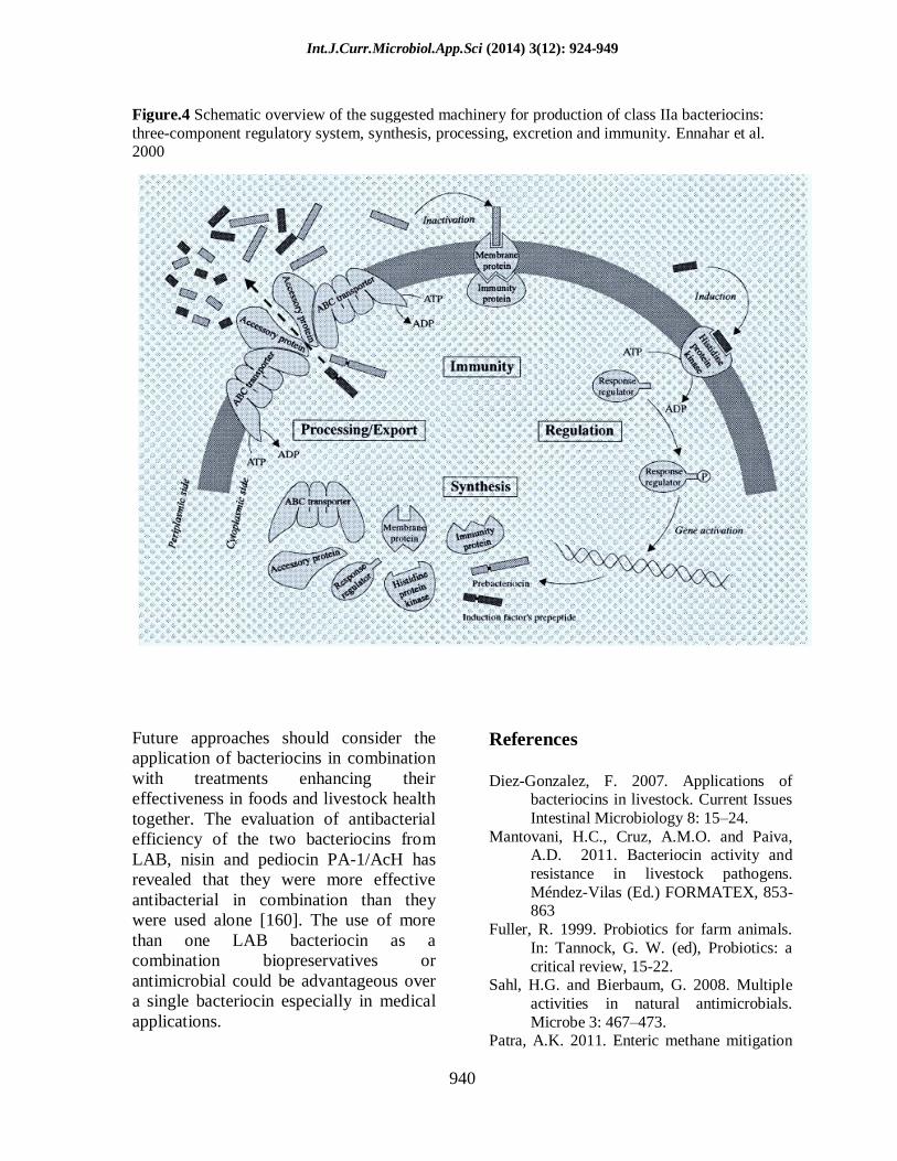
1992). Figure 4 shows the suggested

Int.J.Curr.Microbiol.App.Sci (2014) 3(12): 924-949
931
machinery for production of class IIa
bacteriocins (Ennahar et al. 2000).
Applications of Bacteriocins in Food
Although several methods other than
bacteriocins are employed for the
preservation of food and beverages, an
increasingly health conscious public may
seek to avoid foods that have undergone
extensive processing or which contain
chemical preservatives. Bacterial
fermentation of perishable raw materials has
been used for centuries to preserve the
nutritive value of food and beverages and to
extend shelf-life. Among bacteriocins
produced by many Gram-positive and
Gram-negative species, those produced by
LAB are of particular interest to the food
industry, since these bacteria have generally
been regarded as safe. The production of
bacteriocins by LAB is advantageous for
survival of the producing bacteria in a
competitive ecological niche; therefore they
could be exploited by the food industry as a
tool to control undesirable bacteria in a
food-grade and natural manner, which is
likely to be more acceptable to consumers
(Deegan et al. 2006; Parada et al. 2007).
Many lactic acid bacteria produce a high
diversity of different bacteriocins and
several have been patented for their
applications in foods (Schöbitz et al. 2006).
Listeria monocytogenes is a pathogenic
bacterium that has been involved in several
foodborne outbreaks worldwide and causes
special concern with regard to food safety
due to its psychrotropic and ubiquitous
characteristics. The presence of this
pathogen in fermented sausages and in
vacuum-packaged meat products (Chung
and Hancock 2000) is of particular interest
for food safety, as these two groups of meat
are frequently eaten without reheating
(Vignolo et al. 1996). This pathogen has
shown to survive at pH as low as 3.6 in
foods and in salt concentration of up to 10%,
in the presence of surfactants, sanitizers and
after several cycles of freezing and thawing
(Martinez and de Martinis 2006), being a
serious risk.
Several possible strategies for the
application of bacteriocins in the
preservation of foods may be considered: i)
inoculation of the food with LAB as starter
or protective cultures that produce the
bacteriocin in the product (production in
situ); ii) addition of the purified or semi-
purified bacteriocin as a food preservative,
and iii) use of a product previously
fermented with a bacteriocin-producing
strain as an ingredient in food formulation
(Jeevaratnam et al. 2005).
Bacteriocin production in situ by starter
cultures has a good chance of finding
applications in fermented foods. In non-
fermented refrigerated products, such as
minimally processed meats or prepackaged
vegetable salads, only those strains
producing sufficient and potent amounts of
bacteriocin but no other metabolic
compounds, at levels detrimental to the
sensory quality product, can be applied. The
direct addition of purified bacteriocins
obviously provides a more controllable
preservative tool in such products
(Jeevaratnam et al. 2005).
The use of nisin in foods and foodborne is
the most expected use of this bacteriocin.
There is an enormous amount of information
about its application to inhibit a variety of
pathogenic and spoilage bacteria in many
food products (Delves-Broughton et al.
1996). Nisin is suitable for use in a wide
range of foods (liquid or solid), canned or
packaged, chill or warm ambient storage.
Based on target microorganisms, its usage
falls into three broad categories: i) to

Int.J.Curr.Microbiol.App.Sci (2014) 3(12): 924-949
932
prevent spoilage by Gram-positive
endospore formers (especially in heat
processed food), ii) to prevent spoilage by
LAB and similar organisms like Brocothrix
thermosphacta, and iii) to kill or inhibit
Gram-positive pathogens such Listeria
monocytogenes, and Clostridium botulinum.
It was demonstrated that nisin is best added
as an aqueous solution. Usually, it serves as
the liquid portion of a product during its
processing. It can also be added as a powder,
but it is essential to ensure uniform dispersal
throughout the food matrix in both ways.
The best time to add nisin is at the last
practical stage before heat processing (if this
is a part of the manufacturing process). In
the manufacture of processed cheese, for
instance, nisin is usually added to the heated
cheese at the same time as the melting salts.
Nisin can also be used at high
concentrations as a spray or dip for surface
decontamination. The level of nisin addition
depends on the type of food, severity of heat
process, pH, storage conditions and the
required self-life. Nisin is often used in
acidic foods, but is effective in products
across a wide range of pH values 3.5-8.0. It
is used in variety of products including
pasteurized, flavored and long-life milks,
aged and processed cheeses, and canned
vegetables and soups (Delves-Broughton et
al. 1996; Jeevaratnam et al. 2005). Nisin has
utilized to inhibit undesirable LAB in wine
and beer (Daeschel et al. 1991; Jeevaratnam
et al. 2005; Ogden et al. 1988). Nisin has
also been used in conjunction with other
preservative measures to enhance product
safety or quality. In canned foods such
vegetables, soups and puddings, nisin has
been applied in conjunction with heat to
successfully counter heat-resistant spores of
flat-sour thermophilic bacteria (Chung and
Hancock 2000; Smid and Gorris 1999).
In seafood industry, studies of nisin
indicated that it delayed growth of L.
monocytogenes in cold-smoked salmon
(Bakkal et al. 2012). There has also been
encouraging research into nisin-coated
packaging. The effect of nisin-coated
plastic films on the survival of L.
monocytogenes on vacuum-packed cold
smoked salmon (Neetoo et al. 2008) showed
that nisin-coated plastic films reduced the
number of L. monocytogenes by 3.9 log
CFU/cm2 at 4
oC and 10
oC after 56 and 49
days of incubation, respectively. This study
also showed that nisin-coated plastic films
suppressed the growth of other aerobic and
anaerobic spoilage microorganisms in a
concentration-dependent manner. A
combination of nisin and some lactates has
been demonstrated to be more active against
L. monocytogenes due to synergistic action
(McEntire et al. 2003; Nykänen et al. 2000).
A combinatory treatment of nisin and
listeriophage was found to be effective in
controlling L. monocytogenes, while it was
not effective in model food systems which
reflect the complexity of natural system
(Dykes and Moorhead 2002).
Despite of few studies reported on the
applications of pediocins, pedioci PA-
1/AcH has been demonstrated to effectively
reduce populations of listeria strains in ice
cream mix, sausage mix, fresh and ground
beef and whole milk (Motlagh et al. 1994).
It has been found to be effective against
many strains of sub-lethally stressed Gram-
positive and Gram-negative bacteria. Such
injured bacteria can be present in foods that
have an acid pH (below 6), water activity
below 0.9, or have been given low heat
treatment, subjected to hydrostatic pressure,
or stored at low temperature, including long-
term storage at refrigerated temperature.
Incorporation of pediocins as preservatives
in such foods can help in killing the
normally sensitive and resistant but injured
cells of spoilage and pathogenic bacteria and
ensure longer product shelf-life and greater
consumer safety (Jeevaratnam et al. 2005).

Int.J.Curr.Microbiol.App.Sci (2014) 3(12): 924-949
933
Pediocin PA-1/AcH has a specific
application to control L. monocytogenes in
the production of certain fermented foods,
especially in controlled fermentation where
specific strains of starter cultures are used.
Many refrigerated, vacuum-packaged
processed food products from meat, dairy,
fish and vegetable groups contain normally
psychotropic Gram-positive bacteria strains
such Leuconostoc, Lactobacillus,
Carnobacterium, Brochothrix and
Clostridium. By incorporating pediocin PA-
1/AcH during the formulation of the raw
product, spoilage problems in the final
product could be reduced (Breukink et al.
1997; Ennahar et al. 1998; Yang and Ray
1994). Researchers in several countries have
recognized its potential as a food
preservative, especially for use in certain
specific foods. Pediocins are also
commercially available but are marketed as
fermentates of lactic acid bacteria (LAB)
having GRAS status (Gálvez et al. 2008).
Applications of Bacteriocins in Livestock
Health
The application of bacteriocins in livestock
has been largely achieved by feeding
bacteriocin-producing strains. Feeding
purified bacteriocins to humans for control
of diarrhea was reported in a few
publications during the 1900‟s
(Papavassiliou 1961), but very little
evidence exists in administering of
bacteriocins alone to livestock. Because of
lack of evidence, the use of bacteriocins in
livestock is largely based on those studies
that reported feeding or applying
bacteriocin-producing bacteria (BPB) (Diez-
Gonzalez 2007).
The application of BPB for improvements in
productivity has not been limited to cattle, as
several researchers have explored the use of
probiotic strains capable of producing
bacteriocins to increase the growth rate of
swine. In poultry, the use of BPB has been
mainly targeted for the control salmonella
(Rodriguez et al. 2003). The utilization of
BPB as a pre-harvest food safety strategy is
considered as one of the most viable
interventions for reducing the
gastrointestinal colonization of livestock by
foodborne pathogens (Callaway et al. 2004;
Gillor et al. 2004; Renter and Sargeant
2002). The BPB can easily be administered
to animals by mixing dried or wet cultures
with feed or drinking water, and depending
on the ability of the particular probiotic
strain to colonize the gastrointestinal tract
they could be fed sporadically or
continuously. The feeding of BPB can have
a direct effect on reducing the existing
populations of foodborne pathogens such as
salmonella and Escherichia coli, and long-
term colonization with BPB would prevent
further re-introduction of the pathogenic
bacteria (Diez-Gonzalez 2007).
Many different types of LAB bacteriocins
have been studied and characterized, but the
most widely known are: nisin, lacticin,
enterocin, pediocin, and plantaricin. These
have been extensively studied for their
application in foods, but just a few of them
have been used in livestock (Ray and
Bhunia 2013). The well-known, most and
likely use among of them is nisin. One of the
most promising applications of nisin is on
the control of Listeria monocytogenes in
ready-to-eat meats (Ariyapitipun et al.
2000). Nisin has also successfully been used
to control respiratory tract infection by
Staphylococcus aureus in animal model (De
Kwaadsteniet et al. 2008). One of the major
disease in dairy cattle is „bovine mastitis‟
(Ruegg 2003) induced by Staphylococcus
aureus that is the one of the most pathogen
agent implicated in clinical and subclinical
mastitis infections (Kerro et al. 2002).
Several bacteriocins including nisin have

Int.J.Curr.Microbiol.App.Sci (2014) 3(12): 924-949
934
been tested against the causative bacteria of
bovine mastitis. The positive results have
been reported for in vivo studies performed
with intra-mammary formulations
containing bacteriocins like germicidal
preparation for cow‟s teats (Sears et al.
1992; Wu et al. 2007). Therapeutic
formulations containing nisin reduced
considerably the viability of S. aureus and
E. coli at 3.9 and 4.2 log cycles, respectively
(Sears et al. 1992). Other studies using
treatment with nisin Z have shown a
significant increase in cure rates of
infections caused by S. agalactiae, S.
aureus, and other mastitis pathogens (90.1
%, 50 % and 65.2 %). Moreover, after 48
hours of treatment, no bacteriocin residue
was detected in milk (Wu et al. 2007).
Nisin was able to decrease the methane
production in vitro in ruminal fermentation.
For instance, the reduction of methane
emissions in sheep had been reported with
the combinations of this bacteriocin with
nitrate (Sar et al. 2005). Nisin has shown an
inhibitory effect against common rumen
anaerobes (Kišidayová et al. 2003;
Mantovani and Russell 2001). In vitro, this
bacteriocin affected ruminal fermentation in
the similar way to monensin, the most
common ionophore used as feed additive in
cattle rations (Callaway et al. 1997).
Moreover, the introduction of nisin into an
artificial rumen system brought some
changes in fermentation parameters, such as
an increase in hemicellulose degradation and
acetate and propionate production, which
contributed to the improvement of microbial
balance in this environment. (Jalc and
Lauková 2001; Santoso et al. 2006; Zendo
2013).
Applications of Bacteriocins in
Aquaculture
Aquatic cultures are facing with the same
problems with animal farming. They are
continuously exposed to a wide range of
microorganisms, some of which are
pathogenic (Reilly and Kaeferstein 1998).
Many efforts were undertaken to prevent
and control this dilemma: husbandry
techniques and the use of vaccines
(Corripio-Miyar and Mazorra de Quero) and
antibiotics (Smith 2007). These methods can
create several negative problems. They
cannot prevent disease (husbandry
techniques). Laborious, costly, and highly
stressful to the animals (vaccines) and
especially the selection for antibiotic-
resistant bacteria and active residues of the
drugs remain long after use (Lauková et al.
2003; Matyar 2007; Zhou and Wang 2012).
An alternative approach to disease
prevention in aquaculture is the use of
bacteriocin-producing bacteria, BPB
(Lauková et al. 2003). It means use these
bacteria as probiotic because in aquaculture,
aquatic animal and microorganisms share
the same ecosystem in the aquatic
environment and it suggested that the
interaction between the microbiota,
including probiotics, and the host is not
limited to the intestinal tract (Zhou and
Wang 2012). Many works reported that the
administration of BPB as probiotic exclude
competitively pathogenic bacteria through
the production of inhibitory compounds,
improve water quality, enhance the immune
response of host species, and enhance the
nutrition of host species through the
production of supplemental digestive
enzymes (Taoka et al. 2006; Wang 2007).
Most probiotics used in aquaculture belong
to the lactic acid bacteria, of the genus
Bacillus, to the photosynthetic bacteria or to
the yeast, although other genera or species
have also been mentioned. Many studies
have reported promising results using a
single beneficial bacterial strain as probiotic
in the culture of many aquatic species (Zhou

Int.J.Curr.Microbiol.App.Sci (2014) 3(12): 924-949
935
and Wang 2012)but it is important to
consider the possibility of using different
species. The effect of probiotics,
photosynthetic bacteria (Rhodobacter
sphaeroides) and Bacillus sp. (B.
coagulans), on growth performance and
digestive enzyme activity of the shrimp,
Penaeus vannamei, was investigated and the
results showed that the effects were related
with supplementation concentrations of
probiotics and thus use of a 10g/kg (wet
weight) supplement of probiotics in shrimp
diet was recommended to stimulate
productive performance (Wang 2007).
Some study showed that nutrient and water
enrichment with commercial BPB,
designated Alchem Poseidon™ (a mixture
of Bacillus subtilis, L. acidophilus,
Clostridium butyricum, and Saccharomyces
cerevisiae) significantly improved lysozyme
activity, lowered levels of mucosal proteins
and also improved survival after bacterial
immersion challenge with Vibrio
anguillarum (Taoka et al. 2006). BPB has
the potential to serve as an efficacious long-
term solution, as the administered bacteria
become established in the host and/or the
aquatic environment.
Early attempts to use probiotic species in
aquaculture usually employed BPB
developed for terrestrial animals, which
contained the facultative or obligate Gram-
positive anaerobes found in the GI tract,
specifically of the genera Bifidobacterium,
Lactobacillus, and Streptococcus (Callaway
et al. 2004; Gatesoupe 2007; Gatesoupe
1999). Production of BPB specifically for
the use in aquaculture is now a more popular
approach, as these strains are more likely to
establish in aquatic communities (Irianto
and Austin 2002). Bacteriocin producing
strains should be developed to be more
effective for aquaculture than the regular
probiotic strains in the future.
Applications of Bacteriocins in Medicine
Bacteriocins have interest in medicine
because they are made by non-pathogenic
bacteria that normally colonize the human
body. Loss of these harmless bacteria
following antibiotic use may allow
opportunistic pathogenic bacteria to invade
the human body. As the narrow spectrum of
bacteriocins produced by LAB represent an
important limitation for the application of
these bacteriocins as clinical drugs or as
food preservatives (Acuña et al. 2012),
some examples of bacteriocins and their
pharmaceutical applications are (a) the use
of microcins derived from enterobacteria to
control Gram negative bacteria (Duquesne et
al. 2007).
Similarly to pediocin-like bacteriocins,
microcins belonging to class IIa such as
microcin V are linear polypeptides, and the
removal of the leader peptide is the unique
posttranslational modification that they
undergo before being secreted by the cells.
In order to obtain a peptide with a broader
antimicrobial spectrum, recent works fused
by asymmetrical PCR the required portions
of genes encoding enterocin CRL35 and
microcin V, namely munA and cvaC.
The hybrid bacteriocin purified from E. coli
extracts, named Ent35eMccV, showed
inhibitory activity against
enterohemorrhagic E.coli, L.
monocytogenes, and other pathogenic Gram-
positive and Gram-negative bacteria (Acuña
et al. 2012). In this field, several lantibiotics
are used in pharmaceutical applications (van
Kraaij et al. 1999). Some have been used in
dental caries treatment (mutacin-producing
strain) (Hillman et al. 2000; Hillman 2002)
used to control vaginal microbiota with
significantly reducing the adherence of the
urogenital pathogen Staphylococcus aureus
(Zárate and Nader-Macias 2006). So far,

Int.J.Curr.Microbiol.App.Sci (2014) 3(12): 924-949
936
nisin is the most promising in this medical
field. The intravenous use of nisin has not
been further developed since nisin shows a
low stability at physiological pH. However,
several protein-engineered derivatives of
nisin Z have been generated in recent years
that show improved stability and these or
others may extend the medical application of
nisin (Kuipers et al. 1991, (Severina et al.
1998).
Nisin was also applied in the treatment of
respiratory tract infections. Some study
reported the capacity of nisin to develop
resistance in respiratory tract to prevent
growth of resistant Staphylococcus aureus
or Streptococcus pneumonia (De
Kwaadsteniet et al. 2009). Also, recent work
reported that Nisin F inhibits
Staphylococcus aureus in the nasal cavities
of immunosuppressed rats (De Kwaadsteniet
et al. 2009). Many studies report the
efficiency of nisin against several diseases
responsible in digestive tract especially
Clostridium species that can induce
diarrhea: C. botulinum, C. tyrobutyricum
and C. difficile (De Carvalho et al. 2007;
Delves-Broughton et al. 1996; Irianto and
Austin 2002) and gastric ulcers:
Helicobacter pylori (Delves-Broughton et
al. 1996; Kim et al. 2003).
Recently, the study of the therapeutic
properties of nisin F in mice infected by S.
aureus Xen 36 appeared to be promising to
control the disease (Brand 2013). The
resistance of spontaneous mutants to
bacteriocins have also been reported, that
may be related to changes in membrane and
cell wall, such as alterations in the electrical
potential, fluidity, membrane lipid
composition and load or cell wall thickness
or even a combination of all factors. These
changes may occur following cell exposure
to low concentrations of bacteriocins or as
part of an adaptive response to some other
stress.
The resistance of L. monocytogenes to nisin
is related to variation in fatty acid
composition of cell membranes, reducing
the concentration of phospholipids,
hindering the formation of pores. The
mechanism of resistance to subclass IIa
bacteriocins appears to be linked to reduced
expression of mannose permease of the
phosphotransferase system (Vadyvaloo et al.
2002).
Commercial Production of Bacteriocins
Several bacteriocin-producing bacteria have
been patented, but to the end of 2005 none
of them were at the commercialization stage
(Brown et al. 1999; Shotts Jr and Wooley
2000), the only commercially produced
bacteriocins are the group of nisin produced
by Lactoccocus lactis (Jones et al. 2005) and
pediocin PA-1 by Pediococcus acidilactici
(Gálvez et al. 2008). Nisin is the most
commercially important member of a large
class of bacteriocins produced by bacteria
that can kill or inhibit the growth of other
bacteria.
This phase of the Nisin Market Study
analyzes the characteristics of the current
market for nisin and competing bacteriocins
in four main sections highlighting: (1) the
general market characteristics for
antimicrobial preservatives; (2) current
producers and sellers of commercial grade
nisin; (3) current users of nisin and
competing bacteriocins; and (4) implications
for the market opportunities for nisin
production in the U.S.
The global leader in the antimicrobial
preservatives industry is Danisco A/S, a
Danish company, with Royal DSM
(Netherlands), and Kerry Bio-Sciences
(Ireland) considered being their peer
competitors in the bio-preservatives sector
(Jones et al. 2005). Danisco‟s Nisaplin™ is

Int.J.Curr.Microbiol.App.Sci (2014) 3(12): 924-949
937
generally considered to be the most
commercially available form of nisin for
food preservative uses. Danisco‟s strategic
focus for their nisin product line is the U.S.
meat and deli food sector in order to take
advantage of the FDA approval status of
nisin as a natural ingredient. Other players in
the global nisin market include Rhodia, S.A.
(France) along with numerous producers and
providers of various antimicrobial products
based in China. Some of these Chinese
sources are in joint ventures or alliances
with European-based corporate entities.
Bacteriocin preservatives are part of the $22
billion global food additives market that has
grown at 2-3% per annum through 2007 to
$24 billion. The Genencor division of
Danisco has manufacturing locations in the
United States, Finland, Belgium, China, and
Argentina (http://www.genencor.com).
More than half of Genencor's $410 million
yearly sales are outside the United States
(Law 2005). Key competitors to Genencor
have been identified as Diversa, Novo
Nordisk, and DSM (Royal DSM NV)
(www.hoovers.com ). Several attempts have
also been tried to express and secrete
pediocin PA-1 in other L. lactis hosts,
resulting in the enhanced production of
pediocin PA-1 and to coproduce the
lantibiotic nisin A and pediocin PA-1 and
develop novel expression system for large-
scale production and purification of
recombinant class IIa bacterions and its
application to Piscicolin (Gibbs et al. 2004).
More recently the bacteriocin sakacin A
(SakA) and two SakA-derived expressed as
chimeras in lactic acid bacteria (LAB) and
the yeast Pichia pastoris and Kluyveromyces
lactis (Jiménez et al. 2013).
Concluding remarks
Lactic acid bacteria have been recognized as
safe, and bacteriocins produced by these
microorganisms have been a good model to
use in food industries. Bacteriocins may
further be a good solution to the problem of
resurgence of resistant strains to antibiotics.
It is now evident that the bacteriocin-like
products of Gram-positive bacteria,
especially those with a relatively broad anti-
bacterial spectrum, will continue to be an
active area of applied research.
The potential for either the discovery or
genetic engineering of novel peptides with
commercially desirable antibacterial
activities offers an irresistible lure (Jack et
al. 1995; Jones et al. 2005; Liu and Hansen
1990).
The utilization of bacteriocins or
bacteriocin-producing bacteria in livestock
is a field with enormous possibilities for
both research and commercialization, but it
has been very limited research in this area.
As more countries develop antibiotic-
limiting policies, the need for alternative
antimicrobial will probably be the main
driving force to continuously identifying
novel bacteriocins and testing existing ones.
Because of the relative specificity of
bacteriocins as compared with antibiotics, it
can be anticipated that the identification of
broader spectrum bacteriocins will be an
active research endeavor. Novel LAB
bacteriocins and their bioengineering will be
useful in applications, but more details of
their actions mechanisms and biosynthetic
mechanisms must be determined for further
application in food and livestock health. On
the other hand, other techniques such as
screening must further be undertaken to
discover novel bacteriocins. This could help
in the control of undesirable bacteria and in
designing more powerful and more selective
antimicrobial peptides.
Int.J.Curr.Microbiol.App.Sci (2014) 3(12): 924-949
938
ABU = Amino butyric acid
DHA = Dehydroalanine
ALA-S-ALA= Lanthionine
DHB = Dehydrobutyrine (β-Methyldehydroalanine)
ABU-S-ALA = β-Methyl lanthionine
Hinge
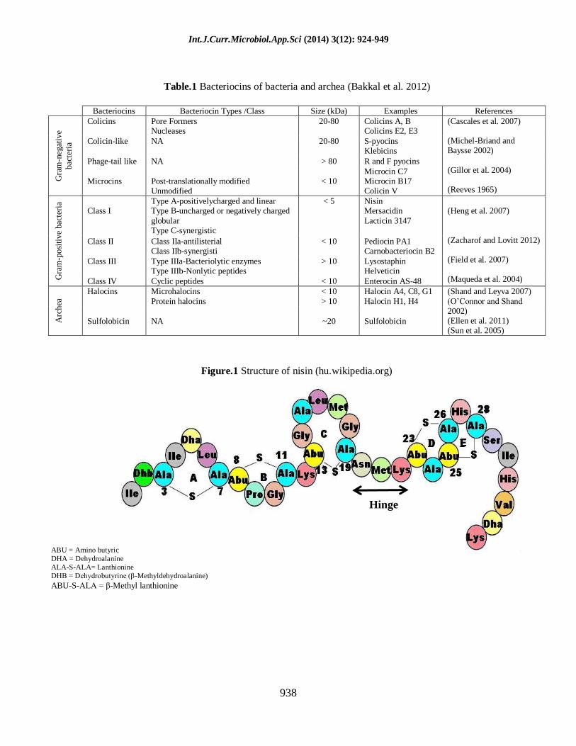
Table.1 Bacteriocins of bacteria and archea (Bakkal et al. 2012)
Bacteriocins Bacteriocin Types /Class Size (kDa) Examples References
Gra
m-n
egat
ive
bac
teri
a
Colicins Pore Formers Nucleases
20-80 Colicins A, B Colicins E2, E3
(Cascales et al. 2007) (Michel-Briand and Baysse 2002)
(Gillor et al. 2004) (Reeves 1965)
Colicin-like NA 20-80 S-pyocins Klebicins
Phage-tail like NA > 80 R and F pyocins
Microcins
Post-translationally modified Unmodified
< 10
Microcin C7 Microcin B17 Colicin V
Gra
m-p
osi
tive
bac
teri
a Class I
Type A-positivelycharged and linear Type B-uncharged or negatively charged
globular Type C-synergistic
< 5 Nisin Mersacidin
Lacticin 3147
(Heng et al. 2007)
(Zacharof and Lovitt 2012) (Field et al. 2007) (Maqueda et al. 2004)
Class II Class IIa-antilisterial Class IIb-synergisti
< 10 Pediocin PA1 Carnobacteriocin B2
Class III Type IIIa-Bacteriolytic enzymes Type IIIb-Nonlytic peptides
> 10 Lysostaphin Helveticin
Class IV Cyclic peptides < 10 Enterocin AS-48
Arc
hea
Halocins Microhalocins
Protein halocins
< 10
> 10
Halocin A4, C8, G1
Halocin H1, H4
(Shand and Leyva 2007)
(O‟Connor and Shand 2002) (Ellen et al. 2011) (Sun et al. 2005)
Sulfolobicin
NA
~20
Sulfolobicin
Figure.1 Structure of nisin (hu.wikipedia.org)

Int.J.Curr.Microbiol.App.Sci (2014) 3(12): 924-949
939
Immunity
Regulated
gene
expression
Leader
Processing Translocation
Modification
NisT
NisP
NisB
NisC
NisI
NisG
NisE
NisF
NisF
NisK
NisR
P
P’
A
B A
T A
C A
I A
P A
R A
K A
F A
E A
G A
P’
Nisin precursor
Mature nisin
Signal peptide Antimicrobial peptide
P1
Figure.2 Structure of Pediocin PA-1 (Desriac et al. 2010)
Figure 3. Model for the biosynthesis of nisin. The nisin precursor is modified by the putative
enzymes NisB and NisC and translocated across the membrane by the exporter NisT. The precursor is
extracellularly processed by NisP, resulting in the release of mature nisin. NisK senses the presence of nisin in the medium and autophosphorylates. The phosphate-group is transferred to NisR, which
activates transcription of the genes nisABTCIP and nisFEG. NisI, F, E, and G protect the cell from
the bacteriocidal activity of nisin. P: promoter region, P*: nisin-regulated promoters ( Van et al.2009).
Int.J.Curr.Microbiol.App.Sci (2014) 3(12): 924-949
940
Figure.4 Schematic overview of the suggested machinery for production of class IIa bacteriocins:
three-component regulatory system, synthesis, processing, excretion and immunity. Ennahar et al. 2000
Future approaches should consider the
application of bacteriocins in combination
with treatments enhancing their
effectiveness in foods and livestock health
together. The evaluation of antibacterial
efficiency of the two bacteriocins from
LAB, nisin and pediocin PA-1/AcH has
revealed that they were more effective
antibacterial in combination than they
were used alone [160]. The use of more
than one LAB bacteriocin as a
combination biopreservatives or
antimicrobial could be advantageous over
a single bacteriocin especially in medical
applications.
References
Diez-Gonzalez, F. 2007. Applications of
bacteriocins in livestock. Current Issues
Intestinal Microbiology 8: 15–24.
Mantovani, H.C., Cruz, A.M.O. and Paiva, A.D. 2011. Bacteriocin activity and
resistance in livestock pathogens.
Méndez-Vilas (Ed.) FORMATEX, 853-863
Fuller, R. 1999. Probiotics for farm animals.
In: Tannock, G. W. (ed), Probiotics: a
critical review, 15-22. Sahl, H.G. and Bierbaum, G. 2008. Multiple
activities in natural antimicrobials.
Microbe 3: 467–473. Patra, A.K. 2011. Enteric methane mitigation

Int.J.Curr.Microbiol.App.Sci (2014) 3(12): 924-949
941
technologies for ruminant livestock: a
synthesis of current research and future directions. Environ Monit Assess 184:
1929–1952.
Mellon, M., Benbrook, C. and Benbrook, K.L.
2001. Hogging it: estimates of antimicrobial abuse in livestock. Union
of Concerned Scientists.
http://www.ucsusa.org/index.html .Accessed Oct. 2002.
Bedford, M. 2000. Removal of antibiotic
growth promoters from poultry diets: implications and strategies to minimize
subsequent problems. World‟s Poultry
Science Journal 56: 347–365.
Wierup, M. 2000. The control of microbial diseases in animals: alternatives to the
use of antibiotics. Antimicrobial Agents
14: 315–319. Doyle, M.E. 2001. Alternatives to antibiotic
use for growth promotion in animal
husbandry. FRI briefings, 1-17. Cleveland, J., Montville, T.J., Nes, I.F.,
Chikindas, M.L. 2001. Bacteriocins:
Safe, natural antimicrobials for food
preservation. International Journal of Food Microbiology 71: 1–20.
Kumar, B., Praveen, P., Kaur, B. and Garg, N.
2011. Cloning and expression of bacteriocins of Pediococcus spp. A
review. Arch Clin Microbiol 2, 1-18.
Zendo, T. 2013. Screening and
characterization of Novel Bacteriocins from Lactic Acid Bacteria. Bioscience
and Biotechnology Biochemistry 77:
893-899. Papagianni, M. and Anastasiadou, S. (2009)
Pediocins: The bacteriocins of
Pediococci. Sources, production, properties and applications. Microbial
Cell Fact 8, 1-16.
Joerger, RD. (2003) Alternatives to
Antibiotics, Bacteriocins, Antimicrobial Peptides and Bacteriophages. Poultry
Sci 82, 640–647.
Riley, MA. (2009) Bacteriocins, Biology, Ecology, and Evolution. Encyclopedia
of Microbiology. Moselio Schaechter
(ed), 32-44. Papavassiliou, J. (1961) Biological
characteristics of colicine X. Nature
190, 110.
El-Khatib, T. and El-Rahman, H.A. (1987) A research note – Effect of garlic and
Lactobacillus plantarum on growth of
Salmonella typhimurium in Egyptian
sausage and beef burger. J Food Prot 50, 310-314.
Gupta, S. and Savaliya, C.V. (2012)
Application of biotechnology to improve livestock products. Vet World
5, 634-638.
Mohanasirivasan, V., Suganthi, V., Selvarajan, E. and Subathradevi, C. (2012)
Lantibiotic Nisin: natural preservative
from Lactococcus lactis. IRJP 3, 13-19.
Gillor, O. (2007) Bacteriocins‟ role in bacterial communication. In: Riley MA,
Chavan M, eds. Bacteriocins: ecology
and evolution. Springer, 135–146. Gobbetti, M., De Angelis, M., Di Cagno, R.,
Minervini, F. and Limitone, A. (2007)
Cell–cell communication in food related bacteria. Int J Food Microbiol 120, 34–
45.
Wang, W.L., Liu, J., Huo, Y.B. and Ling, J.Q.
(2013) Bacteriocin immunity proteins play a role in quorum-sensing system
regulated antimicrobial sensitivity of
Streptococcus mutans UA159. Arch Oral Biol 58, 384-390.
Heng, N.C.K., Wescombe, P.A., Burton, J.P.,
Jack, R.W. and Tagg, J.R. (2007) The
Diversity of Bacteriocins in Gram-positive bacteria, in Bacteriocins:
Ecology and Evolution, Riley MA,
Chavan MA. (Eds.), pp 45-92. Reeves, P. (1965) The bacteriocins. Bacteriol
Rev 29, 24-45.
Delves-Broughton, J., Blackburn, P., Evans, R.J., Hugenholtz, J. (1996) Applications
of the bacteriocin nisin. Antonie van
Leeuwenhoek 69, 193-202.
FDA (US Food and Drug Administration). (1988) Nisin preparation: affirmation of
GRAS status as a direct human food
ingredient. Federal Register 53, 11247-11251.
FDA (US Food and Drug Administration).
Department of Health and Human Services. Agency Response Letter
GRAS Notice nº

Int.J.Curr.Microbiol.App.Sci (2014) 3(12): 924-949
942
GRN000065.2001.Available at:
http://www.accessdata.fda.gov/scripts/fcn/gras_notices/grn0065.pdf. 2011.
Takala, T.M. (2005) Nisin Immunity and
Food-Grade Transformation in Lactic
Acid Bacteria. Academic Dissertation in Microbiology, 1-46.
Liu, W. and Hansen, N. (1990) Some chemical
and physical properties of nisin, a small-protein antibiotic produced by
Lactococcus lactis. Appl Environ
Microbiol 56, 2551-2558. Davies, E.A., Bevis, H.E., Potter, R., Harris,
J., Williams, G.C. and Delves-
Broughton, J. (1998) The effect of pH
on the stability of nisin solution during autoclaving. Lett Appl Microbiol 27,
186-187.
Nolan, E.M. and Walsh, C.T. (2009) How nature morphs peptide scaffolds into
antibiotics. Chem BioChem 10, 34-53.
Papagianni, M. (2003) Ribosomally synthesized peptides with antimicrobial
properties: biosynthesis, structure,
function, and applications. Biotechnol
Adv 21, 465-499. Zacharof, M.P. and Lovitt, R.W. (2012)
Bacteriocin produced by Lactic Acid
Bacteria. APCBEE Procedia 00, 1-6. Drider, D., Fimland, G., Hechard, Y.,
McMullen, L.M. and Prevost, H. (2006)
The continuing story of class IIa
bacteriocins. Microbiol Mol Biol Rev 70, 564-582.
Chen, Y., Ludescher, R.D. and Montville, T.J.
(1997)n Electrostatic interactions but not the YGNGV consensus motif,
govern the binding of pediocin PA-1
and its fragments to phospholipids vesicles. Appl Environ Microbiol 63,
4770-4777.
Miller, K.W., Schamber, R., Osmanagaoglou,
O. and Ray, B. (1998) Isolation and characterization of pediocin AcH
chimeric protein mutants with altered
bactericidal activity. Appl Environ Microbiol 64, 1997-2005.
Kazazic, M., Nissen-Meyer, J. and Fimland,
G. (2002) Mutational analysis of the role of charged residues in target-cell
binding, potency and specificity of the
pediocin-like bacteriocin sakacin P.
Microbiology 148, 2019-2027. Uteng, M., Hauge, H.H., Markwick, P.R.,
Fimland, G., Mantzilas, D., Nissen-
Meyer, J. and Muhle-Goll, C. (2003)
Three-dimensional structure in lipid micelles of the pediocin-like
antimicrobial peptide sakacin P and a
sakacin P variant that is structurally stabilized by an inserted C-terminal
disulphide bridge. Biochemistry 42,
11417-11426. Fimland, G., Blingsmo, O.R., Sletten, K.,
Jung, G., Nes, I.F. and Nissen-Meyer, J.
(1996) New biologically active hybrid
bacteriocins constructed by combining regions from various pediocin-like
bacteriocins: the C-terminal region is
important for determining specificity. Appl Environ Microbiol 62, 3313-3318.
Fimland, G., Jack, R., Jung, G., Jung, G., Nes,
I.F. and Nissen-Meyer, J. (1998) The bactericidal activity of pediocin PA-1 is
specifically inhibited by a 15-mer
fragment that spans the bacteriocin from
the center toward the C terminus. Appl Environ Microbiol 64, 5057-5060.
Abee, T. (1995) Pore-forming bacteriocins of
Gram-positive bacteria and selfprotection mechanisms of producer
organisms. FEMS Microbiol Lett 129,
1-10.
Martinez, R.C.R. and De Martinis, E.C.P. (2006) Effect of Leuconosoc
mesenteroides11 bacteriocin in the
multiplication control of Listeria monocytogenes. Ciênc Tecnol Aliment
26, 52-55.
Deegan, L.H., Cotter, P.D., Hill, C. and Ross, P. (2006) Bacteriocins: Biological tools
for bio-preservation and shelf-life
extension. Int Dairy J 16, 1058-1071.
Van Kraaij, C., de Vos, W.M., Siezen, R.J. and Kuipers, O.P. (1999) Lantibiotics:
biosynthesis, mode of action and
applications. Nat Prod Rep 16, 575–587.
Pag, U. and Sahl, H.G. (2002) Multiple
activities in lantibiotics - models for the design of novel antibiotics? Curr
Pharmaceut Design 8, 815–833.

Int.J.Curr.Microbiol.App.Sci (2014) 3(12): 924-949
943
Hsu, S.T., Breukink, E., de Kruijff, B.,
Kaptein, R., Bonvin, A.M. and van Nuland, N.A. (2002) Mapping the
targeted membrane pore formation
mechanism by solution NMR: the nisin
Z and Lipid II interaction in SDS micelles. Biochemistry 41, 7670-7676.
Breukink, E., van Kraaij, C., Demel, A.,
Siezen, R.J., Kuipers, O.P. and De Kruijff, B. (1997) The C-terminal
region of nisin is responsible for the
initial interaction of nisin with the target membrane. Biochemistry 36, 6968-
6976.
Brötz, H., Bierbaum, G., Leopold, K.,
Reynolds, P.E. and Sahl, H.G. (1998) The lantibiotic mersacidin inhibits
peptidoglycan synthesis by targeting
lipid II. Antimicrobial Agents Chemotherapy 42, 154-160.
Wiedemann, I., Breukink, E., van Kraaij, C.,
Kuipers, O.P., Bierbaum, G., de Kruijff, B. and Sahl, H.G. (2001) Specific
binding of nisin to the peptidoglycan
precursor lipid II combines pore
formation and inhibition of cell wall biosynthesis for potent antibiotic
activity. J Biol Chem 276, 1772–1779.
Breukink, E., Wiedemann, I., van Kraaij, C., Kuipers, O.P., Sahl, H.G. and De
Kruijff, B. (1999) Use of the cell wall
precursor lipid II by a pore-forming
peptide antibiotic. Science 286, 2361–2364.
Hasper, H.E., de Kruijff, B. and Breukink, E.
(2004) Assembly and stability of nisin-Lipid II pores. Biochemistry 43, 11567-
11575.
Héchard, Y. and Sahl, H.G. (2002) Mode of action of modified and unmodified
bacteriocins from Gram-positive
bacteria. Biochimie 84, 545-557.
Gut, I.M., Prouty, A.M., Ballard, J.D., van der Donk, W.A. and Blanke, S.R. (2008)
Inhibition of Bacillus anthracis spore
outgrowth by nisin. Antimicrob Agents Chemo 52, 4281-4288.
Ray, B. (1995) Pediococcus in Fermented
Foods. In Food Biotechnology: Microorganisms Edited by Hui YH and
Khachatourians G. Wiley-VCH, pp 745-
795.
Chikindas, M.L., Garcia–Garcera, M.J., Driessen, A.J.M., Ledeboer, A.M. and
Nissen–Meyer, J. (1993) Pediocin PA–
1, a bacteriocin from Pediococcus
acidilactici PAC1.0, forms hydrophilic pores in the cytoplasmic membrane of
target cells. Appl Environ Microbiol 59,
3577–3584. Manuel, N., Rafael, M., Miguel, A. and
Castanho, R.B. (2009) Antimicrobial
peptides: linking partition, activity and high membrane–bound concentrations.
Nature Rev Microbiol 7, 245–250.
Bhunia, A.K., Johnson, M.C., Ray, B. and
Kalchayanand, N. (1991) Mode of action of pediocin AcH from
Pediococcus acidilactici H on sensitive
bacterial strains. J Appl Bacteriol 70, 25–33.
Mashal. (2007) Biopermeabilization and
antimicrobial applications of purified pediocin CP2 produced from P.
acidilactici MTCC 5101. A project
report, Department of Biotechnology,
Punjabi University, Patiala, Punjab. Miller, K.W., Schamber, R., Chen, Y. and
Ray, B. (1998) Production of active
chimeric pediocin AcH in Escherichia coli in the absence of processing and
secretion genes from the Pediococcus
Pap operon. Appl Environ Microbiol
64, 14–20. Coderre, P.E. and Somkuti, G.A. (1999)
Cloning and expression of the pediocin
operon in Streptococcus thermophiles and other lactic fermentation bacteria.
Curr Microbiol 39, 295–301.
Osmanagaoglu, O., Beyatli, Y. and Gündüz, U. (2000) Cloning and expression of a
plasmid–linked pediocin determinant
trait of Pediococcus acidilactici F. J
Basic Microbiol 40, 41–49. Tominaga, T. and Hatakeyama, Y. (2007)
Development of innovative pediocin
PA–1 by DNA shuffling among class IIa bacteriocins. Appl Environ
Microbiol 73, 5292–5299.
Belkum, M.J., Hayema, B.J., Geis, A., Kok, J. and Venema, G. (1998) Cloning of two
bacteriocin genes from a lactococcal

Int.J.Curr.Microbiol.App.Sci (2014) 3(12): 924-949
944
bacteriocin plasmid. Appl Environ
Microbiol 55, 1187-1191. Buchman, G., Banerjee, S. and Hansen, J.
(1998) Structure, expression and
evolution of gene encoding the
precursor of nisin, a small protein antibiotic. J Biol Chem 263, 16260-
16266.
Jack, R.W., Tagg, J.R. and Ray, B. (1995) Bacteriocins of Gram-positive bacteria.
Microbiol Rev 59, 171-200.
Rauch, P.J.G., Beerthuyzen, M.M. and De Vos, W.M. (1991) In Nisin and Novel
Lantibiotics, eds. Sahl H.G and Jung
G.ESCOM, Leiden, pp 243.
Mierau, I. and Kleerebezem, M. (2005) 10 years of the nisin-controlled gene
expression system (NICE) in
Lactococcus lactis. Appl Microbiol Biotechnol. 68(6), 705-717.
Cotter, P.D., Hill, C. and Ross, R.P. (2005)
Bacteriocins: developing innate immunity for food. Nat Rev Microbiol
3, 777–788.
Lubelski, J., Rink, R., Khusainov, R., Moll,
G.N. and Kuipers, O.P. (2008) Biosynthesis, immunity, regulation,
mode of action and engineering of the
model lantibiotic nisin. Cell Mol Life Sci 65, 455–476.
Cortés, J., Appleyard, A.N. and Dawson, M.J.
(2009) Whole-cell generation of
lantibiotic variants. Methods Enzymol 458( 22), 559–574.
Field, D., Hill, C., Cotter, P.D. and Ross, R.P.
(2010b) The dawning of a „Golden era‟ in lantibiotic bioengineering. Mol.
Microbiol 78, 1077–1087.
Rouse, S., Des, F., Daly, K.M., O‟Connor, P.M., Cotter, P.D., Hill, C. and Ross,
R.P. (2012) Bioengineered nisin
derivatives with enhanced activity in
complex matrices. Microbial Biotechnol 5, 501–508.
Yuan, J., Zhang, Z.Z., Chen, X.Z., Yang, W.
and Huan, L.D. (2004) Site-directed mutagenesis of the hinge region of nisin
Z and properties of nisin Z mutants.
Appl Microbiol Biotechnol 64, 806–815.
Field, D., O‟Connor, P.M., Cotter, P.D., Hill,
C. and Ross, R.P. (2008) The generation
of nisin variants with enhanced activity against specific Gram-positive
pathogens. Mol Microbiol 69, 218–230.
Field, D., Quigley, L., O‟Connor, P.M., Rea,
M.C., Daly, K. and Cotter, P.D. (2010a) Studies with bioengineered Nisin
peptides highlight the broad spectrum
potency of Nisin V. Microbiol Biotechnol 3, 473–486.
Havarstein, L.S., Holo, H. and Nes, I.F. (1995)
The leader peptide of colicin V shares consensus sequences with leader
peptides that are common amongst
peptide bacteriocins produced by Gram-
positive bacteria. Microbiology 140, 2383-2389.
Nes, I.F., Diep, D.B., Havarstein, L.S.,
Brurberg, M.B., Eijsink, V. and Holo, H. (1996) Biosynthesis of bacteriocins
in lactic acid bacteria. Int J Gen Mol
Microbiol 70, 113-128. Klaenhammer, T.R. (1993) Genetics of
bacteriocins produced by lactic acid
bacteria. FEMS Microbiol Rev 12, 39-
86. Ennahar, S., Sashihara, T., Sonomoto, K. and
Ishizaki, A. (2000) Class IIa
bacteriocins: biosynthesis, structure and activity. FEMS Microbiol Rev 24, 85-
106.
Marugg, J.D., Gonzalez, C.F., Kunka, B.S.,
Ledeboer, A.M. and Pucci, M.J. (1992) Cloning, expression and nucleotide
sequence of genes involved in
production of pediocin PA–1, a bacteriocin from Pediococcus
acidilactici PAC1.0. Appl Environ
Microbiol 58, 2360–2367. Bukhtiyarova, M., Yang, R. and Ray, B.
(1994) Analysis of pediocin AcH gene
cluster from plasmid pSMB74 and its
expression in a pediocin–negative Pediococcus acidilactici strain. Appl
Environ Microbiol 603, 405–3408.
Motlagh, A., Bukhtiyarova, M. and Ray, B. (1994) Complete nucleotide sequences
of pSMB74, a plasmid encoding
production of pediocin AcH in Pediococcus acidilactici. Lett Appl
Microbiol 18, 305–312.

Int.J.Curr.Microbiol.App.Sci (2014) 3(12): 924-949
945
Nieto–Lozano, J.C.N., Nissen–Meyer, J.,
Sletten, K., Pelaz, C. and Nes, I.F. (1990) Purification and amino acid
sequence of a bacteriocin produced by
Pediococcus acidilactici. J Gen
Microbiol 138, 1985–1990. Henderson, J.T., Chopko, A.L. and van
Wassenaar, P.D. (1992) Purification and
primary structure of pediocin PA–1 produced by Pediococcus acidilactici
PAC1.0. Arch Biochem Biophys 29,
55–12. Parada, J.L., Caron, C.R., Medeiros, A.B.P.
and Soccol, C.R. (2007) Bacteriocins
from lactic acid bacteria: Purification,
properties and use as biopreservatives. Brazil Arch Biology Technol 50, 521-
542.
Schöbitz, R.P., Bórquez, P.A., Costa, M.E., Ciampi, L.R. and Brito, C.S. (2006)
Bacteriocin like substance production
by Carnobacterium piscicola in a continuous system with three culture
broths. Study of antagonism against
Listeria monocytogenes in vacuum
packaged salmon. Braz J Microbiol 37, 52-57.
Chung, K., Dickson, J. and Creouse, J.
(1989) Effects of nisin on growth of bacteria attached to meat. Appl Environ
Microbiol 55, 1329-1333.
Vignolo, G., Fadda, S., De Kairuz, M.N., De
Ruiz Holgado, A.A. and Oliver, G. (1996) Control of Listeria
monocytogenes in ground beef by
lactocin705, a bacteriocin produced by Lactobacillus casei. Food Microbiol
CRL 705, 29, 397-402.
Jeevaratnam, K., Jamuna, M. and Bawa, A.S. (2005) Biological preservation of foods
– Bacteriocins of lactic acid bacteria.
Indian J Biotechnol 4, 446-454.
Delves-Broughton, J. (1990) Nisin and its uses as preservative. Food Technol 44, 100-
117.
Ogden, K., Waites, M.J. and Hammond, J.R.M. (1988) Nisin and brewing. J Inst
Brew 94, 233-238.
Deaschel, M.A., Jung, D.S. and Watson, B.T. (1991) Controlling wine malolactic
fermentation with nisin and nisin-
resistant strains of Leuconostoc oenos.
Appl Environ Microbiol 57, 601-603. Smid, E.J. and Gorris, G.M. (1999) Natural
Antimicrobials for food preservation.
Handbook of food preservation, Part 9,
285-308. Chung, W. and Hancock, R.E.W. (2000)
Action of lysozyme and nisin mixtures
against lactic acid bacteria. Int J Food Microbiol 60, 25-32.
Bakkal, S., Robinson, S.M. and Riley, M.A.
(2012) Bacteriocins of Aquatic Microorganisms and Their Potential
Applications in the Seafood Industry. In
book: Health Environ Aquaculture, 303-
328. Neetoo, H., Ye, M., Chen, H., Joerger, R.D.,
Hicks, D.T. and Hoover, D.G. (2008)
Use of nisin-coated plastic films to control Listeria monocytogenes on
vacuum-packaged cold-smoked salmon.
Int J Food Microbiol 122, 8-15. Nykanen, A., Weckman, K. and
Lapvetelainaen, A. (2000) Synergistic
inhibition of Listeria monocytogenes on
cold-smoked rainbow trout by nisin and sodium lactate. Int J Food Microbiol
61, 63-72.
McEntire, J.C., Montville, T.J. and Chikindas, M.L. (2003) Synergy between nisin and
select lactate against L. monocytogenes
is due to the metal cations. Int J Food
Microbiol 66, 1631-1636 Dykes, G.A. and Moorhead, S.M. (2002)
Combined antimicrobial effect of nisin
and a listeriophage against Listeria monocytogenes in broth but not in
buffer or on raw beef. Int J Food
Microbiol 73, 71-81. Motlagh, A.M., Holla, S., Johnson, M.C., Ray,
B. and Field, R.A. (1992) Inhibition of
Listeria spp. in sterile food systems by
pediocin AcH, a bacteriocin produced by Pediococcus acidilactici H. J Food
Prot 55, 337–343.
Yang, R. and Ray, B. (1994) Factors influencing production of bacteriocins
by lactic acid bacteria. Food Microbiol
11, 281-291. Bennik, M.H.J. (1997) Vegetable-associated
Pediococcus parvulus produces

Int.J.Curr.Microbiol.App.Sci (2014) 3(12): 924-949
946
pediocin PA-1. Appl Environ Microbiol
63, 2074-2076. Ennahar, S., Assobhel, D. and Hasselmann, C.
(1998) Inhibition of Listeria
monocytogenes in a smear-surface soft
cheese by Lactobacillus plantarum WHE92 a pediocin AcH producer. J
Food Prot 61, 86-191.
Galvez, A., Lopez, R.L., Abriouel, H., Valdivia, E. and Omar, N.B. (2008)
Application of bacteriocins in the
control of food borne pathogenic and spoilage bacteria. Crit Rev Biotechnol
28, 125-152.
Rodriguez, E., Arques, J.L., Rodriguez, R.,
Nunez, M. and Medina, M. (2003) Reuterin production by lactobacilli
isolated from pig feces and evaluation
of probiotic traits. Lett Appl Microbiol 37, 259–263.
Renter, D.G. and Sargeant, J.M. (2002)
Enterohemorrhagic Escherichia coli O157: epidemiology and ecology in
bovine production environments. Anim
Health Res Rev 3, 83–94.
Callaway, T.R., Anderson, R.C., Edrington, T.S., Genovese, K.J., Harvey, R.B.,
Poole, T.L. and Nisbet, D.J. (2004)
Recent pre-harvest supplementation strategies to reduce carriage and
shedding of zoonotic enteric bacterial
pathogens in food animals. Anim Health
Res Rev 5, 35–47. Gillor, O., Kirkup, B.C. and Riley, M.A.
(2004) Colicins and microcins: the next
generation of antimicrobials. Adv Appl Microbiol 54, 129–146.
Timmerman, H.M., Koning, C.J., Mulder, L.,
Rombouts. F.M. and Beynen, A.C. (2004) Monostrain, multistrain and
multispecies probiotics--A comparison
of functionality and efficacy. Int J Food
Microbiol 96, 219–233. Ray, B. (2003) Fundamental Food
Microbiology. Boca Raton, FL, CRC
Press 9, 103-120. Ariyapitipun, T., Mustapha, A. and Clarke,
A.D. (2000) Survival of Listeria
monocytogenes on vacuum-packaged raw beef treated with polylactic acid,
lactic acid, and nisin. J Food Prot 63,
131–136.
De Kwaadsteniet, M., Ten, Doeschate, K. and Dicks, L.M.T. (2008) Characterization
of the structural gene encoding nisin F,
a new lantibiotic produced by a
Lactococcus lactis subsp. lactis isolate from freshwater fish (Clarius
gariepinus). Appl Environ Microbiol 74,
547–549. Kerro-Dego, O., van Dijk, J.E. and
Nederbragt, H. (1992) Factors involved
in the early pathogenesis of bovine Staphylococcus aureus mastitis with
emphasis on bacterial adhesion and
invasion. A review. Vet Quartely 24,
181-198. Sears, P.M., Smith, B.S., Stewart. W.K.,
Gonzalez, R.N., Rubino, S.D., Gusik,
S.A., Kulizek, E.S., Projan, S.J. and Blackburn, P. (1992) Evaluation of a
nisin-based germicidal formulation on
teat skin of live cows. J Dairy Sci 75, 3185–3190.
Wu, J., Hu, S. and Cao, L. (2007) Therapeutic
effect of nisin Z on subclinical mastitis
in lactating cows. Antimicrob Agents Chemother 51, 3131–3135.
Sar, C., Mwenya, B., Pen, B., Morikawa, R.,
Takaura, K., Kobayashi, T. and Takahashi, J. (2005) Effect of nisin on
ruminal methane production and
nitrate/nitrite reduction in vitro. Aust J
Agric Res 56, 803-810. Mantovani, H. and Russell, J.B. (2001) Nisin
resistance of Streptococcus bovis. App
Environ Microbiol 67, 808-813. Kišidayová, S., Siroka, P. and Laukova, A.
(2003) Effect of nisin on two cultures of
rumen ciliates. Folia Microbiologica 48, 408-412.
Callaway, T.R., Melo, A.M.S.C. and Russell,
J.B. (1997) The effect of nisin and
monensin on ruminal fermentation in vitro. Current Microbiol 35, 90-96.
Jalc, D. and Laukove, A. (2002) Effect of
nisin and monensin on rumen fermentation in artificial rumen.
Berliner und Munchener Tierärztliche
Wochenschrift 115, 6–10. Santoso, B., Mwenya, B., Sar, C. and
Takahashi, J. (2006) Ruminal
Int.J.Curr.Microbiol.App.Sci (2014) 3(12): 924-949
947
fermentation and nitrogen metabolism
in sheep fed a silage-based diet supplemented with Yucca schidigera or
Y. schidigera and nisin. Animal Feed
Sci Technol 129, 187-195.
Takahashi, J., Mwenya, B., Santoso, B., Sar, C., Umetsu, K., Kishimoto, T.,
Nishizaki, K., Kimura, K. and Zendo, T.
(2013) Screening and characterization of novel bacteriocins from Lactic Acid
Bacteria. Biosci Biotechnol Biochem
77, 893-899. Reilly, A. and Kaferstein, F. (1999) Food
safety and products from aquaculture. J
Appl Microbiol 85, 249S–257S.
Corripio-Myar, Y., Mazorra de Quero, C., Treasurer, J.W., Ford, L., Smith, P.D.
and Secombes, C.J. (2007) Vaccination
experiments in the gadoid haddock, Melanogrammus aeglefinus L., against
the bacterial pathogen Vibrio
anguillarum. Vet Immunol Immunopathol 118, 147–153.
Smith, P. (2007) Antimicrobial use in shrimp
farming in Ecuador and emerging multi-
resistance during the cholera epidemic of 1991: a re-examination of the data.
Aquaculture 271, 1–7.
Alderman, D.J. and Hastings, T.S. (1998) Antibiotic use in aquaculture:
development of antibiotic resistance
potential for consumer health risks. Int J
Food Sci Technol 33, 139–155. Prater, D.A. (2005) Judicious use of
antimicrobials for aquatic veterinarians.
FDA, Veterinarian Newsletter, 20-25. Matyar, F. (2007) Distribution and
antimicrobial multi-resistance in Gram-
negative bacteria isolated from Turkish sea bass farm. Ann Microbiol 57, 35–
38.
Laukova, A., Guba, P., Nemcova, R. and
Vasilkova, Z. (2003) Reduction of Salmonella in gnotobiotic Japanese
quails caused by the enterocin A
producing EK13 strain of Enterococcus faecium. Vet Res Commun 27, 275–
280.
Zhou, X. and Wang, Y. (2012) Probiotics in Aquaculture - Benefits to the Health,
Technological Applications and Safety.
Health Environ Aquaculture 8, 215-226.
Thompson, F.L., Abreu, P.C. and Cavalli, R. (1999) The use of microorganisms as
food source for Penaeus paulensis
larvae. Aquaculture 174, 139–153.
Verschuere, L., Rombaut, G., Sorgeloos, P. and Verstraete, W. (2000) Probiotic
bacteria as biological control agents in
aquaculture. Microbiol Mol Biol Rev 64, 655–671.
Wang, Y. (2007) Effect of probiotics on
growth performance and digestive enzyme activity of the shrimp Penaeus
vannamei. Aquaculture 269, 259-264.
Taoka, Y., Maeda, H., Jo, J.Y., Jeon, M.J.,
Bai, S.C., Lee, W.J., Yuge, K. and Koshio, S. (2006) Growth, stress
tolerance and non-specific immune
response of Japanese flounder Paralichthys olivaceusto probiotics in a
closed recirculating system. Fish Sci 72,
310–321. Gatesoupe, F.J. (1999) The use of probiotics
in aquaculture. Aquaculture 180, 147–
165.
Gatesoupe, F.J. (2008) Updating the Importance of Lactic Acid Bacteria in
Fish Farming: Natural Occurrence and
Probiotic Treatments. J Mol Microbiol Biotechnol 14, 107–114.
Irianto, A. and Austin, B. (2002) Probiotics
in aquaculture. J Fish Dis 25, 633–642.
Acuña, L., Picariello, G.F., Sesma, F., Morero, R.D. and Bellomio, A. (2012) A new
hybrid bacteriocin, Ent35eMccV,
displays antimicrobial activity against pathogenic Gram- positive and
Gram-negative bacteria. Fed Eur
Biochem Soc Open BIO. 2, 12-19. Duquesne, S., Destoumieux-Garzón, D.,
Peduzzi, J. and Rebuffat, S. (2007)
Review: Microcins, gene-encoded
antibacterial peptides from enterobacteria. Nat. Prod. Rep. 24, 708-
734.
Hillman, J.D., Brooks, T.A., Michalek, S.M., Harmon, C.C., Snoep, J.L. and van Der
Weijden, C.C. (2000) Construction and
characterization of an effector strain of Streptococcus mutans for replacement
therapy of dental caries. Infect Immun

Int.J.Curr.Microbiol.App.Sci (2014) 3(12): 924-949
948
68, 543–549.
Hillman, J.D. (2002) Genetically modified Streptococcus mutans for the prevention
of dental caries. Antonie Van
Leeuwenhoek 82, 361–366.
Zarate, G. and Nader-Macias, M.E. (2006) Viability and biological properties of
probiotic vaginal lactobacilli after
lyophilization and refrigerated storage into gelatin capsules. Process Biochem
41, 1779–1785.
Kuipers, O.P., Yap, W.M.G.J., Rollema, H.S., Beerthuyzen, M.M., Siezen, R.J. and de
Vos, W.M. (1991) Nisin and novel
lantibiotics, eds. Sahl H-G and Jung G,
ESCOM, Leiden, pp. 250. Rollema, H.S., Kuipers, O.P., Both, P., de
Vos, W.M. and Siezen, R.J. (1995)
Biotechnological applications of microbes. Appl Environ Microbiol 61,
2873-2878.
Severina, E., Severin, A. and Tomasz, A. (1998) Antibacterial efficacy of nisin
against multidrug-resistant Gram-
positive pathogens. J Antimicrob
Chemother 41, 341-347. De Kwaadsteniet, M., Ten Doeschate, K.T.
Dicks, L.M.T. (2009) Nisin F in the
treatment of respiratory tract infections caused by Staphylococcus aureus. Lett
Appl Microbiol 48, 65-70.
Bartoloni, A., Mantella, A., Goldstein, B.P.,
Dei, R., Benedetti, M., Sbaragli, S. and Paradisi, F. (2004) In-vitro activity of
nisin against clinical isolates of
Clostridium difficile. J Chemother 6, 119-121.
De Carvalho, A.A., Mantovani, H.C. and
Vanetti, M.C. (2007) Bactericidal effect of bovicin HC5 and nisin against
Clostridium tyrobutyricum isolated
from spoiled mango pulp. Lett Appl
Microbiol 45, 68-74. Kim, T.S., Hur, J.W., Yu, M.A., et al.
(2003) Antagonism of Helicobacter
pylori by bacteriocins of lactic acid bacteria. J Food Prot 66, 3–12.
Brand, M.A. (2013) Therapeutic properties of
the lantibiotic nisin F. Dissertation presented for the degree of Doctor of
Science in the Faculty of Science at
Stellenbosch University. Stellenbosch
University, 7-32. Vadyvaloo, V., Hastings, J.W., Van Der
Merwe, M.J. and Rautenbach, M.
(2002) Membranes of class IIa
bacteriocin-resistant L. monocytogenes cells contain increased levels of
desaturated and snort-acyl-chain
phosphatidylglycerols. Appl. Environ.Microbiol. 68, 5223-5230.
Doyle, M.P., Zhao, T., Harmon, B.G. and
Brown, C.A. (1999) Control of entero hemorrhagic E. coli O157:H7 in cattle
by probiotic bacteria and specific strains
of E. coli. In Official Gaz. U.S. Pat
Tradem. Off. Pat. (USA). Wooley, R.E. and Shotts Jr, E.B. (2000)
Biological control of food pathogens in
livestock. In Official Gaz. U.S. Pat. Tradem. Off. Pat. (USA)
Jones, E., Salin, V, and Williams, G.W. (2005)
Nisin and the market for commercial bacteriocins. TAMRC Consumer and
Product Research Report No. CP-01-05,
1-19.
Law, A. (2005). www.hoovers.com. Gibbs, G.M., Davidson, B.E. and Hillier, A.J.
(2004) Novel expression system for
large-scale production and purification of recombinant class IIa
bacteriocins and its application to
Piscicolin 126. Appl Environ Microbiol
70, 3292-3297. Jiménez, J.J., Borrero, J., Diep, D.B., Gútiez,
L., Nes, I.F., Herranz, C., Cintas, L.M.
and Hernández, P.E. (2013) Cloning, production, and functional expression of
the bacteriocin sakacin A (SakA) and
two SakA-derived chimeras in lactic acid bacteria (LAB) and the yeasts
Pichia pastoris and Kluyveromyces
lactis. J Ind Microbiol Biotechnol 40,
977-993. Ralph, W.J., Tagg, J.R. and Ray, B. (1995)
Bacteriocins of Gram-positive bacteria.
Microbiol Rev 59, 171–200. Hanlin, M.B., Kalchayanand, N., Ray, P. and
Ray, B. (1993) Bacteriocins of lactic
acid bacteria in combination have a greater antibacterial activity. J Food
Prot 56, 252-255.

Int.J.Curr.Microbiol.App.Sci (2014) 3(12): 924-949
949
Desriac, F., Defer, D., Bourgougnon, N.,
Brillet, B., Le Chevalier, P. and Fleury, Y. (2010) Bacteriocin as weapons in the
marine animal-associated bacteria
warfare: Inventory and potential
applications as an aquaculture probiotic. Mar Drugs 8, 1153-1177.
Cascales, E., Buchanan, S.K., Duche, D.,
Kleanthous, C., Lloubes, R., Postle, K., Riley, M., Slatin, S. and Cavard, D.
(2007) Colicin biology. Microbiol Mol
Biol Rev 71,158-229. Michel-Briand, Y. and Baysse, C. (2002) The
pyocins of Pseudomonas aeruginosa.
Biochimie 84, 499-510.
Field, D., Cotter, P., Hill, C. and Ross, R.P. (2007) Bacteriocin Biosynthesis,
Structure, and Function, In Research
and Applications in Bacteriocins, Riley, M.A., Gillor, O. (Eds.), pp 5-41.
Maqueda, M., Galvez, A., Bueno, M.M.,
Sanchez-Barrena, M.J., Gonzalez. C., Albert, A., Rico, M. and Valdivia, E.
(2004) Peptide AS-48: prototype of a
new class of cyclic bacteriocins. Curr
Prot Peptide Sci 5, 399-416. Shand, R.F. and Leyva, K.J. (2007) Peptide
and Protein Antibiotics fromthe Domain
Archaea: Halocins and Sulfolobicins, In Bacteriocins: Ecology and Evolution,
Riley MA, Chavan, M.A. (Eds.), pp 93-
109.
O'Connor, E.M. and Shand, R.F. (2002) Halocins and sulfolobicins: the
emerging story of archaeal protein and
peptide antibiotics. J Ind Microbiol Biotechnol 28, 23-31.
Ellen, A.F., Rohulya, O.V., Fusetti, F.,
Wagner, M., Albers, S.V. and Driessen, A.J. (2011) The sulfolobicin genes of
Sulfolobus acidocaldarius encode novel
antimicrobial proteins. J Bacteriol 193,
4380-4387. Sun, C., Li, Y., Mei, S., Lu, Q., Zhou, L. and
Xiang, H. (2005) A single gene directs
both production and immunity of halocin C8 in a haloarchaeal strain
AS7092. Mol Microbiol 57, 537-549.