Interfaces development for medical imaging Applications … · Interfaces development for medical...
25
Interfaces development for medical imaging Applications in the framework of the Multiple Scleoris disease Erik Pernod 1 1 Student engineer in “Calcul Scientifique”, ISITV, Toulon 2 Asclépios Project team, INRIA, Sophia Antipolis Supervisor: Jean-Christophe Souplet 2 Internship presentation Thursday the 4 th of September, 2008
Transcript of Interfaces development for medical imaging Applications … · Interfaces development for medical...
Interfaces development for medical imagingApplications in the framework
of the Multiple Scleoris disease
Erik Pernod1
1Student engineer in “Calcul Scientifique”, ISITV, Toulon
2Asclépios Project team, INRIA, Sophia Antipolis
Supervisor: Jean-Christophe Souplet2
Internship presentationThursday the 4th of September, 2008
• Institut National de Recherche en Informatique et en automatique.
• Goals: - Dedicated to fundamental and applied research.
INRIA http://www.inria.fr
Internship presentationThursday the 4th of September, 2008
- Dedicated to fundamental and applied research.- Play a major role in technology transfer.
Asclepios• In medical image processing domain.
• Goals:- Analysis of medical and biomedical images with advanced
geometrical, statistical, physical and functional models.- Provides optimized tools to clinicians
http://www-sop.inria.fr/asclepios/
• A three years ANR scientific project (2007-2009)
• Goal: Federate medical data and algorithms, and sharing computing resources on grid infrastructure.
NeuroLOG http://neurolog.polytech.unice.fr
Internship presentationThursday the 4th of September, 2008
• On three different pathologies:- Multiple Sclerosis- Brain Stroke- Tumours
• Partners from different disciplines:- Software technologies- Databases and knowledge- Medical imaging
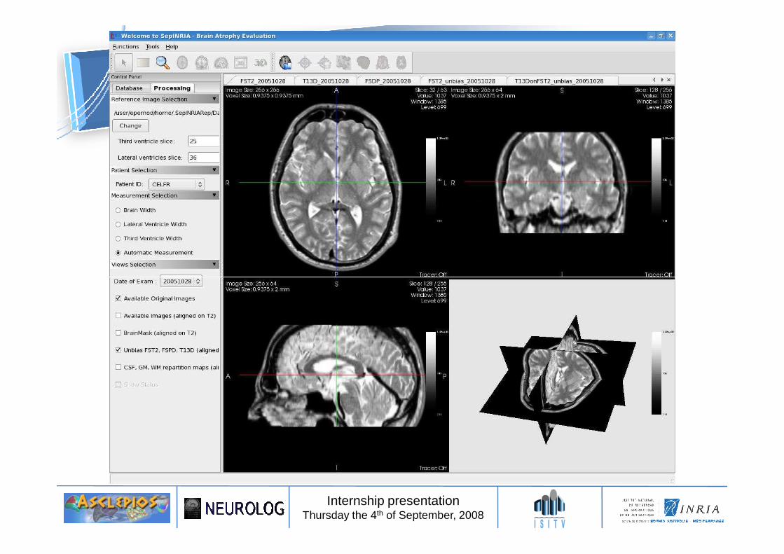
• To understand an application of brain MRI segmentation by implementing tools in the software SepINRIA.
• To deploy this application on the EGEE computational grid
My work
Internship presentationThursday the 4th of September, 2008
Plan• Brain MRI segmentation pipeline
• SepINRIA software
• Workflow deployment on the EGEE grid
• Time performance and study of a method’s parameter influence
• Conclusion
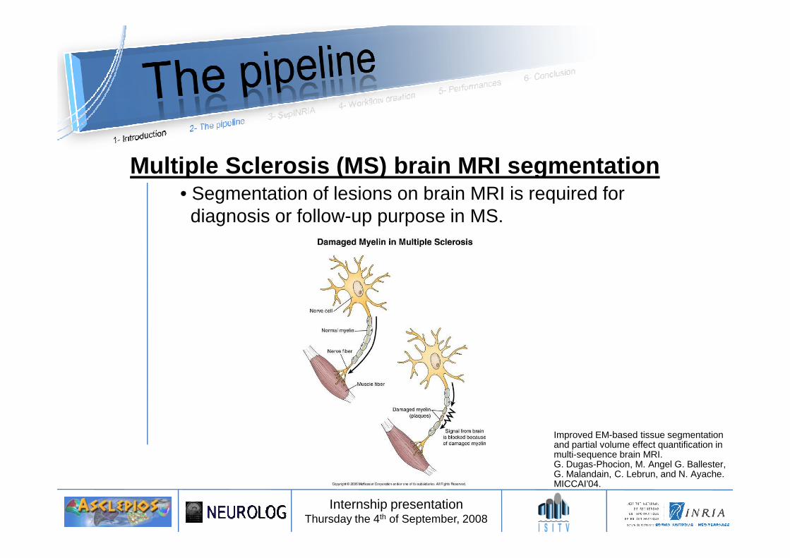
Multiple Sclerosis (MS) brain MRI segmentation• Segmentation of lesions on brain MRI is required fordiagnosis or follow-up purpose in MS.
Internship presentationThursday the 4th of September, 2008
Improved EM-based tissue segmentation and partial volume effect quantification in multi-sequence brain MRI.G. Dugas-Phocion, M. Angel G. Ballester, G. Malandain, C. Lebrun, and N. Ayache.MICCAI’04.
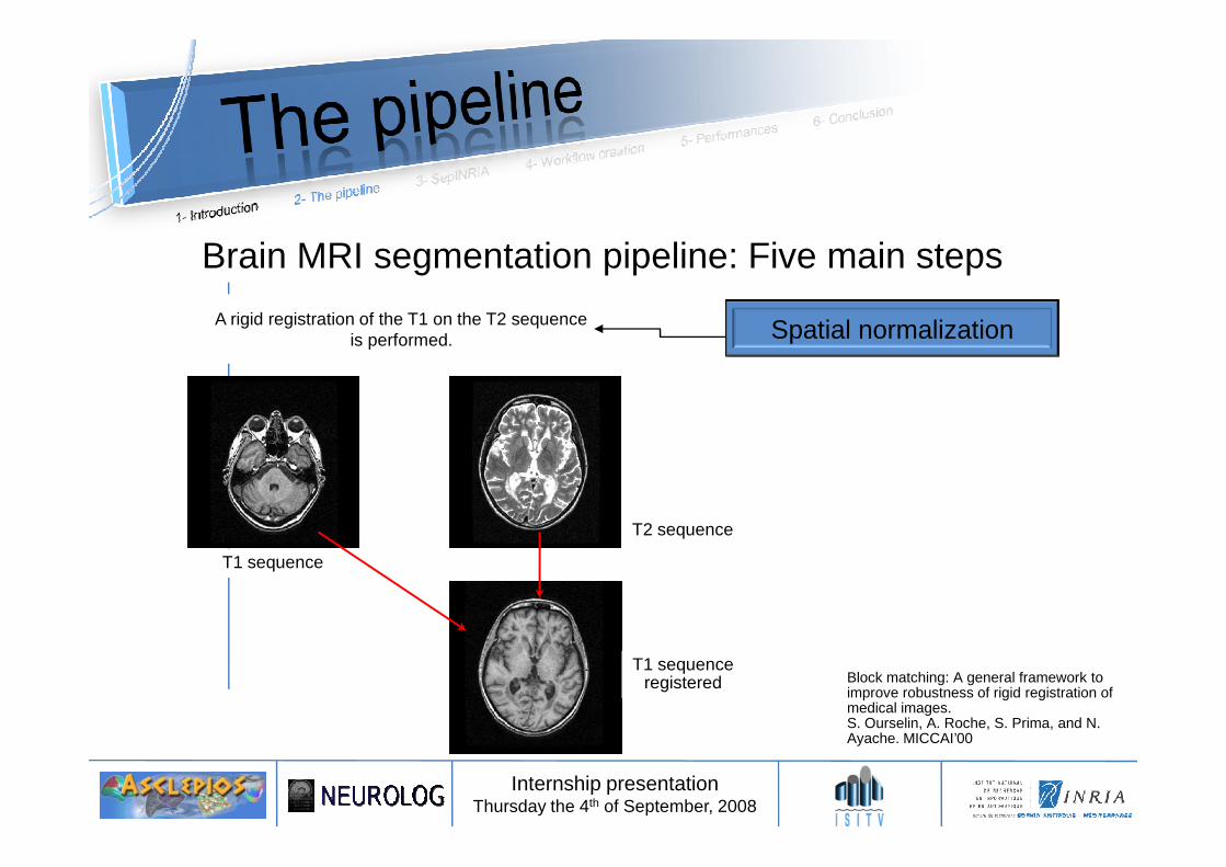
Brain MRI segmentation pipeline: Five main steps
Spatial normalizationA rigid registration of the T1 on the T2 sequenceis performed.
A rigid registration of the T1 on the T2 sequenceis performed.
Internship presentationThursday the 4th of September, 2008
T1 sequence
T2 sequence
T1 sequenceregistered Block matching: A general framework to
improve robustness of rigid registration of medical images. S. Ourselin, A. Roche, S. Prima, and N. Ayache. MICCAI’00
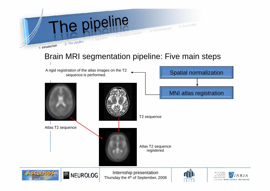
Brain MRI segmentation pipeline: Five main steps
Spatial normalization
MNI atlas registration
A rigid registration of the atlas images on the T2sequence is performed.
A rigid registration of the atlas images on the T2sequence is performed.
Internship presentationThursday the 4th of September, 2008
MNI atlas registration
T2 sequence
Atlas T2 sequenceregistered
Atlas T2 sequence
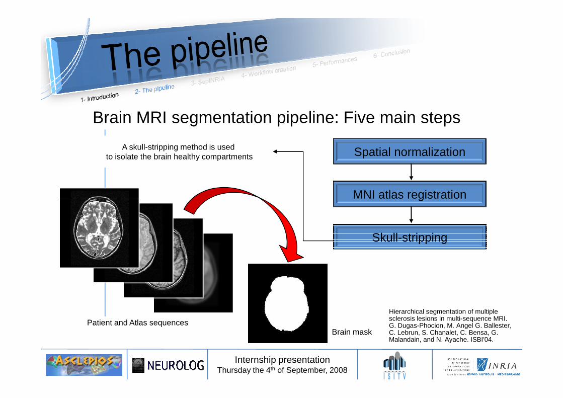
Spatial normalization
MNI atlas registration
A skull-stripping method is used to isolate the brain healthy compartments
A skull-stripping method is used to isolate the brain healthy compartments
Brain MRI segmentation pipeline: Five main steps
Internship presentationThursday the 4th of September, 2008
MNI atlas registration
Skull-stripping
Patient and Atlas sequencesBrain mask
Hierarchical segmentation of multiple sclerosis lesions in multi-sequence MRI.G. Dugas-Phocion, M. Angel G. Ballester, C. Lebrun, S. Chanalet, C. Bensa, G. Malandain, and N. Ayache. ISBI’04.
Spatial normalization
MNI atlas registration
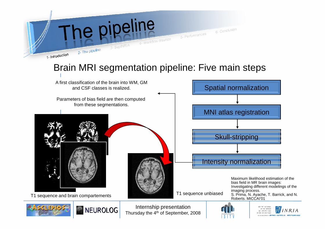
A first classification of the brain into WM, GMand CSF classes is realized.
Parameters of bias field are then computed from these segmentations.
A first classification of the brain into WM, GMand CSF classes is realized.
Parameters of bias field are then computed from these segmentations.
Brain MRI segmentation pipeline: Five main steps
Internship presentationThursday the 4th of September, 2008
MNI atlas registration
Skull-stripping
Intensity normalization
T1 sequence and brain compartements T1 sequence unbiased
Maximum likelihood estimation of the bias field in MR brain images: Investigating different modelings of the imaging process.S. Prima, N. Ayache, T. Barrick, and N. Roberts. MICCAI’01
Brain MRI segmentation pipeline: Five main steps
Spatial normalization
MNI atlas registration
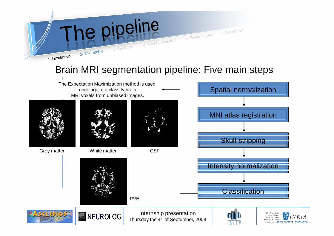
The Expectation Maximization method is used once again to classify brain
MRI voxels from unbiased images.
The Expectation Maximization method is used once again to classify brain
MRI voxels from unbiased images.
Internship presentationThursday the 4th of September, 2008
MNI atlas registration
Skull-stripping
Intensity normalization
Classification
Grey matter White matter CSF
PVE
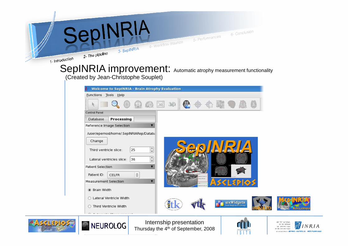
SepINRIA improvement: Automatic atrophy measurement functionality(Created by Jean-Christophe Souplet)
Internship presentationThursday the 4th of September, 2008
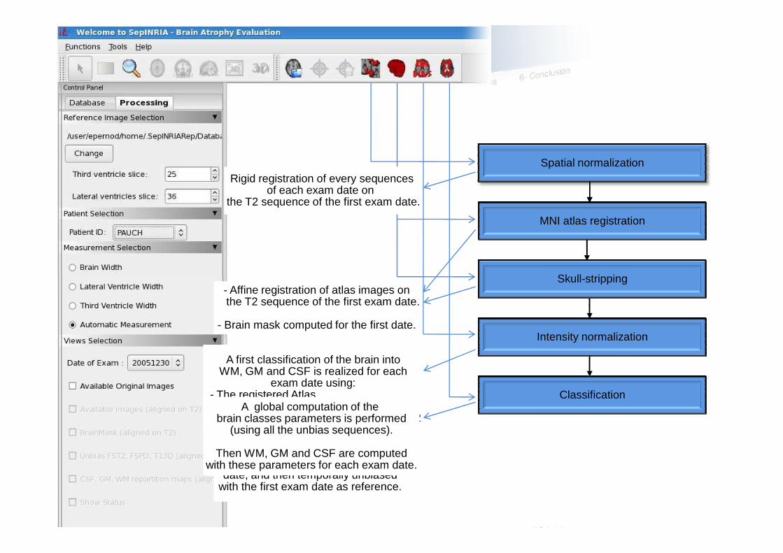
Connection with the pipeline:Spatial normalization
MNI atlas registrationMNI atlas registration
Rigid registration of every sequencesof each exam date on
the T2 sequence of the first exam date.
Skull-strippingSkull-stripping
Internship presentationThursday the 4th of September, 2008
Intensity normalizationIntensity normalization
ClassificationClassification
- Affine registration of atlas images onthe T2 sequence of the first exam date.
- Brain mask computed for the first date.
A first classification of the brain intoWM, GM and CSF is realized for each
exam date using: - The registered Atlas- The brain mask of the first date.- Sequences registered on the first date T2
Sequences are then unbiased for each date, and then temporally unbiased
with the first exam date as reference.
A global computation of the brain classes parameters is performed
(using all the unbias sequences).
Then WM, GM and CSF are computedwith these parameters for each exam date.
Skull-strippingSkull-stripping
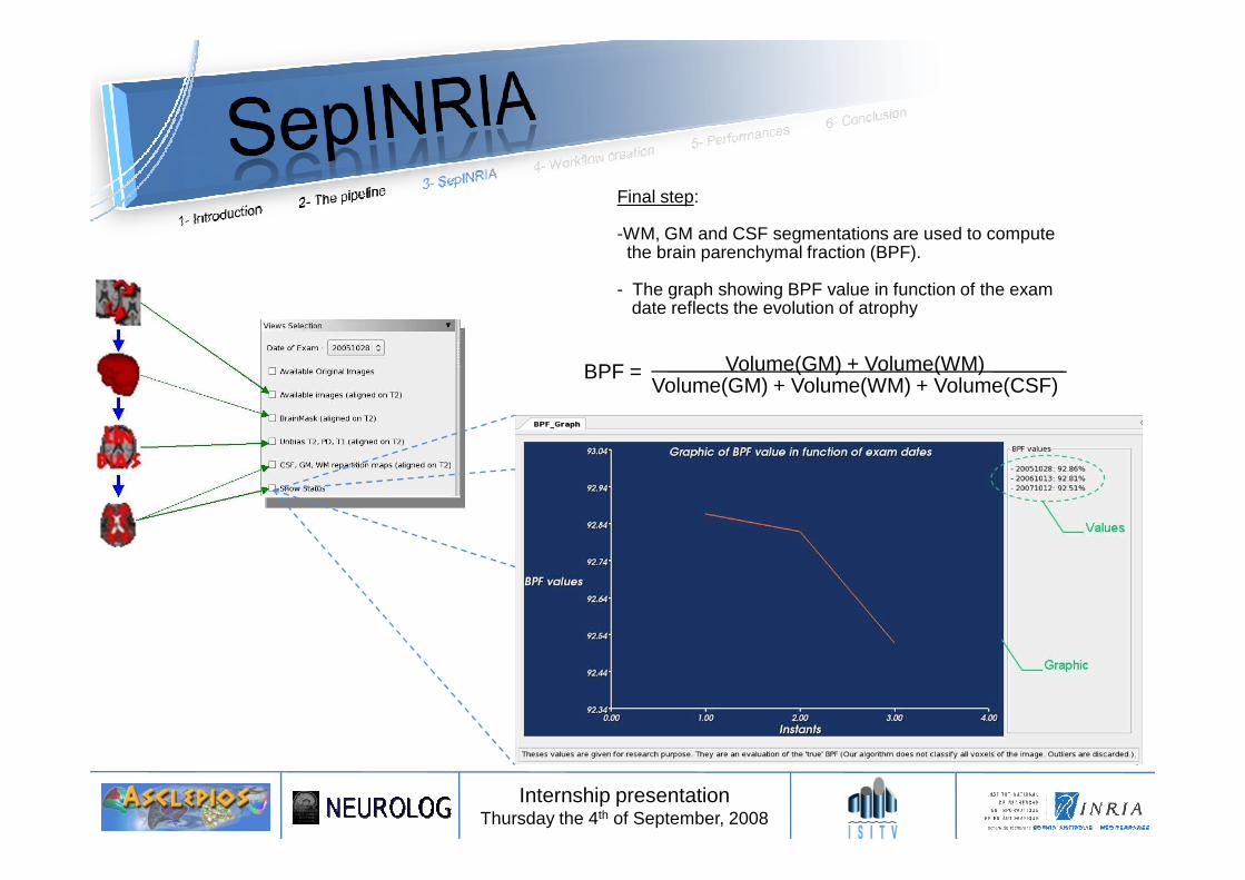
Volume(GM) + Volume(WM)Volume(GM) + Volume(WM) + Volume(CSF)
BPF =
Final step:
-WM, GM and CSF segmentations are used to compute the brain parenchymal fraction (BPF).
- The graph showing BPF value in function of the examdate reflects the evolution of atrophy
Internship presentationThursday the 4th of September, 2008
Internship presentationThursday the 4th of September, 2008
• Needed transformations of the pipeline ?
Problematic: How to deploy and parallelize an algorithm on a grid?
Internship presentationThursday the 4th of September, 2008
• How to create and execute a workflow ?
• Performances ?
• First, what is a grid?- Network of shared computing resources.- Different from a cluster.
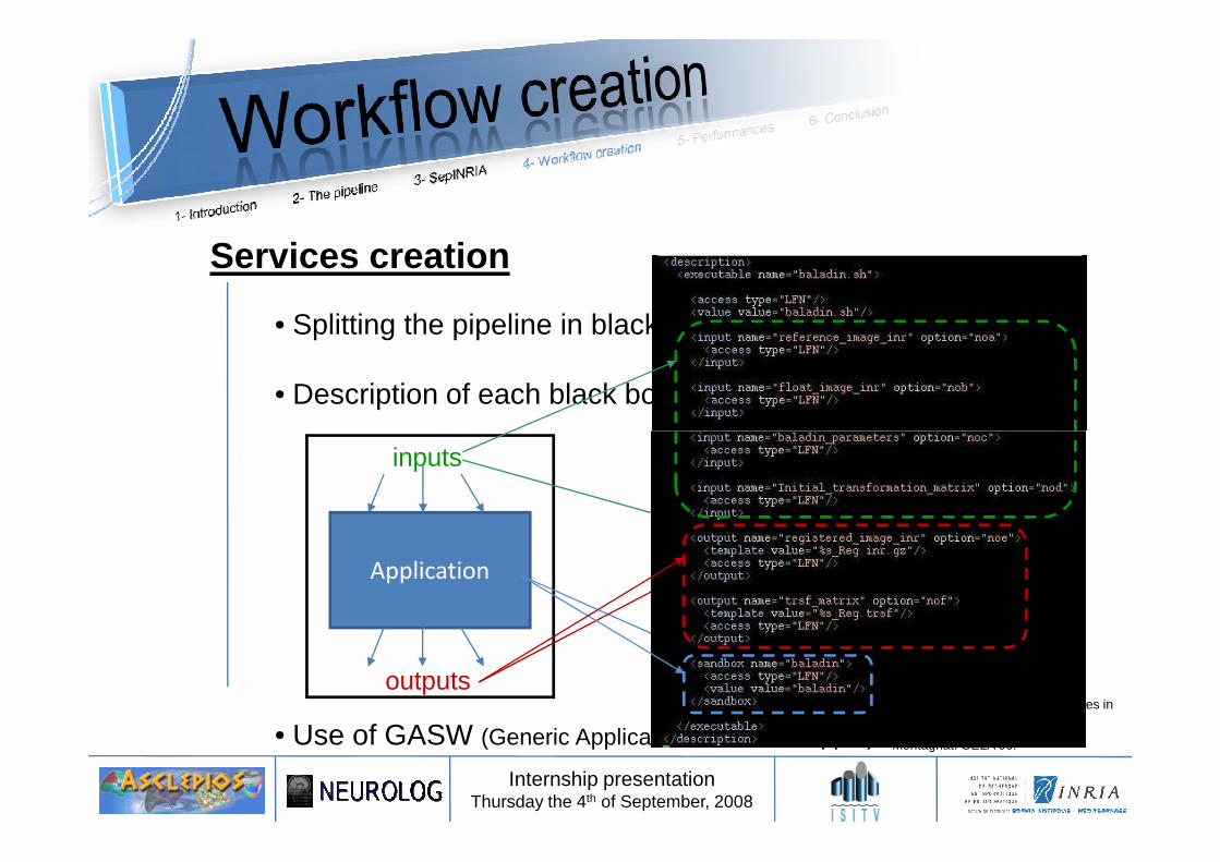
Services creation
• Splitting the pipeline in black boxes
• Description of each black boxes (XML files)
Internship presentationThursday the 4th of September, 2008
• Use of GASW (Generic Application Service Wrapper)
XML files
Inputs
…
Outputs
…
Files
…
Application
inputs
outputs Generic web service wrapper for efficient embedding of legacy codes in service-based workflows.T. Glatard, D. Emsellem, and J. Montagnat. GELA’06.
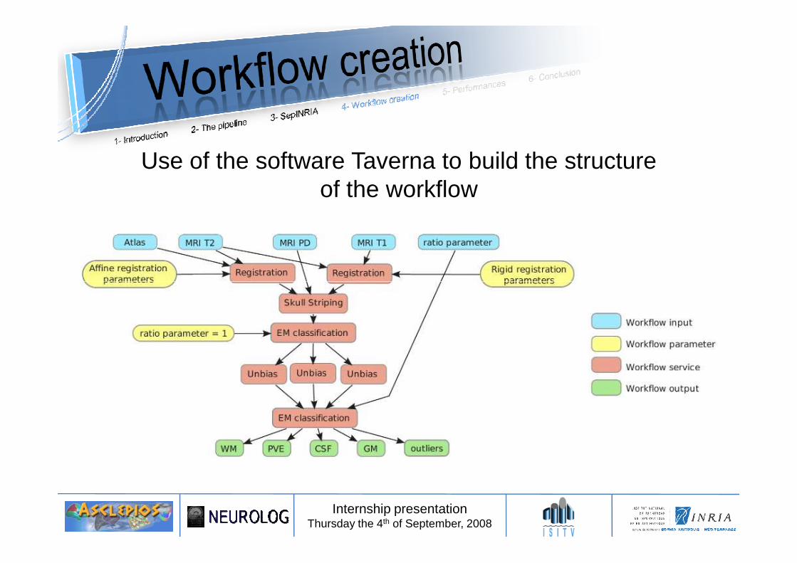
Use of the software Taverna to build the structureof the workflow
Internship presentationThursday the 4th of September, 2008
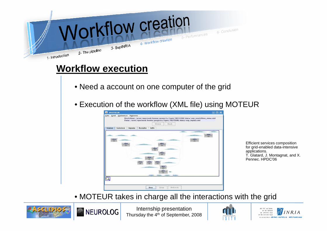
• Need a account on one computer of the grid
• Execution of the workflow (XML file) using MOTEUR
Workflow execution
Internship presentationThursday the 4th of September, 2008
• MOTEUR takes in charge all the interactions with the grid
Efficient services composition for grid-enabled data-intensive applications.T. Glatard, J. Montagnat, and X. Pennec. HPDC’06
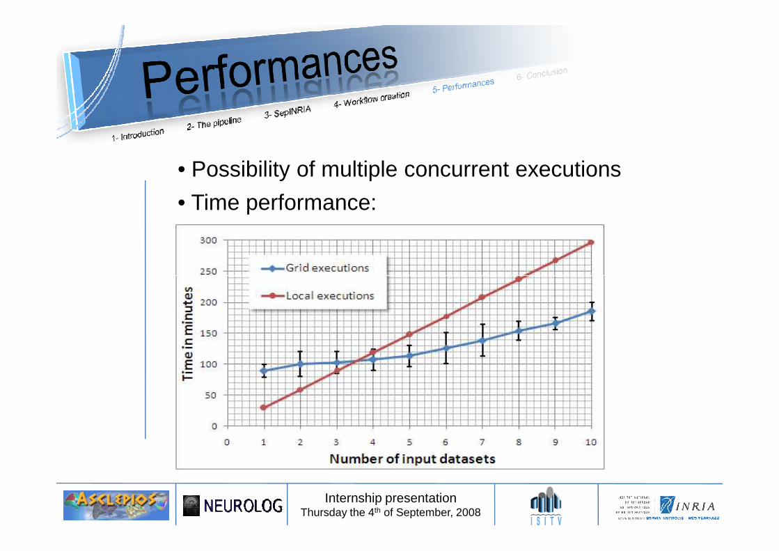
• Possibility of multiple concurrent executions
• Time performance:
Internship presentationThursday the 4th of September, 2008
• The EGEE grid use the gLite middleware.
• In this framework, the Resource Broker is responsible for thematchmaking between job requests and resources.
http://glite.web.cern.ch/glite/
Potential issues
Internship presentationThursday the 4th of September, 2008
- Fastest responding resources are chosen. (after filtration)
- Not necessarily the most powerful- Nor directly available.
• Workload management could becomes a bottleneck.
• Validation of the deployment on the grid• Comparison with a sequential execution on one single
computer: identical results
Parameter-sweep test
Internship presentationThursday the 4th of September, 2008
• The power of the grid allows to perform parameter sweeping in a reasonable amount of time
• Goal: To find a good compromise between accuracy and speed in the EM method. Study of the performance for different percentage of points used to estimate brain classes.
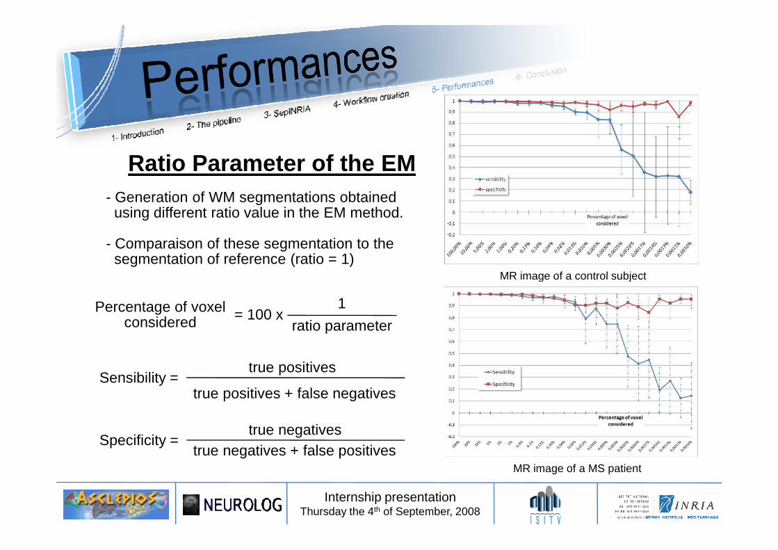
- Generation of WM segmentations obtained using different ratio value in the EM method.
- Comparaison of these segmentation to thesegmentation of reference (ratio = 1)
MR image of a control subject
Ratio Parameter of the EM
Internship presentationThursday the 4th of September, 2008
Specificity =true negatives
true negatives + false positives
Sensibility =true positives
true positives + false negatives
Percentage of voxelconsidered
1
ratio parameter= 100 x
MR image of a control subject
MR image of a MS patient
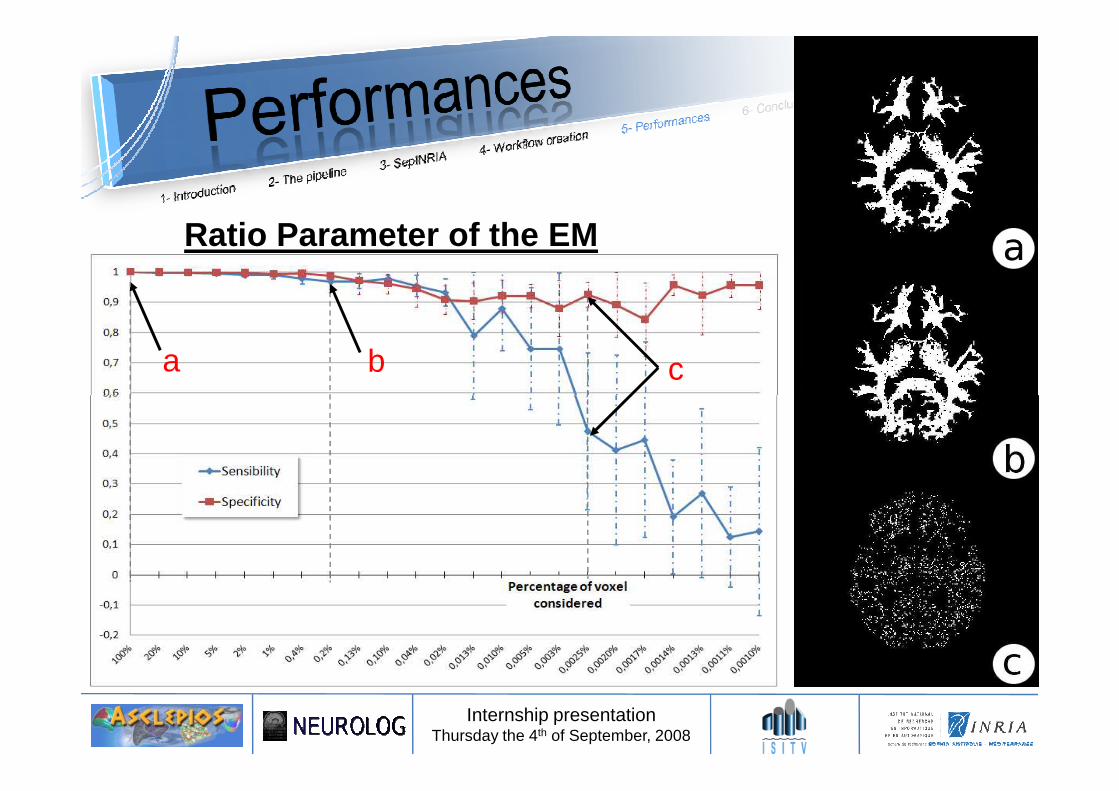
a b c
Ratio Parameter of the EM
Internship presentationThursday the 4th of September, 2008
• Using only 1% of the image voxels in the EM method: - Divides the execution time of the method by ~3- Still provides segmentation of sufficient quality
• Taking less than 1% of the voxels may leads to poor results
Ratio Parameter of the EM
Internship presentationThursday the 4th of September, 2008
• Taking less than 1% of the voxels may leads to poor results
• ~ 210 workflow executions (10 per ratio value) have been computed (per image set).
- Local execution time (sequential): ~ 100 hours (estimated)- Grid execution time: ~ 40 hours (4 hours per bunch of 21 workflow executions)
Conclusion:• Implemented tools in the software SepINRIA have been validated and recognized useful for
clinician research and application (article accepted) :
• Deployment demonstration of a “real” medical image processing solution on the grid:
Erik Pernod, Jean-Christophe Souplet, Mikael Cohen, Nicolas Toussaint, Christine Lebrun et Grégoire Malandain.SepINRIA v1.7.2: Multiple Sclerosis Brain MRI: visualisation, comparison and analysis Software. In World Congressfor Treatment and Research in Multiple Sclerosis (WCTRIMS), Montreals, Canada, September 2008.
Internship presentationThursday the 4th of September, 2008
• Deployment demonstration of a “real” medical image processing solution on the grid:- The power of the grid allows multiple concurrent executions and a sizeable gain of time.
- As a consequence, it allows computation costly tests, e.g. parameter sweeping.- This implementation on the grid leads to the article:
Future work:• Generalization of the services to support more image formats
• Does not require to modify the workflow nor the web service descriptions• Can be done directly at the application level• Allows the workflow diffusion to other research groups
• Add new services to the workflow to get the lesions segmentation.
Erik Pernod, Jean-Christophe Souplet, Javier Rojas Balderrama, Diane Lingrand et Xavier Pennec.Multiple Sclerosis Brain MRI Segmentation Workow deployment on the EGEE Grid.In MICCAI-Grid Workshop (MICCAI-Grid), New York, NY, USA, September 2008