
insight review articles Plasticity in single neuron and ...aurel/nature insight04/plasticity in...
7
T he nervous system shows considerable plasticity, allowing animals to adapt to changing internal and external environments. During development, learning and in ongoing behaviour, individual neurons, synapses and the circuits they form show short-term and long-term changes as a result of experience. Plasticity occurs at all levels, from the behaviour of single ion channels to the morphology of neurons and large circuits and over timescales ranging from milliseconds to years. Because plasticity in the brain occurs at so many levels of organization and over so many timescales, theoretical and computational methods are required to understand how adaptive change to brain function and behaviour is brought about. Many studies of plasticity in the brain have focused on memory storage and retrieval. However, plasticity and neuromodulation also have crucial roles in altering excitability in the brain and regulating behavioural states, such as the transitions between sleep and wakeful activity. Theoretical work is also needed to understand the computational consequences of these various plasticity and modulation mechanisms. Here, we illustrate the use of combined theoretical and experi- mental approaches for understanding neuronal and circuit dynamics, using examples from both small invertebrate and large vertebrate circuits. The building blocks of circuit plasticity Neurons communicate with each other by means of chemi- cal and electrical synapses. It is now clear that the strengths of many, if not most, synapses are altered by either the tem- poral pattern of firing of the presynaptic neuron and/or by amines or neuropeptides delivered hormonally or by neuromodulatory neurons 1 . Some synapses show short- term depression in which the amplitude of successive synaptic potentials progressively decreases. Others show rapid facilitation in which successive synaptic potentials grow in amplitude. (For a detailed discussion of the compu- tational potential of short-term plasticity of synaptic strength, see review in this issue by Abbott and Regehr, page 796.) Much attention has been paid to the computational consequences of long-term use-dependent changes in synaptic strength, such as that seen in long-term depression (LTD) and in long-term potentiation (LTP). It is also clear that the specific timing of activation of presynaptic and post-synaptic activity is crucial for the induction of plas- ticity 2,3 . Synaptic strength can be modulated by amines and neuro-peptides that act on presynaptic terminals to alter the amount of neurotransmitter released with each action potential 4 . Again, this can result in short-term or long-term modifications of synaptic strength 5 , depending on how often the neuromodulator is applied. Although historically most theoretical studies of memory storage in neural networks focused on changes in synaptic strength as the mechanism for implementing stable changes in network behaviour 6 , it is now evident that changes in the intrinsic firing properties of individual neurons also have important roles in altering circuit behaviour. Because some ion channels have slow kinetics, a neuron’s response to a synaptic input can reflect the neuron’s history of activation 7 . There are numerous use- and modulator-dependent alter- ations in channel number and distribution that can also influence a neuron’s excitability and the way it responds to synaptic inputs 8,9 . Changes in both synaptic strength and a neuron’s intrinsic firing properties will alter circuit dynamics. This is illustrated in Fig. 1 where the dynamic clamp 10 is used to construct a simple two-neuron circuit in which each neuron is inhibited by the other 11 . The dynamic clamp is used to alter the strength of the synapses, or the amount of one of the membrane currents, I H (hyperpolarization-activated inward current). Similar changes in the period of the circuit oscilla- tion were produced by changes in both the synaptic and I H conductances. This illustrates that it is impossible to a priori predict the mechanism that produces a change in network output, and that without theoretical methods, it is difficult to understand how the dynamics of even such small circuits depend on the properties of their underlying neurons and synapses. Much important theoretical work has been done on simplified and small circuits. But understanding how the functions of large circuits in the vertebrate brain are altered by plasticity demands an understanding of how to study those large circuits and how to evaluate and understand changes in their behaviour when synaptic and intrinsic properties are altered. Structural complexity of neurons Cajal 12 showed that individual neurons have extraordinarily complex anatomical forms that are characteristic of a given neuronal cell type. The beauty of these structures makes the implicit promise that they have meaning — a premise that was supported by the influential theoretical work of Rall on integration in passive cables 13–15 . Using Rall’s cable theory, it is possible to predict the attenuation of a given synaptic input as a function of its position in the (passive) dendritic tree. The emergence of visually-guided patch–clamp record- ing techniques has since made it possible to routinely record from dendrites, and to perform multi-site dendritic recordings in the same neuron. These techniques have insight review articles NATURE | VOL 431 | 14 OCTOBER 2004 | www.nature.com/nature 789 Plasticity in single neuron and circuit computations Alain Destexhe 1 & Eve Marder 2 1 Integrative and Computational Neuroscience Unit (UNIC), CNRS, Gif-sur Yvette 91198, France (e-mail: [email protected]) 2 Volen Center, Brandeis University, Waltham, Massachusetts 02454, USA (e-mail: [email protected]) Plasticity in neural circuits can result from alterations in synaptic strength or connectivity, as well as from changes in the excitability of the neurons themselves. To better understand the role of plasticity in the brain, we need to establish how brain circuits work and the kinds of computations that different circuit structures achieve. By linking theoretical and experimental studies, we are beginning to reveal the consequences of plasticity mechanisms for network dynamics, in both simple invertebrate circuits and the complex circuits of mammalian cerebral cortex. ©2004 Nature Publishing Group
Transcript of insight review articles Plasticity in single neuron and ...aurel/nature insight04/plasticity in...
The nervous system shows considerable plasticity,allowing animals to adapt to changing internaland external environments. During development,learning and in ongoing behaviour, individualneurons, synapses and the circuits they form show
short-term and long-term changes as a result of experience.Plasticity occurs at all levels, from the behaviour of single ionchannels to the morphology of neurons and large circuits andover timescales ranging from milliseconds to years. Becauseplasticity in the brain occurs at so many levels of organizationand over so many timescales, theoretical and computationalmethods are required to understand how adaptive change tobrain function and behaviour is brought about. Many studiesof plasticity in the brain have focused on memory storageand retrieval. However, plasticity and neuromodulationalso have crucial roles in altering excitability in the brain andregulating behavioural states, such as the transitionsbetween sleep and wakeful activity. Theoretical work is alsoneeded to understand the computational consequences ofthese various plasticity and modulation mechanisms. Here,we illustrate the use of combined theoretical and experi-mental approaches for understanding neuronal and circuitdynamics, using examples from both small invertebrate andlarge vertebrate circuits.
The building blocks of circuit plasticityNeurons communicate with each other by means of chemi-cal and electrical synapses. It is now clear that the strengthsof many, if not most, synapses are altered by either the tem-poral pattern of firing of the presynaptic neuron and/or byamines or neuropeptides delivered hormonally or byneuromodulatory neurons1. Some synapses show short-term depression in which the amplitude of successivesynaptic potentials progressively decreases. Others showrapid facilitation in which successive synaptic potentialsgrow in amplitude. (For a detailed discussion of the compu-tational potential of short-term plasticity of synapticstrength, see review in this issue by Abbott and Regehr, page796.) Much attention has been paid to the computationalconsequences of long-term use-dependent changes insynaptic strength, such as that seen in long-term depression(LTD) and in long-term potentiation (LTP). It is also clearthat the specific timing of activation of presynaptic andpost-synaptic activity is crucial for the induction of plas-ticity2,3. Synaptic strength can be modulated by amines andneuro-peptides that act on presynaptic terminals to alter theamount of neurotransmitter released with each actionpotential4. Again, this can result in short-term or long-term
modifications of synaptic strength5, depending on howoften the neuromodulator is applied.
Although historically most theoretical studies of memorystorage in neural networks focused on changes in synapticstrength as the mechanism for implementing stable changesin network behaviour6, it is now evident that changes in theintrinsic firing properties of individual neurons also haveimportant roles in altering circuit behaviour. Because someion channels have slow kinetics, a neuron’s response to asynaptic input can reflect the neuron’s history of activation7.There are numerous use- and modulator-dependent alter-ations in channel number and distribution that can alsoinfluence a neuron’s excitability and the way it responds tosynaptic inputs8,9. Changes in both synaptic strength and aneuron’s intrinsic firing properties will alter circuit dynamics.This is illustrated in Fig. 1 where the dynamic clamp10 is usedto construct a simple two-neuron circuit in which each neuronis inhibited by the other11. The dynamic clamp is used to alterthe strength of the synapses, or the amount of one of themembrane currents, IH (hyperpolarization-activated inwardcurrent). Similar changes in the period of the circuit oscilla-tion were produced by changes in both the synaptic and IH
conductances. This illustrates that it is impossible to a prioripredict the mechanism that produces a change in networkoutput, and that without theoretical methods, it is difficult tounderstand how the dynamics of even such small circuitsdepend on the properties of their underlying neurons andsynapses. Much important theoretical work has been doneon simplified and small circuits. But understanding how thefunctions of large circuits in the vertebrate brain are alteredby plasticity demands an understanding of how to studythose large circuits and how to evaluate and understandchanges in their behaviour when synaptic and intrinsicproperties are altered.
Structural complexity of neuronsCajal12 showed that individual neurons have extraordinarilycomplex anatomical forms that are characteristic of a givenneuronal cell type. The beauty of these structures makes theimplicit promise that they have meaning — a premise thatwas supported by the influential theoretical work of Rall onintegration in passive cables13–15. Using Rall’s cable theory,it is possible to predict the attenuation of a given synapticinput as a function of its position in the (passive) dendritictree. The emergence of visually-guided patch–clamp record-ing techniques has since made it possible to routinelyrecord from dendrites, and to perform multi-site dendriticrecordings in the same neuron. These techniques have
insight review articles
NATURE | VOL 431 | 14 OCTOBER 2004 | www.nature.com/nature 789
Plasticity in single neuron and circuit computationsAlain Destexhe1 & Eve Marder2
1Integrative and Computational Neuroscience Unit (UNIC), CNRS, Gif-sur Yvette 91198, France (e-mail: [email protected])2Volen Center, Brandeis University, Waltham, Massachusetts 02454, USA (e-mail: [email protected])
Plasticity in neural circuits can result from alterations in synaptic strength or connectivity, as well as from changes in the excitability of the neurons themselves. To better understand the role of plasticity in the brain, we need to establish how brain circuits work and the kinds of computations that differentcircuit structures achieve. By linking theoretical and experimental studies, we are beginning to reveal the consequences of plasticity mechanisms for network dynamics, in both simple invertebrate circuits and the complex circuits of mammalian cerebral cortex.
14.10 Insight 789 Destexhe 1/10/04 7:46 pm Page 789
© 2004 Nature Publishing Group

revealed that dendrites contain many ion-channel types16–19, andthat they can produce Na� and Ca2� spikes, which propagatetowards the soma or away from it16,19. The presence of dendritic ionchannels may also modify the amplitude and shape of synapticinputs20–22, sometimes correcting for dendritic filtering, or havemore subtle effects like establishing coincidence detection23,24. Theemergence of efficient techniques to perform three-dimensionalmorphological reconstructions of single neurons, and of sophist-icated numerical tools for simulating these morphologies25–27 nowmakes it relatively easy to develop semi-realistic computationalmodels of the complex dendritic structure of neurons26. As thesecomputational models become standard tools in the laboratory25,27,they will increasingly aid our understanding of how changes in thedistribution and number of ion channels over the dendritic treechange the firing properties of neurons and their responses tosynaptic inputs.
Dendritic action potentials probably have a central role in synapticplasticity because they provide the strong depolarization necessary toestablish coincidence of presynaptic and postsynaptic activity, whichis required for inducing synaptic changes23,24. Interestingly, this coin-cidence can be established by local dendritic spikes, withoutparticipation of the soma, which raises the possibility that localdendritic computations, or associations, can occur without partici-pation of the cell body28. These problems are now being heavily inves-tigated; experiments and models are needed to explore the possiblecomputations performed by the exquisite dendritic morphologiesinitially described by Cajal12.
Regulation of intrinsic propertiesA growing body of both theoretical and experimental work arguesthat part of a neuron’s characteristic identity is a ‘set-point’ or targetactivity level that regulates the neuron’s long-term mean activitylevel8,9,29,30. In the intact and functioning brain, when neurons arereceiving and responding to synaptic inputs, homeostatic maint-enance of a neuron’s activity level could be achieved by a globalregulation of the strength of all of its synapses (synaptic scaling)31, byregulation of the excitability of the neuron itself 9,32, or by both. Whenneurons, or the circuits in which they reside, are silenced for one ormore days, individual neurons respond by altering the densities ofone or more ion channels32. Long-term compensation for changes inchannel density or synaptic drive may require many of the samemechanisms that are used to produce changes in synaptic strength8.Moreover, because similar patterns of neuronal activity can be pro-duced by various combinations of channel densities33, it is likely thatcompensations for altered patterns of channel expression34 occurfrequently. Use-dependent alterations in conductance densities canoccur on timescales ranging from minutes to hours8,35, and so cancompensate and be coordinated with similar timescale changes insynaptic efficacy.
Defining circuitsNeurons are connected into circuits by excitatory, inhibitory andelectrical synapses that show a variety of amplitudes, time coursesand time-dependent changes in synaptic strength. How then do westudy the circuits underlying behaviour, and how do we determinehow changes in circuit output depend on altered synaptic andintrinsic membrane properties? These problems have beenapproached differently for small and large circuits. In all cases it hasbecome clear that computational approaches are needed to under-stand how circuit output depends on the properties of its componentsand their interactions.
The premise underlying the study of small invertebrate circuitswas that it would be possible to: (1) characterize a behaviour; (2)identify the neurons participating in the circuit that produce thatbehaviour; (3) determine the connectivity among those neurons;and (4) understand how those neurons and their connections giverise to the behaviour. Towards this end, a number of invertebratepreparations were developed in the 1960s and 1970s. One of thehopes, perhaps naive, of these early workers was that similar circuitdesigns would underlie similar behaviour. As the circuits underlyinga number of invertebrate central-pattern generators weredescribed36, it became clear that similar motor patterns could begenerated by different circuit architectures and underlying cellularmechanisms. Nonetheless, it was possible to describe circuit‘building blocks’ that are generally found to contribute to circuitdynamics in specific ways37. For example, reciprocal inhibition(Fig. 1) is found in many motor circuits, where it often ensures thatfunctional antagonists, such as extensor and flexor motor neurons,fire out of phase. This example illustrates the importance of theory:in the work on motor circuits, reciprocal inhibition is almost uni-versally found to ensure alternation of firing between theneurons38. Nonetheless, theoretical work showed that, dependingon the time course of the inhibition, reciprocal inhibition can also
insight review articles
790 NATURE | VOL 431 | 14 OCTOBER 2004 | www.nature.com/nature
Figure 1 Plasticity of circuit dynamics can arise from modifications of synapticstrength or of intrinsic membrane currents. The dynamic clamp is a method thatallows the investigator to add a programmed conductance to a biological neuron. Inthe example shown here, the dynamic clamp was used to create artificialreciprocal inhibitory synapses between two biological neurons that are notconnected by biological synapses. Additionally, the dynamic clamp was used toadd an IH conductance to both neurons. Because the amount of the programmedconductances is under investigator control, the effect of altering the conductanceon the network’s output can easily be determined.Two biological neurons aresynaptically coupled using the dynamic clamp. Modified from ref. 11.
Synaptic conductance lH conductance
50 ns
60 ns 50 ns
30 ns
80 ns
80 ns
10 mV2 s
10 mV2 s
25
20
15
10
00 20 40 60 80 100
5
Per
iod
(s)
SynapticlH
Conductance (nS)
14.10 Insight 789 Destexhe 1/10/04 7:46 pm Page 790
© 2004 Nature Publishing Group
support in-phase firing39,40 — an insight that may be important incortical dynamics41. This highlights the dangers of extrapolatingthe circuit consequences of even simple circuit configurationswithout fully understanding how circuit dynamics depend on theparameters of the underlying circuit elements.
Lessons from small circuitsA great deal is now known about how the small circuits that generaterhythmic behaviour in invertebrates are organized and about howthey function42,43. This is because it is relatively easy to determinewhich neurons are ‘part of the circuit’ and to identify how they areconnected as these circuits have easily measurable and definable out-puts. Sensory and motor circuits can easily be studied in relation tosensory stimuli or to motor behaviour, but defining circuits becomesmore nebulous as we move further to the higher centres in the brainwhere cognitive processes take place. That said, what has been learnedfrom studies of small circuits and their plasticity that generalizes tolarger and more complex circuits in higher animals and humans?(1) Alterations in circuit function are often achieved by modifica-tions of both intrinsic and synaptic properties. For example, in thepyloric rhythm of the lobster stomatogastric ganglion, the neuro-modulator dopamine influences the strength of many of theinhibitory synapses within the network, and modifies IA (the tran-sient outward K� current) and IH (ref. 44) in several network neurons.In the classic work on the gill and siphon withdrawal reflex in Aplysia,changes in both neuronal excitability and synaptic strength areproduced by serotonin and experience4. (2) Neuromodulation is the rule, not the exception. Individual neu-rons and individual synapses are often modulated by several sub-stances, and many neuromodulatory neurons release a mixture ofseveral cotransmitters43. As the neuromodulatory environmentchanges, so will many properties of the cells and synapses that influ-ence circuit function. As some circuit elements themselves containneuromodulators, when these neurons are active, their releasedmodulators will alter the circuit’s dynamics45. Consequently, as a cir-cuit functions, this will itself alter the properties of its components.
In summary, the temporal dynamics and neuromodulatoryenvironment specify the properties of the circuit which produces aspecific output pattern. Changes in the neuromodulatory environ-ment and changes in the circuit’s own activity can in turn producechanges in output, and these changes contribute to behaviouralplasticity on numerous timescales. However, to measure the propertiesof a single synapse, it is often necessary to silence the preparation so thatthe synapse can be studied in isolation. Likewise, to study the proper-ties of a single neuron, it is customary to isolate it from its synapticinputs. These two commonly implemented procedures mean thatalmost all measurements of synapses and cell properties are madeunder conditions that do not pertain during normal circuit operation.Therefore, it is desirable to use techniques such as the dynamic clamp10
and other modelling techniques to determine how circuit behaviour islikely to depend on the properties of the circuit elements.
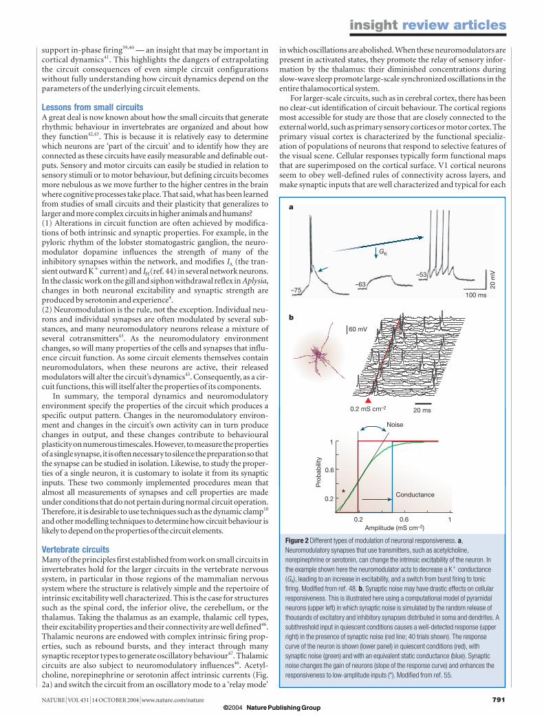
Vertebrate circuitsMany of the principles first established from work on small circuits ininvertebrates hold for the larger circuits in the vertebrate nervoussystem, in particular in those regions of the mammalian nervoussystem where the structure is relatively simple and the repertoire ofintrinsic excitability well characterized. This is the case for structuressuch as the spinal cord, the inferior olive, the cerebellum, or thethalamus. Taking the thalamus as an example, thalamic cell types,their excitability properties and their connectivity are well defined46.Thalamic neurons are endowed with complex intrinsic firing prop-erties, such as rebound bursts, and they interact through manysynaptic receptor types to generate oscillatory behaviour47. Thalamiccircuits are also subject to neuromodulatory influences46. Acetyl-choline, norepinephrine or serotonin affect intrinsic currents (Fig.2a) and switch the circuit from an oscillatory mode to a ‘relay mode’
in which oscillations are abolished. When these neuromodulators arepresent in activated states, they promote the relay of sensory infor-mation by the thalamus: their diminished concentrations duringslow-wave sleep promote large-scale synchronized oscillations in theentire thalamocortical system.
For larger-scale circuits, such as in cerebral cortex, there has beenno clear-cut identification of circuit behaviour. The cortical regionsmost accessible for study are those that are closely connected to theexternal world, such as primary sensory cortices or motor cortex. Theprimary visual cortex is characterized by the functional specializ-ation of populations of neurons that respond to selective features ofthe visual scene. Cellular responses typically form functional mapsthat are superimposed on the cortical surface. V1 cortical neuronsseem to obey well-defined rules of connectivity across layers, andmake synaptic inputs that are well characterized and typical for each
insight review articles
Figure 2 Different types of modulation of neuronal responsiveness. a,Neuromodulatory synapses that use transmitters, such as acetylcholine,norepinephrine or serotonin, can change the intrinsic excitability of the neuron. Inthe example shown here the neuromodulator acts to decrease a K� conductance(GK), leading to an increase in excitability, and a switch from burst firing to tonicfiring. Modified from ref. 48. b, Synaptic noise may have drastic effects on cellularresponsiveness. This is illustrated here using a computational model of pyramidalneurons (upper left) in which synaptic noise is simulated by the random release ofthousands of excitatory and inhibitory synapses distributed in soma and dendrites. Asubthreshold input in quiescent conditions causes a well-detected response (upperright) in the presence of synaptic noise (red line; 40 trials shown). The responsecurve of the neuron is shown (lower panel) in quiescent conditions (red), withsynaptic noise (green) and with an equivalent static conductance (blue). Synapticnoise changes the gain of neurons (slope of the response curve) and enhances theresponsiveness to low-amplitude inputs (*). Modified from ref. 55.
60 mV
0.2 mS cm–2 20 ms
Pro
bab
ility
0.2
0.6
1
0.2 0.6 1Amplitude (mS cm–2)
Conductance
Noise
*
a
b
GK
–75–63
–53
20 m
V
100 ms
NATURE | VOL 431 | 14 OCTOBER 2004 | www.nature.com/nature 791
14.10 Insight 789 Destexhe 1/10/04 7:46 pm Page 791
© 2004 Nature Publishing Group

layer. These data suggest a well-constrained wiring diagram acrosslayers, and has motivated the concept of ‘cortical column’49–52.According to this concept, there is a basic canonical pattern of corticalconnectivity. In this scheme all areas of neocortex would performsimilar computational operations with their inputs53. However,even for the primary sensory cortices, there is no clear paradigm inwhich the distributed activity of neurons, their properties and con-nectivity have been characterized in sufficient detail to allow us torelate structure and function directly (as is the case for oscillationsin small invertebrate preparations or in the thalamus). Nevertheless,using computational models, one can predict generic computa-tions that cortical circuits could perform, a few of which are men-tioned below.
One of the most striking differences between cerebral cortex andinvertebrate networks is that cortical neurons in vivo show a consid-erable degree of apparent randomness in their activity. The membranepotential of cortical neurons shows fluctuating activity, mostly ofsynaptic origin, which is consistent with the extraordinarily denseconnectivity in cortex54. This ‘synaptic noise’ sets the membrane in a‘high-conductance state’, which may affect the integrative propertiesof cortical neurons55. Because studying dendritic integration in vivois technically difficult, computational models are needed to recon-struct in-vivo-like conditions and to evaluate the impact of thissynaptic noise on integrative properties. Such models predict thathigh-conductance states confer several computational advantages tocortical neurons55. First, synaptic noise may boost the response tosynaptic inputs56 (Fig. 2b), in a similar way to stochastic resonancephenomena57. This property was confirmed experimentally usingdynamic clamp58,59. Second, synaptic noise may reduce the dependenceof the efficacy of synaptic inputs on their location in dendrites60,resulting in a more ‘democratic’ dendritic tree in which each synapseexerts a similar vote in firing an action potential in the axon. This is,however, only valid for isolated inputs: the integration of multipleinputs may reveal the existence of ‘dendritic subunits’, as has beensuggested by experiments61 and models62,63. Third, synaptic noisesharpens temporal resolution, allowing cortical neurons to detectcoincidences separated by milliseconds,and therefore to resolve pre-cisely timed inputs55,64. Finally, an obvious consequence of synapticnoise is that cortical neurons show a high trial-to-trial variability intheir responses (Fig. 2b) — a feature often seen in vivo65. Conse-quently, the only sensible measures that can be used to characterizethe activity of a cortical neuron in vivo are probabilities. Indeed,probabilities have been used for decades to characterize responsesrecorded in cortex in vivo, under the form of ‘post-stimulus timehistograms’66. There is also a whole family of computational modelsof cortical coding based on probabilistic models67, some of which arementioned below.
Cortical computations One of the most influential theories of neural computation was pro-posed by Hopfield68, who showed that memories can be stored asstationary states (point attractors) in networks of simplified neurons.One advantage of this model is that it is mathematically similar towell-studied physical systems, and memory storage can be under-stood from the formation of minima in the energy landscape of thesystem. In these models, a hebbian-type learning rule (Box 1) can beused for modifying synaptic weights, and memories are distributedamong the synaptic weights. However, the drawback of Hopfield’stheory is that there are no point-attractors in real networks of neurons,so its direct heuristic value in explaining cortical computations islimited. Nevertheless, this theory had the considerable merit ofmotivating generations of researchers to study computational modelsin neuroscience using the tools of mathematics and physics.
One generic computation of cortical networks may be to detectand extract correlations. Sensory systems must make sense of complexflows of information, in which exactly the same pattern is unlikely tohappen twice. According to Barlow53, the main task of our sensory
system is to detect (and model) correlations; it acts like a detectiveand notes, in the form of neuron firing, ‘suspicious coincidences’ incomplex incoming information. It is these coincidences or correlationsthat may form the ‘objects’ or ‘features’ of our symbolic represent-ations. After being detected by primary sensory areas, such correl-ations can be used for binding elementary features into moreelaborate percepts. This binding problem has been intensely debated(for a recent review see ref. 69), and is based on the concept of neuronalassemblies, which are usually defined as a group of neurons thattransiently undergo synchronous firing70–72. This transient synchronycould form the basis of a common input to later stages of integration,and so promote responses that are specific to a given ensemble offeatures71. Thus, correlated firing serves here to form assemblies ofneurons that are specific to a given feature. Cortical neurons shouldtherefore be very efficient at detecting correlations72, as is indicatedby computational models73.
Another view, not necessarily contradictory, is that the cortexattempts to remove correlations. Probabilistic models have beenproposed based on the observation that the cortex must infer prop-erties from a highly variable and uncertain environment, and anefficient way to do so is to compute probabilities. One of the earliestprobabilistic models proposed that the cortex infers probabilitiesbased on ‘decorrelation’ or ‘redundancy-reduction’ operations53,74,75.The most salient functional consequence of this is that these proba-bilites could be used to build efficient novelty detectors — a featureessential for survival. This redundancy-reduction function is alsosupported by the fact that the sensory system of mammals receivessignals from millions of peripheral receptors sampling differentfeatures of the external world.Because many receptors convey similarinformation, the sensory system may need to reduce this redun-dancy to focus on the interesting aspects of the scene. This paradigmis particularly relevant to the retina, where the number of outputfibres are two orders of magnitude less than the number of photo-receptors. Indeed, experiments provide evidence for redundancyreduction in this system76.
The same ideas have been proposed for central structures such asthe cortex. Here, an efficient way to reduce redundancy is to use synapticinteractions that obey the anti-hebbian rule (see Box 1). This type ofplasticity has been identified in synapses from parallel fibres onPurkinje cells in cerebellum77, and in excitatory synapses betweenparallel fibres and medium ganglionic cells in the electrosensory lobein electric fish78. Networks with hebbian feedforward synapsescombined with anti-hebbian recurrent inhibitory synapses wereshown to efficiently decorrelate inputs, and they perform well invarious un-supervised learning paradigms79. Interestingly, severalmechanisms present in cortical circuits can also have similar roles,such as spike frequency adaptation80 or short-term synapticdepression81. Adaptation or plasticity processes remove correlationsmost efficiently over timescales comparable to their own characteristicrelaxation time constant80. This suggests that a broad range ofdynamic processes is needed to cover the relevant timescales overwhich signals must be decorrelated. This is consistent with the factthat several mechanisms, possibly present in neocortex, such as
insight review articles
792 NATURE | VOL 431 | 14 OCTOBER 2004 | www.nature.com/nature
Hebbian ruleA given link will be strengthened (either by an increase of excitatorygain, or by a decrease of inhibitory gain) if the two units that itconnects are active simultaneously.
Anti-hebbian ruleA given link will be weakened (either by a reduction of excitatory gain,or by an increase of inhibitory gain) if the two units that it connects areactive simultaneously.
Box 1 Definition of hebbian and anti-hebbian rules (from ref. 100)
14.10 Insight 789 Destexhe 1/10/04 7:46 pm Page 792
© 2004 Nature Publishing Group

intrinsic adaptation, short-term synaptic depression, anti-hebbianplasticity, or even long-term changes of intrinsic properties, mighthave equivalent functional roles but complement each other atdifferent timescales.
However, it is not clear that these ideas apply so straightforwardlyto cortex, for several reasons. First, anti-hebbian plasticity has not yetbeen demonstrated in cortical recurrent connections, although itmay be that plasticity of inhibitory connections has a similar func-tional role (see below). Second, in contrast to the retina, the numberof cortical neurons, as well as the number of efferent axons, largelyexceeds the number of ascending ‘input’ fibres82. There is, therefore,no structural constraint, as there is in the retina, which would call forredundancy reduction in cortex. Morphological and physiologicaldata are more consistent with ‘sparse codes’ in which many units areused for coding, but extremely few units are active simultane-ously79,83–85. Third, other mechanisms also present in neocortex, suchas hebbian plasticity86,87 or short-term synaptic facilitation88, have theopposite role of enhancing pre-existing correlations89 (Fig. 3). Thus,the cortex possesses mechanisms that are compatible with eitherreducing or enhancing correlations, and it is unclear whether thesemechanisms coexist or whether they are expressed differentiallyaccording to context or cortical area. Neocortical circuits dominatedby anti-hebbian and depressing mechanisms may serve as noveltydetectors by decorrelating afferent inputs and therefore function in a‘search mode’. This mode would be a priori compatible with primarysensory areas. However, other cortical circuits, dominated by hebbianand facilitating mechanisms, might function in a ‘convergencemode’, compatible with the type of operation performed in associationor motor areas. It is not clear, however, whether these modes areseparate or whether they coexist everywhere in cortex. In the lattercase, any neocortical area would be equipped to function in bothmodes simultaneously or to switch between these modes dependingon activity levels or neuromodulation.
Rather than attempting to explain cortical function on the basis ofgeneric cellular and synaptic properties or stereotyped circuits, thediversity of cortical neurons and their highly complex synaptic con-nectivity can be used to propose a different computational paradigm.Cortical neurons show a wide diversity of intrinsic properties90. Like-wise, synaptic dynamics are richly variable and show properties thatrange from those of facilitating to depressing synapses88. Indeed, theessential feature of cortical anatomy may be that there is no canonicalpattern of connectivity, consistent with the considerable apparentrandom component of cortical connectivity templates54,91. Takingthese observations together, the cortex may be seen as a circuit thatmaximizes its own complexity, both at the single-cell level and at thelevel of its connectivity. In support of this view, computational modelsare now emerging in which the goal is to take advantage of the specialinformation processing-capabilities, and memory, of such a complexsystem. Such large-scale networks can transform temporal codes intospatial codes by self-organization92, and computing frameworks havebeen proposed which exploit the capacity of such complex networksto cope with complex input streams93 (Fig. 4). In these examples,information is stored in the ongoing activity of the network, inaddition to its synaptic weights. A given output can be provided at anytime within this ongoing activity, rather than requiring the system toconverge towards predefined attractors. The concept of the cortex as a‘large network of identical units’ should be replaced with the idea thatthe cortex consists of ‘large networks of diverse elements’, where cellularand synaptic diversity are important for computation.
Towards understanding the many facets of plasticitySeveral issues must be considered when linking plasticity mech-anisms with neuronal computations. First, the rules that govern theplasticity at many inhibitory synapses are unknown. One possibilityis that the inhibitory feedback from local interneurons obeys anti-hebbian plasticity, which would be consistent with the predictions ofmodels of redundancy reduction. In contrast to the very large number
of studies modelling memory storage in networks using changes inexcitatory synapses, few models implement learning rules forinhibitory synapses. Nonetheless, recent work showing that thebalance of inhibition and excitation can be important for gain mod-ulation56,58, and in the genesis of functional selectivity94, illustratesthe importance of determining the rules that control the strength ofinhibitory synapses.
Second, plasticity mechanisms are likely to depend on behav-ioural state, such as deep sleep or aroused states. Most experimentalstudies of the mechanisms underlying synaptic plasticity have beendone in slices or in anesthetized preparations. However, these prep-arations differ from aroused and attentive animals, during whichcortical networks are in high-conductance states55, maintained by therelease of a number of neuromodulators, such as acetylcholine andnorepinephrine95. These substances may considerably affect theplasticity mechanisms of cortical circuits96,97. It is, therefore, imperativeto verify that the plasticity mechanisms found in slices apply to the
insight review articles
NATURE | VOL 431 | 14 OCTOBER 2004 | www.nature.com/nature 793
Figure 3 The type of transformations realized by synaptic plasticity. a, Facilitatingsynapses enhance existing correlations. When an image, such as the natural sceneshown here, is processed by a neural network with facilitating synapses,correlations are reinforced, or equivalently, the spatial power spectrum is morestructured (see graph). The result is an output image which has enhanced contrast.Enhancement of correlations can also be obtained using hebbian synaptic plasticity.b, Similar model with depressing synapses. Here, the transformation results fromreducing correlations, or equivalently, reducing redundancy. This redundancyreduction corresponds to whitening the spatial spectrum of the image (see graph).The reduction of existing correlations leads to an output image in which many detailsare lost. A decorrelation can also be obtained using anti-hebbian synapses oradaptation mechanisms.
Input image Output imageFacilitating synapses
Depressing synapses
a
b
log(frequency)
log(
pow
er)
log(frequency)
log(
pow
er)
14.10 Insight 789 Destexhe 1/10/04 7:46 pm Page 793
© 2004 Nature Publishing Group

activated brain. The relative ease of inducing and consolidatingplasticity in slices may also indicate that these mechanisms are bestexpressed during states of low release of neuromodulators, such asduring slow-wave sleep. This would corroborate recent evidence thatslow-wave sleep is actively implicated in the consolidation of mem-ory traces98, and models of learning that require a ‘sleep’ phase99.Consistent with this idea, the widely synchronized oscillationscharacteristic of slow-wave sleep are likely to constitute an optimalsignal for inducing plastic changes in the network47. Relating plasticitymechanisms to the state of the network constitutes an essential pieceof information that should be targeted by appropriate experimentsand theories.
OutlookHow far have we come in understanding how neuronal circuitsproduce behaviour? Certainly, considerable progress has been madefor some relatively simple, small circuits4,42,43,45. These small circuitsprovide ideal platforms for understanding which circuit parametersare genetically specified, and how circuit properties are modified byexperience. A more daunting challenge is to link circuitry withbehaviour for more complex networks, such as cerebral cortex,because the computational operations in cortex are still largelyunknown. It is clear that cortical neurons possess complex intrinsicproperties and that their rich and diverse synaptic connections aresubject to plasticity, modulation and noise over many timescales.Many of the concepts arising from studies of small networks mayextrapolate directly to the cortex, and our present inability to under-stand cortical function could be just a matter of complexity arisingfrom its large size and multiple cell types. If so, we need to develop
appropriate conceptual, physiological and computational tools tohandle this complexity. Alternatively, we may be missing a fundamental‘building block’ that is required to understand cortical function. Ineither case, there is presently no coherent theory of cortical computa-tions, and constructing one will be possible only through a tight com-bination of experimental and theoretical approaches. ■■
doi:10.1038/nature03011
1. Marder, E. & Thirumalai, V. Cellular, synaptic and network effects of neuromodulation. Neural Netw.
15, 479–493 (2002).
2. Abbott, L. F. & Nelson, S. B. Synaptic plasticity: taming the beast. Nature Neurosci. 3 (suppl.),
1178–1183. (2000).
3. Sjostrom, P. J. & Nelson, S. B. Spike timing, calcium signals and synaptic plasticity. Curr. Opin.
Neurobiol. 12, 305–314 (2002).
4. Kandel, E. R. The molecular biology of memory storage: a dialogue between genes and synapses.
Science 294, 1030–1038 (2001).
5. Martin, K. C. & Kosik, K. S. Synaptic tagging — who’s it? Nature Rev. Neurosci. 3, 813–820 (2002).
6. Dayan, P. & Abbott, L. F. in Theoretical Neuroscience (MIT, Cambridge, 2001).
7. Marder, E., Abbott, L. F., Turrigiano, G. G., Liu, Z. & Golowasch, J. Memory from the dynamics of
intrinsic membrane currents. Proc. Natl Acad. Sci. USA 93, 13481–13486 (1996).
8. Zhang, W. & Linden, D. J. The other side of the engram: experience-driven changes in neuronal
intrinsic excitability. Nature Rev. Neurosci. 4, 885–900 (2003).
9. Daoudal, G. & Debanne, D. Long-term plasticity of intrinsic excitability: learning rules and
mechanisms. Learn. Mem. 10, 456–465 (2003).
10.Prinz, A. A., Abbott, L. F. & Marder, E. The dynamic clamp comes of age. Trends Neurosci. 27,
218–224 (2004).
11.Sharp, A. A., Skinner, F. K. & Marder, E. Mechanisms of oscillation in dynamic clamp constructed
two-cell half-center circuits. J. Neurophysiol. 76, 867–883 (1996).
12.Cajal, R. S. Histologie du Système Nerveux de l’Homme et des Vertébrés (Maloine, Paris, 1909).
13.Rall, W. Distinguishing theoretical synaptic potentials computed for different soma-dendritic
distributions of synaptic input. J. Neurophysiol. 30, 1138–1168 (1967).
14.Rall, W. Time constants and electrotonic length of membrane cylinders and neurons. Biophys. J.
9, 1483–1508 (1969).
15.Rall, W. & Rinzel, J. Branch input resistance and steady attenuation for input to one branch of a
insight review articles
794 NATURE | VOL 431 | 14 OCTOBER 2004 | www.nature.com/nature
Figure 4 Computing with network complexity. a, Scheme of a computational model that uses a network in which diverse cell types and synaptic interactions are taken into account. The activity of a few cells are fed into ‘readouts’ (blue), which extract the response from the complex dynamics of the network.
b, Example of computation of different spoken words. The ongoing network activity is apparently random and similar in each case, but it contains information about the input, which can be retrieved by the readout. Modified from ref. 93.
Input pattern
Response
ReadoutsNetwork
Time (s)
Network activity
Input pattern 'one' Input pattern 'two' Input pattern 'three'
Readout response
40
1
50
1
135
1
40
1
50
1
135
1
40
1
50
1
135
1
ch. n
o.nr
n no
.nr
n no
.
0 0.25 0.5 0 0.25 0.5 0 0.25 0.5
a
b
14.10 Insight 789 Destexhe 1/10/04 7:46 pm Page 794
© 2004 Nature Publishing Group

dendritic neuron model. Biophys. J. 13, 648–687 (1973).
16. Johnston, D., Magee, J. C., Colbert, C. M. & Cristie, B. R. Active properties of neuronal dendrites.
Annu. Rev. Neurosci. 19, 165–186 (1996).
17.Migliore, M. & Shepherd, G. M. Emerging rules for the distributions of active dendritic
conductances. Nature Rev. Neurosci. 3, 362–370 (2002).
18.Yuste, R. & Tank, D. W. Dendritic integration in mammalian neurons, a century after Cajal. Neuron
16, 701–716 (1996).
19.Stuart, G., Spruston, N. & Hausser, M. Dendrites (MIT, Cambridge, Massachusetts, 2000).
20.Schwindt, P. C. & Crill, W. E. Amplification of synaptic current by persistent sodium conductance in
apical dendrite of neocortical neurons. J. Neurophysiol. 74, 2220–2224 (1995).
21.Magee, J. C. Dendritic Ih normalizes temporal summation in hippocampal CA1 neurons. Nature
Neurosci. 2, 508–514 (1999).
22.Williams, S. R. & Stuart, G. J. Site independence of EPSP time course is mediated by dendritic I(h) in
neocortical pyramidal neurons. J. Neurophysiol. 83, 3177–3182 (2000).
23.Magee, J. C. & Johnston, D. A synaptically controlled, associative signal for Hebbian plasticity in
hippocampal neurons. Science 275, 209–213 (1997).
24.Markram, H., Lübke, J., Frotscher, M. & Sakmann, B. Regulation of synaptic efficacy by coincidence
of postsynaptic APs and EPSPs. Science 275, 213–215 (1997).
25.Hines, M. L. & Carnevale, N. T. The NEURON simulation environment. Neural Comput. 9,
1179–1209 (1997).
26.Koch, C. & Segev, I. Methods in Neuronal Modeling (MIT, Cambridge, 1998).
27.Bower, J. & Beeman, D. The Book of GENESIS (Springer, Berlin, 1994).
28.Golding, N. L., Staff, N. P. & Spruston, N. Dendritic spikes as a mechanism for cooperative long-term
potentiation. Nature 418, 326–331 (2002).
29.Turrigiano, G. G. & Nelson, S. B. Homeostatic plasticity in the developing nervous system. Nature
Rev. Neurosci. 5, 97–107 (2004).
30.Marder, E. & Prinz, A. A. Modeling stability in neuron and network function: the role of activity in
homeostasis. Bioessays 24, 1145–1154 (2002).
31.Turrigiano, G. G., Leslie, K. R., Desai, N. S., Rutherford, L. C. & Nelson, S. B. Activity-dependent
scaling of quantal amplitude in neocortical neurons. Nature 391, 892–896 (1998).
32.Desai, N. S., Rutherford, L. C. & Turrigiano, G. G. Plasticity in the intrinsic excitability of cortical
pyramidal neurons. Nature Neurosci. 2, 515–520. (1999).
33.Goldman, M. S., Golowasch, J., Marder, E. & Abbott, L. F. Global structure, robustness, and
modulation of neuronal models. J. Neurosci. 21, 5229–5238 (2001).
34.MacLean, J. N., Zhang, Y., Johnson, B. R. & Harris-Warrick, R. M. Activity-independent homeostasis
in rhythmically active neurons. Neuron 37, 109–120 (2003).
35.Golowasch, J., Abbott, L. F. & Marder, E. Activity-dependent regulation of potassium currents in
an identified neuron of the stomatogastric ganglion of the crab Cancer borealis. J. Neurosci. 19,
RC33 (1999).
36.Selverston, A. I. Are central pattern generators understandable? Behav. Brain Sci. 3, 535–571 (1980).
37.Getting, P. A. Emerging principles governing the operation of neural networks. Annu. Rev. Neurosci.
12, 185–204 (1989).
38.Friesen, W. O. Reciprocal inhibition: a mechanism underlying oscillatory animal movements.
Neurosci. Biobehav. Rev. 18, 547–553 (1994).
39.Wang, X.-J. & Rinzel, J. Alternating and synchronous rhythms in reciprocally inhibitory model
neurons. Neural Comput. 4, 84–97 (1992).
40.Van Vreeswijk, C., Abbott, L. F. & Ermentrout, G. B. When inhibition not excitation synchronizes
neural firing. J. Comput. Neurosci. 1, 313–321 (1994).
41.White, J. A., Chow, C. C., Ritt, J., Soto-Trevino, C. & Kopell, N. Synchronization and oscillatory
dynamics in heterogeneous, mutually inhibited neurons. J. Comput. Neurosci. 5, 5–16 (1998).
42.Marder, E. & Calabrese, R. L. Principles of rhythmic motor pattern generation. Physiol. Rev. 76,
687–717 (1996).
43.Nusbaum, M. P. & Beenhakker, M. P. A small-systems approach to motor pattern generation. Nature
417, 343–350 (2002).
44.Harris-Warrick, R. M. et al. Distributed effects of dopamine modulation in the crustacean pyloric
network. Ann. N Y Acad. Sci. 860, 155–167 (1998).
45.Katz, P. S. & Frost, W. N. Intrinsic neuromodulation: altering neuronal circuits from within. Trends
Neurosci. 19, 54–61 (1996).
46.Steriade, M., Jones, E. G. & McCormick, D. A. Thalamus (Elsevier, Amsterdam, 1997).
47.Destexhe, A. & Sejnowski, T. J. Interactions between membrane conductances underlying
thalamocortical slow-wave oscillations. Physiol. Rev. 83, 1401–1453 (2003).
48. McCormick, D. A. Cholinergic and noradrenergic modulation of thalamocortical processing. Trends
Neurosci. 12, 215–221 (1989).
49.Mountcastle, V. B. in The Neurosciences: Fourth Study Program (eds Schmidt, F. O. & Worden, F. G.)
21–42 (MIT Press, Cambridge, 1979).
50.Hubel, D. H. & Wiesel, T. N. Shape and arrangement of columns in cat’s striate cortex. J. Physiol. 165,
559–568 (1963).
51.Douglas, R. J. & Martin, K. A. A functional microcircuit for cat visual cortex. J. Physiol. 440,
735–769 (1991).
52.Szentagothai, J. The modular architectonic principle of neural centers. Rev. Physiol. Biochem.
Pharmacol. 98, 11–61 (1983).
53.Barlow, H. in Models of the Visual Cortex (eds Rose, D. & Dobson, V.) 37–46 (Wiley, Chichester, 1985).
54.Braitenberg, V. & Schuz, A. Cortex: statistics and geometry of neuronal connectivity (Springer, Berlin,
1998).
55.Destexhe, A., Rudolph, M. & Pare, D. The high-conductance state of neocortical neurons in vivo.
Nature Rev. Neurosci. 4, 739–751 (2003).
56.Ho, N. & Destexhe, A. Synaptic background activity enhances the responsiveness of neocortical
pyramidal neurons. J. Neurophysiol. 84, 1488–1496 (2000).
57.Wiesenfeld, K. & Moss, F. Stochastic resonance and the benefits of noise: from ice ages to crayfish and
SQUIDs. Nature 373, 33–36 (1995).
58.Chance, F. S., Abbott, L. F. & Reyes, A. D. Gain modulation from background synaptic input. Neuron
35, 773–782 (2002).
59.Shu, Y., Hasenstaub, A., Badoual, M., Bal, T. & McCormick, D. A. Barrages of synaptic activity control
the gain and sensitivity of cortical neurons. J. Neurosci. 23, 10388–10401 (2003).
60.Rudolph, M. & Destexhe, A. A fast-conducting, stochastic integrative mode for neocortical neurons
in vivo. J. Neurosci. 23, 2466–2476 (2003).
61.Wei, D. S. et al. Compartmentalized and binary behavior of terminal dendrites in hippocampal
pyramidal neurons. Science 293, 2272–2275 (2001).
62.Shepherd, G. M. & Brayton, R. K. Logic operations are properties of computer-simulated interactions
between excitable dendritic spines. Neuroscience 21, 151–165 (1987).
63.Mel, B. W. Information processing in dendritic trees. Neural Comput. 6, 1031–1085 (1994).
64.Softky, W. Sub-millisecond coincidence detection in active dendritic trees. Neuroscience 58, 13–41
(1994).
65.Shalden, M. N. & Newsome, W. T. The variable discharge of cortical neurons: implications for
connectivity, computation, and information coding. J. Neurosci. 18, 3870–3896 (1998).
66.Moore, G. P., Perkel, D. H. & Segundo, J. P. Statistical analysis and functional interpretation of
neuronal spike data. Annu. Rev. Physiol. 28, 493–522 (1966).
67.Rao, R., Olshausen, B. & Lewicki, M. Probabilistic Models of the Brain (MIT, Cambridge, 2002).
68.Hopfield, J. J. Neural networks and physical systems with emergent collective computational abilities.
Proc. Natl Acad. Sci. USA 79, 2554–2558 (1982).
69.Roskies, A. The binding problem: special issue. Neuron 24, 7–125 (1999).
70.von der Malsburg, C. & Schneider, W. A neural cocktail-party processor. Biol. Cybern. 54, 29–40
(1986).
71.Engel, A. K., Fries, P. & Singer, W. Dynamic predictions: oscillations and synchrony in top-down
processing. Nature Rev. Neurosci. 2, 704–716 (2001).
72.Abeles, M. Corticonics: Neuronal Circuits of the Cerebral Cortex (Cambridge University Press,
Cambridge, 1991).
73.Rudolph, M. & Destexhe, A. Correlation detection and resonance in neural systems with distributed
noise sources. Phys. Rev. Lett. 86, 3662–3665 (2001).
74.Barlow, H. B. in Sensory Communications (ed. Rosenblith, W.) Ch. 13, 217–234 (MIT, Cambridge,
1961).
75.Barlow, H. & Foldiak, P. in The Computing Neuron Ch. 4 (eds Durbin, R., Miall, C. & G, M.) 54–72
(Addison-Wesley, New York, 1989).
76.Srinivasan, M. V., Laughlin, S. B. & Dubs, A. Predictive coding: a fresh view of inhibition in the retina.
Proc. R. Soc. Lond. B 216, 427–459 (1982).
77. Ito, M. Cerebellar long-term depression: characterization, signal transduction, and functional roles.
Physiol. Rev. 81, 1143–1195 (2001).
78.Bell, C. C., Han, V. Z., Sugawara, Y. & Grant, K. Synaptic plasticity in a cerebellum-like structure
depends on temporal order. Nature 387, 278–281 (1997).
79.Foldiak, P. Forming sparse representations by local anti-Hebbian learning. Biol. Cybern. 64, 165–170
(1990).
80.Wang, X. J., Liu, Y., Sanchez-Vives, M. V. & McCormick, D. A. Adaptation and temporal decorrelation
by single neurons in the primary visual cortex. J. Neurophysiol. 89, 3279–3293 (2003).
81.Goldman, M. S., Maldonado, P. & Abbott, L. F. Redundancy reduction and sustained firing with
stochastic depressing synapses. J. Neurosci. 22, 584–591 (2002).
82.Peters, A. & Yilmaz, E. Neuronal organization in area 17 of cat visual cortex. Cereb. Cort. 3, 49–68
(1993).
83.Olshausen, B. A. & Field, D. J. Emergence of simple-cell receptive field properties by learning a sparse
code for natural images. Nature 381, 607–609 (1996).
84.Perez-Orive, J. et al. Oscillations and sparsening of odor representations in the mushroom body.
Science 297, 359–365 (2002).
85.Hahnloser, R. H., Kozhevnikov, A. A. & Fee, M. S. An ultra-sparse code underlies the generation of
neural sequences in a songbird. Nature 419, 65–70 (2002).
86.Baranyi, A. & Feher, O. Conditioned changes of synaptic transmission in the motor cortex of the cat.
Exp. Brain Res. 33, 283–298 (1978).
87.Kirkwood, A. & Bear, M. F. Hebbian synapses in visual cortex. J. Neurosci. 14, 1634–1645 (1994).
88.Thomson, A. M. Facilitation, augmentation and potentiation at central synapses. Trends Neurosci. 23,
305–312 (2000).
89.Hebb, D. O. The Organization of Behavior (Wiley, New York, 1949).
90.Gupta, A., Wang, Y. & Markram, H. Organizing principles for a diversity of GABAergic interneurons
and synapses in the neocortex. Science 287, 273–278 (2000).
91.Silberberg, G., Gupta, A. & Markram, H. Stereotypy in neocortical microcircuits. Trends Neurosci. 25,
227–230 (2002).
92.Buonomano, D. V. & Merzenich, M. M. Temporal information transformed into a spatial code by a
neural network with realistic properties. Science 267, 1028–1030 (1995).
93.Maass, W., Natschlager, T. & Markram, H. Real-time computing without stable states: a new
framework for neural computation based on perturbations. Neural Comput. 14, 2531–2560
(2002).
94.Monier, C., Chavane, F., Baudot, P., Graham, L. J. & Fregnac, Y. Orientation and direction selectivity
of synaptic inputs in visual cortical neurons: a diversity of combinations produces spike tuning.
Neuron 37, 663–680 (2003).
95.Steriade, M. & McCarley, R. W. Brainstem Control of Wakefulness and Sleep (Plenum, New York,
1990).
96.Bear, M. F. & Singer, W. Modulation of visual cortical plasticity by acetylcholine and noradrenaline.
Nature 320, 172–176 (1986).
97.Shulz, D. E., Sosnik, R., Ego, V., Haidarliu, S. & Ahissar, E. A neuronal analogue of state-dependent
learning. Nature 403, 549–553 (2000).
98. Stickgold, R., Hobson, J. A., Fosse, R. & Fosse, M. Sleep, learning, and dreams: off-line memory
reprocessing. Science 294, 1052–1057 (2001).
99. Hinton, G. E., Dayan, P., Frey, B. J. & Neal, R. M. The ‘wake-sleep’ algorithm for unsupervised neural
networks. Science 268, 1158–1161 (1995).
100. Frégnac, Y. in Handbook of Brain Theory and Neural Networks (ed. Arbib, M. A.) 515–522 (MIT,
Cambridge, 2002).
Acknowledgements We thank M. Rudolph and Y. Fregnac for comments on themanuscript. The authors’ research was supported by the NIH (E.M.), CNRS, HFSP andthe European Union (Future and Emerging Technologies).
Competing interests statement The authors declare that they have no competing financialinterests.
insight review articles
NATURE | VOL 431 | 14 OCTOBER 2004 | www.nature.com/nature 795
14.10 Insight 789 Destexhe 1/10/04 7:46 pm Page 795
© 2004 Nature Publishing Group


![a] [Ptuprints.ulb.tu-darmstadt.de/420/3/habil_elektr_3.pdf · gle crystal plasticity, multi{surface plasticity, textur development 6.1 Introduction The treatment of single crystal](https://static.fdocuments.net/doc/165x107/5f0f53897e708231d4439b67/a-gle-crystal-plasticity-multisurface-plasticity-textur-development-61-introduction.jpg)

