Innovations for Wound Bed Preparation: The Role of … for Wound Bed Preparation: The Role of...
28
Innovations for Wound Bed Preparation: The Role of Drawtex Hydroconductive Dressings 2 Innovations for Wound Bed Preparation: The Role of Drawtex Hydroconductive Dressings: An Introduction Martin C. Robson, MD 3 In Vivo and In Vitro Evaluation of the Properties of Drawtex LevaFiber Wound Dressing in an Infected Burn Wound Model Rachel T. Ortiz, MS; Lauren T. Moffatt, PhD; Martin C. Robson, MD, FACS; Marion H. Jordan, MD, FACS; Jeffrey W. Shupp, MD 6 Evaluation of Mechanisms of Action of a Hydroconductive Wound Dressing (Drawtex) in Chronic Wounds Diane Ochs, RN; M. Georgina Uberti, MD; Guillermo A. Donate, DPM; Melissa Abercrombie, DPM; Rudolph J Mannari, PA-C; Wyatt G. Payne, MD 9 Analysis of Wound Bed Documentation in Advanced Wound Care Using Drawtex, a Hydroconductive Dressing With LevaFiber Technology Tom Wolvos, MS, MD 11 Detoxification of Venous Ulcers With a Novel Hydroconductive Wound Dressing That Absorbs and Transports Chronic Wound Fluid Away From the Wound Martin Wendelken, DPM, RN; Phillip Lichtenstein, BS; Kathryn DeGroat, BS; Oscar M. Alvarez, PhD 14 The Effects of a Hydroconductive Dressing on Wound Biofilm Randall D. Wolcott, MD; Stephen Cox, PhD 17 Use of Drawtex in Thermal Injuries Rachel Karlnoski, PhD; C. Wayne Cruse, MD; Kim S. Brown, ARNP; Collin J. Sprenker, BS; David J. Smith, MD 19 Buruli Ulcer: Its Impact and Treatment Worldwide, an Interval Report Terry Treadwell, MD; John Macdonald, MD 21 Advanced Dressings for Pilonidal Disease: A Randomized Trial of Two Dressings LT John S. Maddox, MC, USN; CDR Eric Elster MC, USN; CDR Michael P. McNally, MC, USN; CDR Charmagne G. Beckett MC, USN 23 Roundtable Discussion: The Role of Drawtex Hydroconductive Dressings Martin C. Robson, MD; Terry Treadwell, MD; Randall D. Wolcott, MD; Wyatt G. Payne, MD; Tom Wolvos, MS, MD; Marion H. Jordan, MD, FACS; CDR Eric Elster, MC, USN; David J. Smith, MD This supplement was not subject to WOUNDS ® peer-review process. Supported by SteadMed Medical LLC. Supplement to WOUNDS ® September 2012 Proceedings of a Symposium of Investigators, Held May 4, 2012, in Tampa, FL
Transcript of Innovations for Wound Bed Preparation: The Role of … for Wound Bed Preparation: The Role of...
Innovations for Wound Bed Preparation: The Role of Drawtex Hydroconductive Dressings
2 Innovations for Wound Bed Preparation: The Role of Drawtex Hydroconductive Dressings: An Introduction
Martin C. Robson, MD
3 In Vivo and In Vitro Evaluation of the Properties of Drawtex LevaFiber Wound Dressing in an Infected Burn Wound Model
Rachel T. Ortiz, MS; Lauren T. Moffatt, PhD; Martin C. Robson, MD, FACS; Marion H. Jordan, MD, FACS; Jeffrey W. Shupp, MD
6 Evaluation of Mechanisms of Action of a Hydroconductive Wound Dressing (Drawtex) in Chronic Wounds
Diane Ochs, RN; M. Georgina Uberti, MD; Guillermo A. Donate, DPM; Melissa Abercrombie, DPM; Rudolph J Mannari, PA-C; Wyatt G. Payne, MD
9 Analysis of Wound Bed Documentation in Advanced Wound Care Using Drawtex, a Hydroconductive Dressing With LevaFiber Technology
Tom Wolvos, MS, MD
11 Detoxification of Venous Ulcers With a Novel Hydroconductive Wound Dressing That Absorbs and Transports Chronic Wound Fluid Away From the Wound
Martin Wendelken, DPM, RN; Phillip Lichtenstein, BS; Kathryn DeGroat, BS; Oscar M. Alvarez, PhD
14 The Effects of a Hydroconductive Dressing on Wound Biofilm Randall D. Wolcott, MD; Stephen Cox, PhD
17 Use of Drawtex in Thermal Injuries Rachel Karlnoski, PhD; C. Wayne Cruse, MD; Kim S. Brown, ARNP; Collin J. Sprenker, BS; David J. Smith, MD
19 Buruli Ulcer: Its Impact and Treatment Worldwide, an Interval Report Terry Treadwell, MD; John Macdonald, MD
21 Advanced Dressings for Pilonidal Disease: A Randomized Trial of Two Dressings LT John S. Maddox, MC, USN; CDR Eric Elster MC, USN; CDR Michael P. McNally, MC, USN;
CDR Charmagne G. Beckett MC, USN
23 Roundtable Discussion: The Role of Drawtex Hydroconductive Dressings Martin C. Robson, MD; Terry Treadwell, MD; Randall D. Wolcott, MD; Wyatt G. Payne, MD;
Tom Wolvos, MS, MD; Marion H. Jordan, MD, FACS; CDR Eric Elster, MC, USN; David J. Smith, MD
This supplement was not subject to WOUNDS® peer-review process.Supported by SteadMed Medical LLC.
Supplement to WOUNDS® September 2012
Proceedings of a Symposium of Investigators, Held May 4, 2012, in Tampa, FL

2 September 2012 • WOUNDS® • www.woundsresearch.com
Currently, more than 50 different classes of dressings and more than 3,000 products are available to clinicians for wound care. Each has various properties
designed to enhance the body’s ability to heal a wound. Although mechanisms of action may be quite diverse, many dressing choices and strategies have merit. It is up to the clinician to develop a formulary and select the most suitable dressing for each wound at each evaluation.1,2
In 2011, a novel class of products, hydroconductive dressings (SteadMed Medical’s Drawtex), was introduced at the Symposium on Advanced Wound Care Spring/Wound Healing Society Annual Meeting 2011. These dressings provide a capil-lary action that lifts and moves exudate and debris away from the wound surface. LevaFiber technology, the proprietary name of the Drawtex dressing technology, combines two types of absorbent, cross-action structures that facilitate the ability to move large volumes of fluid and other debris from the wound through the dressing (Figure 1).2
Over the past year, investigators have elucidated many actions of the hydrocon-ductive dressing, Drawtex, in a variety of wounds and in different clinical scenarios. This symposium has brought together a group of those investigators to discuss their findings. The presentations will demonstrate the ability of Drawtex to
• decrease wound exudate;• decrease tissue bacterial levels;• decrease nutrients for biofilm production;• decrease deleterious cytokine levels such as matrix metalloproteinases;• facilitate wound bed preparation;• aid in the care of burn victims; and• serve as a possible alternative to negative pressure wound therapy.After reviewing the investigators’ efforts currently under way, these experts will
consider potential new indications in which hydroconductive dressings might prove useful in the future. n
References1. Broussard C. Dressing decisions. In: Krasner D, Rodeheaver G, Sibbald R, eds. Chronic Wound Care. 4th ed. Mal-
vern, Pa: HMP Communications, LLC; 2007:249–262.2. Couch KS. Discovering hydroconductive dressings. Ostomy Wound Manage. 2012;58(4):2–3.
Innovations for Wound Bed Preparation: The Role of Drawtex Hydroconductive Dressings:An IntroductionMartin C. Robson, MD
Department of Surgery, University of South Florida, Tampa, FL
Address correspondence toMartin C. Robson, MDDepartment of Surgery, University of South FloridaTampa General HospitalTampa, FL [email protected]
HMP 130-208

www.woundsresearch.com • September 2012 • WOUNDS® 3
Innovations for Wound Bed Preparation: The Role of Drawtex Hydroconductive Dressings
IntroductionBurn wounds are dynamic and com-
plex lesions that can be challenging to treat when infected. Initial treatment with topical agents and dressings is designed to create a physical barrier against wound contamination, inhibit bacterial prolifera-tion, provide an environment conducive to healing, and absorb exudate.1–4
Drawtex is a unique hydroconductive dressing that is designed to move large amounts of exudate, bacteria, and wound debris from the wound to the dressing. It has been shown in some case studies to decrease granulation, slough, and es-char from a wound bed.5 Despite these potentially valuable features as a dressing for treating and managing infected burn wounds, Drawtex has not been tested in a controlled infection model. Further, little is known about its true measurable limita-tions and capacity to remove protein and cellular materials from a wound environ-ment. The goals of this pilot study were to demonstrate and measure protein and bacterial absorption by Drawtex through the use of in vivo and in vitro models.
Materials and MethodsBurn Wound Infection ModelAll animal work described herein was
approved by the MedStar Health Re-
search Institute (MHRI) Institutional Animal Care and Use Committee. Ani-mal receipt and husbandry was provided in accordance with standard operating procedures under an animal care and use program accredited by the Associa-tion for Assessment and Accreditation of Laboratory Animal Care.6
Male Sprague-Dawley rats (Harlan Labs, Frederick, MD) were prepared for wound creation as described by Shupp et al.7 Paired burn wounds were created with a 2 cm x 2 cm aluminum billet on each animal ap-
proximately 1 cm lateral to the midline on each side of the spine. Digital images were taken of both wounds, and the animals were returned to clean, sterile cages.
On post-burn day 1, the rats were anesthetized and both burn wounds were inoculated with a virulent strain of methicillin-resistant Staphylococcus aureus (MRSA). From a nutrient broth with 1 x 10⁸ colony forming units (CFU) per ml, a 0.2 ml aliquot was applied to 2 cm x 2 cm non-adhesive gauze squares, and a gauze was then
Drawtex® SOC
Biopsy Sites
2cm
Figure 1. Representative infected burn wounds from animals treated with standard of care (left) and with Drawtex (right) on post-burn day 4.
In Vivo and In Vitro Evaluation of the Properties of Drawtex LevaFiber Wound Dressing in an Infected Burn Wound ModelRachel T. Ortiz, MS1,2; Lauren T. Moffatt, PhD1,2; Martin C. Robson, MD, FACS3; Marion H. Jordan, MD, FACS1,2; Jeffrey W. Shupp, MD1,2
1MedStar Health Research Institute, Washington, DC; 2The Burn Center, Department of Surgery, MedStar Washington Hospital Center, Washington, DC; 3Department of Surgery, University of South Florida, Tampa, FL
Address correspondence to Jeffrey W. Shupp, MDThe Burn Center, MedStar Washington Hospital CenterRoom 3B55110 Irving Street, NWWashington, DC [email protected]
4 September 2012 • WOUNDS® • www.woundsresearch.com
Innovations for Wound Bed Preparation: The Role of Drawtex Hydroconductive Dressings
sutured over each of the paired burn wounds. These gauzes were covered by Mepitel One dressing (Mölnlycke, Gothenburg, Sweden).
On post-burn day 2, approximately 24 hours post-inoculation, the rats were anesthetized and dressings and gauzes were removed. Digital images were taken of both wounds, and 2 mm punch biopsies were collected. On each animal, one wound was cov-ered with Mepilex AG (Mölnlycke, Gothenburg, Sweden) representing standard of care (SOC) dressing, and the remaining wound was covered with Drawtex.
On post-burn days 3 and 4, biopsies were collected from both wounds and from the Drawtex dressing material. Digital images were taken on day 4.
Quantitative CulturesThe Drawtex and wound biopsies
were weighed and homogenized in ster-ile saline using a LabGen Homogenizer (Omni International, Kennesaw, GA).
The homogenates were then serially diluted and plated on mannitol salt agar plates selective for Staphylococcus species. After incubation, yellow colonies (which indicat-ed coagulase positivity and presumptive patho-genic Staphylococcus spe-cies) were counted and CFU per gram calcu-lated. Data were plotted using GraphPad Prism (GraphPad, La Jolla, CA, Version 5.04).
In Vitro Protein AbsorptionTo examine the pro-
tein absorbency of Drawtex, an in vitro ex-periment was conduct-ed. Sterile glass flasks that contained 2 mg/ml, 1 mg/ml, 0.125 mg/ml, or 0.075 mg/ml Bovine Serum Albumin (BSA; Roche USA) in 1X PBS (Phosphate-buffered sa-line) were set up on a rocker. Pieces of Draw-tex, with similar weight
and size, were submerged in each of the flasks and allowed to incubate, with constant gentle rocking for 1 hour. One flask containing 2 mg/ml BSA did not contain any Drawtex and served as a control. At 0, 10, 30, 45, and 60 minutes post-submergence of Drawtex, a sample of the media was collected from each of the flasks and BSA concentration was measured using a bicinchoninic acid (BCA) assay (ThermoFisher Scientific, Waltham, MA). Amount of change over time was compared to concentration at time of submergence (t = 0), and data were plotted using GraphPad Prism. This experiment was done in triplicate (n = 3 for each treatment). Significant differences from control (no Drawtex) were assessed using a two-way ANOVA.
In vitro Bacterial AbsorptionTo examine the bacterial absorption
properties of Drawtex, two sterile glass flasks containing 50 ml of Todd Hewitt (TH) broth with MRSA (1 x 10⁸ CFU per ml) were prepared. A piece of Drawtex was
submerged into the media in one of these two bacteria-containing flasks. A third flask contained TH broth only (without inocu-lum) and also had a piece of Drawtex, equal in weight, submerged in it. The flasks were allowed to sit with gentle rocking for 90 minutes. At 0, 10, 30, 45, and 60 min-utes, samples of Drawtex and TH broth from each flask were collected. Quantita-tive cultures were performed as described above. Amount of change in CFU/g over time was calculated versus CFU/g at t = 1 minute. This experiment was performed in triplicate (n = 3). Significant differences from control (flask with no Drawtex in MRSA culture) were assessed using a two-way ANOVA.
ResultsBurn Wound Infection ModelDigital photographs of wounds on
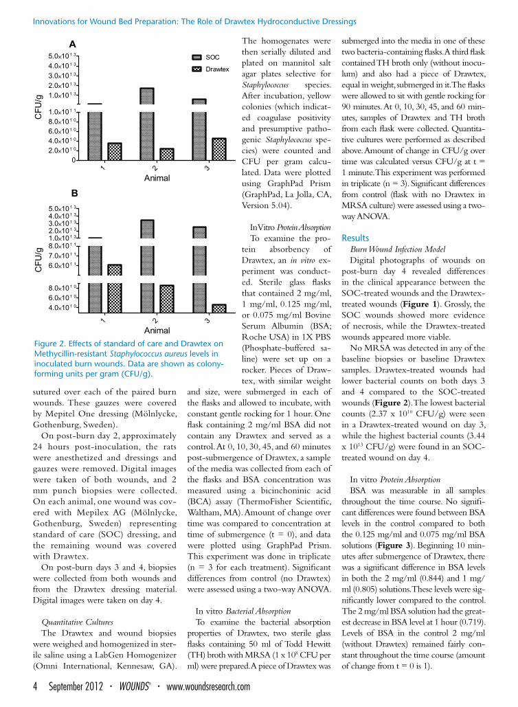
post-burn day 4 revealed differences in the clinical appearance between the SOC-treated wounds and the Drawtex-treated wounds (Figure 1). Grossly, the SOC wounds showed more evidence of necrosis, while the Drawtex-treated wounds appeared more viable.
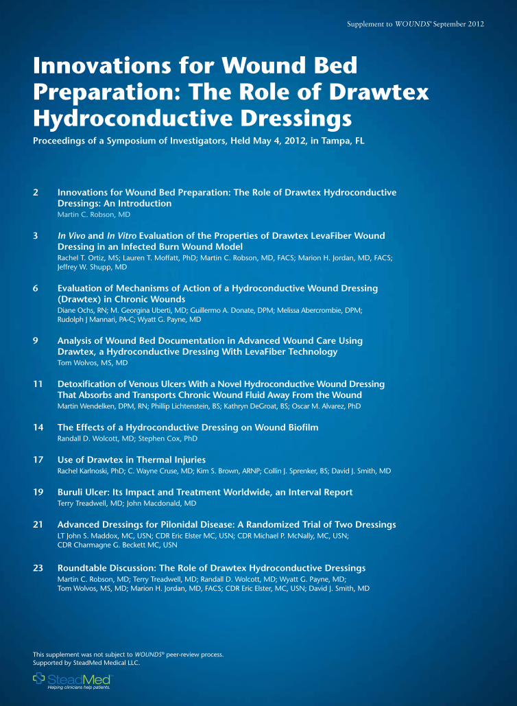
No MRSA was detected in any of the baseline biopsies or baseline Drawtex samples. Drawtex-treated wounds had lower bacterial counts on both days 3 and 4 compared to the SOC-treated wounds (Figure 2). The lowest bacterial counts (2.37 x 10¹⁰ CFU/g) were seen in a Drawtex-treated wound on day 3, while the highest bacterial counts (3.44 x 10¹³ CFU/g) were found in an SOC-treated wound on day 4.
In vitro Protein AbsorptionBSA was measurable in all samples
throughout the time course. No signifi-cant differences were found between BSA levels in the control compared to both the 0.125 mg/ml and 0.075 mg/ml BSA solutions (Figure 3). Beginning 10 min-utes after submergence of Drawtex, there was a significant difference in BSA levels in both the 2 mg/ml (0.844) and 1 mg/ml (0.805) solutions. These levels were sig-nificantly lower compared to the control. The 2 mg/ml BSA solution had the great-est decrease in BSA level at 1 hour (0.719). Levels of BSA in the control 2 mg/ml (without Drawtex) remained fairly con-stant throughout the time course (amount of change from t = 0 is 1).
Figure 2. Effects of standard of care and Drawtex on Methycillin-resistant Staphylococcus aureus levels in inoculated burn wounds. Data are shown as colony-forming units per gram (CFU/g).

www.woundsresearch.com • September 2012 • WOUNDS® 5
Innovations for Wound Bed Preparation: The Role of Drawtex Hydroconductive Dressings
In vitro Bacterial AbsorptionNo MRSA was detected in any of
the baseline Drawtex samples (pre-sub-mergence), or in the uninoculated TH broth throughout the time course. No significant differences in MRSA growth existed between the two MRSA cultures at 1 minute after Drawtex submergence; therefore, data were compared to t = 1 minute to determine the amount of change. Starting 10 minutes after Draw-tex submergence, the MRSA-contain-ing medium with Drawtex submerged showed a significantly lower bacterial count compared to the control MRSA culture (without Drawtex). This cul-ture had the highest amount of change of bacterial count (2.71) at 90 minutes. Correspondingly, significantly higher bacterial counts were measured in the Drawtex material that was submerged in the culture media, also compared to the control, with the lowest amount of change (0.1590) at 90 minutes (P < 0.001, Figure 4).
DiscussionThough Drawtex has been reported
to have exceptional capabilities in ab-sorbing and wicking away exudate and wound debris from wound surfaces, no published in vitro studies have been found that quantify these capabilities. The in vitro experiments described here were aimed at characterizing the absorption ability of Drawtex at both cellular and molecular levels. These experiments demonstrated a significant reduction in bacterial counts in the MRSA-containing media that had Drawtex submerged in it, while simul-taneously showing a significant increase in bacteria in the Drawtex material itself. The logical conclusion is that Drawtex is capable of absorbing bacteria from media to a large extent.
Protein assay data also demonstrated a significant reduction in protein concentra-tion over time in the 2 mg/ml and 1 mg/ml BSA solutions that contained Drawtex, highlighting this property and suggest-ing that this material would be capable of wicking away other proteins, such as viru-lence factors, in a wound environment. Further work will be aimed at determining virulence-factor absorption in vivo.
This study also utilized a reproduc-ible burn wound infection model that has been developed to allow observation
of the effectiveness of wound dressings on local wound infections. Though sev-eral clinical case studies have described the use Drawtex to treat a variety of wounds, there have been no controlled pre-clinical studies published comparing Drawtex to a known dressing in burn wounds. Some of these studies have re-portedly demonstrated a reduction in both eschar and exudate at the wound areas.5 In our model, quantitative cul-tures revealed a reduction of bacterial growth in the Drawtex-treated, MRSA-infected wound area compared to the SOC wound. Further, digital images demonstrated a noticeable difference in viability between the two wounds.
This study demonstrates the ability of Drawtex to reduce bacterial growth in an MRSA-infected burn wound. The in vitro work also demonstrates the ability
of Drawtex to absorb both protein and bacteria. Additional work is needed to further characterize the mechanisms by which Drawtex impacts wound healing, focusing on its absorptive capabilities. n
References1. Aramwit P, Muangman P, Namviriyachote N, Sricha-
na T. In vitro evaluation of the antimicrobial effec-tiveness and moisture binding properties of wound dressings. Int J Mol Sci. 2010;11(8):2864–2874.
2. Atiyeh BS, Gunn SW, Hayek SN. State of the art in burn treatment. World J Surg. 2005 Feb;29(2):131–148. Review.
3. Königová R, Matousková E, Broz L. Burn wound coverage and burn wound closure. Acta Chir Plast. 2000;42(2):64–68.
4. Lansdown AB, Williams A, Chandler S, Benfield S. Silver absorption and antibacterial efficacy of silver dressings. J Wound Care. 2005;14(4):155–160.
5. Steadmed Medical. Drawtex. 2012. Available from: http://www.steadmed.com/product-wound-thera-py-primary-dressings-drawtex.php.
6. Committee for the Update of the Guide for the Care Use of Laboratory Animals, National Research Council. Guide for the Care and Use of Laboratory Ani-mals: Eighth Edition. Washington, DC: The National Academies Press; 2011.
7. Shupp JW, et al. Treatment with an oxazolidinone antibiotic inhibits TSST-1 production in MRSA-in-
0 1 5 10 15 20 25 30 35 40 45 50 55 60 65 70 75 80 85 90
-4
-3
-2
-1
0
1
2
Time (m)
Cha
nge
in C
FU
MRSA Culture (- Drawtex) Quantitative Bx from Drawtex MRSA Culture (+ Drawtex)
Figure 3. Effects of Drawtex in various BSA levels compared to the control data are shown as amount of change in mg/ml from t = 0. Data points are displayed as the mean (n = 3) ± SD. Statistical significance was determined by two-way ANOVA (P < 0.001).
Figure 4. Effects of Drawtex on bacterial counts of MRSA-containing Todd Hewitt Broth. Data are shown as amount of change in colony forming units per gram (CFU/g) from minute 1. Data points are displayed as the mean (n = 3) ± SD. Sta-tistical significance was determined by two-way ANOVA (P < 0.001).

6 September 2012 • WOUNDS® • www.woundsresearch.com
Innovations for Wound Bed Preparation: The Role of Drawtex Hydroconductive Dressings
Wound healing is the result of dy-namic interactive processes that
begin at the moment of wounding and involve soluble mediators, many cell types, and extracellular matrices.1 Unen-cumbered, these processes follow a spe-cific time sequence and chronological order. When a wound proceeds through an orderly and timely reparative process and results in a sustained restoration of anatomic and functional integrity, it has been labeled an acute wound.2 Converse-ly, a chronic wound is one that has failed to proceed through an orderly and timely process to produce anatomic and func-tional integrity or has proceeded through the repair process without establishing a
sustained anatomic and functional result. In chronic wounds, the healing process is prolonged and incomplete, proceeds in an uncoordinated manner and results in a poor outcome.1–3
Chronic wounds have excessive in-flammation, increased pro-inflammatory cytokines, increased proteases such as matrix metalloproteinases (MMPs), and decreased growth factors.3–5 The com-mon chronic wounds of the skin and soft tissues that result in indolent ulcers are similar in that each is characterized by persistent inflammatory stimuli such as repeat trauma, relative ischemia, and bac-terial contamination.5 Common chronic wound ulcers include diabetic foot ulcers
(DFUs), venous stasis ulcers (VSUs), and pressure ulcers (PUs). The standard treat-ment for these chronic wounds has been wound bed preparation by debridement of necrotic tissue, decreasing excessive wound exudate, decreasing bacterial lev-el, removal of deleterious chemical me-diators, and wound closure.
Recently, a hydroconductive, non-adherent dressing has been designed with two types of absorbent cross-sec-tion structure, which might be able to perform the functions of wound bed preparation. This dressing creates the ability to actively move large volumes of fluid and other debris from chronic wounds. These dressings can remove ex-
Evaluation of Mechanisms of Action of a Hydroconductive Wound Dressing, Drawtex, in Chronic WoundsDiane Ochs, RN; M. Georgina Uberti, MD;Guillermo A. Donate, DPM; Melissa Abercrombie, DPM; Rudolph J Mannari, PA-C; Wyatt G. Payne, MD
Bay Pines Veterans Administration Healthcare System, Bay Pines, FL; Division of Plastic Surgery, University of South Florida, Tampa, FL
Address correspondence toWyatt G. Payne, MDSurgical Services (112)Bay Pines VA Healthcare SystemPO Box 500510,000 Bay Pines Blvd.Bay Pines, FL [email protected]
TABLE 1. Example patient with diabetic foot ulcer.
TissueCFUs/gm
DrawtexCFUs/cm2
Tissue MMP-1pg/μg protein
Drawtex MMP-1pg/μg protein
Tissue MMP-9pg/μg protein
Drawtex MMP-9pg/μg protein
Wound size cm2
Day 0 106 2.649 22.38 2.5
Day 7 105 103 0.471 ----- 22.28 ----- 1.8
Day 14 104 104 0.685 0.510 23.26 19.86 1.7
Day 21 <102 104 0.860 0.529 22.20 16.66 1.7
Day 28 <102 104 0.696 0.962 21.78 21.64 1.8

www.woundsresearch.com • September 2012 • WOUNDS® 7
Innovations for Wound Bed Preparation: The Role of Drawtex Hydroconductive Dressings
cessive inflammatory exudate and have been reported to remove matrix metal-loproteinases such as MMP-9.6 As was shown in In Vivo and In Vitro Evaluation of the Properties of Drawtex LevaFiber Wound Dressing in an Infected Burn Wound Model (p. 3), a hydroconductive dressing, specifically Drawtex, can draw bacteria away from an experimental burn wound.
The exact mechanisms of action of Drawtex and its benefits in wound heal-ing have not been elucidated. Therefore, we have designed a study to: 1) evaluate the dressing’s role of decreasing the quan-titative tissue level of bacteria in wounds; 2) determine if the tissue bacteria are drawn into the Drawtex dressing; 3) eval-uate the ability of the dressing to remove deleterious substances such as MMP-9 from chronic wounds; and 4) evaluate the effect of the dressing to decrease wound size and improve wound appearance.
A series of DFUs, VSUs, and PUs are in the process of being studied. For each wound, standardized photographs and tracing for digitized planimetry are obtained on Day 1. A biopsy for quan-titative and qualitative bacteriology is then obtained. Also a biopsy is obtained from the wound for enzyme-linked immunosorbent assay (ELISA) analy-ses. These analyses are run in duplicate. The wounds are treated with Drawtex dressing next to the wound, and the re-mainder of the dressing is completed as determined by the attending phy-sician and as appropriate for the spe-cific chronic ulcer type. If the wound is extremely exudative or consists of a deep cavity, as with a PU, two layers of Drawtex dressings are acceptable.
The dressings are changed twice a week. At alternate dressing changes, a piece of Drawtex from the dressing cen-ter is aseptically placed into transport me-dia for wound culture and sensitivity ex-amination. Similarly, a piece of Drawtex is placed into a test tube with 2 cc of ster-ile saline for ELISA analyses of cytokines. A routine tissue biopsy is then obtained for quantitative and qualitative bacteriol-ogy and another biopsy is obtained from the wound for ELISA analyses.
To date, nine patients have been stud-ied. Their wounds include DFUs, VSUs, and PUs. Tissue bacterial levels ranged as high as 109 CFUs/gm of tissue. Draw-
Figure 1A. Diabetic foot ulcer day 0.
Figure 1B. Diabetic foot ulcer day 14.

8 September 2012 • WOUNDS® • www.woundsresearch.com
Innovations for Wound Bed Preparation: The Role of Drawtex Hydroconductive Dressings
tex dressings decreased bacterial levels to <102 CFUs/gm. Bacterial levels in Drawtex dressings ranged from 103–104
CFUs per square centimeter of dressing. MMP-1 levels in the tissue tended to de-crease as levels increased in the Drawtex dressings. MMP-9 levels similarly tended to decrease slightly as the MMP-9 level appeared in Drawtex dressings.
An example of these cases is this pa-tient with a DFU (Table 1). Bacterial levels in the wound tissue decreased from 106 CFUs/to <102 CFUs/gm of tis-sue. The bacteria appeared to be drawn into the Drawtex dressings. MMP-1 tissue levels decreased from 2.649 pg/ug of protein to 0.696 pg/ug. MMP-9 tissue levels decreased slightly. The level of MMP-1 and MMP-9 in the Drawtex dressings increased. Even in this midfoot ulcer in a foot with mild Charcot defor-mity, these changes resulted in a decrease in size within 14 days (Figure 1).
Measurements of the MMP-1 and MMP-9 levels in the tissue and Drawtex dressings have provided additional in-formation. In a VSU patient, as bacterial levels in the Drawtex dressing increased from 102–104 CFUs/cm2 of dressing over 21 days, the cytokine levels were as fol-lows: MMP-1 in the tissue were 0.549 pg/ug on day 0 and 1.944 pg/ug on day 28, while the MMP-1 levels in the Drawtex dressing were 1.315 pg/ug on day 7 and 0.432 pg/ug on day 28.
The tissue MMP-9 level went from 19.66 pg/ug on day 0 to 23.12 pg/ug on day 28. The Drawtex drew the MMP-9 levels into the dressing at 20.22 pg/ug on day 7 and 15.8 pg/ug on day 28. This VSU did actually increase slightly in size during the 28 days from 16.6 cm2
on day 0 to 18.3 cm2 on day 28 (Figure 2). Muller et al reported that, in poorly healing ulcers such as this, MMP-1 and MMP-9 levels did not appear to decrease, and that the excess of the proteases con-tributed to the lack of healing.7 It ap-pears, despite the ability of the Drawtex to remove these deleterious cytokines from the wound bed, that more proteases were being produced, leading to stagna-tion of the wound.
This interval report demonstrates that clinical results in chronic wounds treat-ed with Drawtex mimic experimental animal results. Drawtex hydroconduc-tive dressings have the ability to draw
bacteria and deleterious cytokines from wound tissue into the dressing. The data to date suggest that Drawtex may be an effective adjunct for debridement of chronic wounds. n
References1. Robson MC, Steed DL, Franz MG. Wound healing:
biologic features and approaches to maximize healing trajectories. Curr Probl Surg. 2001;38(2):61–140.
2. Lazurus GS, Cooper DM, Knighton DR, et al. Definitions and guidelines for assessment of wounds and evaluation of healing. Arch Dermatol. 1994;130(4):489-493.
3. Nwomeh BC, Yager DR, Cohen IK. Physiology of the chronic wound. Clin Plast Surg. 1998;25(3):341–356.
4. Tarnuzzer RW, Schultz GS. Biochemical analysis of acute and chronic wound environments. Wound Re-pair Regen. 1996;4(3):321–325.
5. Mast BA, Schultz GS. Interactions of cytokines, growth factors, and proteases in acute and chronic wounds. Wound Repair Regen. 1996;4(4):411–420.
6. Lichtenstein P, Wendelken M, Alvarez O. Detoxifica-tion of venous ulcers with a novel hydroconductive wound dressing that transfers chronic wound fluid away from the wound. Presented at 24th Annual Sym-posium on Advanced Wound Care and the Wound Healing Society Meeting, Dallas, TX; April 2011.
7. Muller M, Trocme C, Lardy B, et al. Matrix metal-loproteinases and diabetic foot ulcers: the ratio of MMP-1 to TIMP-1 is a predictor of wound healing. Diabet Med. 2008;25(4):419–426.
Figure 2A. Venous stasis ulcer day 0.
Figure 2B. Venous stasis ulcer day 21.

www.woundsresearch.com • September 2012 • WOUNDS® 9
Innovations for Wound Bed Preparation: The Role of Drawtex Hydroconductive Dressings
Analysis of Wound Bed Documentation in Advanced Wound Care Using Drawtex, a Hydroconductive Dressing With LevaFiber TechnologyTom Wolvos MS, MD, FACSScottsdale Healthcare Osborn Medical Center, Scottsdale, AZ
Address correspondence toTom Wolvos, MS, MD, FACSChief of the Division of General SurgeryScottsdale Healthcare Osborn Medical Center7400 East Osborn RoadScottsdale, AZ, [email protected]
The oldest medical text, the Su-merian tablet, written in 1600
BC, contains treatments thought to date back to 3000 BC.1 Because in-fection was almost always present in the wounds at that time, suppuration was considered a necessary phase in wound healing for over 3,500 years. In the 1860s, Lister demonstrated that infections were not a normal event in wound healing. Infection was rec-ognized as caused by bacteria, and he promoted the use of carbolic acid as an antiseptic in surgery and for the treatment of wounds with carbolic acid-soaked dressings.1 His theory was supported by a fall in the infection rate and morality in wounded patients.
The concept of moist wound heal-ing was introduced in 1962 by Winter.2 Maintaining the proper moisture level in a wound has now been accepted as important in promoting a favorable wound healing environment.3
The role for debridement has been advanced via combat wound treat-ment during times of war. Debride-ment may be accomplished not only by sharp excision of necrotic and de-vitalized tissue, but also by using enzy-matic, mechanical, autolytic, or hydro-conductive techniques.
The development of advanced dress-ings is only a relatively recent phenom-enon. In 1979, Turner listed the criteria for an ideal dressing.4 Specialized dress-ings can be grouped into categories ac-cording to their functions and mecha-nisms of action. The overall goal of an advanced dressing is to restore, in a moist setting, the wound microenviron-ment to achieve the normal balance of cytokines, growth factors, and proteo-lytic mediators.
Advancements in wound care pro-tocols and dressings created a need to analyze the results to determine the effectiveness of treatments. Given that serial observation of a wound by clini-cians — the method traditionally used — is a very subjective evaluation, clini-cians have searched for more objective wound evaluation methods.
An advanced pattern recognition software algorithm that uses artificial intelligence to analyze digital wound images to provide accurate wound measurements and tissue analysis has been developed (iCLR technology, powered by Elixr, Imago Care Ltd., London, UK). This technology calcu-lates wound measurements including area, circumference, width, and depth, and analyzes the tissue-type compo-
sition of the wound bed.5 Using the iCLR technology algorithm, wound tissue color features are acquired and parameters of statistical distributions are calculated for the different tissue types in a three-dimensional color space. In the digitized wound photo-graph, this program divides a wound into three tissue-type classifications: necrotic tissue, represented as a black color; fibrin and slough, represented as a yellow color; and granulation tissue, represented as a red color.
A new active hydroconductive non-adherent dressing with LevaFiber tech-nology has been recently introduced (Drawtex, SteadMed Medical LLC, Ft. Worth, TX). Due to its unique propri-ety construction, the ability to remove large amounts of fluid and debris from the wound into the dressing is estab-lished. The dressing actively draws fluid away from the wound up to 150 cc/hour using an active capillary action, and retains its integrity when moist. It can also draw toxic wound exudates into the dressing, in effect detoxifying the wound.
Drawtex can help selectively de-bride wounds by removing adher-ent fibrin, slough and necrotic tissue while leaving healthy granulation tis-
10 September 2012 • WOUNDS® • www.woundsresearch.com
Innovations for Wound Bed Preparation: The Role of Drawtex Hydroconductive Dressings
sue in place. The rapid transfer of fluid into this dressing may sever the fibers of undenatured collagen that anchors the necrotic tissue to the wound sur-face itself. Termed “hydroconductive debridement,” undesirable tissue is selectively removed, leaving healthy tissue intact, and is observed undis-turbed in serial digital photo analy-sis.6 The healthy granulation tissue was preserved over time and actually increased in percentage of the total wound volume, while the necrotic tissue and slough were selectively de-brided, decreasing their volume in the wound analysis.7
The removed exudate may contain factors such as proteases and other factors or toxins that inhibit normal wound healing. In one study, matrix metalloproteinase-9 was demonstrated to be drawn into the dressing and was actively transported up to 7 cm from the wound edge.8 It has been suggested that the fluid removed may also contain the plasma necessary to maintain the viability of a biofilm, helping to lead to its breakdown.9
The dressing was used in a variety of wound types. It was particularly effective in a patient with a highly exudating venous leg ulcer. Initially, it was placed in a double layer un-der compression and changed twice a week. After the drainage decreased, the dressing changes were done once a week until the wound healed. Suc-cess has also been noted in dry wounds. In this situation, wound moisture was maintained with ointments such as sil-ver sulfadiazine. Typically the dressings were changed daily. Similarly necrotic tissue, fibrin, and slough are removed in dry wounds, and the wound fills in with healthy, vascular granulation tis-sue. In a published series of eight pa-tients using independent digital wound analysis, the average area of necrotic tis-sue, fibrin and slough of all the patients were reduced by 36% in 1 week, 52% by week 2, and 77% in 3 weeks.7 There also was a corresponding reduction of the wound area of 15% in 1 week, 35% by week 2, and 47% by week 3.
No adverse effects have been noted in any of the patients treated with this
dressing. Further studies will be needed to find the role of this advance dressing for treating complex wounds. n
References1. Broughton G, Janis J, Attinger C. A brief history
of wound care. Plast Reconstr Surg. 2006; 117(7 Suppl):6S–11S.
2. Winter G. Formation of the scab and the rate of epithelization of superficial Wounds in the skin of the young domestic pig. Nature. 1962;193:293–294.
3. Mendez-Eastman S. Wound dressing categories. Plast Surg Nurs. 2005;25(2):95–99.
4. Turner T. The development of wound manage-ment products. In Krasner D, editor. Chronic Wound Care: A Source Book for Healthcare Professionals. King of Prussia, PA: Health Management Publications; 1990;33.
5. Product information. Application & Validation of a Unique Wound Image Analysis Tool. Elixr & iCLR Technology Developers. Imago Care Ltd. Available from [email protected].
6. Couch KS. Discovering hydroconductive dressings. Ostomy Wound Manage. 2012;5(4):8–10.
7. Livingston M, Wolvos T. Hydroconductive debride-ment: A new perspective in reducing slough and ne-crotic tissue. Presented at 24th Annual Symposium on Advanced Wound Care and the Wound Healing Society Meeting, Dallas, TX; 2011.
8. Lichtenstein P, Wendelken M, Alvarez O. Detoxica-tion of venous ulcers with a novel hydroconduc-tive wound dressing that transfers chronic wound fluid away from the wound. Presented at 24th An-nual Symposium on Advanced Wound Care and the Wound Healing Society Meeting, Dallas, TX; 2011.
9. Wolcott R, Dowd. Drawtex effects on VLU healing and biofilm. Presented at 24th Annual Symposium on Advanced Wound Care and the Wound Healing Society Meeting, Dallas, TX; 2011.
Figure 1C. Wound after 21 days of Drawtex. Figure 1D. Digital analysis of Figure 1C (red = granulation tissue, yellow = slough, black = necrotic tissue).
Figure 1A. Skin tear after 7 days of Drawtex. Figure 1B. Digital analysis of Figure 1A (red = granulation tissue, yellow = slough, black = necrotic tissue).

www.woundsresearch.com • September 2012 • WOUNDS® 11
Innovations for Wound Bed Preparation: The Role of Drawtex Hydroconductive Dressings
Detoxification of Venous Ulcers With a Novel Hydroconductive Wound Dressing That Absorbs and Transports Chronic Wound Fluid Away From the WoundMartin Wendelken DPM, RN1; Phillip Lichtenstein, BS1; Kathryn DeGroat, BS1; Oscar M. Alvarez, PhD1, 2
1Center for Curative and Palliative Wound Care, Calvary Hospital, Bronx, NY2Department of Medicine, New York Medical College, Valhalla, NY
Address correspondence toOscar M. Alvarez, PhDDirector, Center for Curative and Palliative Wound CareCalvary Hospital1740 Eastchester RoadBronx, NY [email protected]
Acknowledgement: We thank the Biochemistry Department at New York Medical College, Valhalla NY for performing the MMP-9 assays.
The chronicity of venous ulcers (VUs) can be defined clinically by
excessive granulation tissue, increased fibrosis, hyperkeratotic wound margins and increased lipodermatosclerosis.1,2 Biochemically, chronicity can be de-fined by significant increases in pro-inflammatory cytokines, proteases, and neutrophil elastase.3–6 Excessive inflam-mation caused by hyperstimulated neu-trophil response has also been suggest-ed as a potential cause for a wound’s chronicity.7,8 It is this protease activity, primarily caused by a specific group of proteases, called matrix metallopro-teinases (MMPs), that is believed to be responsible for the destruction of the provisional matrix (fibronectin, neces-sary for keratinocyte migration) and other extracellular matrix components negatively affecting chemotaxis and cellular migration.8–10
Wound fluid (exudates) from chronic VUs contains excessive levels of MMP-2 and MMP-9. Furthermore, it has been reported that these gelatinases need to be down-regulated to permit healing to take place.11 Down-regulation of in-flammatory cytokines and MMPs 2 and
9 occurs naturally (albeit slowly) when VUs are treated with adequate compres-sion.12,13 It is important to lower the lev-els of MMP-9 in chronic VUs because it breaks down basement membrane col-lagens more than other MMPs do.14,15
It would seem logical that, if a device could transport chronic wound fluid from the ulcer so that it is not trapped within the primary dressing and in con-stant contact with the wound bed, less proteolytic breakdown of the provi-
sional matrix would take place and, thus, improve keratinocyte migration and subsequent healing. The objective in this study was to evaluate a hydroconductive wound dressing (HWD) as a transport medium to detoxify chronic VUs by assisting the displacement of chronic wound fluid away from the wound bed.
Study Design This was a prospective, randomized,
single-center pilot study involving 15
Figure 1. Proportion of wounds healed at 4 weeks.
Wound Healing and Wound Scores at 4 Weeks
*+3 = complete closure at 4 weeks, +2 = > 50% clo-sure, +1 = 25–49% closure, -1 = 1–24% closure, -2 = no improvement, -3 = wound deterioration
Mean Wound Score at 4 Weeks*
HWD 2.1
Control 1.5
60
50
40
30
20
10
0
n = 10
n = 5
12 September 2012 • WOUNDS® • www.woundsresearch.com
Innovations for Wound Bed Preparation: The Role of Drawtex Hydroconductive Dressings
subjects in an outpatient wound care center setting. Each subject with a ve-nous ulcer was randomized (2:1) to re-ceive HWD plus compression therapy or standard care (non-adherent dressing plus compression therapy). Wound healing outcomes were graded using photo-dig-ital planimetry software and a numerical scale of +3 to -3 (+3 = complete clo-sure at 4 weeks, +2 = >50% closure at 4 weeks, +1 = 25–49% closure at 4 weeks, -1 = 1–24% closure at 4 weeks, -2 = no improvement, -3 = wound deteriora-tion). In addition, wound fluid MMP-9 levels were measured in both the wound bed and HWD both proximal and distal to the ulcer. Subjects were followed until healing or for 8 weeks, and the primary endpoint was the proportion of subjects reaching 50% healing within 4 weeks.
Study Participation CriteriaThe inclusion criteria were ages 18–90
years; ability to provide informed con-sent; open VU for at least 1 year with a surface area > to 1.5 cm2; and an ABI > 0.70. The exclusion criteria were: target ulcer not a VU; ABI < 0.7; intermittent claudication, wound infection, cellulitis, or osteomyelitis; known hypersensitivity to cellulose, xylose, cotton, or wool, or any of the study dressings or compres-sion bandages; a subject’s receiving cor-ticosteroids, immunosuppressive agents, radiation therapy, or chemotherapy that might interfere with wound healing; un-controlled diabetes mellitus; immuno-deficiency disorders that interfere with wound healing; a history of sickle cell anemia, thalassemia, vasculitis, rheuma-toid arthritis, lupus scleroderma, or any
hematological, connective tissue, or col-lagen vascular disorder; and wounds that had been treated with an investigational product within the previous 30 days.
MethodsStandard of care compression therapy
was applied once weekly using either a four-layer bandage system (Profore Smith and Nephew, Largo FL) or a modified Unna’s boot (Unna’s paste boot, Vis-copaste, Smith and Nephew, Largo FL, and Coban Cohesive Bandage, 3M, St. Paul, MN). The primary wound dress-ings were the test agent HWD (Draw-tex, SteadMed Medical, Ft. Worth, TX) and Profore WCL, Smith and Nephew, Largo FL). Wounds were measured using PictZar Photodigital Planimetry Software (BioVisual Technologies, Elmwood Park, NJ).16 Wound assessment was performed using a numerical composite scale of +3 to -3 (+3 = complete closure at 4 weeks, +2 = >50% closure at 4 weeks, +1 = 25–49% closure at 4 weeks, -1 = 1–24% closure at 4 weeks, -2 = no improve-ment, -3 = wound deterioration). Wound fluid MMP-9 was measured in both the wound and HWD using a direct enzyme-linked immunosorbant assay (ELISA) as described by Rayment et al.17 Assays were performed at baseline, week 2, and week 4 on four subjects.
Results The proportion of wounds healed and
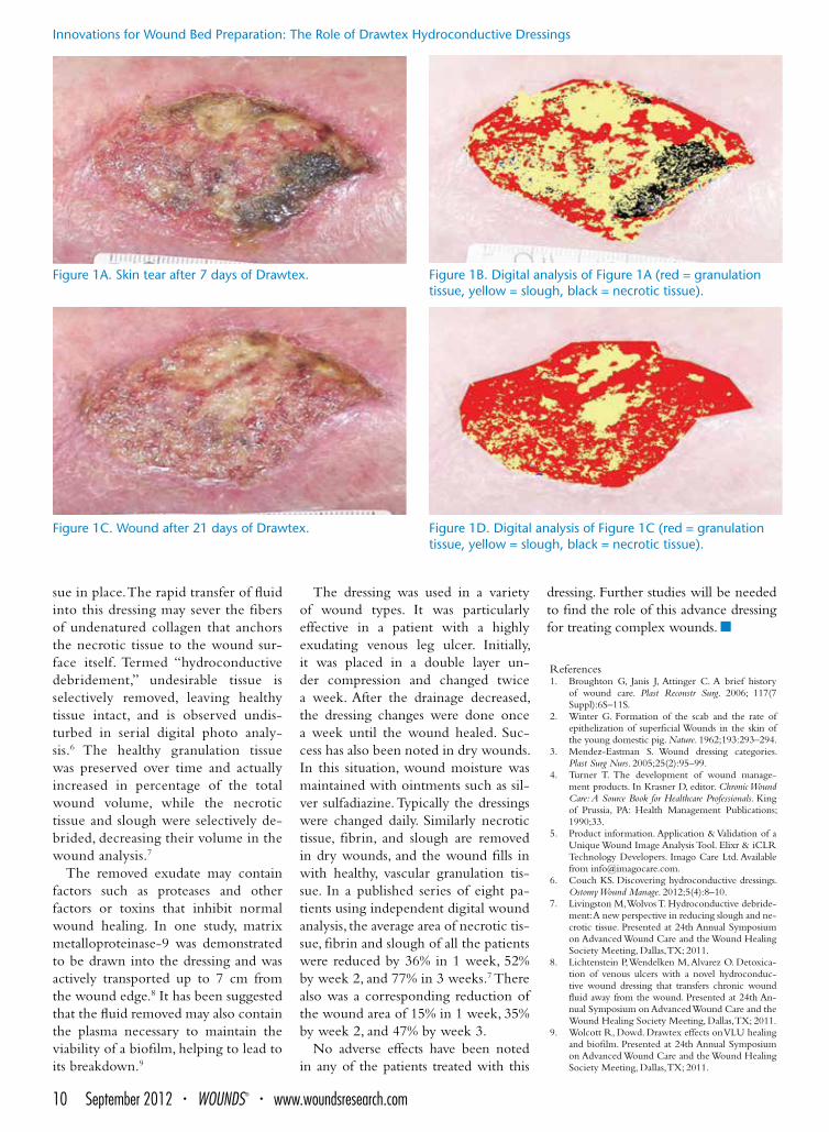
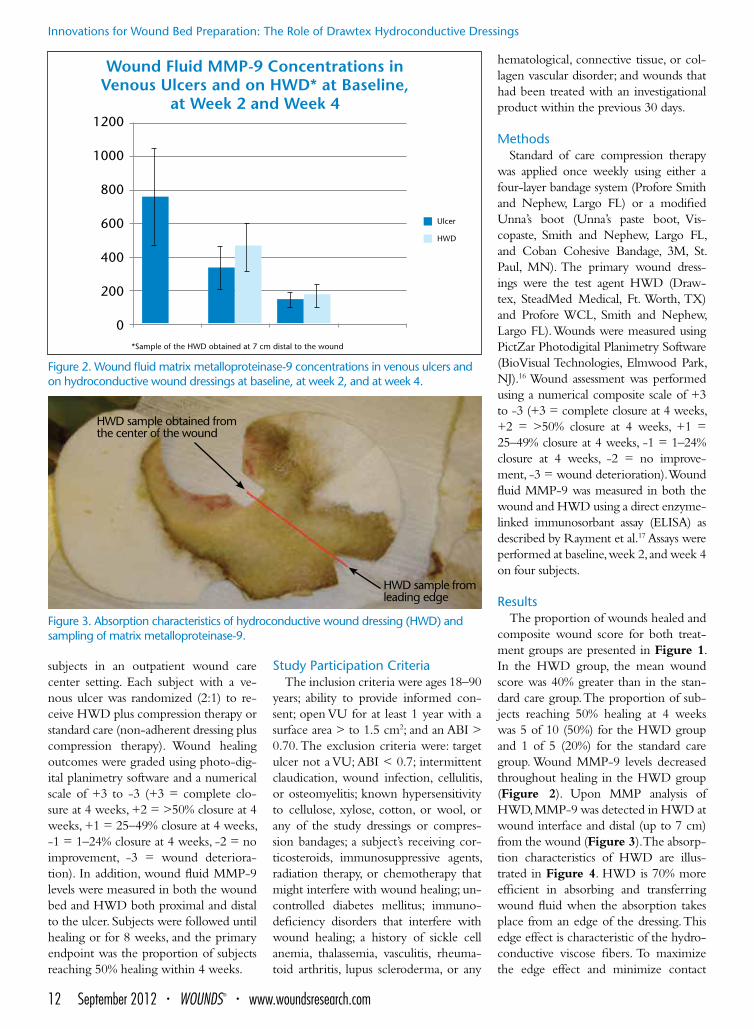
composite wound score for both treat-ment groups are presented in Figure 1. In the HWD group, the mean wound score was 40% greater than in the stan-dard care group. The proportion of sub-jects reaching 50% healing at 4 weeks was 5 of 10 (50%) for the HWD group and 1 of 5 (20%) for the standard care group. Wound MMP-9 levels decreased throughout healing in the HWD group (Figure 2). Upon MMP analysis of HWD, MMP-9 was detected in HWD at wound interface and distal (up to 7 cm) from the wound (Figure 3). The absorp-tion characteristics of HWD are illus-trated in Figure 4. HWD is 70% more efficient in absorbing and transferring wound fluid when the absorption takes place from an edge of the dressing. This edge effect is characteristic of the hydro-conductive viscose fibers. To maximize the edge effect and minimize contact
Figure 2. Wound fluid matrix metalloproteinase-9 concentrations in venous ulcers and on hydroconductive wound dressings at baseline, at week 2, and at week 4.
Figure 3. Absorption characteristics of hydroconductive wound dressing (HWD) and sampling of matrix metalloproteinase-9.
Wound Fluid MMP-9 Concentrations in Venous Ulcers and on HWD* at Baseline,
at Week 2 and Week 4
*Sample of the HWD obtained at 7 cm distal to the wound
1200
1000
800
600
400
200
0
HWD sample obtained from the center of the wound
HWD sample from leading edge
Ulcer
HWD

www.woundsresearch.com • September 2012 • WOUNDS® 13
Innovations for Wound Bed Preparation: The Role of Drawtex Hydroconductive Dressings
with the saturated HWD and the wound bed, the dressings were cut so only the edge of HWD came in contact with the wound margins (Figure 5).
DiscussionHWD effectively transfers chronic
wound fluid away from VUs by a natural vacuum created via the hydroconduc-
tive viscose fibers. This detoxification process resulted in faster healing for VUs in this feasibility study. To our knowl-edge, this is the first time that a pri-mary wound dressing has been shown to sequester and transport elements of chronic wound fluid and isolate them away from the VU.
Reynolds et al conducted a random-ized, multi-center, controlled study to compare HWD to standard wound dressings in chronic wounds of several etiologies. The authors reported wound improvement of 12.7% based on subjec-tive interpretation (nurse perception); however, upon blinded assessment (based on evaluation of digital images), standard dressings were better by 6.6%.18 These authors placed the HWD directly over the wounds. We realize the use of HWD as a primary wound dressing may be counterintuitive, because we avoid cov-ering the wound and use it as a transport medium to evacuate harmful chronic wound fluid away from the ulcer itself.
In this small pilot study, MMP-9 levels were lower in the group treated with HWD at week 2 and at week 4. The viscosity of the wound fluid does impact the absorptive capacity and subsequent transfer of HWD. We found the hydroconductive capacity of HWD is limited by viscous or sero-sanguinous wound fluid.
We recommend wound bed prepa-ration (consisting of thorough selective debridement to remove all devital-ized tissues) before treating the wound with HWD. In our experience, a clean
wound consisting of 90–100% granu-lation tissue produced a less viscous discharge that contained less necrotic cells and solid debris. More studies are needed in a variety of inflamma-tory chronic wounds to investigate the mechanism and effect of this wound fluid transfer phenomenon. n
References1. Trengove NJ, Stacey MC, Macauley S, et al. Analysis
of the acute and chronic wound environments: the role of proteases and their inhibitors. Wound Repair Regen. 1999;7(6):442–452.
2. Stadelmann WK, Digenis AG, Tobin GR. Physiol-ogy and healing dynamics of chronic cutaneous wounds. Am J Surg. 1998;126(2A Suppl) 26S–38S.
3. Chen C, Schultz GS, Bloch M, et al. Molecular and mechanistic validation of delayed healing rat wounds as a model for human chronic wounds. Wound Repair Regen. 1999;7(6):486–494.
4. Tarnuzzer RW, Schultz GS. Biochemical analysis of acute and chronic wound environments. Wound Re-pair Regen. 1996;4(3):321–325.
5. Li WW, Li VW. Therapeutic angiogenesis for wound healing. Wounds. 2003;15(Suppl):3S–12S.
6. Ladwig GP, Robson MC, Liu R, et al. Ratios of ac-tivated matrix metalloproteinase-9 to tissue inhibi-tor of matrix metalloproteinase-1 in wound fluids are inversely correlated with healing of pressure ulcers. Wound Repair Regen. 2002;10(1):26–37.
7. Yager DR, Nwomeh BC. The proteolytic envi-ronment of chronic wounds. Wound Repair Regen. 1999;7(6):433–441.
8. Tomic-Canic M, Agren MS, Alvarez OM. Epider-mal repair and the chronic wound. In: The Epidermis and Wound Healing. Rovee DT, Maibach HI, eds. New York, NY: CRC Press; 2004.
9. Wysocki, AB, Staiano-Coico L, Grinnell F. Wound fluid from chronic leg ulcers contains elevated levels of metalloproteinases MMP-2 and MMP-9. J Invest Dermatol. 1993;101(1):64–68.
10. Grinnell F, Zhu M. Identification of neutrophil elastase as the proteinase in burn wound responsible for degradation of fibronectin. J Invest Dermatol. 1994;103(2):155–161.
11. Fray JM, Dickinson RP, Huggins JP, et al. A potent, selective inhibitor of matrix metalloproteinase-3 for the topical treatment of chronic dermal ulcers. J Med Chem. 2003; 46(16):3514–3525.
12. Beidler SK, Douillet CD, Berndt DF, et al. Inflam-matory cytokine levels in chronic venous insuf-ficiency ulcer tissue before and after compression therapy. J Vasc Surg. 2009;49(4):1013–1020.
13. Beidle SK, Douillet CD, Berndt DF, et al. Multi-plexed analysis of matrix metalloproteinases in leg ulcer tissue of patients with chronic venous insuffi-ciency before and after compression therapy. Wound Repair Regen. 2008;16(5):642–648.
14. Wysocki AB, Kusakabe AO, Chang S, et al. Tempo-ral expression of urokinase plasminogen activator, plasminogen activator inhibitor and gelatinase-B in chronic wound fluid switches from a chronic to acute wound profile with progression to healing. Wound Repair Regen. 1999;7(3):154–165.
15. Okada Y, Gonoji Y, Naka K, et al. Matrix metallo-proteinase-9 (92-kDa gelatinase type-IV collagenase) from Ht-1080 human fibrosarcoma cells — purifica-tion and activation of the precursor and enzymatic properties. J Biol Chem. 1992;267(30): 21712-9.
16. Wendelken ME, Berg WT, Lichtenstein P, et al. Wounds measured from digital photographs using photodigital planimetry software: validation and rater reliability. Wounds. 2011;23(9):267–275.
17. Rayment EA, Upton Z, Shooter GK. Increased ma-trix metalloproteinase-9 (MMP-9) activity observed in chronic wound fluid is related to the clinical sever-ity of the ulcer. Br J Dermatol. 2008;158(5):951–961.
18. Reynolds T, Russell L, Deeth M, et al. A randomised controlled trial comparing Drawtex with stan-dard dressings for exuding wounds. J Wound Care. 2004;13(2):71–74.
Figure 4. Absorption characteristics of a hydroconductive wound dressing (HWD). Wound fluid applied to center (A) or edge (B) of HWD. HWD is more efficient when absorption takes place from the edge. Note when 1 ml of wound fluid is applied to the center, it saturates 86% of the dressing, but when 1 ml of wound fluid is absorbed from the edge, it saturates only 25% of the dressing.
Figure 5. Method illustrating the use of a hydroconductive wound dressing (HWD). Note that, to maximize absorp-tion from the edges (edge effect) and to minimize contact of the saturated HWD with the wound bed, the HWD was cut in a way so that only the edges came in contact with the wound.
A B

14 September 2012 • WOUNDS® • www.woundsresearch.com
Innovations for Wound Bed Preparation: The Role of Drawtex Hydroconductive Dressings
Bacteria possess the ability to cause infection in two very distinct ways.1
The first way is when an individual bacterium with its unique genome uses one portion of its genes to stay a free-floating, motile cell (planktonic pheno-type) that has a strategy in a host envi-ronment to breach and kill cells with its virulence factors to create a source of nutrition. The second way is that the very same bacterium can up-regulate a separate group of genes, which lets it at-tach to a host structure. Once attached to the host, the bacterium secretes a polysaccharide matrix around itself and its progeny. When this small group reaches a sufficient number (quorum), signaling molecules (quorum-sensing molecules) direct the gene expression of each bacterium throughout the col-ony. This lets a community of bacteria develop within the protection of the matrix, which gives colony defenses against host immunity, including anti-bodies and white blood cells.2 Given that a biofilm requires attachment, it cannot use the host tissue to which it is adhered for a nutritional source and, therefore, successful biofilm uses local inflammation to produce plasma exu-date on which it can nourish itself.3
Excess exudate causes poor wound healing outcomes. Many strategies have been employed to decrease wound exu-date including antibiotics, topical anti-septics, edema management and control of inflammation. However, most wound care strategies include removal of the exudate once it is formed. For decades, moist, interactive wound care has been
utilized to manage exudate to improve wound healing. 4,5 It would be of great importance if a dressing had the ability not only to manage the exudate, but also to suppress the formation of the exudate at its source.
Chronic wounds have a large amount of biofilm on their surfaces and acute wounds have very little biofilm.6 The presence of biofilm is sufficient to explain the hyperinflam-matory milieu that is the hallmark for chronic wounds. Chronic wounds have elevated proinflammatory cyto-kines such as tumor necrosing factor, gamma interferon, interleukins 1, 6 and 8, and a host of other inflamma-tory cytokines.7 The chronic wound environment is also highly proteolytic, with elevated levels of matrix metallo-proteinases (MMPs) 2, 8 and 9, along with elastase.8 Additionally, at a cellu-lar level, chronic wounds are associ-ated with excessive neutrophils.9 This biochemical and cellular phenomenon of the chronic wound is also seen in other chronic infections.
Another strong argument for biofilm’s role in the nonhealing of wounds is host cellular senescence. Cellular senescence is evident by host cells that are unable to undergo cell division (shed),10 un-able to migrate11 and, most importantly, unable to apoptose.12 Apoptosis is the strategy the host uses to clear damaged or infected cells. By producing wound bed senescence, the biofilm prevents the host from removing the secure attach-ment for the biofilm while also prevent-ing the wound’s healing.
The activity of biofilm is controlled by quorum sensing molecules that dif-fuse throughout the biofilm community. The nutritional source is host plasma. Therefore, decreasing the dwell time of the plasma and other fluids within the wound biofilm may diminish the abil-ity of biofilm to produce host inflam-mation, host cell senescence and subvert host immunity.
Other technologies targeting rapid re-moval of wound exudate include nega-tive pressure wound therapy. In previous studies, it was shown that the bacterial numbers increased with negative pres-sure wound therapy. Yet, there was sig-nificant improvement in wound healing outcomes. There was no evidence that this was due to decreased dwell time for quorum-sensing molecules or nutrient molecules from the plasma.
Our study focused on the ability of a dressing with the properties of being able to generate high capillary pressures capable of the rapid removal of wound exudate. It was hoped that, with the rapid removal of wound exudate, the biofilm’s ability to produce persistent inflammation and host cellular senes-cence would be diminished. It was also important to determine if rapid removal of exudate decreased bacterial numbers on the surface of the wound.
MethodsTen patients with nonhealing, mod-
erate to highly exudative venous leg ulcers (lasting more than 30 days) were identified and consented to participate in a small cohort study (Western IRB
The Effects of a Hydroconductive Dressing on Wound BiofilmRandall D. Wolcott, MD1; Stephen Cox, PhD2
1Southwest Regional Wound Care Center, Lubbock, TX; 2Research & Testing Laboratory of the South Plains, Lubbock, TX
Address correspondence toRandall D. Wolcott, MDSouthwest Regional Wound Care Center2002 Oxford AvenueLubbox, TX [email protected]

www.woundsresearch.com • September 2012 • WOUNDS® 15
Innovations for Wound Bed Preparation: The Role of Drawtex Hydroconductive Dressings
TABLE 1. A significant reduction is seen in wound volume for nine of the 10 patients in the study. Two patients actually went on to full wound healing within the 4 weeks of the study.
Patient ID Initial Volume (cm2) Final Volume (cm2) % Healed
22517 0.07 0.00 100.0%
22632 1.10 0.09 91.7%
9510 2.18 2.14 1.7%
23008 0.48 0.28 41.6%
23262 9.43 4.75 49.6%
16358 5.58 3.01 46.1%
13711 0.08 0.00 100.0%
3035 1.82 1.11 39.0%
15623 1.18 0.23 80.5%
22822 2.94 0.89 69.7%
Avg 62.0%
TABLE 2. The beginning cycle threshold (CT) numbers compared with the final CT numbers for the 10 evaluable patients are shown. The CT number indicates how many times the sample had to be doubled before a signal could be obtained. The number of doublings required to obtain a signal is directly related to how much of the target DNA is in the original sample. The more bacteria present, the smaller the CT number. Four patients showed an increase in bacteria over the 4 weeks of the study.
Patient ID Initial Cycle Threshold Number
Final Cycle Threshold Number
Bacteria Change
23262 25.73 26.10 Less
15623 28.50 28.31 More
2308 16.81 27.05 Less
3035 18.75 26.99 Less
16358 19.95 18.28 More
9510 28.85 19.78 More
22822 22.85 24.51 Less
22632 27.63 22.24 More
13711 27.11 0 Less
22517 27.41 0 Less

16 September 2012 • WOUNDS® • www.woundsresearch.com
Innovations for Wound Bed Preparation: The Role of Drawtex Hydroconductive Dressings
#20101569). The average age of the study participants was 56.3 years (42 years old to 68 years old). There were six males and four females, and four of the patients were under management for diabetes. There were no other sig-nificant comorbidities.
Each patient was subjected to evalu-ation at each visit (weeks 0, 1, 2, 3, and 4) for a total of five visits over a 4-week period. At weeks 0 and 4, all wound metrics recorded and 5 mm punch bi-opsies were performed for comprehen-sive molecular evaluation (polymerase chain reaction [PCR] and sequencing), plus scanning electron microscopy. The molecular diagnostics were conducted by PathoGenius Laboratories. The bi-opsies were sent for scanning electron microscopy evaluation at the Center for Biofilm Engineering.
All wounds were managed under a general treatment regimen that includ-ed standard-of-care techniques. Mea-surements were obtained using Aranz Silhouette (Aranz Medical) equipment adhering to the manufacturer’s recom-mendation. The venous leg ulcers were assessed clinically, and then cleaned with a nontoxic, non-antimicrobial product. The wounds were then sharply debrid-ed to manage the surface accumulation of slough, devitalized tissue, and any other debris. DrawTex dressings were then applied. A multilayer compres-sion wrap was then applied to provide management of lower-limb edema. The dressings were changed on a Monday/Wednesday/Friday basis until the next clinic visit.
ResultsTable 1 shows that nine of 10 pa-
tients showed 40% or more healing
within the 4-week duration of the study. Only one wound failed to heal, but it did not show any deterioration. Two wounds healed completely, and one wound healed 92%.
To quantify the amount of bacteria on the wound pre- and post-treatment, real-time PCR methods were used. As seen in Table 2, two of the patients healed and, of the remaining eight, four had slight increases in bacterial num-bers, and four had some decreases in the number of bacteria. Given that the real-time PCRs on the pre- and post-treatment samples were run on the same plate, the cycle threshold numbers are comparable.
DiscussionThe use of the Drawtex hydrocon-
ductive dressing did improve clinical outcomes. There was less maceration and less erythema of the wounds. More importantly, their wound healing tra-jectories improved: three wounds were healed or almost healed within the 4-week duration of the study. This is better than expected for these types of chronic wounds.
There did not seem to be a significant correlation between the reduction of wound biofilm and wound healing. This does not preclude the possibility that decreasing dwell time of the exudate di-minished the effect of the biofilm on the host wound. In fact, drying the wound biofilm may artificially increase the den-sity of bacterial cells within the sample taken. This would be reported as an in-crease in bacterial numbers per gram of tissue. Regardless, the positive effects on healing from the rapid removal of wound exudate do not appear to be dependent on the reduction of bacterial numbers.
The ability of the hydroconduc-tive dressing to rapidly remove wound exudate improves wound healing, but not by the mechanism of reducing the number of bacteria present. Therefore, further investigation will need to be conducted, possibly focusing on mi-crobial and host transcriptomes, to de-termine if the rapid removal of exudate is related to nutrient depletion, disrup-tion of quorum sensing, or unknown mechanisms. n
References1. Kim M, Ashida H, Ogawa M, et al. Bacterial inter-
actions with the host epithelium. Cell Host Microbe. 2010;8(1):20–35.
2. Costerton JW, Stewart PS, Greenberg EP. Bacterial biofilms: a common cause of persistent infections. Sci-ence. 1999;284(5418):1318–1322.
3. Wolcott RD, Rhoads DD, Dowd SE. Biofilms and chronic wound inflammation. J Wound Care. 2008;17(8):333–341.
4. Ratliff CR. Wound exudate: an influential factor in healing. Adv Nurse Pract. 2008;16(7):32–35.
5. Hourigan LA, Linfoot JA, Chung KK, et al. Loss of protein, immunoglobulins, and electrolytes in exu-dates from negative pressure wound therapy. Nutr Clin Pract. 2010;25(5):510–516.
6. James GA, Swogger E, Wolcott R et al. Bio-films in chronic wounds. Wound Repair Regen. 2008;16(1):37–44.
7. Gohel MS, Windhaber RA, Tarlton JF, et al. The relationship between cytokine concentrations and wound healing in chronic venous ulceration. J Vasc Surg. 2008;48(5):1272–1277.
8. Trengove NJ, Stacey MC, MacAuley S, et al. Analysis of the acute and chronic wound environments: the role of proteases and their inhibitors. Wound Repair Regen. 1999;7(6):442–452.
9. Diegelmann RF. Excessive neutrophils character-ize chronic pressure ulcers. Wound Repair Regen. 2003;11(6):490–495.
10. Preston GM. Metropolitan microbes: type III se-cretion in multihost symbionts. Cell Host Microbe. 2007;2(5):291–294.
11. Mills E, Baruch K, Charpentier X, et al. Real-time analysis of effector translocation by the type III se-cretion system of enteropathogenic Escherichia coli. Cell Host Microbe. 2008;3(2):104–113.
12. Mimuro H, Suzuki T, Nagai S, et al. Helicobacter py-lori dampens gut epithelial self-renewal by inhibiting apoptosis, a bacterial strategy to enhance colonization of the stomach. Cell Host Microbe. 2007;2(4):250–263.
www.woundsresearch.com • September 2012 • WOUNDS® 17
Innovations for Wound Bed Preparation: The Role of Drawtex Hydroconductive Dressings
Use of Drawtex in Thermal Injuries Rachel Karlnoski, PhD1,2; C. Wayne Cruse, MD1,2; Kim S. Brown, ARNP2; Collin J. Sprenker, BS3; David J. Smith, MD1,2
1Division of Plastic Surgery, University of South Florida, Tampa, FL; 2Tampa General Hospital Regional Burn Center, Tampa, FL; 3Florida Gulf-to-Bay Anesthesiology, Tampa, FL
Address correspondence toDavid J. Smith, MDDivision of Plastic Surgery/Department of SurgeryUniversity of South FloridaTampa General HospitalTampa, FL [email protected]
After resuscitation and treatment of inhalation injury, the treatment
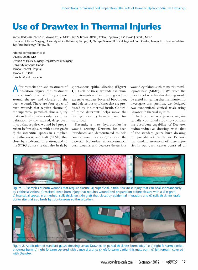
of a victim’s thermal injury centers around therapy and closure of the burn wound. There are four types of burn wounds that require closure: a) the superficial, partial-thickness injury that can heal spontaneously by epithe-lialization; b) the excised, deep burn injury that requires wound bed prepa-ration before closure with a skin graft; c) the interstitial spaces in a meshed split-thickness skin graft (STSG) that close by epidermal migration; and d) the STSG donor site that also heals by
spontaneous epithelialization (Figure 1).1 Each of these wounds has clini-cal deterrents to ideal healing such as excessive exudate, bacterial bioburden, and deleterious cytokines that are pro-duced by the thermal insult. Control of these deterrents helps move the healing trajectory from impaired to-ward ideal.
Recently, a new hydroconductive wound dressing, Drawtex, has been introduced and demonstrated to help control wound exudate, decrease the bacterial bioburden in experimental burn wounds, and decrease deleterious
wound cytokines such as matrix metal-loproteinase (MMP) 9.2 We raised the question of whether this dressing would be useful in treating thermal injuries. To investigate this question, we designed two randomized clinical trials using Drawtex in thermal injuries.
The first trial is a prospective, in-ternally controlled study to compare the absorbent capability of Drawtex hydroconductive dressing with that of the standard gauze burn dressing on partial-thickness burns. Because the standard treatment of these inju-ries in our burn center consisted of
Figure 1. Examples of burn wounds that require closure: a) superficial, partial-thickness injury that can heal spontaneously by epithelialization; b) excised, deep burn injury that requires wound bed preparation before closure with a skin graft; c) interstitial spaces in a meshed, split-thickness skin graft that closes by epidermal migration; and d) split-thickness graft donor site that also heals by spontaneous epithelialization.
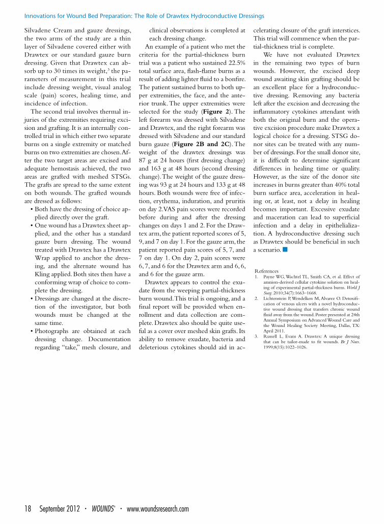
Figure 2. Application of standard gauze dressing versus Drawtex on partial-thickness burns (day 1): a) right forearm partial-thickness burn; b) right forearm covered with gauze dressing; c) left forearm partial-thickness burn; d) left forearm covered with Drawtex.
A
A
B
B
D
D
C
C
18 September 2012 • WOUNDS® • www.woundsresearch.com
Innovations for Wound Bed Preparation: The Role of Drawtex Hydroconductive Dressings
Silvadene Cream and gauze dressings, the two arms of the study are a thin layer of Silvadene covered either with Drawtex or our standard gauze burn dressing. Given that Drawtex can ab-sorb up to 30 times its weight,3 the pa-rameters of measurement in this trial include dressing weight, visual analog scale (pain) scores, healing time, and incidence of infection.
The second trial involves thermal in-juries of the extremities requiring exci-sion and grafting. It is an internally con-trolled trial in which either two separate burns on a single extremity or matched burns on two extremities are chosen. Af-ter the two target areas are excised and adequate hemostasis achieved, the two areas are grafted with meshed STSGs. The grafts are spread to the same extent on both wounds. The grafted wounds are dressed as follows:
• Both have the dressing of choice ap-plied directly over the graft.
• One wound has a Drawtex sheet ap-plied, and the other has a standard gauze burn dressing. The wound treated with Drawtex has a Drawtex Wrap applied to anchor the dress-ing, and the alternate wound has Kling applied. Both sites then have a conforming wrap of choice to com-plete the dressing.
• Dressings are changed at the discre-tion of the investigator, but both wounds must be changed at the same time.
• Photographs are obtained at each dressing change. Documentation regarding “take,” mesh closure, and
clinical observations is completed at each dressing change.
An example of a patient who met the criteria for the partial-thickness burn trial was a patient who sustained 22.5% total surface area, flash-flame burns as a result of adding lighter fluid to a bonfire. The patient sustained burns to both up-per extremities, the face, and the ante-rior trunk. The upper extremities were selected for the study (Figure 2). The left forearm was dressed with Silvadene and Drawtex, and the right forearm was dressed with Silvadene and our standard burn gauze (Figure 2B and 2C). The weight of the drawtex dressings was 87 g at 24 hours (first dressing change) and 163 g at 48 hours (second dressing change). The weight of the gauze dress-ing was 93 g at 24 hours and 133 g at 48 hours. Both wounds were free of infec-tion, erythema, induration, and pruritis on day 2. VAS pain scores were recorded before during and after the dressing changes on days 1 and 2. For the Draw-tex arm, the patient reported scores of 5, 9, and 7 on day 1. For the gauze arm, the patient reported pain scores of 5, 7, and 7 on day 1. On day 2, pain scores were 6, 7, and 6 for the Drawtex arm and 6, 6, and 6 for the gauze arm.
Drawtex appears to control the exu-date from the weeping partial-thickness burn wound. This trial is ongoing, and a final report will be provided when en-rollment and data collection are com-plete. Drawtex also should be quite use-ful as a cover over meshed skin grafts. Its ability to remove exudate, bacteria and deleterious cytokines should aid in ac-
celerating closure of the graft interstices. This trial will commence when the par-tial-thickness trial is complete.
We have not evaluated Drawtex in the remaining two types of burn wounds. However, the excised deep wound awaiting skin grafting should be an excellent place for a hydroconduc-tive dressing. Removing any bacteria left after the excision and decreasing the inflammatory cytokines attendant with both the original burn and the opera-tive excision procedure make Drawtex a logical choice for a dressing. STSG do-nor sites can be treated with any num-ber of dressings. For the small donor site, it is difficult to determine significant differences in healing time or quality. However, as the size of the donor site increases in burns greater than 40% total burn surface area, acceleration in heal-ing or, at least, not a delay in healing becomes important. Excessive exudate and maceration can lead to superficial infection and a delay in epithelializa-tion. A hydroconductive dressing such as Drawtex should be beneficial in such a scenario. n
References1. Payne WG, Wachtel TL, Smith CA, et al. Effect of
amnion-derived cellular cytokine solution on heal-ing of experimental partial-thickness burns. World J Surg. 2010;34(7):1663–1668.
2. Lichtenstein P, Wendelken M, Alvarez O. Detoxifi-cation of venous ulcers with a novel hydroconduc-tive wound dressing that transfers chronic wound fluid away from the wound. Poster presented at 24th Annual Symposium on Advanced Wound Care and the Wound Healing Society Meeting, Dallas, TX: April 2011.
3. Russell L, Evans A. Drawtex: A unique dressing that can be tailor-made to fit wounds. Br J Nurs. 1999;8(15):1022–1026.

www.woundsresearch.com • September 2012 • WOUNDS® 19
Innovations for Wound Bed Preparation: The Role of Drawtex Hydroconductive Dressings
Buruli Ulcer: Its Impact and Treatment Worldwide: An Interval ReportTerry Treadwell, MD, FACS1,2; John Macdonald, MD, FACS2,3
1Institute for Advanced Wound Care, Montgomery, AL; 2World Alliance of Wound and Lymphedema Care, Geneva, Switzerland; 3Wound Program, Hospital Bernard Mevs Project Medishare, Port-au-Prince, Haiti
Address correspondence toTerry Treadwell, MD, FACSInstitute for Advanced Wound Care2167 Normandie DriveMontgomery, AL [email protected]
Buruli ulcer, a devastating disease first described in 1897 by Sir Albert Cook
in Uganda, Africa, is caused by Mycobac-terium ulcerans and is seen in more than 30 mostly under-resourced, countries worldwide.1 More than 70% of the pa-tients affected are children younger than 16,2 with 90% of the ulcers manifested on the limbs.1 The ulceration caused by the microorganism is painless due to the cy-totoxic and immunosuppressive properties of the bacterial toxin mycolactone. Devel-opment of these ulcers is accompanied by marked edema of the affected extremity, and up to 15% of the skin surface can be involved in the ulcerative process.3
Unfortunately, these ulcerative lesions can become very large before treatment is sought because of lack of access to care, lack of funds, superstitious beliefs about the disease, and the stigma of the disease.4 Current treatment entails ad-ministration of two antibiotics (rifampin and streptomycin) for 8 weeks, followed by excision of the ulcerated area and skin grafting if the ulcer does not show signs of healing by secondary intention. Complications of the disease and its treatment can be seen in up to 24.5% of patients and can include amputation, joint contractures, and death.2
Historically, care for Buruli ulcers dur-ing the 8 weeks of antibiotic therapy
has been left to the standards of each healthcare facility treating a patient. This wound care would usually involve wash-ing the wound with water and/or acetic acid and applying Betadine-soaked gauze dressings. In addition, no formal debride-ment would be done, and no attempt would be made to address the edema of the extremity. If these wounds remained unhealed after 8 weeks of antibiotic ther-apy, they would be excised and treated with split-thickness skin grafting. In one series, this treatment required an average of 1.45 operations per patient, and 44% of the patients required blood transfusion at the time of operation.2
In an attempt to improve the healing of this devastating disease and to avoid some of the longstanding complications, a clinical trial using good, basic wound care techniques, dressings, and compres-sion therapy in conjunction with an-tibiotic therapy has been instituted in Ghana, Africa, under the auspices of the World Health Organization.
Methods and MaterialsThe goal to recruit and treat 20 pa-
tients has been undertaken. All patients are treated with rifampin and strepto-mycin for 8 weeks. Due to the need to provide moist wound healing and treatment of the edema, each patient
is treated with Vaseline gauze, Drawtex hydroconductive dressings, and short-stretch compression therapy. The Draw-tex dressing is used because of its superi-or wicking action, which moves wound fluid away from the wound surface, facilitating autolytic debridement of the wound. Short-stretch compression bandages are used to reduce the marked edema seen in the extremities of pa-tients with this disease. Clinic personnel change dressings three times per week. Wounds are measured, photographed, and evaluated weekly for 8 weeks.
ResultsTo date, eight of the 20 projected
patients have completed the study. Im-provement in the wound bed was noted in all patients (Table 1). The amounts of granulation tissue in the wound beds improved from 25% to 75% in two patients and from 25% to 100% in six patients. These improvements in the granulation tissue occurred along with reductions of necrotic tissue and slough in the wound beds and through autolyt-ic debridement facilitated by the Draw-tex hydroconductive dressing.
Drainage from these large wounds is always a problem. Treatment with the Drawtex hydroconductive dressing had the following results: one patient’s
Table 1. Results of Buruli ulcer therapy with Drawtex and short-stretch compression therapy.
Change in %Granulation Tissue
< 25% to 75%:2
< 25% to 100%:6
Change in Drainage Large to Medium:1
Large to Minimal:4
Unchanged:3
Change in Wound Size Increased:3
Decreased:5
20 September 2012 • WOUNDS® • www.woundsresearch.com
Innovations for Wound Bed Preparation: The Role of Drawtex Hydroconductive Dressings
wound drainage decreased from “large” to “medium”; four patients’ drainages decreased from “large” to “minimal”; three patients’ drainages remained un-changed despite improvements in wound bed granulation tissue responses.
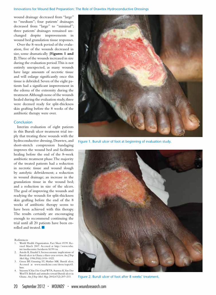
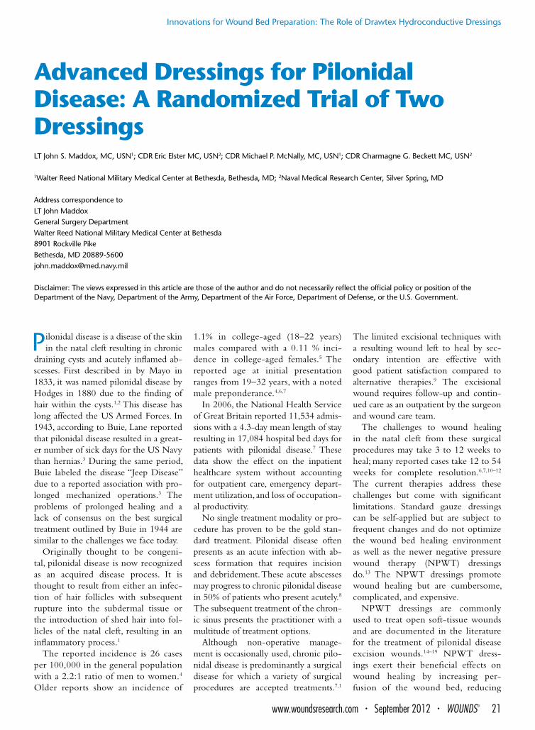
Over the 8-week period of the evalu-ation, five of the wounds decreased in size, some dramatically (Figures 1 and 2). Three of the wounds increased in size during the evaluation period. This is not entirely unexpected, as many wounds have large amounts of necrotic tissue and will enlarge significantly once this tissue is debrided. Seven of the eight pa-tients had a significant improvement in the edema of the extremity during the treatment. Although none of the wounds healed during the evaluation study, three were deemed ready for split-thickness skin grafting before the 8 weeks of the antibiotic therapy were over.
ConclusionInterim evaluation of eight patients
in this Buruli ulcer treatment trial im-ply that treating these wounds with the hydroconductive dressing, Drawtex, and short-stretch compression bandaging improves the wound bed and facilitates healing before the end of the 8-week antibiotic treatment phase. The majority of the treated patients had a reduction in necrotic tissue and wound slough by autolytic debridement; a reduction in wound drainage; an increase in the granulation tissue in the wound bed; and a reduction in size of the ulcers. The goal of improving the wounds and readying the wounds for split-thickness skin grafting before the end of the 8 weeks of antibiotic therapy seems to have been achieved with this therapy. The results certainly are encouraging enough to recommend continuing the trial until all 20 patients have been en-rolled and treated. n
References1. World Health Organization. Fact Sheet #199. Re-
vised March 2007. Accessed at http://www.who.int/mediacentre/factsheets/fs199/en
2. Asiedu K, Etuaful S. Socioeconomic implications of Buruli ulcer in Ghana: a three-year review. Am J Trop Med Hyg. 1996;59(6):1016–1022.
3. Green BP, Gunning ST, Mather MK. Buruli ulcer. Accessed at www.emedicine.com/derm/topic65.htm.
4. Stienstra Y, Van Der Graaf WTA, Asamoa K, Van Der Werf TS. Beliefs and attitudes toward Buruli ulcer in Ghana. Am J Trop Med. Hyg. 2002;67(2):207–213.
Figure 1. Buruli ulcer of foot at beginning of evaluation study.
Figure 2. Buruli ulcer of foot after 8 weeks’ treatment.
www.woundsresearch.com • September 2012 • WOUNDS® 21
Innovations for Wound Bed Preparation: The Role of Drawtex Hydroconductive Dressings
Advanced Dressings for Pilonidal Disease: A Randomized Trial of Two DressingsLT John S. Maddox, MC, USN1; CDR Eric Elster MC, USN2; CDR Michael P. McNally, MC, USN1; CDR Charmagne G. Beckett MC, USN2
1Walter Reed National Military Medical Center at Bethesda, Bethesda, MD; 2Naval Medical Research Center, Silver Spring, MD
Address correspondence toLT John MaddoxGeneral Surgery DepartmentWalter Reed National Military Medical Center at Bethesda8901 Rockville PikeBethesda, MD [email protected]
Disclaimer: The views expressed in this article are those of the author and do not necessarily reflect the official policy or position of the Department of the Navy, Department of the Army, Department of the Air Force, Department of Defense, or the U.S. Government.
Pilonidal disease is a disease of the skin in the natal cleft resulting in chronic
draining cysts and acutely inflamed ab-scesses. First described in by Mayo in 1833, it was named pilonidal disease by Hodges in 1880 due to the finding of hair within the cysts.1,2 This disease has long affected the US Armed Forces. In 1943, according to Buie, Lane reported that pilonidal disease resulted in a great-er number of sick days for the US Navy than hernias.3 During the same period, Buie labeled the disease “Jeep Disease” due to a reported association with pro-longed mechanized operations.3 The problems of prolonged healing and a lack of consensus on the best surgical treatment outlined by Buie in 1944 are similar to the challenges we face today.
Originally thought to be congeni-tal, pilonidal disease is now recognized as an acquired disease process. It is thought to result from either an infec-tion of hair follicles with subsequent rupture into the subdermal tissue or the introduction of shed hair into fol-licles of the natal cleft, resulting in an inflammatory process.1
The reported incidence is 26 cases per 100,000 in the general population with a 2.2:1 ratio of men to women.4 Older reports show an incidence of
1.1% in college-aged (18–22 years) males compared with a 0.11 % inci-dence in college-aged females.5 The reported age at initial presentation ranges from 19–32 years, with a noted male preponderance.4,6,7
In 2006, the National Health Service of Great Britain reported 11,534 admis-sions with a 4.3-day mean length of stay resulting in 17,084 hospital bed days for patients with pilonidal disease.7 These data show the effect on the inpatient healthcare system without accounting for outpatient care, emergency depart-ment utilization, and loss of occupation-al productivity.
No single treatment modality or pro-cedure has proven to be the gold stan-dard treatment. Pilonidal disease often presents as an acute infection with ab-scess formation that requires incision and debridement. These acute abscesses may progress to chronic pilonidal disease in 50% of patients who present acutely.8 The subsequent treatment of the chron-ic sinus presents the practitioner with a multitude of treatment options.
Although non-operative manage-ment is occasionally used, chronic pilo-nidal disease is predominantly a surgical disease for which a variety of surgical procedures are accepted treatments.7,1
The limited excisional techniques with a resulting wound left to heal by sec-ondary intention are effective with good patient satisfaction compared to alternative therapies.9 The excisional wound requires follow-up and contin-ued care as an outpatient by the surgeon and wound care team.
The challenges to wound healing in the natal cleft from these surgical procedures may take 3 to 12 weeks to heal; many reported cases take 12 to 54 weeks for complete resolution.6,7,10–12 The current therapies address these challenges but come with significant limitations. Standard gauze dressings can be self-applied but are subject to frequent changes and do not optimize the wound bed healing environment as well as the newer negative pressure wound therapy (NPWT) dressings do.13 The NPWT dressings promote wound healing but are cumbersome, complicated, and expensive.
NPWT dressings are commonly used to treat open soft-tissue wounds and are documented in the literature for the treatment of pilonidal disease excision wounds.14–19 NPWT dress-ings exert their beneficial effects on wound healing by increasing per-fusion of the wound bed, reducing

22 September 2012 • WOUNDS® • www.woundsresearch.com
Innovations for Wound Bed Preparation: The Role of Drawtex Hydroconductive Dressings
edema, and modulation of the wound biomarkers.20 They also produce me-chanical stress on the wound and sur-rounding tissue and decrease the bio-burden of the wound.20 Due to the complex nature of these dressings, dressing malfunctions, and loss of seal these dressings can require specialized nursing attention. The cost per dress-ing is significant, but less than standard gauze dressings due to the longer time between dressing changes.
Drawtex® is a new wound dressing technology that is designed to man-age exudative wounds and provide an appropriate wound moisture balance. This dressing is increasingly used for the treatment of open wounds with a reported effectiveness similar to that of a variety of dressings including alginate, hydrogel, hydrocellular foam, or hydro-phobic foam.21 Drawtex is engineered to disperse exudative fluid while main-taining the temperature and moisture balance at the wound surface.22
Control of exudative drainage is im-portant for biologic wound healing and for patient compliance. Excess moisture in the wound bed will result in macer-ated tissues at the wound edges, which can lead to slower wound healing,23
whereas a desiccated wound leads to slower wound healing and promotes eschar formation with resultant impedi-ment to full epithelialization.
The Drawtex® dressing is reported to achieve this important wound mois-ture balance while being able to handle the occasionally high volume of exu-dates produced by pilonidal cystectomy wounds. Additionally, this dressing does not require the cumbersome external vacuum pump and canister that are in-tegral to the NPWT dressing.
Balanced wound biomarkers and low bioburden in the wound bed are important for healing pilonidal exci-sion wounds by secondary intention. The initial inflammatory phase serves to promote proteolytic and fibrinolyt-ic factors, which provide for autolytic debridement of the wound bed. This phase progresses into the regenerative and proliferative phase with the expres-sion of growth factors and concomitant
increase in fibroblasts, keratinocytes, and endothelial cells.23
Studies of NPWT dressings have shown a decrease in the inflammatory mediators, up-regulated growth factor expression,24,25 and a reduction of the bioburden within infected wounds.20 This effect may be due to the fluid handling mechanism of the dressing. The effect of Drawtex® on the balance of biomarkers and bioburden within open wounds healing by secondary in-tention has not yet been reported. We believe that the fluid-handling capa-bilities and debridement action of the Drawtex® dressing will exert a similar effect to that of NPWT dressings on the biomarkers of the healing wound bed and result in a similar reduction in bioburden, both of which will increase healing rates and potentially prevent conversion from an acute wound to a chronic wound.
If the Drawtex® dressing dem-onstrates the same wound healing properties as the NPWT dressing, it may provide improved patient satis-faction and lower overall cost to the health care system. Our current pi-lot study is a prospective, randomized open label trial comparing the use of Drawtex® Hydroconductive Wound Dressing to the NPWT dressing in the setting of a pilonidal cystectomy excision wound healing by second-ary intention. As a primary endpoint, we are comparing the time to heal-ing using the Drawtex® dressing ver-sus the time to healing achieved with the use of the current standard of care NPWT dressings. Using digital pla-nimetry, we will track the change in the size of the wound leading up to 100% epithelialization. We will evalu-ate the effects of both dressings on wound healing biomarkers and bac-terial burden within the wound as secondary endpoints. n
References1. Humphries AE, Duncan JE. Evaluation and man-
agement of pilonidal disease. Surg Clin North Am. 2010;90(1):113–124, Table of Contents.
2. Velasco AL, Dunlap WW. Pilonidal disease and hi-dradenitis. Surg Clin North Am. 2009;89(3):689–701.
3. Buie LA. Jeep disease: pilonidal disease of mecha-
nized warfare. Southern Med J. 1944;37(2):103–108.4. Al-Khamis A, McCallum I, King PM, Bruce J. Heal-
ing by primary versus secondary intention after sur-gical treatment for pilonidal sinus. Cochrane Database Syst Rev. 2010 Jan 20;(1):CD006213.
5. Spivak H, Brooks VL, Nussbaum M, Friedman I. Treatment of chronic pilonidal disease. Dis Colon Rectum. 1996;39(10):1136–1139.
6. Solla JA, Rothenberger DA. Chronic pilonidal dis-ease. An assessment of 150 cases. Dis Colon Rectum. 1990;33(9):758–761.
7. Chintapatla S, Safarani N, Kumar S, Haboubi N. Sacrococcygeal pilonidal sinus: historical review, pathological insight and surgical options. Tech Co-loproctol. 2003;7(1):3–8.
8. Søndenaa K, Andersen E, Nesvik I, Søreide JA. Patient characteristics and symptoms in chron-ic pilonidal sinus disease. Int J Colorectal Dis. 1995;10(1):39–42.
9. Mohamed HA, Kadry I, Adly S. Comparison between three therapeutic modalities for non-complicated pi-lonidal sinus disease. Surgeon. 2005;3(2):73–77.
10. Bradley L. Pilonidal sinus disease: a review. Part one. J Wound Care. 2010;19():504–508.
11. Harris CL, Holloway S. Development of an evidence-based protocol for care of pilonidal sinus wounds healing by secondary intent us-ing a modified reactive Delphi procedure. Part one: the literature review(*). Int Wound J. 2012;9(2):156–172.
12. Marks J, Harding KG, Hughes LE, Ribeiro CD. Pi-lonidal sinus excision — healing by open granula-tion. Br J Surg. 1985;72(8):637–640.
13. Woo K, Ayello EA, Sibbald RG. The edge effect: current therapeutic options to advance the wound edge. Adv Skin Wound Care. 2007;20(2):99–117;quiz 8–9.
14. Farrell D, Murphy S. Negative pressure wound therapy for recurrent pilonidal disease: a review of the literature. J Wound Ostomy Continence Nurs. 2011;38(4):373–378.
15. McGuinness JG, Winter DC, O’Connell PR. Vacu-um-assisted closure of a complex pilonidal sinus. Dis Colon Rectum. 2003;46(2):274–276.
16. Lynch JB, Laing AJ, Regan PJ. Vacuum-assisted clo-sure therapy: a new treatment option for recurrent pilonidal sinus disease. Report of three cases. Dis Colon Rectum. 2004;47(6):929–932.
17. Caniano DA, Ruth B, Teich S. Wound management with vacuum-assisted closure: experience in 51 pe-diatric patients. J Pediatr Surg. 2005;40(1):128–132.
18. Bendewald, FP, Cima RR, Metcalf DR, Hassan I. Using negative pressure wound therapy following surgery for complex pilonidal disease: a case series. Ostomy Wound Manage. 2007;53(5):40–46.
19. Saad SA, Shakov E, Sebastian V, Saad A. The use of wound vacuum-assisted closure system in the treat-ment of recurrent or complex pilonidal cyst disease: experience in 4 adolescent patients. Internet J Surg. 2007;11(1):1–4
20. Banwell PE, Musgrave M. Topical negative pressure therapy: mechanisms and indications. Int Wound J. 2004;1(2):95–106.
21. Reynolds T, Russell L, Deeth M, et al. A randomised controlled trial comparing Drawtex with stan-dard dressings for exuding wounds. J Wound Care. 2004;13(2):71–74.
22. Russell L, Evans A. Drawtex: a unique dressing that can be tailor-made to fit wounds. Br J Nurs. 1999;8(15):1022–1026.
23. Okan D, Woo K, Ayello EA, Sibbald G. The role of moisture balance in wound healing. Adv Skin Wound Care. 2007;20(1):39–53.
24. Kopp J, Kneser U, Bach AD, Horch RE. Buried chip skin grafting in neuropathic diabetic foot ulcers fol-lowing vacuum-assisted wound bed preparation: enhancing a classic surgical tool with novel technol-ogies. Int J Low Extrem Wounds. 2004;3(3):168–171.
25. Coerper S, Wagner S, Witte M, Schäffer M, Becker HD. [Temporary expression pattern in wound se-cretions and peripheral wound biopsies]. Zentralbl Chir. 1999;124(Suppl 1):78–80.

www.woundsresearch.com • September 2012 • WOUNDS® 23
Innovations for Wound Bed Preparation: The Role of Drawtex Hydroconductive Dressings
Roundtable Discussion: The Role of Drawtex Hydroconductive DressingsParticipants:Martin C. Robson, MD (Moderator)Terry Treadwell, MDRandall D. Wolcott, MDWyatt G. Payne, MDTom Wolvos, MS, MDMarion H. Jordan, MD, FACSCdr. Eric Elster, MC, USNDavid J. Smith, MD
Dr. Robson: After the questions, we will discuss your ideas for poten-tial new uses for Drawtex that have not been discussed today. I have a question for Dr. Treadwell. If you use compression alone, do you decrease the bacterial count in the wound or, if you use Drawtex alone, do you need compression?
Dr. Treadwell: Compression thera-py will help you with bacteria in the wound and in the subcutaneous tissue because high-protein fluid serves as a nutrient source for all the bacteria. By getting the high-protein fluid out of the extremity and the wound bed, you can reduce the bacteria’s ability to survive. There is nothing in compres-sion therapy alone that will actually kill bacteria, but decreasing the edema increases the arterial capillary inflow to the wound bed, thus increasing the antibiotic concentration in the tissue. Using Drawtex with compression takes care of the surface bacteria and reduces the bioburden. Therefore, it is a combi-nation therapy.
Dr. Robson: Dr. Wolcott, is the concept that compression can decrease bacteria the same that you discuss in saying that compression decreases the nutrient source for bacteria?
Dr. Wolcott: That is a working hy-pothesis. What we see clinically is, if there is edema fluid coming out through the wound, there is more slough and a more robust bioburden. We believe that local edema has a nu-
trient effect on the biofilm, plus, it di-lutes out the host factors that would counteract the biofilm.
Dr. Robson: Dr. Payne, with all the data that Drawtex can decrease the bacterial count in the tissue, could there be a randomized trial in which, when you had a high bacterial count, you used topical antibacterials in one arm and Drawtex in the other arm with no antimicrobial? Could such a
trial be approved by an institutional re-view board (IRB)?
Dr. Payne: I believe so, if you use some of the data presented in this sym-posium. However, since some have used Drawtex in combination with Sil-vadene, I think there should be a third arm of the study.
Dr. Robson: Dr. Wolvos, could you get such a clinical trial through your IRB?
Dr. Wolvos: It sounds like a good study with the three arms, and I believe it would be approved.
Dr. Jordan: I believe we could get such a study approved to study Mepi-
tel and Drawtex versus silver-loaded Mepilex and Drawtex on partial-thickness burns.
Dr. Robson: Dr. Elster, do you have
a question? Dr. Elster: Dr. Wolcott, regarding
your polymerase chain reaction (PCR) data, the markers can be ubiquitous and present in both live and dead bacteria. Do you think that played a role in your results where you did not see a decrease
in the bacterial load? Possibly, you were recording both live and dead bacte-ria. Secondly, when you showed your pathogen-specific PCR, you discussed diversity, but truly how much diversity was there?
Dr. Wolcott: In our studies, if one sees DNA, that bacteria was alive up to at least 24 hours before the sampling. Therefore, we assume it is alive. As for diversity, one of the wounds had over 20 organisms at 1% or less contribu-tion to the community. We consider these as contaminants or transients. So when over 1% is present, quantitative measures equate to 106–108 bacteria. So
By getting the high-protein fluid out of the extremity and the wound bed, you can reduce the bacteria’s ability to survive.
Terry Treadwell, MD

24 September 2012 • WOUNDS® • www.woundsresearch.com
Innovations for Wound Bed Preparation: The Role of Drawtex Hydroconductive Dressings
1% is significant. Therefore, you cannot dismiss that diversity, because it is pret-ty robust. The least amount of diversity was five organisms, demonstrating the bioburden in a venous leg ulcer (VLU) is highly polymicrobial.
Dr. Elster: And that diversity re-mained despite treatment?
Dr. Wolcott: That is correct. The di-versity did not collapse at all, and the species did not change. When we use our topical antimicrobials, the diversity does not change, but the species do. Their usage really changes the quantity of the different constituents. However, we have not seen this change of species with the use of Drawtex.
Dr. Robson: Dr. Wolcott, we previ-ously demonstrated with tissue biop-sies that predominantly a single species achieved a significant tissue level of bac-teria. Dr. Elster’s group recently showed again, in combat wounds, that a single species predominated at significant tis-sue levels. So with the diversity you are
demonstrating on the wound surface, if they do not achieve tissue levels, how do you know which ones are deleteri-ous to healing and which ones let your wounds heal even though you did not eradicate them?
Dr. Wolcott: We’ve shown that the vast majority of the wounds we see have highly diverse bacterial popula-tions, and there are minor colonies. The question is what you do with those minor colonies. We attempt to group the major organisms and treat the major groups with antimicrobials or antiseptics, and just do surveillance on the minor colonies. We try to man-age the biobuden with multiple con-
current therapies because biofilms are tremendously robust. If you put any selective stress on them, because they are polymicrobial, they can morph into whatever is successful in that niche. So if you put just Drawtex on the wound, the bacteria will find a way around it. If you use just antimicrobials in the wound, the bacteria will similarly find a way around the antimicrobials. There-fore, the way I think about Drawtex is to combine it with Silvadene or anoth-er topical antimicrobial.
Dr. Robson: Dr. Elster, do you have another question?
Dr. Elster: Yes. Data were presented that showed a decrease in matrix me-talloproteinase (MMP) levels in the wound itself and absorption by the dressing. Is that a direct effect or just a surrogate marker for either the antibac-terial or the wound modulation effects of the Drawtex?
Dr. Robson: I believe it is a direct effect. Drawtex is actually drawing off
the cytokines because you can measure their decrease in the wound increasing in the Drawtex dressing.
Dr. Elster: By measuring the MMPs, are we just measuring the effect of the dressing either directly on the wound environment, directly on the bacteria, or the combination of both?
Dr. Robson: In a recent paper, MMP levels were shown to be related to the degree of healing in a series of patients. Dr. Payne showed in a few of his patients, their MMP lev-els decreased and, in others, the levels stayed the same. Dr. Payne, you did not show all your data about wound sizes. Did some of your patients’
wounds remain the same size or actu-ally increase in size?
Dr. Payne: Yes, that is correct.Dr. Robson: In those patients, did the
MMP-9s stay level across the 28 days? Dr. Payne: That’s right.Dr. Elster: These data raise the ques-
tion whether you could put some type of chemometric area into the dressing itself, so one could distinguish between responders and non-responders.
Dr. Robson: As you know, there is a diagnostic tool to measure MMP-9 levels that became available recently and can be used diagnostically in pro-spective studies.
Dr. Robson: Dr. Wolvos, Do you have a question?
Dr. Wolvos: Several people have mentioned their study protocols ex-clude people who have a known al-lergy to components in Drawtex. Are there any known allergic reactions to the materials of Drawtex?
Dr. Robson: None have been re-ported to date. Dr. Jordan, do you have a question?
Dr. Jordan: Dr. Smith showed the several kinds of burn wounds, includ-ing a mesh graft, and I believe Dr. Treadwell was talking about the donor-site coverage. Can either of you tell me if you have observed anything regard-ing what Drawtex does to red blood cells that ooze out under split grafts, mesh grafts, or the donor site?
Dr. Treadwell: In my experience, red blood cells have been drawn into the Drawtex, so it is not forming a he-matoma beneath the dressing.
Dr. Payne: In our study patients, in the course of changing the wound dressings, a little bleeding occurs and the blood does seem to be drawn into the dressing.
Dr. Robson: Dr. Jordan, in your study, the serum albumin was drawn into the Drawtex. Do you think the blood would be equally drawn, or do you think the red cell would be different?
Dr. Jordan: I think we are talking about rheology here. On the one hand, you have the albumin, which is in sus-pension and is a fairly sizeable molecule, but the red cell has got size and mass and probably stuck with fibrin as well.
Data were presented that showed a decrease in matrix metalloproteinase levels in the wound itself and absorption by the dressing. Is that a direct effect or just a surrogate for either the antibacterial or the wound modulation effects of the Drawtex?
CDR Eric Elster, MC, USN

www.woundsresearch.com • September 2012 • WOUNDS® 25
Innovations for Wound Bed Preparation: The Role of Drawtex Hydroconductive Dressings
Dr. Robson: One thing that could be done pretty easily in the operating room is applying Drawtex to a bleed-ing surface and seeing what happens.
Dr. Wolvos: I had a patient who developed fairly brisk post-operative bleeding. I applied Drawtex to the wound and it was good at absorbing the blood, but not actually stopping the bleeding. The Drawtex did hold a fair volume of blood.
Dr. Robson: Dr. Smith, did you have a question?
Dr. Smith: Dr. Treadwell, when you use Drawtex to dress donor sites, do you use the Drawtex in isolation?
Dr. Treadwell: No. As I mentioned, we do put a non-adherent dressing ad-jacent to the donor site. We use Xero-form gauze, Vaseline gauze, or Mepitel. Then we apply the Drawtex. If we use Drawtex alone, it will become adher-ent. Generally on the donor sites, we change the dressing in a week and it is usually healed
Dr. Robson: Although a specific Drawtex donor-site dressing is not yet available, people around the country are using Drawtex on donor sites. I have suggested they use either a non-adherent layer under the Drawtex or, if they are going to put it directly on the wound, they allow it to remain in place until the wound heals. I believe if you try to take it off any time before it is totally epithelialized, you are go-ing to remove the epithelium with it. When queried, the panel thought that a non-adherent variant of Drawtex would be beneficial.
Dr. Smith: Dr. Wolvos, did I un-derstand that some of the wounds you presented were dry when you put the Drawtex on?
Dr. Wolvos: That is correct. I think the majority of the wounds I have treated were dry, and Drawtex still worked very effectively. In those dry wounds, I use Sil-vadene, and then the Drawtex dressing.
Dr. Smith: If we believe it is the exudate that Drawtex is removing, how do you hypothesize it is working in dry wounds?
Dr. Wolvos: I think it may be the collagen that helps keep the slough and necrotic tissue in the wound. By help-
ing break that up, even in a dry wound, Drawtex acts to debride the wound.
Dr. Robson: If there are no other questions, I would like to move forward by discussing the potential new uses for Drawtex that have not been discussed.
Dr. Payne, you showed patients with diabetic foot ulcers and VLUs. Do you think Drawtex will also be of use in pressure ulcers (PUs)?
Dr. Payne: Quite possibly. It might be interesting to use a combination of some topical antimicrobial with the Drawtex because of the high biobur-den in pressure ulcers.
Dr. Robson: Would you use the stacked Drawtex in a deep wound, like a PU, versus your more superficial wounds?
Dr. Payne: I think I would.Dr. Robson: Has anybody used
Drawtex in PUs?Dr. Wolcott: Yes. It has been very ef-
fective for cavernous wounds because
if you use alginates or things without structure, they fall to the bottom of the wound. However, if you can pack the ulcer with the Drawtex on top of an an-timicrobial gel, it holds its shape. I like Drawtex’s property of physically staying out over the wound and not bunching up like the alginates do.
Dr. Robson: If you have sinuses at the bottom of a PU, do you cut the Drawtex into ribbons and pack it?
Dr. Wolcott: Yes, but we try to open all sinuses during the first few debridements.
Dr. Robson: Drs. Smith and Jor-dan both care for burn wounds. How about traumatic wounds like road rash, which although not thermal, certainly
present similar wounds? Dr. Jordan, do you think Drawtex could be used to remove the cinders and trash left in af-ter a road rash injury?
Dr. Jordan: Well, I think it would be an interesting thing to try. We take those patients to the operating room and put them to sleep so we can radi-cally remove all the debris from the wound. We have made headway us-ing the pulse irrigator as an adjunct to sharp debridement. I do believe the particulate matter could be drawn up into Drawtex. I think it could be tested in a laboratory model to see how much of the grit and black particles could be drawn up by the Drawtex. That might
decrease cellulitis, which sometimes occurs at day 2 or 3 with these injuries.
Dr. Smith: Our patients who have road rash usually have multiple injuries. We see the patients later when it is dif-ficult to remove the road rash debris. Having a dressing like Drawtex that could be applied while the other inju-ries are being attended to would be a nice way to temporize before you had to move to the operating room.
I do believe the particulate matter could be drawn up into the Drawtex. I think it could be tested in a laboratory model to see how much of the grit and black particles could be drawn up by the Drawtex. Marion H. Jordan, MD, FACS
Having a dressing like Drawtex that could be applied while the other injuries are being attended to would be a nice way to temporize before you had to move to the operating room.
David J. Smith, MD

26 September 2012 • WOUNDS® • www.woundsresearch.com
Innovations for Wound Bed Preparation: The Role of Drawtex Hydroconductive Dressings
Dr. Robson: Dr. Elster, is there a role for Drawtex as a holding mecha-nism in combat wounds?
Dr. Elster: Sure. I think the study we are going to do with pilonidal dis-ease will demonstrate effectiveness of Drawtex versus negative pressure wound therapy (NPWT). NPWT is our current standard of care for these acute combat wounds. If Drawtex is equally effective, the advantages are ob-
vious. One of the disadvantages: We are using the NPWT cannisters as a way to sample the local wound environ-ment by collecting the effluent. The inability to sample local wound envi-ronment may be a disadvantage for us using Drawtex. However, if one is not going to evaluate the effluent, then a lot of Drawtex’s properties would seem to be a nice fit for wound treatment, both combat and non-combat.
Dr. Robson: So you are doing bio-markers on the effluent that reach the vacuum-assisted closure reservoir?
Dr. Elster: That is correct. By sam-pling systemic and local wound bio-markers, we feel that we get a really good read on systemic response and local responses.
Dr. Wolvos: And what exactly are you looking for?
Dr. Elster: We have pared this down to cytokines and chemokines. They are measured from both serum and wound effluent. We are able to make timing decisions regarding wound closure re-gardless of the sampling times.
Dr. Robson: Their retrospective
and prospective studies have been pub-lished and are pretty spectacular using the biomarkers to predict the success in their wound closure. Dr. Treadwell, we do not have a lot of Buruli ulcers in the United States, but we do have sickle cell ulcers. Can you give us your
opinion as to whether Drawtex will work in sickle cell ulcers and whether it would be worthwhile investigating?
Dr. Treadwell: I think sickle cell ul-cers should be one of the next things we study. There are a great many sickle cell patients in Haiti, so we are begin-ning a Drawtex trial there. Eventually the trial could be repeated in the United States. A dressing that would painlessly allow the debridement and removal of
the bacterial bioburden should certainly benefit patients and, hopefully, would allow the wound microenvironment to be more in a healing mode rather than a hostile environment. We have had suc-cess increasing the microcirculation in the wound beds of patients with sickle cell ulcers, which seems to improve the healing. Adding that to the use of Draw-tex would be very interesting.
Dr. Robson: Dr. Wolvos, you showed one case with the abdominal fistulae. Have you used Drawtex in the open abdomen, where most people now are using NPWT?
Dr. Wolvos: I have not used it. I would be very hesitant unless there was a non-adherent layer over the in-testines. Drawtex alone may result in a higher chance of fistula formation.
If you were to use Drawtex to absorb the fluid from the open abdomen, you would have to really be sure that the Drawtex did not come in contact with the bowel.
Dr. Robson: If you were doing a de-layed primary closure after fascia approx-imation, would you use Drawtex there?
Dr. Wolvos: Sure. Typically my prac-tice is to use NPWT, which also helps to contract the wound. Drawtex would not do that.
Dr. Robson: Drs. Jordan and Smith,
do you think toxic epidermal necrolysis would be an indication for Drawtex?
Dr. Jordan: Our internists are man-aging these patients with immunoglob-ulins, which has made a huge difference in the natural course of the disease. We consult on these patients for wound management and are using one of the silver membrane materials. It tends to stick to the wounds, causing significant pain. I think a non-adherent Draw-tex would be of benefit. If we had big sheets of something that would absorb fluid, reduce bacteria, and not stick to those healing dermal surfaces, it should be quite beneficial.
Dr. Smith: I think if you put Draw-tex on in the current form that the adherence would be a major problem. A non-adherent Drawtex has a lot of potential in that type of wound.
Dr. Robson: Dr. Smith, how about Drawtex as a dressing for dermabrasion?
Dr. Smith: I think it goes back to the answer on the donor sites. If the blood is nicely absorbed the way Dr. Treadwell said, I think it could be a very nice dressing for dermabrasion. I
It might be interesting to use a combination of some topical antimicrobial with the Drawtex because of the high bioburden in pressure ulcers.
Wyatt G. Payne, MD
If you use Drawtex to absorb the fluid from the open abdomen, you would have to be really sure that the Drawtex did not come in contact with the bowel.
Tom Wolvos, MS, MD

www.woundsresearch.com • September 2012 • WOUNDS® 27
Innovations for Wound Bed Preparation: The Role of Drawtex Hydroconductive Dressings
just have no experience using Drawtex in that particular kind of wound.
Dr. Treadwell: I think adherence would also be another issue on the face.
Dr. Robson: You would definitely have to have a non-adherent Drawtex. We have heard a lot of clinicians say a non-adherent type of Drawtex would be very useful in their practices. With no non-adherent Drawtex currently avail-able, we need data as to what interface dressing is best to use with Drawtex.
Dr. Wolvos: And the question is whether the non-adherent layer is go-ing to affect the degree of absorption of the hydroconductive dressing.
Dr. Robson: That is correct. Obvi-ously, if Drawtex does not draw off the deterrents to wound healing that have been demonstrated, it would not be very helpful at all. If a Vaseline gauze or Xeroform gauze is used as the non-adherent layer, then the excess oint-ment must be rubbed out. A perforated Telfa-type layer or Mepitel have been reported to work with Drawtex.
Dr. Treadwell: When we use Xero-form gauze or Adaptic, we use no more than two layers, two thicknesses, and we do wipe the excess off.
Dr. Robson: I have always removed the excess of Xeroform or Scarlet Red, so one could see through all the inter-stices. The Xeroform was only there as a wetting agent for the gauze. If this is not done, I doubt that Drawtex can be ef-fective at removing deterrents to healing.
Dr. Robson: Does anybody have any other examples of conditions for which we should try Drawtex as a treatment or study its possible effectiveness?
Dr. Smith: We not infrequently get consulted on calciphylaxis wounds. I think having something like Drawtex for that type of wound would be a dis-tinct advantage.
Dr. Robson: Dr. Treadwell, is there a role for Drawtex in the treatment of pyoderma gangrenosum?
Dr. Treadwell: We have treated one patient with pyoderma gangrenosum with Drawtex. However, if you don’t treat the underlying disease, it will not be successful. If you satisfactorily treat the underlying disease, leaving only the wound to treat, then Drawtex works
fine. Once one gets the underlying dis-ease under control with steroids or im-mune modulators, the inflammatory ex-udate is well controlled with Drawtex.
Dr. Robson: I assume the same would be true for any kind of vasculitis. Once the underlying disease is treated, the wound can be treated with Drawtex.
Dr. Elster: Another thought is wound failure after renal transplantation. Those are typically right or left lower-quadrant incisions. When they do fail, they be-
come chronic wounds. Sometimes there is a lymphocele component, so a dress-ing like Drawtex with a great amount of absorption would aid in getting the patients out of the hospital.
Dr. Robson: Do you have enough of those patients to do a real study?
Dr. Elster: A couple of centers in Baltimore, like the Johns Hopkins Hospital or the University of Mary-land, have many of these patients get-ting NPWT, waiting for these wounds to close. If Drawtex could get those patients out of the hospital and reduce their infection risks, I think that would be very valuable.
Dr. Robson: The present armed conflict has resulted in many traumatic amputations and many of those do not heal per primum. Presently there is a study underway at Walter Reed Medi-cal Center evaluating the use of Draw-tex on these wounds. Another disease for consideration of Drawtex treatment is Fournier’s gangrene. Once the radi-cal debridement has been performed, is there a place for the use of Drawtex? It is a difficult place to attempt NPWT. Dr. Elster, do you see patients with necrotizing fasciitis?
Dr. Elster: Yes. We have seen in-creasing fungal infections in the dia-betic population. We also see them in the combat wounds. In our patients
with the high double amputations with destroyed pelvises and open perinea, the wounds require multiple debride-ments to prevent necrotizing fasciitis before you can perform some type of coverage. The only question I would have about a dressing like Drawtex in those wounds is that NPWT has been a very useful device to get a dressing on these complex, three-dimensional wounds. Some of these wounds will track posteriorly up into the buttocks,
with tunnels that can track into the retroperitoneum. Experience in us-ing the Drawtex in that situation that would be interesting.
Dr. Jordan: Is Drawtex radio-opaque?Dr. Wolvos: I don’t think so.Dr. Jordan: If we are considering
packing under a wound edge, but cer-tainly within the retroperitoneum, I think we need to consider a radio-opaque strip on the Drawtex and develop some assur-ance that the dressing will not fragment or pull apart during retrieval.
Dr. Robson: Drawtex does come in a roll, and I would certainly think if you’re going to use it in those kinds of wounds, you would want to use the roll and not multiple individual dressings.
Dr. Robson: We have probably raised more questions than we have provided answers. One of the things we have been trying to do for the hydroconductive dressing Drawtex is develop hard data wound care professionals can rely on. If answers to the types of questions posed today can be obtained, then the data can be used to develop a second and third generation of the dressing. If there are no other questions and no other potential uses for Drawtex to discuss, I would like to personally thank all of you for all of work you have done and for your partici-pation in this symposium. n
One of the things we have been trying to do for the hydroconductive dressing Drawtex is develop hard data wound care professionals can rely on.
Martin C. Robson, MD (Moderator)

Drawtex®, with its hydroconductive action, lifts and moves exudate, slough and debris away from the wound surface. Clinical results have shown it to decrease wound exudate, tissue bacterial levels, nutrients for biofi lm production and deleterious cytokine levels such as matrix metalloproteinases (MMP-9).1,2
Based on these actions, Drawtex® facilitates effective wound bed preparation and serves as a possible alternative to passive absorptive products, like calcium alginates, hydrofi bers, foams and super absorbers.3 In addition, at times it can replace some enzymatic, antimicrobial and negative-pressure wound therapy (NPWT).4
Advancing the science of wound bed preparation. Drawtex® draws out:
Drawtex and LevaFiber are trademarks of Beier Drawtex Healthcare (Pty) Ltd.SteadMed and the SteadMed logo are trademarks of SteadMed Medical, LLC.
Distributed by: SteadMed Medical, LLC, Fort Worth, TX 76107Call 1-855-888-8273 or visit us at www.SteadMed.com
Helping clinicians help patients.
References: 1. Couch K. Discovering hydroconductive dressings. Ostomy Wound Manage. 2012;58(4):8-10. 2. Wolcott RD. The effect of a hydroconductive dressing on the suppression of wound biofi lm. Wounds. 2012;24(5):132-137. 3. Spruce P. Preparing the wound to heal using a new hydroconductive dressing. Ostomy Wound Manage. 2012;58(7):2-3. 4. Scott RG. A hydroconductive dressing as a potential alternative to negative pressure wound therapy. Ostomy Wound Manage. 2012;58(5):10.
#88-326