


Initial Management of Complex Pelvic Fractures
70
Initial Management of Complex Pelvic Fractures Jeffrey Anderson, MD Saint Mary’s Trauma Center 05 May 2011
Transcript of Initial Management of Complex Pelvic Fractures

Initial Management of Complex
Pelvic Fractures
Jeffrey Anderson, MDSaint Mary’s Trauma Center
05 May 2011

Course Objectives
• Identify high risk pelvic fractures
• Attain basic knowledge of biomechanics involved
with pelvic fractures
• Understand initial management strategy for
complex pelvic fractures
• Awareness of potential pitfalls in management
• Understand which patients require angiographic
studies versus exploratory laparotomy

Overview of Problem
• Common: 5-10% of all high-speed MVC
occupants will sustain pelvic fractures
• High incidence of serious associated
injuries
• Highly lethal: some studies show a mortality
approaching 50%
• Good outcomes require a rapid and
multidisciniplary team approach

Bony Pelvic Anatomy

Pelvic Ligaments

Pelvic Ligaments

PELVIC VASCULAR ANATOMY

PELVIC VASCULAR ANAOTMY
With high impact pelvic
fractures approximately
15-20% of bleeding is
arterial; from branches of
internal iliac arteries

PELVIC STABILITY
• Bony pelvis has no inherent stability
– pelvic stability highly dependent on ligaments
• Symphysis and sacral-iliac ligaments
• Fractures of the pelvis imply high energy forces
• MVC, MCC
• pedestrian vs auto
• fall from height
• pelvis is a bony ring and hence fractures typically occur at
two or more sites

PELVIC STABILITY
• Anterior stability
– Pubic symphysis and pubic bones act as a strut
• sectioning of the symphysis creates a diastasis of less than 2.0
cm
• Posterior stability
– stability depends on integrity of sacroiliac complex
• sectioning of the symphysis and the anterior sacroiliac
ligaments allows symphysis to separate > 2.5 cm
• sectioning of the symphysis and both anterior and posterior
sacroiliac ligaments allows for vertical instability
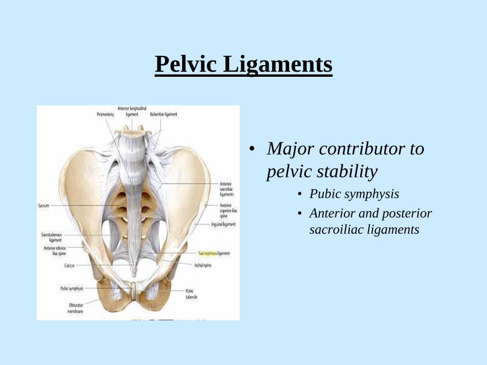
Pelvic Ligaments
• Major contributor to
pelvic stability• Pubic symphysis
• Anterior and posterior
sacroiliac ligaments

Young and Burgess Classification
of Pelvic Fractures
• Useful in the clinical setting
• Addresses injury mechanism and
• Seeks to quantify forces involved
– anterior posterior compression
– lateral compression
– vertical shear
– combination

• Young and Burgess Classification Pelvic Fractures
– AP compression (APC) - direct anterior force
• Type 1: disruption of pubic symphysis < 2.0 cm
– low energy forces (sports)
– stable
• Type 2: symphysis > 2.0 cm and disruption of
anterior SI ligaments
– high energy, “open book”; MVC, ped vs auto
– unstable
– high risk hemorrhage
• Type 3: symphysis > 2.0 cm and disruption of
anterior and posterior SI ligaments
– very unstable
– highest incidence of major hemorrhage

Anterior-Posterior Compression (APC)
• APC 1
– anterior force of mild-
moderate force (sports)
– symphysis separation
< 2cm
– stretching of anterior
sacroiliac ligaments
– stable fracture

Anterior-Posterior Injury
• Grade I
• Symphysis < 2cm
• SI joints intact

Anterior-Posterior Compression (APC)
• APC 2
– anterior force of high
energy
– “open book”
– symphysis > 2cm
– tearing of sacroiliac
ligaments
– unstable fracture

Anterior-Posterior Compression (APC)
• APC 3
– high energy force
– hemipelvis rotates
externally
– symphysis >2cm
– rupture of anterior &
posterior sacroiliac
ligaments
– highest incidence of
major hemorrhage
– unstable fracture

Open Book Pelvic Fractures
– Symphysis > 2cm
– Disruption SI joint
– High incidence hemorrhage
– Mortality approaches 50%

• Young and Burgess
– lateral compression (LC)
• Type 1: unilateral rami fracture and ipsilateral
sacroiliac compression - stable
• Type 2: unilateral rami fracture and ipsilateral
posterior sacroiliac fracture
– unstable fracture
– high risk for hemorrhage
• Type 3: type 2 plus injury to contralateral
hemipelvis
– unstable fracture
– high risk for hemorrhage

Lateral Compression Fractures
• Type I:
• Pubic rami fracture
• Sacral compression
• stable
• Type 2:
• Pubic rami fracture
• Iliac fracture
• Unstable
• Higher incidence
hemorrhage

Lateral compression fractures
• Bilateral pubic rami fractures
• Sacral deformity / fracture
• Lateral compression type fractures are usually stable and rarely hemodynamically unstable
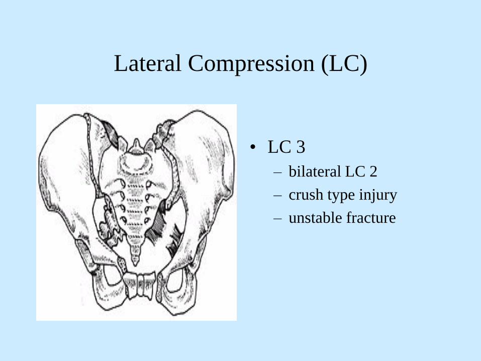
Lateral Compression (LC)
• LC 3
– bilateral LC 2
– crush type injury
– unstable fracture

Classification Pelvic Fractures
• Young and Burgess
– vertical shear (VS): fall
from a height
• Vertical displacement
of hemipelvis
• unstable
• high incidence of
hemorrhage
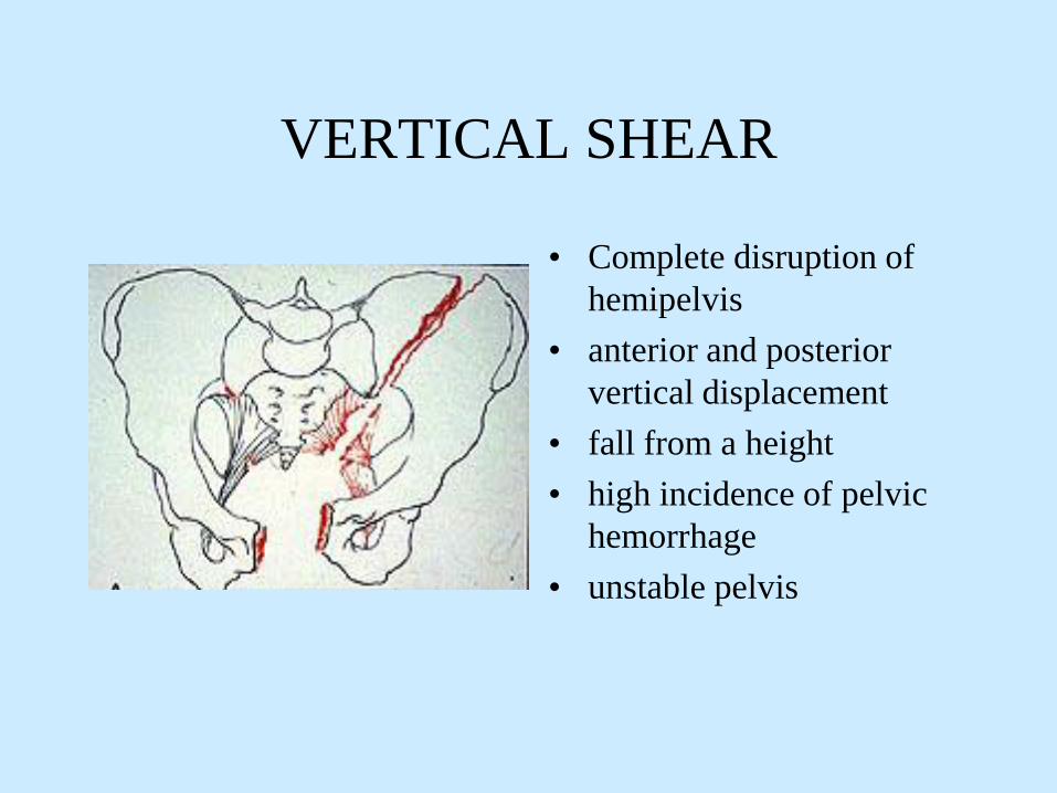
VERTICAL SHEAR
• Complete disruption of
hemipelvis
• anterior and posterior
vertical displacement
• fall from a height
• high incidence of pelvic
hemorrhage
• unstable pelvis

UNSTABLE PELVIS
• APC2, APC3, LC2, LC3,
VS, and combination
injuries are all unstable pelvic
fractures and are associated with a
higher incidence of vascular
disruption and hemodynamic
compromise
• this does not imply that bony
instability equates to hemodynamic
instability

Pelvic Fractures
• High energy
– MVC, falls from
height, crush injury
– 75% associated injuries
– 15-25% intra-
abdominal injuries
– often hemodynamically
unstable
– mortality up to 55%
• Low energy
– falls from standing
• Lateral compression
– elderly / osteopenic
– associated injuries
uncommon
– hemodynamically
stable
– mortality < 1%

AP Pelvis Radiograph
• Indicators of potential
vasculature injury:• diastasis symphysis >
2.0 cm
• fractures all 4 rami
• widening SI joint >0.5
cm
• vertical displacement at
the SI joint

High Energy Pelvic Fractures
• MVC, auto-pedestrian, motorcycle accidents
• 5-10% of high speed MVCs will sustain pelvic fractures
• mortality correlates highly to hemodynamic stability
• stable < 4%
• unstable > 50%
• mortality:
• 50% acute hemorrhage
• 25% associated injuries
• 25% sepsis / MODS
• associated injuries: TBI, thoracic, intra-abdominal

Physical findings may indicate pelvic
fractures
• Labial, scrotal, perineal swelling / ecchymosis
• deformities lower extremities
• open wounds - rectal / vaginal blood necessitates
sigmoidoscopy / speculum examinations
• urethral injuries• blood at meatus
• high riding prostate
• scrotal/labial hematoma
• sacral nerve root injuries

• Physical maneuvers to establish pelvic
stability are of questionable benefit
• pelvic rock, springing, compression, distraction are
crude and insensitive
• exacerbate hemorrhage and soft tissue injury
• painful and unnecessary maneuvers
– initial pelvic radiographs better indicator of stability

Routine Pelvic X-rays
• Examination of the pelvis is extremely unreliable;
especially in the obtunded, intoxicated, or obese patients
• Routine A-P pelvic and chest x-ray is still indicated in the
multiply or obtunded injured patient

• Hemorrhage in pelvic fractures:
– Venous bleeding (85%)
• fracture surfaces of cancellous bone
• venous plexuses
– Arterial bleeding (15%) - from branches of the superior
gluteal artery ( fracture through sciatic notch) or other branches of
internal iliac artery
• Most common source of significant hemorrhage in
pelvic fractures is NOT the pelvis

Pelvic Stabilization
• Purpose:• controls non-arterial hemorrhage
• aids in clot formation
• decreases fracture site movement and clot
dislodgement
• decreases volume of pelvis and promotes tamponade
• exact mechanism in which stabilization decreases
hemorrhage has not been elucidated

Pelvic Stabilization
• Three basic methods:• non-invasive techniques
• external fixation
• open reduction / internal fixation

Pelvic Stabilization
• 1. Non-invasive techniques:– Sheet wrap
– Proprietary devices / binders
• Most appropriate in the trauma bay for unstable
pelvic fractures
• temporary measures
• Controls hemorrhage as effectively as external
fixation

Pelvic Stabilization
• Sheet wrap• Inexpensive
• Fast
• Effective
• Temporary

Pelvic Binders
• T POD
– Cost: $125.00
– Fast
– Efficient
– Temporary

T-Pod: Non-invasive Pelvic Stabilization

Pelvic Stabilization
• External fixation devices:• anterior fixation device - ideally suited for “open-book”
deformities
• C-clamp device - ideally suited for posterior disruptions
– unstable pelvic fractures associated with hypotension
– can be placed in the trauma bay, OR, ICU
– should be placed prior to skin incision in patients
needing a laparotomy
– complications / drawbacks:

Pelvic Stabilization
• External Fixation:• “Fast”
• Orthopedic consultation
• Effective
• Typically temporary

External Fixation
• Should be applied as soon as possible with unstable pelvis
• May be applied in trauma bay, OR, or TICU
• Anterior bridging bars placed low over pelvis so as not to interfere with laparotomy incision
• Ideally placed prior to laparotomy

External Fixation

Pelvic Stabilization
• Internal stabilization:• limited value in the
acute setting
– occasionally used in
“open-book”
deformities after
laparotomy in the
stable patient
• reserved for patients
who are
hemodynamically stable
• definitive treatment

• No convincing data to support one method of pelvic stabilization over another in the acute setting:
• all methods equally effective
• T-POD HAS GAINED WIDE EXCETANCE
• But studies do support some form of bony stabilization
• decreases hemorrhage
• decreases transfusion requirements

PRINCIPLES OF ANGIOGRAPHY AND
EMBOLIZATION
• Used to control bleeding that cannot be corrected with
surgery
• Purpose is to slow bleeding rather than create large areas
of ischemia and necrosis
• Limit areas of ischemia and necrosis to smallest extent
possible
• Must be done expeditiously prior to onset of “lethal triade”

Angiography
• HEMORRHAGIC SHOCK:
• Surgically correctable injuries directly to operating room
• Non-surgically correctable injuries to angiographic department

Pelvic Angiogram

Angiography
• Approximately 7-11% of pelvic fractures will require
embolization to control arterial bleeding.
• lateral compression fractures: 2%
• anteroposterior compression: 20%
• vertical shear injury: 20%
• combination: 20%
• Approximately 25-40% of cases will require embolization of more
than one artery
• Segina, D., Agnew,S., OTA Annual Meeting 2000

Angiographic Embolization
• Embolization is only
effective for arterial
source of hemorrhage
-90% effective

When To Transfer to Angiography ?
• IMMEDIATELY:• Patients who are
hemodynamically
unstable as the result of
their pelvic fractures
AND if laparotomy is
not indicated

Hypotension and Associated High
Energy Pelvic Fracture
• Etiology of hypotension will be secondary
to non-pelvic sources at least 50% of time• thorax
• abdomen
• long bone fractures
• externally / at the scene

5 Major Sites of Blood Loss
• Chest
• Abdomen
• Retroperitoneum
• Muscle compartment of thigh
• Injury scene

Problem!
• Where is the source of hemorrhage / hypotension?
– If solely related to the pelvic fracture(s) - then
angiography and embolization is the best and initial
therapeutic option
– If hemorrhage secondary to an abdominal injury then
laparotomy is the best and initial therapeutic option
– If hemorrhage secondary to a thoracic injury then tube
thoracostomy and possibly thoracotomy is indicated
• How to determine source of bleeding?

Diagnosing intra-abdominal hemorrhage
• FAST
• Diagnostic peritoneal lavage (DPL)
• Diagnostic peritoneal tap: supra-umbilical
approach appears to be the most reliable test for intra-
abdominal hemorrhage, which requires laparotomy
• CT scan
• E.A.S.T., Practice Management Guidelines, 2001

Focused Abdominal Sonography for Trauma
• FAST
• Ultrasound examination to
determine presence of free
fluid in the pericardial or
peritoneal cavities
• High sensitivity /
specificity

Diagnostic Peritoneal Lavage
Diagnostic Peritoneal Tap
• Catheter introduced
through a supraumbilical
incision
• 5-10cc gross blood is
positive tap and an
indication for immediate
laparotomy
• 1 liter crystalloid solution
infused

Critical Questions ?
• Which patients warrant early pelvic
stabilization?
• Which patients warrant pelvic angiography
and possible embolization?
• Which patients warrant emergent
laparotomy?

Early Pelvic Stabilization
• Patients with unstable pelvic fractures
associated with hypovolemia
• All patients with “unstable pelvis”
diastasis of symphysis > 2.0
cm
fractures all 4 rami
widening SI joint >0.5 cm
vertical displacement at the
SI joint

Early Angiography
• Pelvic fractures with signs of ongoing hemorrhage after non-pelvic sources of blood loss have been ruled-out
• Patients with pelvic fractures with ongoing hemorrhage that cannot be controlled at laparotomy
• Arterial extravasation noted in pelvis on CT scan

Emergent Laparotomy
• Patients with hypotension and gross blood
in the abdomen or evidence intestinal
perforation
• FAST / DPL are most reliable diagnostic
tests

Initial Management
• initial assessment: “ABCDE’s”
• radiographic assessment: CXR, A-P pelvis, FAST
• hemodynamically unstable patients:
– 50% of patients with severe pelvic fractures are
bleeding from sources other than the pelvis
– associated injuries very common
– determine source of hemorrhage!!

• “patients in hemorrhagic shock with a
surgically correctable lesion should be
transported to the OR”
• “patient in hemorrhagic shock with an
unknown source of bleeding, as well as,
lesions best treated by embolization should
be transported to the angiography suite”
• Bassam, D., Am. Surg., 1998, 862-867
When to perform laparotomy
• Indications for laparotomy in the face of
hypotension and pelvic fractures remain the same
• intra-abdominal hemorrhage
• intestinal perforation
• peritoneal signs
Intraoperative Management
• If laparotomy is required for ongoing bleeding:
• control non-pelvic sources of hemorrhage
• if pelvic bleeding identified; enlarging hematoma
– do not open retroperitoneum
– pack pelvis
– exercise damage control
– escort patient to angiography suite: embolization is the
best method to control pelvic arterial bleeding; effective in
90% of cases

Open Pelvic Fractures
• Fracture site communicates through the skin, rectum,
vagina
• High incidence of associated injuries, mortality approaches
80%
• Vital to make diagnosis early - blood in rectum / vagina
necessitates further evaluation
• basic tenants apply to all open fractures - thorough irrigation
and debridement, prophylactic antibiotics, fracture stabilization

Open Pelvic Fractures
• Treatment same
• Early prophylactic
antibiotics• ancef / gentamycin

CONCLUSION
• Do not delay treatment
• Priorities remain the same: ABCDE’s
• Pelvic stabilization mandatory
• Rule –out other sources of bleeding
• Exploratory laparotomy: indications remain the same
• Angiography / embolization: must be considered
early to mobilize appropriate personnel
• early transfer to a trauma center may be life
saving


