Influence of Trichoderma isolates and Phlebiopsis …eeb.lu.lv/EEB/201512/EEB_13_Alksne.pdfInfluence...
10
Influence of Trichoderma isolates and Phlebiopsis gigantea on the growth of Heterobasidion parviporum and wood decay of Norway spruce in controlled conditions Laura Alksne 1 , Vizma Nikolajeva 1 *, Zaiga Petriņa 1 , Daina Eze 2 , Tālis Gaitnieks 3 1 Department of Microbiology and Biotechnology, Faculty of Biology, University of Latvia, Jelgavas 1, Riga LV–1004, Latvia 2 Microbial Strain Collection of Latvia, Faculty of Biology, University of Latvia, Jelgavas 1, Riga LV–1004, Latvia 3 Forest Sector Competence Center, LLC “MNKC”, Dzerbenes 27, Riga LV–1006, Latvia *Corresponding author, E-mail: [email protected] Abstract Heterobasidion parviporum-caused root rot is a destructive infection to conifers, especially Norway spruce Picea abies. e disease can be limited by the biocontrol agent fungus Phlebiopsis gigantea. e aim of this work was to study the impact of four previously selected psychrotrophic fast-growing strains of Trichoderma, P. gigantea and combinations of both agents on the growth of H. parviporum in spruce wood blocks and on the structure and chemical composition of wood in laboratory conditions. Stained wood cuts were examined under a microscope and the chemical composition of wood was determined. All investigated Trichoderma strains belonging to species T. viride and T. viridescens inhibited the growth of H. parviporum in the wood. Protective efficiency of Trichoderma spp. against spruce dry weight losses reached 77 to 79%. e study indicated a need for development of combined Trichoderma spp. and P. gigantea plant protection products for biocontrol of root rot. We propose that fast-growing Trichoderma could restrict the colonization of wood surface by rot fungi, while P. gigantea competes with them in deeper parts of wood. Key words: biocontrol, Heterobasidion, Norway spruce, Phlebiopsis, Trichoderma, wood. Abbreviations: CFU, colony-forming units; MSCL, Microbial Strain Collection of Latvia; r, Pearson correlation coefficient; W, moisture content. Environmental and Experimental Biology (2015) 13: 159–168 Original Paper Introduction Root rot caused by Heterobasidion annosum s.l. is a common and economically important disease of coniferous trees in the Northern Hemisphere (Woodward et al. 1998). H. annosum (Fr.) Bref. s.s. preferentially attacks pine, while H. parviporum Niemelä & Korhonen has a preference for spruce (Mason, Huber 2002). e main route of infection is by basidiospores germinating on stumps aſter tree felling (Tainter, Baker 1996). Biological methods of controlling the pathogen involve application of commercial preparations on fresh stumps. Among them, the PG Suspension was developed in the UK, PG IBL in Poland and Rotstop in Finland (Pratt et al. 2000). ese products contain asexual spores (oidia) of Phlebiopsis gigantea (or 2001), a saprotrophic and antagonistic basidiomycete (Kües 2007). PG Suspension and PG IBL are applied to pine stumps, while Rotstop is as effective on both pine and Norway spruce stumps (Pratt et al. 2000). Saprotrophic soil fungi from the genus Trichoderma/ Hypocrea (Ahmad, Baker 1986; Kubicek, Harman 1998) are well-known fungal antagonists which also work on wood decay fungi (Highley, Ricard 1988). Trichoderma has a high rate of growth and sporulation, variable metabolism, mycoparasitism and the ability to produce volatile and soluble antibiotics (Monte 2001; Shalini, Kotasthane 2007). Trichoderma can inhibit pathogens on plants and plant remnants, and in the soil (Shalini, Kotasthane 2007). Trichoderma strains are able to restrict H. annosum s.l. in vitro (Nicolotti, Varese 1996; Schubert et al. 2008; Nikolajeva et al. 2012). However, the effectiveness has been low in field conditions (Kallio 1971; Nicolotti et al. 1999; Berglund et al. 2005). It is known that Trichoderma species degrade cellulose (Song et al. 2010) while P. gigantea degrades both cellulose and lignin (Mgbeahuruike et al. 2011; Hori et al. 2014). To the best of our knowledge, the effect of combined suspensions of Trichoderma and P. gigantea on H. annosum has not been previously investigated. Gymnospermous wood consists primarily of tracheids (90 to 95% of cells) and uniseriate xylem rays (5 to 10% of cells) (Sjöström 1993). e wood cell wall is organized in layers: a thin primary wall and much thicker secondary wall, consisting of three layers. e cell wall consists of cellulose, hemicellulose and the matrix material lignin. e walls of adjacent cells are bonded together by the middle lamella, consisting of lignin, calcium and pectic substances (Albersheim et al. 2010). e typical composition of Norway spruce Picea abies dry wood is 27.5% lignin, 159 Environmental and Experimental Biology ISSN 2255-9582
Transcript of Influence of Trichoderma isolates and Phlebiopsis …eeb.lu.lv/EEB/201512/EEB_13_Alksne.pdfInfluence...
Influence of Trichoderma isolates and Phlebiopsis gigantea on the growth of Heterobasidion parviporum and wood decay of Norway spruce in controlled conditions
Laura Alksne1, Vizma Nikolajeva1*, Zaiga Petriņa1, Daina Eze2, Tālis Gaitnieks3
1Department of Microbiology and Biotechnology, Faculty of Biology, University of Latvia, Jelgavas 1, Riga LV–1004, Latvia2Microbial Strain Collection of Latvia, Faculty of Biology, University of Latvia, Jelgavas 1, Riga LV–1004, Latvia3Forest Sector Competence Center, LLC “MNKC”, Dzerbenes 27, Riga LV–1006, Latvia
*Corresponding author, E-mail: [email protected]
Abstract
Heterobasidion parviporum-caused root rot is a destructive infection to conifers, especially Norway spruce Picea abies. The disease can be limited by the biocontrol agent fungus Phlebiopsis gigantea. The aim of this work was to study the impact of four previously selected psychrotrophic fast-growing strains of Trichoderma, P. gigantea and combinations of both agents on the growth of H. parviporum in spruce wood blocks and on the structure and chemical composition of wood in laboratory conditions. Stained wood cuts were examined under a microscope and the chemical composition of wood was determined. All investigated Trichoderma strains belonging to species T. viride and T. viridescens inhibited the growth of H. parviporum in the wood. Protective efficiency of Trichoderma spp. against spruce dry weight losses reached 77 to 79%. The study indicated a need for development of combined Trichoderma spp. and P. gigantea plant protection products for biocontrol of root rot. We propose that fast-growing Trichoderma could restrict the colonization of wood surface by rot fungi, while P. gigantea competes with them in deeper parts of wood.
Key words: biocontrol, Heterobasidion, Norway spruce, Phlebiopsis, Trichoderma, wood.Abbreviations: CFU, colony-forming units; MSCL, Microbial Strain Collection of Latvia; r, Pearson correlation coefficient; W, moisture content.
Environmental and Experimental Biology (2015) 13: 159–168 Original Paper
Introduction
Root rot caused by Heterobasidion annosum s.l. is a common and economically important disease of coniferous trees in the Northern Hemisphere (Woodward et al. 1998). H. annosum (Fr.) Bref. s.s. preferentially attacks pine, while H. parviporum Niemelä & Korhonen has a preference for spruce (Mason, Huber 2002). The main route of infection is by basidiospores germinating on stumps after tree felling (Tainter, Baker 1996). Biological methods of controlling the pathogen involve application of commercial preparations on fresh stumps. Among them, the PG Suspension was developed in the UK, PG IBL in Poland and Rotstop in Finland (Pratt et al. 2000). These products contain asexual spores (oidia) of Phlebiopsis gigantea (Thor 2001), a saprotrophic and antagonistic basidiomycete (Kües 2007). PG Suspension and PG IBL are applied to pine stumps, while Rotstop is as effective on both pine and Norway spruce stumps (Pratt et al. 2000).
Saprotrophic soil fungi from the genus Trichoderma/Hypocrea (Ahmad, Baker 1986; Kubicek, Harman 1998) are well-known fungal antagonists which also work on wood decay fungi (Highley, Ricard 1988). Trichoderma has a high rate of growth and sporulation, variable metabolism,
mycoparasitism and the ability to produce volatile and soluble antibiotics (Monte 2001; Shalini, Kotasthane 2007). Trichoderma can inhibit pathogens on plants and plant remnants, and in the soil (Shalini, Kotasthane 2007). Trichoderma strains are able to restrict H. annosum s.l. in vitro (Nicolotti, Varese 1996; Schubert et al. 2008; Nikolajeva et al. 2012). However, the effectiveness has been low in field conditions (Kallio 1971; Nicolotti et al. 1999; Berglund et al. 2005). It is known that Trichoderma species degrade cellulose (Song et al. 2010) while P. gigantea degrades both cellulose and lignin (Mgbeahuruike et al. 2011; Hori et al. 2014). To the best of our knowledge, the effect of combined suspensions of Trichoderma and P. gigantea on H. annosum has not been previously investigated.
Gymnospermous wood consists primarily of tracheids (90 to 95% of cells) and uniseriate xylem rays (5 to 10% of cells) (Sjöström 1993). The wood cell wall is organized in layers: a thin primary wall and much thicker secondary wall, consisting of three layers. The cell wall consists of cellulose, hemicellulose and the matrix material lignin. The walls of adjacent cells are bonded together by the middle lamella, consisting of lignin, calcium and pectic substances (Albersheim et al. 2010). The typical composition of Norway spruce Picea abies dry wood is 27.5% lignin,
159Environmental and Experimental Biology ISSN 2255-9582
39.5% cellulose, 30.6% hemicellulose and 2.1% extractives (Sjöström 1993).
Fungal decay types fall into three categories according to their mode of degradation of the woody cell walls: brown rot, white rot and soft rot (reviewed in Schwarze 2007). However, the latest analyses of basidiomycete genomes demonstrate inadequacy of the prevailing paradigm of white vs brown rot in the understanding of the diversity of fungal wood decay mechanisms (Riley et al. 2014). Lignin presents the most significant barrier to wood decay, but white rot fungi such as Heterobasidion spp. (Peek et al. 1972) and P. gigantea (Hori et al. 2014) are known to degrade it.
During early stages of colonization, most hyphae grow in the cell lumina (Schweingruber 2007). Heterobasidion shows typical delignification within the compound middle lamella between tracheids without any alterations in the primary and secondary walls. In Norway spruce, delignification occurs from the lumina of the latewood tracheids. Sheaths of the inner secondary wall detach and peel off towards the lumina (Peek et al. 1972). Individual hyphae grow transversely via minute bore holes to adjacent cells and produce chlamydospores in the cell lumen (Schwarze et al. 2012). Soft rot fungal hyphae penetrate the secondary wall and erode channels, which usually develop into cavities (reviewed in Blanchette 2010). Ascomycetes and deuteromycetes, including Trichoderma species, cause soft rot. Soft rots are chemically more similar to brown rots than white rots, since cellulose and hemicellulose is decomposed while lignin is modified slightly (Schwarze 2007; Schweingruber 2007). Water saturation of wood impedes the availability of oxygen necessary for fungal colonization and wood decay (Boddy 1992). Formation of chlamydospores may be crucial to survival under dry or very moist conditions, or other adverse conditions for a range of wood decay fungi (Powell 2002).
The aim of this work was to study the impact of four
previously selected psychrotrophic fast growing strains of Trichoderma spp. (Nikolajeva et al. 2012), P. gigantea and combinations of both genera, on the growth of H. parviporum in spruce wood blocks and on the structure and chemical composition of wood in laboratory conditions.
Materials and methods
Biological materialThe studied microorganisms were obtained from the Microbial Strain Collection of Latvia (MSCL). One H. parviporum (MSCL 1023), one P. gigantea (MSCL 702) and four Trichoderma strains (MSCL 472, 585, 945 and 969) were used. For long-term storage, fungi were maintained frozen in liquid nitrogen in the MSCL, University of Latvia. Two types of fresh cut wood blocks of Norvay spruce Picea abies (Olaine, Latvia) were used, i.e. 2 × 1 × 1 cm blocks, and blocks 8 to 9 cm long and 7 to 9 cm in diameter.
Fungal growth studiesFungal growth was observed on 8 to 9 cm long and 7 to 9 cm diameter wood blocks. Cell suspensions were made from fungi after 14-day long incubation on DifcoTM Malt Extract Agar (Becton & Dickinson, USA) at 21 °C. Concentrations of inocula were as follows: T. viridescens 472 105 colony-forming units (CFU) mL–1; T. viride 585 109 CFU mL–1; T. viride 945 107 CFU mL–1; T. viride 969 107 CFU mL–1; P. gigantea 702 105 CFU mL–1, and H. parviporum 1023 105 CFU mL–1. The blocks were sterilized twice in an autoclave at 121 °C for 1 h and placed in sterile polypropylene plant tissue culture containers. At first, top surfaces of blocks were moistened with sterile water where appropriate and then inoculated with fungal suspensions according to the experimental design (Table 1). Each treatment had five replications. The inoculated blocks were incubated in darkness at 20 ± 2 °C.
Table 1. Experiment setup of treatment of wood blocks for observation of fungal growth
Treatment Amount of suspension (mL) Amount of water (mL)H. parviporum 1023 0.3 0.6T. viridescens 472 0.3 0.6T. viride 585 0.3 0.6T. viride 945 0.3 0.6T. viride 969 0.3 0.6P. gigantea 702 0.3 0.6H. parviporum 1023 + T. viridescens 472 0.3 + 0.3 0.3H. parviporum 1023 + T. viride 585 0.3 + 0.3 0.3H. parviporum 1023 + T. viride 945 0.3 + 0.3 0.3H. parviporum 1023 + T. viride 969 0.3 + 0.3 0.3H. parviporum 1023 + P. gigantea 702 0.3 + 0.3 0.3H. parviporum 1023 + P. gigantea 702 + T. viridescens 472 0.3 + 0.3 + 0.3 0.0H. parviporum 1023 + P. gigantea 702 + T. viride 945 0.3 + 0.3 + 0.3 0.0H. parviporum 1023 + P. gigantea 702 + T. viride 969 0.3 + 0.3 + 0.3 0.0Control 0.0 0.9
L. Alksne, V. Nikolajeva, Z. Petriņa, D. Eze, T. Gaitnieks
160
Visual observations of blocks were performed every week. After nine months, one block from each treatment was rinsed with sterile water. The bark was damaged and dislodged during washing. Fungal spores and/or mycelium were carefully scraped from the surface where appropriate and one block from each variant was sawn in half. The bottom part was kept in a closed container and fungi developed on the top surface were examined under a microscope with 400× magnification and identified by micromorphological features after one week. After incubation for 6 and 15 months, one block from each treatment was used for histological studies of wood. After 16 and 26 months, one block from each treatment was used for chemical analyses.
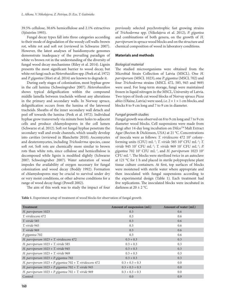
Histological preparationsAfter 6 and 15 months of incubation with fungal isolates, wood blocks 8 to 9 cm long and 7 to 9 cm in diameter were washed under running water, 1-cm thick discs were cut from the upper surface and samples (1 × 1 × 1 cm) were made from the cut disc under this (Fig. 1). Samples were fixed with a solution of formaldehyde-acetic acid-ethanol-water (2:1:10:7), gradually dewatered according to the histovax/terc-butanol method and paraffinized (Ruzin 1999). Tangential and radial sections (35-μm thick) were cut using a Leica 23-TMB (Germany) rotary microtome, and 60-μm thick non-paraffinized sections were cut using a hand microtome.
Sections were stained with either a solution of astra blue and safranin (1:5) or toluidine blue (Braune et al. 1999). Stained sections were examined using a Leica DM 2000 microscope under 400× magnification and images were recorded digitally with a Leica DFC 420 camera (Germany) and processed with software Image-Pro Express v. 6.0. Observations were made on several sections for the staining experiments.
Wood degradation studiesWood degradation studies were carried out using wood blocks, 2 × 1 × 1 cm each. The blocks were sterilized in an autoclave at 121 °C for 1 h. Cell suspensions were made from
Trichoderma spp. and P. gigantea after 14-day incubation on DifcoTM Malt Extract Agar (Becton & Dickinson, USA) at 21 °C. Concentrations of inocula were as follows: T. viride 585 108 CFU mL–1; T. viride 945 108 CFU mL–1; T. viride 969 104 CFU mL–1; and P. gigantea 702 107 CFU mL–1. Each wood block was weighed and numbered. Then each block was immersed in one of the fungal suspensions, or in sterile water (control), for 2 s and placed to flow off on napkins in aseptic conditions. The treated blocks were placed in Petri dishes on top of the mycelium of 18-day old H. parviporum 1023, but sterile control blocks were placed on water agar (agar 2%, Bio-Rad Laboratories, USA). Five blocks were placed in each Petri dish (Schubert et al. 2008). Three replications were used. Petri dishes were sealed with parafilm and incubated in the dark at 20 ± 2 °C. Samples were removed after 6, 12, 18, 24 and 30 weeks of incubation. Fungal spores and/ or mycelium were scraped from the surface where appropriate. The blocks were weighed, dried at 105 °C for 48 h and repeatedly weighed.
Five blocks were weighed before and after immersion in water to estimate the average amount of absorbed water. Moisture content of blocks W was calculated as:
W = (A – B – C) / (A – B) × 100%, where A is weight before treatment with fungi, g; B is average amount of absorbed water, g; and C is weight after drying, g. Five blocks were left untreated and weighed without immersion in water or suspension, and average weight loss was estimated after drying. Percentage of dry weight loss (M) of wood blocks was calculated as follows:
M = (Mb – Mav – Md) / (Mb – Mav) × 100%, where Mb is weight before treatment with fungi, g; Mav is average amount of water, g; Md is weight after drying, g. Fungal biocontrol effectiveness (E) was calculated as:
E = (Minf – MT) / Minf × 100%, where Minf is dry weight loss of control blocks immersed in water and treated with H. parviporum; MT is dry weight loss of blocks immersed in suspension of Trichoderma strain and treated with H. parviporum.
Chemical analysesAfter 16 and 26 months of incubation with fungal isolates, wood blocks, 8 to 9 cm long and 7 to 9 cm diameter, were quickly washed under running water, 1-cm thick discs were cut from the upper surfaces and chips (<0.4 mm for determination of lignin and >0.4 mm for determination of cellulose) were made from the next discs. The chips were oven dried at 105 °C for 18 h and the percentage moisture content was calculated.
Extractives were isolated from wood chips with acetone in a Soxhlet extractor for 8 h. Concentration of cellulose was estimated according to the Kirshner-Hofer method (Abelson et al. 1988). Concentration of lignin was estimated according to the Klason method (Wood, Kellogg 1988). Hemicellulose was determined by subtracting the values of cellulose and lignin (%) from that of dry matter without extractives (100). Fig. 1. Schematic illustration of the histological sample cut.
Influence of Trichoderma on Heterobasidion parviporum and wood decay
161
Statistical analysisAn F-test was used for estimation of homogeneity of samples and for comparison of variance. The significance of the difference in values was determined at a significance level of 0.05. Program R.2.12.1. was used for Pearson correlation analysis.
Results
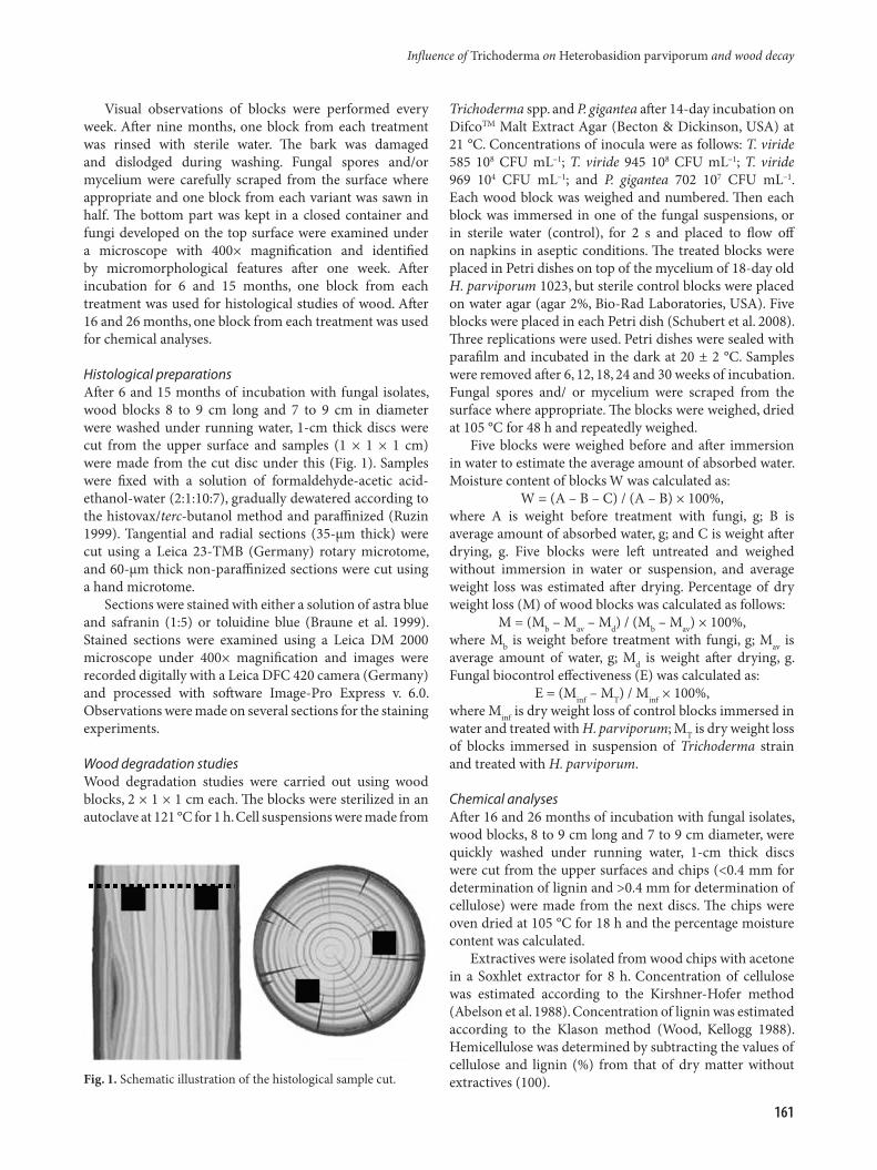
Fungal growth on spruce wood blocksTrichoderma spp. mycelium was observed on inoculated wood blocks as of one week after the beginning of incubation when these fungi had colonized about half of the block surface. At that time, colonies of P. gigantea were scarcely visible. Growth of H. parviporum was noticed at two weeks (Fig. 2 a to d). P. gigantea later developed branched mycelium in all of the P. gigantea-containing variants and Trichoderma strains were overgrown by P. gigantea after two months (Fig. 2 e to h). P. gigantea overgrew the surface and sides of blocks after three months and one week, and retained dense mycelium and artrospores throughout the experiment.
We observed mycelium of H. parviporum only in the treatment with single H. parviporum (Fig. 2h) but not in coculture with Trichoderma and/or P. gigantea during the entire experiment period of nine months. Trichoderma strains, in single cultures as well as in cocultures with H. parviporum, developed a dense cover of mycelium and conidia which was visible during all nine months (Fig. 2).
Trichoderma spp. formed conidia first on the sapwood (Fig. 2b) and later on heartwood. P. gigantea-caused brown colouration was observed mainly in the centre of the heartwood (Fig. 2).
After incubation for nine months, the inoculated fungi
were identified in 4 to 4.5 cm deep layers of spruce blocks with the exception of three treatments, i.e. T. viride 969 + H. parviporum 1023, and P. gigantea 702 + H. parviporum 1023 where Heterobasidion was not observed, and T. viridescens 472 + P. gigantea 702 + H. parviporum 1023 where Trichoderma was not observed (Table 2).
Histological studiesWood blocks (8 to 9 cm long and 7 to 9 cm diameter) used for fungal growth studies were also used for histological studies. Structural changes of tracheids and presence of fungal cells were investigated. The structure of blocks incubated with P. gigantea was too much degraded, deformed and soft that we did not achieve good quality of cuts.
Observations under a light microscope showed that spruce blocks after 6 and 15 months of incubation were colonized by Trichoderma hyphae located in the tracheid lumen (Fig. 3a), the hyphae grew through bordered pits (Fig. 3c) and formed chlamydospores (Fig. 3b, Fig. 4a), and occasionally conidiospores (Fig. 3d) also. Similarly, hyphae in the lumen and penetration through bordered pits were observed in wood after incubation with H. parviporum; also dipyramidal probably oxalate crystals were observed (Fig. 3e). H. parviporum also grew perpendicularly to the tracheids through lateral pores of tracheids and showed degraded bordered pits with hyphae in the middle (Fig. 4 b to d).
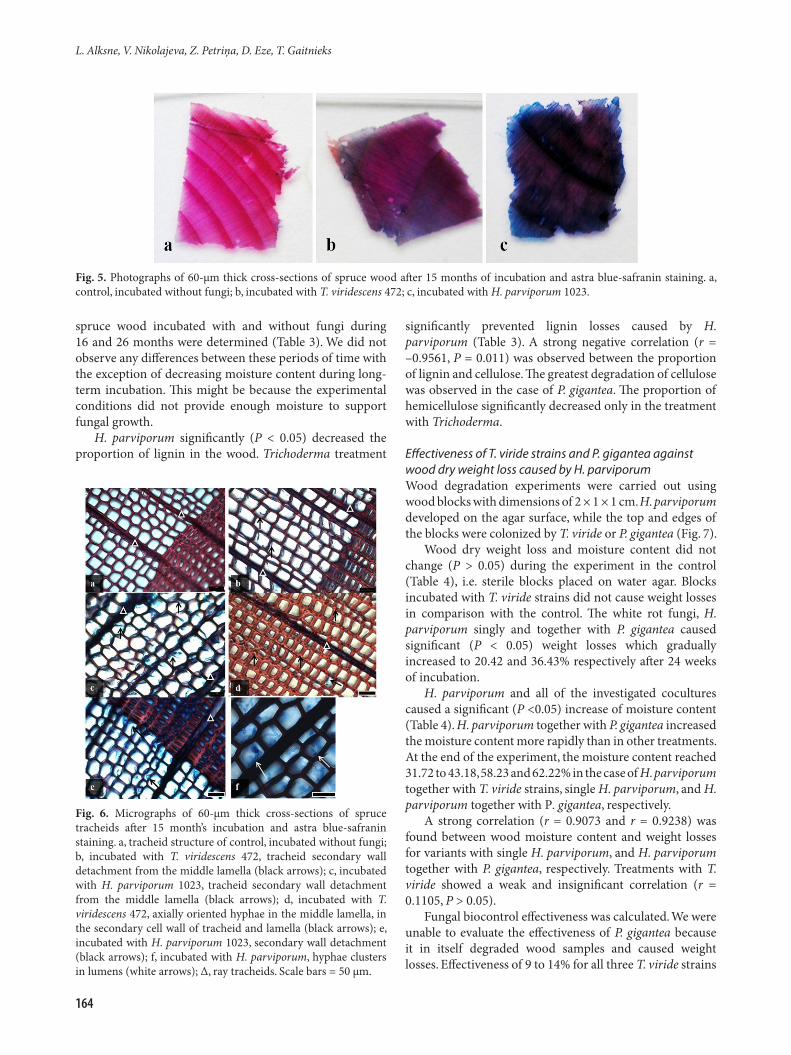
Stained cross-sections demonstrated visual differences between wood inoculation treatments (Fig. 5). Colour varied from red (safranin) to blue (astra blue), which are indicative of lignin and cellulose in the absence of lignin, respectively. Only the control showed bright rot borders in the latewood zone. Trichoderma and particularly H. parviporum decreased rot colouration.
Fig. 2. Fungal growth on spruce wood blocks after two weeks (a to d) and after nine months (e to h) of incubation. a, H. parviporum 1023 + P. gigantea 702; b, H. parviporum 1023 + P. gigantea 702 + T. viride 945; c, T. viride 945; d, P. gigantea 702; e, H. parviporum 1023 + T. viride 945; f, H. parviporum 1023 + P. gigantea 702; g, H. parviporum 1023 + P. gigantea 702 + T. viride 945; h, H. parviporum 1023; black arrows show P. gigantea coloration; white arrows show T. viride conidia.
L. Alksne, V. Nikolajeva, Z. Petriņa, D. Eze, T. Gaitnieks
162
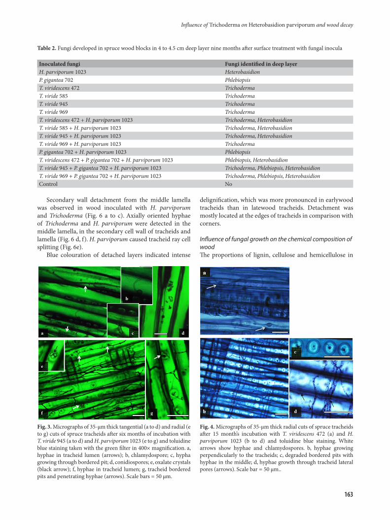
Secondary wall detachment from the middle lamella was observed in wood inoculated with H. parviporum and Trichoderma (Fig. 6 a to c). Axially oriented hyphae of Trichoderma and H. parviporum were detected in the middle lamella, in the secondary cell wall of tracheids and lamella (Fig. 6 d, f). H. parviporum caused tracheid ray cell splitting (Fig. 6e).
Blue colouration of detached layers indicated intense
delignification, which was more pronounced in earlywood tracheids than in latewood tracheids. Detachment was mostly located at the edges of tracheids in comparison with corners.
Influence of fungal growth on the chemical composition of woodThe proportions of lignin, cellulose and hemicellulose in
Fig. 3. Micrographs of 35-μm thick tangential (a to d) and radial (e to g) cuts of spruce tracheids after six months of incubation with T. viride 945 (a to d) and H. parviporum 1023 (e to g) and toluidine blue staining taken with the green filter in 400× magnification. a, hyphae in tracheid lumen (arrows); b, chlamydospore; c, hypha growing through bordered pit; d, conidiospores; e, oxalate crystals (black arrow); f, hyphae in tracheid lumen; g, tracheid bordered pits and penetrating hyphae (arrows). Scale bars = 50 μm.
Fig. 4. Micrographs of 35-μm thick radial cuts of spruce tracheids after 15 month’s incubation with T. viridescens 472 (a) and H. parviporum 1023 (b to d) and toluidine blue staining. White arrows show hyphae and chlamydospores. b, hyphae growing perpendicularly to the tracheids; c, degraded bordered pits with hyphae in the middle; d, hyphae growth through tracheid lateral pores (arrows). Scale bar = 50 μm..
Table 2. Fungi developed in spruce wood blocks in 4 to 4.5 cm deep layer nine months after surface treatment with fungal inocula
Inoculated fungi Fungi identified in deep layerH. parviporum 1023 HeterobasidionP. gigantea 702 PhlebiopsisT. viridescens 472 TrichodermaT. viride 585 TrichodermaT. viride 945 TrichodermaT. viride 969 TrichodermaT. viridescens 472 + H. parviporum 1023 Trichoderma, HeterobasidionT. viride 585 + H. parviporum 1023 Trichoderma, HeterobasidionT. viride 945 + H. parviporum 1023 Trichoderma, HeterobasidionT. viride 969 + H. parviporum 1023 TrichodermaP. gigantea 702 + H. parviporum 1023 PhlebiopsisT. viridescens 472 + P. gigantea 702 + H. parviporum 1023 Phlebiopsis, HeterobasidionT. viride 945 + P. gigantea 702 + H. parviporum 1023 Trichoderma, Phlebiopsis, HeterobasidionT. viride 969 + P. gigantea 702 + H. parviporum 1023 Trichoderma, Phlebiopsis, HeterobasidionControl No
Influence of Trichoderma on Heterobasidion parviporum and wood decay
163
spruce wood incubated with and without fungi during 16 and 26 months were determined (Table 3). We did not observe any differences between these periods of time with the exception of decreasing moisture content during long-term incubation. This might be because the experimental conditions did not provide enough moisture to support fungal growth.
H. parviporum significantly (P < 0.05) decreased the proportion of lignin in the wood. Trichoderma treatment
significantly prevented lignin losses caused by H. parviporum (Table 3). A strong negative correlation (r = –0.9561, P = 0.011) was observed between the proportion of lignin and cellulose. The greatest degradation of cellulose was observed in the case of P. gigantea. The proportion of hemicellulose significantly decreased only in the treatment with Trichoderma.
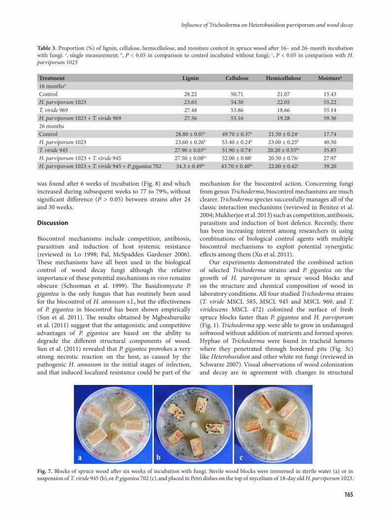
Effectiveness of T. viride strains and P. gigantea against wood dry weight loss caused by H. parviporumWood degradation experiments were carried out using wood blocks with dimensions of 2 × 1 × 1 cm. H. parviporum developed on the agar surface, while the top and edges of the blocks were colonized by T. viride or P. gigantea (Fig. 7).
Wood dry weight loss and moisture content did not change (P > 0.05) during the experiment in the control (Table 4), i.e. sterile blocks placed on water agar. Blocks incubated with T. viride strains did not cause weight losses in comparison with the control. The white rot fungi, H. parviporum singly and together with P. gigantea caused significant (P < 0.05) weight losses which gradually increased to 20.42 and 36.43% respectively after 24 weeks of incubation.
H. parviporum and all of the investigated cocultures caused a significant (P <0.05) increase of moisture content (Table 4). H. parviporum together with P. gigantea increased the moisture content more rapidly than in other treatments. At the end of the experiment, the moisture content reached 31.72 to 43.18, 58.23 and 62.22% in the case of H. parviporum together with T. viride strains, single H. parviporum, and H. parviporum together with P. gigantea, respectively.
A strong correlation (r = 0.9073 and r = 0.9238) was found between wood moisture content and weight losses for variants with single H. parviporum, and H. parviporum together with P. gigantea, respectively. Treatments with T. viride showed a weak and insignificant correlation (r = 0.1105, P > 0.05).
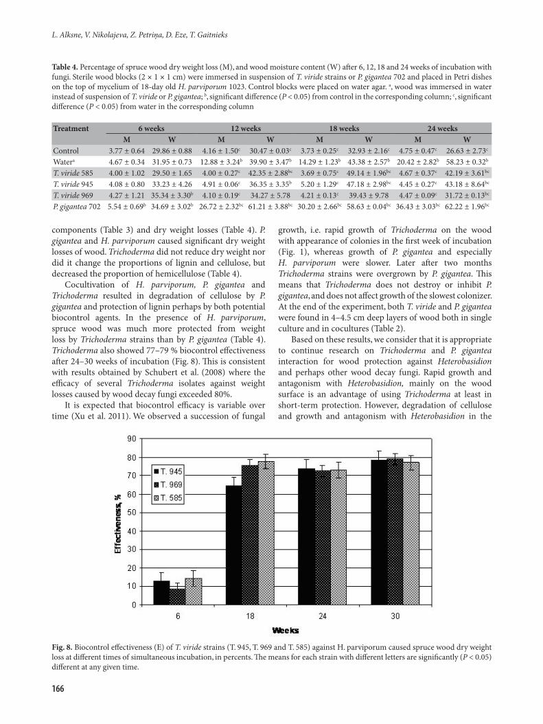
Fungal biocontrol effectiveness was calculated. We were unable to evaluate the effectiveness of P. gigantea because it in itself degraded wood samples and caused weight losses. Effectiveness of 9 to 14% for all three T. viride strains
Fig. 5. Photographs of 60-μm thick cross-sections of spruce wood after 15 months of incubation and astra blue-safranin staining. a, control, incubated without fungi; b, incubated with T. viridescens 472; c, incubated with H. parviporum 1023.
Fig. 6. Micrographs of 60-μm thick cross-sections of spruce tracheids after 15 month’s incubation and astra blue-safranin staining. a, tracheid structure of control, incubated without fungi; b, incubated with T. viridescens 472, tracheid secondary wall detachment from the middle lamella (black arrows); c, incubated with H. parviporum 1023, tracheid secondary wall detachment from the middle lamella (black arrows); d, incubated with T. viridescens 472, axially oriented hyphae in the middle lamella, in the secondary cell wall of tracheid and lamella (black arrows); e, incubated with H. parviporum 1023, secondary wall detachment (black arrows); f, incubated with H. parviporum, hyphae clusters in lumens (white arrows); ∆, ray tracheids. Scale bars = 50 μm.
L. Alksne, V. Nikolajeva, Z. Petriņa, D. Eze, T. Gaitnieks
164
was found after 6 weeks of incubation (Fig. 8) and which increased during subsequent weeks to 77 to 79%, without significant difference (P > 0.05) between strains after 24 and 30 weeks.
Discussion
Biocontrol mechanisms include competition, antibiosis, parasitism and induction of host systemic resistance (reviewed in Lo 1998; Pal, McSpadden Gardener 2006). These mechanisms have all been used in the biological control of wood decay fungi although the relative importance of these potential mechanisms in vivo remains obscure (Schoeman et al. 1999). The Basidiomycete P. gigantea is the only fungus that has routinely been used for the biocontrol of H. annosum s.l., but the effectiveness of P. gigantea in biocontrol has been shown empirically (Sun et al. 2011). The results obtained by Mgbeahuruike et al. (2011) suggest that the antagonistic and competitive advantages of P. gigantea are based on the ability to degrade the different structural components of wood. Sun et al. (2011) revealed that P. gigantea provokes a very strong necrotic reaction on the host, as caused by the pathogenic H. annosum in the initial stages of infection, and that induced localized resistance could be part of the
mechanism for the biocontrol action. Concerning fungi from genus Trichoderma, biocontrol mechanisms are much clearer. Trichoderma species successfully manages all of the classic interaction mechanisms (reviewed in Benitez et al. 2004; Mukherjee et al. 2013) such as competition, antibiosis, parasitism and induction of host defence. Recently, there has been increasing interest among researchers in using combinations of biological control agents with multiple biocontrol mechanisms to exploit potential synergistic effects among them (Xu et al. 2011).
Our experiments demonstrated the combined action of selected Trichoderma strains and P. gigantea on the growth of H. parviporum in spruce wood blocks and on the structure and chemical composition of wood in laboratory conditions. All four studied Trichoderma strains (T. viride MSCL 585, MSCL 945 and MSCL 969, and T. viridescens MSCL 472) colonized the surface of fresh spruce blocks faster than P. gigantea and H. parviporum (Fig. 1). Trichoderma spp. were able to grow in undamaged softwood without addition of nutrients and formed spores. Hyphae of Trichoderma were found in tracheid lumens where they penetrated through bordered pits (Fig. 3c) like Heterobasidion and other white rot fungi (reviewed in Schwarze 2007). Visual observations of wood colonization and decay are in agreement with changes in structural
Fig. 7. Blocks of spruce wood after six weeks of incubation with fungi. Sterile wood blocks were immersed in sterile water (a) or in suspension of T. viride 945 (b), or P. gigantea 702 (c), and placed in Petri dishes on the top of mycelium of 18-day old H. parviporum 1023.
Table 3. Proportion (%) of lignin, cellulose, hemicellulose, and moisture content in spruce wood after 16– and 26–month incubation with fungi. a, single measurement; b, P < 0.05 in comparison to control incubated without fungi; c, P < 0.05 in comparison with H. parviporum 1023
Treatment Lignin Cellulose Hemicellulose Moisturea
16 monthsa
Control 28.22 50.71 21.07 15.43H. parviporum 1023 23.65 54.30 22.05 55.22T. viride 969 27.48 53.86 18.66 55.14H. parviporum 1023 + T. viride 969 27.56 53.16 19.28 59.3626 monthsControl 28.80 ± 0.07c 49.70 ± 0.37c 21.50 ± 0.24c 17.74H. parviporum 1023 23.60 ± 0.26b 53.40 ± 0.24b 23.00 ± 0.25b 40.50T. viride 945 27.90 ± 0.03bc 51.90 ± 0.74c 20.20 ± 0.55bc 35.85H. parviporum 1023 + T. viride 945 27.50 ± 0.08bc 52.00 ± 0.88c 20.50 ± 0.76c 27.97H. parviporum 1023 + T. viride 945 + P. gigantea 702 34.3 ± 0.49bc 43.70 ± 0.40bc 22.00 ± 0.42c 39.20
Influence of Trichoderma on Heterobasidion parviporum and wood decay
165
components (Table 3) and dry weight losses (Table 4). P. gigantea and H. parviporum caused significant dry weight losses of wood. Trichoderma did not reduce dry weight nor did it change the proportions of lignin and cellulose, but decreased the proportion of hemicellulose (Table 4).
Cocultivation of H. parviporum, P. gigantea and Trichoderma resulted in degradation of cellulose by P. gigantea and protection of lignin perhaps by both potential biocontrol agents. In the presence of H. parviporum, spruce wood was much more protected from weight loss by Trichoderma strains than by P. gigantea (Table 4). Trichoderma also showed 77–79 % biocontrol effectiveness after 24–30 weeks of incubation (Fig. 8). This is consistent with results obtained by Schubert et al. (2008) where the efficacy of several Trichoderma isolates against weight losses caused by wood decay fungi exceeded 80%.
It is expected that biocontrol efficacy is variable over time (Xu et al. 2011). We observed a succession of fungal
growth, i.e. rapid growth of Trichoderma on the wood with appearance of colonies in the first week of incubation (Fig. 1), whereas growth of P. gigantea and especially H. parviporum were slower. Later after two months Trichoderma strains were overgrown by P. gigantea. This means that Trichoderma does not destroy or inhibit P. gigantea, and does not affect growth of the slowest colonizer. At the end of the experiment, both T. viride and P. gigantea were found in 4–4.5 cm deep layers of wood both in single culture and in cocultures (Table 2).
Based on these results, we consider that it is appropriate to continue research on Trichoderma and P. gigantea interaction for wood protection against Heterobasidion and perhaps other wood decay fungi. Rapid growth and antagonism with Heterobasidion, mainly on the wood surface is an advantage of using Trichoderma at least in short-term protection. However, degradation of cellulose and growth and antagonism with Heterobasidion in the
Table 4. Percentage of spruce wood dry weight loss (M), and wood moisture content (W) after 6, 12, 18 and 24 weeks of incubation with fungi. Sterile wood blocks (2 × 1 × 1 cm) were immersed in suspension of T. viride strains or P. gigantea 702 and placed in Petri dishes on the top of mycelium of 18-day old H. parviporum 1023. Control blocks were placed on water agar. a, wood was immersed in water instead of suspension of T. viride or P. gigantea; b, significant difference (P < 0.05) from control in the corresponding column; c, significant difference (P < 0.05) from water in the corresponding column
Treatment 6 weeks 12 weeks 18 weeks 24 weeksM W M W M W M W
Control 3.77 ± 0.64 29.86 ± 0.88 4.16 ± 1.50c 30.47 ± 0.03c 3.73 ± 0.25c 32.93 ± 2.16c 4.75 ± 0.47c 26.63 ± 2.73c
Watera 4.67 ± 0.34 31.95 ± 0.73 12.88 ± 3.24b 39.90 ± 3.47b 14.29 ± 1.23b 43.38 ± 2.57b 20.42 ± 2.82b 58.23 ± 0.32b
T. viride 585 4.00 ± 1.02 29.50 ± 1.65 4.00 ± 0.27c 42.35 ± 2.88bc 3.69 ± 0.75c 49.14 ± 1.96bc 4.67 ± 0.37c 42.19 ± 3.61bc
T. viride 945 4.08 ± 0.80 33.23 ± 4.26 4.91 ± 0.06c 36.35 ± 3.35b 5.20 ± 1.29c 47.18 ± 2.98bc 4.45 ± 0.27c 43.18 ± 8.64bc
T. viride 969 4.27 ± 1.21 35.34 ± 3.30b 4.10 ± 0.19c 34.27 ± 5.78 4.21 ± 0.13c 39.43 ± 9.78 4.47 ± 0.09c 31.72 ± 0.13bc
P. gigantea 702 5.54 ± 0.69b 34.69 ± 3.02b 26.72 ± 2.32bc 61.21 ± 3.88bc 30.20 ± 2.66bc 58.63 ± 0.04bc 36.43 ± 3.03bc 62.22 ± 1.96bc
Fig. 8. Biocontrol effectiveness (E) of T. viride strains (T. 945, T. 969 and T. 585) against H. parviporum caused spruce wood dry weight loss at different times of simultaneous incubation, in percents. The means for each strain with different letters are significantly (P < 0.05) different at any given time.
L. Alksne, V. Nikolajeva, Z. Petriņa, D. Eze, T. Gaitnieks
166
deeper layers of wood are advantages of using P. gigantea. Such combined preparations would correspond to biological control with different protection mechanisms and opportunities (Xu et al. 2011). Furthermore, P. gigantea may have limitations when applied at a lower temperature (Zhao 2013). Our selected psychrotrophic fast growing Trichoderma strains have the ability to completely overgrow Heterobasidion at temperatures in the range of 4 to 21 °C (Nikolajeva et al. 2012). We must accept that attempts at biologically controlling basidiomycetes in forests on a commercial scale have met with limited success and the environmental factors influencing efficacy must be taken into account (Schoeman et al. 1999). We propose to examine combinations of selected Trichoderma strains and P. gigantea in further experiments in field conditions.
Acknowledgements
We thank Kristine Dokane, Dace Megre and Iluta Dauskane for assistance with histological preparations, Edgars Kuka and Eduards Bakis for chemical analyses, and Ilze Kreismane for language editing. This study was partly financed by European Regional Development Fund project “Methods and technologies for increasing forest capital value” (No. L-KC-11-0004).
References
Abelson J.N., Simon N.I., Wood W.A., Kellogg S.T. (eds.) 1988. Methods in Enzymology. Vol. 161. Biomass. Part B: Lignin, Pectin, and Chitin. Academic Press, San Diego. 574 p.
Ahmad J.S., Baker R. 1986. Rhizosphere competence of Trichoderma harzianum. Phytopathology 77: 182–189.
Albersheim P., Darvill A., Roberts K., Sederoff R., Staehelin A. 2010. Plant Cell Walls: from Chemistry to Biology. Garland Science, Taylor and Francis Group, New York. 430 p.
Benitez T., Rincon A.M., Limon M.C., Codon A.C. 2004. Biocontrol mechanisms of Trichoderma strains. Int. Microbiol. 7: 249–260.
Berglund M., Rönnberg J., Holmer L., Stenlid J. 2005. Comparison of five strains of Phlebiopsis gigantea and two Trichoderma formulations for treatment against natural Heterobasidion spore infections on Norway spruce stumps. Scand. J. For. Res. 20: 12–17.
Blanchette R.A. 2010. Microbial degradation of wood from aquatic and terrestrial environments. In: Mitchell R., McNamara C.J. (eds) Cultural Heritage Microbiology: Fundamental Studies in Conservation Science. ASM Press, Washington, DC, pp. 179–206.
Boddy L. 1992. Microenvironmental aspects of xylem defences to wood decay fungi. In: Blanchette R.A., Biggs A.R. (eds) Defence Mechanisms of Woody Plants Against Fungi. Springer Verlag, Berlin, New York, pp. 96–132.
Braune W., Leman A., Taubert H. 1999. Pflanzenanatomisches Praktikum. I. Spektrum. Akademischer Verlag, Berlin, Heidelberg. 368 S.
Brown H.L., Bruce A. 1999. Assessment of the biocontrol potential of a Trichoderma viride isolate: Part I: Establishment of field and fungal cellar trials. Int. Biodeter. Biodegr. 44: 219–223.
Highley T.L., Ricard J. 1988. Antagonism of Trichoderma spp. and Gliocladium virens against wood decay fungi. Mater.
Organismen 23: 157–169.Hori C., Ishida T., Igarashi K., Samejima M., Suzuki H., Master
E., Ferreira P., Ruiz-Dueñas F.J., Held B., Canessa P., Larrondo, L.F., Schmoll M., Druzhinina I.S., Kubicek C.P., Gaskell J.A., Kersten P., John F.St., Glasner J., Sabat G., BonDurant S.S., Syed K., Yadav J., Mgbeahuruike A.C., Kovalchuk A., Asiegbu F.O., Hoffmeister D., Rencoret J., Gutiérrez A., Sun H., Lindquist E., Barry K., Riley R., Grigoriev I.V., Henrissat B., Kües U., Berka R.M., Martínez A.T., Covert S.F., Blanchette R.A., Cullen D. 2014. Analysis of the Phlebiopsis gigantea genome, transcriptome and secretome provides insight into its pioneer colonization strategies of wood. PLoS Genetics 10: e1004759.
Kallio T. 1971. Protection of spruce stumps against Fomes annosus (Fr.) Cooke by some wood inhabiting fungi. Acta For. Fenn. 117: 1–20.
Kubicek C.P., Harman G.E. 1998. Trichoderma & Gliocladium. Vol. 1. Basic Biology, Taxonomy and Genetics. CRC Press, London. 293 p.
Kües U. 2007. Wood Production, Wood Technology, and Biotechnological Impacts. Universitätsverlag Göttingen, Göttingen. 635 p.
Lo C.-T. 1998. General mechanisms of action of microbial biocontrol agents. Plant Pathol. Bull. 7: 155–166.
Mason P.G., Huber J.T. 2002. Biological Control Programmes in Canada, 1981-2000. CABI Publishing, Wallingford. 583 p.
Mgbeahuruike A.C., Sun H., Fransson P., Kasanen R., Daniel G., Karlsson M., Asiegbu F.O. 2011. Screening of Phlebiopsis gigantea isolates for traits associated with biocontrol of the conifer pathogen Heterobasidion annosum. Biol. Control 5: 118–129.
Monte E. 2001. Understanding Trichoderma: between biotechnology and microbial ecology. Int. Microbiol. 4: 1–4.
Mukherjee P.K., Horwitz B.A., Singh U.S., Mukherjee M., Schmoll M. 2013. Trichoderma: Biology and Applications. CABI, Wallingford. 344 p.
Nicolotti G., Gonthier P., Varese G.C. 1999. Effectiveness of some biocontrol and chemical treatments against Heterobasidion annosum on Norway spruce stumps. For. Pathol. 29: 339–346.
Nicolotti G., Varese G.C. 1996. Screening of antagonistic fungi against air-borne infection by Heterobasidion annosum on Norway spruce. For. Ecol. Manage. 88: 249–257.
Nikolajeva V., Petrina Z., Vulfa L., Alksne L., Eze D., Grantina L., Gaitnieks T., Lielpetere A. 2012. Growth and antagonism of Trichoderma spp. and conifer pathogen Heterobasidion annosum s.l. in vitro at different temperatures. Adv. Microbiol. 2: 295–302.
Pal K.K., McSpadden Gardener B. 2006. Biological control of plant pathogens. The Plant Health Instructor doi:10.1094/PHI-A-2006-1117-02.
Peek V.R.-D., Liese W., Parmeswaran N. 1972. Infektion und Abbau der Wurzelrinde von Fichte durch Fomes annaus. Eur. J. For. Pathol. 2: 104–115.
Powell M.R. 2002. A model for probalistic assessment of phytosanitary risk reduction measures. Plant Dis. 86: 552–557.
Pratt J.E., Niemi M., Sierota Z.H. 2000. Comparison of tree products based on Phlebiopsis gigantea for the control of Heterobasidion annosum in Europe. Biocontrol Sci. Technol. 10: 467–477.
Riley R., Salamov A.A., Brown D.W., Nagy L.G., Floudas D., Held B.W., Levasseur A., Lombard V., Morin E., Otillar R., Lindquist E.A., Sun H., LaButti K.M., Schmutz J., Jabbour D., Luo H., Baker S.E., Pisabarro A.G., Walton J.D., Blanchette R.A.,
Influence of Trichoderma on Heterobasidion parviporum and wood decay
167
Henrissat B., Martin F., Cullen D., Hibbett D.S., Grigoriev I.V. 2014. Extensive sampling of basidiomycete genomes demonstrates inadequacy of the white-rot/brown-rot paradigm for wood decay fungi. Proc. Natl. Acad. Sci., USA 111: 9923–9928.
Ruzin S.E. 1999. Plant Microtechnique and Microscopy. Oxford University Press, New York. 322 p.
Schoeman M.W., Webber J.F., Dickinson D.J. 1999. The development of ideas in biological control applied to forest products. Int. Biodeter. Biodegr. 43: 109–123.
Schubert M., Fink S., Scwarze F.W.M.R. 2008. In vitro screening of an antagonistic Trichoderma strain against wood decay fungi. Arboric. J. 31: 227–248.
Schwarze F.W.M.R. 2007. Wood decay under the microscope. Fungal Biol. Rev. 21: 133–170.
Schwarze F.W.M.R., Mattheck C., Engels J. 2012. Fungal Strategies of Wood Decay in Trees. Springer, Heidelberg. 185 p.
Schweingruber F.H. 2007. Wood Structure and Environment. Springer, Berlin. 279 p.
Shalini S., Kotasthane A.S. 2007. Parasitism of Rhizoctonia solani by strains of Trichoderma spp. Electron. J. Environ. Agric. Food Chem. 6: 2272–2281.
Sjöström E. 1993. Wood Chemistry: Fundamentals and Applications. 2nd ed. Academic Press, San Diego. 292 p.
Song F., Tian X., Fan X., He X. 2010. Decomposing ability of filamentous fungi on litter is involved in a subtropical mixed
forest. Mycologia 102: 20–26.Sun H., Paulin L., Alatalo E., Asiegbu F.O., Peter G. 2011. Response
of living tissues of Pinus sylvestris to the saprotrophic biocontrol fungus Phlebiopsis gigantea. Tree Physiol. 31: 438–451.
Tainter F.H., Baker F.A. 1996. Principles of Forest Pathology. John Wiley and Sons, New York. 805 p.
Thor M. 2001. Operational stump treatment against Heterobasidion annosum in European forestry – current situation. In: Laflamme G., Bérubé J., Bussières G. (eds) Root and Butt Rot of Forest Trees. Proceedings of the IUFRO Working Party 7.02.01 Quebec City, Canada, September 16-22, 2001. Canadian Forest Service, Information Report LAU-X-126, pp. 170–175.
Wood W.A., Kellogg S.T. (eds.) 1988. Methods in Enzymology. Vol. 160. Biomass, Part A. Cellulose and Hemicellulose. Academic Press, San Diego. 774 p.
Woodward S., Stenlid J., Karjalainen R., Hutterman A. (eds.). 1998. Heterobasidion annosum: Biology, Ecology, Impact, and Control. CAB International, Cambridge. 589 p.
Xu X.-M., Jeffries P., Pautasso M., Jeger M.J. 2011. Combined use of biocontrol agents to manage plant diseases in theory and practice. Phytopathology 101: 1024–1031.
Zhao A. 2013. Effects of Temperature and Heterobasidion Species on the Biological Control Efficacy of Phlebiopsis gigantea. Master Thesis. Swedish University of Agricultural Sciences, Uppsala. 27 p.
L. Alksne, V. Nikolajeva, Z. Petriņa, D. Eze, T. Gaitnieks
168
Received 12 September 2015; received in revised form 17 November 2015; accepted 15 December 2015


![Morphological Characterization of Biocontrol Isolates of ... · the development of species are still very slow [14, 3, 4, and 5]. Rifai classified the Trichoderma into nine species](https://static.fdocuments.net/doc/165x107/5eb5b0181ca5d35838571c6e/morphological-characterization-of-biocontrol-isolates-of-the-development-of.jpg)