Inducible Prophage Mutant of Escherichia coli Can Lyse New ...
13
ORIGINAL RESEARCH published: 01 February 2017 doi: 10.3389/fmicb.2017.00147 Edited by: Peter Mullany, University College London, UK Reviewed by: Weili Liang, Chinese Center for Disease Control and Prevention, China Victor Krylov, I. I. Mechnikov Research Institute of Vaccines and Sera, Russia *Correspondence: Wei Zhang [email protected] Specialty section: This article was submitted to Antimicrobials, Resistance and Chemotherapy, a section of the journal Frontiers in Microbiology Received: 16 November 2016 Accepted: 20 January 2017 Published: 01 February 2017 Citation: Chen M, Zhang L, Xin S, Yao H, Lu C and Zhang W (2017) Inducible Prophage Mutant of Escherichia coli Can Lyse New Host and the Key Sites of Receptor Recognition Identification. Front. Microbiol. 8:147. doi: 10.3389/fmicb.2017.00147 Inducible Prophage Mutant of Escherichia coli Can Lyse New Host and the Key Sites of Receptor Recognition Identification Mianmian Chen, Lei Zhang, Sipei Xin, Huochun Yao, Chengping Lu and Wei Zhang* College of Veterinary Medicine, Nanjing Agricultural University, Nanjing, China The use of bacteriophages as therapeutic agents is hindered by their narrow and specific host range, and by a lack of the knowledge concerning the molecular mechanism of receptor recognition. Two P2-like coliphages, named P88 and pro147, were induced from Escherichia coli strains K88 and DE147, respectively. A comparison of the genomes of these two and other P2-like coliphages obtained from GenBank showed that the tail fiber protein genes, which are the key genes for receptor recognition in other myoviridae phages, showed more diversity than the conserved lysin, replicase, and terminase genes. Firstly, replacing hypervariable region 2 (HR2: amino acids 716– 746) of the tail fiber protein of P88 with that of pro147 changed the host range of P88. Then, replacing six amino acids in HR2 with the corresponding residues from pro147 altered the host range only in these mutants with changes at position 730 (leucine) and 744 (glutamic acid). Thus, we predicted that these amino acids are vital to establish the host range of P88. This study provided a vector of lysogenic bacteria that could be used to change or expand the phage host range of P88. These results illustrated that, in P2- like phage P88, the tail fiber protein determined the receptor recognition. Amino acids 716–746 and the amino acids at positions 730 and 744 were important for receptor recognition. Keywords: prophage, Escherichia coli, tail fiber protein, receptor recognition, host range expansion INTRODUCTION Escherichia coli is an important pathogen in intestinal and extraintestinal infections (Johnson and Russo, 2002; Kaper et al., 2004; Kim, 2012; Croxen et al., 2013). Antibiotics are commonly used to treat colibacillosis (infection with a bacteria called E. coli); however, antibiotic resistance among bacteria, especially that of multi-drug-resistant strains (Kim et al., 2007; Hasan et al., 2011; Liu et al., 2016) have led to the reconsideration of bacteriophages as alternative therapeutic agents (Brussow, 2012; Keen, 2012; Young and Gill, 2015; Gorski et al., 2016). Compared with traditional antibiotic therapy, phages can target specific bacteria and reduce damage to the normal flora of the host (Clark and March, 2006). Unfortunately, the main drawback of phages as therapeutic agents for bacterial infection is their narrow host range (Haq et al., 2012). Frontiers in Microbiology | www.frontiersin.org 1 February 2017 | Volume 8 | Article 147 brought to you by CORE View metadata, citation and similar papers at core.ac.uk provided by Frontiers - Publisher Connector
Transcript of Inducible Prophage Mutant of Escherichia coli Can Lyse New ...

fmicb-08-00147 January 30, 2017 Time: 15:15 # 1
ORIGINAL RESEARCHpublished: 01 February 2017
doi: 10.3389/fmicb.2017.00147
Edited by:Peter Mullany,
University College London, UK
Reviewed by:Weili Liang,
Chinese Center for Disease Controland Prevention, China
Victor Krylov,I. I. Mechnikov Research Institute
of Vaccines and Sera, Russia
*Correspondence:Wei Zhang
Specialty section:This article was submitted to
Antimicrobials, Resistanceand Chemotherapy,
a section of the journalFrontiers in Microbiology
Received: 16 November 2016Accepted: 20 January 2017
Published: 01 February 2017
Citation:Chen M, Zhang L, Xin S, Yao H,
Lu C and Zhang W (2017) InducibleProphage Mutant of Escherichia coli
Can Lyse New Host and the Key Sitesof Receptor Recognition Identification.
Front. Microbiol. 8:147.doi: 10.3389/fmicb.2017.00147
Inducible Prophage Mutant ofEscherichia coli Can Lyse New Hostand the Key Sites of ReceptorRecognition IdentificationMianmian Chen, Lei Zhang, Sipei Xin, Huochun Yao, Chengping Lu and Wei Zhang*
College of Veterinary Medicine, Nanjing Agricultural University, Nanjing, China
The use of bacteriophages as therapeutic agents is hindered by their narrow and specifichost range, and by a lack of the knowledge concerning the molecular mechanism ofreceptor recognition. Two P2-like coliphages, named P88 and pro147, were inducedfrom Escherichia coli strains K88 and DE147, respectively. A comparison of thegenomes of these two and other P2-like coliphages obtained from GenBank showedthat the tail fiber protein genes, which are the key genes for receptor recognition inother myoviridae phages, showed more diversity than the conserved lysin, replicase,and terminase genes. Firstly, replacing hypervariable region 2 (HR2: amino acids 716–746) of the tail fiber protein of P88 with that of pro147 changed the host range of P88.Then, replacing six amino acids in HR2 with the corresponding residues from pro147altered the host range only in these mutants with changes at position 730 (leucine) and744 (glutamic acid). Thus, we predicted that these amino acids are vital to establish thehost range of P88. This study provided a vector of lysogenic bacteria that could be usedto change or expand the phage host range of P88. These results illustrated that, in P2-like phage P88, the tail fiber protein determined the receptor recognition. Amino acids716–746 and the amino acids at positions 730 and 744 were important for receptorrecognition.
Keywords: prophage, Escherichia coli, tail fiber protein, receptor recognition, host range expansion
INTRODUCTION
Escherichia coli is an important pathogen in intestinal and extraintestinal infections (Johnson andRusso, 2002; Kaper et al., 2004; Kim, 2012; Croxen et al., 2013). Antibiotics are commonly usedto treat colibacillosis (infection with a bacteria called E. coli); however, antibiotic resistance amongbacteria, especially that of multi-drug-resistant strains (Kim et al., 2007; Hasan et al., 2011; Liu et al.,2016) have led to the reconsideration of bacteriophages as alternative therapeutic agents (Brussow,2012; Keen, 2012; Young and Gill, 2015; Gorski et al., 2016). Compared with traditional antibiotictherapy, phages can target specific bacteria and reduce damage to the normal flora of the host(Clark and March, 2006). Unfortunately, the main drawback of phages as therapeutic agents forbacterial infection is their narrow host range (Haq et al., 2012).
Frontiers in Microbiology | www.frontiersin.org 1 February 2017 | Volume 8 | Article 147
brought to you by COREView metadata, citation and similar papers at core.ac.uk
provided by Frontiers - Publisher Connector

fmicb-08-00147 January 30, 2017 Time: 15:15 # 2
Chen et al. Receptor Recognition Sites Identification
In general, phages can undergo either a lytic or lysogeniclifecycle, and the genome of a lysogenic phage is integrated intothe host chromosome and replicated passively as the host genomeis replicated. Lysogenic phages can exist as phage particles andas part of the host genome. When in the host chromosome, thephage genome can be modified by manipulating the bacterialgenome using molecular biological techniques (Posfai et al., 1999;Datsenko and Wanner, 2000; Yu et al., 2008; Blank et al., 2011).
In the Myoviridae adsorption process, such as that ofphage T4, binding to cell membrane surfaces during theinfection processes has two steps: first, specific host recognitionoccurs through a reversible interaction of the tail fibers withlipopolysaccharides or with the outer membrane porin proteinC (Yu and Mizushima, 1982; Bartual et al., 2010); second, thetail spikes (also called short tail fibers) extend and irreversiblybind to the lipopolysaccharides (Riede, 1987; Thomassen et al.,2003). The host selectivity of a phage is determined by its tail fiberspecificity, meanwhile tail spike specificity is as important forthe infection process and host recognition as tail fiber specificity(Kageyama et al., 2009). Tail fiber proteins of many phages havebeen identified as important regions for receptor recognitiondetermination experimentally (Riede et al., 1985; Montag et al.,1990; Yoichi et al., 2005; Mahichi et al., 2009; Bartual et al., 2010).In P2 like phages, only the tail spike has been proven to be theimportant region for the receptor recognition (Kageyama et al.,2009), while the involvement of the tail fiber protein has notbeen proven experimentally. Thus, in the present study, we isolateP2-like phages and determined whether the tail fiber proteinwas the receptor recognition region. The different parts of thetail fiber of one phage were replaced with the correspondingparts of another phage, and then the host ranges of the phagemutants were analyzed. We aimed to identify the specific bindingsequences that determine the phage host range and to alteror expand the phage host range in the vector of lysogenicbacteria.
MATERIALS AND METHODS
Bacterial Strains, Plasmids, and GrowthConditionsStrains used for prophage induction and phage isolation werelisted in Supplementary Tables S1 and S2, respectively. StrainK88 came from a swine source. In addition, the 101 avianpathogenic E. coli strains used in these experiments were isolatedfrom the brains of ducks with clinical signs of septicemiaand neurological symptoms at different times and in differentareas in the east of China, as previously described (Maet al., 2013) (Supplementary Table S2). All E. coli strainswere grown in Luria-Bertani (LB) medium at 37◦C. Whennecessary, the LB medium was supplemented with appropriateantibiotic: ampicillin (100 µg/mL), kanamycin (50 µg/mL),chloramphenicol (30 µg/mL), or tetracycline hydrochloride(5 µg/mL). The plasmids used in this study were listed inSupplementary Table S3. The Red- and I-SceI-expressing plasmidpWRG99 was a derivative of pKD46 (Datsenko and Wanner,2000; Blank et al., 2011).
Prophage Induction from theChromosome of 54 E. coli StrainsBacteriophages were obtained following mitomycin C (Sigma,St. Louis, MO, USA) induction of 54 lysogenic strains(Supplementary Table S1). Each strain was cultured for 5 h inLB medium and then diluted 1:100 in 5 mL of fresh medium.When the optical density at 600 nm (OD600) of the culturesreached 0.2, mitomycin C was added to a final concentration of500 ng/mL. The cultures were then incubated at 37◦C for 10 h.The lysates were centrifuged for 10 min at 3,500 × g at 4◦C, andthe supernatants were collected for follow-up experiments. Thedouble-layer agar plate method (Adams, 1959) was used to findthe host of the induced bacteriophage from the 108 E. coli strains(Supplementary Table S2).
Optimal Multiplicity of Infection (MOI)and One-Step Growth Curve of P88Optimal MOIEarly log phase cells were infected with P88 at five different ratios(0.1, 0.01, 0.001, 0.0001, and 0.00001). After incubation for 3.5 hat 37◦C, the phage lysates was centrifuged at 3,500 × g for 5 min.The supernatant was diluted to determine the phage titer (Luet al., 2003).
One-Step Growth CurveThe phage and bacteria were mixed. Samples only containingabsorbed phage were taken at 5 min intervals and then diluted todetermine the plaque forming units (pfu) using the double-layerplate method (Chow et al., 1988; Ma and Lu, 2008; Hejnowiczet al., 2009).
Electron MicroscopyThe phage filtrate was applied to a copper grid before negativestaining with phosphotungstic acid (PTA, 2% w/v). Electronmicrographs were observed using an H_7650 transmissionelectron microscope (TEM; Hitachi, Japan).
Phage DNA ExtractionPhage DNA extraction was performed as previously described(Ma and Lu, 2008) with some modifications. The phage pellet wasdigested with 2 µg/mL DNase and 10 µg/mL RNase for 30 min at37◦C. Phages were purified with NaCl-polyethylene glycol (PEG)8000, and the DNA was isolated using SDS-Proteinase K. Toanalyze the quality of the DNA fragments, the molecules wereseparated by 0.8 % (w/v) agarose gel electrophoresis in TAEbuffer (40 mM Tris-HCl, 500 mM sodium acetate, 50 mM EDTA,pH 7.2). The DNA was then suspended in ultrapure water.
Genome SequencingThe lysogenic bacteria K88, host bacteria DE048, and phageP88 were analyzed by high-throughput sequencing to confirmif the phage P88 genome is located in the K88 genome. Thephage P88 and pro147 genomes were sequenced by Roche 454Sequencing, and reads were assembled into contigs using theNewbler (version 2.8). Strain K88 and the DE048 genomes were
Frontiers in Microbiology | www.frontiersin.org 2 February 2017 | Volume 8 | Article 147
fmicb-08-00147 January 30, 2017 Time: 15:15 # 3
Chen et al. Receptor Recognition Sites Identification
sequenced by Illumina MiSeq Sequencing. Reads were assembledinto contigs using the Newbler.
Functional Analysis of Phage P88 andpro147RAST (Aziz et al., 2008; Overbeek et al., 2014; Brettin et al., 2015)was used to predict the open reading frames (ORFs) of sequencesand to analyze their corresponding functions. BLASTp was usedto analyze the putative ORFs against the NCBI non-redundantproteins (NR) database. The phylogenetic trees of several selectedgenes were constructed with MEGA5.2 (Tamura et al., 2011)using the Neighbor-Joining algorithm. The structural model wasgenerated using the Phyre1 server1 (Kelley et al., 2015).
Comparative Genome AnalysisNucleic acid sequences of 10 P2-like phages [P88, pro147,P2 (NC_001895.1), Wphi, PsP3, 186, L-413C, Fels-2, fiAA91-ss, and P2 (KC618326.1)] were downloaded from GenBank.Comparisons of nucleic acid sequences were carried out withBLASTn. Comparisons of nucleic acid sequences of P88 (orpro147) with other P2-like phages were performed. Comparisonsof nucleic acid sequences of P88 with pro147 were alsoperformed. Phylogenetic trees based on the amino acid sequences(the lysins, the replicase, the tail fiber protein and the ATPasesubunit of the terminase) were constructed with MEGA toanalyze their evolutionary relationships.
Comparison of Tail Fiber Amino AcidSequencesAmino acid sequences of the tail fiber proteins of 10 P2-likephages [P88, pro147, P2 (NC_001895.1), Wphi, PsP3, 186, L-413C, Fels-2, fiAA91-ss, and P2 (KC618326.1)] were downloadedfrom the NCBI database and comparisons of amino acidsequences were carried out with ClustalX (version 2.1). Thephylogenetic tree of the tail fiber proteins showed that P88 had aclose evolutionary relationship with pro147, P2 (NC_001895.1),and Wphi. The amino acid sequences of the tail fiber proteins ofP88, pro147, P2 (NC_001895.1), and Wphi were then comparedto analyze which segments of encoding DNA could be selectedfor gene manipulation. The amino acid sequences of the tailfiber proteins of P88 and pro147 were compared to select thereplacement sections.
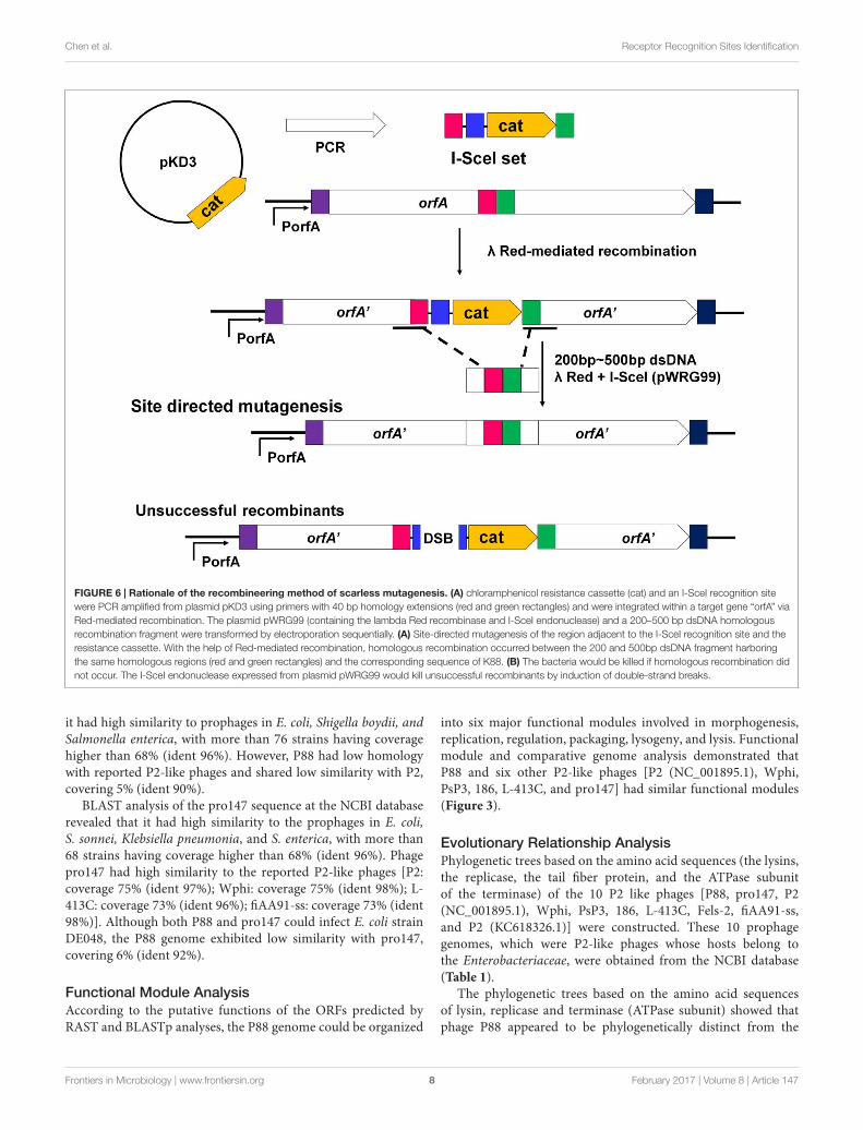
Construction of Gene Mutants toAnalyze the Key Regions and SitesThe recombineering method of scarless mutagenesis was carriedout according to the protocol described previously (Blank et al.,2011) with some modifications. The recombineering method ofscarless mutagenesis was divided into two steps. In the first step,the target gene was replaced with a chloramphenicol resistancecassette (cat) gene and the I-SceI recognition site, which wasPCR-amplified from plasmid pKD3 (Supplementary Table S3).The primers (Supplementary Table S3) were homologousto the flanking region of the corresponding target gene.
1http://www.sbg.bio.ic.ac.uk/phyre2/html/
The PCR products were then transformed by electroporationinto K88 containing the lambda Red recombinase expressionplasmid pKD46 (Datsenko and Wanner, 2000; Wang et al.,2011). After electroporation, samples were incubated at 30◦Cfor 90 min in SOC broth (Datsenko and Wanner, 2000)and plated on LB agar with chloramphenicol to select fortarget gene mutants. Mutants were confirmed by PCR andsequencing using primers k1 and k2 (c1 and c2) (SupplementaryTable S3). In the second step, the PCR products containingregions homologous to the flanking regions of the resistancegene for homologous recombination were transformed byelectroporation into K88 containing the lambda Red recombinaseexpression and I-SceI endonuclease expression plasmid pWRG99(Supplementary Table S3). After electroporation, samples wereincubated at 30◦C for 90 min in SOC broth and plated onLB agar with tetracycline hydrochloride. Selection of successfulrecombinants was mediated by the sequential expressionof I-SceI endonuclease after Red-mediated recombination. Ifrecombination occurred, the 200–500 bp dsDNA fragmentharboring the same homologous region would replace thecorresponding sequence of K88. If no recombination occurred,the unique I-SceI restriction site within the genome wouldstill be present and the I-SceI endonuclease expressed fromplasmid pWRG99 would kill unsuccessful recombinants byinduction of double-strand breaks. Finally, pWRG99 was curedby streaking colonies and incubated at 37◦C. Mutants wereconfirmed by PCR and sequencing using primers check1 andcheck2 (Supplementary Table S3).
Induction, Lytic Capacity Analysis, andHost Range Test of P88 MutantsEach K88 mutant strain was cultured for 5 h in LB mediumand then diluted 1:100 in 5 mL fresh medium. When the culturereached an OD600 of 0.2, mitomycin C was added and then wasincubated at 30◦C for 10 h. The lysates were centrifuged and usedto test their lytic capacity by the double-layer agar plate methodwith DE048, the host of P88.
Phage P88 could not infect the bacteria if the phage suspensionwas dropped onto the LB agar plate spread with bacteria, so thehost range analysis of each P88 mutant strain constructed by thescarless mutagenesis method was carried out by the double-layeragar plate method with DE048, MC1061, DH5a and BL21.
RESULTS
Two Prophages of P88 and pro147 WereInduced from 54 E. coli StrainsPhage induction of 54 E. coli strains was investigated bythe addition of mitomycin C. After induction, cell lysateswere prepared and tested for their ability to lyse variousE. coli hosts in plaque assays using the 108 strains listed inSupplementary Table S2. Out of the 54 E. coli strains used, thelysates of E. coli strain K88 and DE147 produced phagesthat could lyse the clinical isolates of avian pathogenicE. coli (APEC) strain DE048. These two phages were
Frontiers in Microbiology | www.frontiersin.org 3 February 2017 | Volume 8 | Article 147

fmicb-08-00147 January 30, 2017 Time: 15:15 # 4
Chen et al. Receptor Recognition Sites Identification
designated as P88 and pro147. After growth on DE048 indouble agar LB plates at 37◦C for 12 h, P88 formed clearand round plaques of approximately 0.06 cm in diameter(Figure 1A); pro147 formed clear and round plaques ofapproximately 0.13 cm in diameter (Figure 1B). Amongthe 108 E. coli strains tested, P88 specifically lysed DE048,while pro147 specifically lysed DE048, DH5a, BL21, andMC1061.
Optimal MOI and One-Step GrowthCurve of P88After incubation for 3.5 h, P88 had the highest concentration(1.1 × 10¹⁰) at the ratio of 0.01 (pfu/cfu); therefore, the ratio of0.01 (pfu/cfu) was considered as the optimal MOI. The one-stepgrowth curve obtained for P88 showed a latent period of about20 min, a rise period of 25 min, and an average burst size of about65 (Figure 1C).
FIGURE 1 | Fundamental characteristics of P2-like phages. (A) Plaques formed by phage P88. (B) Plaques formed by phage pro147. Growth of the phages(P88 and pro147) on Escherichia coli DE048 in double agar LB plates for 6 h. P88 formed clear and round plaques of approximately 0.06 cm in diameter. Pro147formed clear and round plaques of approximately 0.13 cm in diameter. (C) One-step growth curve. Phage and bacteria were mixed, and samples containing onlyabsorbed phage were taken at 5 min intervals and diluted. The double-layer plate method was used to determine the plaque forming units (pfu). One-step growthcurve for P88 showed a latent period of about 20 min, a rise period of 25 min, and an average burst size of about 65. (D) Electron microscopy image of P88.(E) Electron microscopy image of pro147. Electron microscopy showed that the heads of P88 and pro147 were icosahedrons, and the tails was long and couldshrink. For P88, the diameter of its capsid head was estimated at 50 nm and the tail length was estimated at 140 nm. For pro147, the diameter of its capsid headwas estimated at 50 nm and the tail length was estimated at 130 nm.
Frontiers in Microbiology | www.frontiersin.org 4 February 2017 | Volume 8 | Article 147
fmicb-08-00147 January 30, 2017 Time: 15:15 # 5
Chen et al. Receptor Recognition Sites Identification
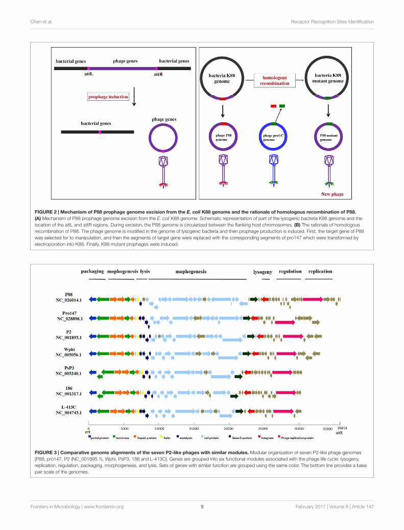
FIGURE 2 | Mechanism of P88 prophage genome excision from the E. coli K88 genome and the rationale of homologous recombination of P88.(A) Mechanism of P88 prophage genome excision from the E. coli K88 genome. Schematic representation of part of the lysogenic bacteria K88 genome and thelocation of the attL and attR regions. During excision, the P88 genome is circularized between the flanking host chromosomes. (B) The rationale of homologousrecombination of P88. The phage genome is modified in the genome of lysogenic bacteria and then prophage production is induced. First, the target gene of P88was selected for to manipulation, and then the segments of target gene were replaced with the corresponding segments of pro147 which were transformed byelectroporation into K88. Finally, K88 mutant prophages were induced.
FIGURE 3 | Comparative genome alignments of the seven P2-like phages with similar modules. Modular organization of seven P2-like phage genomes[P88, pro147, P2 (NC_001895.1), Wphi, PsP3, 186 and L-413C]. Genes are grouped into six functional modules associated with the phage life cycle: lysogeny,replication, regulation, packaging, morphogenesis, and lysis. Sets of genes with similar function are grouped using the same color. The bottom line provides a basepair scale of the genomes.
Frontiers in Microbiology | www.frontiersin.org 5 February 2017 | Volume 8 | Article 147
fmicb-08-00147 January 30, 2017 Time: 15:15 # 6
Chen et al. Receptor Recognition Sites Identification
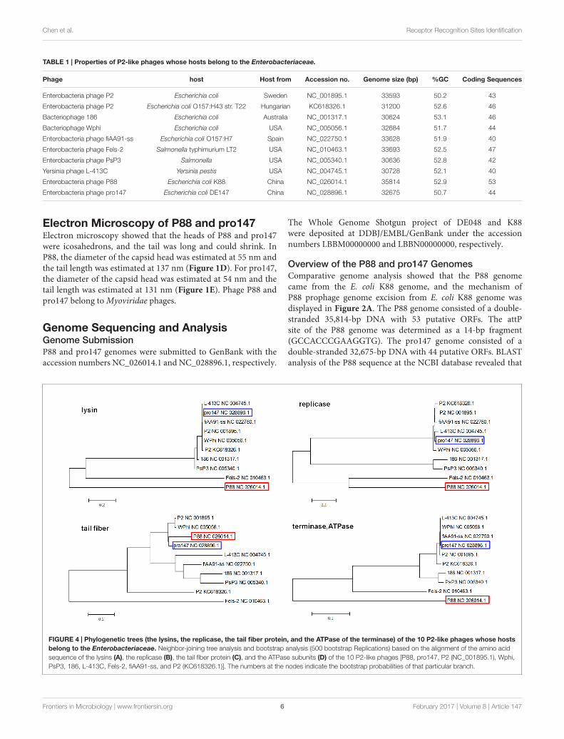
TABLE 1 | Properties of P2-like phages whose hosts belong to the Enterobacteriaceae.
Phage host Host from Accession no. Genome size (bp) %GC Coding Sequences
Enterobacteria phage P2 Escherichia coli Sweden NC_001895.1 33593 50.2 43
Enterobacteria phage P2 Escherichia coli O157:H43 str. T22 Hungarian KC618326.1 31200 52.6 46
Bacteriophage 186 Escherichia coli Australia NC_001317.1 30624 53.1 46
Bacteriophage Wphi Escherichia coli USA NC_005056.1 32684 51.7 44
Enterobacteria phage fiAA91-ss Escherichia coli O157:H7 Spain NC_022750.1 33628 51.9 40
Enterobacteria phage Fels-2 Salmonella typhimurium LT2 USA NC_010463.1 33693 52.5 47
Enterobacteria phage PsP3 Salmonella USA NC_005340.1 30636 52.8 42
Yersinia phage L-413C Yersinia pestis USA NC_004745.1 30728 52.1 40
Enterobacteria phage P88 Escherichia coli K88 China NC_026014.1 35814 52.9 53
Enterobacteria phage pro147 Escherichia coli DE147 China NC_028896.1 32675 50.7 44
Electron Microscopy of P88 and pro147Electron microscopy showed that the heads of P88 and pro147were icosahedrons, and the tail was long and could shrink. InP88, the diameter of the capsid head was estimated at 55 nm andthe tail length was estimated at 137 nm (Figure 1D). For pro147,the diameter of the capsid head was estimated at 54 nm and thetail length was estimated at 131 nm (Figure 1E). Phage P88 andpro147 belong to Myoviridae phages.
Genome Sequencing and AnalysisGenome SubmissionP88 and pro147 genomes were submitted to GenBank with theaccession numbers NC_026014.1 and NC_028896.1, respectively.
The Whole Genome Shotgun project of DE048 and K88were deposited at DDBJ/EMBL/GenBank under the accessionnumbers LBBM00000000 and LBBN00000000, respectively.
Overview of the P88 and pro147 GenomesComparative genome analysis showed that the P88 genomecame from the E. coli K88 genome, and the mechanism ofP88 prophage genome excision from E. coli K88 genome wasdisplayed in Figure 2A. The P88 genome consisted of a double-stranded 35,814-bp DNA with 53 putative ORFs. The attPsite of the P88 genome was determined as a 14-bp fragment(GCCACCCGAAGGTG). The pro147 genome consisted of adouble-stranded 32,675-bp DNA with 44 putative ORFs. BLASTanalysis of the P88 sequence at the NCBI database revealed that
FIGURE 4 | Phylogenetic trees (the lysins, the replicase, the tail fiber protein, and the ATPase of the terminase) of the 10 P2-like phages whose hostsbelong to the Enterobacteriaceae. Neighbor-joining tree analysis and bootstrap analysis (500 bootstrap Replications) based on the alignment of the amino acidsequence of the lysins (A), the replicase (B), the tail fiber protein (C), and the ATPase subunits (D) of the 10 P2-like phages [P88, pro147, P2 (NC_001895.1), Wphi,PsP3, 186, L-413C, Fels-2, fiAA91-ss, and P2 (KC618326.1)]. The numbers at the nodes indicate the bootstrap probabilities of that particular branch.
Frontiers in Microbiology | www.frontiersin.org 6 February 2017 | Volume 8 | Article 147
fmicb-08-00147 January 30, 2017 Time: 15:15 # 7
Chen et al. Receptor Recognition Sites Identification
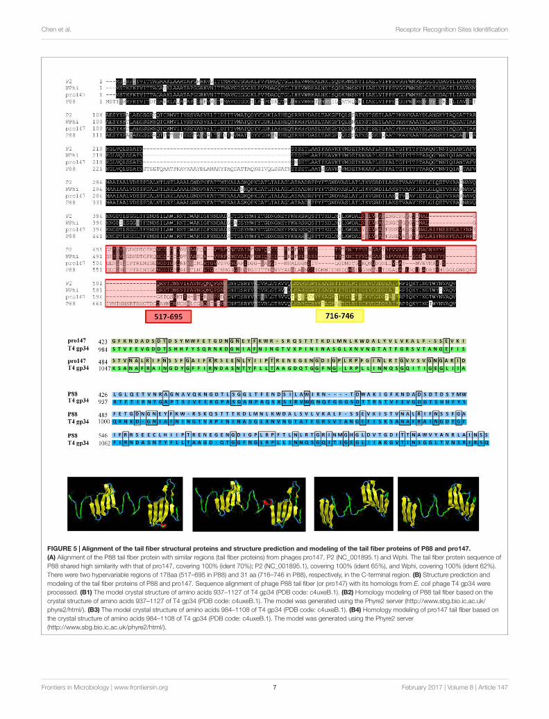
FIGURE 5 | Alignment of the tail fiber structural proteins and structure prediction and modeling of the tail fiber proteins of P88 and pro147.(A) Alignment of the P88 tail fiber protein with similar regions (tail fiber proteins) from phages pro147, P2 (NC_001895.1) and Wphi. The tail fiber protein sequence ofP88 shared high similarity with that of pro147, covering 100% (ident 70%); P2 (NC_001895.1), covering 100% (ident 65%), and Wphi, covering 100% (ident 62%).There were two hypervariable regions of 178aa (517–695 in P88) and 31 aa (716–746 in P88), respectively, in the C-terminal region. (B) Structure prediction andmodeling of the tail fiber proteins of P88 and pro147. Sequence alignment of phage P88 tail fiber (or pro147) with its homologs from E. coli phage T4 gp34 wereprocessed. (B1) The model crystal structure of amino acids 937–1127 of T4 gp34 (PDB code: c4uxeB.1). (B2) Homology modeling of P88 tail fiber based on thecrystal structure of amino acids 937–1127 of T4 gp34 (PDB code: c4uxeB.1). The model was generated using the Phyre2 server (http://www.sbg.bio.ic.ac.uk/phyre2/html/). (B3) The model crystal structure of amino acids 984–1108 of T4 gp34 (PDB code: c4uxeB.1). (B4) Homology modeling of pro147 tail fiber based onthe crystal structure of amino acids 984–1108 of T4 gp34 (PDB code: c4uxeB.1). The model was generated using the Phyre2 server(http://www.sbg.bio.ic.ac.uk/phyre2/html/).
Frontiers in Microbiology | www.frontiersin.org 7 February 2017 | Volume 8 | Article 147
fmicb-08-00147 January 30, 2017 Time: 15:15 # 8
Chen et al. Receptor Recognition Sites Identification
FIGURE 6 | Rationale of the recombineering method of scarless mutagenesis. (A) chloramphenicol resistance cassette (cat) and an I-SceI recognition sitewere PCR amplified from plasmid pKD3 using primers with 40 bp homology extensions (red and green rectangles) and were integrated within a target gene “orfA” viaRed-mediated recombination. The plasmid pWRG99 (containing the lambda Red recombinase and I-SceI endonuclease) and a 200–500 bp dsDNA homologousrecombination fragment were transformed by electroporation sequentially. (A) Site-directed mutagenesis of the region adjacent to the I-SceI recognition site and theresistance cassette. With the help of Red-mediated recombination, homologous recombination occurred between the 200 and 500bp dsDNA fragment harboringthe same homologous regions (red and green rectangles) and the corresponding sequence of K88. (B) The bacteria would be killed if homologous recombination didnot occur. The I-SceI endonuclease expressed from plasmid pWRG99 would kill unsuccessful recombinants by induction of double-strand breaks.
it had high similarity to prophages in E. coli, Shigella boydii, andSalmonella enterica, with more than 76 strains having coveragehigher than 68% (ident 96%). However, P88 had low homologywith reported P2-like phages and shared low similarity with P2,covering 5% (ident 90%).
BLAST analysis of the pro147 sequence at the NCBI databaserevealed that it had high similarity to the prophages in E. coli,S. sonnei, Klebsiella pneumonia, and S. enterica, with more than68 strains having coverage higher than 68% (ident 96%). Phagepro147 had high similarity to the reported P2-like phages [P2:coverage 75% (ident 97%); Wphi: coverage 75% (ident 98%); L-413C: coverage 73% (ident 96%); fiAA91-ss: coverage 73% (ident98%)]. Although both P88 and pro147 could infect E. coli strainDE048, the P88 genome exhibited low similarity with pro147,covering 6% (ident 92%).
Functional Module AnalysisAccording to the putative functions of the ORFs predicted byRAST and BLASTp analyses, the P88 genome could be organized
into six major functional modules involved in morphogenesis,replication, regulation, packaging, lysogeny, and lysis. Functionalmodule and comparative genome analysis demonstrated thatP88 and six other P2-like phages [P2 (NC_001895.1), Wphi,PsP3, 186, L-413C, and pro147] had similar functional modules(Figure 3).
Evolutionary Relationship AnalysisPhylogenetic trees based on the amino acid sequences (the lysins,the replicase, the tail fiber protein, and the ATPase subunitof the terminase) of the 10 P2 like phages [P88, pro147, P2(NC_001895.1), Wphi, PsP3, 186, L-413C, Fels-2, fiAA91-ss,and P2 (KC618326.1)] were constructed. These 10 prophagegenomes, which were P2-like phages whose hosts belong tothe Enterobacteriaceae, were obtained from the NCBI database(Table 1).
The phylogenetic trees based on the amino acid sequencesof lysin, replicase and terminase (ATPase subunit) showed thatphage P88 appeared to be phylogenetically distinct from the
Frontiers in Microbiology | www.frontiersin.org 8 February 2017 | Volume 8 | Article 147
fmicb-08-00147 January 30, 2017 Time: 15:15 # 9
Chen et al. Receptor Recognition Sites Identification
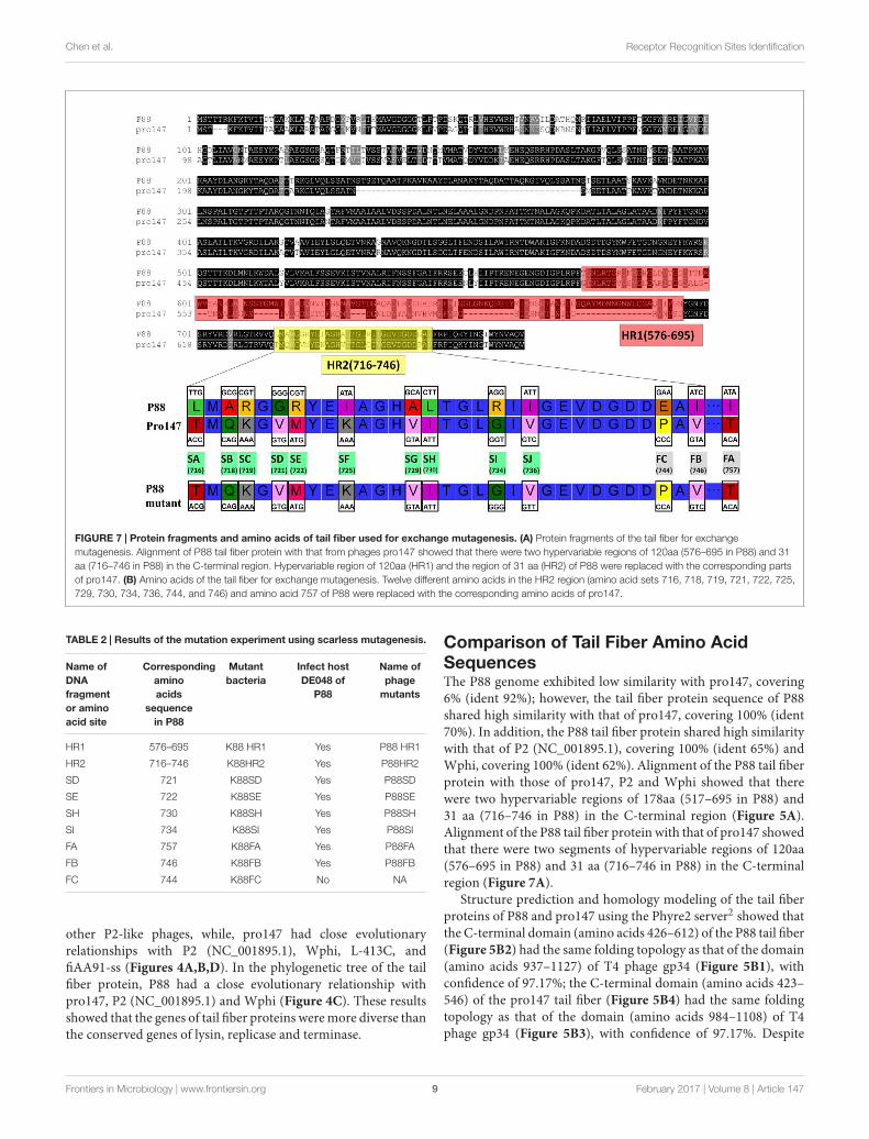
FIGURE 7 | Protein fragments and amino acids of tail fiber used for exchange mutagenesis. (A) Protein fragments of the tail fiber for exchangemutagenesis. Alignment of P88 tail fiber protein with that from phages pro147 showed that there were two hypervariable regions of 120aa (576–695 in P88) and 31aa (716–746 in P88) in the C-terminal region. Hypervariable region of 120aa (HR1) and the region of 31 aa (HR2) of P88 were replaced with the corresponding partsof pro147. (B) Amino acids of the tail fiber for exchange mutagenesis. Twelve different amino acids in the HR2 region (amino acid sets 716, 718, 719, 721, 722, 725,729, 730, 734, 736, 744, and 746) and amino acid 757 of P88 were replaced with the corresponding amino acids of pro147.
TABLE 2 | Results of the mutation experiment using scarless mutagenesis.
Name ofDNAfragmentor aminoacid site
Correspondingaminoacids
sequencein P88
Mutantbacteria
Infect hostDE048 of
P88
Name ofphage
mutants
HR1 576–695 K88 HR1 Yes P88 HR1
HR2 716–746 K88HR2 Yes P88HR2
SD 721 K88SD Yes P88SD
SE 722 K88SE Yes P88SE
SH 730 K88SH Yes P88SH
SI 734 K88SI Yes P88SI
FA 757 K88FA Yes P88FA
FB 746 K88FB Yes P88FB
FC 744 K88FC No NA
other P2-like phages, while, pro147 had close evolutionaryrelationships with P2 (NC_001895.1), Wphi, L-413C, andfiAA91-ss (Figures 4A,B,D). In the phylogenetic tree of the tailfiber protein, P88 had a close evolutionary relationship withpro147, P2 (NC_001895.1) and Wphi (Figure 4C). These resultsshowed that the genes of tail fiber proteins were more diverse thanthe conserved genes of lysin, replicase and terminase.
Comparison of Tail Fiber Amino AcidSequencesThe P88 genome exhibited low similarity with pro147, covering6% (ident 92%); however, the tail fiber protein sequence of P88shared high similarity with that of pro147, covering 100% (ident70%). In addition, the P88 tail fiber protein shared high similaritywith that of P2 (NC_001895.1), covering 100% (ident 65%) andWphi, covering 100% (ident 62%). Alignment of the P88 tail fiberprotein with those of pro147, P2 and Wphi showed that therewere two hypervariable regions of 178aa (517–695 in P88) and31 aa (716–746 in P88) in the C-terminal region (Figure 5A).Alignment of the P88 tail fiber protein with that of pro147 showedthat there were two segments of hypervariable regions of 120aa(576–695 in P88) and 31 aa (716–746 in P88) in the C-terminalregion (Figure 7A).
Structure prediction and homology modeling of the tail fiberproteins of P88 and pro147 using the Phyre2 server2 showed thatthe C-terminal domain (amino acids 426–612) of the P88 tail fiber(Figure 5B2) had the same folding topology as that of the domain(amino acids 937–1127) of T4 phage gp34 (Figure 5B1), withconfidence of 97.17%; the C-terminal domain (amino acids 423–546) of the pro147 tail fiber (Figure 5B4) had the same foldingtopology as that of the domain (amino acids 984–1108) of T4phage gp34 (Figure 5B3), with confidence of 97.17%. Despite
Frontiers in Microbiology | www.frontiersin.org 9 February 2017 | Volume 8 | Article 147
fmicb-08-00147 January 30, 2017 Time: 15:15 # 10
Chen et al. Receptor Recognition Sites Identification
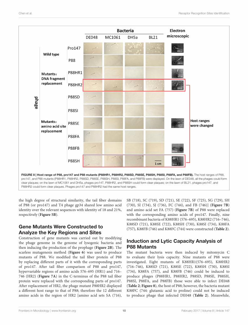
FIGURE 8 | Host range of P88, pro147 and P88 mutants (P88HR1, P88HR2, P88SD, P88SE, P88SH, P88SI, P88FA, and P88FB). The host ranges of P88,pro147, and P88 mutants (P88HR1, P88HR2, P88SD, P88SE, P88SH, P88SI, P88FA, and P88FB) were displayed. On the lawn of DE048, all the phages could formclear plaques; on the lawn of MC1061 and DH5a, phages pro147, P88HR2, and P88SH could form clear plaques; on the lawn of BL21, phages pro147, andP88HR2 could form clear plaques. Phages pro147 and P88HR2 had the same host ranges.
the high degree of structural similarity, the tail fiber domainsof P88 (or pro147) and T4 phage gp34 shared low amino acididentity over the relevant sequences with identity of 18 and 21%,respectively (Figure 5B).
Gene Mutants Were Constructed toAnalyze the Key Regions and SitesConstruction of gene mutants was carried out by modifyingthe phage genome in the genome of lysogenic bacteria andthen inducing the production of the prophage (Figure 2B). Thescarless mutagenesis method (Figure 6) was used to producemutants of P88. We modified the tail fiber protein of P88by replacing different parts of it with the corresponding partsof pro147. After tail fiber comparison of P88 and pro147,hypervariable regions of amino acids 576–695 (HR1) and 716–746 (HR2) (Figure 7A) in the C-terminus of the P88 tail fiberprotein were replaced with the corresponding parts of pro147.After replacement of HR2, the phage mutant P88HR2 displayeda different host range to that of P88; therefore the 12 differentamino acids in the region of HR2 [amino acid sets SA (716),
SB (718), SC (719), SD (721), SE (722), SF (725), SG (729), SH(730), SI (734), SJ (736), FC (744), and FB (746)] (Figure 7B)and amino acid set FA (757) (Figure 7B) of P88 were replacedwith the corresponding amino acids of pro147. Finally, ninerecombinant bacteria of K88HR1 (576–695), K88HR2 (716–746),K88SD (721), K88SE (722), K88SH (730), K88SI (734), K88FA(757), K88FB (746) and K88FC (744) were constructed (Table 2).
Induction and Lytic Capacity Analysis ofP88 MutantsThe mutant bacteria were then induced by mitomycin Cto evaluate their lysis capacity. Nine mutants of P88 wereinvestigated. Eight mutants of K88HR1(576–695), K88HR2(716–746), K88SD (721), K88SE (722), K88SH (730), K88SI(734), K88FA (757), and K88FB (746) could be induced toproduce phages (P88HR1, P88HR2, P88SD, P88SE, P88SH,P88SI, P88FA, and P88FB) those were able to infect DE048(Table 2; Figure 8), the host of P88; however, the bacteria mutantK88FC (744: glutamic acid to proline) could not be inducedto produce phage that infected DE048 (Table 2). Meanwhile,
Frontiers in Microbiology | www.frontiersin.org 10 February 2017 | Volume 8 | Article 147

fmicb-08-00147 January 30, 2017 Time: 15:15 # 11
Chen et al. Receptor Recognition Sites Identification
K88FC could not be induced to produce phage to infect MC1061,DH5a, and BL21. The phage particles induced from the mutantsthat could lyse the E. coli strain DE048 were enriched for thesubsequent experiment of host range determination and electronmicroscopy.
The Host Range Test of P88 MutantsThe host ranges of P88, pro147 and P88 mutants (P88HR1,P88HR2, P88SD, P88SE, P88SH, P88SI, P88FA, and P88FB) wereanalyzed. After incubation at 37◦C for 10 h, on the lawn ofDE048, all the phages could form clear plaques; on the lawn ofMC1061 and DH5a, phages pro147, P88HR2 and P88SH (730:leucine to isoleucine) could form clear plaques; on the lawn ofBL21, phages pro147 and P88HR2 could form clear plaques;pro147 and P88HR2 had the same host ranges (Figure 8). P88,pro147 and all the P88 mutants could not infect DE048 whenincubated at 28◦C.
Electron micrographs showed that compared with those of theoriginal P88, the head and the tail of the phage mutants withaltered host ranges (P88HR2 and P88SH) and with no changein host range (P88HR1, P88SE, and P88FB) did not have visibledifferences (Figure 8).
DISCUSSION
We screened two P2-like coliphages, named P88 and pro147,and identified their corresponding host bacteria. Analysis of thekey regions and sites that might determine receptor recognitionshowed that the host range of P88 was altered after replacingthe hypervariable regions HR2 (amino acids 716–746) of thetail fiber protein of P88 with that of pro147; the host rangesof P88 with replacement of amino acid 730 (leucine) and 744(glutamic acid) were changed after replacing six amino acidsin HR2 with the corresponding amino acids of pro147. Theseresults showed that, in P88, the tail fiber determined the receptorrecognition; the region of amino acids 716–746 was importantfor receptor recognition; amino acids 730 (leucine) and 744(glutamic acid) were the key amino acids of P88 that determinethe host range.
Replacing regions of the tail fiber protein in P88 with thoseof pro147 changed the host range, which indicated that thecorresponding regions of pro147 would determine its receptorrecognition. Furthermore, the high homology of the tail fibersequences in P2-like phages P88, pro147, P2 (NC_001895.1), andWphi (005056.1) caused us to speculate that the correspondingparts in P2 and Wphi would also determine their receptorrecognition specificity.
For temperate phages, when the phage genome is modifiedby manipulating the bacterial genome, the mutant strainsconstructed could be confirmed by PCR and the prophage couldthen be introduced and characterized. If the phage genome wasmodified in the host cell when it was separated from the bacterialchromosome by homologous recombination with a homologousfragment in the form of plasmid or DNA fragment, like lyticphages (Yoichi et al., 2005; Mahichi et al., 2009; Thomasonet al., 2009), a plaque assay was used to screen the mutantphages; however, the recombinant phage would not be selected
without a sensitive host strain. Hence, to construct mutants oftemperate phages, modifying the phage genome by manipulatingthe bacterial genome was preferred.
The lysogeny module is required to form stable lysogens(Court et al., 2007). It is thought that a phage should be lytic andnot lysogenic for use as an antibacterial agent (Chan et al., 2013).Here, the phage modification experiment was conducted onlysogenic bacteria (phage in the lysogenic state). In other words,the induced phage was temperate and able to perform lysogenicinfections. Therefore, to disrupt lysogenic infection, the genesof lysogeny module could be deleted without disrupting thelytic ability of the phage (Zhang et al., 2013). Thus, comparedwith wild temperate phage strains, the novel modified phagesare expected to minimize potential hazards to be integratedinto the bacteria in normal flora if used as antibacterialagents.
In the next step of our research, we could create a homologoussequence library that contains sufficient allelic sequences tomodify specific binding sequences by gene synthesis andhomologous sequences enriched from natural samples. Thecrystal structure of the tail fiber protein with the key sites forreceptor binding would be analyzed to understand how P2-likephages recognize their host.
CONCLUSION
This study located the specific binding sequences that determinedthe phage host range of P88 and increased our understandingof the function of the tail fibers in P2-like phages. The resultrevealed that lysogenic bacteria could be used as vectors tochange or broaden a phage’s host range. It might also bepossible to modify the genomes to analyze gene function orchange/expand the host range of other bacteriophages using thismethod.
AUTHOR CONTRIBUTIONS
WZ and MC conceived the study and designed experiments. MC,HY, and CL performed the data analysis. MC, LZ, and SX carriedout experiments. MC and LZ analyzed experimental results. MCand WZ wrote the manuscript.
ACKNOWLEDGMENTS
This work was supported by grants from National NaturalScience Foundation of China (31322054), Priority AcademicProgram Development of Jiangsu Higher Education Institutionsand the Special Fund for Public Welfare Industry of ChineseMinistry of Agriculture (201303041).
SUPPLEMENTARY MATERIAL
The Supplementary Material for this article can be found onlineat: http://journal.frontiersin.org/article/10.3389/fmicb.2017.00147/full#supplementary-material
Frontiers in Microbiology | www.frontiersin.org 11 February 2017 | Volume 8 | Article 147
fmicb-08-00147 January 30, 2017 Time: 15:15 # 12
Chen et al. Receptor Recognition Sites Identification
REFERENCESAdams, M. H. (1959). Bacteriophages. New York: Interscience.Aziz, R. K., Bartels, D., Best, A. A., Dejongh, M., Disz, T., Edwards, R. A., et al.
(2008). The RAST server: rapid annotations using subsystems technology. BMCGenomics 9:75 doi: 10.1186/1471-2164-9-75
Bartual, S. G., Otero, J. M., Garcia-Doval, C., Llamas-Saiz, A. L., Kahn, R., Fox,G. C., et al. (2010). Structure of the bacteriophage T4 long tail fiber receptor-binding tip. Proc. Natl. Acad. Sci. U.S.A. 107, 20287–20292. doi: 10.1073/pnas.1011218107
Blank, K., Hensel, M., and Gerlach, R. G. (2011). Rapid and highly efficient methodfor scarless mutagenesis within the Salmonella enterica chromosome. PLoSONE 6:e15763. doi: 10.1371/journal.pone.0015763
Brettin, T., Davis, J. J., Disz, T., Edwards, R. A., Gerdes, S., Olsen, G. J., et al. (2015).RASTtk: a modular and extensible implementation of the RAST algorithm forbuilding custom annotation pipelines and annotating batches of genomes. Sci.Rep. 5:8365 doi: 10.1038/srep08365
Brussow, H. (2012). What is needed for phage therapy to become a realityin Western medicine? Virology 434, 138–142. doi: 10.1016/j.virol.2012.09.015
Chan, B. K., Abedon, S. T., and Loc-Carrillo, C. (2013). Phage cocktails andthe future of phage therapy. Future Microbiol. 8, 769–783. doi: 10.2217/fmb.13.47
Chow, J. J., Batt, C. A., and Sinskey, A. J. (1988). Characterization of Lactobacillusbulgaricus bacteriophage ch2. Appl. Environ. Microbiol. 54, 1138–1142.
Clark, J. R., and March, J. B. (2006). Bacteriophages and biotechnology: vaccines,gene therapy and antibacterials. Trends Biotechnol. 24, 212–218. doi: 10.1016/j.tibtech.2006.03.003
Court, D. L., Oppertheim, A. B., and Adhya, S. L. (2007). A new look atbacteriophage lambda genetic networks. J. Bacteriol. 189, 298–304. doi: 10.1128/JB.01215-06
Croxen, M. A., Law, R. J., Scholz, R., Keeney, K. M., Wlodarska, M., andFinlay, B. B. (2013). Recent advances in understanding enteric pathogenicEscherichia coli. Clin. Microbiol. Rev. 26, 822–880. doi: 10.1128/CMR.00022-13
Datsenko, K. A., and Wanner, B. L. (2000). One-step inactivation of chromosomalgenes in Escherichia coli K-12 using PCR products. Proc. Natl. Acad. Sci. U.S.A.97, 6640–6645. doi: 10.1073/pnas.120163297
Gorski, A., Miedzybrodzki, R., Weber-Dabrowska, B., Fortuna, W., Letkiewicz, S.,Rogoz, P., et al. (2016). Phage therapy: combating infections with potential forevolving from merely a treatment for complications to targeting diseases. Front.Microbiol. 7:1515. doi: 10.3389/fmicb.2016.01515
Haq, I. U., Chaudhry, W. N., Akhtar, M. N., Andleeb, S., and Qadri, I. (2012).Bacteriophages and their implications on future biotechnology: a review. Virol.J. 9:9 doi: 10.1186/1743-422X-9-9
Hasan, B., Faruque, R., Drobni, M., Waldenstrom, J., Sadique, A., Ahmed, K. U.,et al. (2011). High prevalence of antibiotic resistance in pathogenic Escherichiacoli from large- and small-scale poultry farms in Bangladesh. Avian Dis. 55,689–692. doi: 10.1637/9686-021411-Reg.1
Hejnowicz, M. S., Golebiewski, M., and Bardowski, J. (2009). Analysis ofthe complete genome sequence of the lactococcal bacteriophage bIBB29.Int. J. Food Microbiol. 131, 52–61. doi: 10.1016/j.ijfoodmicro.2008.06.010
Johnson, J. R., and Russo, T. A. (2002). Uropathogenic Escherichia coli as agents ofdiverse non-urinary tract extraintestinal infections. J. Infect. Dis. 186, 859–864.doi: 10.1086/342490
Kageyama, Y., Murayama, M., Onodera, T., Yamada, S., Fukada, H., Kudou, M.,et al. (2009). Observation of the membrane binding activity and domainstructure of gpV, which comprises the tail spike of bacteriophage P2.Biochemistry 48, 10129–10135. doi: 10.1021/bi900928n
Kaper, J. B., Nataro, J. P., and Mobley, H. L. T. (2004). Pathogenic Escherichia coli.Nat. Rev. Microbiol. 2, 123–140. doi: 10.1038/nrmicro818
Keen, E. C. (2012). Phage therapy: concept to cure. Front. Microbiol. 3:238 doi:10.3389/fmicb.2012.00238
Kelley, L. A., Mezulis, S., Yates, C. M., Wass, M. N., and Sternberg, M. J. (2015). ThePhyre2 web portal for protein modeling, prediction and analysis. Nat. Protoc.10, 845–858. doi: 10.1038/nprot.2015.053
Kim, K. S. (2012). Current concepts on the pathogenesis of Escherichia colimeningitis: implications for therapy and prevention. Curr. Opin. Infect. Dis. 25,273–278. doi: 10.1097/QCO.0b013e3283521eb0
Kim, T. E., Jeong, Y. W., Cho, S. H., Kim, S. J., and Kwon, H. J. (2007).Chronological study of antibiotic resistances and their relevant genes in Koreanavian pathogenic Escherichia coli isolates. J. Clin. Microbiol. 45, 3309–3315.doi: 10.1128/JCM.01922-06
Liu, Y. Y., Wang, Y., Walsh, T. R., Yi, L. X., Zhang, R., Spencer, J., et al.(2016). Emergence of plasmid-mediated colistin resistance mechanism MCR-1 in animals and human beings in China: a microbiological and molecularbiological study. Lancet Infect. Dis. 16, 161–168. doi: 10.1016/S1473-3099(15)00424-7
Lu, Z., Breidt, F., Fleming, H. P., Altermann, E., and Klaenhammer, T. R. (2003).Isolation and characterization of a Lactobacillus plantarum bacteriophage,Phi JL-1, from a cucumber fermentation. Int. J. Food Microbiol. 84,225–235.
Ma, J., Sun, M., Bao, Y., Pan, Z., Zhang, W., Lu, C., et al. (2013). Genetic diversityand features analysis of type VI secretion systems loci in avian pathogenicEscherichia coli by wide genomic scanning. Infect. Genet. Evol. 20, 454–464.doi: 10.1016/j.meegid.2013.09.031
Ma, Y. L., and Lu, C. P. (2008). Isolation and identification of a bacteriophagecapable of infecting Streptococcus suis type 2 strains. Vet. Microbiol. 132,340–347. doi: 10.1016/j.vetmic.2008.05.013
Mahichi, F., Synnott, A. J., Yamamichi, K., Osada, T., and Tanji, Y. (2009).Site-specific recombination of T2 phage using IP008 long tail fiber genesprovides a targeted method for expanding host range while retaining lyticactivity. FEMS Microbiol. Lett. 295, 211–217. doi: 10.1111/j.1574-6968.2009.01588.x
Montag, D., Hashemolhosseini, S., and Henning, U. (1990). Receptor-recognizing proteins of T-even type bacteriophages. The receptor-recognizingarea of proteins 37 of phages T4 TuIa and TuIb. J. Mol. Biol. 216,327–334.
Overbeek, R., Olson, R., Pusch, G. D., Olsen, G. J., Davis, J. J., Disz, T., et al.(2014). The SEED and the rapid annotation of microbial genomes usingsubsystems technology (RAST). Nucleic Acids Res. 42, D206–D214. doi: 10.1093/nar/gkt1226
Posfai, G., Kolisnychenko, V., Bereczki, Z., and Blattner, F. R. (1999). Markerlessgene replacement in Escherichia coli stimulated by a double-strand break inthe chromosome. Nucleic Acids Res. 27, 4409–4415. doi: 10.1093/nar/27.22.4409
Riede, I. (1987). Receptor specificity of the short tail fibres (gp12) of T-eventype Escherichia coli phages. Mol. Gen. Genet. 206, 110–115. doi: 10.1007/BF00326544
Riede, I., Degen, M., and Henning, U. (1985). The receptor specificity ofbacteriophages can be determined by a tail fiber modifying protein. EMBO J.4, 2343–2346.
Tamura, K., Peterson, D., Peterson, N., Stecher, G., Nei, M., and Kumar, S. (2011).MEGA5: molecular evolutionary genetics analysis using maximum likelihood,evolutionary distance, and maximum parsimony methods. Mol. Biol. Evol. 28,2731–2739. doi: 10.1093/molbev/msr121
Thomason, L. C., Oppenheim, A. B., and Court, D. L. (2009). Modifyingbacteriophage lambda with recombineering. Methods Mol. Biol. 501, 239–251.doi: 10.1007/978-1-60327-164-6_21
Thomassen, E., Gielen, G., Schutz, M., Schoehn, G., Abrahams, J. P., Miller, S., et al.(2003). The structure of the receptor-binding domain of the bacteriophage T4short tail fibre reveals a knitted trimeric metal-binding fold. J. Mol. Biol. 331,361–373. doi: 10.1016/S0022-2836(03)00755-1
Wang, S., Niu, C., Shi, Z., Xia, Y., Yaqoob, M., Dai, J., et al. (2011).Effects of ibeA deletion on virulence and biofilm formation of avianpathogenic Escherichia coli. Infect. Immunol. 79, 279–287. doi: 10.1128/IAI.00821-10
Yoichi, M., Abe, M., Miyanaga, K., Unno, H., and Tanji, Y. (2005).Alteration of tail fiber protein gp38 enables T2 phage to infect Escherichiacoli O157:H7. J. Biotechnol. 115, 101–107. doi: 10.1016/j.jbiotec.2004.08.003
Young, R., and Gill, J. J. (2015). Phage therapy redux - What is to be done? Science350, 1163–1164. doi: 10.1126/science.aad6791
Frontiers in Microbiology | www.frontiersin.org 12 February 2017 | Volume 8 | Article 147

fmicb-08-00147 January 30, 2017 Time: 15:15 # 13
Chen et al. Receptor Recognition Sites Identification
Yu, B. J., Kang, K. H., Lee, J. H., Sung, B. H., Kim, M. S., and Kim, S. C. (2008).Rapid and efficient construction of markerless deletions in the Escherichia coligenome. Nucleic Acids Res. 36:e84 doi: 10.1093/nar/gkn359
Yu, F., and Mizushima, S. (1982). Roles of lipopolysaccharide and outermembrane protein OmpC of Escherichia coli K-12 in the receptor function forbacteriophage T4. J. Bacteriol. 151, 718–722.
Zhang, H., Fouts, D. E., Depew, J., and Stevens, R. H. (2013). Genetic modificationsto temperate Enterococcus faecalis phage Ef11 that abolish the establishment oflysogeny and sensitivity to repressor, and increase host range and productivityof lytic infection. Microbiology 159, 1023–1035. doi: 10.1099/mic.0.067116-0
Conflict of Interest Statement: The authors declare that the research wasconducted in the absence of any commercial or financial relationships that couldbe construed as a potential conflict of interest.
Copyright © 2017 Chen, Zhang, Xin, Yao, Lu and Zhang. This is an open-accessarticle distributed under the terms of the Creative Commons Attribution License(CC BY). The use, distribution or reproduction in other forums is permitted, providedthe original author(s) or licensor are credited and that the original publication in thisjournal is cited, in accordance with accepted academic practice. No use, distributionor reproduction is permitted which does not comply with these terms.
Frontiers in Microbiology | www.frontiersin.org 13 February 2017 | Volume 8 | Article 147