In vivo processing and antibiotic activity of microcin B17 analogs ...
In Vivo Performance of Antibiotic Embedded Electrospun PCLMembranes for Prevention of Abdominal Adhesions
N. Bolgen,1 _I. Vargel,2 P. Korkusuz,3 Y. Z. Menceloglu,4 E. Piskin1
1 Chemical Engineering Department and Bioengineering Division and TUB_ITAK-USAM-Biyomedtek, Hacettepe University,Beytepe, Ankara, Turkey
2 Faculty of Medicine, Department of Plastic and Reconstructive Surgery, Kırıkkale University, Kırıkkale, Turkey
3 Faculty of Medicine, Department of Histology and Embryology, Hacettepe University, Ankara, Turkey
4 Faculty of Engineering and Natural Sciences, Sabancı University, Tuzla, _Istanbul, Turkey
Received 12 January 2006; revised 9 July 2006; accepted 18 July 2006Published online 13 October 2006 in Wiley InterScience (www.interscience.wiley.com). DOI: 10.1002/jbm.b.30694
Abstract: The aim of this study was to prepare nonwoven materials from poly(e-caprolactone)(PCL) and their antibiotic containing forms by electrospinning, so as to prevent postsurgery
induced abdominal adhesions in rats. e-Caprolactone was first polymerized by ring-opening
polymerization, and then it was processed into matrices composed of nanofibers by
electrospinning. Amodel antibiotic (Biteral1) was embeddedwithin a group of PCLmembranes.
In the ratmodel, defects on the abdominal walls in the peritoneumweremade to induce adhesion.
The plain or antibiotic embedded PCL membranes were implanted on the right side of the
abdominal wall. No membrane implantation was made on the left side of the abdominal wall that
served as control. Macroscopical and histological evaluations showed that using these barriers
reduces the extent, type, and tenacity of adhesion. The antibiotic embedded membranes
significantly eliminated postsurgery abdominal adhesions, and also improved healing. ' 2006
Wiley Periodicals, Inc. J Biomed Mater Res Part B: Appl Biomater 81B: 530–543, 2007
Keywords: abdominal adhesion; animal model; electrospinning; nonwoven membranes;
biodegradable nanofibers; poly(e-caprolactone)
INTRODUCTION
Abdominal adhesions are defined as pathological fibrotic
bands developed between any surfaces in the peritoneal cav-
ity.1 Adhesion formation is a well-known complication of ab-
dominal surgery, which not only renders future operations
more difficult but also is the most common cause of small
bowel obstruction, female infertility, and chronic debilitating
pain.2,3 Adhesion formation is estimated to occur in over 90%
of all abdominal surgical procedures.4 Trauma, foreign bodies,
ischemia, and infections are major factors associated with the
formation of postsurgery adhesions. Histological studies of ex-
perimental adhesion formation have demonstrated that adhe-
sions result from the normal peritoneal wound healing
response following surgery. It begins with tissue inflammation
and fibrin deposition within an inflammatory exudate. The or-
ganization of fibrin with fibroblast invasion and collagen for-
mation are followed by maturation of collagen forming a
bridge between peritoneal surfaces.5 Fibrous bands and
newly formed capillaries remain at the site and these struc-
tures form the permanent fibrotic adhesions.1
A wide variety of approaches have been demonstrated in
animal models and clinical practice to reduce or prevent ad-
hesion by improving surgical procedures and using antiadhe-
sion materials.6–8 The use of fibrinolytic agents such as
anticoagulants, anti-inflammatory agents, and antibiotics has
also been invistigated.9–11 One of the techniques that has been
studied extensively and has demonstrated the most promising
results is placing a physical barrier between the injured site
and the adjacent tissues to prevent adhesion. With this barrier
technique, surgically traumatized surfaces are kept covered
during mesothelial regeneration, thus preventing adherence of
adjacent structures and reducing adhesion formation.12 It
was shown that commercially available synthetic polymeric
materials such as silicon and polytetrafluoroethylene sheets
can effectively reduce/prevent adhesion.13 However, such
materials are nondegradable, and need to be removed by a
second surgery, which is not desirable. It has been reported
that a variety of polymer solutions such as hyaluronic acid,14
Correspondence to: E. Piskin, (e-mail: [email protected] and [email protected])
' 2006 Wiley Periodicals, Inc.
530
dextran,15 polyvinylpyrolidone, carboxymethylcellulose,16 and
polyethylene glycol17 have been used as potential agents to
prevent adhesion; however, their effectiveness is low in most
cases.9 Hyaluronic acid has been considered a good antiadhe-
sive agent. However, it disappears from the injured site very
quickly, which limits its efficacy as an adhesion preventative
therapy.18 Products using carboxymethylcellulose have also
been reported to prevent adhesion formation in experimental
models.19,20 Seprafilm1 is a bioresorbable membrane barrier
composed of sodium hyaluronate and carboxymethylcellulose.
Interceed1 is a knitted fabric composed of oxidized regener-
ated cellulose. Although both Seprafilm1 and Interceed1 bar-
riers have been shown to be safe and effective in all human
clinical trials, their use does not eliminate adhesions in all
patients.8,13 Seprafilm1 is claimed to have limitations in
application and handling difficulties because of its poor me-
chanical properties within the surgical field. Similarly, the ef-
ficacy of Interceed1 may be significantly reduced in the
presence of blood, which is very frequently associated in the
surgical setting.21–23
Besides the limitations of the existing commercial prod-
ucts, use of membranes is thought to be the most effective
method for adhesion prevention. There are many ongoing de-
velopmental studies to design novel biomaterials that have
less complications and high efficiencies. It is generally agreed
that an antiadhesive membrane should be designed to stay at
the injured site during the postoperative wound healing phase
and should be absorbed by biodegradation, and also should
have the following characteristics: mechanically strong for
better handling and flexiblity, and should be biocompatible in
general sense (nontoxic, nonallergic, nonmutagenic, noncan-
cerogenic, etc.).12,24
Poly(e-caprolactone) (PCL), a semicrystalline biodegrad-
able polyester, belongs to poly(a-hyroxy acids) family. Its
copolymers with lactides and glycolide are getting increasing
attention because of their controllable biodegradation rates in
the desirable ranges (usually slower) and also more suitable
and tailor-made mechanical properties (usually more flexible
and softer) for some applications such as long-term drug
delivery and tissue repair and regeneration.25–29
One novel method of processing of biodegradable poly-
mers including poly(a-hydroxy acids) is electrospinning,
which is a unique method that produces polymer fibers with
diameters usually in nanoscale and nonwoven fibrous struc-
tures composed of these fibers.30–32 The processing of bioma-
terials by conventional means (such as film casting and
foaming) often imposes several limitations in the optimiza-
tion of their final properties. The PCL cast films are not suita-
ble for cell scaffolding because they are not porous and do
not allow cell ingrowth. In addition, the cast films can be too
brittle to be handled. In contrast, electrospun nonwoven mate-
rials have small and controllable pore size, high porosity, and
high surface area; therefore, they can be used in a wide vari-
ety of biomedical applications, such as for scaffolds in tissue
engineering.33–36
In our recent related studies, we have first synthesized
PCL with desirable molecular weight that can be electrospun
into nonwoven membranes formed of PCL nanofibers. De-
tails of polymers synthesis/characterization, and preparation/
properties of the electrospun membranes were provided in
our previous article.37 Here, we attempted to use these mem-
branes and their antibiotic embedded forms as mechanical
barrier between surgically damaged surfaces to prevent post-
surgery induced abdominal adhesions in rats.
MATERIALS AND METHODS
Membrane Preparation
e-Caprolactone (Aldrich, Germany) was dried on a molecular
sieve for about 24 h. The catalyst, stannous octoate (Sigma,
USA), and other agents were analytical grade and used as
received. The polymerization system and procedure has been
described in detail elsewhere.38 Briefly, polymerization was
performed in a glass reactor under nitrogen atmosphere for
24 h at 1208C. The monomer/catalyst ratio was 1700:1 (mol/
mol) in the homopolymerization of e-caprolactone. Low-mo-
lecular-weight residuals were removed by a dissolution-pre-
cipitation method in which chloroform and methanol were
used as the solvent and precipitant, respectively.
The number and weight average molecular weights (Mn
and Mw) and polydispersity index were determined by gel-
permeation chromatography (Shimadzu, LC 10A, Japan) in
chloroform at ambient temperature. Molecular weight of the
polymer was determined relative to narrow molecular weight
polystyrene standards.
Electrospun PCL membranes were fabricated according to
procedures previously described.37 Briefly, the membrane
was prepared by using a PCL solution in a mixture of chloro-
form and dimethylformamide (DMF), with a PCL concentra-
tion of 13 g/100 mL and a DMF content of 70%. For the
process of electrospinning, polymer solution was placed in a
glass Pasteur pipette. The copper probe of the high voltage
generator was inserted into the capillary. The grounded alu-
minum sheet was positioned opposite to the tip of the capil-
lary at a distance of 10 cm. An electrical field of 13 kV was
applied by a high voltage power supply (CPS, 2594). The
fluid jet was ejected from the capillary. As the jet accelerated
toward the grounded collector, the solvent evaporated and the
polymer nanofibers were deposited on the collector in the
form of a nonwoven fabric, that is, the PCL membrane.
Membrane Characterization
The fiber morphology of the electrospun nanofibrous struc-
tures were investigated with a scanning electron microscope
(SEM) (LEO, Supra 35VP). The SEM images were taken after
the deposition of a conductive gold coating on the electrospun
films with a sputter coater (Emitech, K950X, USA).
For mechanical tests, a Universal Test Machine (Lloyd
Instruments, LR 5K Serensworth Fareham, UK) was used.
The specimens that were cut from the nonwoven matrices
(0.5 3 5 cm2 in size and *25 mm in thickness) were uti-
531ELECTROSPUN PCL MEMBRANES FOR PREVENTION OF ABDOMINAL ADHESIONS
Journal of Biomedical Materials Research Part B: Applied BiomaterialsDOI 10.1002/jbmb
lized in the tests in which a crosshead speed 5 mm/min at
room temperature was applied.
Drug Loading and In Vitro Release
The antibiotic that was used in this study was a commercial
product, that is, Biteral1 (Roche, France), which was a drug
solution (in an ampule) containing 500 mg active substance
(i.e., ornidazole) in 3 mL (900 mg absolute alcohol and
1600 mg propylene glycol) as reported in its prospectus.
0.15 mL drug solution (containing 25 mg ornidazole) was
taken with an injector from the ampule and was dropped
slowly (evenly distributed) on the each electrospun nonwo-
ven membrane specimen (2 3 3 cm2 in size and *25 mm in
thickness). Note that because of their unique structure, the
electrospun membranes absorbed the whole drug solution.
To obtain in vitro release kinetics, each drug-loaded elec-
trospun membrane specimen was put in one flask containing
10 mL distilled water. Two milliliter solution was taken from
the medium (and 2 mL fresh water was added) every 3 h in
the first 12 h and then every 6 h for total 36 h. The drug re-
leased within the medium was measured spectrophotometri-
cally at 270 nm. Five parallel studies were conducted to obtain
the average release data.
Animal Model and Surgical Protocols
Twenty seven Wistar-Albino female rats weighing between
250 and 300 g were used. All rats were fed food and water
ad libitum. They were maintained in a temperature and humid-
ity controlled environment at the Animal Research Center of
Hacettepe University. The following study was conducted after
receiving permission/approval from the Animal Ethical Commit-
tee of the University (Approval number: B.30.2.HAC.0.01.00.05,
Approval date: April 1, 2004).
Sterile surgical technique was applied throughout the
study. The electrospun nonwoven membranes produced in the
previous step were cut into specimens (2 3 3 cm2 in size and
approximately 25 mm in thickness) and sterilized by gamma
radiation (2.5 Mrad). Animals were anesthetized by intraperi-
toneal injection with a mixture of ketamine HCl (Parke Davis,
50 mg/mL, Taiwan) and Rompun (Bayer, 2%, Germany). An
area (about 15 cm2) in the abdomen of the test animal was
shaved and disinfected with Baticon solution (Droksan, 10%,
Turkey). A longitudinal incision (*6 cm long) was made on
the midline of the abdominal wall using a blade (No. 11), and
both abdominal walls (right and left side) were reflected and
similar adhesion models were made on each of the abdominal
walls. A 2 3 3 cm2 template was put on the internal surface
(on the peritoneum) of the abdominal wall, and the perito-
neum was injured by creating vertical and horizontal lines,
which formed about 10 small rectangles with roughly the
same size, using a No. 11 blade. A tweezer was covered with
a gauze, and the outer surfaces of the internal organs exactly
seeing this defected abdominal wall area were abraded/
brushed gently to trigger the adhesion process between these
defected surfaces. The plain or antibiotic embedded PCL
membrane was fixed with 6-0 prolene suture at four corners
on the right side of the abdominal wall to cover the injured
area. The left side of the abdominal wall was defected in a
similar procedure but left as it is (no material was put), which
served as control. Then, the middle line incision was closed
using 4-0 silk suture.
Macroscopical Evaluations
The test animals were sacrificed at the selected time inter-
vals (after 14, 30, 45, 60, and 90 days of postimplantation),
and the abdominal cavity was opened and the injured sites
were first observed for the incidence of adhesion by naked
eye. The adhered sites on the injured area were marked with
a black marker. The tissue specimens (2 3 3 cm2) were sur-
gically removed from both the membrane-treated (the tissue
with the membrane) and the control (only the tissue) sites.
The removed sites (2 3 3 cm2) were put onto milimetric
papers to obtain the size of the marked area. The percent of
adhesions were then calculated from these papers using the
following data: the sum of the adhered area and the total
specimen area. The extent, type, and tenacity of the adhe-
sions were graded according to the adhesion scale described
by Haney et al.13
A statistical analysis was done by using the macroscopi-
cal observations data with the Pearson’s Chi-Square test. A
p value < 0.05 was considered significant.
Histological Evaluations
Tissue specimens removed from the injured areas were fixed
in 10% phosphate buffered formalin (pH 7.0) at room tem-
perature, rinsed in buffer, and dehydrated in a graded series
of ethanol before embedding in paraffin. Five- to seven-mi-
crometer-thick sections were cut with a rotary microtome
(Microm, HM 360, Germany). Hematoxylin and Eosin, and
Mallory Trichrome stained sections were investigated for
overall morphology, adhesion, and tissue response to the
biomaterial. The stained sections (a minimum of 10 sections
obtained from different levels of each tissue) were examined
by at least two independent and blinded investigators with a
Leica DMR microscope (Germany). The images were cap-
tured via Leica DC500 digital camera (Germany). Histologi-
cal findings were evaluated and scored in two subcategories;
consisting of cell and tissue morphology of capsule and the
surrounding tissue components of the capsule, according to
An and Friedman.39 Mean of score of two independent
investigators were taken. The scoring system is summarized
in Table I. According to this system, tissue response that
takes place in implant surrounding site (the capsule and the
surrounding connective tissue) consisting of an acute and/or
chronic inflammatory process was evaluated with its cellular
content. The capsular fibroblastic layers were counted.
Inflammatory cells (macrophages, polymorphonuclear leu-
kocytes, lymphocytes, and plasma cells) locations, presence
of giant cells, and the blood vessels were separately eval-
532 BOLGEN ET AL.
Journal of Biomedical Materials Research Part B: Applied BiomaterialsDOI 10.1002/jbmb

uated. Total tissue response was scored and expressed as
means 6 standard deviations.
A statistical analysis was done by using the surrounding
tissue and capsule formation scores with the Pearson’s Chi-
Square test. A p value < 0.05 was considered significant.
RESULTS
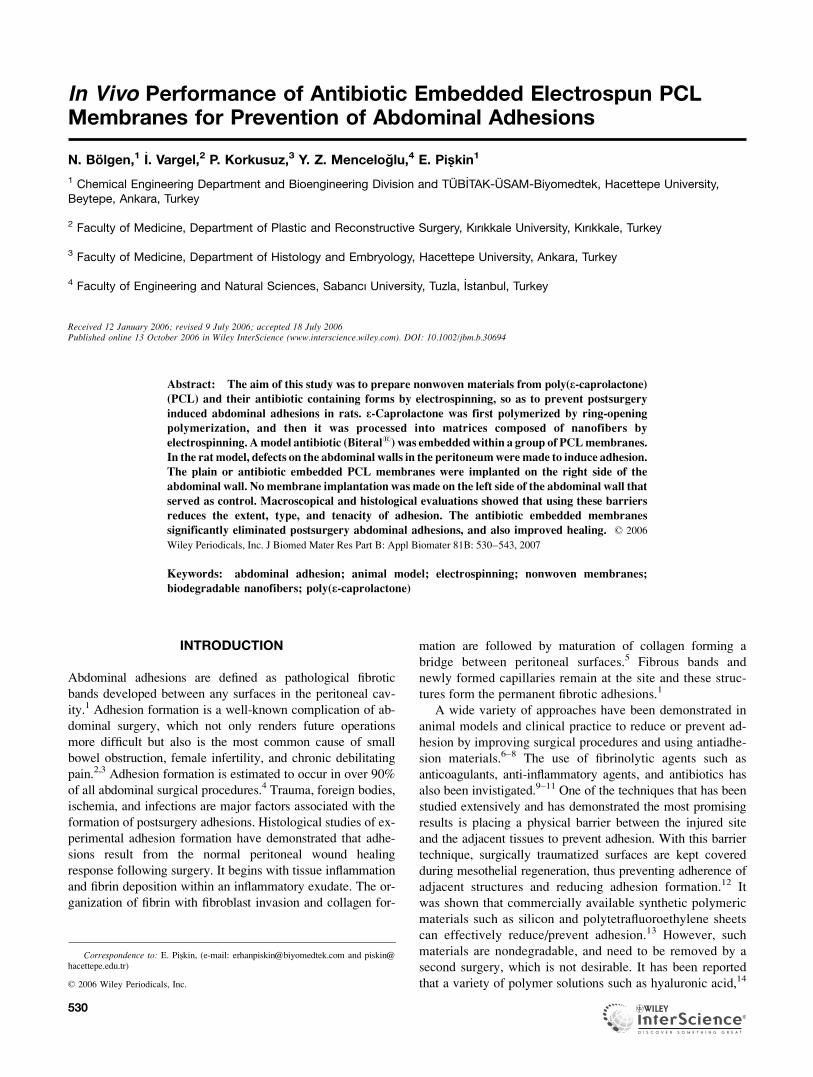
Membrane Properties
On the basis of our previous studies, we selected one of the
nonwoven PCL membranes that we produced from PCL
with different molecular weights37 and used it in our present
study as a barrier material to prevent abdominal adhesions.
Mw, Mn, and polydispersity index (Mw/Mn) of the PCL syn-
thesized and used for preparation of this material (obtained
by gel-permeation chromatography) are 84387, 51172, and
1.64, respectively. This homopolymer was dissolved in a
mixture of chloroform (30%) and DMF (70%), with a con-
centration of 13 g/100 mL, and nonwoven membranes were
prepared by electrospinning with an applied voltage and tube
tip-collector distance of 13 kV and 10 cm, respectively. The
mechanical properties, namely, elongation at break, ultimate
strength, and Young modulus of this fibrous material (with a
thickness of 25 mm) were 69.0%, 16.6 MPa, and 3.7 MPa, re-
spectively. Figure 1(A,B) gives representative images (SEM
and optical micrographs, respectively) of this membrane.
Drug Loading and In Vitro Release
In the present study, 25 mg ornidazole (‘‘Biteral1’’) was
loaded in each membrane specimen by following a very sim-
ple and effective method as described before. Then, drug
release from the electrospun membranes was followed by
measuring the drug concentration within the medium (i.e.,
distilled water), spectrophotometrically. From the release data
obtained spectrophotometrically, ‘‘percent release’’ was cal-
culated, which was the amount of the drug release at certain
TABLE I. Histological Scoring System Consists of Two Categories: 1st for the Capsule and 2nd for the Surrounding Tissue
Parameters
Scores
4 3 2 1
Capsule localization Capsule on
two sides
present
Capsule on one (lower)
side present
Capsule on one
(upper/dermis)
side present
No capsule
present
Capsule formation Dense Loose fibroadipose or
loose adipose
Loose
fibroelastic
No capsule
present
Capsule cellular
features
Fibroblast thickness More than
30 layers
10–30 layers 0–10 layers 0 layer
Fibroblast contacting
surface
No Yes
Acute/chronic
inflammatory process
Chronic Acute
Severity of inflammatory
process
Severe Moderate Mild None
Inflammatory cells
location
Inflammatory cells location End and
middle
Middle End None
Macrophages contacting
surface
No Yes
Giant cells contacting
surface
No Yes
Polymorphonuclear
leucocytes contacting
surface
No Yes
Plasma cells contacting
surface
No Yes
Blood vessels present No Yes
Capsule surrounding
tissues
Acute/chronic inflammatory
process
Chronic Acute
Severity of inflammatory
process
Severe Moderate Mild None
Macrophages No Yes
Giant cells No Yes
Polymorphonuclear
leucocytes
No Yes
Plasma cells No Yes
Blood vessels present No Yes
533ELECTROSPUN PCL MEMBRANES FOR PREVENTION OF ABDOMINAL ADHESIONS
Journal of Biomedical Materials Research Part B: Applied BiomaterialsDOI 10.1002/jbmb
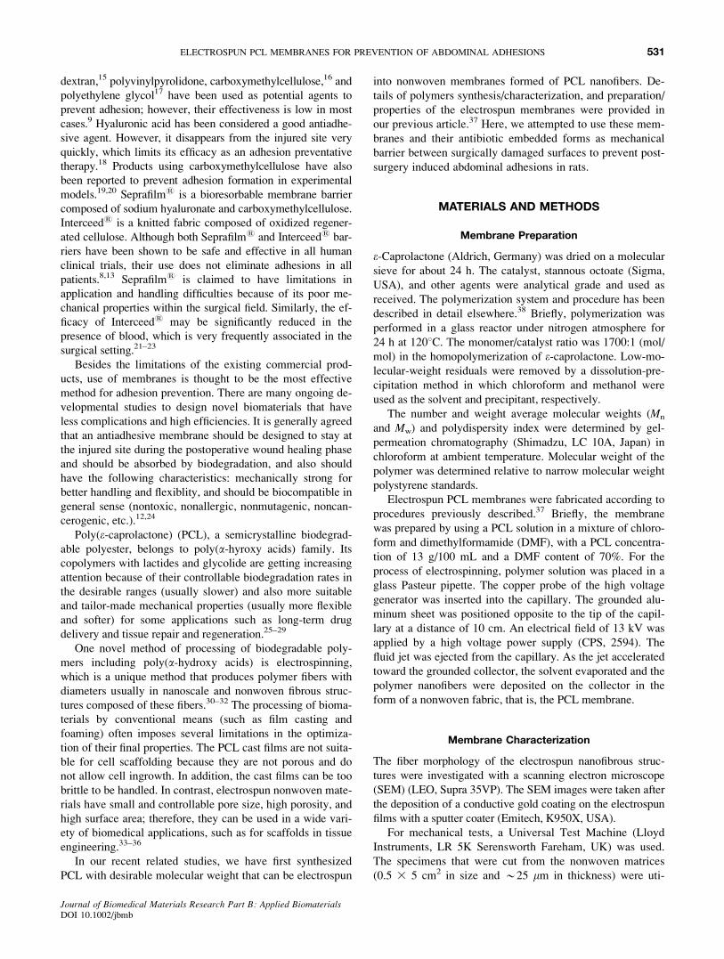
time divided by the total amount (25 mg) multiplied by 100.
The average values of five parallel studies and standard devi-
ations are given in Figure 2. As seen here, about 80% of the
drug was released in 3 h, and the release was completed
almost in 18 h.
Membrane Performance by Macroscopical Evaluation
In our animal model, defects were created by using 2 3 3 cm2
templates in two sites in the abdominal cavity. The electro-
spun membranes were implanted in one site, and the other
injured area was left as control. The adhesions, both at the
control and membrane-implanted sites in the abdominal cavity
of the experimental rats, were first evaluated macroscopically
as described in the previous sections. The extent, type, and te-
nacity of the adhesions were graded on a scale to demonstrate
the adhesion prevention efficacy of the membranes. The adhe-
sions were investigated after 14, 30, 45, 60, and 90 days of
postimplantation. The grades of adhesions for the control site
and the sites in which the plain and antibiotic embedded PCL
membranes were implanted are summarized in Table II, as the
collection of this several time points. Some selected images of
Figure 1. Representative images of the nonwoven electrospun PCL membrane: (A) a SEM micro-
graph and (B) an optical micrograph. [Color figure can be viewed in the online issue, which is avail-able at www.interscience.wiley.com.]
Figure 2. In vitro release of ornidazole from electrospun mem-branes.
TABLE II. The Extent, Type, and Tenacity of the Adhesionson the Control Site and the Sites in Which the Plainand Antibiotic Embedded PCL Membranes Were Used
Grade
Control (%)
(n ¼ 27)a
Plain
PCL (%)
(n ¼ 14)a
Antibiotic
Embedded
PCL (%)
(n ¼ 13)a
Extent (adhesion area)
No adhesion 40.7 14.3 46.1
�25% 37.0 64.3 38.5
25–50% 18.5 21.4 15.4
50–75% 0.0 0.0 0.0
�75% 3.8 0.0 0.0
Type
No adhesion 40.8 14.3 46.1
Filmy, transparent,
avascular 0.0 0.0 0.0
Opaque, translucent,
avascular 0.0 14.3 15.4
Opaque, capillaries
present 18.4 42.8 38.5
Opaque, larger vessels
present 40.8 28.6 0.0
Tenacity
No adhesion 40.7 14.3 46.1
Adhesions fall apart 0.0 14.3 7.8
Adhesions lysed
with traction 3.7 50.0 46.1
Adhesions
sharply dissected 55.6 21.4 0.0
a n, the number of the sites that were evaluated.
534 BOLGEN ET AL.
Journal of Biomedical Materials Research Part B: Applied BiomaterialsDOI 10.1002/jbmb
the control and PCL membrane implanted sites are given in
Figures 3 and 4, respectively.
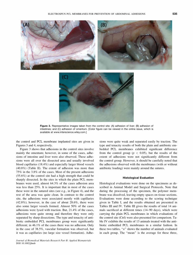
Figure 3 shows that adhesions in the control sites involve
mainly the omentum; however, in some of the cases, adhe-
sions of intestine and liver were also observed. These adhe-
sions were all over the dissected area and usually involved
blood capillaries (18.4%) and especially larger blood vessels
(40.8%) (Table II). The extent of adhesion was more than
75% in the 3.8% of the cases. Most of the present adhesions
(55.6%) at the control site had a high strength that could be
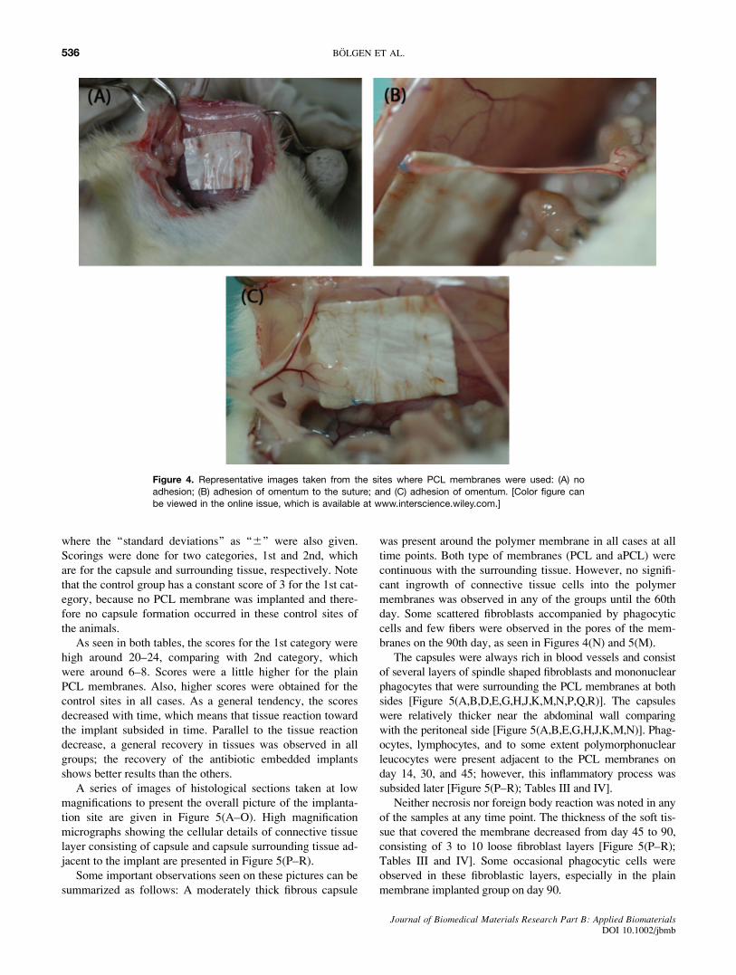
sharply dissected. In the sites in which the plain PCL mem-
branes were used, almost 64.3% of the cases adhesion area
was less than 25%. It is important that in most of the cases
these were in the sutured sites (see e.g., in Figure 4), and the
rest of the area was quite clean. In contrast to the control
site, the adhesions were associated mostly with capillaries
(42.8%); however, in the case of about 28.6%, there were
also some larger vessels formed. Almost 50% of the cases
adhesions were lysed with traction. But, 21.4% of the cases,
adhesions were quite strong and therefore they were only
separated by sharp dissections. The type and tenacity of anti-
biotic embedded PCL membranes group were significantly
different; in 46.1% of the cases there was no adhesion. Only
in the case of 38.5%, vascular formation was observed, but
it was as capillaries (no large size vessel formation). Adhe-
sions were quite weak and separated easily by traction. The
type and tenacity results of both the plain and antibiotic em-
bedded PCL membranes exhibited significant difference
from the control group (p < 0.05), but the results of the
extent of adhesions were not significantly different from
the control group. However, it should be carefully noted that
the adhesions observed with the membranes (with or without
antibiotic loading) were mainly around the sutures.
Histological Evaluation
Histological evaluations were done on the specimens as de-
scribed in Animal Model and Surgical Protocols. Note that
during the processing of the specimen, the polymer mem-
brane was dissolved, leaving empty spaces on tissue sections.
Evaluations were done according to the scoring technique
given in Table I, and the results obtained are presented in
Tables III and IV. Table III gives the results of total 14 ani-
mals sacrificed at different times (14–90 days), which were
carrying the plain PCL membranes in which evaluations of
the control site (Ctrl) were also presented for comparison. Ta-
ble IV exhibits the results of 13 animals treated with the anti-
biotic embedded PCL membranes in a similar fashion. In
these two tables, ‘‘n’’ shows the number of animals evaluated
in each group. The ‘‘mean’’ is the average for these three,
Figure 3. Representative images taken from the control site: (A) adhesion of liver; (B) adhesion of
intestines; and (C) adhesion of omentum. [Color figure can be viewed in the online issue, which is
available at www.interscience.wiley.com.]
535ELECTROSPUN PCL MEMBRANES FOR PREVENTION OF ABDOMINAL ADHESIONS
Journal of Biomedical Materials Research Part B: Applied BiomaterialsDOI 10.1002/jbmb
where the ‘‘standard deviations’’ as ‘‘6’’ were also given.
Scorings were done for two categories, 1st and 2nd, which
are for the capsule and surrounding tissue, respectively. Note
that the control group has a constant score of 3 for the 1st cat-
egory, because no PCL membrane was implanted and there-
fore no capsule formation occurred in these control sites of
the animals.
As seen in both tables, the scores for the 1st category were
high around 20–24, comparing with 2nd category, which
were around 6–8. Scores were a little higher for the plain
PCL membranes. Also, higher scores were obtained for the
control sites in all cases. As a general tendency, the scores
decreased with time, which means that tissue reaction toward
the implant subsided in time. Parallel to the tissue reaction
decrease, a general recovery in tissues was observed in all
groups; the recovery of the antibiotic embedded implants
shows better results than the others.
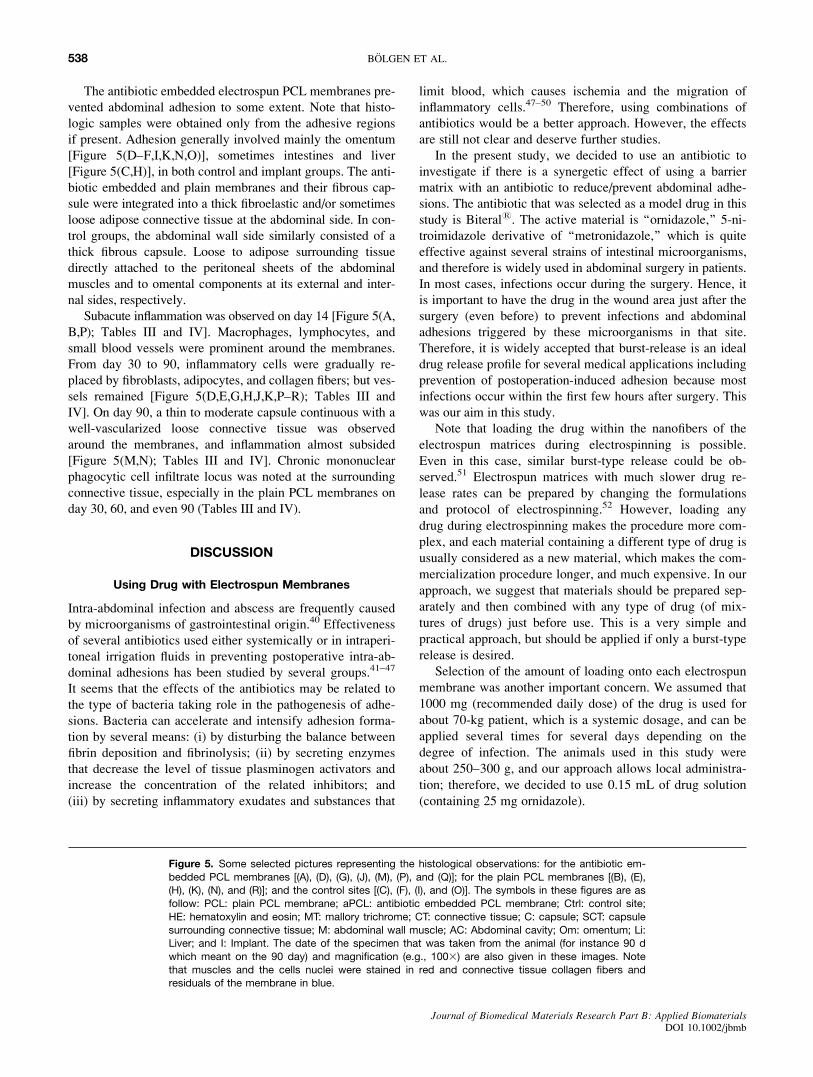
A series of images of histological sections taken at low
magnifications to present the overall picture of the implanta-
tion site are given in Figure 5(A–O). High magnification
micrographs showing the cellular details of connective tissue
layer consisting of capsule and capsule surrounding tissue ad-
jacent to the implant are presented in Figure 5(P–R).
Some important observations seen on these pictures can be
summarized as follows: A moderately thick fibrous capsule
was present around the polymer membrane in all cases at all
time points. Both type of membranes (PCL and aPCL) were
continuous with the surrounding tissue. However, no signifi-
cant ingrowth of connective tissue cells into the polymer
membranes was observed in any of the groups until the 60th
day. Some scattered fibroblasts accompanied by phagocytic
cells and few fibers were observed in the pores of the mem-
branes on the 90th day, as seen in Figures 4(N) and 5(M).
The capsules were always rich in blood vessels and consist
of several layers of spindle shaped fibroblasts and mononuclear
phagocytes that were surrounding the PCL membranes at both
sides [Figure 5(A,B,D,E,G,H,J,K,M,N,P,Q,R)]. The capsules
were relatively thicker near the abdominal wall comparing
with the peritoneal side [Figure 5(A,B,E,G,H,J,K,M,N)]. Phag-
ocytes, lymphocytes, and to some extent polymorphonuclear
leucocytes were present adjacent to the PCL membranes on
day 14, 30, and 45; however, this inflammatory process was
subsided later [Figure 5(P–R); Tables III and IV].
Neither necrosis nor foreign body reaction was noted in any
of the samples at any time point. The thickness of the soft tis-
sue that covered the membrane decreased from day 45 to 90,
consisting of 3 to 10 loose fibroblast layers [Figure 5(P–R);
Tables III and IV]. Some occasional phagocytic cells were
observed in these fibroblastic layers, especially in the plain
membrane implanted group on day 90.
Figure 4. Representative images taken from the sites where PCL membranes were used: (A) no
adhesion; (B) adhesion of omentum to the suture; and (C) adhesion of omentum. [Color figure canbe viewed in the online issue, which is available at www.interscience.wiley.com.]
536 BOLGEN ET AL.
Journal of Biomedical Materials Research Part B: Applied BiomaterialsDOI 10.1002/jbmb

TABLE
III.
HistologicalEvaluationfortheAnim
als
WiththePlain
PCLMembranes
Anim
al
Group/
Categories
Plain
PCLandControl
14days
30days
45days
60days
90days
PCL
Ctrl
PCL
Ctrl
PCL
Ctrl
PCL
Ctrl
PCL
Ctrl
1st
2nd
1st
2nd
1st
2nd
1st
2nd
1st
2nd
1st
2nd
1st
2nd
1st
2nd
1st
2nd
1st
2nd
124
83
921
73
720
63
619
63
719
63
6
223
73
922
83
720
63
720
83
6–
––
–
322
73
10
21
73
8–
––
–19
63
6–
––
–
422
73
10
22
7–
––
––
––
––
––
––
–
Mean6
SD
22.86
1.0
7.36
0.5
3.06
0.0
9.56
0.6
21.56
0.6
7.36
0.5
3.06
0.0
7.36
0.6
20.06
0.0
6.06
0.0
3.06
0.0
6.56
0.7
19.36
0.0
6.76
1.2
3.06
0.0
6.36
0.6
19.0
6.0
3.0
6.0
n4
44
32
23
31
1
TABLE
IV.HistologicalEvaluationfortheAnim
als
WiththeAntibioticEmbeddedPCLMembranes
Anim
al
Group/
Categories
AntibioticEmbedded
PCLandControl
14days
30days
45days
60days
90days
PCL
Ctrl
PCL
Ctrl
PCL
Ctrl
PCL
Ctrl
PCL
Ctrl
1st
2nd
1st
2nd
1st
2nd
1st
2nd
1st
2nd
1st
2nd
1st
2nd
1st
2nd
1st
2nd
1st
2nd
122
63
819
63
918
63
818
53
618
63
6
221
63
11
18
63
818
53
718
53
6–
––
–
320
73
918
53
718
53
718
53
7–
––
–
4–
––
––
––
––
––
––
––
––
––
–
Mean6
SD
21.06
1.0
6.36
0.6
3.06
0.0
9.36
1.5
18.36
0.6
5.76
0.6
3.06
0.0
8.06
1.0
18.06
0.0
5.36
0.6
3.06
0.0
7.36
0.6
18.06
0.0
5.06
0.0
3.06
0.0
6.36
0.6
18.0
6.0
3.0
6.0
n3
33
33
33
31
1
537ELECTROSPUN PCL MEMBRANES FOR PREVENTION OF ABDOMINAL ADHESIONS
Journal of Biomedical Materials Research Part B: Applied BiomaterialsDOI 10.1002/jbmb
The antibiotic embedded electrospun PCL membranes pre-
vented abdominal adhesion to some extent. Note that histo-
logic samples were obtained only from the adhesive regions
if present. Adhesion generally involved mainly the omentum
[Figure 5(D–F,I,K,N,O)], sometimes intestines and liver
[Figure 5(C,H)], in both control and implant groups. The anti-
biotic embedded and plain membranes and their fibrous cap-
sule were integrated into a thick fibroelastic and/or sometimes
loose adipose connective tissue at the abdominal side. In con-
trol groups, the abdominal wall side similarly consisted of a
thick fibrous capsule. Loose to adipose surrounding tissue
directly attached to the peritoneal sheets of the abdominal
muscles and to omental components at its external and inter-
nal sides, respectively.
Subacute inflammation was observed on day 14 [Figure 5(A,
B,P); Tables III and IV]. Macrophages, lymphocytes, and
small blood vessels were prominent around the membranes.
From day 30 to 90, inflammatory cells were gradually re-
placed by fibroblasts, adipocytes, and collagen fibers; but ves-
sels remained [Figure 5(D,E,G,H,J,K,P–R); Tables III and
IV]. On day 90, a thin to moderate capsule continuous with a
well-vascularized loose connective tissue was observed
around the membranes, and inflammation almost subsided
[Figure 5(M,N); Tables III and IV]. Chronic mononuclear
phagocytic cell infiltrate locus was noted at the surrounding
connective tissue, especially in the plain PCL membranes on
day 30, 60, and even 90 (Tables III and IV).
DISCUSSION
Using Drug with Electrospun Membranes
Intra-abdominal infection and abscess are frequently caused
by microorganisms of gastrointestinal origin.40 Effectiveness
of several antibiotics used either systemically or in intraperi-
toneal irrigation fluids in preventing postoperative intra-ab-
dominal adhesions has been studied by several groups.41–47
It seems that the effects of the antibiotics may be related to
the type of bacteria taking role in the pathogenesis of adhe-
sions. Bacteria can accelerate and intensify adhesion forma-
tion by several means: (i) by disturbing the balance between
fibrin deposition and fibrinolysis; (ii) by secreting enzymes
that decrease the level of tissue plasminogen activators and
increase the concentration of the related inhibitors; and
(iii) by secreting inflammatory exudates and substances that
limit blood, which causes ischemia and the migration of
inflammatory cells.47–50 Therefore, using combinations of
antibiotics would be a better approach. However, the effects
are still not clear and deserve further studies.
In the present study, we decided to use an antibiotic to
investigate if there is a synergetic effect of using a barrier
matrix with an antibiotic to reduce/prevent abdominal adhe-
sions. The antibiotic that was selected as a model drug in this
study is Biteral1. The active material is ‘‘ornidazole,’’ 5-ni-
troimidazole derivative of ‘‘metronidazole,’’ which is quite
effective against several strains of intestinal microorganisms,
and therefore is widely used in abdominal surgery in patients.
In most cases, infections occur during the surgery. Hence, it
is important to have the drug in the wound area just after the
surgery (even before) to prevent infections and abdominal
adhesions triggered by these microorganisms in that site.
Therefore, it is widely accepted that burst-release is an ideal
drug release profile for several medical applications including
prevention of postoperation-induced adhesion because most
infections occur within the first few hours after surgery. This
was our aim in this study.
Note that loading the drug within the nanofibers of the
electrospun matrices during electrospinning is possible.
Even in this case, similar burst-type release could be ob-
served.51 Electrospun matrices with much slower drug re-
lease rates can be prepared by changing the formulations
and protocol of electrospinning.52 However, loading any
drug during electrospinning makes the procedure more com-
plex, and each material containing a different type of drug is
usually considered as a new material, which makes the com-
mercialization procedure longer, and much expensive. In our
approach, we suggest that materials should be prepared sep-
arately and then combined with any type of drug (of mix-
tures of drugs) just before use. This is a very simple and
practical approach, but should be applied if only a burst-type
release is desired.
Selection of the amount of loading onto each electrospun
membrane was another important concern. We assumed that
1000 mg (recommended daily dose) of the drug is used for
about 70-kg patient, which is a systemic dosage, and can be
applied several times for several days depending on the
degree of infection. The animals used in this study were
about 250–300 g, and our approach allows local administra-
tion; therefore, we decided to use 0.15 mL of drug solution
(containing 25 mg ornidazole).
Figure 5. Some selected pictures representing the histological observations: for the antibiotic em-
bedded PCL membranes [(A), (D), (G), (J), (M), (P), and (Q)]; for the plain PCL membranes [(B), (E),(H), (K), (N), and (R)]; and the control sites [(C), (F), (I), and (O)]. The symbols in these figures are as
follow: PCL: plain PCL membrane; aPCL: antibiotic embedded PCL membrane; Ctrl: control site;
HE: hematoxylin and eosin; MT: mallory trichrome; CT: connective tissue; C: capsule; SCT: capsule
surrounding connective tissue; M: abdominal wall muscle; AC: Abdominal cavity; Om: omentum; Li:Liver; and I: Implant. The date of the specimen that was taken from the animal (for instance 90 d
which meant on the 90 day) and magnification (e.g., 1003) are also given in these images. Note
that muscles and the cells nuclei were stained in red and connective tissue collagen fibers andresiduals of the membrane in blue.
538 BOLGEN ET AL.
Journal of Biomedical Materials Research Part B: Applied BiomaterialsDOI 10.1002/jbmb
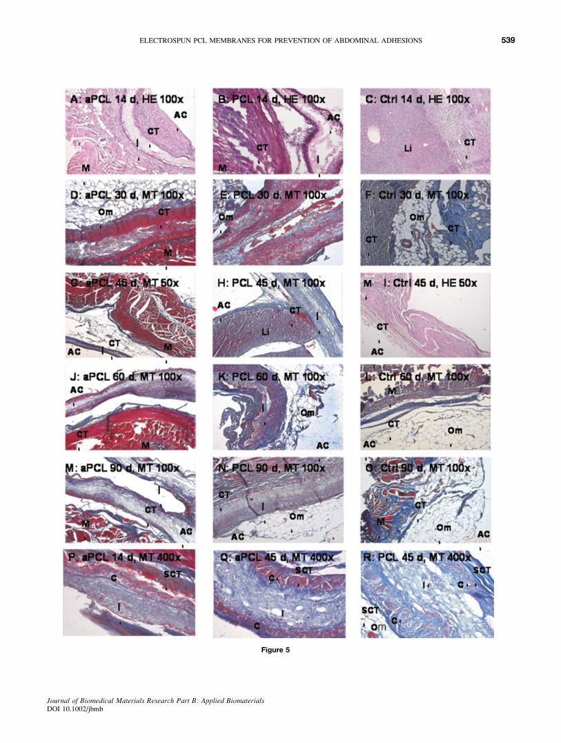
Figure 5
539ELECTROSPUN PCL MEMBRANES FOR PREVENTION OF ABDOMINAL ADHESIONS
Journal of Biomedical Materials Research Part B: Applied BiomaterialsDOI 10.1002/jbmb
To obtain in vitro release kinetics, we put each drug-
loaded electrospun membrane specimen in one flask contain-
ing 10 mL distilled water, and followed the drug release spec-
trophotometrically. This was of course not the best model of
release study to mimic the situation in vivo, since we do not
know the exact consumption rate of the drug in the abdominal
cavity that would of course significantly control the drug
release (because it is by diffusion). However, we thought that
these tests would at least allow us to demonstrate if the
release was fast enough or not, as we have expected/aimed.
As shown in Figure 2, the drug release was quite fast, like
a typical burst-type release that was expected. Note that
Zong et al. have reported similar release kinetics for
mefoxin delivery with their electrospun materials, in which
drug was loaded during electrospinning process.51 They
showed that most of the drug was released because of the
concentration gradient in 3 h, and the release was completed
in the first 48 h. They claimed that a large amount of drug
molecules is aggregated on or near the fiber surface by elec-
trospinning; therefore, they have reached burst-type release,
similar to what we have observed in this study.
Performances of Electrospun Membranesin Animal Model
Creating reproducible wounds/injuries on the abdominal wall
to study surgical adhesions in animal models is an important
consideration to obtain statistically meaningful results. There
are several approaches in the related literature, not only one
that are agreed on and applied. For instance, Zong et al.
excised a 1 3 1 cm2 of abdominal wall muscle to create a
cecal wound. The celiotomy was then closed in two layers
immediately (control), after a barrier was laid in between the
cecum and the abdominal wall.53 Wang et al. stripped a 3 34 cm2 patch of parietal peritoneum corresponding to the cecal
and also scraped an area of about 3 3 4 cm2 at both sides of
peritoneum along the abdomen incision.54 Pestieau et al. first
made a midline incision in the abdomen using a scalpel.55
Then, they burned a 2-cm circle of parietal peritoneum
located laterally 2 cm from the incision line and directly op-
posite the cecum by electrocoagulation until there was a uni-
form coagulum of abdominal surface. In our animal models,
defects were created by using 2 3 3 cm2 templates. Injuries
as small rectangular shapes were created on the abdominal
wall at both the control and membrane-implanted sites as
described in the previous sections, which were quite reproduc-
ible, and we were very careful to reach high reproducibility.
Macroscopical Observations
The adhesions, both at the control and membrane-implanted
sites, in the abdominal cavity of the experimental rats were
first evaluated macroscopically to obtain the extent, type,
and tenacity of the adhesions. As mentioned before, the
adhesions were investigated after selected time periods up to
90 days of postimplantation. It should be noted that the mac-
roscopical observations did not give clear idea about the
progress of intra-abdominal adhesion formation and preven-
tion. Therefore, in these macroscopical examinations, we
have examined the results by pooling the observations
obtained at different times. The progress of adhesion forma-
tion/prevention has been evaluated by only microscopical
(histological) observations/tests given in the later part.
As demonstrated in detail in the previous section, we have
observed severe and strong adhesions in the control sites, all
over the dissected area, involving not only the omentum but
also, in some cases, intestine and liver. We have observed
blood capillaries and especially larger blood vessels in the
control site. Covering the injured area with the plain PCL
membranes reduced the overall adhesion profile. Interestingly,
most of the adhesions were around the sutured sites, and the
rest of the area was quite clean. However, even in this case,
there were still blood vessels that were mostly as capillaries in
contrast to the control site. Using the antibiotic with the bar-
rier membrane significantly reduced the extent of adhesion
and there were only capillaries. In addition, the adhesions
were quite weak and, therefore, separated easily by traction.
However, there were still adhesions around the sutures.
It should be noted that the electrospun PCL membranes
produced in this study are hydrophobic, soft, elastic, very
light like a butterfly wing, and comfortable but mechanically
strong enough to handle; therefore, we were able to place
them easily/tightly on the wound surface during surgical use
because of their unique nonwoven nanofibrous structure.37
However, by considering the experience of Zong et al. (who
pointed out the shrinkage problem of hydrophobic PLGA
electrospun membranes because of their nanofibrous struc-
tures, which lead displacement of the barrier from the abdom-
inal wall) regarding electrospun membranes,53 we decided to
suture our PCL membranes to eliminate the risk of displace-
ment that may occur in the long-term stay in the abdominal
cavity of the animals. But the data discussed above clearly
showed that this was not a very good choice, and created
problems. It seems that in these kind of surgery these barriers
should not be sutured, may be only dressed on the defected
area by using some kind of tissue glue, or some other means.
The mechanism of postsurgical peritoneal adhesion for-
mation and reformation remain poorly understood. The ex-
perimental evidence suggest that adhesions form between
two surgically traumatized surfaces in natural apposition
during the healing interval because it is more efficient to
combine two sites of tissue repair into a single healing site,
resulting in coalescing adhesions between peritoneal surfa-
ces.56 In theory, surgical barriers separate contacting perito-
neal surfaces during healing to enable reconstitution of the
surgically traumatized surfaces separately without coalesc-
ing adhesions. However, according to our results, when we
compared the macroscopic observations for the plain and an-
tibiotic embedded PCL membranes, it seems that placing
only a physical barrier is not enough to prevent adhesion.
This was also one of our initial concerns; therefore, we have
used an antibiotic. The electrospun membranes were loaded
(embedded) with the antibiotic with a very easy technique,
540 BOLGEN ET AL.
Journal of Biomedical Materials Research Part B: Applied BiomaterialsDOI 10.1002/jbmb
by dropping the antibiotic solution on the membranes just
before the surgery.
Our macroscopic observations demonstrated that using
antibiotics with this type of physical barrier (the electrospun
membrane) is quite an effective way of treatment. It seems
that the microorganisms may contaminate the wound area
during the surgical operations and play a very important role
in the adhesion process. In our case, the antibiotic mole-
cules, which were loaded in the matrix by simple absorption,
were released (diffuse out) quite easily from the matrix and
create a burst effect in the abdominal cavity, which most
probably prevented infections in the wounds locally in the
earlier phase of healing process. Therefore, this fast release
in the beginning of healing strongly affected the success of
our barrier system. Zong et al. have also reached a similar
conclusion in their animal studies with a very similar
approach on postsurgical adhesions.53
Histological Evaluations
Histological evaluations were done according to the scoring
technique (see Table I). A series of images of histological
sections have also been taken to present the overall picture of
the implantation site. As we have also observed in our previ-
ous study, the PCL membranes maintain their integrity (both
appearance and mechanical properties) in 90 days in vivoeven if the Mw drops from about 85,000 to 62,000.37 During
routine histological processing, the PCL membranes partly
dissolve by well-shaped blocks from different regions. There
were no cells or tissue components neighboring these regions.
Note that if there were particle-form degradation products
around, we would observe these particles that are almost
always surrounded by reactive and regenerating cells.
Abdominal wall healing was slower in the control sites
compared with the sites where the PCL membranes were
used. Healing process improved with the antibiotic embedded
PCL membranes. Both a decrease in the the tissue reaction
and a general recovery in tissues were observed in all groups;
the recovery of the antibiotic-containing implants showed
better results than the others. These findings were in accord-
ance with macroscopical observations. Statistical analysis
done by using the surrounding tissue showed that only the
membranes containing antibiotics were significantly different
from the control group (p < 0.05). Since no PCL membrane
was implanted to the control site, no capsule formation was
observed in the control sites of the animals. The comparison
of capsule scores of PCL and aPCL groups exhibited that
there is a significance difference between these two (p <0.05). These analyses clearly demonstrated that the antibi-
otic-containing PCL membranes are successful in the preven-
tion of abdominal adhesions. Note once again that the
undesirable events observed both in macroscopical and histo-
logical examinations were around the suturing area. Most
probably, without suturing, much successful (statistically sig-
nificant) improvements may be obtained even with the plain
PCL membranes, as mentioned earlier.
CONCLUSIONS
PCL, as a biocompatible and biodegradable polymer, is a
very good candidate in the preparation of polymeric implants
for several biomedical applications. It can easily be polymer-
ized by ring-opening polymerization with different molecular
weights and therefore with different in vivo degradation
rates.25–27,37,38 Nonwoven PCL membranes (or films) can
easily be prepared with different thicknesses which can be
formed from nanofibers with different diameters, therefore
with different degradation rates.30,31,33–35,37,53 Recently, we
also synthesized PCL with different molecular weights and
prepared a series of nonwoven membranes by electrospin-
ning.37 Here, we investigated their possible use as a physical
barrier for prevention of adhesions after abdominal surgery.
We thought that they will be a very suitable matrix for these
types of biomedical applications because of their excellent
physical properties, such as elasticity, softness, and lightness.
Note that they were strong enough to be handled. One impor-
tant advantage of these nonwoven matrices was their high
absorption capacities. We were able to load antibiotics easily
by simply dropping antibiotic solution on the membranes.
Note that the others use more sophisticated methods for drug
loading within the polymeric materials, such as they do elec-
trospun polymer fibers with the antibiotic solutions.53
Both macroscopical and histological observations showed
that the PCL membranes reduce abdominal adhesions and
even if we observe some adhesions they were loose. Antibi-
otic loading significantly affected the type of adhesion and
the tenacity. In the case of antibiotic-loaded PCL membranes,
only capillaries were formed which were mostly on the edges
of the membrane in which sutures were applied. The statisti-
cal analysis done both by using the macroscopical and histo-
logical observations exhibited that only the PCL membranes
carrying antibiotic are significantly different from the con-
trolled sites. However, we believe that this is due to suturing.
These observations clearly demonstrated that using the plain
PCL membranes changes the type and tenacity of adhesion in
a positive direction and improves healing. The suturing
should be eliminated in the use of synthetic barriers. Antibiot-
ics should be included in the formulations. The use of mem-
brane and antibiotic together synergistically affects the
healing processes and makes the process better and faster,
and reduces abdominal adhesions.
The biodegradation rate of our membranes is too slow
comparing the healing rate in the abdominal cavity; there-
fore, we concluded that thinner PCL membranes formed
of nanofibers with smaller diameters, and made of PCL
with much lower molecular weights, should be used to match
the healing rate in vivo. Our related studies in this direction
for production of PCL membranes (and/or their copolymers
with lactides) are under investigation before the clinical
applications.
Erhan Piskin is supported by Turkish Academy of Sciences as afull member. Nimet Bolgen is supported by Turkish Scientific andTechnical Research Council as a Ph.D. student.
541ELECTROSPUN PCL MEMBRANES FOR PREVENTION OF ABDOMINAL ADHESIONS
Journal of Biomedical Materials Research Part B: Applied BiomaterialsDOI 10.1002/jbmb
REFERENCES
1. Hellebrekers B, Trimbos-Kemper T, Trimbos J, Emeis J,Kooistra T. Use of fibrinolytic agents in the prevention of post-operative adhesion formation. Fertil Steril 2000;74:203–212.
2. DeCherney A, Dizerega G. Clinical problem of intraperitonealpostsurgical adhesion formation following general surgery andthe use of adhesion prevention barriers. Surg Clin North Am1997;77:671–688.
3. Menzies D. Peritoneal adhesions: Incidence, cause and pre-vention. Surg Annu 1992;24:27–45.
4. Menzies D. Postoperative adhesions: Their treatment and rele-vance in clinical practise. Ann R Coll Surg Engl 1993;75:147–153.
5. Milligan DW, Raftery AT. Observations on the pathogenesisof peritoneal adhesions: A light and electron microscopicalstudy. Br J Surg 1974;61:274–280.
6. Wiseman D. Polymers for prevention of surgical adhesions.In: Domb AJ, editor. Polymeric Site-Spesific Pharmacother-apy. Arlington, TX: Wiley; 1994. pp 370–421.
7. Rodgers K, Cohn D, Hotovely A, Pines E, Diamond MP,DiZerega G. Evaluation of polyethylene glycol/polylactic acidfilms in the prevention of adhesions in the rabbit adhesion for-mation and re-formation sidewall models. Fertil Steril 1998;69:403–408.
8. Becker JM, Dayton MT, Fazio VW, Beck DE, Stryker SJ,Wexner SD, Wolf BG, Roberts PL, Smith LE, Sweeney SA,Moore M. Prevention of postoperative abdominal adhesions bya sodium hylarunate-based bioresorbable membrane: A prospec-tive, randomized, double-blind multicenter study. J Am CollSurg 1996;183:297–306.
9. diZerega GS. Contemporary adhesion prevention. Fertil Steril1994;61:219–235.
10. Pijlman BM, Dorr PJ, Brommer EJP, Vemer HM. Preventionof adhesions. Eur J Obstet Gynecol Reprod Biol 1994;53:155–163.
11. Bakkum EA, Trimbos JB, Trimbos-Kemper TCM. Postsurgi-cal adhesion formation and prevention-recent developments.Reprod Med Rev 1996;5:1–13.
12. Hellebrekers BWJ, Trimbos-Kemper GCM, Blitterswijk CA,Bakkum EA, Trimbos JBMZ. Effects of five different barriermaterials on postsurgical adhesion formation in the rat. HumReprod 2000;15:1358–1363.
13. Haney AF, Hesla J, Hurst BS, Kettel ML, Murphy AA, RockJA, Rowe G, Schlaff WD. Expanded polytetrafluoroethylene(Gore-Tex Surgical Membrane) is superior to oxidized regen-erated cellulose (Interceed TC7þ) in preventing adhesions.Fertil Steril 1995;63:1021–1026.
14. Urman B, Gomel V. Effect of hyaluronic acid on postopera-tive intraperitoneal adhesion formation and reformation in therat model. Fertil Steril 1991;56:568–570.
15. Diamond MP, DeCherney AH, Linsky CB, Cunningham T,Constantine B. Assesment of carboxymethylcellulose and %32Dextran 70 for prevention of adhesions in a rabbit uterine hornmodel. Int J Fertil 1998;33:278–282.
16. Yaacobi Y, Israel AA, Goldberg EP. Prevention of postopera-tive abdominal adhesions by tissue precoating with polymersolutions. J Surg Res 1993;55:422–426.
17. O’Sullivan D, O’Riordain M, O’Connell RP, Dinen M, BradyMP. Peritoneal adhesion formation after lysis: Inhibition bypolyethylene glycol 4000. Br J Surg 1991;78:427–429.
18. Johns DB, Rodgers KE, Donahue WD, Kiorpes TC, diZeregaGS. Reduction of adhesion formation by postoperative admin-istration of ionically cross-linked hyaluronic acid. Fertil steril1997;68:37–42.
19. Hay WP, Mueller PO, Harmon B, Amoroso L. One percent so-dium carboxymethylcellulose prevents experimentally inducedabdominal adhesions in horses. Vet Surg 2001;30:223–227.
20. Osada H, Minai M, Tsunoda I, Fujii TK, Tsubata K, Satoh K.The effect of hyaluronic acid-carboxymethylcellulose in reducingadhesion re-formation in rabbits. J Int Med Res 1999;27:292–296.
21. Wiseman DM, Gottlick Larkowski L, Kamp L. Effect of dif-ferent barriers of oxidized regenerated cellulose (ORC) oncecal and sidewall adhesions in the presence and absence ofbleeding. J Invest Surg 1999;12:141–146.
22. Burns JW, Colt MJ, Burgees LS. Preclinical evaluation ofSeprafilm bioresorbable membrane. Eur J Surg Suppl 1997;577:40–48.
23. De Iaco PA, Muzzupapa G, Bigon E. Efficacy of a hyalur-onan derivative gel in postsurgical adhesion prevention in thepresence of inadequate hemostatis. Surgery 2001;130:60–64.
24. Matsuda S, Se N, Iwata H, Ikada Y. Evaluation of the antiad-hesion potential of UV cross-linked gelatin films in a ratmodel. Biomaterials 2002;23:2901–2908.
25. Piskin E. Biodegradable polymers in medicine. In: Scott G, editor.Degradable Polymers: Principles and Applications. Dordrecht:Kluwer; 2002. pp 321–377.
26. Fambri L, Migliaresi C, Kesenci K, Piskin E. Biodegradablepolymers. In: Barbucci R, editor. Integrated Biomaterials Sci-ence. New York: Kluwer; 2002. pp 119–187.
27. Pitt CG. Poly(e-caprolactone) and its copolymers. In: Chasin M,Langer R, editors. Biodegradable Polymers as Drug DeliverySystems. New York: Marcel Dekker; 1990. pp 71–120.
28. Ishaug-Riley SL, Okun LE, Prado G, Applegate MA, Ratcliffe A.Human articular chondrocyte adhesion and proliferation on syn-thetic biodegradable polymer films. Biomaterials 1999;20:2245–2256.
29. Ekholm M, Hietanen J, Lindqvist C, Rautavuori J, Santavirta S,Suuronen R. Histological study of tissue reactions to e-caprolac-tone-lactide copolymer in paste form. Biomaterials 1999;20:1257–1262.
30. Huang ZM, Zhang YZ, Kotaki M, Ramakrishna S. A review onpolymer nanofibers by electrospinning and their applications innanocomposites. Comp Sci Tech 2003;63:2223–2253.
31. Frenot A, Cronakis IS. Polymer nanofibers assembled by elec-trospinning. Curr Opin Colloid Interface Sci 2003;8:64–75.
32. Larrondo L, Manley RSJ. Electrostatic fiber spinning from poly-mer melts. I. Experimental observations on fiber formation andproperties. J Polym Sci Polym Phys Ed 1981;19:909–920.
33. Smith LA, Ma PX. Nanofibrous scaffolds for tissue engineer-ing. Colloids Surf B 2004;39:125–131.
34. Li W, Laurencin C, Caterson E, Tuan R, Ko F. Electrospunnanofibrous structure: A novel scaffold for tissue engineering.J Biomed Mater Res 2002;60:613–621.
35. Xu CY, Inai R, Kotaki M, Ramakrishna S. Aligned biode-gradable nanofibrous structure: A potential scaffold for bloodvessel engineering. Biomaterials 2004;25:877–886.
36. Mo XM, Xu CY, Kotaki M, Ramakrishna S. ElectrospunP(LLA-CL) nanofiber: A biomimetic extracellular matrix forsmooth muscle cell and endothelial cell proliferation. Bioma-terials 2004;25:1883–1890.
37. Bolgen N, Menceloglu YZ, Acatay K, Vargel _I, Piskin E.In vitro and in vivo degradation of non-woven materials madeof poly(e-caprolactone) nanofibers prepared by electrospinningat different conditions. J Biomater Sci Polym Ed 2005;16:1537–1555.
38. Ural E, Kesenci K, Fambri L, Migliaresi C, Piskin E. Poly(D,L-lactide/e-caprolactone)/hydroxyapatite composites as bonefiller: Preparation and characterization. Biomaterials 2000;21:2147–2154.
39. Jansen JA. Animal models for studying soft tissue biocompati-bility of biomaterials. In: An YH, Friedman RJ, editors. Ani-mal models in orthopedic research. Boca Raton, Florida, USA:CRC Press; 1999. p 393–405.
542 BOLGEN ET AL.
Journal of Biomedical Materials Research Part B: Applied BiomaterialsDOI 10.1002/jbmb
40. Tzianabos AO, Cisneros RL, Gershkovich J, Johnson J,Miller RJ, Burns JW, Onderdonk AB. Effect of surgical ad-hesion reduction devices on the propagation of experimen-tal intra-abdominal infection. Arch Surg 1999;134:1254–1259.
41. Philips RKS, Dudley HAF. The effect of tetracycline lavageand trauma on visceral and parietal peritoneal ultrastructureand adhesion formation. Br J Surg 1984;71:537–539.
42. Rappaport WD, Holcomd M, Valente J, Chvapil M. Antibioticirrigation and formation of intra-abdominal adhesions. Am JSurg 1989;158:435–437.
43. Barie PS, Vogel SB, Dellinger EP, Rotstein OD, Solomkin JS,Yang JY, Baumgartner TF. A randomized, double-blinded clini-cal trial comparing cefepime plus metronidazole with imipenem–cilastin in the treatment of complicated intra-abdominal infec-tions. Arch Surg 1997;132:1294–1302.
44. Rambo WM. Irrigation of the peritoneal cavity with cephalo-thin. Am J Surg 1972;123:192–195.
45. Brolin J, Lahnborg G, Nord CE. The effect of one prophy-lactic dosage of antibiotics on experimentally induced lethalintra-abdominal sepsis. Acta Chir Scand 1984;150:239–244.
46. Berne TV, Yellin AE, Appleman MD, Heseltine PN, Gill MA. Aclinical comparison of cefepime and metronidazole versus genta-micin and clindamycin in the antibiotic management of surgi-cally treated advanced appendicitis. Surg Gynecol Obstet 1993;177:35–40.
47. Oncel M, Kurt N, Remzi FH, Sensu SS, Vural S, Gezen CF,Cincin TG, Olcay E. The effectiveness of systemic antibioticsin preventing postoperative, intraabdominal adhesions in ananimal model. J Surg Res 2001;101:52–55.
48. Tito WA, Sorr MG. Intestinal obstructions. In: Zuidema GD,Nyhus LM, editors. Schakelford’s Surgery of Alimentary Tract,Vol. 5. Philadelphia: Saunders; 1996. pp 375–416.
49. Holtz G. Prevention and management of peritoneal adhesions.Fertil Steril 1984;41:497–507.
50. Goor HV, Graaf JS, Grond J, Sluiter WJ, Meer J, Bom VJ,Bleichrodt RP. Fibrinolytic activity in the abdominal cavity ofrats with faecal peritonitis. Br J Surg 1994;81:1046–1049.
51. Zong X, Kima K, Fang D, Rana S, Hsiaoa BS, Chua B.Structure and process relationship of electrospun bioabsorb-able nanofiber membranes. Polymer 2002;43:4403–4412.
52. Kenawy ER, Bowlin GL, Mansfield K, Layman J, Simpson DG,Sanders EH, Wnek GE. Release of tetracycline hydrochloridefrom electrospun poly(ethylene-co-vinylacetate), poly(lactic acid),and a blend. J Control Release 2002;81:57–64.
53. Zong X, Li S, Chen E, Garlick B, Kim K, Fang D, Chiu J,Zimmerman T, Brathwaite C, Hsiao BS, Chu B. Prevention ofpost-surgery induced abdominal adhesions by electrospun bio-absorbable nanofibrous poly(lactide-co-glycolide) based mem-branes. Ann Surg 2004;240:910–915.
54. Wang XC, Gui CQ, Zheng QS. Combined therapy of allantoin,metronidazole, dexamethasone on the prevention of intra-abdom-inal adhesion in dogs and its quantitative nalysis. World J Gastro-enterol 2003;9:568–571.
55. Pestieau SR, Marchettini P, Stuart OA, Chang D, Sugarbaker PH.Prevention of intraperitoneal adhesions by intraperitoneal lavageand intraperitoneal 5-fluorouracil: Experimental studies. Int Surg2002;87:195–200.
56. Haney AF, Doty E. The formation of coalescing peritonealadhesions requires injury to both contacting peritoneal surfa-ces. Fertil Steril 1999;61:767–775.
543ELECTROSPUN PCL MEMBRANES FOR PREVENTION OF ABDOMINAL ADHESIONS
Journal of Biomedical Materials Research Part B: Applied BiomaterialsDOI 10.1002/jbmb