Persistent Luminescence Strontium Aluminate Nanoparticles ...

In-vitro study of antibiotic and
strontium release from
hydroxyapatite spheres and its
PMMA composite
Martin Zarazua Mujo
Degree project 30 credits
Chemical Engineering Programme
Martin Zarazua Mujo
Supervisors: Dr. Wei Xia & Dr. Cecilia Persson
Department of Engineering Sciences; Applied Materials Science
Uppsala University

1
Table of Contents
1.0. Aim ......................................................................................................................................................... 2
1.1. Introduction ........................................................................................................................................ 2
1.2. Materials used in the study ................................................................................................................ 3
1.3. Antibiotics and ion of interest in the study ........................................................................................ 5
2. Materials & Methods ................................................................................................................................ 5
2.1. Drug loading of hydroxyapatite .......................................................................................................... 6
2.2. Loading into PMMA ............................................................................................................................ 6
2.3. Drug release from hydroxyapatite ..................................................................................................... 7
2.4. Drug release from hydroxyapatite-PMMA composite groups ........................................................... 7
2.5. Mechanism study of drug and Sr ion release ..................................................................................... 7
3. Results and discussion ............................................................................................................................... 8
3.1. Release from hydroxyapatite spheres ................................................................................................ 8
3.2. Release from PMMA composites ..................................................................................................... 14
4. Conclusion ............................................................................................................................................... 22
5. Acknowledgement ................................................................................................................................... 23
6. References ............................................................................................................................................... 23

2
1.0. Aim The purpose of this master thesis project was to investigate the drug release from
hydroxyapatite spheres and its polymethylmethacrylate (PMMA) composite in vitro. The
drugs that were studied were the antibiotics cephalothin and vancomycin. The
hydroxyapatite spheres were to be made containing strontium and the ion release from the
spheres would also be measured. The study was to be done with phosphate buffer saline
solution (PBS) with differing pH value to see what effect it might have on the release. To
simulate blood condition a pH value of 7.4 was desired and a pH of 8 to simulate intestinal
fluid. The extracellular compartment where bone resorption occurs tends to be in an acidic
environment therefore the release was also to be studied at a pH value of 41.
1.1. Introduction
Prosthesis-related infection is a severe problem that can occur when dealing with cemented
orthopedic implants and the use of antibiotic-loaded bone cement is just one of many
methods applied to prevent and treat this type of infection. Rise of infections can develop
through different ways; either through direct contamination of the biomaterial or the
surrounding tissue; by blood-bone contamination or by the spread of a superficial
infection2. Direct contamination is most likely the cause for the greater part of prostheses-
related infections. Infected cemented prosthesis lead to a weakened function and pain for
the patient. An infection can sometimes require multiple operations and the severity of the
problem sometimes lead to amputations in worst cases. Diagnosis of an infection around an
implant can be difficult when dealing with less open infections and to replace an old
prosthesis with a new one on an infected area without proper antibacterial treatment is
obviously dangerous. It has therefore been suggested that every loose prostheses is to be
assumed as infected until proven otherwise3. However, it has been noted that the risk of
infection increases after revision arthroplasty compared to the primary arthroplasty. The
cause for prosthesis failure might be due to a combination of mechanical and infectious
loosening4.
To obtain certainty of an infection the causative organism from the implant side is cultured.
This can be problematic because a negative culture from the tissue does not necessarily rule
out an infection. A negative result can be from failure to get a correct sample but it may also
3
be due to changed bacterial metabolism. Bacteria in a biomaterial-related infection can be
difficult to culture because these bacteria do not grow exponentially; instead they are slow-
growing. Furthermore, these bacteria can produce an extracellular matrix that can protect
them from inhospitable environment. This allows the bacteria to evade the host immune
system and antibiotic treatment, as well as making detection difficult by standard
laboratory methods5.
Treatment of prosthesis-related infection is difficult, and antibiotic treatment generally
focuses on removal of the foreign body material combined with surgical removal of infected
tissue around the prostheses area. Thereafter antibiotics aimed at the causative bacteria are
administrated. To enable a both locally and systematically administrated antibiotic
treatment, an antibiotic-loaded bone cement can be implemented. This method aims to
maintain a level of local antibiotic concentration which cannot be achieved by using a
systemic administration without side effects6.
Published in vitro studies show that the release of antibiotics from hydroxyapatite and
PMMA follows a typical bi-phasic fashion; a burst release gives a peak which is followed by
a long, tail of low release that goes on from days up to months. In addition, the amount of
antibiotics released increases with increased surface area and also relates with the surface
roughness7.
1.2. Materials used in the study
Hydroxyapatite (Ca10(PO4)6(OH)2; HA) is a bioactive bone material that has been widely
used in filling bone defects because of its chemical similarities with the inorganic phase of
bone. The advantage of using HA as bone material is its formidable osteoconductivity, non-
toxicity and ability to form a direct bond with bone8 . HA is the main mineral component of
bones and teeth and has superior adsorption of many molecules making it an excellent
choice as an adsorbent9. Other advantages are that it does not cause any foreign body
reaction or an immunological response. Antibiotic-loaded bone cements have been used in
clinical procedures for a long time now and hollow HA has been used as a local drug
delivery system because of its porous structure that can allow a high loading capacity10.
Hydroxyapatite has also shown promise as filler for dental composites not just because of it

4
being a natural component in teeth but also because it is naturally radio-opaque and highly
resistant to moisture11. Moreover, hydroxyapatite has the ideal hardness which is used as a
standard for composite resin filler particles.
Another, common material for bone substitution and prosthesis fixation is
polymethylmethacrylate (PMMA). This bone cement consists of a mixture of liquid
methylmethacrylate (MMA) and pre-polymerized PMMA powder which is polymerized in
the patient when used in clinic. The mixture of the components gives a viscous paste which
gives the advantage of molding and the ability to support prosthesis during insertion. The
initial presence of PMMA makes it possible to use less monomer to obtain the same amount
of end product. The end result also means less undesirable effects of polymerization. The
use of less monomer enables less heat production, reduced volume and increased density
because the molecule take up less space in a polymer than their liquid counterpart.
Feng Ye et al. used hydroxyapatite hollow nanoparticles loaded with vancomycin that had a
narrow sized distribution, which reportedly gave a high drug loading ratio. These particles
were then morphology changed into nanotubes by adding citric acid as a co-surfactant. This
would further improve the drug load efficiency and could form a pH-controlled drug
release. The effectiveness of hydroxyapatite particles loaded with vancomycin in treatment
of Staphylococcus aureus showed impressive results12. Uwe Joosten et al. studied the
release both in-vitro and in vivo using rabbits for their model. The in vitro study, where
hydroxyapatite was mixed with a high concentration vancomycin, showed a high level of
vancomycin release over a prolonged period more than sufficient in treating an infection. In
the animal study no histological evidence of an infection was seen after 42 days of
incubation on the group that been administrated vancomycin loaded hydroxyapatite, nor
was any systemic side effects seen.
In vitro release studies of antibiotics from PMMA follows a similar fashion to release from
hydroxyapatite with an early burst release followed by a lower release after. Currently,
antibiotic loaded PMMA beads are being used clinically and have shown to deliver a high
concentration locally and with minor systemic effects13. Diefenbeck et al. summarized non-
controlled, non-randomized clinical studies of the efficacy of the antibiotic gentamicin

5
loaded PMMA beads for treating osteomyelitis and showed an infection control rate of
almost 90 %. From experimental and clinical data they suggested that the most reliable
approach for treatment would be achieved by combining local and systemic antibiotic
therapy. The release mechanism for antibiotics from bone cement is not clearly proved;
some authors have found that the release depends on the diffusion of antibiotics either
through the entire polymer matrix or through capillaries within the matrix. However, most
authors have come to the conclusion that the release is dependent on the surface of the
bone cement14
1.3. Antibiotics and ion of interest in the study
Vancomycin is a glycopeptide antibiotic, applicable against Gram-positive bacteria and are
therefore often used clinically against multi-resistant bacteria strains such as
Staphylococcus aureus. Staphylococcus aureus is one of the major types of infections that
affect bone causing inflammatory destruction of joint and bones. The infections that arise
can be acute or chronic and affect vertebrae, native- and prosthetic joints.
Cepalothin is a first generation cephalosporin antibiotic, which is classed as β-Lactam
antibiotics. It has a large volume of distribution, a protein binding of nearly 65 % and is
mainly eliminated by renal excretion15.
Strontium increase bone mass and decrease bone resorption, the use of the drug strontium
ranelate increase bone calcium which has shown reduction of incidence of fractures in
osteoporotic patients. It also enhances bone cell replication and bone formation.
Administered strontium is mostly deposited into bone. Although Strontium only makes up a
0.035 of the calcium content in skeleton its behaviors are similar, they both have strong
bone-seeking properties and the excretion from the body is similar. Strontium can replace
calcium in the hydroxyapatite structure and it is also the only trace metal in bone that was
correlated with bone compression strength16.
2. Materials & Methods The hydroxyapatite spheres containing strontium were made in the laboratory. The spheres
were made by dissolving strontium nitrate (1.0mM) into a solution containing Na+ (145

6
mM), K+ (4.3 mM), Mg2+ (0.49 mM), Ca2+ (0.91 mM), Cl- (143 mM) and HPO42- (9.6 mM). The
solution, made in a glass bottle, was then covered with a lid and put into an oven at 100°C
for 24 hours. Thereafter, the solution was centrifuged and the hydroxyapatite spheres were
separated from the solution. The vancomycin and cephalothin, both in powder from, were
bought from Sigma. Shimadzu 1800 UV-Vis spectrophotometer was used to measure the
antibiotic concentration and an inductively coupled plasma atomic emission spectroscopy
(ICP-AES) Spectro Ciros CCD was used to quantify the amount of strontium in the samples.
The concentrated strontium solution of 1000 ppm that was used for making stock solutions
was bought from Analytical Standards AB. The PMMA that was used was the radiopaque
bone cement for vertebroplasty Osteopal V from Heaues Medical. PBS was made from
tablets that were bought from Sigma and the pH was adjusted with hydrochloric acid and
sodium hydroxide.
2.1. Drug loading of hydroxyapatite
The drug loading was carried out at room temperature for 24 hours under stirring. 500 mg
of hydroxyapatite was immersed into 20 ml drug solution. For the vancomycin and the
cephalothin, the drug solutions were at a concentration of 10 mg/ml. After 24 hours the
samples were centrifuged at 3000 rpm. The estimation of amount of antibiotic loaded into
the hydroxyapatite spheres was done by an indirect method, where the amount of drug in
the solution before and after the loading was found. Percentage of the drug loading was
then calculated using the formula17:
Where X is the amount of drug in the solution before loading and Y is the amount of drug in
the solution after the loading. The drug solution was analyzed by UV-vis spectrophotometer
at a wavelength of 280 nm for the vancomycin and at a wavelength of 237 nm for the
cephalothin. Drug content was determined by comparing with the standard curves of
vancomycin and cephalothin. The standard curves were made from vancomycin and
cephalothin solutions in Milli-Q water with concentration between 10 µg/ml and 125
µg/ml.
2.2. Loading into PMMA
For the PMMA release study four groups were prepared in 6 mm × 12 mm molds:

7
Group 1: PMMA + Hydroxyapatite (10 wt %)18
Group 2: PMMA + Vancomycin (2.5 wt %)19
Group 3: PMMA + Vancomycin loaded hydroxyapatite (10 wt %)
Group 4: PMMA + Hydroxyapatite (10 wt %) + Vancomycin (2.5 wt %)
The groups were prepared by adding respective solid materials into a cup and mixing them
for one minute in a cap vibrator. After the mixing the liquid monomer was added and the
mixture was mixed for one minute using a spatula. The obtained cement mix was then
injected into the molds. The molds were approximately loaded with four gram of obtained
mixture.
2.3. Drug release from hydroxyapatite
20 ml PBS with differing pH value (4, 7.4 & 8) was added to 50 mg drug loaded HA particles
and the release was carried out at 37°C on an orbital shaker. 3 ml samples of the release
medium were collected at specific time intervals, new buffer was added and the
concentration of the drug was then measured using the UV-spectrophotometer. These
measurements were carried out in triplicate. Collected samples were also measured in ICP-
AES at a wavelength of 407,771 nm to determine the amount of strontium released. The
strontium samples were determined by comparing them to a standard curve made from
strontium in concentrations from 0 ppm to 10 ppm. The stock solutions were prepared by
diluting a concentrated solution of strontium (1000 ppm) with the respective PBS solutions.
2.4. Drug release from hydroxyapatite-PMMA composite groups
20 ml PBS of pH value 4, 7.4 and 8 was added to the PMMA groups and the release was
carried out at 37⁰C on an orbital shaker. Sample collecting and detection was carried out in
the same way as the drug release from the hydroxyapatite particles.
2.5. Mechanism study of drug and Sr ion release
To describe the mechanism of the drug release the collected data was implemented to the
Korsmeyer Peppas model20: Mt / M∞ = ktn
Where Mt / M∞, is the fraction of drug released, t is the release time, k is a constant, and n
is the diffusional exponent characteristic of the release. The value of the diffusion exponent
n is used to describe the release mechanism as seen on table 1. A drug release that is
described as Fickian diffusion requires that the amount of substance goes from regions of

8
high concentrations to regions of low concentration to an extent that is proportional to the
concentration gradient. If the release does not follow this it is said to be non-Fickian
diffusion and if the drug release is independent of its concentration it is classified as a zero-
order release.
Table 1. Diffusion exponent and drug release mechanism.
Diffusion exponent (n) Drug release mechanism
0.43 Fickian diffusion
0.43 - 1.00 Anomalous (non-Fickian) transport
1.00 Zero-order release (release is independent
of the drug’s concentration)
3. Results and discussion
3.1. Release from hydroxyapatite spheres
The UV-vis results from the drug loading combined with the equation on the section about
drug loading gave the percentage of loaded antibiotic in the hydroxyapatite spheres. The
percentage loaded cephalothin in the hydroxyapatite was 93.3 % and percentage loaded
vancomycin was found to be 91.5 %. Thus each milligram of hydroxyapatite could load
approximately 0.37mg of antibiotics. Figure 1 shows the in-vitro release of vancomycin
from HA particles carried out in 37°C in PBS with different pH values. An initial burst
release of the antibiotic appears in the first two hours, and then a slow release continues on
the proceeding hours. The results achieved when studying the in-vitro release of
cephalothin, carried out the same way as the vancomycin release, can be seen in figure 2.
The cephalothin release also has an initial burst release within the first 2 hours, and is
slightly continued until after the first 24 hours to then have a continued slow release over
the next hours.

9
Figure 1. Release of vancomycin from hydroxyapatite in PBS (pH 4, pH 7.4 and pH 8) with standard error bars.
Figure 2. Release of cephalothin from hydroxyapatite particles in PBS (pH 4, 7.4, 8).
Table 2 shows the amount of vancomycin and cephalothin released within the first 24
hours. The burst release within the first 24 hours might be due to the amount of antibiotic
on the surface of the hydroxyapatite particles and table 2 gives an indication of how much
of the antibiotic was on the surface.
0
100
200
300
400
500
600
0 8 16 24 32 40 48 56 64 72 80 88 96 104 112 120
cum
ula
tive
van
com
ycin
re
leas
ed
(u
g/m
l)
time (h)
VM pH 4
VM ph 7,4
VM pH 8
0
50
100
150
200
250
0 8 16 24 32 40 48 56 64 72 80 88 96 104 112 120
cum
ula
tive
ce
ph
alo
thin
re
leas
ed
(u
g/m
l)
time (h)
CP pH 4
CP pH 7,4
CP pH 8

10
Table 2. Amount of vancomycin and cephalothin released within the first 24 hours.
Vancomycin Cephalothin
pH 4 pH 7.4 pH 8 pH 4 pH 7.4 pH 8
0.968 mg 1.027 mg 0.959 mg 0.366 mg 0.434 mg 0.416 mg
Figure 3. Strontium release from vancomycin loaded hydroxyapatite in PBS (pH 4, 7.4, 8).
Figure 4. Strontium release from cephalothin loaded hydroxyapatite in PBS (pH 4, 7.4, 8)
0
20
40
60
80
100
120
140
160
180
200
0 8 16 24 32 40 48 56 64 72 80 88 96 104 112 120
Cu
mu
lati
ve S
r re
leas
e (
pp
m)
Time (h)
Sr-VM pH 4
Sr-VM pH 7.4
Sr-VM pH 8
0
50
100
150
200
250
0 8 16 24 32 40 48 56 64 72 80 88 96 104 112 120
Cu
mu
lati
ve S
r re
leas
e (
pp
m)
Time (h)
Sr-Cp pH 4
Sr-CP pH 7.4
Sr-CP pH 8

11
In figure 3, the strontium release from the vancomycin loaded hydroxyapatite particles with
different pH values is shown. The strontium release for the PBS medium with a pH value of
four showed a higher burst release rate than the ones with higher pH value. The pH four
release curve also shows a higher release behavior on the following hours compared to the
other curves. This could be due to the acid environment making the hydroxyapatite
dissolve. The release profiles for the PBS with pH values seven and eight show that the
release increases a bit initially the first hours to then come to a slow release. The strontium
release profile for the cephalothin loaded hydroxyapatite particles is shown in figure 4. It
shows a similar release profile, where the release curve for pH value four has a high release
rate and an erratic behavior while the other two release profiles have a small increase of
release in the beginning and then have a more slow release.
Figure 5 and figure 6 show Korsmeyer Peppas model for release behavior of vancomycin
and cephalothin loaded hydroxyapatite respectively. Regression lines were applied to both
the vancomycin and cephalothin releases. Furthermore, both antibiotics gave a diffusion
exponent n that is just below explainable release mechanisms but closest to Fickian
diffusion, thus it’s the closest explanation to the release behavior. The reason for a low
diffusion exponent might be due to instability in the sample or that the Korsmeyer Peppas
model is not sufficient enough to explain the release behavior. A summary of the release
parameter values are shown in table 3.

12
Figure 5. Korsmeyer Peppas model of the vancomycin release from hydroxyapatite in PBS (pH 4, 7.4, 8)
Figure 6. Korsmeyer Peppas model of the cephalothin release from hydroxyapatite in PBS (pH 4, 7.4, 8)
Table 3. Release parameter values for the antibiotics using Korsmeyer Peppas model.
0
0,5
1
1,5
2
2,5
0 0,5 1 1,5 2 2,5
log
van
com
ycin
re
leas
ed
log time
VM pH4
VM pH 7.4
VM pH 8
0
0,5
1
1,5
2
2,5
0 0,5 1 1,5 2 2,5
log
cep
hal
oth
in r
ele
ase
d
log time
CP pH 4
CP pH 7.4
CP pH 8
Vancomycin Cephalothin
pH 4 pH 7,4 pH 8 pH 4 pH 7,4 pH 8
R² 0,8969 0,8972 0,8972 0,9169 0,913 0,9185
n 0,3681 0,3645 0,3775 0,3733 0,3691 0,3833

13
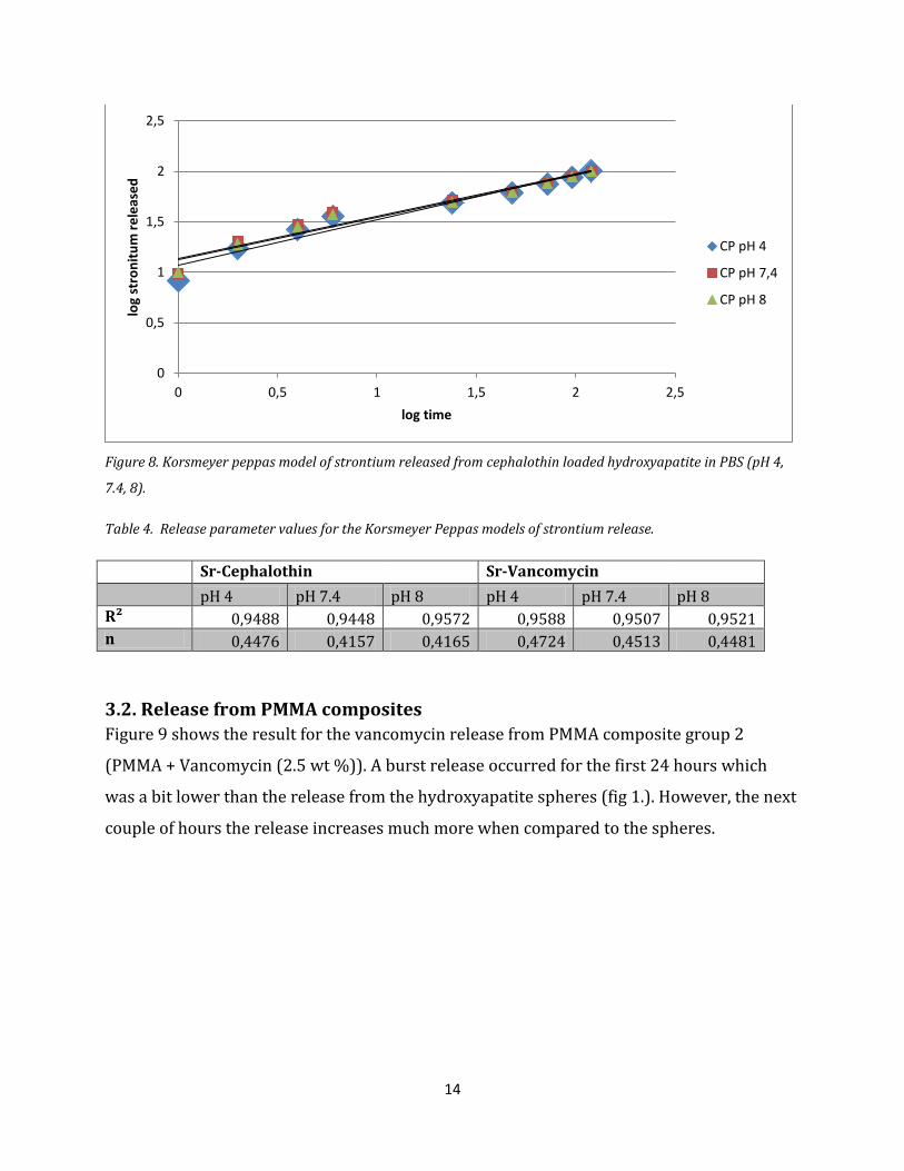
In figure 7 and figure 8 the Korsmeyer Peppas models for the release of strontium from
vancomycin and cephalothin loaded hydroxyapatite particles respectively are shown. Table
4 shows the release parameter values for the Korsmeyer Peppas models of strontium
release. Similar to the antibiotic release, the predictable strontium release for both the
vancomycin loaded particles as well as the cephalothin loaded particles are improved by
using a regression line. The diffusion exponent for all of the releases of strontium from
vancomycin loaded particles are just over 0.43, and are therefore described as anomalous
release. On the other hand, the diffusion exponent from the strontium release from the
cephalothin loaded particles are just below 0.43 with the exception of the release profile at
pH 4, which is just over 0.43. It could be assumed that release mechanisms would most
accurately be described by Fickian diffusion. The exception being the strontium release
from the vancomycin loaded hydroxyapatite spheres at pH 4 which is dependent on the
diffusion on the spheres therefore having a higher diffusion exponent.
Figure 7. Korsmeyer peppas model of strontium released from vancomycin loaded hydroxyapatite in PBS (pH 4,
7.4, 8).
0
0,5
1
1,5
2
2,5
0 0,5 1 1,5 2 2,5
log
stro
nti
um
re
leas
ed
log time
VM pH 4
VM pH 7,4
VM pH 8
14
Figure 8. Korsmeyer peppas model of strontium released from cephalothin loaded hydroxyapatite in PBS (pH 4,
7.4, 8).
Table 4. Release parameter values for the Korsmeyer Peppas models of strontium release.
Sr-Cephalothin Sr-Vancomycin
pH 4 pH 7.4 pH 8 pH 4 pH 7.4 pH 8 R² 0,9488 0,9448 0,9572 0,9588 0,9507 0,9521 n 0,4476 0,4157 0,4165 0,4724 0,4513 0,4481
3.2. Release from PMMA composites
Figure 9 shows the result for the vancomycin release from PMMA composite group 2
(PMMA + Vancomycin (2.5 wt %)). A burst release occurred for the first 24 hours which
was a bit lower than the release from the hydroxyapatite spheres (fig 1.). However, the next
couple of hours the release increases much more when compared to the spheres.
0
0,5
1
1,5
2
2,5
0 0,5 1 1,5 2 2,5
log
stro
nit
um
re
leas
ed
log time
CP pH 4
CP pH 7,4
CP pH 8

15
Figure 9. Vancomycin release profiles for PMMA composite group 2 in PBS (pH 4, 7.4 and 8).
The PMMA composite group 3 (PMMA + Vancomycin loaded hydroxyapatite (10 wt %)) had
a low burst release and a continued stable slow release the following hours compared to the
release profiles of the hydroxyapatite and the other PMMA composite groups. Moreover,
the estimated amount of vancomycin in group 3 was about 50 % higher than the other
groups which would indicate that loading vancomycin into hydroxyapatite spheres affected
the release rate to be slower. The release profiles do not differ much between the different
PBS pH values. The release profiles are shown in figure 10.
Figure 10. Vancomycin release profiles for PMMA composite group 3 in PBS (pH 4, 7.4 and 8).
0
100
200
300
400
500
600
700
0 8 16 24 32 40 48 56 64 72 80 88 96 104 112 120
Cu
mu
lati
ve v
anco
myc
in r
ele
ase
d (
ug/
ml)
Time (h)
pH 4
pH 7,4
pH 8
0
50
100
150
200
250
300
350
400
0 8 16 24 32 40 48 56 64 72 80 88 96 104 112 120
Cu
mu
lati
ve v
anco
myc
in r
ele
ase
(u
g/m
l)
Time (h)
pH 4
pH 7,4
pH 8

16
The release profiles for PMMA composite group 4 (PMMA + Hydroxyapatite (10 wt %) +
Vancomycin (2.5 wt %)), shown in figure 11, are similar to those of PMMA composite group
2. A relative low burst release which then had a high increase of release in the proceeding
time. The vancomycin release is slightly higher than the other PMMA groups.
Figure 11. Vancomycin release profiles for PMMA composite group 4 in PBS (pH 4, 7.4 and 8).
In table 5 the amount of vancomycin released within the first 24 hours from PMMA
composite group 2, 3 and 4. This amount gives an estimation of much of the antibiotic was
on the outer surface of the PMMA.
Table 5. Amount of vancomycin releasedwithin the first 24 hours from PMMA composite groups 2, 3 and 4.
Group 2 Group 3 Group 4
pH 4 pH 7.4 pH 8 pH 4 pH 7.4 pH 8 pH 4 pH 7.4 pH 8
1.145
mg
1.073
mg
1.360
mg
0.771
mg
0.724
mg
0.722
mg
1.539
mg
1.608
mg
1.780
mg
The Korsmeyer Peppas model was implemented to describe the release mechanisms for the
vancomycin release from the PMMA groups. The release parameter values are summarized
in table 6.
0
100
200
300
400
500
600
700
800
900
0 8 16 24 32 40 48 56 64 72 80 88 96 104 112 120
Cu
mu
lati
ve v
anco
myc
in r
ele
ase
(u
g/m
l)
Time (h)
pH 4
pH 7,4
pH 8

17
Table 6. Release parameter values for the vancomycin release from the PMMA composite groups.
Group 2 Group 3 Group 4
pH 4 pH 7,4 pH 8 pH 4 pH 7,4 pH 8 pH 4 pH 7,4 pH 8
R² 0,9284 0,9243 0,922 0,9085 0,9017 0,9048 0,9376 0,9419 0,9364
n 0,4406 0,4625 0,4552 0,4669 0,4637 0,4609 0,5108 0,5086 0,4684
Figure 12, 13 and 14 show the Korsmeyer Peppas model for the PMMA composite groups 2,
3 and 4. All of the profiles have release mechanisms that should be classified as non-Fickian
diffusion. However, the diffusion exponents are rather close to the value of 0.43 which
indicates Fickian diffusion, especially for the release profile of group 2 at pH 4 while the
release profiles for group 4 tend to be more anomalous than the others and should be
interpreted that the diffusion is more dependent on the diffusion and degradation of the
hydroxyapatite spheres.
Figure 12. Korsmeyer Peppas profiles for the vancomycin release from PMMA group 2 in PBS (pH 4, 7.4 and 8)
0
0,5
1
1,5
2
2,5
0 0,5 1 1,5 2 2,5
log
van
com
ycin
re
leas
ed
log time
pH 4 pH 7,4 pH 8

18
Figure 13. Korsmeyer Peppas profiles for the vancomycin release from PMMA group 3 in PBS (pH 4, 7.4 and 8)
Figure 14. Korsmeyer Peppas profiles for the vancomycin release from PMMA group 4 in PBS (pH 4, 7.4 and 8)
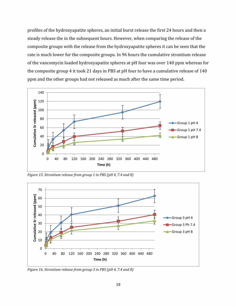
In figure 15 the strontium release from PMMA composite group 1 is shown and the
strontium release for group 3 and 4 are shown in figure 16 and 17. The release rate of
group 1 is similar to group 4, whereas the release rate for group 3 is lower than the other
two which it also had for the vancomycin release when compared to the other PMMA
composite groups. The release medium had a similar effect on all the three PMMA
composite groups in which the more acidic solution gave a higher release rate.
Furthermore, all the three PMMA composite groups had, just as the strontium release
0
0,5
1
1,5
2
2,5
0 0,5 1 1,5 2 2,5
log
van
com
ycin
re
leas
ed
log time
pH 4
pH 7.4
pH 8
0
0,5
1
1,5
2
2,5
0 0,5 1 1,5 2 2,5
log
van
com
ycin
re
leas
ed
log time
pH 4 pH 7.4 pH 8
19
profiles of the hydroxyapatite spheres, an initial burst release the first 24 hours and then a
steady release the in the subsequent hours. However, when comparing the release of the
composite groups with the release from the hydroxyapatite spheres it can be seen that the
rate is much lower for the composite groups. In 96 hours the cumulative strontium release
of the vancomycin loaded hydroxyapatite spheres at pH four was over 140 ppm whereas for
the composite group 4 it took 21 days in PBS at pH four to have a cumulative release of 140
ppm and the other groups had not released as much after the same time period.
Figure 15. Strontium release from group 1 in PBS (pH 4, 7.4 and 8)
Figure 16. Strontium release from group 3 in PBS (pH 4, 7.4 and 8)
0
20
40
60
80
100
120
140
0 40 80 120 160 200 240 280 320 360 400 440 480
Cu
mu
lati
ve S
r re
leas
ed
(p
pm
)
Time (h)
Group 1 pH 4
Group 1 pH 7.4
Group 1 pH 8
0
10
20
30
40
50
60
70
0 40 80 120 160 200 240 280 320 360 400 440 480
Cu
mu
lati
ve S
r re
leas
ed
(p
pm
)
Time (h)
Group 3 pH 4
Group 3 Ph 7.4
Group 3 pH 8

20
Figure 17. Strontium release from group 4 in PBS (pH 4, 7.4 and 8)
The Korsmeyer Peppas model release parameters for the strontium releases from the
PMMA composite groups are shown in table 7. From the diffusion exponents it can be seen
that for group 1 and 4 the release mechanism at pH four and 7.4 was controlled by the
diffusion of the hydroxyapatite spheres but at pH 8 the release mechanism was controlled
by Fickian diffusion. Group 3, which consisted of PMMA and vancomycin loaded
hydroxyapatite, has values under 0.43 which would indicate that the release mechanism is
best described as Fickian diffusion, whereas the vancomycin release for the same group
turned out to be non-Fickian diffusion. The Korsmeyer Peppas profiles for the PMMA
composite groups are shown in figure 18, 19 and 20.
Table 7. Release parameter values for the strontium release from PMMA composite group 1, 3 and 4.
Group 1 Group 3 Group 4
pH 4 pH 7.4 pH 8 pH 4 pH 7.4 pH 8 pH 4 pH 7.4 pH 8
R² 0.979 0.9758 0.9776 0.9756 0.9767 0.9729 0.9836 0.9837 0.9794
n 0.4648 0.463 0.4092 0.417 0.4128 0.3922 0.485 0.443 0.4032
0
20
40
60
80
100
120
140
160
0 40 80 120 160 200 240 280 320 360 400 440 480
Cu
mu
lati
ve S
r re
leas
ed
(p
pm
)
Time (h)
Group 4 pH 4
Group 4 pH 7.4
Group 4 pH 8

21
Figure 18. Korsmeyer Peppas model of the strontium release from group 1 in PBS (pH 4, 7.4 and 8)
Figure 19. Korsmeyer Peppas model of the strontium release from group 3 in PBS (pH 4, 7.4 and 8)
0
0,5
1
1,5
2
2,5
0 0,5 1 1,5 2 2,5 3
log
stro
nti
um
re
leas
ed
log time
Group 1 ph 4
Group 1 pH 7,4
Group 1 pH 8
0
0,5
1
1,5
2
2,5
0 0,5 1 1,5 2 2,5 3
log
stro
nti
um
re
leas
ed
log time
Group 3 pH 4
Group 3 pH 7.4
Group 3 pH 8

22
Figure 20. Korsmeyer Peppas model of the strontium release from group 4 in PBS (pH 4, 7.4 and 8)
4. Conclusion Hydroxyapatite spheres containing strontium were prepared for in vitro release study. The
hollow spheres were found to have a high loading capacity for the antibiotics vancomycin
and cephalothin. The loading capacity was approximately 0.37 mg antibiotics per mg
hydroxyapatite. The antibiotics vancomycin and cephalothin were loaded separately into
hydroxyapatite spheres and the in vitro release under different pH values for the release
medium PBS was evaluated. All of the releases showed the expected burst release followed
by a slow release. The amount of antibiotic released during the burst release was about 5 %
of the loaded amount for the vancomycin and 3 % for the cephalothin. The pH value of the
release medium had influence on the release rate to some extent for the antibiotic release
and the acidic solution had a more significant impact on the strontium release. The
cephalothin loaded spheres had higher release rate than the vanomycin loaded spheres in
both antibiotic and strontium release rate. The release mechanism for hydroxyapatite
spheres was found to be best explained by Fickian diffusion.
Four hydroxyapatite-PMMA composite groups’ antibiotic and strontium release were
studied. Group 3 consisting of PMMA and vancomycin loaded hydroxyapatite (10 wt %) had
the lowest vancomycin release rate and the lowest strontium release rate of all the groups.
However, it was also the group that was estimated to be most loaded with vancomycin
0
0,5
1
1,5
2
2,5
0 0,5 1 1,5 2 2,5 3
log
stro
nti
um
re
leas
ed
log time
Group 4 pH 4
Group 4 pH 7.4
Group 4 pH 8

23
suggesting that loading vancomycin into hydroxyapatite spheres affected the release rates.
Group 4 (PMMA + Hydroxyapatite (10 wt %) + Vancomycin (2.5 wt %)) had the highest
rates both in vancomycin release and strontium release. The influence of the release
medium was similar to the influence on the hydroxyapatite spheres which is to say that an
acidic environment gave a higher release rate. The release mechanism for the vancomycin
release from hydroxyapatite-PMMA composite groups showed signs that could best be
explained by the surface phenomenon of the hydroxyapatite. The strontium release showed
similar signs with the exception of PMMA composite group 3 which were controlled by
Fickian diffusion. All of the composite groups had a much lower strontium release rate than
the strontium release from the hydroxyapatite spheres. However, group 2 and group 4 had
similar or higher vancomycin release rate than the ones from the hydroxyapatite spheres.
5. Acknowledgement I would like to thank my supervisors Dr. Wei Xia and Dr. Cecilia Persson, at the Department
of Engineering Sciences; Applied Materials Science, for giving me the opportunity to do this
master’s thesis project and for all valuable help during the course of this project.
6. References 1 Baron, R., Neff, L., Louvard, D., & Courtoy, P. Cell-mediated Extracellular Acidification and Bone Resorption: Evidence for a Low pH in Resorbing Lacunae and Localization of a 100-kD Lysosomal Membrane Protein at the Osteoclast Ruffled Border. Journal of Cell Biology, 101 (1985), 2210-2222. 2 J.G.E. Hendriks, J.R. van Hornb, H.C. van der Meia, H.J. Busscher, Backgrounds of antibiotic-
loaded bone cement and prosthesis-related infection, Biomaterials 25 (2004) 545–556. 3 Hunter GA, Welsh RP, Cameron HU, Bailey WH, The results of revision of total hip arthroplasty, J Bone Jt Surg Br 61 (1979) 419–21. 4 Mariani BD, Tuan RS, Advances in the diagnosis of infection in prosthetic joint implants, Mol Med Today 4 (1998)207–13. 5 John A.Wright, SeanP.Nair, Interaction of staphylococci with bone, International Journal of Medical Microbiology 300(2010)193–204 6 Uwe Joostena, Alexander Joistb, G. Goshegerc, Ulf Liljenqvistc, Burkhard Brandtd, Christof von Eiff, Effectiveness of hydroxyapatite-vancomycin bone cement in the treatment of Staphylococcus aureus induced chronic osteomyelitis, Biomaterials 26 (2005) 5251–5258. 7 Van de Belt H, Neut D, Uges DRA, Schenk W, Van Horn JR, Van der Mei HC, Busscher HJ. Surface roughness, porosity and wettability of gentamicin-loaded bone cements and their antibiotic release. Biomaterials 21 (2000) 1981–7.

24
8 Sang-Hoon Rhee, Synthesis of hydroxyapatite via mechanochemical treatment, Biomaterials(2002), 23(4), 1147-1152. 9 Yutaka Mizushima, Toshiyuki Ikoma, Jyunzo Tanaka, Keiko Hoshi, Tsutomu Ishihara, Yasuaki Ogawa, Akinori Ueno, Injectable porous hydroxyapatite microparticles as a new carrier for protein and lipophilic drugs, Journal of Controlled Release 110 (2006) 260 – 265. 10 Feng Ye , Haifeng Guo, Haijiao Zhang, Xiulan He, Polymeric micelle-templated synthesis of hydroxyapatite hollow nanoparticles for a drug delivery system, Acta Biomaterialia 6 (2010) 2212–2218. 11 R. Labellaa, M. Bradena, S. Deb, Novel hydroxyapatitenext term-based previous termdentalnext term composites, Biomaterials 15 (1994) 1197-1200. 12 Uwe Joostena, Alexander Joistb, G. Goshegerc, Ulf Liljenqvistc, Burkhard Brandtd, Christof von Eiff, Effectiveness of hydroxyapatite-vancomycin bone cement in the treatment of Staphylococcus aureus induced chronic osteomyelitis, Biomaterials 26 (2005) 5251–5258. 13 Michael Diefenbeck, Thomas Mückley, Gunther O Hofmann, Prophylaxis and treatment of implant-related infections by local application of antibiotics, Injury, Int. J. Care Injured (2006) 37, S95—S104. 14 H. van de Belt, D. Neut, D.R.A. Uges, W. Schenk, J.R. van Horn, H.C. van der Mei, H.J. Busscher, Surface roughness, porosity and wettability of gentamicin-loaded bone cements and their antibiotic release, Biomaterials 21 (2000) 1981-1987 15 Salwa R. El-Shaboury, Gamal A. Saleh ∗, Fardous A. Mohamed, Azza H. Rageh, Analysis of cephalosporin antibiotics, Journal of Pharmaceutical and Biomedical Analysis 45 (2007) 1–19. 16 S. Pors Nielsen, The biological role of strontium, Bone 35 (2004) 583– 588. 17 M. Sivakumar, I. Manjubala, K. Panduranga Rao, Preparation, characterization and in vitro release of gentamicin from coralline hydroxyapatite–gelatin composite microspheres, Biomaterials, Volume 23, Issue 15, August 2002, Pages 3175-3181. 18 K.T. Chu, Y. Oshida, E.B. Hancock, M.J. Kowolik, T. Barco and S.L. Zunt, Hydroxyapatite/PMMA composites as bone cements, Bio-Medical Materials and Engineering 14 (2004) 87–105. 19 Daniela Cerretani, Giorgio Giorgi, Paolo Fornara, Luigi Bocchi, Laura Neri, Remo Ceffa, Franco Ghisellini, and Merrill A. Ritter, The In Vitro Elution Characteristics of Vancomycin Combined With Imipenem-Cilastatin in Acrylic Bone–Cements, The Journal of Arthroplasty 17 (2002) 5 20 Philip L. Ritger and Nikolaos A. Peppas, A simple equationnext term for previous termdescription of solute release I. Fickian and non-fickian release from non-swellable devicesnext term in the previous termform of slabs, spheres, cylinders or discs, Journal of Controlled Release, 5 (1987) 23-36.