Improving Long-Term Patient Outcomes for Exudative … · Improving Long-Term Patient Outcomes for...
24
A Report Based on a National Expert Summit and Expert Roundtable Convened in 2017 by the Angiogenesis Foundation Improving Long-Term Patient Outcomes for Exudative Age-Related Macular Degeneration Version1.0
Transcript of Improving Long-Term Patient Outcomes for Exudative … · Improving Long-Term Patient Outcomes for...

A Report Based on a National Expert Summit and Expert Roundtable Convened in 2017 by the Angiogenesis Foundation
Improving Long-Term Patient Outcomes for Exudative Age-Related Macular Degeneration
Version1.0

Copyright © 2017 by The Angiogenesis Foundation. All Rights Reserved.2
Authors 3
Executive Summary 4
Introduction 6• Exudative AMD Analysis• Paradigm Change: Antiangiogenic Therapies • Past Summits: Identifying Unmet Needs • Mandate for Better Long-Term Outcomes • National Multistakeholder Expert Summit • Further Analysis by Expert Roundtable
Real-World Evidence on Long-Term Patient Outcomes 11 • Role of the Angiogenesis Foundation • Real-World Evidence on Long-Term Patient Outcomes • Differential Outcomes of Anti-VEGF Therapy: Short-Term vs. Long-Term Outcomes • Differential Outcome of Anti-VEGF Therapy: Clinical Trial Settings vs. Real-World
Settings
Probable Causes for Differential Long-Term Patient Outcomes • Issues Related to Clinical Practice • The Patient and Caregiver Experiences with the Disease and Its Treatment • Knowledge Gaps
Desired Future State: Barriers to Improved Long-Term Visual Outcomes 15 • Physician Practice-Related Barriers • Patient- and Caregiver-Related Barriers • Knowledge-Related Barriers
References 21
Participants 22
Acknowledgements 23
Table of Contents
13

Copyright © 2017 by The Angiogenesis Foundation. All Rights Reserved.3
Table of Contents White Paper Report Developed By:
Mark G. Ackermann, M.S.Lighthouse Guild InternationalNew York, NY
Andrew N. Antoszyk, M.D.Charlotte Eye Ear Nose & Throat AssociatesCharlotte, NC
Odysseus Argy, M.D.Patient AdvocateSouth Dartmouth, MA
Carl W. Baker, M.D.Paducah Retinal CenterPaducah, KY
Dan BerkeryPatient AdvocateOsterville, MA
Elizabeth BerkeryPatient AdvocateOsterville, MA
W. Lloyd Clark, M.D.Palmetto Retina CenterW. Columbia, SC
Pravin U. Dugel, M.D.Retinal Consultants of ArizonaPhoenix, AZ
Roger A. Goldberg, M.D., M.B.A.Bay Area Retina AssociatesWalnut Creek, CA
Juan Grunwald, M.D.University of PennsylvaniaWynnewood, PA
Jeffrey S. Heier, M.D.Ophthalmic Consultants of BostonBoston, MA
Allen C. Ho, M.D.Wills Eye HospitalPhiladelphia, PA
Michelle JenneyPatient AdvocateBoston, MA
Peter Jenney, M.D.Patient AdvocateBoston, MA
Matthew Levine, M.F.A.American Macular Degeneration FoundationNorthampton, MA
Vincent W. Li, M.D., M.B.A.The Angiogenesis Foundation Cambridge, MA
William W. Li, M.D.The Angiogenesis FoundationCambridge, MA
Quan Dong Nguyen, M.D., M.Sc.Byers Eye InstitutePalo Alto, CA
Sunil S. Patel, M.D., Ph.D.West Texas Retina ConsultantsAbilene, TX
John S. Pollack, M.D.Rush University Medical CenterChicago, IL
Diana SavilleSummit Co-ChairThe Angiogenesis FoundationCambridge, MA
Rishi P. Singh, M.D.Summit Co-ChairCleveland Clinic Cole Eye InstituteShaker Heights, OH
Michael W. Stewart, M.D.Mayo ClinicJacksonville Beach, FL
Ivan J. Suner, M.D., M.B.A.Retina Associates of FloridaTampa, FL
Demetrios G. Vavvas, M.D., Ph.D.Massachusetts Eye and EarBoston, MA
Charles C. Wykoff, M.D., Ph.D.Retina Consultants of HoustonHouston, TX

Copyright © 2017 by The Angiogenesis Foundation. All Rights Reserved.4
Age-related macular degeneration (AMD), which primarily affects people over the age of 50, is the leading cause of blindness in the world. An estimated 11.2 million Americans are affected by AMD, and that number is expected to rise significantly in the coming years as the U.S. population continues to age.
During the past 15 years, new therapies, primarily in the form of VEGF-targeted antiangiogenic drugs, have produced a true paradigm shift in the treatment of exudative AMD, also known as wet AMD, which is an advanced form of the disease and can cause rapid loss of visual function. Patients now have effective treatment options that can help prevent vision loss, and in some cases, even restore vision. Modern diagnostic techniques, including home-based monitoring, have also helped patients by supporting early diagnosis of wet AMD.
As the number of people diagnosed with wet AMD increases, so does the demand for anti-vascular endothelial growth factor (anti-VEGF) treatments. Meeting this demand efficiently and economically has been a challenge for retina specialists and others in the ophthalmology community. In addition, research into the treatment of wet AMD — particularly studies that focus on long-term and real-world outcomes — has recently begun to reveal new evidence about the relationship between patient outcomes and dosing frequency over the long term. As more researchers, clinicians, and patients use antiangiogenic therapies and as more studies involving these treatments are published, new questions have arisen about the best practices. These questions center on the timeliness of initial dosing and the optimal dosing frequency of the treatments, as well as on concerns about the diagnosis and long-term management of the disease. One major concern involves the undertreatment of patients over the long term, which may lead to devastating outcomes.
The Angiogenesis Foundation is focused on optimizing the clinical benefits of antiangiogenesis therapies. On August 16, 2017, the Foundation brought together representatives from key wet AMD stakeholder groups from across the United States — clinicians, patients with wet AMD, caregivers, researchers, and patient advocates — for a day-long national summit. The purpose: to determine how clinicians can better meet the needs of patients with wet AMD to ensure their vision is preserved as long as possible with anti-VEGF therapies as well as explore novel approaches to assist wet AMD patients in retaining functional vision. For additional analysis, the Angiogenesis Foundation convened an expert roundtable on October 18, 2017, consisting of six leading clinicians who provided further insights on the summit findings.
The following key actions were recommended:
1. Increase public awareness of AMD.
• Initiate a major, ongoing, multi-faceted awareness campaign for AMD so the at-risk age population, specifically patients with intermediate AMD who are at the highest risk for progression to wet AMD, is aware of the need for regular vision examinations and the role they play in maintaining functional vision should they develop wet AMD.
• Work with medical societies and medical schools to raise awareness of intermediate and wet AMD among trainees, primary care providers, and general ophthalmologists. Emphasize the critical role primary care providers and general ophthalmologists can play in helping patients retain good vision should they convert to wet AMD.
2. Improve early detection of wet AMD.
• Raise awareness of the availability and opportunity to benefit from early detection with home-monitoring devices. The ForeseeHome device, as well as other home-monitoring systems in development, support earlier detection and treatment of patients transitioning from dry to wet AMD. More patients need to be empowered to use these devices to detect disease progression from the convenience of their own home between routine examinations.
Executive Summary

Copyright © 2017 by The Angiogenesis Foundation. All Rights Reserved.5
Executive Summary3. Develop and adopt evidence-based clinical practice guidelines.
• Use currently available registrational and long-term, real-world data to formulate a set of recommended retinal practice guidelines for anti-VEGF therapy.
• Enlist professional retina organizations to develop treatment protocol guidelines for using anti-VEGF agents for the long-term management of wet AMD.
• Share best practices and guidelines through an information clearinghouse to benefit the community of vision health care providers.
4. Acknowledge and address undertreatment as a cause of long-term vision loss.
• Increase awareness of the consequences of undertreatment and/or the interruption of treatment in clinician and patient communities.
• Identify and share ways to streamline retina specialist practices to improve treatment efficiency and reduce undertreatment.
• Package the reimbursement of treatment to include more services than the injection.
5. Treat the “whole patient.”
• Provide better, more holistic educational information to patients and their caregivers in the retina specialist’s office.
• Use support agencies to perform outreach to patients after their diagnosis and throughout treatment.
• Work with organizations such as the American Society of Retina Specialists, the American Academy of Ophthalmology, the Retina Society, the Macula Society, the Lighthouse Guild, and the American Macular Degeneration Foundation to develop a new standard of care for patients with wet AMD that takes a patient-centered approach to treatment.
6. Develop a real-world, long-term, late-stage research agenda.
• Mine existing registries for useful, valid data on long-term outcomes.
• Establish a real-world, long-term, late-stage treatment protocol study.
Copyright © 2017 by The Angiogenesis Foundation. All Rights Reserved.6
Exudative AMD Analysis
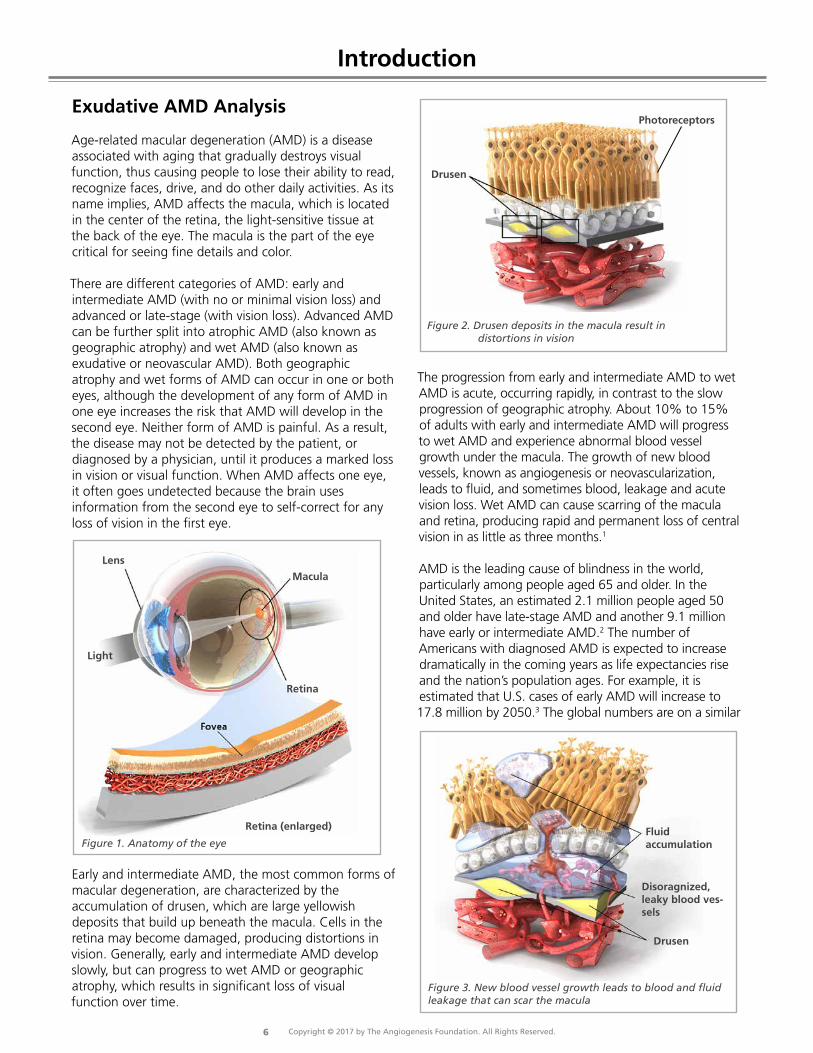
Age-related macular degeneration (AMD) is a disease associated with aging that gradually destroys visual function, thus causing people to lose their ability to read, recognize faces, drive, and do other daily activities. As its name implies, AMD affects the macula, which is located in the center of the retina, the light-sensitive tissue at the back of the eye. The macula is the part of the eye critical for seeing fine details and color. There are different categories of AMD: early and intermediate AMD (with no or minimal vision loss) and advanced or late-stage (with vision loss). Advanced AMD can be further split into atrophic AMD (also known as geographic atrophy) and wet AMD (also known as exudative or neovascular AMD). Both geographic atrophy and wet forms of AMD can occur in one or both eyes, although the development of any form of AMD in one eye increases the risk that AMD will develop in the second eye. Neither form of AMD is painful. As a result, the disease may not be detected by the patient, or diagnosed by a physician, until it produces a marked loss in vision or visual function. When AMD affects one eye, it often goes undetected because the brain uses information from the second eye to self-correct for any loss of vision in the first eye.
Early and intermediate AMD, the most common forms of macular degeneration, are characterized by the accumulation of drusen, which are large yellowish deposits that build up beneath the macula. Cells in the retina may become damaged, producing distortions in vision. Generally, early and intermediate AMD develop slowly, but can progress to wet AMD or geographic atrophy, which results in significant loss of visual function over time.
The progression from early and intermediate AMD to wet AMD is acute, occurring rapidly, in contrast to the slow progression of geographic atrophy. About 10% to 15% of adults with early and intermediate AMD will progress to wet AMD and experience abnormal blood vessel growth under the macula. The growth of new blood vessels, known as angiogenesis or neovascularization, leads to fluid, and sometimes blood, leakage and acute vision loss. Wet AMD can cause scarring of the macula and retina, producing rapid and permanent loss of central vision in as little as three months.1
AMD is the leading cause of blindness in the world, particularly among people aged 65 and older. In the United States, an estimated 2.1 million people aged 50 and older have late-stage AMD and another 9.1 million have early or intermediate AMD.2 The number of Americans with diagnosed AMD is expected to increase dramatically in the coming years as life expectancies rise and the nation’s population ages. For example, it is estimated that U.S. cases of early AMD will increase to 17.8 million by 2050.3 The global numbers are on a similar
Figure 2. Drusen deposits in the macula result in distortions in vision
Drusen
Photoreceptors
Figure 3. New blood vessel growth leads to blood and fluid leakage that can scar the macula
Fluid accumulation
Disoragnized, leaky blood ves-sels
Drusen
Figure 1. Anatomy of the eye
Retina
Macula
Retina (enlarged)
Lens
Light
Introduction

Copyright © 2017 by The Angiogenesis Foundation. All Rights Reserved.7
Introduction trend line: Experts estimate that 196 million people will be affected by AMD by 2020, a number that is projected to climb to 288 million by 2040.4
Paradigm Change: Antiangiogenic Therapies
Angiogenesis research, which escalated in the early 1980s, made dramatic advances in the late 1990s. Those advances culminated in the identification of specific antiangiogenic approaches to treating a variety of diseases, including cancer, skin diseases, and blinding disorders such as wet AMD. More than 10,000 laboratories around the world are currently involved in angiogenesis research. In the United States, the National Institutes of Health (NIH) funds approximately USD $60 million of angiogenesis-related grants per year. This rapidly developing field has witnessed important advances, particularly in the last 15 years, which have had a major impact on the lives of patients. Just 10 years ago, patients diagnosed with wet AMD almost always became functionally blind. Today, wet AMD is a highly treatable condition and many people maintain their vision because of anti-VEGF therapies. In fact, antiangiogenic drugs have led to a 50% plunge in the incidence of legal blindness attributable to wet AMD.5
In December 2004, the U.S. Food and Drug Administration (FDA) approved pegaptanib, the first angiogenesis inhibitor for wet AMD. Clinical trials showed that intravitreal injections of pegaptanib slowed the rate of vision loss caused from wet AMD.6 This antiangiogenic therapy became recognized as an entirely new class of disease treatment. An even more effective drug, ranibizumab, was approved for the treatment of wet AMD in the United States in late 2006. Ranibizumab, as well as pegaptanib, interferes with a small protein known as vascular endothelial growth factor (VEGF). This growth factor stimulates angiogenesis and promotes vascular permeability (the passage of water and other small molecules through a blood vessel’s wall), two processes that play a major role in the development of wet AMD.
Clinical trials demonstrated that 95% of patients treatedwith a once-monthly intravitreal injection of ranibizumab maintained their vision as long as the injections continued over the course of the trial. “Maintaining vision” meant that their ability to read a vision chart declined by no more than 15 Early Treatment DiabeticRetinopathy Study (ETDRS) letters, or three lines. In addition, up to 40% of those treated with monthly ranibizumab for a year experienced an improvement of 15 or more letters (3 lines) in visual acuity.7,8,9
For the first time, physicians could offer people the opportunity to preserve their vision, and, in some cases, reverse a portion of their vision loss. The major drawback to this new therapy, however, was its price tag, costing about USD $2,000 per injection per eye. Additionally, the monthly in-office injection places transportation and logistical burdens on patients and their caregivers.
Before ranibizumab was approved, retina specialists began experimenting with another anti-VEGF agent, bevacizumab, which was approved by the FDA for the treatment of colorectal cancer in 2004 (and later for other types of cancer). Bevacizumab is a larger molecule, known as a monoclonal antibody, from which ranibizumab, a monoclonal antibody fragment, is derived.
Bevacizumab is not indicated for eye diseases, and has not been FDA-approved for use in the eye. Nonetheless, it has been shown to be clinically effective for the treatment of wet AMD, in a large randomized clinical trial funded by the National Eye Institute,8 and is used off-label for this purpose at a cost of about USD $50 per intravitreal injection. (A drug is used “off-label” when it is prescribed for a use not approved by a country’s regulatory agency.) Because it is produced in large vials for cancer treatments, bevacizumab must be divided by a compounding pharmacy into the much smaller quantities for treating the eye. There have been numerous documented cases of infection from bevacizumab’s use in the eye, likely due to the preparation of the solution and not to the molecule itself. In addition, research has found significant variations in the concentration of proteins in bevacizumab samples taken from various pharmacies.10,11 When treatments are used off-label, patients should be properly informed of safety risks. Clinical trials comparing ranibizumab with bevacizumab have suggested that both drugs are similarly effective at stopping disease progression and restoring visual acuity, at least when dosed monthly during the first two years of treatment.8,12
In 2011, the FDA approved a third antiangiogenic drug, called aflibercept, for the treatment of wet AMD.13 It is based on a novel drug technology that fuses binding domains from two proteins (VEGFR1 and VEGFR2) to the Fc fragment of an IgG molecule to neutralize not only VEGF-A (like ranibizumab and bevacizumab), but also proteins such as VEGF-B and placental growth factor
Figure 4. Anti-VEGF Treatment

Copyright © 2017 by The Angiogenesis Foundation. All Rights Reserved.8
(PIGF). Aflibercept can be administered by intravitreal injection every other month, following three initial monthly injections. Clinical trials comparing ranibizumab with aflibercept show both drugs are similarly effective at stopping disease progression and restoring some portion of visual acuity, with fewer total injections for aflibercept.14
Past Summits: Identifying Unmet Needs
By 2009, anti-VEGF therapies had revolutionized the treatment of wet AMD — and the field of ophthalmology. Given these remarkable treatment advances, the Angiogenesis Foundation convened the AMD stakeholder community to review the progress made, the challenges faced, and the questions that need to be answered to best meet the needs of people living with wet AMD.
As its first major global step, the Angiogenesis Foundation assembled an interdisciplinary group of international leaders in AMD treatment and translational science. The Foundation convened the International Expert Summit for Age-Related Macular Degeneration in Berlin, Germany, in November 2011. The success of that meeting led to three other global events: the Latin American Wet AMD Coalition Expert Summit in Bogota, Colombia, held in March 2012 in partnership with the Pan-American Retina & Vitreous Society; the Australian Wet Age-Related Macular Degeneration Coalition Expert Summit in Sydney, held in July 2012 in partnership with the Macular Disease Foundation Australia; and the Asia-Pacific Wet AMD Coalition Expert Summit, held in Hong Kong in February 2013. Then, in June 2013, a second international summit was held in Berlin: Advocating for the Improved Treatment and Outcomes for Wet Age-Related Macular Degeneration. It focused on the advances that had occurred by that date in the treatment of wet AMD, as well as on other matters regarding the diagnosis and management of the disease. Each summit resulted in a white paper that provided an overview of the group’s discussions and the key steps necessary for advancing the treatment of wet AMD using anti-VEGF therapies to maximize impact and help the most individuals possible.
Mandate for Better Long-Term Outcomes
As the number of people diagnosed with wet AMD increases, so does the demand for anti-VEGF treatments. Meeting this demand has been a challenge for retina specialists and others in the ophthalmology community. In addition, research into the treatment of wet AMD — particularly studies that focus on long-term and real-world outcomes — has revealed new evidence about how vision is maintained over the long term, based on treatment patterns and the importance of good absolute visual acuity at the time wet AMD treatment is initiated. That research has shown significant and troubling disparities in short-term versus long-term patient outcomes and in clinical trial versus real-world patient outcomes. As a result, new, important questions have arisen about the optimal dosing frequency of the treatments over the long term, as well as concerns about diagnosis, monitoring, and long-term management of the disease.
National Multistakeholder Expert Summit
By mid-2017, the Angiogenesis Foundation determined it was time for a U.S-specific expert summit on wet AMD that would delve deeply into the latest research on long-term, real-world outcomes. That summit, the National Multistakeholder Expert Summit: Improving Long-Term Patient Outcomes for Exudative Age-Related Macular Degeneration, convened in Boston, Massachusetts, on August 16, 2017. The summit included representatives from all key stakeholder groups: clinicians, patients, caregivers, researchers, and patient advocates. As with the earlier summits, this event was an interactive, professionally moderated set of short presentations and extensive roundtable discussions aimed to establish a dialogue and consensus among the participants.
The summit opened with two short presentations on anti-VEGF therapies for wet AMD. One provided an up-to-date summary of the evidence from clinical trials regarding short-term versus long-term outcomes as well as differences in outcomes from dosing, while the other presentation offered a similar review of the evidence regarding differential outcomes in clinical trial settings versus real-world settings. Under the direction of the moderator, the assembled experts then spent the rest of the day engaged in a series of discussions that defined and prioritized actions for better long-term patient outcomes in wet AMD. A graphical recorder captured key points of the discussion enabling the participants to visually review the content in real time.

Copyright © 2017 by The Angiogenesis Foundation. All Rights Reserved.9
Dr. W. Lloyd Clark
Dr. Quan Dong Nguyen Dr. John S. Pollack
Dr. Jeffrey Heier Dr. William W. Li
Dr. Charles Wykoff
Figure 5. Participants at the National Multistakeholder Expert Summit: Improving Long-Term Patient Outcomes for Exudative Age-Related Macular Degeneration. Participants from Expert Roundtable shown below.
Figure 6. Participants in the Expert Roundtable: Improving Long-Term Patient Outcomes for Exudative Age-Related Macular Degeneration

Copyright © 2017 by The Angiogenesis Foundation. All Rights Reserved.10
Further Analysis by Expert Roundtable
As a follow-up to the summit, on October 18, 2017, the Angiogenesis Foundation convened an expert roundtable of six additional leading clinicians to provide an analysisand further reflections on the summit findings. The clinicians were given a synopsis of the August 16 summit and then asked to provide their insights and contributions on the following topics:
1. Real-world versus clinical trial settings and impact on long-term patient outcomes for anti-VEGF treatments for wet AMD.
2. Variations in care, especially undertreatment, as a barrier to optimal long-term vision outcomes.
3. Actions to improve long-term patient outcomes, including future research using real-world evidence (RWE).
This white paper report provides an overview and highlights the key points raised during both the national summit and the subsequent expert roundtable discussion.
The Role of the Angiogenesis Foundation
Founded in 1994 and headquartered in Cambridge, Massachusetts, the Angiogenesis Foundation is focused on advancing the clinical benefits of angiogenesis and antiangiogenesis therapy. The Foundation is the premier nonprofit organization dedicated to helping people lead healthier, longer lives through angiogenesis-based treatment and prevention. As a nonprofit, third-party scientific organization, the Angiogenesis Foundation is independent of any individual, institution, or commercial entity.
The Angiogenesis Foundation has been supporting innovation in angiogenesis-based medical therapies for nearly a quarter century. Thanks in part to its research, advocacy and education, there are now more than 32 FDA-approved drugs and medical devices used to control angiogenesis. Angiogenesis-related research continues across a variety of fields where the control of the vasculature is critical, including cancer, chronic wounds, ophthalmology and cardiovascular disease.
Figure 7. Dr. Vincent Li welcomes participants during the summit opening roundtable discussion.
Real-World Evidence on Long-Term Patient Outcomes

Copyright © 2017 by The Angiogenesis Foundation. All Rights Reserved.11
The National Multistakeholder Expert Summit opened with welcoming remarks from Dr. Vincent Li, the Angiogenesis Foundation’s Chief Operating Officer and Scientific Director. He provided a brief description of the Foundation’s work in the field of ophthalmology, including convening international summits on anti-VEGF treatments for wet AMD. The two co-chairs of the summit — Diana Saville, Chief Innovation Officer of the Angiogenesis Foundation, and Rishi P. Singh, M.D. Associate Professor of Ophthalmology and Staff Surgeon at the Cleveland Clinic Cole Eye Institute — welcomed the stakeholders and introduced the primary purpose of the summit: to determine how clinicians can better meet the needs of patients with wet AMD to ensure their vision is preserved as long as possible. The opening remarks were followed by two brief presentations on the evidence that has emerged in recent years on differential outcomes for anti-VEGF therapies for wet AMD. Dr. Singh described differences in the short-term and long-term outcomes for such therapies, while Sunil Patel, M.D., a retina specialist with the West Texas Retina Consultants, explained how the results of anti-VEGF therapies differ in clinical-trial versus real-world settings.
Differential Outcomes of Anti-VEGF Therapy: Short-Term vs. Long-Term Outcomes
Few retina specialists follow the aggressive dosing and strict follow-up treatment regimens of the registration trials for anti-VEGF agents. Such regimens can impose a burden on patients with wet AMD and their caregivers, both in terms of the number of injections they must receive and in terms of the social and economic costs (e.g., journeying to see the doctor and taking time off from work or other responsibilities). In real-world settings, clinicians tend to treat patients using a variety of dosing frequencies — monthly, bi-monthly, quarterly, as-needed (PRN), and, more recently, treat-and-extend. Short-term studies (≤ 2 years) have been offered in support of each of these strategies, Dr. Singh noted. The 2-year HARBOR study found, for example, that PRN and monthly dosing produced equivalent visual outcomes.15 Other studies, however, have shown that quarterly and PRN regimens with less frequent monitoring and dosing generally led to limited improvement in visual outcomes at one and two years.16,17,18
Long-term clinical outcomes (≥ 3 years) of anti-VEGF agents in patients with wet AMD reveal variable outcomes based on the frequency and consistency of treatment.19 Such findings are concerning, particularly given that the average age of diagnosis in the United
States is 71 years, a factor that means patients with wet AMD often require ongoing treatment and maintenance for 10 or more years. Long-term studies (≥ 5 years) have revealed a correlation between greater visual declines and fewer injections. In the extended CATT study, for example, visual gains during the first 2 years were not maintained at 5 years; patients experienced, on average, a loss of 12 to 13 ETDRS letters in visual acuity at 5 years.20 A study that looked at 7-year outcomes for the ANCHOR, MARINA, and HORIZON studies, found that the patients had experienced, on average, an almost 9-letter decline in visual acuity from baseline.21 Those studies, however, reported a direct correlation between the number of injections and visual acuity during the extended phase: the more aggressive the dosing, the better the outcome. The VIEW 1 extension study also demonstrated the benefits of frequent, regimented dosing.22 Patients who received an anti-VEGF injection every 8 weeks from year 2 through year 5 finished with an average 7-letter gain in visual acuity.
In a small, investigator-initiated observational study, conducted among patients in Dr. Singh’s practice, patients were converted from PRN to bi-monthly
treatment.23 Most of the patients had been diagnosed and treated for 2 years prior to the dosing switch. At 24 months, the patients had gained, on average, 10 ETDRS letters in visual acuity, and 71% had 20/40 vision or better. Some patients then returned to as-needed treatment. At 5 years post-diagnosis, those who returned to as-needed dosing lost vision over time, while those in the bi-monthly treatment group continued to have vision gain. Multiple factors influence treatment decisions for patients with wet AMD. Patients must balance receiving too few treatments and the risk of devastating progressive vision loss with the potential of receiving unnecessary treatments and the burdens on both patients and caregivers that such treatments can impose.
Figure 8. Dr. Rishi P. Singh, Summit Co-chair presents data on differential outcomes of anti-VEGF therapy.
Real-World Evidence on Long-Term Patient Outcomes

Copyright © 2017 by The Angiogenesis Foundation. All Rights Reserved.12
Probable Causes for Differential Long-Term Patient OutcomesYet the evidence suggests there is a direct correlation between frequency of injections and long-term visual acuity — and undertreatment of patients with wet AMD has become a major barrier to optimal patient outcomes.
Differential Outcome of Anti-VEGF Therapy: Clinical Trial Settings vs. Real-World Settings
Clinical trials of anti-VEGF therapies for wet AMD do not reflect real-world practice for a variety of reasons. Clinical trials have strict inclusion and exclusion criteria, aggressive dosing frequencies, strict protocol adherence, extensive infrastructure, and generous funding. Real-world treatment settings, on the other hand, pose logistical and economic challenges that make it difficult for patients and clinicians to follow such a strict treatment regimen. As a result, very few retina specialists are treating patients at the monthly frequencies used in drug-registration trials. In a global survey conducted in 2014 by the American Society of Retina Specialists (ASRS), 16% of U.S. retina specialists said they used a PRN treatment approach with their wet AMD patients, and 78% said they used a treat-and-extend approach.24 In this respect, the survey’s U.S. respondents were no different than those in other regions of the world. Indeed, more than 90% of retina specialists surveyed in European, Central and South American, Asian-Pacific, and African-Middle Eastern countries cited PRN and treat-and-extend as their primary anti-VEGF treatment strategies for wet AMD. Yet, there is no universally agreed-upon definition of PRN or treat-and-extend. The definition varies from practitioner to practitioner. It’s not surprising, therefore, that an analysis of electronic health records (EHRs) from a large integrated U.S. health system database revealed that wet AMD patients in clinical practice received fewer anti-VEGF injections than patients in pivotal clinical trials.25 Indeed, in routine clinical practice, 65% of wet AMD patients receive 6 or fewer injections during the first year of treatment.25,26
The quantity of anti-VEGF injections matters in terms of outcomes. Two studies conducted in Canada27 and the United Kingdom28 reported that visual acuity gained bypatients during the first year of treatment was lost during the second year as injections tapered off. Data from CATT,
ANCHOR, and other extension studies suggest that the “sweet spot” for both gaining and retaining visual acuity (about 7 ETDRS letters, on average) appears to be 6-8 injections per year, a regimen that is difficult for many patients to maintain. The EXCITE study found that wet AMD patients who received monthly anti-VEGF injections gained an average of about 8 ETDRS letters of visual acuity, while those given quarterly injections gained an average of about 4.18 In a retrospective chart-review study, 93.2% of patients who received a fixed interval dosing (FIDO) of anti-VEGF injections every 4-8 weeks for at least 5 years experienced vision stabilization or improvement.29 At 7 years, the patients in the study were able to maintain an average of 12 ETDRS letters.
Yet, because of the strain placed on clinics overloaded with patients needing injections, and the burdens imposed on patients and caregivers by frequent dosing, most clinicians are using a treat-and-extend approach, which is not clearly defined in the ophthalmology community. With this regimen, the anti-VEGF drugs are often administered monthly until all signs of choroidal neovascularization (CNV) activity are resolved. Treatment is then slowly extended, usually in 2-week intervals. If leakage recurs, the intervals between treatments are shortened. However, there is no agreed-upon protocol outlining the exact dosing intervals used in each phase of this treatment approach. As a result, the treat-and-extend approach varies from one retina specialist to another. Several studies have shown that using various versions of the treat-and-extend approach result in outcomes similar to monthly injections, although careful monitoring is required.30,31,32 Yet, other studies suggest that after the first year of treatment, some gains in visual acuity are lost with treat-and-extend.32,33 To produce optimal results, therefore, this approach may require 6-9 injections per year, indefinitely.
The treatment of patients with wet AMD may be transformed in the coming years as new molecules, as well as new ways of delivering those drugs, become available. One of those promising molecules is brolucizumab, a single-chain antibody fragment, which has been designed as a long-lasting drug, to be administered by intravitreal injection at intervals of 8 or 12 weeks. Early research has shown that brolucizumab at 8 and 12 week intervals is not inferior to aflibercept at 8-week intervals.34

Copyright © 2017 by The Angiogenesis Foundation. All Rights Reserved.13
Although anti-VEGF therapies have made a remarkable difference in the lives of millions of people with wet AMD, discrepancies exist between the visual outcomes reported in clinical trials and those that occur in real-world practice. Furthermore, current evidence suggests that many, if not most, patients with the disease are being undertreated. Summit and expert roundtable participants addressed possible causes of these discrepancies. The discussions focused on three factors that may play a role:
1) Issues related to clinical practice
2) The patient and caregiver experience
3) Gaps in our knowledge of wet AMD
Issues Related to Clinical Practice
• Undertreatment is a widespread issue. The Medicare database suggests that, on average, patients with wet AMD may be undertreated.
• Late diagnosis of wet AMD contributes to poorer outcomes because patients have already lost vision that, in many cases, cannot be restored.
• Retina specialists are overwhelmed with patients who need treatment for wet AMD. Every aspect of a specialist’s practice, including waiting room space, is overloaded. Indeed, delivering anti-VEGF injections takes almost twice as long — and requires almost twice as many employees — as other types of ophthalmology appointments. This factor often leads to major scheduling bottlenecks, which, in turn, may cause clinicians to stretch the time between patients’ visits. Some clinicians are moving their practices into larger spaces, but such expansions are expensive and accompanied by economic uncertainties. Furthermore, although the infrastructure required for the treatment of wet AMD is massively greater than in the past, the reimbursement revenue from the treatment has decreased in recent years.
• Clinicians may say they adhere to specific approaches or treatment schedules, but in reality are treating patients with a variety of approaches on a case-by-case basis.
• Treatments are varied among different retina specialists and among different practices, and among different geographic regions.
• Clinicians often engage in de facto triaging of their patients. For example, they tend to be more aggressive in ensuring monocular patients (those who have already lost vision in one eye) return at regular, frequent intervals for anti-VEGF treatment, while putting less emphasis on strict treatment scheduling and adherence for patients whose opposing eye is unaffected.
• Treatments that do not produce expected outcomes right away sometimes wear on clinicians, as they do on patients, leading to treatment fatigue.
• Some clinicians may not be aware of the latest long- term and real-world outcomes research, and thus may not have adjusted their treatment practices regarding PRN.
• It is unlikely that one formula and dosing interval will work best for all patients.
• PRN and treat-and-extend mean different things to different clinicians. No evidence exists to universally define the terms in regard to best practices. Participants in the expert roundtable suggested that treatment guidelines, such as “PRN should still include monthly monitoring” and “treat-and-extend should not go beyond 12 weeks,” would be helpful for the community.
• The paperwork involved in getting approvals and reimbursements from public and private insurance companies for anti-VEGF treatments is quite burdensome and can play a role in how frequently patients get treated.
Patient and Caregiver Experiences with the Disease and Treatment
• When clinics do not give wet AMD patients a predictable, pleasant experience, the patients may choose to return at less frequent intervals.
• The importance of receiving frequent treatments and of adhering to treatments over the long term is often not sufficiently conveyed to patients and caregivers.
• Many patients find the “the cage” — the eyelid speculum — used during treatment to be
“torturous.” Their dread of it can cause stress, and may also result in patients requesting fewer treatments.
• Sometimes clinicians do not wait long enough between applying the numbing solution to the eye and delivering the anti-VEGF injection. Subsequently, patients may experience pain and then fear future treatments.
• Clinicians often fail to establish reasonable expectations with patients about the number of treatments they may need before they experience any improvement in their vision. As a result, many patients drop out of treatment too early.
• Many patients, particularly those with lower incomes, do not have support from family. For these patients, getting to treatment visits can be problematic.
• For caregivers, the burden of getting the patient to treatments — particularly, repeatedly taking time off from work — can be disruptive and burdensome.
Probable Causes for Differential Long-Term Patient Outcomes

Copyright © 2017 by The Angiogenesis Foundation. All Rights Reserved.14
Patients often begin to feel guilty for asking their caregivers for assistance and may, therefore, request fewer treatments.
• Age plays a role in how often a patient comes in for treatment. The frailer the patient, the more difficult it is to get to the clinic for treatment.
• Patients are often “cherry-picked” for participation in anti-VEGF clinical trials. For example, patients with other chronic diseases tend to be excluded from studies. This selection bias may explain some of the discrepancies between the results of clinical trials and the experiences of patients and clinicians in real-world settings.
• Because patients with AMD tend to be older, they are often juggling additional health problems requiring medical care, a factor that can further complicate scheduling appointments, getting transportation, and balancing financial costs.
Knowledge Gaps
• Wet AMD’s patient-subtypes have not yet been identified, and therefore clinicians don’t yet have the capability to know which patients will benefit — and which will not — from the various anti-VEGF therapies or frequencies.
• The best predictor for visual improvement in patients with wet AMD is baseline visual acuity, yet the current tools for detecting the onset of wet AMD
early and rapidly are either insufficient or not broadly adopted. In particular, many clinicians do not utilize personalized home-monitoring of the intermediate form of the disease.
• There is a research lag in the development of other molecules that work independently or in combination with anti-VEGF therapies.
• The current clinical endpoints for the treatment of wet AMD — improvements in best-corrected visual acuity (BCVA) — are not optimal. For example, they do not reflect the benefit in retaining good vision at the time of diagnosis. Patients with good functional vision cannot “regain” vision that has not yet been lost, but they have the best absolute vision outcomes with treatment.
• An effective treatment for dry AMD, which can progress concurrently with wet AMD, has not yet been developed.
• Early preventive treatments for the disease in high-risk patients have yet to be identified. (It was pointed out, however, that such a trial is currently underway.) The cumulative benefits of lifestyle and nutritional adjustments have not been sufficiently emphasized as early preventive measures.
• Wet AMD is primarily a wound-healing response, yet scientists understand little about that process, including anti-VEGF’s effect on it. More research is needed on how to mitigate the wound-healing response so that blood vessel leakage is contained, but not to the point that healing is hindered (the “Goldilocks” dilemma).
Figure 9. Participants identified barriers to optimal long-term patient outcomes.

Copyright © 2017 by The Angiogenesis Foundation. All Rights Reserved.15
What barriers stand in the way of better long-term visual outcomes for patients with wet AMD? The summit and expert roundtable revealed three major categories of such barriers: those that are physician practice-related, patient and caregiver-related, and knowledge-related.
Physician Practice-Related Barriers
• A significant physician practice-related barrier has to do with the logistics of treating patients with AMD. Retina specialists have become overwhelmed with the growing number of patients who need anti-VEGF treatment. Their practices, however, often don’t have the physical space or the personnel to handle clinic overload. The concept of using “physician extenders” to meet this need is not an acceptable option within the retina specialty given that anti-VEGF treatments are a surgical procedure with inherent risks and the need for expert interpretation and judgment for each injection.
• While the cost of monitoring and treating patients with wet AMD has increased in recent years, the reimbursement received by retina specialists for such treatment has decreased. Physicians are also restricted by the government regarding which drugs they can use. For some practices, the economics of the situation is, therefore, unsustainable.
• Clinicians’ diagnostic and prognostic tools are inadequate, a factor that hinders their ability to get patients into treatment early enough to effectively prevent vision loss and to know how many injections patients will need to retain and improve their vision. The inability to identify and categorize subtypes of the disease is an additional impediment to clinicians’ ability to personalize treatments; as a result, clinicians currently treat to the mean, a factor that often leads to undertreatment.
• One of the strongest predictors of long-term patient outcomes is the visual acuity of the patient when treatment begins.35 Patients who are diagnosed and treated early in the progression of wet AMD have a much better chance of preserving their vision. However, the transition from dry to wet AMD can be rapid and asymptomatic, and most patients are only monitored annually for progression from dry to wet AMD. Late diagnosis of patients is a significant barrier to better long-term outcomes.
• Poorly organized clinical pathways, which can delay treatment, is yet another practice-related barrier to optimal outcomes. Sometimes treatment is delayed because of the poor clinical competence of referring physicians (general ophthalmologists and optometrists), who fail to recognize the
early signs of wet AMD. Other times it is delayed because the patient turns first to ineffective and/or harmful interventions promoted by practitioners. These include sub-threshold laser therapy, stem-cell therapies, microcurrent stimulation, acupuncture, and multi-vitamins (promoted as a cure, not just as a possible preventive measure).
• Clinicians tend to use a monolithic, non-holistic strategy when treating patients with wet AMD, one that focuses on the retina, not the whole patient. Such a strategy can cause patients to become discouraged with an aggressive treatment schedule. When a patient becomes fatigued with treatment, their clinician may do the same, letting the patient persuade them to reduce their number of treatments.
• Clinicians and patients value different measures of vision improvement, creating a disconnect that can impede the patient’s commitment to an aggressive treatment strategy. Clinicians use best-corrected visual acuity (BCVA) as their measure, while patients care most about functional improvements, such as being able to read or drive. When patients experience little functional improvement, they may become less committed to frequent treatment sessions.
• Clinicians may say they adhere to specific approaches or treatment schedules, but in reality are treating patients with a variety of approaches on a case-by-case basis. When using PRN or treat-and-extend approaches, physicians do not necessarily define clear follow-ups for their patients. There is no established guideline defining time for appointment follow-ups, for example, many clinicians agreed that patients should go no longer than 8-12 weeks between appointments, even after the patient’s fluid levels have stabilized.
• Although expert retina specialists may be familiar with current evidence guidelines and tips to streamline practice, many community clinicians may not. There is a need for an information clearinghouse resource for best practices, and the Angiogenesis Foundation could help serve in this role.
Patient and Caregiver-Related Barriers
• Patients with wet AMD often experience fear, anxiety, depression, and/or guilt — emotions that can negatively affect how they approach and carry through with their treatment.
• Ongoing treatment of the disease carries a significant
Desired Future State

Copyright © 2017 by The Angiogenesis Foundation. All Rights Reserved.16
logistical and/or financial burden for many patients and caregivers, which can lead to a suboptimal dosing schedule.
• Patients are often diagnosed late with wet AMD because they are not continually monitored. Because one eye can compensate for vision loss in the other eye, patients may not realize they are experiencing vision loss, or they may wait for their annual exam to ask their doctor about the problem.
• Patients and physicians may not be aware of the emerging home-monitoring devices that are helping to rapidly detect the transition from dry to wet AMD. Therefore, people who would be eligible to start injections may not even know that they need treatment.
• Patients often struggle to find reliable, inexpensive transportation to a clinician’s office for treatment. Not all patients have a caregiver who can drive them to their treatments. Others may have caregivers for whom frequently taking a day off from work or other responsibilities becomes increasingly problematic as the patient’s treatments continue.
• The amount of information that newly diagnosed patients and their caregivers must absorb about wet AMD and its treatment can be overwhelming. This “cognitive overload” is exacerbated by the low level of awareness of the disease among the
general public, a factor that also keeps high-risk patients from making behavioral changes (e.g., not smoking, taking AREDS vitamins) that may decrease their risk. Poor public awareness of the disease also contributes to late diagnosis and treatment, which is associated with poorer outcomes.
• Many patients adopt a “polite mode” when given information about their disease and may not ask their physician for explanations or clarifications. Thus, they may not make decisions that are optimal for their care. Patient passivity also often translates into the patient not taking an active role in planning and implementing his or her treatment strategy. Having a caregiver present during discussions with the patient’s clinician can help, but many patients do not have such support or their caregivers are not aware that they can attend those discussions.
• There is a general lack of awareness among patients and providers about transportation, mobility training, and other assistance that various state agencies and national nonprofit organizations, such as the Lighthouse Guild (www.lighthouseguild.org), provide to patients with wet AMD. Transportation services, such as Rides in Sight (www.ridesinsight.org) are available in many communities, but patients and providers are often unaware of them. Not all communities, however, provide easy access to patient services.
Figure 10. Participants identified barriers to optimal long-term patient outcomes.

Copyright © 2017 by The Angiogenesis Foundation. All Rights Reserved.17
Knowledge-Related Barriers
• The pathophysiology of wet AMD is currently poorly characterized. Scientists know little of the disease’s subtypes, for example, or of other ways to precisely describe the disease. Research has revealed limited information about which genes are associated with a higher risk of developing wet AMD, and how those genes cause that increased risk remains unclear. Similarly, scientists have been unable to correlate genetic risk with effective early prevention and treatment of the disease. Research to date has provided a poor understanding of how to best manage macular atrophy, including whether anti-VEGF treatment may worsen the condition over time.
• The current evidence base for what constitutes best practices for the treatment of wet AMD is inadequate. There is no clear understanding of how to translate existing evidence into practices that would lead to optimal outcomes for individual patients.
• Due to a lack of funding for real-world, long-term, late-stage studies of wet AMD, clinicians have an inadequate evidence base to inform long-term treatment practices. Pharmaceutical companies are not necessarily incentivized to run such clinical trials, particularly given their high cost. Research-eligible patients are also difficult to find and recruit.
• It was noted that existing data on long-term outcomes from registries may be difficult to use for validation that undertreatment is the primary contributor to poorer real-world outcomes. Because patients are not being monitored as frequently as they should be with optical coherence tomography (OCT), there could be a progressive dry AMD component that is contributing to the less optimal real-world outcomes. Exploring this possibility would require better OCT practices and access to granular electronic medical records (EMR) and electronic health records (EHR) data, including imaging. However, collecting and standardizing this data would be onerous and may be impractical due to the need to access and validate the panoply of individual clinicians’ disparate record systems.
Figure 11. Participants voted on the top barriers to optimal long-term patient outcomes using a dot voting system.
Breakthrough in Early DetectionOn average, patients are diagnosed with wet AMD when the disease has been present for 7.7 months, following the fastest period of lesion growth.36,37 In the CATT trial, mean lesion size at diagnosis was 2.9 Disk Areas (DA = 2.54 mm2) and 3.7 DA in IVAN.8,38 A large, randomized, controlled trial has shown that use of a home-based monitoring system can support earlier detection, where the median lesion size was 0.23 DA at wet AMD diagnosis.39

Copyright © 2017 by The Angiogenesis Foundation. All Rights Reserved.18
In the final session of the summit and expert roundtable, the participants developed high-level recommendations for actions to overcome top-priority barriers to optimal care for patients with exudative AMD. During that conversation, the groups focused on six key actions:
• Increase public awareness of AMD
• Improve early detection
• Develop and adopt evidence-based clinical practice guidelines
• Acknowledge and reduce undertreatment
• Treat the “whole patient”
• Develop a real-world, long-term, late-stage research agenda.
1. Increase public awareness of AMD
Initiate a major, ongoing, multi-faceted awareness campaign for AMD.
• National and local government institutions, patient advocacy groups, professional medical societies, and other organizations supporting aging (like AARP) should work together to present a single, coordinated awareness campaign. The campaign should include a wide variety of media platforms, including brochures, public service ads (with celebrity spokespeople), telethons, and social media initiatives (e.g. the “ice-bucket challenge” that raised awareness and funding for amyotrophic lateral sclerosis). In addition, the campaign should have unified branding, be repetitive and consistent, and contain honest messaging. A major focus of the messaging should be on reducing behavioral risk factors.
• Develop a low-vision-friendly website that offers comprehensive information for AMD patients and their caregivers. This site should be prominently publicized through the multi-factorial awareness campaign.
• Work with medical societies and medical schools to raise greater awareness about AMD with primary care providers and medical students. Currently, medical schools do not include AMD in their curricula.
2. Improve early detection.
Raise awareness of the availability and opportunity to benefit from home-monitoring devices.
• Pioneering home-monitoring devices, such as ForeseeHome, have been shown to detect the progression of dry AMD to wet AMD within days, which then allows a person who had no treatment options for dry AMD to be eligible for vision-saving anti-VEGF intervention. The ForeseeHome clinical trial was ended early because it was so effective in early detection, it was deemed unethical for patients in the control arm to not have access to the device. The emergence of home-monitoring devices — ForeseeHome as well as others in development — are making the earliest possible detection of wet AMD a reality, and more patients need to be empowered to detect disease progression from the convenience of their own home. Portable, low-cost OCT devices are also in development for home-monitoring.
• Telemedicine offers a particular benefit to patients who would otherwise be unable to come to the retina specialist for regular office visits. This strategy would benefit from deep learning algorithms, which could quantify each patient’s data to dictate when treatment is needed. Remote monitoring would also enable rapid detection and earlier treatment of patients transitioning from dry to wet AMD, thereby improving their likelihood of a good treatment response. These technologies need to be made affordable so all at-risk patients can benefit.
Recommended Actions

Copyright © 2017 by The Angiogenesis Foundation. All Rights Reserved.19
Recommended Actions 3. Develop and adopt evidence-based clinical practice guidelines.
Use the current evidence base and the consensus recommendations of leading retina specialists to develop and disseminate clinical practice guidelines for wet AMD.
• Clinicians would greatly benefit from a set of evidence-based, clinical practice guidelines. Any guidelines that are established should preserve patient and physician choices, as it is unlikely that one formula or one fixed interval will work for every patient.
• The American Society of Retina Specialists and the Angiogenesis Foundation have both expressed a willingness to partner on the development of the guidelines, with the Foundation spearheading the convening of stakeholder groups such as the American Academy of Ophthalmology, the Retina Society, the Macula Society, the Lighthouse Guild and the American Macular Degeneration Foundation. The guidelines should be inclusive of all treatment paradigms supported by clinical evidence.
• Monitoring in both the home and ophthalmology clinic should be included in the development of guidelines.
4. Acknowledge and address undertreatment as a cause of long-term vision loss.
Identify and share ways to streamline retina specialist practices to reduce undertreatment.
• Some practitioners set aside specific days on which they only administer anti-VEGF injections. Another possibility is to use other sites, such as ambulatory surgery centers or hospitals, for injections. Others have streamlined the reimbursement process to help their offices to run more smoothly. It is incumbent on physicians to be responsible to define clear follow-ups for their patients. There should be a defined interval for the follow-up appointment — for example, no longer than 8 to 12 weeks.
Establish a clearinghouse resource for best practices.
• The American Society of Retina Specialists and the Angiogenesis Foundation could work together to share new evidence, guidelines, operational streamlining tips, and best practices with the vision health care community.
5. Treat the “whole patient.”
Provide better, more holistic educational information to patients and their caregivers in the physician’s office.
• Use support agencies to conduct outreach to patients after their diagnosis. Create a communication “loop” with the agencies: The physician office provides basic information about the patient’s diagnosis to the agency, and the agency provides feedback to the physician’s office after the outreach. The American Macular Degeneration Foundation may be uniquely positioned to support this suggestion, and it falls squarely within its mission.
• Clinicians can make use of the Lighthouse Guild’s website, which can steer individual patients to services for the visually impaired in their community. The Lighthouse Guild is also developing an app that physician offices will be able to use to identify and print out those local services for their patients.
• Utilize the time spent waiting in the clinic to have nurses and/or physician assistants educate patients about wet AMD. The information will need to be low-vision friendly, and repeated throughout the treatment process. Receiving all of the information at once when the disease is newly diagnosed can be overwhelming to patients and caregivers alike, leading to a “cognitive overload.”
• Work with organizations such as the American Society of Retina Specialists, the American Academy of Ophthalmology, the Retina Society, the Macula Society, the Lighthouse Guild and the American Macular Degeneration Foundation to develop a standard of care for wet AMD that treats the whole patient.

Copyright © 2017 by The Angiogenesis Foundation. All Rights Reserved.20
6. Develop a real-world, long-term, late-stage research agenda.
Summit participants suggested two approaches to developing a real-world, long-term, late-stage research agenda for wet AMD. One approach would be to mine the long-term data in existing registries. The other would be to conduct a long-term (≥ 5 years) prospective clinical trial. Both approaches could be used to identify best practices, but these approaches are not without challenges.
Mine registries for useful data on long-term outcomes.
• Two large electronic health record (EHR) registries are available for data mining: the Vestrum Health Retina Research Dataset, which is an independent registry, and the IRIS (Intelligent Research in Sight) Registry, which is administered by the American Academy of Ophthalmology. The Foundation Fighting Blindness also has a patient registry, the International Registry for Individuals and Families Affected by Retinal Degenerative Disease. The data in the three registries has been de-identified so that researchers can use it without breaching any individual patient’s privacy. Researchers who are already working with these databases, such as Robert Massof at Johns Hopkins University, could be approached about expanding existing research to include the long-term effects of various anti-VEGF treatments.
• The expert roundtable group had concerns about the feasibility and challenges of mining existing data for a long-term study. The process would be extremely onerous, if not impossible, and even if it was achieved it could be inaccurate, depending on how the data is interpreted. The accuracy of data also depends significantly on interpreting imaging that would be difficult to pull from disparate clinician EMR/EHRs. Exploring this possibility would require better OCT practices and other modalities of imaging for progressive dry AMD changes, such as fundus autofluorescence, and access to electronic medical records (EMR) and electronic health records (EHR) data including imaging. The group also stressed that the useful real-world data is hard to summarize in an Excel box. There is also the problem of the clinician documentation in EMR/EHRs. Because clinicians are already overburdened, they may not be documenting everything that would be relevant for data interpretation (e.g. state of a patient’s drusen). The group concluded that data-mining was fraught with challenges.
Establish a real-world, long-term, late-stage prospective treatment protocol study.
• Evidence about best treatment practices could come from a well-structured prospective clinical trial. The trial should be long-term (≥ 5 years), include two or three treatment arms, and take place in real-world clinical settings. Any long-term RCT study would be extraordinarily expensive, and pharmaceutical companies are unlikely to fund it. A more likely source of funding would be the National Eye Institute. To keep costs down, the study could use only bevacizumab, which has a significantly lower price. The Diabetic Retinopathy Clinical Research Network (DRCRnet) has expressed interest in such a study, but the network has worries about its cost. The data mining of existing EHR registries would help provide hypotheses about best treatment practices for wet AMD, to help with trial design.
• The expert roundtable group also outlined some of the challenges that exist with a real-world, long-term, prospective treatment protocol study. They highlighted the expense and the issues of heterogeneous EMR/EHRs. This type of study would be expensive and potentially difficult to manage. It would require real OCT data, not summary data, and data interpretation would need to be rigorous and validated. Pragmatic issues related to data storage and responsibility for maintenance would also need to be considered and factored into the research costs.
Participants

Copyright © 2017 by The Angiogenesis Foundation. All Rights Reserved.21
Participants
Summit Co-Chairs:
Rishi P. Singh, M.D.Cleveland Clinic Cole Eye InstituteShaker Heights, OH
Diana SavilleThe Angiogenesis FoundationCambridge, MA
Summit Participants:
Mark G. Ackermann, M.S. Lighthouse Guild InternationalNew York, NY
Andrew N. Antoszyk, M.D.Charlotte Eye Ear Nose & Throat AssociatesCharlotte, NC
Odysseus Argy, M.D.Patient AdvocateSouth Dartmouth, MA
Carl W. Baker, M.D.Paducah Retinal CenterPaducah, KY
Dan BerkeryPatient AdvocateOsterville, MA
Elizabeth BerkeryPatient AdvocateOsterville, MA
Pravin U. Dugel, M.D.Retinal Consultants of ArizonaPhoenix, AZ
Roger A. Goldberg, M.D., M.B.A.Bay Area Retina AssociatesWalnut Creek, CA
Juan Grunwald, M.D.University of PennsylvaniaWynnewood, PA
Jeffrey S. Heier, M.D.*Ophthalmic Consultants of BostonBoston, MA
Allen C. Ho, M.D.*Wills Eye HospitalPhiladelphia, PA
Michelle JenneyPatient AdvocateBoston, MA
Peter Jenney, M.D.Patient AdvocateBoston, MA
Matthew Levine, M.F.A.American Macular Degeneration FoundationNorthampton, MA
Vincent W. Li, M.D., M.B.A.The Angiogenesis FoundationCambridge, MA
Quan Dong Nguyen, M.D., M.Sc.*Byers Eye InstitutePalo Alto, CA
Sunil S. Patel, M.D., Ph.D.West Texas Retina ConsultantsAbilene, TX
Michael W. Stewart, M.D.Mayo ClinicJacksonville Beach, FL
Ivan J. Suner, M.D., M.B.A.Retina Associates of FloridaTampa, FL
Demetrios G. Vavvas, M.D., Ph.D.Massachusetts Eye and EarBoston, MA
Expert Roundtable Participants:
W. Lloyd Clark, M.D.Palmetto Retina CenterW. Columbia, SC
Jeffrey Heier, M.D.Ophthalmic Consultants of BostonBoston, MA
William W. Li, M.D.The Angiogenesis FoundationCambridge, MA
Quan Dong Nguyen, M.D., M.Sc.Byers Eye InstitutePalo Alto, CA
John S. Pollack, M.D.Rush University Medical CenterChicago, IL
Charles C. Wykoff, M.D., Ph.D.Retina Consultants of HoustonHouston, TX
Other ContributorsLisa AroraRobert MittmanSusan Perry
*provided input in advance of the summit

Copyright © 2017 by The Angiogenesis Foundation. All Rights Reserved.22
References
1. Chakravarthy U, Klein R, et al. The natural history of prognosis of neovascular age related macular degeneration. Ophthalmology, 2008;115(9):1524.
2. Rein DB, Wittenborn JS, Zhang X, et al, for the Vision Health Cost- Effectiveness Study Group. Forecasting age-related macular degeneration through the year 2050: the potential impact of new treatments. Archives of Ophthalmology, 2009;127(4):553-540.
3. Wong WK, Su X, Li X, et al. Global prevalence of age-related macular degeneration and disease burden projection for 2020 and 2040: a systematic review and meta-analysis. The Lancet, 2014;2(2):e106-e116.
4. Lim LS, Mitchell P. Seddon JM, Holz FG, Wong TY. Age-related maculardegeneration. Lancet, 2012;379(9827):1728-1738.
5. Bloch SB, Larsen M, Munch IC. Incidence of legal blindness from age-related macular degeneration in Denmark: year 2000 to 2010. American Journal of Ophthalmology, 2012;153(2):209-213.
6. Gradoudas ES, Adamis AP, Cunningham ET, Feinsod M, Guyer DR, for the VEGF Inhibition Study in Ocular Neovascularization Clinical TrialGroup. Pegaptanib for neovascular age-related macular degeneration. New England Journal of Medicine, 2004;351(27):2805-2806.
7. Rosenfeld PJ, Brown DM, Heier JS, et al., for the MARINA Study Group. Ranibizumab for neovascular age-related macular degeneration.New England Journal of Medicine, 2006;355(14):1419-1431.
8. Martin DF, Maguire MG, Ying GS, et al., for the CATT Research Group. Ranibizumab and bevacizumab for neovascular age-related macular degeneration. New England Journal of Medicine, 2011;364(20);1897-1908.
9. Brown DM, Michels M, Kaiser PK, Heier JS, et al. Ranibizumab versus Verteporfin Photodynamic Therapy for Neovascular Age-Related Macular Degeneration: Two-Year Results of the ANCHOR Study. Ophthalmology, 2009;116(1): 57-65.
10. Goldberg RA, Flynn HW Jr, Miller D, Gonzalez S, Isom RF. Streptococcusendophthalmitis outbreak after intravitreal injection of bevacizumab: one-year outcomes and investigative results. Ophthalmology, 2013;120(7):1448 53.
11. Yannuzzi NA, Klufas MA, Quach L, et al. Evaluation of Compounded Bevacizumab Prepared for Intravitreal Injection. JAMA Ophthalmology, 2015;133(1):32-39.
12. Martin DF, Maguire MG, Fine SL, et al., for the CATT Research Group. Ranibizumab and bevacizumab for neovascular age-related macular degeneration: two year results. New England Journal of Medicine, 2012;119(7):1388-1398.
13. U.S. Food & Drug Administration. FDA approves Aflibercept for eye disorder in older people. Silver Spring, MD: FDA; Nov. 18, 2011.
14. Heier JS, Brown DM, Chong V, et al. Intravitreal aflibercept (VEGF trap-eye) in wet age-related macular degeneration. Ophthalmology, 2012;119(12):2537-2548.
15. Ho AC, Busbee BG, Regillo CD, et al, for the HARBOR Study Group. Twenty-fourmonth efficacy and safety of 0.5 mg or 2.0 mg ranibizumab in patients with subfoveal neovascular age-related macular degeneration. Ophthalmology, 2014;121(11):2181-2192.
16. Regillo CD, Brown DM, Abraham P, et al. Randomized, double-masked, shamcontrolled trial of ranibizumab for neovascular age-related macular degeneration: PIER Study year 1. American Journal of Ophthalmology, 2008;145(2):239-248.
17. Bhisitkul RB, Stewart JM. Alternative Anti-VEGF Treatment Regimens in Exudative Age-related Macular Degeneration. Expert Review of Ophthalmology, 2010;5(6):799-809.
18. Schmidt-Erfurth U, Eldem B, Guymer R, et al., for the EXCITE study group. Efficacy and safety of monthly versus quarterly ranibizumab treatment in neovascular age-related macular degeneration: the EXCITE study. Ophthalmology, 2011;118(5):831-839.
19. Qin VL, Young J, Silva FG, Conti FF, Singh RP. Outcomes of patients with exudative age-related macular degeneration treated with antivascular endothelial growth factor therapy for three or more years: a review of current outcomes. Retina, 2017; June 30 [Epub ahead of print].
20. Maguire MG, Martin DF, et al. Comparison of Age-Related Macular Degeneration Treatments Trials (CATT) Research Group, Five-year outcomes with anti-VEGF treatment of neovascular age-related macular degeneration (AMD): the comparison of age-related macular degeneration treatments trials. Ophthalmology, 2016; 123(8);1751- 1761.
21. Rofagha S, Bhisitkul RB, Boyer DS, Sadda SVR, Zhang K, for the SEVEN- UP Study Group. Seven-year outcomes in ranibizumab-treated patients in ANCHOR, MARINA, and HORIZON: a multicenter cohort study (SEVEN-UP). Ophthalmology, 2013. 120(11):2292-2299.
22. Kaiser PK, Singer M, Tolentino M, et al. Long-term safety and visual outcome of intravitreal aflibercept in neovascular age-related macular degeneration: VIEW 1 extension study. Ophthalmology Retina, 2017. 1(4):304-313.
23. Singh RP. Unpublished data.
24. Rezaei KA, Stone TW, eds. 2014 Global Trends in Retina Survey: Chicago, IL. American Society of Retina Specialists; 2014.
25. Almony A. Clinical utilization of anti-vascular endothelial growth factor (VEGF) therapy for neovascular age-relate U.S. health system database. Presented at the Retina Society 48th Annual Meeting: October 7-11, 2015.
26. Holekamp, N. Real world vision outcomes in RVO treated with anti-VEGF injections—an analysis of EMR data from a large integrated U.S. health system. Presented at the Retina Society 47th Annual Meeting: September 11-14, 2014.
27. Devenyi R, Maberley D, Sheldow TG, et al. Real-world utilization of ranibizumab in wet age-related macular degeneration patients from Canada. Canadian Journal of Ophthalmology, 2016;51(2):55-57
28. Writing Committee for the UK Age-Related Macular Degeneration EMR Users Group. The neovascular age-related macular degeneration database: multicenter study of 92 976 ranibizumab injections: report 1: visual acuity. Ophthalmology, 2014;121(5):1092-1101.
29. Peden MC, Suner IJ, Hammer ME, Grizzard WS. Long-term outcomes in eyes receiving fixed-interval dosing of antivascular endothelial growth factor agents for wet age-related macular degeneration. Ophthalmology, 2015;122(4):803-808.
30. Lalwani, GA, Rosenfeld PJ, Fung AE, et al. A variable-dosing regimen with intravitreal ranibizumab for neovascular age-related macular degeneration: year 1 of the PrONTO study. American Journal of Ophthalmology, 2009;148(1):43-58.
31. Berg K, Hadzalic E, Gjertsen I, et al. Ranibizumab or bevacizumab for neovascular age-related macular degeneration according to the Lucentis compared to Avastin study treat-and-extend protocol: two year results. Ophthalmology, 2016;123(1):51-59.
32. Wykoff CC, Croft DE, Brown DM, et al., for the TREAT-AND-EXTEND-AMD Study Group. Prospective trial of treat-and-extend versus monthly dosing for neovascular agerelated macular degeneration: TREX-AMD 1-year results. Ophthalmology, 2015; 122(12):2514-2522.
33. Decroos FC, Reed D, Adam MK, et al. Treat-and-extend therapy using aflibercept for neovascular age-related macular degeneration: a prospective trial. American Journal of Ophthalmology, 2017;180:143-150.
34. Novartis. Novartis RTH258 (brolucizumab) demonstrates robust visual gains in wAMD patients with a majority on a 12-week injection interval. Basel, Switzerland: Novartis. June 20, 2017.
35. Finger, RP, Wickremasinghe SS, Baird PN, Guymer RH. Predictors of anti-VEGF treatment response in neovascular age-related macular degeneration. Survey Ophthalmology, 2014; 59(1):1-18.
36. Ying G, Huang J, Maguire MG, Jaffe GJ, et. al, on behalf of The CATT Research Group. Baseline Predictors for One Year Visual Outcomes with Ranibizumab or Bevacizumab for Neovascular Age-related Macular Degeneration. Ophthalmology, 2013 January; 120(1): 122–129.
37. Shah AR, Del Priore LV. Natural history of predominantly classic, minimally classic, and occult subgroups in exudative age-related macular degeneration. Ophthalmology, 2009 Oct;116 (10):1901-7.
38. Chakravarthy U, Harding SP, Rogers CA, et al; IVAN Study Investigators. Ranibizumab versus bevacizumab to treat neovascular age-related macular degeneration: one-year findings from the IVAN randomized trial. Ophthalmology, 2012;119(7):1399-1411.
39. Chew EY, Clemons TE, Bressler SB, Elman MJ, Danis RP, Domalpally A, Heier JS et al., for the AREDS2-HOME Study Research Group, Randomized trial of a home monitoring system for early detection of choroidal neovascularization home monitoring of the Eye (HOME) study. Ophthalmology, 2014 Feb;121(2):535-44.

Copyright © 2017 by The Angiogenesis Foundation. All Rights Reserved.23
Acknowledgements
This publication was supported by unrestricted grants from Regeneron, Boeing, andNotal Vision, which had no influence on the scope or content of the document,
and gifts from Henry and Elizabeth Mellon and Winston and Katy Ko.
References

One Broadway, 14th Floor, Cambridge, Massachusetts 02142 USA617.401.2779 | [email protected] | www.angio.org