
“An exceedingly curious man” Alma 63 Hagoth. Hagoth Alma 63.

J O U R N A L O F T H E M E C H A N I C A L B E H AV I O R O F B I O M E D I C A L M A T E R I A L S 6 ( 2 0 1 2 ) 5 3 – 6 2
Available online at www.sciencedirect.com
journal homepage: www.elsevier.com/locate/jmbbm
Research paper
Improved mechanical performance and delayed corrosionphenomena in biodegradable Mg–Zn–Ca alloys throughPd-alloying
S. Gonzáleza,∗, E. Pellicera,∗, J. Fornella, A. Blanquerb, L. Barriosb, E. Ibáñezb, P. Solsonaa,S. Suriñacha, M.D. Baróa, C. Noguésb, J. Sortc
aDepartament de Física, Facultat de Ciències, Universitat Autònoma de Barcelona, E-08193 Bellaterra, SpainbDepartament Biologia Cel·lular, Fisiologia i Immunologia, Universitat Autònoma de Barcelona, E-08193 Bellaterra, Spainc Institució Catalana de Recerca i Estudis Avançats (ICREA) and Departament de Física, Universitat Autònoma de Barcelona, E-08193Bellaterra, Spain
A R T I C L E I N F O
Article history:
Received 22 June 2011
Received in revised form
23 September 2011
Accepted 25 September 2011
Published online 4 November 2011
Keywords:
Nanoindentation
Biomaterial
Corrosion
Elastic properties
A B S T R A C T
The influence of partial substitution of Mg by Pd on the microstructure, mechanical
properties and corrosion behaviour of Mg72−xZn23Ca5Pdx (x = 0, 2 and 6 at.%) alloys,
synthesized by copper mould casting, is investigated. While the Mg72Zn23Ca5 alloy is
mainly amorphous, the addition of Pd decreases the glass-forming ability, thus favouring
the formation of crystalline phases. From a mechanical viewpoint, the hardness increases
with the addition of Pd, from 2.71 GPa for x = 0 to 3.9 GPa for x = 6, mainly due to
the formation of high-strength phases. In turn, the wear resistance is maximized for
an intermediate Pd content (i.e., Mg70Zn23Ca5Pd2). Corrosion tests in a simulated body
fluid (Hank’s solution) indicate that Pd causes a shift in the corrosion potential towards
more positive values, thus delaying the biodegradability of this alloy. Moreover, since the
cytotoxic studies with mouse preosteoblasts do not show dead cells after culturing for 27 h,
these alloys are potential candidates to be used as biomaterials.c⃝ 2011 Elsevier Ltd. All rights reserved.
d
1. Introduction
Most of conventional orthopaedic implants used for repairingjoint and bone fractures consist of metallic biomaterials withpolycrystalline microstructure that exhibit good corrosionresistance, high hardness and excellent fatigue and wearresistance. Amongst metallic biomaterials, austenitic steels,Co–Cr–Mo, titanium and Ti–6Al–4V alloys have been themost frequently employed (Geetha et al., 2009). In spite of
∗ Corresponding authors. Tel.: +34 93 581 14 01; fax: +34 93 581 215E-mail addresses: [email protected], sergiogsanchez10@ya
1751-6161/$ - see front matter c⃝ 2011 Elsevier Ltd. All rights reservedoi:10.1016/j.jmbbm.2011.09.014
5.hoo.es (S. González), [email protected] (E. Pellicer).
their extended use, the Young’s modulus of these materialstypically ranges between 120 and 250 GPa and is thus muchhigher than the Young’s modulus of human bone (20–30 GPa)(Black and Hasting, 1998). As a consequence, the loadtransfer from the implant to the adjacent bone is considerablyprevented, leading to the so-called “stress shielding effect”,which can cause bone resorption, cell death and implantloosening (Nagels et al., 2003). For certain specific applications(e.g., dental prostheses, heart valves, hip prostheses, etc.)
.

54 J O U R N A L O F T H E M E C H A N I C A L B E H AV I O R O F B I O M E D I C A L M A T E R I A L S 6 ( 2 0 1 2 ) 5 3 – 6 2
ceramic implants such as alumina, bioglass or hydroxyapatite(calcium phosphate) are also used (Whitters et al., 2005).These ceramics exhibit low Young’s modulus (up to 30 GPa)but are usually rather brittle compared to that of metallicimplants.
In most cases, once the patient has recovered from thetraumatic injury, a revision surgery is required in order to re-move the implant from the body and avoid problems associ-ated with joint wear, osteopenia or inflammation of adjacenttissues that could ultimately cause sarcoma. To circumventpost-extraction of the implant, intensive efforts have beenmade in recent years to develop the so-called “biodegradableimplants”, i.e., non-toxic materials that become reabsorbedby the human body after a certain period of time. These im-plants are usually based on organic molecules that polymer-ize to form fibres and solid composites. Some examples are:polyglycol acid, poly(dl-lactic acid), poly(p-dioxanone) or poly-beta-hydroxybutyric acid (Ambrose and Clanton, 2004). Themain drawbacks of these materials are their relatively highcost and their rather low mechanical strength. Sometimesthese organic polymers can also react with human tissues,leading to osteolysis and eventually synovitis. For these rea-sons, it is highly desirable to develop cost-effective metallicalloys, with better mechanical performance than polymers,to be used as biodegradable implants.
Owing to their good biocompatibility and rather lowmechanical stiffness (i.e., low Young’s modulus), Mg-alloysare potential candidates for biodegradable implants (Staigeret al., 2006; Witte et al., 2005). However, the problem withsome magnesium alloys is their exceedingly high corro-sion rates in physiological conditions, which makes theirbiodegradability to be faster than the time required to healthe bone (Li et al., 2008). For this reason it is important to de-crease the degradation rate of magnesium alloys using differ-ent techniques such as purification, alloying, surface coating(Song, 2007) or amorphization (Zberg et al., 2009). AmorphousMg-alloys, also termed Mg-based metallic glasses, are usu-ally produced by rapid solidification techniques. Because ofthe lack of grain boundaries, which can favour the occur-rence of galvanic pairs, the corrosion rate in metallic glassesis typically slower than in crystalline alloys. However, most ofMg-based metallic glasses obtained over the years containharmful elements like Ni and La (González et al., 2009). Thesubstitution of these elements by non-toxic elements suchas Zn and Ca has permitted the fabrication of biocompatibleglassy Mg-based alloys (Zberg et al., 2009; Gu et al., 2005) withpotential use as biomaterials. It has been recently reportedthat the corrosion resistance of Mg–Zn–Ca metallic glassescan be tuned by adjusting the alloy composition (Zberg et al.,2009). However, its kinetic biodegradation is still too fast forbiomedical applications.
In this work we show that the addition of Pd constitutesa proper and effective way to improve the mechanicalbehaviour and, simultaneously, delay the beginning ofcorrosion phenomena in Mg–Zn–Ca alloys. Palladium hasbeen used for many years as major component in dentalcasting alloys due to its high corrosion resistance and lowtoxic activity (Wataha, 2000). There is a dearth of literatureabout the elimination of Pd from the body and it mostly refersto Pd(II) compounds (Kielhorn et al., 2002). The excretion ofPd via faeces and urine has been demonstrated in in-vivo
studies with rats and rabbits. Remarkably, small addition ofPd, in the 2–6 at.% range, already results in a significantincrease of hardness and wear resistance. The obtainedresults are correlated with the microstructural differencesexisting among the investigated compositions.
2. Material and methods
Ingot alloys were obtained by melting a mixture of pureelements (>99.9 at.%) of Mg, Zn, Ca and Pd in an inductionfurnace. Rod samples with diameter of 2 mm and up to20 mm length were obtained by remelting the master alloyin a quartz tube and subsequent injection into copper mouldin an inert gas atmosphere. The structure of the as-castsamples was studied by X-ray diffraction (XRD) (PhillipsX’Pert) with monochromated Cu Kα radiation (30◦–90◦ 2θ
range, counting time: 10 s, step size: 0.030◦) and the thermalstability was investigated by differential scanning calorimetry(DSC) (Perkin-Elmer DSC-7) at a constant heating rate of40 K/min up to 575 K. The microstructure was observed witha scanning electronmicroscope (SEM) (Zeiss Merlin) equippedwith energy dispersive X-ray (EDX) analysis.
The elastic properties were evaluated at room temperatureby means of ultrasonic measurements (pulse-echo overlaptechnique), using an ultrasonic pulser–receiver model 5072PRand an oscilloscope model TDS 2022B Textronix, along withdensity assessment (Archimedes’ method). Nanoindentationexperiments were also carried out in load control mode,at room temperature, on the discs’ cross section, atapproximately half the radius distance from the disc centre,using a diamond Berkovich-type tip. Prior to nanoindentation,the specimens were polished until the surface exhibited amirror-like appearance. The indentation function consistedof a loading segment of 150 s, to a maximum load of500 mN, followed by a load holding segment of 50 s and anunloading segment of 150 s. This high load was selected sothat the indentation impression size was sufficiently largeto embrace the different existing phases. The thermal driftwas kept below 0.05 nm s−1. The results shown in this workcorrespond to the average of a total of 50 indentations foreach sample. The hardness (H) and reduced elastic modulus(Er) values were derived from these load–displacement curvesusing the method of Oliver and Pharr (1992). The elastic(Uel) and total energies (Utot) during nanoindentation werecalculated as the areas between the unloading curve and thex-axis and between the loading curve and x-axis, respectively.The indentation plastic energy (Upl), is the differenceUtot − Uel.
The corrosion behaviour of the samples was studiedqualitatively by potentiodynamic polarization tests in aHank’s solution (simulated body fluid, SBF, purchased fromAldrich) at 37 ◦C. The electrochemical experiments wereperformed in a thermostatized, one-compartment three-electrode cell. A double junction Ag | AgCl reference electrodewas used with 3 M KCl inner solution and 1 M NaCl outersolution. A Pt sheet acted as a counter electrode. Initially,the specimens were immersed in the Hank’s solution todetermine the open circuit potential (OCP). The potentialbecame usually stable after 3 h immersion. Immediatelyafterwards, the potential was swept at a rate of 0.1 mV s−1
from 300 mV below the OCP towards 300 mV above the OCP.
J O U R N A L O F T H E M E C H A N I C A L B E H AV I O R O F B I O M E D I C A L M A T E R I A L S 6 ( 2 0 1 2 ) 5 3 – 6 2 55
The corrosion current density (jcorr) values were determinedby extrapolation of the anodic and cathodic Tafel slopes to thecorrosion potential (Ecorr).
Mouse preosteoblasts (MC3T3-E1, ATCC) were used in thecytotoxicity study. They were cultured in alpha minimumessential medium (Invitrogen) supplemented with 10% foetalbovine serum at 37 ◦C in a humidified atmosphere of 5%CO2. The discs of the three compositions, Mg72−xZn23Ca5Pdx(x = 0, 2 and 6), were placed individually on glass coverslipsintroduced into a 4-multiwell culture plate and sterilized byUV for at least 2 h. Then 50,000 preosteoblasts were seededinto each well and cultured for 27 h. Control cells wereseeded directly on the glass coverslip in the absence of thealloy. Alloy cytotoxicity was assessed using the LIVE/DEADViability/Cytotoxicity Kit for mammalian cells (Invitrogen)according to the manufacturer’s protocol. This kit detects theactivity of intracellular esterases, allowing to distinguish livecells (able to hydrolyze calcein AM) from dead ones. Sampleswere analysed under an Olympus IX71 inverted microscopeequipped with epifluorescence.
3. Results and discussion
3.1. Morphology and microstructure
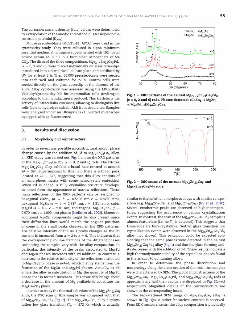
In order to reveal any possible microstructural and/or phasechange caused by the addition of Pd to Mg72Zn23Ca5 alloy,an XRD study was carried out. Fig. 1 shows the XRD patternsof the Mg72−xZn23Ca5Pdx (x = 0, 2 and 6) rods. The Pd-freeMg72Zn23Ca5 alloy exhibits a broad halo centred at around2θ = 39◦. Superimposed to this halo there is a broad peaklocated at 2θ ∼ 37◦, suggesting that this alloy consists ofan amorphous matrix with some nanocrystals embedded.When Pd is added, a fully crystalline structure develops,as noted from the appearance of narrow reflections. Thesemain reflections of the XRD patterns can be assigned tohexagonal CaZn5 (a = b = 0.5406 nm, c = 0.4280 nm),hexagonal MgZn (a = b = 2.557 nm, c = 1.814 nm), cubicMg6Pd (a = b = c = 2.01 nm) and trigonal Mg6Ca2Zn3 (a =
0.970 nm, c = 1.000 nm) phases (Jardim et al., 2002). Moreover,additional Mg–Zn compounds might be also present sincetheir diffraction lines would match the angular positionsof some of the small peaks observed in the XRD patterns.The relative intensity of the XRD peaks changes as the Pdcontent is increased from x = 2 to x = 6. This indicates thatthe corresponding volume fractions of the different phasescomposing the samples vary with the alloy composition. Inparticular, the intensity of the peaks associated to Mg6Pdand MgZn phases increases with Pd addition. In contrast, adecrease in the relative intensity of the reflections attributedto Mg6Ca2Zn3 phase is noted, which mainly stems from theformation of the MgZn and Mg6Pd phases. Actually, as Pdenters the alloy in substitution of Mg, the quantity of Mg6Pdphase that is formed increases. This invariably brings abouta decrease in the amount of Mg available to constitute theMg6Ca2Zn3 phase.
In order to study the thermal behaviour of theMg72Zn23Ca5alloy, the DSC scan of this sample was compared with thatof Mg70Zn23Ca5Pd2 (Fig. 2). The Mg72Zn23Ca5 alloy displaysrather low glass transition (Tg ∼ 372 K), which is actually
Fig. 1 – XRD patterns of the as-cast Mg72−xZn23Ca5Pdx(x = 0, 2 and 6) rods. Phases detected: ∗CaZn5, ◦ MgZn,+ Mg6Pd, ∆Mg6Zn3Ca2.
Fig. 2 – DSC scans of the as-cast Mg72Zn23Ca5 andMg70Zn23Ca5Pd2 rods.
similar to that of other amorphous alloys with similar compo-sition (e.g. Mg70Zn25Ca5 and Mg66Zn30Ca4) (Gu et al., 2010).Several exothermic peaks are observed at higher tempera-tures, suggesting the occurrence of various crystallizationevents. In contrast, the scan of the Mg70Zn23Ca5Pd2 sample isalmost featureless (i.e. no Tg is detected). This suggests thatthese rods are fully-crystalline. Neither glass transition norcrystallization events were observed in the Mg66Zn23Ca5Pd6alloy (not shown). This behaviour could be expected con-sidering that the same phases were detected in the as-castMg70Zn23Ca5Pd2 alloy (Fig. 1) and that the glass forming abil-ity decreases with the addition of Pd. These results indicate ahigh thermodynamic stability of the crystalline phases foundin the as-cast Pd-containing alloys.
In order to determine the phase distribution andmorphology along the cross-section of the rods, the sampleswere characterized by SEM. The global microstructures of theMg72Zn23Ca5, Mg70Zn23Ca5Pd2 and Mg66Zn23Ca5Pd6 rods atapproximately half their radius are displayed in Fig. 3(a)–(c)respectively. Magnified details of the microstructure areshown in the corresponding insets.
The backscattered SEM image of Mg72Zn23Ca5 alloy isshown in Fig. 3(a). A rather featureless contrast is observed.From EDX measurements, the alloy composition is practically

56 J O U R N A L O F T H E M E C H A N I C A L B E H AV I O R O F B I O M E D I C A L M A T E R I A L S 6 ( 2 0 1 2 ) 5 3 – 6 2
a
b
c
Fig. 3 – SEM images (backscattered electrons) of(a) Mg72Zn23Ca5, (b) Mg70Zn23Ca5Pd2 and (c)Mg66Zn23Ca5Pd6 rod. The insets show the magnifieddetails of the microstructure of the Mg70Zn23Ca5Pd2 andMg66Zn23Ca5Pd6 samples (in panels (b) and (c)).
the same throughout the sample and is similar to the nominalcomposition.
Ca Zn O
Pd
Mg
Fig. 4 – SEM image (backscattered electrons) ofMg66Zn23Ca5Pd6. Compositional X-ray mappingscorresponding to Ca, Zn, O, Pd and Mg.
Completely differentmicrostructures, consisting of severalregions with different contrast are observed in the SEMimage (using backscattered electrons) of the Mg70Zn23Ca5Pd2and Mg66Zn23Ca5Pd6 alloys (Fig. 3(b) and (c)). These resultsindicate a decrease of glass forming ability of the Mg–Zn–Casystem with the addition of Pd. The Mg66Zn23Ca5Pd6 alloyconsists of small dark areas surrounded by a grey phase,along with white dendrites (inset of Fig. 3(c)). Although themicrostructure and phase distribution present in the two Pd-containing alloys appear to be similar, the volume fractionof dark regions and white dendrites as well as the size ofdendrites is larger in the alloy with x = 6.
In order to identify which crystalline phase composeseach of the regions observed in the SEM images, elementalEDX mappings were obtained from the disc-centre of theMg66Zn23Ca5Pd6 rod (Fig. 4). The results show that the greyregions are rich in Ca and Zn, the white dendrites in Pdand Mg and the dark regions mainly correspond to Mg andZn. This suggests that the dendrites could correspond toMg6Pd, the grey regions to CaZn5 and the black regions toMgZn. Considering that the weight fraction of Mg6Zn3Ca2 islower than that of MgZn it could be expected that the scarceMg6Zn3Ca2 phase present in the Mg66Zn23Ca5Pd6 alloy couldcorrespond to the areas of darkest tonality that lie within thedark regions associated to MgZn.
3.2. Acoustic measurements and nanoindentation studies
The elastic properties of the alloys have been evaluatedat room temperature by means of acoustic measurements.Table 1 lists the values of density (ρ), shear modulus (G),bulk modulus (B), Young’s modulus (E), G/B ratio and Poisson’sratio (ν) of the investigated alloys. The density values of thesealloys are rather low although ρ increases progressively withthe increase of Pd content, as could be expected considering

J O U R N A L O F T H E M E C H A N I C A L B E H AV I O R O F B I O M E D I C A L M A T E R I A L S 6 ( 2 0 1 2 ) 5 3 – 6 2 57
Table 1 – Elastic properties evaluated from acoustic measurements at room temperature. Note that ρ, G, B, E and ν denote,respectively, the density, shear modulus, bulk modulus, Young’s modulus and Poisson’s ratio of the investigated alloys.
Properties Mg72Zn23Ca5 Mg70Zn23Ca5Pd2 Mg66Zn23Ca5Pd6
ρ (g cm−3) 2.84 3.01 3.40G (GPa) 19.47 25.60 28.97B (GPa) 40.66 43.5 50.54E (GPa) 50.38 64.20 72.98G/B 0.48 0.58 0.57ν 0.29 0.25 0.26
a b
Fig. 5 – (a) Load–displacement (P–h) nanoindentation curves of Mg72−xZn23Ca5Pdx (x = 0, 2 and 6) alloys and(b) backscattered SEM image showing some indents made close to the centre of the Mg70Zn23Ca5Pd2 rod.
that the density of Pd (12.023 g cm−3) is much higher thanthat of Mg (1.738 g cm−3). Remarkably, the Young’smodulus ofthese alloys raises significantly with an addition of 2 at.% Pd,while only a moderate further increase is observed for x = 6.This cannot be simply explained in terms of compositionalvariations among the samples but is probably mainly relatedto the aforementioned microstructural changes that occurwhen small amounts of Pd are added to the system. Indeed,the presence of only 2 at.% Pd causes a drastic change in themicrostructure, from a mainly amorphous one (for x = 0) to afully crystalline one (for x = 2), thus inducing large variationsin E. The relatively low Young’s modulus of the Mg72Zn23Ca5alloy can be attributed, at least in part, to the presence of freevolume within the glassy matrix, which causes the averageinteratomic spacing to be larger than in fully crystallinealloys. The so-called elastic-softening of metallic glassesalso depends on the type of atomic displacements thatoccur during elastic deformation of these materials. Namely,contrary to crystalline alloys, elastic deformation in metallicglasses not only involves the nearest-neighbour atomic shellbut also more distant shells, resulting in overall larger elasticstrains (i.e., lower Young’s modulus) than in crystalline alloys(Hufnagel et al., 2006). Similar to the evolution of E, the valuesof G and B also increase with the addition of Pd becauseall these parameters depend on the atomic bonding; E isindicative of the elastic resistance in the direction of theapplied load, B represents the elastic resistance to volumecompression and G denotes the elastic resistance to shearstress.
Considering that Mg-based bulk metallic glasses and crys-talline Mg alloys generally exhibit low ductility and poor
macroscopic deformability at room temperature, nanoinden-tation tests have been performed. Since nanoindentation is avery local technique the influence of porosity or other flawsis avoided and permits to get reliable results when study-ing the influence of Pd addition on the mechanical prop-erties. The load–displacement (P–h) nanoindentation curves,with a maximum load of 500 mN, are shown in Fig. 5(a). Pop-in events are detected in the loading segment of all threespecimens. However, some differences can be observed de-pending on the alloy composition. In the mainly amorphousalloy (x = 0) a large amount of uniformly distributed pop-insare detected and their displacement tends to increase withthe applied load. Conversely, in the fully crystalline alloys,the load at which pop-ins are detected and their displace-ment appear to be more random, which is consistent withthe microstructure. Actually, pop-in events in amorphousalloys are considered to be related to the motion of in-dividual shear bands; since the distance shear bands canpropagate increases with the applied load, this causes theindentation serrations to become more pronounced. How-ever, for crystalline alloys, the length of the pop-ins is relatedto the distance dislocations propagate, which dependsmainlyon the grain size and the distance between the indenter tipand grain boundaries with which dislocation pile-ups caninteract. During nanoindentation of a crystalline alloy, theinitial stress-field generated during loading can create a dis-location source, hence initiating a pop-in event. The processcontinues until the dislocation source stops operating due tothe inverse pileup stress that reduces the net stress to zero. Anew pop-in will be generated only when the indentation loadis sufficiently increased to create a new dislocation source.

58 J O U R N A L O F T H E M E C H A N I C A L B E H AV I O R O F B I O M E D I C A L M A T E R I A L S 6 ( 2 0 1 2 ) 5 3 – 6 2
Table 2 – Hardness (H), reduced Young’s modulus (Er), plastic energy (Upl), Upl/Utot ratio and maximum indentationdepth (hmax) for the investigated alloys after being indented up to a maximum load of 500 mN.
Mg72Zn23Ca5 Mg70Zn23Ca5Pd2 Mg66Zn23Ca5Pd6
H (GPa) 2.71 3.56 3.90Er (GPa) 51.01 59.10 77.70H/Er 0.053 0.060 0.050H3/E2r (GPa) 0.008 0.013 0.001Upl (GPa) 398.3 326.7 337.2
Upl/Utot 0.696 0.628 0.695
hmax (µm) 3.204 2.905 2.673
The stochastic nature of this process makes the indentationserrations to be distributed in a less regular manner than forthe glassy Mg72Zn23Ca5 alloy.
Some of the indentation impressions obtained at halfthe radius of the Mg70Zn23Ca5Pd2 rod are shown inFig. 5(b). The image reveals that the indentation marksfor a maximum load of 500 mN are sufficiently large toembrace all the existing crystalline phases, suggesting thatthe obtained mechanical properties will be representative ofthe average behaviour of the composite material. The valuesof H,Er,Upl,Upl/Utot ratio and maximum indentation depth(hmax) for the three investigated alloys are listed in Table 2.Interestingly, addition of 2 at.% Pd has a large influence onthe hardness, which increases from 2.71 GPa (for x = 0) to3.56 GPa (for x = 2). The hardness increase can be mainlyattributed to the lack of free volume in the crystalline alloy(x = 2) as compared to the mainly amorphous one (x = 0).Also the large volume fraction of Mg6Ca2Zn3 phase mayhave a hardening effect but not be as much as that reportedfor Mg–Zn–Ca (Bamberger et al., 2002; Park et al., 2001) andMg–Zn–Ca–Zr alloys (Shepelev et al., 2009) since the distancebetween intermetallic particles is relatively large and thus thedislocation arrest due to the Orowan mechanism would notbe so effective. The hardness further increases for x = 6, upto 3.90 GPa. This is consistent with the decrease of maximumindentation depth from 2.905 µm (for x = 2) to 2.673 µm (forx = 6). However, from the relative intensity of XRD peaksit can be inferred that the volume fraction of Mg6Ca2Zn3phase decreases when the concentration of Pd varies from 2to 6 at.%. Hence, the large hardness in the alloy with x = 6suggests that the MgZn and Mg6Pd phases also contribute toinduce mechanical hardening, in an analogous way as theMg6Ca2Zn3 phase. Note that the reduced Young’s modulusincreases with the Pd content, in agreement with the resultsfrom acoustic measurements.
Since most biomaterials are subjected to wear, it isnecessary to have materials with high wear resistance tominimize wear debris. Hardness is usually regarded to be animportant property for evaluating wear resistance. However,some authors have shown that the wear resistance can bebetter estimated from the H/Er ratio (Oberle, 1951), ratherthan the hardness itself. This ratio is related to the elasticstrain to failure. Another parameter, also related to thewear performance, is the resistance to plastic deformation,which is proportional to H3/E2r (Tsui et al., 1995). Table 2shows that the maximum H/Er and H3/E2r values areattained by the Mg70Zn23Ca5Pd2 alloy, indicating that thisalloy exhibits the highest wear resistance. Furthermore,nanoindentation also allows extracting some information
about the intrinsic plasticity of the alloy, from the ratiobetween the indentation plastic energy (Upl) and the totalindentation energy (Utot). From the obtained results (seeTable 2), no pronounced differences in plasticity are observedfor the three investigated alloys. It is also worth mentioningthat the values of Upl/Utot for the investigated Mg-basedalloys are similar to those reported for Zr50Cu40Al10 andCu46.25Zr45.25Al7.5Er1 metallic glasses (Upl/Utot ∼ 0.6 (Tekayaet al., 2009) and Upl/Utot ∼ 0.7 (Yang et al., 2009), respectively)and clearly larger than for brittle Fe-based glasses (Upl/Utot ∼
0.37 Fornell et al., 2010). In fact, the ratio Upl/Utot has beensometimes used to characterize the mechanical behaviourof brittle materials which are often difficult to test usingmacroscopic tensile tests (Milman, 2008). Remarkably, mostbrittle materials (like ceramics) show relatively low Upl/Utotvalues (around 0.3–0.4 for SiC or Al2O3), whereas ductilemetals (like Cu or Al) can show Upl/Utot values larger than0.9 (Milman, 2008).
3.3. Corrosion behaviour in simulated body fluid andcytotoxicity
Fig. 6(a) shows the equilibrium potential reached in sam-ples containing different amounts of Pd in the Hank’s solu-tion at 37 ◦C. The OCP shifts towards more positive valuesas the Pd content increases, owing to the noble character ofPd, in the following sequence: Mg72Zn23Ca5 base (−1.15 V) >
Mg70Zn23Ca5Pd2 (−1.04 V) > Mg66Zn23Ca5Pd6 (−0.93 V). Onewould expect the Pd-containing alloys to be more proneto corrosion than the Pd-free partly glassy alloy due toboth the crystallinity and the existence of noble phases,such as Mg6Pd, that would promote the well-known gal-vanic corrosion phenomena. However, it rather seems thatboth factors do not play a major role. Instead, the ability toform thicker, more stable passive layers onto the speci-mens’ surface would contribute to the enhanced corrosionresistance, at least at the working time scales used. Thisshift towards more positive potential can thus be ascribedto the formation of a thicker oxide/hydroxide film ontothe specimen’s surface, as commonly observed in this typeof alloys (Fekry and Ameer, 2011). However, the effective-ness of this passive Mg(OH)2 layer in preventing corrosionis rather limited because the layer is destroyed and dis-solved in solutions containing chloride ions, as it is thecase of the Hank’s solution (Ng et al., 2010). The observedtrends in the OCP values are consistent with the poten-tiodynamic polarization curves shown in Fig. 6(b). As ex-pected, the corrosion potential (Ecorr) shifts towards moreanodic values with an increase in the Pd content, point-ing to a clear delay of the corroding phenomena. The

J O U R N A L O F T H E M E C H A N I C A L B E H AV I O R O F B I O M E D I C A L M A T E R I A L S 6 ( 2 0 1 2 ) 5 3 – 6 2 59
Fig. 6 – (a) Time dependence of the OCP and (b) potentiodynamic polarization curves of Mg72−xZn23Ca5Pdx (x = 0, 2 and 6)alloys.
a b
Fig. 7 – SEM images (secondary electrons) showing the surface of corroded (a) Mg72Zn23Ca5 and (b) Mg66Zn23Ca5Pd6 alloysafter potentiodynamic polarization tests. Insets: magnified images of the surface morphologies.
corrosion current density values obtained by the Tafel extrap-olation method become slightly larger as Pd is progressivelyadded to the alloy: Mg72Zn23Ca5 (1.7 mA cm−2) < Mg70Zn23Ca5Pd2 (2.1 mA cm−2) < Mg66Zn23Ca5Pd6 (2.7 mA cm−2). Itis noteworthy that although the reasonability and reliabilityof the corrosion current density values obtained from elec-trochemical techniques in Mg-based alloys is a source of de-bate due to the so-called “abnormal polarization behaviour”,the potentiodynamic polarization tests still remain one of themost employed methods (Ng et al., 2010; Song, 2005). Com-pared to the weight-loss method, the electrochemical tech-niques provide real time information of the corrosion raterather than an average rate. Hence, they still can be consid-ered valid from the qualitative viewpoint. Our results are inagreement with the beneficial effect attributed to Pd when al-loyed with other biomedical-oriented glassy alloys, such asthe Ti-based ones (Oak and Inoue, 2008).
The SEM surface morphologies of Mg72Zn23Ca5 andMg66Zn23Ca5Pd6 as-cast alloys after the corrosion tests areshown in Fig. 7(a) and (b), respectively. At first sight, thesurface of Mg72Zn23Ca5 appears to be slightly rougher, show-ing a higher density of microcracks. In fact, when the in-ternal or external tensile stresses are combined with theeffect of the corrosion agents, a pressure corrosion crackingcommonly appears. These results suggest a higher vulner-ability of the protective oxide/hydroxide film formed onto
the Mg72Zn23Ca5 surface, in agreement with the trends ob-served in both the OCP and Ecorr values. Moreover, someholes can also be observed, while the Mg66Zn23Ca5Pd6 sur-face is virtually free from these holes and just exhibitsmacrocraking. This macrocracking can be ascribed to thecontracting forces occurring upon drying (Qin et al., 2009).From the OCP and Ecorr trends it can be deduced thatthe passivation behaviour of Mg70Zn23Ca5Pd2 is interme-diate between that of Mg72Zn23Ca5 and Mg66Zn23Ca5Pd6alloys (Fig. 6) and thus also the surface. A homogeneousdistribution of bright particles throughout the cross-sectionof the samples is observed for both alloys. Oxygen, cal-cium and phosphorus elements coming out from the Hank’ssolution have been identified in the EDX spectra (Fig. 7). Bear-ing in mind that these elements are present in hydroxya-patite (i.e. Ca10(PO4)6(OH)2), one might conclude that thealloys have a large apatite-forming ability and, thus, goodbioaffinity. Remarkably, a significant decrease of the relativepercentage of Mg is encountered, likely due to the anodic dis-solution of Mg. In aqueous solutions Mg dissolves accordingto the following reactions:
Anodic reaction : Mg → Mg2++ 2e− (1)
Mg2++ 2OH−
→ Mg(OH)2 (2)
Cathodic reaction : 2H2O + 2e− → 2OH−+ H2. (3)

60 J O U R N A L O F T H E M E C H A N I C A L B E H AV I O R O F B I O M E D I C A L M A T E R I A L S 6 ( 2 0 1 2 ) 5 3 – 6 2
Fig. 8 – EDX spectra of the (a) Mg72Zn23Ca5 and(b) Mg66Zn23Ca5Pd6 alloys after the potentiodynamicpolarization tests in Hank’s solution.
In the presence of Cl− ions, Mg(OH)2 transforms intosoluble MgCl2, resulting in an excess of hydroxide ions. Inaddition to the expected formation of a Mg(OH)2 layer, otherinsoluble products are likely to precipitate onto thespecimen’s surface provided that other ions (such as Ca+
and H2PO−
4 ) are present in the solution. In particular, MgxCay(PO4)z compounds have been identified as key corrosion
Table 3 – Semi-quantitative atomic percentagesdetermined by EDX analyses on the corroded surface ofMg72Zn23Ca5 and Mg66Zn23Ca5Pd6 alloys.
at.% Mg72Zn23Ca5 Mg66Zn23Ca5Pd6
Oxygen 68.98 70.1Magnesium 11.79 5.68Phosphorous 7.01 9.18Chlorine 0.32 0.45Calcium 6.67 9.18Zinc 5.23 5.05Palladium – 0.36
products in Mg-based alloys immersed in simulated bodyfluids (Zhang et al., 2010). The percentages of O, Mg, P,Ca elements encountered on the surface of Mg72Zn23Ca5and Mg66Zn23Ca5Pd6 alloys after the potentiodynamicpolarization tests are relatively high, above 5 at.%, whichis consistent with the formation of MgxCay(PO4)z products(Table 3). These phosphates containing Mg and Ca ionshave been identified in magnesium implants tested in vivo(Zhang et al., 2009). In our case, the (Mg + Ca)/P ratio is 2.63and 1.61 for the Mg72Zn23Ca5 and Mg66Zn23Ca5Pd6 alloys,respectively. Meanwhile, the Ca/P ratio is close to 1 in bothcases, which is not far from the Ca/P ratio of hydroxyapatite(i.e., 1.66) (Fig. 8).
To better understand the potential use of these biodegrad-able implants as biomaterials, their cytotoxicity has beenstudied. The viability of preosteoblasts cultured for 27 hon a sterile coverslip without sample (Fig. 9(a)) and withMg66Zn23Ca5Pd6 sample (Fig. 9(b)) was compared. No differ-ences were observed, thus confirming that the alloying ele-ments are not cytotoxic and thus the alloys can be used asbiomaterials.
4. Conclusions
The influence of Pd addition on the microstructure, mechan-ical properties and corrosion behaviour of Mg–Zn–Ca alloyshas been investigated. The main results can be summarizedas follows:
a b
Fig. 9 – Preosteoblasts cultured (a) on a sterile glass coverslip without alloy or (b) with Mg66Zn23Ca5Pd6 alloy. Live cells arestained with Calcein-AM (green) and dead cells are stained with EthD-1 (red). No dead cells are observed. (For interpretationof the references to colour in this figure legend, the reader is referred to the web version of this article.)

J O U R N A L O F T H E M E C H A N I C A L B E H AV I O R O F B I O M E D I C A L M A T E R I A L S 6 ( 2 0 1 2 ) 5 3 – 6 2 61
– The microstructure of the Mg72Zn23Ca5 alloy, whichconsists of an amorphous matrix with finely dispersedcrystallites, drastically changes with small Pd contents,i.e., 2 at.% Pd is sufficient to obtain a fully crystallinemicrostructure.
– The addition of Pd increases the hardness of the alloy,mainly due to the formation of mechanically hardcrystalline phases, which are virtually not present in theglassy Mg72Zn23Ca5 alloy.
– The values of the elastic constants (E,B and G) raisewith the increase of Pd content, which is consistent withthe increase of reduced Young’s modulus measured bynanoindentation.
– The hardness and Young’s modulus increase at a differentrate with the Pd content. For this reason the maximumwear resistance, estimated from the H/Er,H3/E2r ratios,is attained in the Mg70Zn23Ca5Pd2 alloy (i.e., for anintermediate Pd content).
– The corroding phenomena are greatly delayed with Pdaddition, as evidenced from the shift in the corrosionpotential towards more positive values. The surface ofthe corroded samples is smoother and contains loweramounts of holes in the Pd-containing alloys.
– Cytotoxicity tests do not show the presence of death cellsafter culturing for 27 h, which confirms that these alloysare not toxic and thus can be used as potential implants.
In summary, this work shows that by optimizing the Pdcontent it is possible to modify and improve the propertiesof biocompatible Mg–Zn–Ca–Pd alloys, thus making thesematerials potential candidates to be used as biodegradableimplants.
Acknowledgments
We acknowledge O. Castell and E. Rossinyol (Servei deMicroscopia, UAB) for their technical support with the SEMobservations. This work has been partially financed by the2009-SGR-1292, 2009-SGR-282, MAT2011-27380-C02-01 andMINAHE3 (TEC2008-06883-C03-03) research projects. S.G. andJ.F. acknowledge the Juan de la Cierva contract and thePh.D. Fellowship from the Spanish Ministry of Science andInnovation, respectively. M.D.B. was partially supported by anICREA Academia award.
R E F E R E N C E S
Ambrose, C.G., Clanton, T.O., 2004. Bioabsorbable implants: reviewof clinical experience in orthopedic surgery. Ann. Biomed. Eng.32, 171–177.
Bamberger, M., Jardim, P.M., Solórzano, G., Sande, J.V., 2002.In: Kaplan, H.I. (Ed.), Magnesium Technology. TMS (TheMinerals, Metals & Materials Society).
Black, J., Hasting, G.W., 1998. Handbook of Biomaterials Properties.Chapman and Hall, London.
Fekry, A.M., Ameer, M.A., 2011. Electrochemistry and impedancestudies on titanium and magnesium alloys in Ringer’ssolution. Int. J. Electrochem. Sci. 6, 1342–1354.
Fornell, J., González, S., Rossinyol, E., Suriñach, S., Baró, M.D.,Louzguine-Luzgin, D.V., Perepezko, J.H., Sort, J., Inoue, A., 2010.Enhanced mechanical properties due to structural changesinduced by devitrification in Fe–Co–B–Si–Nb bulk metallicglass. Acta Mater. 58, 6256–6266.
Geetha, M., Singh, A.K., Asokamani, R., Gogia, A.K., 2009. Ti-basedbiomaterials, the ultimate choice for orthopaedic implants. Areview. Prog. Mater. Sci. 54, 397–425.
González, S., Figueroa, I.A., Zhao, H., Davies, H.A., Todd, I., Adeva,P., 2009. Effect of mischmetal substitution on the glass-formingability of Mg–Ni–La bulk metallic glasses. Intermetallics 17,968–971.
Gu, X., Shiflet, G.J., Guo, F.Q., Poon, S.J., 2005. Mg–Ca–Zn bulkmetallic glasses with high strength and significant ductility.J. Mater. Res. 20, 1935–1938.
Gu, X., Zheng, Y., Zhong, S., Xi, T., Wang, J., Wang, W., 2010.Corrosion and cellular responses to Mg–Zn–Ca bulk metallicglasses. Biomaterials 31, 1093–1103.
Hufnagel, T.C., Ott, R.T., Almer, J., 2006. Structural aspects ofelastic deformation of a metallic glass. Phys. Rev. B 73, 064204.
Jardim, P.M., Solórzano, G., Sande, V., John, B., 2002. Pre-cipitate crystal structure determination in melt-spunMg–1.5wt%Ca–6wt%Zn alloy. Microsc. Microanal. 8, 487–496.
Kielhorn, J., Melber, C., Keller, D., Mangelsdorf, I., 2002. Palladium–a review of exposure and effects to human health. Int. J. Hyg.Environ. Health 205, 417–432.
Li, Z.J., Gu, X.N., Lou, S.Q., Zheng, Y.F., 2008. The development ofbinary Mg–Ca alloys for use as biodegradable materials withinbone. Biomaterials 29, 1329–1344.
Milman, Y.V., 2008. Plasticity characteristic obtained by indenta-tion. J. Phys. D: Appl. Phys. 41, 074013.
Nagels, J., Stokdijk, M., Rozing, P.M., 2003. Stress shielding andbone resorption in shoulder arthroplasty. J. Shoulder ElbowSurg. 12, 35–39.
Ng, W.F., Chiu, K.Y., Cheng, F.T., 2010. Effect of Pd on the in vitrocorrosion rate of magnesium degradable implant material.Mater. Sci. Eng. C 30, 898–903.
Oak, J.-J., Inoue, A., 2008. Formation, mechanical properties andcorrosion resistance of TiPd base glassy alloys. J. Non-Cryst.Solids 354, 1828–1832.
Oberle, T.L., 1951. Properties influencing wear of metals. J. Met. 3,438–439.
Oliver, W.C., Pharr, G.M., 1992. An improved technique fordetermining hardness and elastic modulus using load anddisplacement sensing indentation measurements. J. Mater.Res. 7, 1564–1580.
Park, W.-W., You, B.S., Moon, B.-G., Park, J.-G., Yang, S.-C., 2001. Agehardening phenomena of rapidly quenched non-combustibleAg–Al–Si–Ca and Mg–Zn–Ca alloys. Met. Mater. Int. 7, 9–13.
Qin, F.X., Wang, X.M., Wada, T., Xie, G.Q., Asami, K., Inoue, A.,2009. Formation of hydroxyapatite on Ti-coated Ti–Zr–Cu–Pdbulk metallic glass. Mater. Trans. 50, 605–609.
Shepelev, D., Bamberger, M., Katsman, A., 2009. Precipitationhardening of Zr-modified Mg–Ca–Zn alloy. J. Mater. Sci. 44,5627–5635.
Song, G.L., 2005. Recent progress in corrosion and protection ofmagnesium alloys. Adv. Eng. Mater. 7, 563–586.
Song, G.L., 2007. Control of biodegradation of biocompatablemagnesium alloys. Corros. Sci. 49, 1696–1701.
Staiger, M.P., Pietak, A.M., Huadmai, J., Dias, G., 2006. Magnesiumand its alloys as orthopedic biomaterials. Biomaterials 27,1728–1734.
Tekaya, A., Labdi, S., Benameur, T., Jellad, A., 2009. Quasi-staticcyclic loadings induced inelastic deformation in a Zr-basedbulk metallic glass under nanoindentation. J. Mater. Sci. 44,4930–4938.
Tsui, T.Y., Pharr, G.M., Oliver, W.C., Bhatia, C.S., White, R.L.,Anders, S., Anders, A., Brown, I.G., 1995. Nanoindentation and

62 J O U R N A L O F T H E M E C H A N I C A L B E H AV I O R O F B I O M E D I C A L M A T E R I A L S 6 ( 2 0 1 2 ) 5 3 – 6 2
nanoscratching of hard carbon coatings for magnetic disks.Mater. Res. Soc. Symp. Proc. 383, 447–452.
Wataha, J.C., 2000. Biocompatibility of dental casting alloys: areview. J. Prosthet. Dent. 83, 223–234.
Whitters, C.J., Strang, R., Brown, D., Clarke, R.L., Curtis, R.V.,Hatton, P.V., Ireland, A.J., Lloyd, C.H., McCabe, J.F., Nicholson,J.W., Scrimgeour, S.N., Setcos, J.C., Sheriff, K., Yang, B., Vehoff,H., 2005. Grain size effects on the mechanical properties ofnanonickel examined by nanoindentation. Mater. Sci. Eng. A400–401, 467–470.
Witte, F., Kaese, V., Haferkamp, H., Switzer, E., Meyer-Lindenberg,A., Wirth, C., Windhagen, H., 2005. In vivo corrosion offour magnesium alloys and the associated bone response.Biomaterials 26, 3557–3563.
Yang, F., Li, D., Yang, M.X., Li, R., Jiang, W., Wang, G., Zhang, T.,Liaw, P.K., 2009. Localized deformation of a Cu46.25Zr45.25Al7.5Er1 bulk metallic glass. J. Phys. D: Appl. Phys. 42,065401.
Zberg, B., Uggowitzer, P.J., Löffler, J.F., 2009. MgZnCa glasses with-out clinically observable hydrogen evolution for biodegradableimplants. Nat. Mater. 8, 887–891.
Zhang, E., Xu, L., Yu, G., Pan, F., Yang, K., 2009. In vivo evaluationof biodegradable magnesium alloy bone implant in the first6 months of implantation. J. Biomed. Mater. Res. Part A 90,882–893.
Zhang, S., Zhang, X., Zhao, C., Li, J., Song, Y., Xie, C., Tao, H., Zhang,Y., He, Y., Jiang, Y., Bian, Y., 2010. Research on a Mg–Zn alloy asa degradable biomaterial. Acta Biomater. 6, 626–640.


![Beyond Chemical - Exotic Energetics/ Propulsion · PDF fileBeyond Chemical - Exotic Energetics/ Propulsion ... time scales] and is exceedingly ... a collective proton resonance in](https://static.fdocuments.net/doc/165x107/5aa89cbb7f8b9a6c188bc37b/beyond-chemical-exotic-energetics-propulsion-chemical-exotic-energetics-propulsion.jpg)
