
Implant Overdentures for mandibular edentulous patients - … · 2017-06-22 · Implant...
80
1 Journal of Quintessence of Dental Technology Vol.34 No.5 and 6, 2009 (Japanese) MONTHLY FOCUS Considering prosthodontics in the aging society Round-table discussion Implant Overdentures for mandibular edentulous patients - First part: Searching its application and boundary Yoshinobu Maeda, Born in 1951 with clinical background of 30 years. Professor, Department of Prosthodontics and Oral Rehabilitation, Graduate School of Dentistry, Osaka University Writing contributions to academic journals and commercial publications. Major books: “Overdentures applied to clinical cases”, “Implant clinical cases using magnetic appliances (co- authorship)” (both published from our publishers) Jiro Abe, Born in 1955 with 27 years clinical experience. Private practice director of Abe Dental Clinic, Chofu, Tokyo. Many lectures, books and articles on “Suction theory of the mandibular complete denture retention”. Representative of a study group, “Japan Denture Association”. Certified instructor of BPS Ivoclar Vivadent. Yukio Kameda , Born in 1963 with 20 years clinical experience. Private practice director of Kameda Dental Clinic, Kawaguchi, Saitama. Variety of writing contributions including prosthodontics, periodontics and occlusion science. Member of executive board of the Japan Academy of Gnathology and Occlusion, of executive board of the Japan Clinical Periodontal Group, member of Tentomushi Study Group, and of JDA.
Transcript of Implant Overdentures for mandibular edentulous patients - … · 2017-06-22 · Implant...

1
Journal of Quintessence of Dental Technology Vol.34 No.5 and 6, 2009 (Japanese)
MONTHLY FOCUS
Considering prosthodontics in the aging society
Round-table discussion
Implant Overdentures for mandibular edentulous patients
- First part: Searching its application and boundary
Yoshinobu Maeda,
Born in 1951 with clinical background of 30 years.
Professor, Department of Prosthodontics and Oral Rehabilitation,
Graduate School of Dentistry, Osaka University
Writing contributions to academic journals and commercial publications.
Major books: “Overdentures applied to clinical cases”, “Implant clinical
cases using magnetic appliances (co-authorship)” (both published from our publishers)
Jiro Abe,
Born in 1955 with 27 years clinical experience. Private practice director of
Abe Dental Clinic, Chofu, Tokyo. Many lectures, books and articles on
“Suction theory of the mandibular complete denture retention”.
Representative of a study group, “Japan Denture Association”. Certified
instructor of BPS Ivoclar Vivadent.
Yukio Kameda,
Born in 1963 with 20 years clinical experience. Private practice director of
Kameda Dental Clinic, Kawaguchi, Saitama. Variety of writing
contributions including prosthodontics, periodontics and occlusion science.
Member of executive board of the Japan Academy of Gnathology and
Occlusion, of executive board of the Japan Clinical Periodontal Group,
member of Tentomushi Study Group, and of JDA.

2
Introduction
Kameda (Moderator): In these recent years of high blooming of dental implant
therapy, world attention has been on overdentures supported with implant
structures as they call an implant overdenture (hereafter called IOD) as a new
alternative option of restoring the mandible edentulous jaws.
Now here we are at the discussion table with three of us, Prof.Yoshinobu Maeda
from Osaka University, who is one of leading researchers in Japan for overdentures
supported by natural teeth or implant structures, and Dr.Jiro Abe, who has been
well known as a private practitioner of complete denture therapy as well as other
prosthetic therapies keeping in mind of periodontic concern. And Yukio Kameda, I
myself have been practically involved with IOD therapies in edentulous mandibular
jaws.
We are going to discuss on current reality and future possibility of IOD specially
focusing on “edentulous mandibular jaws”.
Objects designed for this discussion (First part)
☞ To know that implant supported prosthetics should rest on a denture base.
☞ To know how to determine the grade of difficulty in an edentulous ridge case.
☞ Current ideas of applying implant supported overdentures from mutual views of
literature and practice.
3
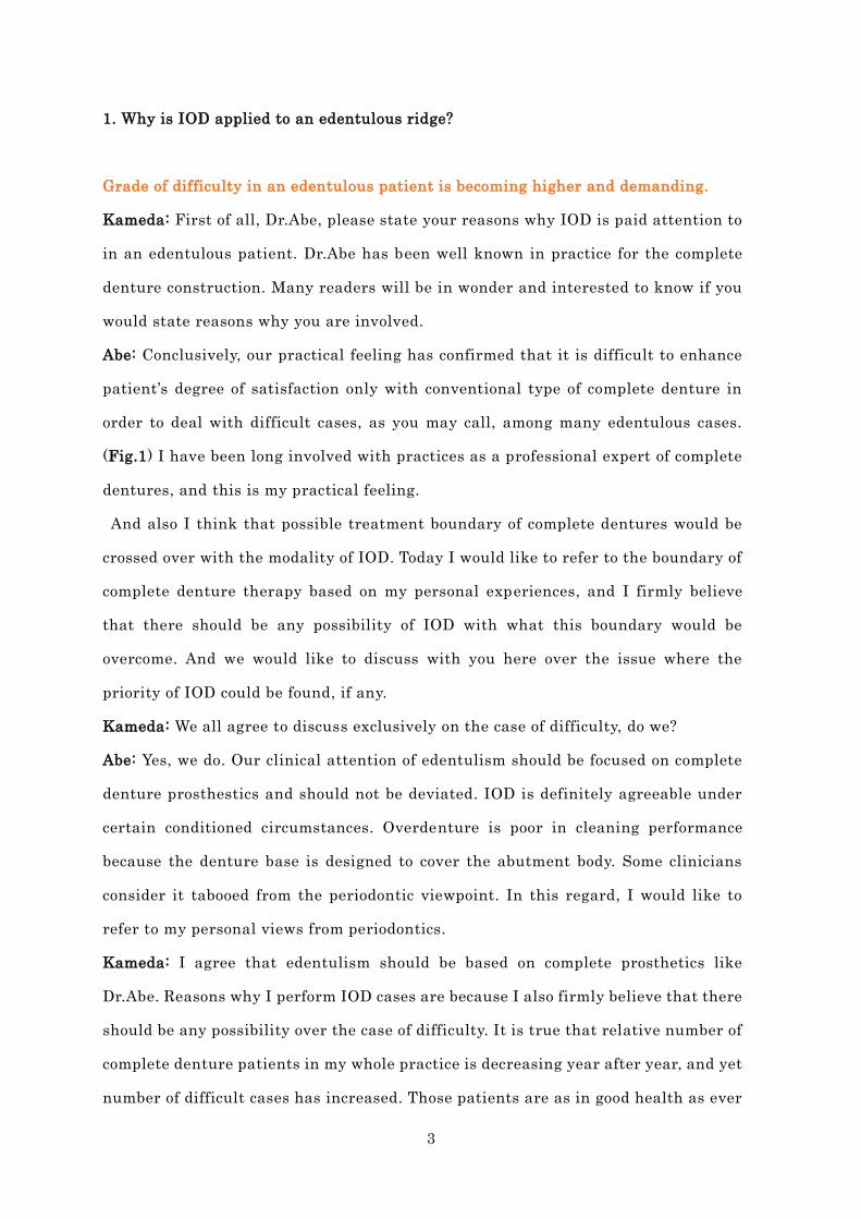
1. Why is IOD applied to an edentulous ridge?
Grade of difficulty in an edentulous patient is becoming higher and demanding.
Kameda: First of all, Dr.Abe, please state your reasons why IOD is paid attention to
in an edentulous patient. Dr.Abe has been well known in practice for the complete
denture construction. Many readers will be in wonder and interested to know if you
would state reasons why you are involved.
Abe: Conclusively, our practical feeling has confirmed that it is difficult to enhance
patient‟s degree of satisfaction only with conventional type of complete denture in
order to deal with difficult cases, as you may call, among many edentulous cases.
(Fig.1) I have been long involved with practices as a professional expert of complete
dentures, and this is my practical feeling.
And also I think that possible treatment boundary of complete dentures would be
crossed over with the modality of IOD. Today I would like to refer to the boundary of
complete denture therapy based on my personal experiences, and I firmly believe
that there should be any possibility of IOD with what this boundary would be
overcome. And we would like to discuss with you here over the issue where the
priority of IOD could be found, if any.
Kameda: We all agree to discuss exclusively on the case of difficulty, do we?
Abe: Yes, we do. Our clinical attention of edentulism should be focused on complete
denture prosthestics and should not be deviated. IOD is definitely agreeable under
certain conditioned circumstances. Overdenture is poor in cleaning performance
because the denture base is designed to cover the abutment body. Some clinicians
consider it tabooed from the periodontic viewpoint. In this regard, I would like to
refer to my personal views from periodontics.
Kameda: I agree that edentulism should be based on complete prosthetics like
Dr.Abe. Reasons why I perform IOD cases are because I also firmly believe that there
should be any possibility over the case of difficulty. It is true that relative number of
complete denture patients in my whole practice is decreasing year after year, and yet
number of difficult cases has increased. Those patients are as in good health as ever

4
and their demand is higher rather than before, when accepting complete dentures.
To meet those patients‟ demand, IOD‟s are effective, and their degree of satisfaction
would be as comparatively high as complete denture wearers. In spite of their low
invasiveness and low costing among various kinds of implant therapies, I think that
their effectiveness is gravely significant.
Now, Prof.Maeda, you already have taken careful attention for quite some time to
overdentures to be supported by natural teeth or implant structures. Could you
please illustrate how and when you became involved with this?
Fig.1 a~e Before we take an option of IOD, let us brush up properly the
construction skills of complete dentures! (Abe)
In order to construct a highly sophisticated denture within the three-dimensional
space of edentulous patient oral cavity, exact impression taking, proper occlusal
registration, error-controlled resin polymerization are extremely important. When
once one of these vital items should fail, score of this denture would be down to point
zero.
In order to perform a series of process perfectly without errors, it is advised to learn
the technique of BPS (Bio-functional Prosthetic System, Ivoclar Vivadent AG.
imported by Hakusui Trading Co.) that has been currently approved as a teaching

5
system by 23 dental schools out of totaling 49 in the United States.
IOD should be definitely based on the denture base.
Maeda: When I was a graduate student, my senior researcher took a degree with
thesis in overdentures. And in this opportunity I took an example of clinical case and
was impressed with its usefulness after that. In those years there was yet still
substantial number of patients who lost teeth extensively among not only aged
people. At that time I was involved with issues how to conserve multiple tooth loss
seeking solutions of overdentures supported by natural tooth abutment.
Both of you already demonstrated your interest in IOD from the view of complete
dentures, and I did not enter in the realm of overdenture through the implant
therapy, either. My entry was not originally for positive introduction of dental
implants into overdentures, but my long-time quest was whether or not there was a
way to cope with „a similar jaw condition like a few remaining natural teeth‟. It was
like a denture but incorporated with implants within. In other words, I meant “a
denture is an ultimate prosthetic base”.
In fact, conventional prosthetic modality should be deeply rooted and it is
important to take an implant into account on this principle.
Abe: Yes, definitely. Generally speaking, as long as an edentulous ridge is sound in
shape, most patients may be satisfied with complete prostheses. In a case of stable
ridge and occlusion, if an implant is occasionally introduced only for reasons that a
dentist cannot construct with a complete denture, it must be a dangerous case. If
this dentist cannot perform basic therapeutic procedures of impression, occlusion,
teeth arrangement and adjustment to a simple case, he or she could never perform
procedures of IOD. (Fig.1) Low level of techniques of impression taking and
provision of occlusion will raise a serious problem of denture mobility involved with
implant abutment, and giving way to collapse.
My belief is that “A complete denture should be constructed three -dimensionally in
the oral cavity.” And technically speaking, “A denture should be stabilized as much

6
as possible on the ridge mucosa.” This must be consistent with IOD.
Maeda: You are exactly right. Unless you have proper technique of complete denture
construction, there is no chance of IOD. Our conception should not be like making a
denture in an accidental place where an implant lies. On the contrary, ideas should
be well thought that an implant is in place within range of denture.
Summary
- IOD has possibilities of reaching beyond boundaries of complete denture
therapies. (Abe)
- IOD is effective amid growing demand of patients while difficult cases of
complete denture are increasing. (Kameda)
- IOD is, however, never possible without mastering technique of complete denture
construction. (Maeda, Abe and Kameda)
2. IOD: Historical background
History of IOD is old.
Kameda: Now, next, Prof.Maeda will illustrate historical background of IOD and
current status of researches.
Maeda: When we track back of origin of IOD, it is not a new one as a matter of fact
and was already available in 1970‟s at the time when a dental implant was also
developing. But IOD was used as a last possible alternative means before the early
part of 1980‟s in clinical case where multiple implant fixtures were not placed.
Serious study of IOD was in fact started from the late 1980‟s. Majority of studies,
however, were limited to the survival rates of implants placed, and what was worse,
its survival was rated low. There were many various reasons for its low rate.
① No definitive studies of effectiveness were processed through taking
comprehensive account from clinical data like in the present time.
② Survival rates were studied mixed with maxillary cases.
These facts can be included among many reasons. That is, general information was

7
not yet organized.
In the meantime, at McGill University Canada in 2002, a workshop session on IOD
was opened and there was a consensus statement presented (hereafter called McGill
consensus statement) as in “Implant overdentures: The standard of care for
edentulous patients”, and same name of book was published. (Fig.2)
Fig.2 a,b “The McGill Consensus Statement” defines “IOD as first choice standard
of care for mandibular edentulous patients”.
☟
- Conventional types of removable dentures are no longer best proper choice in
prosthetic therapy.
- First choice of prosthetic care for mandibular edentulous jaw case is a removable
type of two-implant supported overdenture.
Fig.2 a,b The book, “Implant Overdentures, The standard of care for edentulous
patients” published by Quintessence Publishing Co.,Inc. (U.S.A.) contains part of
summary (a) of “McGill Consensus Statement” in “IOD is first choice of mandibular
edentulous ridge therapy” and in context referring to IOD workshops and details of
McGill Consensus Statement.(b)
“McGill Consensus Statement” that organized IOD.
Abe: Yes, Dr.Kameda and myself read the book, and this McGill Consensus offered
for me a good opportunity to take interest in IOD for treating edentulous mandibular
jaws. I have a long question how much worldwide appreciation was about this
consensus.

8
Maeda: That is a hard question. I heard directly some attendants speaking to me
“There is a question whether every opinion has been dealt with justly.” Or
“Somewhat biased. That was a conference where IOD has to be justified.”
However, it has been highly valued that IOD and conventional complete dentures
were compared from various angles. Particular attention to the following two items
would be significant.
① They have concluded that IOD is significantly effective in case of mandible.
② This opportunity has been taken first time for evaluating IOD from the viewpoint
of „the degree of patient satisfaction‟.
Moreover, it is true that Randomized Clinical Control Trial (RCT) *1 has become
more active than before since the McGill Consensus Statement, including
comparative clinical studies of IOD. This consensus did undoubtedly promote active.
Increasing demand of IOD
Abe: I feel that IOD cases are getting more active throughout the world. I have been
appointed as certified instructor of a complete denture construction system, “BPS”
from Ivoclar Vivadent *2, and I had a discussion last year-end with leader of
headquarter there over the issue. Then and there he said, “Our present goal of BPS
does not rest on complete dentures but on IOD‟s”. This is what surprised me most. In
addition to this, some famous dental technician in the United States referred to the
fact, “40% of edentulism in the States are restored with conventional complete
dentures, and the rest of 60% receive fixed implant superstructures or IOD‟s.”
In this case, however, I have no idea whether this is applied to my stated indication
of IOD, or “a case of difficulty in edentulous ridge”. Even though the McGill
Consensus Statement has concluded, stating “Mand ibular two-implant overdentures
as first choice standard of care for edentulous patients”, my feeling has hinted that
there might be lacking some word in it.
When we read carefully the book summing up on the McGill Consensus, the
mandibular edentulous cases listed here are all equivalent to the level of “difficulty”.

9
So I thought it better that might be written clearly in “the mandibular edentulous
cases of difficulty”.
Maeda: You are exactly right. I think also that does not mean “all cases of edentulous
jaws”.
Kameda: In any case, IOD is attracting worldwide attention undoubtedly and we
practitioners should think it vital to have clear indication criteria.
Summary
- IOD itself was a method even before 1970‟s, but since the McGill Consensus
Statement in 2002, its clinical application and research has been definitely
advanced.
Key Word for better understanding of this discussion
*1 What is RCT?: RCT is abbreviated from Randomized Clinical Trial. In this
test, trial subjects are randomly discriminated into groups (randomization) and they
are tested in one method as Method A and in the other as Method B, and test results
are assessed for comparison. For this reason, this method is regarded as highly
defined in evidence level.
*2 What is BPS?: BPS is a denture construction system developed by Ivoclar
Vivadent AG (Liechtenstein) based on studies of Dr.Rainer Strack, Dr.Eugen
Schleich and other researchers (Tubingen University) and is abbreviated from
Biofunctional Prosthetic System. Currently 23 dental schools out of totaling 49 in
the United States have approved as teaching system.

10
3. Indication criteria of IOD in contrast with a complete denture
Case of difficulty in edentulism is indicated to IOD
Kameda: Now we would like to discuss over the issue of IOD indication criteria.
Previous discussion has already confirmed, “It will be indicated to a difficult case of
an edentulous mandible”. But in practical cases there may be differences in thinking
about one case of difficulty from another among clinicians. And moreover, facts have
already indicated that their techniques and skills are different to some degree.
Abe: The question is displeasing to anyone. (laughter) When we try to design high
degree of function, shape and restoration of structures, every case of edentulous
ridge would belong to case of difficulty. Roughly speaking, whether or not the ridge
shape is favorable, all edentulous ridges are potentially indicated to difficulty. It is
same again even from the degree of patient satisfaction. If a patient is not satisfied,
no other than fixed type implant substructures or IOD will be selected.
And, therefore, when we are asked, “In what specific case is IOD needed?”, then the
answer might be “In every case” if theoretically speaking. But if you set condi tion,
like “Only a dentist who can make a denture three-dimensionally in the mouth
should place an implant”, then the case of difficulty in edentulism is indicated to the
criteria of IOD. Now being based on this discussion, here we have the principles of
difficult cases as in the following.
① Patient whose ridge resorption is so much advanced with unstable occlusion.
② Patient whose function cannot be restored well.
③ Patient whose degree of satisfaction is low.
Evaluation of IOD criteria from the residual ridge
Abe: Let me present an example case. First of all, the case of ① in Fig.3 is good for
the ridge condition, and it might be simple to construct a complete denture, and so
IOD criteria will exclude this kind of case.
Kameda: Yes, right. Even if a patient wishes an implant placement in this case of ①
in Fig.3, the placement will not be in the anterior region but in the posterior site,

11
and a fixed type of bone anchored bridge will be designed.
Abe: The case of ② in Fig.3 is a case with normal residual ridge. This type of ridge
would be controversial whether a complete denture be given or IOD, depending on
the skills of an operator. But, to my personal opinion, for a dentist who is not sure of
the complete denture construction on this type of ridge, top priority should be to
learn the skills first for constructing a complete denture. So if we do not mention to
patient‟s wish or degree of patient satisfaction, I would think the case of ② in Fig.3
not indicated to of IOD criteria.
Kameda: Even if the case of ② in Fig.3 is taken up to place an implant according to
a patient‟s wish, IOD is not taken account of as the best choice, like the case of ① in
Fig.3.
Abe: Now as for the case of ③ in Fig.3, this would be all agreeable with the case of
difficulty for constructing a complete denture. Around here in such a case the
priority of IOD would be found.
Kameda: Yes, I agree. The case of ③ in Fig.3 shows the ridge resorption and we
presume the ridge is hardly present. This is exactly a case of difficulty of complete
denture. Even in case of placing implants, the fixed bone anchored bridge will not be
sufficient if an extensive implant surgery is not performed.
Maeda: No, it will not. It will not be impossible with the fixed type of implant
placement in this case of ③ in Fig.3, but any type of bone anchored bridge would
build up a crown restoration away from the ridge level, and its crown form would be
difficult. In other words, in a case like this, IOD will be better indicated for
satisfactory reasons of form restoration and speech articulation. Also this option
would be easier.

12
Fig.3 Consideration on IOD indications from different residual ridge conditions
(Abe)
① Case where bone resorption is rarely seen. ☞ Not considered as IOD indication.
② Case where bone resorption is in the medium degree. ☞ IOD is possibly
indicated, but if complete denture construction is possible with confidence in this
degree of ridge condition, IOD should not be opted.
③ Case where bone resorption is highly advanced. ☞ IOD is indicated.
Fig.3 IOD indications considered from different residual ridge conditions.
Recent suggestions to GBR for enhancing esthetics and to a bone augmentation for
improving anatomical conditions as well as to “All-on-4 dental implants” and
immediate dental implant loading for establishing early functional restoration are
all in topics in the news. But as far as oral functions such as mastication,
articulation and swallowing are concerned, restoring with a denture plate over the

13
missing part of the ridge might be more reasonable, natural and effective.
Kameda: Let me summarize here. We believe that any case that retains the residual
ridge can be properly opted with a complete denture. In case an implant placement is
applied, options are available including the fixed type of bone anchored bridge.
Meanwhile, in such a case as the ridge resorption is highly advanced, IOD should be
indicated. And it happens to be more often efficient in restoring functions than a
complete denture. Also in such a case, IOD would be more advantageous in esthetics
and articulation than a bone anchored bridge.
Maeda: On the contrary, patients‟ ages of reaching edentulous jaws at present will
not be same as those who are reaching edentulism in the future. For the future after
more people are advanced in ages, they will become edentulous completely in later
ages, exhibiting resorbed ridges and impaired adaptability to use new dentures.
These cases will belong to the case of difficulty especially for younger dentists and
they will be all the more difficult cases of edentulous jaws. For this reason, earlier
option of IOD should be better taken up at an early stage of less amount of ridge
resorption before entering a difficult case.
However, it is admitted from present situation that patients of difficult case are in
great troubles. And there may be still clinicians who can manage these cases, but in
the future, there will be less number of experts who can handle them.
Abe: By all means, in the future, there will be increasing cases of implant
application to cases of difficulty. Here what we have to pay attention to is that, even
if a case is indicated and a dentist is adaptable with skills, this is not enough for
opting toward difficult cases. The more difficult is the case, the more difficult the
construction of superstructures will be. Dental management of cleaning and
maintenance will be furthermore difficult, and any one of factors are essential and, if
lacking, implant failure will be increased.
In fact, in addition to mastering the construction skills of complete dentures
together with dental technicians, both patients and dental hygienists should be
skillful with cleaning IOD, and finally comprehensive strength within the dental

14
office should be essential.
Summary
- IOD is specifically meaningful for the case of difficulty in the edentulous
mandible. (Abe)
- IOD is especially effective in case when grave invasive intervention is to be
avoided or when morphology provision would be difficult with a bone anchored
bridge. (Maeda)
- Long-term maintenance of IOD requires comprehensive strength of dental office
including dental technicians and dental hygienists.
4. What is the case of difficulty of complete denture that requires IOD?
Patient satisfaction of Complete denture / IOD is verified from chewing force.
Kameda: We are now going to discuss how IOD contributes to the complete denture
case of difficulty from case presentation of Dr.Abe and myself. First here in my case,
although the suction effect of the complete denture was attained, patient‟s
satisfaction was low, and complained, “It cannot bite.” And so IOD was provided to
this patient‟s edentulous mandibular jaw. As Prof.Maeda already referred previously,
recent studies of IOD demonstrated “patient satisfaction” as an important index.
And so I paid attention to occlusal center of gravity and chewing force or bite force
among many studies above. By using “Dental Prescale” and Occluser (GC), chewing
forces were assessed. (Fig.4-15, Fig.4b-d, 5-8, 9b, 10-12, 13c, 14 photos excerpt by
permission of Journal of PRACTICES IN PROSTHODONTICS 2009: 42(2): 196-211,
Y.Kameda, “Functional Restoration with Implant Overdentures - Discussion on
Desired Denture Border Configuration”)

15
Fig.4 a~d
Presentation 1 Case where chewing force is verified using IOD on an edentulous jaw
(Kameda)
① Initial visit
Fig.4 a~d The patient was a 69-year-old female, and her major complaints at her
initial visit were mandibular denture pain and chewing disorders. First sight
seemed to indicate decreased distance of vertical dimension and intermaxillary
distance. Her old dentures (d) were joined with metallic bladed teeth and tapping
was tested very stable. The mandibular denture was additionally retained by suction
effect, and it made a sound when the denture was tried dislodged. But the patient
was still not satisfied with them.
Fig.5 The panoramic radiograph at the initial visit revealed the alveolar bone in
the posterior region almost reduced to the mandibular canal.

16
Fig.6 a~c Observation confirmed the reduced mandibular ridge and the alveolar
mucosa almost mobile. Especially the alveolar crest on the left side was discontinued
and not well defined.
Fig.7 a,b “Dental Prescale” assessment. Detected bite force at this stage was 43.5N,
which was very low values.
Literature from Aichi Gakuin University reported that detected bite force of
dentate healthy adults at the age of 22 were about 900N for males and about 800N
for females. And another data reported approximately similar results for those who
had achieved 8020 Movement. So this patient had only about 1/20 weaker bite force
than normal healthy dentate mouth.
咬合力表示面積 Bite force display area
平均圧 Mean pressure
最大圧 Maximum pressure
咬合力 Bite force
全体 Total
右 Right
左 Left
咬合力表示面積 Bite force display area
平均圧 Mean pressure

17
最大圧 Maximum pressure
咬合力 Bite force
② Examination prior to implant placement ~ after placement
Fig.8 a,b In this case, as shown in Fig.4~8, it was viewed that it would be difficult
to enhance patient satisfaction only with new construction of another lower complete
denture. So a metal plate complete denture for the upper jaw and two-implant
supported IOD for the lower jaw were designed as a treatment plan. But the
tomographic images (a: right canine region, b: left canine region) showed the
anterior jaw bone resorption was extensive over the left and right sides and at risk
highly for surgical intervention. And the patient desired minimal invasive surgery.
For these reasons, narrow type implants were introduced with the knowledge of the
fact that their evidences were not yet established.
Fig.9 a,b Four units of MDI Mini Dental Implant (IMTEC Sendax MDI, IMTEC,
imported by IS Corp.) in diameter 1.8mm x length 10mm were placed.

18
③ IOD construction through BPS procedures
Fig.10 a,b
Upper and lower jaws impression taking with custom trays mounted on
Gnathometer M (Ivoclar Vivadent AG. Imported by Hakusui Trading Co.). On
wearing IOD, denture mobility in function will not be preferable from reasons of
damaging effects on implant bodies. So, BPS procedures were employed for
proceeding functional impression taking under the closed mouth position.
Fig.11 a,b As above, Gnathometer M was used for Gothic arch tracing. In this case,
the construction was based on the apex points because the apex and tapping points
were both coincided.
④ Finish and insertion of IOD through BPS procedures
Fig.12 a,b Finished maxillary metal plate complete denture (a) and mandibular

19
IOD (b)
Fig.13 a~c Left side views on insertion (a) and facial views (b, c).
Fig.14 a~c
⑤ Postoperative bite forces evaluation by Occluser.
One week after insertion Two weeks after insertion One month after insertion
Fig.14 a~c Changes of measurement results by Dental Prescale over the periods

20
from one week after IOD insertion to one month. Note different changes from the
course of Fig.8. In accordance, occlusal centers of gravity were being consolidated.
Bite forces were, from 43.5N preoperatively (Fig.8), restored to 76N in one week and
144N in one month.
Old denture One month after insertion
Fig.15 Data from parameters such as Bite force display area, Mean pressure,
Maximum pressure and Bite force were taken and compared between old denture
and one-month-period after IOD insertion. Significant improvement is noted.
Increased bite force with left-right balance has suggested to the patient‟s well
balanced bite habit.
Kameda: As described above I assessed the degree of patient satisfaction and actual
feeling of practice by taking support from the Prescale unit. In the meantime, I think
the data may be sufficient to support the assessment. How would you think about
this kind of approach, Prof.Maeda?
Maeda: As for your trials to attest the variation of occlusal center of gravity *3,
studies are frequently reported using T Scan*3. In those studies, dynamic
21
observation has confirmed that, if dentures are stable, functions are improving to
achieve the right and left balancing, and occlusal center of gravity is shifting to the
center. In this presented case, magnitude of bite force assessed by the Prescale unit
and differences of right and left positions over the time changes have already proved
it.
By the way, have you used Mini Dental Implant? The literature is limited with less
evidence, but under condition of minimum space and volume of bone as well as
patient‟s wish for minimally surgical invasion, this kind of option cannot be denied
easily. There should be any solution for such a case. In the North American countries,
it is very popular. Our concern is its number of distribution and strength. If this
concern is well established, your present case will be acceptable and fulfilled
extensively.
Possibility of IOD toward the case of difficulty of complete dentures
Kameda: Now we would like to have Dr.Abe‟s presentation.
Abe: This case is not joined with IOD, but this case suggests its possibility. (Fig.16 –
25)
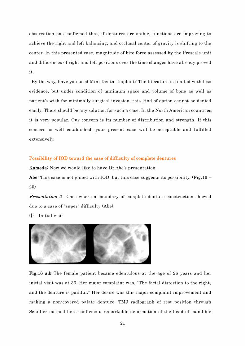
Presentation 2 Case where a boundary of complete denture construction showed
due to a case of “super” difficulty (Abe)
① Initial visit
Fig.16 a,b The female patient became edentulous at the age of 26 years and her
initial visit was at 36. Her major complaint was, “The facial distortion to the right,
and the denture is painful.” Her desire was this major complaint improvement and
making a non-covered palate denture. TMJ radiograph of rest position through
Schuller method here confirms a remarkable deformation of the head of mandible

22
and an anterior displacement of the head of mandible.
Fig.17 a, b Mandibular movement disorders are shown here at the initial visit. The
figure a shows the mandibular displacement to the right, and the figure b shows
abnormal jaw movements. Contrary to these symptoms, however, restoration of
chewing and esthetics would be easier to the author‟s mind as the patient was at
younger ages and her QOL would be improved relatively at earlier stage. And then
treatment procedures were initiated by providing a complete denture in mind,
“Organic problems like TMJ will be restored through the process of remodeling, and,
as a result, abnormality such as jaw movements will be better restored to normality.”
② Correction of jaw positions through treatment denture
Fig.18 a~c Treatment denture delivered. As previously described, the patient was
aged relatively younger and so easier correction of jaw positions and functional
restorations are prospected at first. But, in fact, three years have passed after
changes repeated several times every time treated but returned at once due to larger
displacement of jaw positions.
③ Finish and insertion of newly made dentures

23
Fig.19 a~d Newly made dentures in the year 1999, three years after the initial
visit.
Fig.20 The facial profile on insertion of new dentures. Although the treatment
lasted over a long period of time, the author thought the final dentures of both upper
and lower jaws with good suction effect and pleasant insertion. After treatment
finished, the patient was happily married having a child birth, and the author was
confident that esthetics and masticatory function were correctly restored according
to her original wish.
④ Changes of mandibular jaw positions after one year of new dentures insertion.
Fig.21 a, b Changes of mandibular jaw positions after one year of new dentures
insertion. The results were repeated displacement of mandibular jaw positions and

24
necessary correction of dentures.
⑤ Patient‟s assessment after one year of new dentures insertion.
Fig.22 a, b Results of Sato‟s assessment method of denture satisfaction and
chewing that was obtained from the patient after one year of new dentures insertion.
Reality is severe, and, contrary to a dentist‟s confidence, it was known that the
degree of patient satisfaction was considerably low.
Chewing Function Assessment Chart
とうふ Tofu soybean curd, 卵焼き Fried egg,
煮たじゃがいも Boiled potato 煮たにんじん Boiled carrot
もやし Bean sprout かまぼこ Steamed fish paste
ポテトチップ Potato chips ごぼう Boiled burdock root
あられ Cubic rice crackers 焼肉 Grilled meat
ピーナッツ peanuts たくわん Yellow pickled radish
堅いビスケット Hard biscuit 堅いせんべい Hard rice cracker
古たくわん Old pickled radish とり貝 Japanese cockle
するめ Dried cuttlefish 貝柱の干物 Dried scallop ligament
チューインガム chewing gum リンゴ丸かじり apple biting
Question about 20 different foods listed on the left
What is able to eat normally ○
What is able to eat with additional preparation (small cut or soft cook) △
What is not able to eat ☓
Please write other foods that are hard to eat.
いちご strawberry
なし pear
生野菜(レタスやキャベツ) fresh vegetable (lettuce, cabbage)
野菜炒め sautéed vegetables
What kind of food do you wish to become easy to eat?
生野菜 fresh vegetable

25
ガム chewing gum
For dentist‟s use, count number of ○
Scores
Degree of Denture Satisfaction Assessment Chart
For dentists use
● Can you chew well? 1.Satisfactory, 2.Neutral, 3.Unsatisfactory
● Can you test taste of food?
● How well can you talk?
● Any pain?
Upper jaw
Lower jaw
● How do you like appearance?
● Does the denture fit well to the gum?
Upper jaw
Lower jaw
● Does the denture sit well?
Upper jaw
Lower jaw
● Any ill-feeling with the denture?
Upper jaw
Lower jaw
● Do you like the denture now?
Totaled scores
26
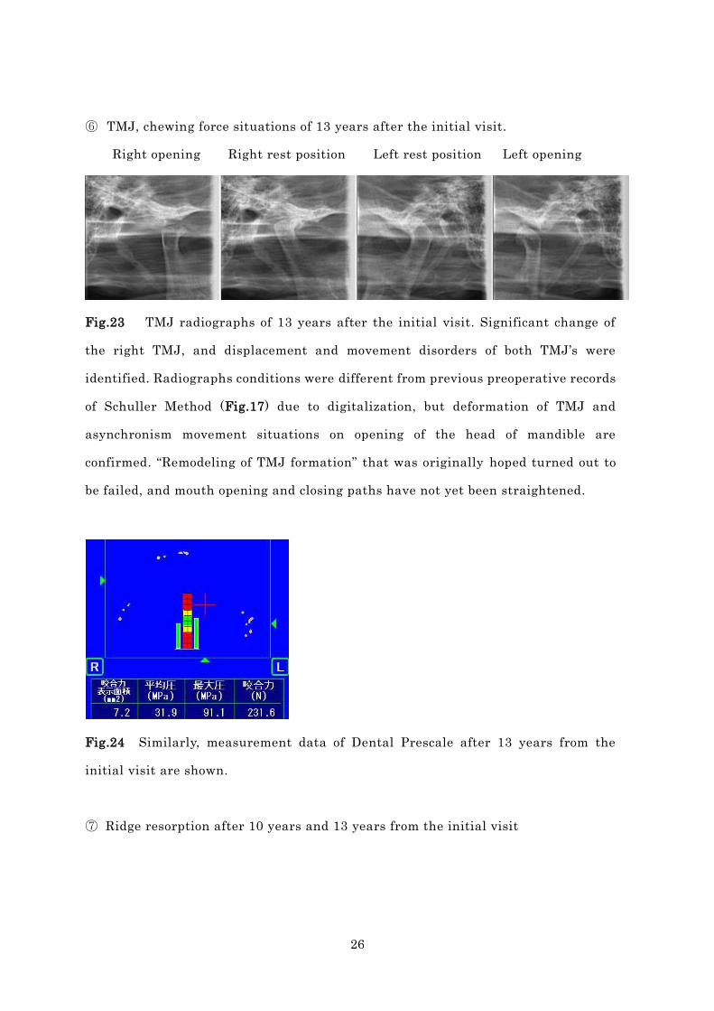
⑥ TMJ, chewing force situations of 13 years after the initial visit.
Right opening Right rest position Left rest position Left opening
Fig.23 TMJ radiographs of 13 years after the initial visit. Significant change of
the right TMJ, and displacement and movement disorders of both TMJ‟s were
identified. Radiographs conditions were different from previous preoperative records
of Schuller Method (Fig.17) due to digitalization, but deformation of TMJ and
asynchronism movement situations on opening of the head of mandible are
confirmed. “Remodeling of TMJ formation” that was originally hoped turned out to
be failed, and mouth opening and closing paths have not yet been straightened.
Fig.24 Similarly, measurement data of Dental Prescale after 13 years from the
initial visit are shown.
⑦ Ridge resorption after 10 years and 13 years from the initial visit

27
Fig.25 a, b Panoramic radiograph after 10 years from the initial visit (a), and
cepharogram after 13 years from the visit (b). Although a therapy of complete
denture was properly given, the photos demonstrate that the ridge resorption is not
yet controlled.
Key Word for better understanding of this discussion
*3 What is occlusal center of gravity?: This is a method to determine the bite
force distribution throughout the dental arch. Each tooth exerts its force, and from
this force, comprehensive gravity center is determined. In case right and left
balancing of bite force is developed, this center will be located often approximately
in the center of antero-posterior and right and left of the dental arch.
*4 What is T Scan?: In 1980‟s Maness and others developed it in Boston. This is
an occlusal force measuring system with a contact sensor sheet, and their dynamic
changes can be observed in digital movies. Also changes of occlusal gravity center
over time can be analyzed.
Abe: Looking back at this case, I find the patient still young and thought that any
favorable recovery would be possible with a good quality of denture and every
problem will be restored. With this hope in mind I entered into the therapy. But what
I finally could achieve was quite limited to esthetic restoration, enhancement of
chewing ability to a certain degree, and not complete but some extent of position
normalization of the head of the mandible. As a dentist I could not solve the patient‟s
confronting problem. In this case that confronting problem meant the normalization

28
of jointed structures. In fact, in this case what complete denture therapy I could
treat was limited to ad hoc treatment … I must say that the patient condition will be
worsened stage by stage. (Fig.26)
The major reason for the failure of normalization of jointed structures was the
weakness of bite force that could exert with wearing a complete denture. And this
weakness made functional pressure weaker toward the temporomandibular joint
area. And I think that this weakness inhibited remodeling *6 within the body.
Undoubtedly an edentulous patient has porous quality of trabecular bone on the
head bone of the mandible. It is said that, in comparison with a dendate jaw,
functional pressure that is loaded to the temporomandibular joint area of complete
denture patient is reduced to about 40 – 50%, and especially in a case of highly
reduced residual ridge is limited only to 10 – 20% of whole dentate mouth. (Fig.27)
Fig.26 Boundaries that are shown from long-term prognosis of difficult case of
complete denture therapy, and new hope for IOD.
Complete dentures have following …
□ Chewing capacity and esthetics will be restored fairly.
□ Early correction of displaced mandibular position will be less possible.
□ Paths of mouth opening and closing will be less possibly straightened.
□ Bone remodeling of edentulous jaw patients will be little hoped.
Then and now, IOD will be hoped for …
Fig.26 Boundaries that are shown from the case in Fig.16 ~ 25, and new hope for
IOD.
Fig.27 Literatures that discuss over morphological abnormality and relative causes
of edentulous TMJ and jaw bones.
① Kawashima T.: Study on structures of TMJ and surrounding bones, Shika
Gakuho 1996; 96(9); 911 – 949 (Japanese)
☟

29
- Tooth loss will cause to exert 20 ~ 50% less of pressure on the jaw bones.
- Morphological abnormality is confirmed in TMJ or jaw bone of 60% population
over 40 years of age.
② Hongo T.: Morphometrical study on trabecular bone structures of Japanese
mandibular articular process, Shika Gakuho 1987; 87(12); 1583 – 1611
(Japanese)
☟
- Trabecular bone continuity of edentulous jaw is less compared to dentate jaw.
- Contributing factors of dynamic environmental changes loaded on the bone is
greater than those of aging process as for trabecular bone structure changes of
mandibular articular process in dentate and edentulous jaw bones.
③ Abe S., Ide Y.: Jaw bone changes by aging process, the 4th, TMJ anatomy and
morphological changes after tooth loss, Shika Gakuho 1999; 99; 435 – 443
(Japanese)
☟
- Abnormal deformation of the head of mandible will be caused in non-denture
wearers or improper denture wearers.
- As a result of comparison in interior trabecular bone changes within articular fossa
between 70-year-old dentate and 50-year-old edentulous patients, cortical bone of
edentulous patient became thinner and interior trabecular bone became more porous
in complementary density.
Fig.27 Literatures that discuss over morphological abnormality and relative causes
of edentulous TMJ and jaw bones. Meanwhile, the reference in ③ where major
cause of jaw bone changes are caused by tooth loss rather than aging process is
consistent with the report from Iwakata et al. (Iwakata S, Nishi k, Kono S, Ishioka
Y,: Study on the movements of head of mandible of aged population, Japanese
Journal of Gerontology 1994; 9; 89 – 96 (Japanese)). This report has that, in

30
comparison of dentate aged and younger population, sagittal condylar paths
decreased in 6 ~ 8 ° , while the mandibular movement paths showed little
differences.
Does IOD promote remodeling of TMJ?
Kameda: Let me summarize roughly. You had a major object to stabilize the
mandibular jaw position through the way of a complete denture toward a patient
whose jaw position was irregular and unstable. But it did not result in normalization
of jointed structures which was fundamental to stabilization of the jaw position
because there were limitations of bite force increase by the provision of a complete
denture.
Key Word for better understanding of this discussion
*5 What is Sato‟s assessment method of denture satisfaction and chewing?: An
assessment method of both clinicians and patients toward denture wearing in the
form of questionnaire and score sheet. It includes assessment of chewing function of
the denture, of patient satisfaction and of QOL. For details of sheet and study
results please refer to, Kohno M, Sato Y, Kitagawa N et al.: Outcome assessment
immediately after insertion in the therapy of newly made complete denture, J the
Japan Prosthodontic Society, 2007: 51(2): 260~269 (Japanese)
*6 What is remodeling?: Bone metabolism has repeated cycles of osteoclastic
cells to resorb and osteoblastic cells to create bone, and in about 200 days, bone is
newly formed. New bone formation will change shapes, and in this discussion, proper
pressure loading on the TMJ region allows to restore proper bone shape formation,
which means remodeling in this discussion.
Abe: Exactly. To stabilize the jaw position does depend on how we can increase bite
force. In order to fulfill this object, an implant therapy can be added to treatment
options for better increase of bite force. I think that IOD will contribute greatly for

31
that purpose. And then an increase of bite force will stimulate the
temporomantibular joint and will bring an organic change to the trabecular bone.
What do you think about an idea around this, Prof.Maeda? By all means, stable
removability of a mandibular denture has to be achieved in order to fulfill the
methodology of normal restoration of jaw position, and if it is possible, this
attainment may be easier.
Maeda: I once had a chance to construct a treatment denture in a case where a single
denture*7 was inserted in the mandible while the maxillary was dentate but had
tooth mobility due to periodontal disease. But we had a hard time to attain normal
restoration of jaw position. And IOD was suggested, and finally this denture does
protect the remaining teeth to the present. It is true that unstable mandibular
denture will likely make the jaw position irregular. And so the implantation would
stabilize a denture and eventually the jaw position will help some stability.
Kameda: What do you think with Dr.Abe‟s idea on improvement of bite force and
functional pressure by means of IOD, and then on promotion of remodeling with the
temporomandibular joint? Will it be possible? Is there any literature on it? We would
appreciate your suggestions.
Key Word for better understanding of this discussion
*7 What is single denture?: One side of edentulous jaw while the other opposing
arch is dentate. In this case of complete denture on one jaw is called “Single denture”.
This kind of pressure loading and its acceptance will cause frequently a denture base
fracture or underlying ridge resorption.
Maeda: I think it is possible. Remodeling in the TMJ region is related with formation
and resorption of the temporal bone or mandibular jaw bone. There should be
certainly of consensus based on their extensive supporting literatures regarding
facts that TMJ‟s are the loading joints that are to share some amount of loading. And
they state that the magnitude of loading will maintain within the range of

32
homeostatis, in other words, within the range of balancing of formation and
resorption.
As for the relation between its magnitude of loading and formation/resorption in
the joint area, there have been some theories, and one of them is shown in Fig.28 as
an example. This idea was organized by ex-Prof.Tsutsumi from Kyoto University,
who studied the behavior of experimental animal bones in the orthopedic field under
Prof.Kummer, Germany. The vertical axis indicates bone volume change, showing
bone formation in the positive area and resorption in the negative. And the
horizontal axis indicates stress applied to the bone, showing tension of the bone in
the positive area and compression in the negative. Here in this case, whichever
stress, positive or negative, is to become more than certain degree of magnitude,
resorption will occur, and even if the value is in the smaller range close to zero,
resorption will occur again. Both in tension and compression of either case, bone will
be added, if the value is within this range.
So when Dr.Abe mentioned, “An edentulous case would decrease bite force to lower
strength, and the trabecular bone quality would become porous. This is because any
mechanical stimulation (bite force) is decreased”, this value range would correspond
to “the smaller range close to zero”.
Fig.28 Relation of stress and bone formation / resorption
Fig.28 Relation of stress and bone formation / resorption. Vertical axis has bone

33
volume change, while horizontal axis show magnitude of stress on the bone. Whether
tension stress or compression stress would create bone resorption when stress
becomes greater than certain degree of magnitude, and, on the contrary, its stress
value is, even within small range close to zero, resorption will occur again. In
between these limited range of stress values whether tension stress or compression
would create bone formation. (Quoted and modification approved courtesy of
Prof.Sadami Tsutsumi.)
Meanwhile, in my analysis of the study on relation between the posterior occlusal
support and TMJ loading, (Fig.29,30) the clenching of the remaining dentition after
eliminating the most posterior region demonstrated larger displacement of the head
of mandible within articular fossa, and pressure load increased accordingly. But, in
reality, it is reasonable that muscle force should increase in response to the
condition of missing dentition, and accordingly loading onto the temporomandibular
joint turns out to be smaller.
For these reasons, in case of edentulous ridge, loading onto the joint area will
become smaller, taking into consideration of lower muscle activity to exert bite force.
On the contrary, application of IOD will increase bite force exertion and possibly
raise the stress level of TMJ area into the range of bone formation.
Fig.29 a, b Relation of posterior occlusal support and loading exerted on TMJ ①
Fig.29 a, b By the two-dimensional computer simulation using the concept of Fig.28,
relation with the area of occlusal support and bone remodeling (resorption /

34
formation) of TMJ area was studied. As a result, here in this case, occlusal support
was valid to the second premolar region namely in the case of shortened dental arch,
there was no change seen in TMJ region (the left figure a in the middle row), and
when the support was shortened than this, changes were seen as shown. (The left
figure a in the lower row.) This model is based on mean values produced with the
lateral profile standardized radiograph of 60 dentate subjects. (The right figure b)
(The chart was quoted and modified from the literature, Maeda Y et al. Form and
function of stomatognathic system in shortened dental arch situation: a
biomechanical simulation. In: Morimoto et al (Eds). Brain and oral functions
(International Congress Series 1079). Amsterdom: Excerpta Medica, 1995: 511 –
514)
Fig.30 Relation of posterior occlusal support and loading exerted on TMJ ②
Fig.30 Relation with TMJ loading and treatment options of distal extension cases of
only anterior arch remaining was studied. As a result, contrary to the case ① where
remaining maxillo-mandibular jaws were valid in occlusion to the second molars
(chart ①), loading is greater with a mucosal born denture (chart ③), but in case
with implants placed underneath the denture base, loading was decreased (chart ④),
close to the values measured in a case of fixed prostheses supported by implants
(charts ⑤, ⑥). (The chart was quoted and modified from the literature, Maeda Y,
Sogo M, Tsutsumi S. Efficacy of a posterior implant support for extra shortened
dental arches: A Biomechanical Model Analysis, J Oral Rehab 2005: 32(9): 656 – 660)

35
Does an implantation on edentulous ridge delay ridge resorption than complete
denture?
Abe: Now this patient is still 48 years old, but cepharogram and panoramic
radiograph confirm advanced ridge resorption. (Fig.25) As a matter of fact, my
denture construction in this case was found of unstable occlusion, denture collision,
chewing difficulty due to displaced mandibular position, and so on. I think I had
tried many various possibilities.
This case story is not exclusive to IOD, but I think a priority to implantation is
given here in an implant case so that the mandibular position should be stabilized
and that the ridge resorption should be controlled. In other words, minimizing
denture mobility will possibly delay the speed of ridge resorption to some extent. A
book written by Prof.Maeda *8 has already referred, “Finally it will absorb but can
delay it.”
Kameda: I admit that resorption of residual ridge or jaw bone of a complete denture
wearer cannot be stopped in the process of change over time. And it is almost true
that ridge resorption of a denture wearer may be larger than that of an implant
placed patient.
For this purpose, I have researched reviewing literatures on the resorption of
complete denture wearers. (Fig.31) Among them, Tallgren observed complete
denture wearers over the period of 25 years and concluded that the denture wearing
period had definitely played decisive roles on resorption of alveolar ridges. Atwood
suggested that factors of ridge resorption were complex with factors of both general
body system and local regions. And factors of local regions might be likely to some
amount of force factors that work on underlying denture bearing ridges.
And Kalk concluded from various studies that ridge resorption of edentulous
patients were extensively influenced by very existing dentures. So wearing denture
itself might be involved with ridge resorption.
Maeda: As far as ridge resorption is concerned, extensive researches are being made
to detect any promoting factors of bone resorption on a genetic level. That is, even
under similar denture wearing condition, any individual who carries such promoting

36
genes would likely proceed the resorption while another who has no likely genes will
reduce the change. If this study is advanced, one can take blood sample from a
patient‟s tooth extraction site and inform, “Your risk of bone resorption will be high
in the future and need any preventive measure from now”.
Fig.31 List of literatures on discussion in reference to relation between ridge
resorption of edentulous patients (complete denture wearers) and dentures.
① J Prosthet Dent. 1972 Feb;27(2):120-32
The continuing reduction of the residual alveolar ridges in complete denture wearers: a
mixed-longitudinal study covering 25 years.
Tallgren A.
☟
- Over observation period of 25 years, duration of tooth missing time (denture
wearing period) has played decisive roles in the alveolar ridge resorption.
② J Prosthet Dent. 1971 Sep;26(3):266-79
Reduction of residual ridges: a major oral disease entity.
Atwood DA.
☟
- Ridge resorption is consequences of various factors including those of both
general body system and local regions.
- Factors of local regions include that of force exertion toward the residual ridge
(dentures and others).
③ J Dent. 1989 Aug;17(4):162-5. Links
Some factors connected with alveolar bone resorption.
Kalk W, de Baat C.
☟

37
- Ridge resorption volume is proportionate to duration time of edentulism and of
denture wearing
- The very existence of denture itself influence on the degree of ridge resorption.
Fig.31 Literatures above referring to causes of ridge resorption of edentulous
patients (complete denture wearers). Literature ① has been derived from the
long-term follow-up of complete denture wearers over the period of 25 years. And the
literature ③ refers to the results of investigating 92 edentulous patients with
connections of ridge resorption and age, duration of edentulism, number of remade
dentures, and denture wearing habit of day-and-night.
Key Word for better understanding of this discussion
*8 What is Porf.Maeda‟s book?: “Overdentures applied to clinical cases” written
by Y.Maeda, 2003 published from our publishers, B5 size edition in 120 pages, price
¥6,200. Overdentures have different advantages and are applicable to various
clinical scenes. So the author describes basics and applications of design and
construction of overdentures supported by natural teeth or implants, as well as
significance of maintenance. He also adds a series of necessary information on
reference books.
*9 What is Kelly Combination Syndrome?: In 1972, Kelly E published paper
“Changes caused by a mandibular removable partial denture opposing a maxillary
complete denture, J Prosthet Dent 1972: 27(2): 140 – 150. Five clinical signs are seen
in a case where maxillary jaw is edentulous and mandible has remaining anterior
teeth exclusively. The signs are ① hyperkeratinized maxillary palate, ② increased
fibrous tissues in maxillary tuberosity, ③ inflammation of maxillary anterior ridge,
④ elongation of mandibular anterior remaining teeth, ⑤ Distal extension bone

38
resorption in the mandibular partial denture. These conditions are called as Kelly
Combination Syndrome
Unfortunately until present it is not yet well defined, and current tendency suggests
that resorption occurs in the case of loading with a denture from above without any
established evidence. This might lead to the notion that a complete denture should
be to blame. Is it truly bad to wear a denture? Never, I say. Some patient is triggered
with a complete denture to induce displacement of mandibular position or bone
resorption, and some are not after all. There is no way to validate it at the moment
but only misleading complexity.
IOD is beyond indications when the maxillary arch is anteriorly open and reduced.
Abe: In another case, there will some great problem be raised, when an implant is
placed in the mandibular anterior region. Cases shown in Fig.32 are all with
edentulous in the maxillary and with a partial denture in the mandible. One of the
upper two cases of three, namely cases ① of the maxillary single denture plus the
mandibular AGC-made telescopic denture is seated on a good ridge form in the
mandible posterior region, expecting significant mucosal bearing capability in the
posterior region. And, as you may call, a stable posterior bite habit is established.
On the other hand, the lower case ② in Fig.32 shows an extensive ridge resorption
in the mandibular posterior region, while the maxillary ridge is reduced in the
anterior and superior direction.
Fig.32 Cases that are prone to Kelly Combination Syndrome and that are not.
(Abe)

39
① The case where the mandibular ridge condition is good in the posterior region
and the posterior bite habit is established.
☞ Not prone to Kelly Combination Syndrome → Indicated for IOD
② The case where the mandibular posterior ridge resorption is extensive and the
maxillary arch is anteriorly open and reduced.
☞ Prone to Kelly Combination Syndrome → Beyond indication for IOD?
Fig.32 Like in this case of missing arch pattern with implant placement in the
mandibular anteriors, the anterior bite habit is easily formed with the lower
anterior implants because the maxillary jaw is open and reduced. This is prone to
Kelly Combination Syndrome.
This kind of patient, as Prof.Maeda describes, has genes potential likely to induce
ridge resorption. Since the posterior alveolar ridge does not accept the loading force,
this patient tends to bite in the anterior region as the teeth are planted exclusively
in the anterior arch. This tendency promotes rapid resorption in the maxilla

40
anteriors to reach a state of flabby gum finally. This extreme case is called Kelly
Combination Syndrome *9, and nothing can be done to most serious case.
So, even in a case of IOD in the mandible with upper-lower edentulous ridges, when
the maxillary arch is anteriorly open and reduced, mandibular anterior implantation
has tendency to bite in the anteriors and to transfer to a flabby gum of the maxilla to
a worsened condition. So, what is important is that a supporting implantation in the
posterior region should be given in an early stage in order to establish the posterior
bite habit. For this reason, therefore, a case with anteriorly reduced maxillary ridge
might be excluded from indications of IOD.
Kameda: As for this case of Kelly Combination Syndrome, I think, for example, in an
edentulous mandible case, IOD would be better than a bone anchored bridge because
the maxillary anterior bone resorption will be controlled with less pressure loading
factors. How do you think?
Maeda: Yes, this is what I have been wondering about its validity. There might be no
researchers who have determined in the process change over years. How about over
years the upper case in Fig.32 presented by Dr.Abe? Has the maxillary anterior
turned out to be flabby?
Abe: No, in this case it has not. That is because any possible valid schemes of
occlusion have been designed by giving metallic occlusal surfaces limited to both
upper and lower second premolars and first molars, and by giving high s trength of
intercuspation there, while leaving the rest teeth untouched and guiding to the
posterior chewing. Sometimes there is a contact to the maxillary anterior arch, but
there is no sign of flabby gum. This patient might belong to one whose osteoclast
activity is low.
Maeda: There might be some extent to relieve onset of Kelly Combination Syndrome
by changing a case into an overdenture. But this is an empirical rule and there is no
definite evidence here to support. Now in a case of a mandibular implantation with
fixed superstructures, there may be strong possibility of Kelly Combination
Syndrome in the maxilla. There will be possibly natural.
Abe: As a conclusion, if multiple numbers of implants are placed in the mandibular

41
anterior region, there may be created conversely the trend of anterior biting habit.
After implantation, patients are likely to bite in the front with less mobile implant
site rather than on the mobile distal extension of denture base. So in due course of
forming chewing habit, bodily system will respond to the implant support area and
not to the mucosal support of denture base. So this discussion will raise a question of
how many implants and what part of region should be placed after all.
Kameda: Now our next discussion will go to proper implant placement site, design
of superstructures and, moreover, to maintenance of them in the next issue.
(continued)
Summary
- It is suggested that use of IOD would improve bite force and occlusal center of
gravity. (Kameda)
- It is our feeling that IOD may be possible in case when bite force restoration is
not predicted with a complete denture and when remodeling of the head of
mandible is not available. (Abe)
- IOD is potentially promising for stable occlusion and residual ridge maintenance
in a difficult case of establishing mandibular jaw position or of single denture.
(Maeda)

42
MONTHLY FOCUS
Considering prosthodontics in the aging society
Round-table discussion
Implant Overdentures for mandibular edentulous patients
- Second part: Considering its design and cleaning performance
,
,
Yoshinobu Maeda Jiro Abe Yukio Kameda
Introduction
Kameda (Moderator): In this discussion, implant supported overdentures (hereafter
called IOD) will be focused on as a new treatment option for the mandibular
edentulous jaw especially concentrating on its possibility and practice. In the first
part, we discussed on general idea of IOD, indications depending on different cases,
and implant placement regions. Now in this part, as in the second part, practical
implant placement for IOD, design of superstructures and maintenance will be
featured..
Objects designed for this discussion (Second part)
☞ To search commonness and difference in designing complete denture and IOD.
☞ To discuss prosthodontics in dental implants based on periodontics.

43
5. Implant placement in the edentulous mandible. Where and how many should be
placed?
One implant in the median of the mandible is the point of starting IOD.
Kameda: Now we would like to discuss an implant placement site for IOD.
Maeda: When we discuss over placing implants in IOD in the edentulous mandible,
one piece of implant placement will be a starting point. And its placement site will
be in the median line. Reason for this is that, according to our research, rotation is
minimum in the midline of the mandible about mobility of denture in back and forth
around. And deformation of denture body as well as jaw bone is least in the midline.
In other words, it is in good reason for dynamics science and realistic according to
our research. Apart from our research study, similar reports have been common
recently. This should be, of course, presumed to remain with some amount of bone
volume in the anterior implant site. It may be impossible with extremely resorbed
jaw bone. But anyway the final denture will be retentive with this support and also
stability will be added.
Ideal pattern will be two in the posterior and another two in the anterior regions.
Maeda: For the purpose of established occlusal support and long-term stability,
minimally essential number of implants will be different from above.
In another research results from ours, when the last ultimate ideal of occlusal
support is conditioned under the clenching of 14 pieces of implants in the mandible,
it is studied how situations are accordingly by reducing its number of placement and
what minimum number of placement is valid for maintaining stable occlusal support.
Fig.1 shows the mandibular jaw bone deformation in this research, and Fig.1 a is a
deformation with the 14 pieces of implants placed. Then afterward its number is
reduced. Fig.1 b indicates 8 pieces, Fig.1 c shows with 6 pieces in the anterior
portion. In Fig.1 c, the jaw bone also deforms a little as expected. Fig.1 d is at the
time of only 4 pieces, or 2 in the posterior and 2 in the anterior regions.

44
In comparison of Fig.1 a to those of Fig.1 b~d, 14 pieces may be ideal to our mind.
With less deformity, stable biting is established. And situations of 14 pieces
placement may be comparatively close to 8 pieces or 4 pieces of placement. But in
case of 6 pieces, although the deformation is less, the posterior region is knowingly
deformed.
Interestingly, a bone anchored bridge, as it is often true to design the posterior
portion as cantilevered, would reportedly create new bone under this cantilever. This
means in reality that the jaw bone there may be formed through remodeling after the
jaw bone deformed. We think this is only an occasion of new bone formation through
remodeling in an edentulous ridge. But this does not always mean that another new
stability of occlusion could be obtained through this process.
From this discussion, it should be definitely effective in the sense of occlusal
support that 2 pieces in the posterior and 2 in the anterior regions are placed. This,
however, is absolutely a theoretical matter, and some would have a question, for
instance, “What happens if all forces are down to these implants only?” Now we are
researching with our graduate students regarding force distribution on implants and
bearing mucosa of IOD. Until now it is evidenced limitedly, but the research suggests
30 or 40% of mucosal bearing will be effective. In case of IOD with 4 pieces
placement, the base area is bearing in effect. With this effect, although they are only
4 pieces, loading onto the implant structures might be relieved and they are
supportive to each other.

45
In case of 14 pieces of implants In case of 8 pieces of implants
In case of 6 pieces of implants In case of 4 pieces of implants
Fig.1 a~d Mandibular jaw bone deformation depending on implant number and
placed site (Maeda)
Fig.1 a~d Data collected of jaw bone deformation through clenching muscles force
as vectorial component exerted onto the occlusal support of assumed implant
placement. As the color is reddened, deformation is larger, and bluish smaller. Cases
are when the support is valid till the second molar in this control model (a), when 4
pieces in the anterior and 4 pieces in the posterior (b), when 6 pieces in the anterior
according to Branemark original protocol (c), and when 2 pieces in the anterior and 2
pieces in the posterior (d). They are compared and deformation is largest for
condition (c).
So, 2 pieces in the posterior and 2 in the anterior regions totaling 4 pieces of

46
implants might be basic and minimally essential. When we counsel with our patients,
therefore, if conditions permit, our suggestions be, “Totaling 4 pieces at least in the
anterior and posterior regions will be ideal. Then a denture will be stable, less
changeable in the future and ideal.”
Nevertheless it is absolutely ideal. In our present discussion over the case of
difficulty for IOD, even 4 pieces will be difficult to be placed. In such a case, 2 pieces
in the anteriors are advisable. If a patient declines even to this, I would suggest,
“Even one piece would be worth trying.”
So in this case one piece in the median line will be decided. Even with this, as we
suggested before, the case will be effective enough.
Abe: The truth is that there are many dentists who can do nothing to prevent a
mandibular complete denture from lifting up, not even its suction effect. In the
previous part I mentioned, “A complete denture must be mastered first, then IOD
comes.” But, although contradicting to this, if such a dentist above could keep his
denture from lifting up by placing an implant in the middle, it cannot be helped as
long as patient satisfaction is improving.
If two implants in the anterior arch, then where in the anterior to be placed?
Abe: In an edentulous case of difficulty, there will often be limited clinically to
place implants in the anterior arch. The question would be to place them at the site
corresponding to the lateral incisor site or the canine site. Where to be best placed?
This is what most readers will be interested in.
Kameda: For example, as the McGill Consensus Statement has 2-implant as first
choice standard of care, where should be the placement site? Also what attachments
would be better to be used together?
Maeda: As Dr.Abe already discussed, there may be any question raised in the
anterior arch, whether be at the lateral incisor site or the canine site. Some
consideration will be possible to this question.
First, consider the curvature of an arch. In case where the ridge between both
canine sites is straightened, or a type of rectangular arch, it can be placed within the

47
arch and retained by a bar attachment. But in a curved arch between them, implants
are placed at the canine sites and then, if retained with a connector, the tongue
space will be interrupted. So a single retainer will be given independently. Or
otherwise, they will be placed in the lateral incisor areas. (Fig.2)
Fig.2 a~c Bar connection with two implants in the mandibular anterior arch is of
most advantage in between the lateral incisor and the canine. (Maeda)
Fig.2 a~c Placement of 2 implants in the mandibular anterior arch becomes
advantageous in between the lateral incisor and the canine (a). When placed
posterior to the premolars, bar itself narrows the tongue space and promotes denture
rotation (b), creating troubles of bending moment on the bar.(c) (Quoted and
modified from the literature, Misch CE, Dental Implant Prosthetics. St.Louis: Mosby
2004: 214 – 216)
Next, what we often discuss is about lateral force. This depends on the distance of
implants in between. Fig.3 is also from our research results, and C refers to the sites
corresponding to both the canine teeth, and LI refers to the lateral incisors. There,
when the ball anchors were joined, the lateral force was greater as distance is
farther.

48
And denture retention should be discussed. This also depends relatively on the
distance of implants in between. The research demonstrates that, if both implants
are farther in distance, retainers are abrasive and less retentive due to incorrect
parallelism. There is a report that refers to the relationship of attachment distance
and retention, or implants distance in between. Overall studies of them indicate that
the shorter distance will help the case easier like in the lateral incisor sites on both
sides or in between the lateral incisors to canine teeth sites. If the distance is given
more than that, they will become less useful for dentist and patient.
Kameda: In the McGill book (ref. to No.5, p.15), 2 implants are placed after all at
the sites corresponding to the lateral incisors. They reason that, if placed at the
canine teeth sites, the bar connector would become a cantilever toward the anterior
direction.
Maeda: Yes, I think so, too.
Fig.3 Relation of implant site and lateral force (Maeda)
Fig.3 IOD with 2 implants is assumed, and placed at the lateral incisors on both
sides (LI) or the canines (C) retained with magnets (Flat and Dome types) as well as
Ball, and occlusal force is given on the first molar area for measurement of lateral
forces on the models. Results indicated the placement on the lateral incisor is likely
known to create less lateral force. (Chart drawn from the literature, Horisaka M,
Maeda Y, Sogo M, Okada M. Overdenture movements and lateral forces to
non-splinted implant abutment with different types of attachment: A model study,

49
Dent Japan 2006; 42: 177 – 180)
Selection of implant fixtures and insertion.
Kameda: What about an implant body selection for IOD case? Does it have to be
longer and thicker as everyone expects?
Maeda: It should not be confined to the matter of IOD, but it was originally
thought that any one thick or long as possible had been advised in the limited bone
volume as a basis reason that one as long and as thick as available may be safer.
But recently it does not necessarily mean that a longer one is always better, but
some report has that there might not be any difference from some certain length. As
a matter of fact, in a fixed type of implant prosthetics, too short of length will be
disadvantageous due to the question of crown to root length ratio.
As for the case of IOD, so called the question of crown to root ratio is improved by
use of attachment in order to control the lateral force, and even a shorter one will be
acceptable. Note, however, that there is an issue of initial fixation, some amount of
bearing space area will be needed. This will therefore be likely to become a shorter
and thicker one.
Kameda: For example, when multiple number of implants are placed, their
parallelism is important, and how do you think with them?
Maeda: Yes, it is important. But in case of two implants placement at the lateral
incisors site, precise parallelism of two implants will not be effected. Theoretically it
may be possible, but in fact the jaw bone anatomy is inclined, and so the inserted
direction is somewhat angled finally. This can be compensated within a certain level
by a technical device of machine-made attachment and hand skills of dental
technicians. But if the degree of inclination is too much excessive, correction will not
be possible. Various problems have been potentially raised for this reason, and so, it
should be in advance prepared how best could it be responded.
Kameda: How do you think it should be prepared?

50
Maeda: For example, when two implants are placed in the anterior arch and they
are inclined, they can be helped by connecting the implants with a bar attachment.
But this kind of connection is not possible with placing at the canine site on both
sides but possible with the incisor area. If a bar attachment is used on the canine
teeth area, the denture base coverage will become bulky at the lingual surface. But
this problem will be kept in minimum if they are connected on the lateral incisor
basis. Even if they are not parallel, they can be helped by all means. In this sense,
placement at the lateral incisor area can be useful. What is more, one single implant
placement at the midline is easy without paying attention to parallelism.
Factors of attachment selection to think about
Kameda: What about selection of attachments? Previous discussion on bar
attachment selection is related with some recovery solution of implant placement
angles. Now what are basic considerations?
Maeda: Our initial object is to finish a denture base and we first try to arrange
artificial teeth without relation of implants. From this stage we start to select a
proper size of attachments that can be accommodated within this denture base. In
addition to this, in case of one or two implants, any rotation should be a matter of
consideration, and so the selection of attachment depends on the rotation
mechanism of attachment design. (Fig.4)
Fig.4 a, b Parallelism must be secured for setting up of 2 ball attachments. (Maeda)

51
Fig.4 a, b In order to realize primary rotation allowance with 2 ball attachment,
they should be set up in parallel (a) to establish one single rotational axis. Otherwise
inclined, rotation allowance is limited (b). (a is from Bredent AG, b from Astratech co.
Quoted and modified from brochures.)
And its selection will need to consider patient‟s jaw position and age. Some patient
would say, “I do not like any tightly assembled attachment. Even if it is not so
strongly retentive, I would prefer simple denture seating.” But any patients who
cannot stabilize the jaw position have no ways to keep the denture stable if its
attachment assembly is weakly retentive. In such a case some high attachment, a
bar or a ball, should be used to be retentive against the lateral force. But an old
patient cannot put off a denture with a bar attachment because of manual dexterity.
For such a patient, magnetic attachment would be better as they are retentive at the
final instance of denture wearing but easy to take off. And attachments should be
easy for maintenance, replacement and relining for both a patient and an operator.
Otherwise it will be in trouble, later on.
In this way, attachment selection criteria must have many considerations.
Kameda: How do you think about types of dental attachments, for example,
magnetic retention is given or simple support is given without any retentive
mechanism? In such a case, simple type of attachment design is given in a way. A
root cap type may be possible. Otherwise, a domed type or inner crown telescopic
type may be possible.
Maeda: For retention of IOD, an ideal type should be a spherical one, or a perfect ly
half of a ball. This type will permit any directional forces to distribute toward the
axial direction of implant bodies, but machine-made attachments are limitedly
available. So, for that purpose, I modify the shape of healing abutments and polish
them in round type when a coping root cap is needed.
Another IOD advantage is flexibility of implant insertion and prosthetic design.

52
Kameda: Now let us return to the previous discussion on placement site. According
to the McGill book, if only placement is done at the site of the lateral incisors on both
sides, another 3 implants will be available totaling 5 pieces to accept the fixed type
restoration against any patient‟s complaint. I think that IOD design should have an
insertion surgery plan that is flexible about patient‟s differed wishes.
Abe: This is a good offer to a patient. Once a good denture is established, and then
even better one is proposed.
Maeda: Yes, certainly. I have just referred to one single implant placement. Even if
one piece is accepted, any patient would like to have another function with another
implant insertion. Just one piece of insertion is easy to accept for a trial, saying,
“Just try to find whether or not it is improved with one insertion.” When a patient is
satisfied with improvement, another 2 pieces are advised to retain IOD with 3 pieces
of triangle formation. Otherwise 4 pieces are added to shape in pentagonal formation.
We are now trying to respond to patient‟s wish to increase insertion pieces. Of course
it is a matter of ridge conditions and patient‟s desire. An aged patient is only
acceptable with one piece, and this is a sufficient reason. (Fig.5)
In order to avoid misunderstanding in advance, increase of implant number is not
to place 14 pieces, but to set up a goal by placing another ones on one piece jaw to
form a pentagonal shape with 5 pieces, or by placing another 2 pieces on already
insertion of 2 pieces to shape in rectangular form. I learned this idea from Dr.Yataro
Komiyama, an implant expert.
Also Dr.Misch has demonstrated similar ideas. He started with 2 pieces. Now he
started with a minimum number that is needed, and if needed more, the case will
turn out to be a fixed type possibly. This way of thinking is very important because
so many cases are shown finished up with implantation for a fixed type. I worry
about future outcome of these fixed type restorations. If any problem is raised with a
fixed type case, where will this case go to end? Will this end in backward direction?
Namely using 5 pieces for a fixed case, and, granting that one or two pieces happen
to fail, will this patient accept another additional pieces of placement? Probabely not.

53
Whether will any prosthetic design case be ready for transferring to IOD or not, and
how will it be done otherwise? I have no intention to deny designing of a fixed type
implant superstructure at all, but it is more important rather than IOD that any
prosthetic design should be planned through calculating risks carefully.
Fig.5 Staged increase of implant numbers coping with patient‟s desire and
condition (Maeda)
Fig.5 Minimum limit number of implants for implant supported overdenture is one
for mandible and two for maxillar. But depending on patient‟s wish and
circumstances, staged addition of implants will secure occlusal support. Or
alternatively can be changed to fixed superstructure design is possible. In case of
mandible, you can first start from 2 pieces.
Summary
- From the viewpoint of occlusal support in a mandibular IOD, 2 pieces of implants
in the anterior and another 2 pieces in the posterior region is an ideal implant
placement. (Maeda)
- In a case where no implant placement is possible in the posterior region due to
extensive ridge resorption, when IOD is to be introduced here, it is important in
the anterior arch to know how many implants and where to be placed. (Abe)
- Selection of attachments for IOD should not be done uniformly but different uses
be selected depending on cases. (Kameda)

54
6. IOD design – From comparison with complete dentures
Denture base design of IOD
Abe: As Prof.Maeda has explained before that 30 or 40% of mucosal is born by the
denture base. In other words, about two thirds will be born with implants, and one
third of division of role is taken care of by the denture base.
Meanwhile, there are roughly two different ways of thinking about impression
taking of denture base shape. (Fig.2) One method is, as taught in dental schools, the
way to take impression in order to obtain the denture base area as broad as possible.
This idea is based on myology that no pain is raised as long as a denture base does
not push on the muscles attached to the bone. In other words, the base can be
extended as far as to this pain free range. While thinking over muscle movements
individually, impression compound is parted to build up to shape the impression.
I think this method is essential to school education because students can learn
prosthodontics in relation to such basic sciences as anatomy and histology and can
improve chewing function even for beginners. But when it is too much extended
beyond the limits of reason, such a denture will flip up and dislodge by the movable
mucosa through the mouth opening. And some patient will feel about mouthful
within the oral space to complain ill-feeling. This will be experienced by anyone after
graduating from school education.
Now the second way of thinking is to take a shape of the mouth at once in functions
as accordingly as the mouth is molded and folded with oral mucosa layers, there on
the muscles, then on blood vessels and nerves, then on fat tissues, connective tissues
and on. As discussed before, it includes muscles to be added with another factor of
submucosal tissues. These overall tissues on the muscles move together in function,
and so the total sum of these movements are all to be taken impression. In practice,
in an object to permit the oral movements under the closed mouth condition, using a
custom made tray joined with wax rim, the patient is advised to follow “pronouncing
woo”, “pronouncing eee”, “tipping the upper lip with tongue”, ”pushing the back of
lower wax rim with tongue” and “swallowing”, and all of these are for taking

55
impression of functional shapes. The suction effective impression taking for a
mandibular complete denture, which I have been introducing, is also one of these
impression taking. IOD, however, can prevent from dislodging with an implant, and
there may be no need of suction.
Details will follow later but here let me summarize that, as a conclusion, these two
impression taking methods, provision of outlines for custom trays are different. One
disadvantage of my idea of outline of functional impression taking is to lose the
surface area of buccal shelf a little. On the other hand, advantage is that the lower
denture would hardly flip up during functions. In overall considerations including
artificial teeth arrangement, the issue would be whichever is better for impression
taking of IOD from these two methods.
①
②
Fig.6 Which should be appropriate for impression taking to determine IOD base

56
border mold. (Abe)
① Muscle trimming border molding impression
② Functional impression
Fig.6 Impression mold morphology differences are shown performed on an identical
patient through border molding impression and functional impression. Border
molding ① is developed to secure stable occlusion (in major consideration of muscle
activities) by extending denture base near around the muscle attached area.
Functional impression ② is an impression taking method of oral cavity shapes that
are composed with muscles and movable mucosal tissues by way of patient‟s
functional activities. The impression mold develops shapes of largely displacing
movable mucosa, too.
Kameda: I do not mean that I have nothing in common to share ideas with Dr.Abe,
but I think that IOD impression taking should take priority of functional impression
taking. This is because, according to a literature using testing units like
Sirognathograph, there can be any changes of chewing patterns and functions by
changing a complete denture to IOD.
Also changing a complete denture into IOD can increase bite force, and so my
practical feeling is that there may be relatively changes of movements of
surrounding tissues and tongue. Supporting ideas are here shown. Fig.7 ① shows
an old denture that is previously referred about that patient, and the lingual surface
of the denture base is made of a metallic plate. The patient was advised to use an old
complete denture as temporary denture in the healing period after implant insertion.
At this instance the lingual frenum was damaged in decubital ulcer from the denture
base margin, and so the metal plate should be cut shortened. (Fig.7 ②) Then after
tissue conditioning, impression was taken. At that time the denture base border at
around the tongue frenum became clearly settled in different position and shape
from the old denture. (Fig.7 ③)

57
The reason that I used BPS for constructing a superstructure is that there should
be some sort of denture base is needed after taking careful considerations of
functions in this case. In this occasion I learned an experience that the shapes of
tongue frenum area became a similar shape after tissue conditioning at the time of
final impression taking. (Fig.7 ④)
And moreover, I tried to find out from experimental stone models how much
different the denture border positions and shapes were, when complete dentures
were transferred to IOD. Cut section samples of each test model at the median line
and approximately at the first molars were compared in Fig.8, 9. Denture base
morphology observed over the time process of change showed that its width became a
little shorter at the time of tissue conditioning. When these are tested for
identification at the alveolar ridge crest, our finding confirmed that the lingual side
denture border became shorter rather than the buccal border. At the same time,
changes in the posterior region were measured, but changes of the width was found
minimum in this case.
For reasons above, impression taking procedure will have to be changed from those
of complete dentures. There is no objection that the border seal of complete denture
is majority for retention, but in case of IOD, some idea should be close to partial
dentures and there may be no need of sealing for the border seal.
Meanwhile, my idea is attained through the findings of this present case and I
think that IOD denture base border itself should be extensive enough on the alveolar
ridge with minimum compression loading displacement and that broad mobile
mucosal area should be avoided.

58
① ②
③ ④
Fig.7 Case example where enhanced function after wearing IOD indicates an
altered denture border mold. (Kameda)
① Old denture at the initial visit.
② Temporary IOD ① was used but decubital ulcer developed at the tongue frenum
area and the border was cut reduced.
③ Temporary IOD during process of tissue conditioning.
④ Final impression taken.
Fig.7 Alteration of denture border mold at the tongue frenum developed from an
old denture through final impression taking in case of IOD that was presented in the
last issue. Following the implant placement, the old denture was used as temporary
IOD, but decubital ulcer developed frequently and reduction was made accordingly.
(Photos ② ,③ , are taken quoted by permission, from the literature, Kameda Y,
Functional restoration with implant overdentures – Discussion on desired denture
border configuration. Practice of prosthodontics 2009: 42(2): 207)

59
Fig.8
① Old denture insertion
② At the process of tissue conditioning
③ Final impression of IOD
計測点 Measurement baseline
本図での計測点 Measurement baseline in this photo
舌側 Lingual side
唇側 Labial side
歯槽頂 Alveolar ridge crest
Fig.8 Each denture impression taking through stone model fabrication as shown in
Fig.7 were observed at the median line cross section. The labiolingual distance at the
lower denture median line is clearly decreased after IOD insertion. The area
indicates the tongue frenum area and especially decreased distance refers from the
alveolar ridge crest to the lingual base border. (Photos are taken quoted by
permission, from the literature, Kameda Y, Functional restoration with implant
overdentures – Discussion on desired denture border configuration. Practice of
prosthodontics 2009: 42(2): 209)

60
Fig.9 Case example where enhanced function after wearing IOD suggests altered
border mold. (Kameda, continued from previous page)
① Old denture insertion
② Final impression taking
計測点 Measurement baseline
右側 Right side
左側 Left side
舌側 Lingual side
頬側 Buccal side
歯槽頂 Alveolar ridge crest
Fig.9 Stone models taken in Fig.8 is the cross section at the molars. The
buccolingual distance alteration at the molars was not confirmed. (Photos are taken
quoted by permission, from the literature, Kameda Y, Functional restoration with
implant overdentures – Discussion on desired denture border configuration. Practice
of prosthodontics 2009: 42(2): 209)
Abe: As stated from Dr.Kameda as above, change from complete denture to IOD
promotes muscle activities, and thereby interfering contacts are developed here and
there. Although this time of impression taking was never tried to force down the
muscles under, functions are enhanced to exhibit new interferences. Based on this
observation, I think that it might be proper to use a method of functional impression
to take a whole body at once. So you might say, “Otherwise, you cannot do better with
the border molding impression using compound material.” (laughter)

61
Now, humans have their own functions that are built-in with everyone. The
function that I am specially paying attention to is the following function, that is,
“Whether or not edentulous or dentate, posterior border seal can be accomplished by
contacting closely with the buccal mucosa and the tongue on the retromolar pad at
the time of closed mouth.” (Fig.10) Thanks to this function, food bolus is not carried
over beyond the second molar. This close contact is termed by myself as “BTS Point”,
namely, the abbreviation of Buccal mucosa and Tongue Side wall. This
physiologically natural function should not be inhibited, and the factors that inhibit
the creation of BTC Point will become a dislodging force of denture at the time of
mouth opening. There is a literature that says, “Even if the border is extended to the
outside beyond the bone external oblique ridge line at the buccal shelf region, it will
not work as a denture dislodging force”. This literature suggests that an increased
surface area of denture base would stabilize it, but the literature does not refer to
the creation of BTC Point. Factors that will inhibit creation of BTC Point are;
① Overextension of denture base toward the buccal shelf.
② Overextention of denture base toward the retromylohyoid fossa.
③ Too much thickness of denture base existing in the retromolar pad.
④ Artificial teeth arrangement distributed to the area where BTC Point is to be
inhibited.
The endoscopic video (Fig.11) explains how BTC Point is created by extending
excessively the denture base intentionally with utility wax toward the buccal side
direction and toward the direction of the retromylohyoid fossa region. As expected,
overextension of denture base inhibited creation of BTC Point.
BTC Point

62
Fig.10 Buccal mucosa and tongue contact on the retromolar pad at the mouth
closing whether or not edentulous / dentate. (Abe)
Fig.10 The posterior denture border seal is accomplished with the buccal mucosa
and tongue contact on the retromolar pad as shown here. This function is common to
dentate and edentulous jaws. (video image excerpt)
(Built-in endoscope) ① ②
③
④
Fig.11 Overextension of denture base will inhibit creation of BTC Point. (Abe)
① Denture base not extended: at the mouth opening
② Denture base not extended: at the mouth closing
③ Denture base extended (buccal side): at the mouth closing

63
④ Denture base extended (lingual side): at the mouth closing
Fig.11 Photos show the endoscope observation over influences on BTC Point
creating (posterior border seal) from denture overextension. Photos ① and ②
show the mouth opening and closing with the denture that patient found no problem.
On the contrary, photo③ shows an extended base to the buccal side with utility wax
on the denture shown in photo ①, and photo ④ also with an extended base to the
buccolingual side with utility wax but knowingly inhibited BTC Point creation.
In this sense, in case of IOD, too, in order to allow these inhibiting factors to be
excluded at the stage of impression taking, custom tray design and wax resin
position and fabrication must be modified innovatively. In other words, it is
important to fabricate a custom tray that enables us to create BTC Point easily at
the time of impression taking. For instance, outline of custom tray should be given
as follows;
① Follow the retromolar pad as it is shaped as an outline of custom tray, and then
cover it thinly with a resin base. The buccal mucosa should not be prevented from
trying to cover the retromolar pad. Also, if the retromolar pad is thickly covered
with a resin base, the buccal mucosa cannot contact with the tongue on the
retromolar pad. (Fig.12)
Fig.12 a~c Custom tray construction that does not inhibit BTC Point creation ①
Outline faithfully according to the retromolar pad.

64
Fig.12 a~c In order not to inhibit BTC Point creation, it is important to trace the
outline of retromolar pad contour faithfully as the outline of custom tray. And in the
photo a, white dotted line shows a border line of ridge mucosa and buccal mucosa.
From this line, the buccal mucosa turns to cover the retromolar pad. At the stage of
designing a custom tray, it is important not to cross over this line in order to
establish the BTC Point.
② The function should be arranged necessarily so that the tongue side wall is to
suppress the lingual side polished surface of the denture. So, outline a custom
tray over about 2-3 mm beyond the mylohyoid ridge line. Whether the line is
possible to be extended beyond that or not will depend on the space room
allowable at the retromylohyoid ridge fossa of individual patient. Even if you
could take deeply enough there, you do not have to fix the outline of custom tray
accordingly as deeply as it was. Overextension of denture base pushes the tongue
root area strongly, and the tongue is difficult to contact with the buccal mucosa.
(Fig.13)
Fig.13 a~c Custom tray construction that does not inhibit BTC Point creation ②
Outline should be about 2-3 mm beyond the mylohyoid ridge line.
mylohyoid line
from 2mm below
avoid Someya‟s sinew string
frenum is variable to movement
bottom of mucobuccal fold

65
shallowly in 2mm
Fig.13 a~c In order to fulfill a function of suppressing the denture polished surface
with the tongue side wall, outline should be given about 2-3 mm beyond the
mylohyoid ridge line. And in general, custom tray outline is taught to be given in the
middle or outside of external oblique line, but, in order to accomplish a function of
containing a denture with the buccal mucosa, it should be standardized to give the
line inside of external oblique line or the lowest point of buccal side mucobuccal fold.
Next, as for the arrangement position of the artificial teeth, which should not inhibit
the movements of the buccal mucosa and tongue, it is important to take impression
by building up the wax rim in the middle of ridge width where the molars and
premolars are to be occupied. The idea is based on the rules of teeth arrangement of
Pound Line that represent former positions of natural teeth, as well as other
concepts of Piezography*1 or Neutral zone. These ideas and concepts are originally
different from one another, but final arrangement will become approximately close to
others. Dental school education in majority seem to take priority of controlling
denture from dislodgement and keeping stability, teaching, “Teeth should be
arranged within common zone that cross the allowances of mandibular and
maxillary common zone”, rather than teaching negation of inhibition of
physiologically natural functions. (Fig.14) Some are still teaching the arrangement
along the alveolar ridge crest. But the arrangement along the ridge crest in the
mandibular denture would deviate to the lingual side and narrow the tongue space.
As a result the tongue would be retruded and inhibited to create BTC Point.

66
Fig.14 “Common zone” is called for the area where teeth arrangement is allowed on
both upper and lower alveolar ridges and is overlapped each other. (green line area
in the chart) By arranging teeth within this belt zone, denture stability is
considered. The idea is based on the study of Takanashi and Yanagawa. (Takanashi
K : Study on criteria of posterior artificial teeth arrangement of removable partial
dentures from clinical case analysis, Shika Gakuho, 68 (6): 855, 1968 (Japanese),
Yanagawa H : Condition of alveolar ridge about criteria of posterior artificial teeth
arrangement of removable partial dentures, Shika Gakuho, 69(5): 721, 1969
(Japanese))
Kameda: Precautions are almost same with suction of lower complete denture, are
they not?
Abe: Yes, they are. Denture border seal in the posterior region will be established
by controlling inhibition of physiological functions. It means immediately to
minimize any possible denture to flip up at the mouth opening.
Kameda: What schemes of occlusion do you think to provide?
Abe: As far as the lower IOD is concerned, provision of balanced occlusion is
general and fundamental. It may not be much difference whether this occlusion
refers to the lingualized occlusion with contacts of only lingual cusps or the full
balanced occlusion with multiple cusp contacts. In consideration of tolerating human
errors, I think the lingualized occlusion would be easier with minimum chance of
error.
Summary

67
- Contouring and occlusion of IOD should be based on those of complete denture.
(Maeda)
- When we consider the fact that implant will bear more than half of pressure
loading, impression taking of IOD is desirable to take a functional impression
from patient‟s movements rather than with the border molding impression using
compound material for expanding the denture base area intentionally. As for
designing a custom tray, attention should be paid not to damage BTC Point so
that flipping up of denture will be prevented from the strength of the movable
mucosa at the mouth opening. (Abe)
- Application of IOD will enhance functions with the tongue and surrounding
tissue activities. Therefore the denture border morphology should be reflected
from the enhancement. (Kameda)
Key Word for better understanding of this discussion
*1 What is Piezography?: Piezography is a term proposed by Dr.Pierre Klein
of France in 1970, a kind of impression taking method in a complete denture therapy
mainly using patient‟s speech and articulation in order to mold the denture base
border, and denture base polished surface, and furthermore to mold labial, buccal,
and lingual surfaces of artificial teeth. Features are that any functional pressure on
speaking is taken with highly flowing impression material to mold denture base.
7. IOD maintenance
Prosthetics is destroyed with periodontic care.
Kameda: Now we would like to discuss IOD maintenance. It has been long
discussed over the issue, “Overdentures are poor in cleaning as the supporting
abutments are covered with denture base and so contraindicated.”
Abe: Nowadays patient‟s hygiene level is improved as well as operator‟s standard of
periodontic care level, that is no comparison with previous years. Situations are
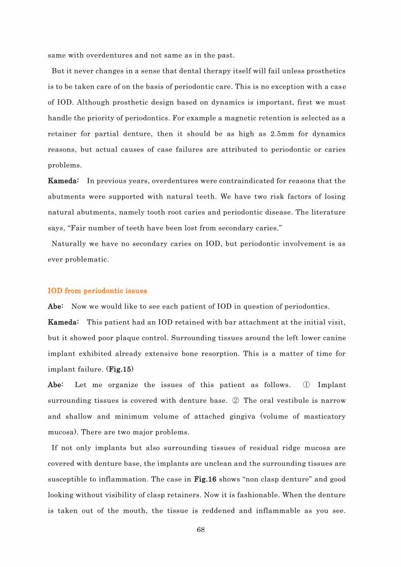
68
same with overdentures and not same as in the past.
But it never changes in a sense that dental therapy itself will fail unless prosthetics
is to be taken care of on the basis of periodontic care. This is no exception with a case
of IOD. Although prosthetic design based on dynamics is important, first we must
handle the priority of periodontics. For example a magnetic retention is selected as a
retainer for partial denture, then it should be as high as 2.5mm for dynamics
reasons, but actual causes of case failures are attributed to periodontic or caries
problems.
Kameda: In previous years, overdentures were contraindicated for reasons that the
abutments were supported with natural teeth. We have two risk factors of losing
natural abutments, namely tooth root caries and periodontic disease. The literature
says, “Fair number of teeth have been lost from secondary caries.”
Naturally we have no secondary caries on IOD, but periodontic involvement is as
ever problematic.
IOD from periodontic issues
Abe: Now we would like to see each patient of IOD in question of periodontics.
Kameda: This patient had an IOD retained with bar attachment at the initial visit,
but it showed poor plaque control. Surrounding tissues around the left lower canine
implant exhibited already extensive bone resorption. This is a matter of time for
implant failure. (Fig.15)
Abe: Let me organize the issues of this patient as follows. ① Implant
surrounding tissues is covered with denture base. ② The oral vestibule is narrow
and shallow and minimum volume of attached gingiva (volume of masticatory
mucosa). There are two major problems.
If not only implants but also surrounding tissues of residual ridge mucosa are
covered with denture base, the implants are unclean and the surrounding tissues are
susceptible to inflammation. The case in Fig.16 shows “non clasp denture” and good
looking without visibility of clasp retainers. Now it is fashionable. When the denture
is taken out of the mouth, the tissue is reddened and inflammable as you see.

69
Fundamental requirement of denture that should not touch the periodontal tissues
is lacking here. IOD, too, has no other ways than to cover the peri -implant tissues
with denture base.
Next, this case shows an implant failure indicated for extraction finally as the oral
vestibule was shallow and narrow to prevent brushing. (Fig.17) This case shows no
oral vestibule or attached gingiva, and the movable mucosa is proximally
neighboring to the implant. In this case you must pull open the lip for making room
of brush cleaning. Compared with anterior remaining natural teeth, cervical line
runs low in level and is difficult to accept the brushing. There are many difficult
conditions for cleaning in this case.
Fig.15 a~c Prognosis of IOD case with poor plaque control is difficult. (Kameda)
Fig.15 a~c At the initial visit, IOD was already inserted, but plaque was deposited
surrounding the implant with gingival inflammation. Radiograph c indicates bone
loss. It is difficult to design an open type base to IOD on an edentulous ridge as it
covers fully surrounding the implant in not only this case but also in any case. For
this reason, thorough and efficient control of plaque is definitely needed.

70
Fig.16 a, b “Principles of cleaning performance” viewed from prognosis of non clasp
denture. (Abe)
Fig.16a, b Ginigival inflammation caused from wearing non clasp denture.
Principle is that an abutment surrounding should not be covered with denture base
as much as possible in order to avoid periodontitis caused by denture. This kind of
denture is lacking in this sense. But IOD ought to be designed like this, and so
thorough attention should be made to cleaning performance.
Fig.17 Reasons for created difficulties of cleaning surrounding implants. (Abe)
Fig.17 The oral vestibule is so narrow and shallow that brushing cannot be
effective, finally leading to an implant failure in this case. Photo shows problems, ①
the oral vestibule is narrow and shallow, ② loss of attached gingiva, and ③ low
level of cervical line. Difficult conditions are already there for cleaning surrounding
implants.
In fact, a patient in this case is my hygienist‟s father and she devoted her efforts to
his plaque control, but finally the implants were forced for extraction at a dental

71
school hospital because pus discharge did not stop. In this regard, clinic
environment that can maintain both patient‟s self care and professional care must be
established. Otherwise future of IOD will be in danger.
Next we would like to see Dr.Kameda‟s case of Mini implant. (Fig.18) The drawback
of the implant body include, in addition to two drawbacks previously described, have
a shorter radicular attachment. A shorter radicular attachment has been long
complained for brushing difficulty.
In order to overcome this drawback, height of abutment has been becoming higher
gradually and so an inner crown like the height of conical crown has been developed.
It is clear that this inner crown type of abutment is easier for brushing. (Fig.19) As
this vertical height is increased, another dynamic problem will be raised. But as I
described it before, I think that lacking of plaque control should be prior to dynamic
problems and that as high attachment as possible should be used.
Maeda: It is a shame that prosthodontist including myself is limited in the
knowledge of periodontal care. At our prosthdotontics department, even at the time
of prosthetics therapy, a probe instrument is always near at hand in an instrument
tray, keeping in mind, “Thorough probing for periodontic treatment should be
verified for its necessity.” We think we are in the process of improving, although
some difficulties are still present.
Fig.18 Particular attention to Mini Implant is needed for cleaning performance.
(Kameda)
Fig.18 Mini Implant has the merit of minimal intervention, but necessary height
cannot be gained for attachment female part and hard to be cleaned.

72
Hard to be brushed. Easy to be brushed
Fig .19 Relation of radicular attachment height and brushing capacity. (Abe).
Fig.19 The chart shows relations of radicular attachment height and brushing
performance. Like most left ones with low radicular attachment, brush bristle ends
are not fixed well and hurt as the ends directly pushed inside the gingival pockets.
On the contrary, like most right ones, conical crown copings can help stabilize the
bristle ends and brush well and consequently reach to the cervical areas to clean .
Even so in fact, the IOD base coverage of abutments cannot be avoided although it
is true that cleaning performance is definitely limited. Therefore, unless patient can
clean for oneself, IOD is not indicated.
Under this assumption, I advise my patient to perform 3Ds. For example,
preventive medicine for periodontal disease is applied to the attachment retained
surface.
By the way, as Dr.Abe said previously, how high of radicular attachment should be

73
proper. One collaborative study joined with a preventive surgeon indicated a result,
that is, “Minimum height with easy cleaning under brushing pressure should be over
2 mm and more.” I keep this as a rule of more than 2 mm for a long time. And in case
of overdenture supported with natural support root length, I try to make it longer
enough to that measurement.
Abe: Background support from literature is important and IOD patients are likely
in advanced age with less dexterity of fingers and hands, and so it may be the higher
the better.
Kameda: Yes, it may be. I also have a sour memory when a magnetic attachment
was given to a coping without sufficient height, I advised the patient to brush
thoroughly, but this patient complained, saying, “Brush bristle ends hurt, and I
cannot brush well”. Low height coping and attachment cannot be cleaned by all
means before we discuss over cleaning performance on this attachment. We believe
we need to prepare good environment for patients to clean well. Also regular
cleaning performed by hygienists must be essential after insertion.
In this opportunity, let us discuss over cleaning method of attachments and
retainers. For example, how do you advise patients to clean a ball attachment?
Maeda: A cleaning brush is difficult to use. We think better to use clean gauze. A
ribbon-like gauze may fit well. No specific cleaning item is not available.
Can any post-operative periodontal risk with specific case be prospected in advance
at a pre-operative stage?
Kameda: In case of overdenture supported by natural tooth abutment, it is said
that, for a patient of strong caries tendencies, tooth loss is attributed to secondary
caries even after wearing overdenture. IOD is caries free as it is, but is there any
risk of periodontal disease to an IOD patient even after tooth loss from periodontal
disease?
Maeda: Yes, there is. It is reported recently that not only IOD but also implant
insertion case of 10 years or 15 years happened to start bone resorption suddenly
within last years. Those patients have been traced back of disease history to indicate

74
the tooth loss originally from severely advanced periodontal disease, although the
causes were not all of them.
In other words, it is not exactly wrong to prospect future disease depending
whether the patient is of caries type or of periodontal disease type. Therefore, IOD is
on a patient of tooth loss from periodontal disease, there may be higher risk of
peri-implantitis or bone resorption than a caries type patient, and this must be
prepared.
Abe: I hear that, even after becoming edentulous, P.g. is still there inactively.
Kameda: In previous years, total tooth extraction would clear out A.a. or P.g., but
they are reportedly found in the back of tongue or buccal mucosa even in edentulism.
At the stage of denture insertion following tooth extraction, is there any procedure of
sterile filtration of pathogens that are of high risk of periodontal disease?
Maeda: On the contrary, it is not yet effective. Those attached may be part of
microorganism resident inhabitants, and if they are forced to change, others will
grow like microbial substitution. But when microbial kind is established at early
stage, specific sterilization on this microbe is reported recently. Any possible support
will be necessary.
How to deal with need of nursing care.
Kameda: IOD patients are at any rate of advanced age, and there may be situations
when they will be possibly bedridden in the near future or contraindicated to surgery
procedure. What do you think, Prof.Maeda?
Maeda: This story is not only about IOD but also about any implant therapy. It may
be difficult for neither dental maintenance nor implant extraction is available. Even
IOD wearer happens to become in need of nursing care, there may be another
problem raised.
But as far as IOD is concerned, there may be small possibility of coping with such a
problem. Because IOD is, in comparison with various prosthetic means of
implantation, to deal with changes easily as its implant number is small. And also
its basis of removability is easy for cleaning. Even if a patient is in need of nursing

75
care, its maintenance is taken care of similarly with maintenance of conventional
denture.
In my opinion, those patients in need of nursing care will be difficult for cleaning,
and IOD will need additional care to control plaque. But not all cases happen to be
involved with inflammation and to permit rapid changes. There may drastic change
occur when some unknown factor extra is added. Some systemic change may change
metabolism to induce rapid bone resorption according to my feeling.
Kameda: The aging society is not easy with full of mixed problems. (laughter)
Maeda: This morning I already visited a hospital and treated an old aged patient
there. More number of patients has more remaining teeth than before. Thanks to the
“8020” Movements, which are grateful both to dentists and patients.
Another reality also has that a patient with multiple remaining teeth turns out in
need of nursing care with extensive of carious lesions, but fails to communicate in
vain for treating. In this sense, only remaining teeth are not always good. Yes, losing
teeth is not welcome, or denture trouble is not welcome, either. Even more troubled
is maintenance for the remaining teeth.
Abe: No conclusion may be reached whether we accept implants for present days in
healthy and happiness, or we do not accept it for future safety.
Kameda: Now in this age, I think it is important to discuss over these realities as
dentists.
Summary
- In case of IOD, regular checkup and maintenance on implant abutments and
denture base are essential. (Maeda)
- Not only IOD but also every prosthetic treatment should be based on periodontal
cares. Establishment of healthy surrounding tissues will contribute to long-term
stability of prostheses. (Abe)
- In case of IOD, morphology should be given to accommodate easy cleaning for
patient oneself, and regular cleaning by hygienists is needed. (Kameda)

76
8. Future of IOD
In case of partial missing arch, IOD be given with Minimal Intervention
Kameda: As for future outlook of IOD, how will it be developed and promoted in
your opinion?
Maeda: Currently an implant therapy performed on an edentulous jaw would
design fixed implant superstructure very frequently. But in future, as I described
before, minimum number of implants will be placed for gradual improvement,
increasing implant number depending on patient‟s personal wish. This way of
treatment will be valid with thanks to IOD.
And in case of dentate jaw, in order to control increasing number of missing teeth
starting from the stage of free-end arch, future outlook will include the application
of IOD as a strategic implant prosthetics for preventing expansion of tooth loss. Of
course, in order to inhibit defect expansion, fixed type implant structure can be an
alternative option, too. But this modality requires more numbers of implants and the
ridge bone augmentation needs more extensively. I personally am free to advocate,
“Minimal intervention be promoted with implants”. (laughter) Simply speaking, this
is a way of idea to convert a free-end denture to an intermediate denture. (Refer to a
column at page 34.)
Background of this idea is derived from the fact of recent years that implant
prosthetics accompanied with large scale of bone augmentation has been presented
luxuriously, but the truth is that no bone is created at the site of no reason for bone
growth.
As a result, there seems to be many people are becoming to realize that removable
type of prosthesis like IOD is better for restoration and is easier to cope with
patient‟s fundamental desire. Future will be prospected in this direction.
Abe: It would be necessary for those who view from restoring with implants to
visualize from another angle of restoring with dentures. Those viewers in the past
were extremely deviated giving weight on implants. This will have to be neutralized
in the future.

77
Kameda: From the dental practitioner‟s view point, there are overwhelmingly more
patients of partial missing teeth than edentulous patients. So if we could reach a
certain amount of consensus as practitioners when detailed studies are conducted in
this regard at dental schools. For example, implant length or diameter is not yet
established in consensus, taking time before conclusion. How progressive in dental
schools in Japan regarding IOD studies like above.
Maeda: As I demonstrated it before, IOD‟s basic, dynamic and biomechanical
studies have been conducted extensively, including myself. In other words,
theoretical studies are fairly well advanced, such as, “Where to be implanted, how to
be developed”. But here in Japan, clinical researches are still a few, and multiple
long-term results are not available. This differs largely from other countries.
Clinical cases themselves are not presented well.
Kameda: Is IOD introduced into school education now?
Maeda: As far as dental schools are concerned, dental implants are taught at
almost all dental schools among totaling 29 schools nationwide, but IOD may not be
included yet. Osaka University has a course of implants joined with crown and
bridge restorations, and, from last year for the first time, theory and practice of
complete denture course is followed by IOD, which is converted from the previous
complete denture. This is an idea to invite students to think over an implant as a
new current source coming from complete denture.
Summary
- Using the advantage of ideas on IOD, a strategic treatment is possible for
controlling expansion of missing arch. (Maeda)
- IOD is demanded in the aging society. The key to success is the combination of
implants with periodontics and denture prosthetics. (Abe)
- From now in Japan, IOD application is expecting in the partial missing arch.
(Kameda)

78
Conclusion of our discussion
Kameda: We are now concluding our discussion. To summarize, could we have your
comments for readers?
Maeda: In summary, as for the case of mandibular edentulous arch, as you both
suggested mainly in the last part, so called a difficult case where the mandibular
edentulous ridge resorption will be best opted. When a complete denture does not
work well, any implant presence will make a considerable difference.
And as I mentioned in the last chapter, IOD is potentially possible to apply not only
to an edentulous ridge but also to a partially missing arch. In whatever the case is,
most prerequisites are basic knowledge and skills of complete denture construction,
and we would be happy if we could covey proper information to our readers.
Abe: I have the same idea. I sincerely hope that our readers would appreciate
implant therapy from a basis of complete denture therapy. Again my emphasis is on
periodontal care that has to go together. If these two are not taken care of rightly as
a dentist, a patient will be in disaster as a consequence. And we wish to our readers
that these two basics are learned well and practiced successfully.
Kameda: Today we think we have been able to deepen our discussion about IOD
that is attracting both overseas and domestic attention. When general idea of
implantation is combined with denture construction, specific cautions of IOD have
been highlighted. In contrast, facts are about many unresolved issues. We are
looking forward to new researches continued for years to come. Thanks for your
attendance.
(end of discussion)
【Column】
Suggestions to Minimal intervention (MI) procedures with dental implants
In case of mandibular distal extension missing case with occlusion effective to the
second premolars, concept of shortened dental arch is valid and so conditions are
comparatively stable. But once any premolar bone support is deteriorated or lost, the

79
circumstances will become destabilized immediately.
Fig.30 at page 30 of last issue shows research results based on stresses loaded on
TMJ regions of computer simulated models depending on different occlusal support
following various prosthetic options, once they are lost like above. In case of fixed
type prostheses supported with implants, it is known that occlusal support can be
restored effectively equivalent with those of the second molars. On the other hand,
only with mucosal born prostheses, larger stress will be loaded, but this stress will
be eased when implants are used underneath the base for support. This is an
effective idea of conversion of intermediate denture with the help of implants.
If this idea is used as a strategic plan, as shown in Fig.20, an implant is inserted in
the molar region if the case becomes a free-end missing arch in order to control
another tooth loss and to convert it an intermediate denture. And even if the tooth
loss becomes extensive unfortunately, another implant is added like one in the
anterior and one in the molar regions together with denture base, totaling 4 pieces
on the both sides to secure the occlusal support.
This idea is not new, and reportedly another implant is inserted under a free-end
saddle. It was started around in 1993 and is increasing in these two years. This,
however, is being ready for the worst final case, and it is important to conserve
remaining teeth with conversion of intermediate denture from lateral free-end
saddle.
Fig.20 Implant application sequence in order to control increasing missing arch.
Fig.20 Concept of controlling expansion of missing arch by using implant and
denture jointly. In case of distal extension missing case with premolar tooth loss, an
implant is inserted in the distal region strategically and preventively, and it can be
converted to an intermediate missing arch. If tooth loss is extended to the anterior

80
arch, another implant is in the canine region for support in order to control
expansion of tooth loss and bone loss.
(end)


![A Systematic Approach to Problem Solving for Elderly Patients … · adapt to new complete dentures particularly the mandibular denture [17,18]. Nearly 50% will have been edentulous](https://static.fdocuments.net/doc/165x107/5e855f128b3d144fe7698422/a-systematic-approach-to-problem-solving-for-elderly-patients-adapt-to-new-complete.jpg)

