Silk fibroin/nanohydroxyapatite porous scaffolds for bone ...
RESEARCH Open Access
Impact of nanohydroxyapatite on enamelsurface roughness and color change afterorthodontic debondingShabnam Ajami, Hamid Reza Pakshir and Neda Babanouri*
Abstract
Background: The aim of this prospective in vitro study was to evaluate the effect of nanohydroxyapatite (nanoHAP)serum on the enamel surface roughness and tooth color stability after orthodontic debonding procedure.
Methods: The crowns of 30 premolars were embedded in acrylic blocks with a 4 mm× 5-mm-sized window on themiddle third of buccal surfaces. Primary roughness values were evaluated by an atomic force microscope (AFM). Afterbracket debonding, and polishing procedures, the second roughness parameters were recorded. Specimens were thenrandomly assigned to two equal groups. NanoHAP serum and HAP toothpaste were applied for 10 days in the first andsecond groups, respectively. Then, after the third AFM, initial color parameters were measured. Following 1-weekimmersion in the coffee solution, second color assessment was performed. The fourth AFM was registered after2 months of aging process.
Results: All roughness parameters were elevated following debonding procedure. There was no statisticallysignificant reduction in roughness parameters after 10 days of nanoHAP serum or HAP toothpaste application.Both groups showed significant color change after immersion in the coffee solution.
Conclusions: NanoHAP serum with the protocols used in this study could not restore enamel surfaces to theiroriginal condition.
Keywords: Nanohydroxyapatite, Surface roughness, Debonding
BackgroundThe final procedure in the fixed orthodontic treatment isthe removal of bonded attachments on the teeth andrestoring the enamel surface as closely as possible to theoriginal pretreatment state. Bonding and debondingprocedures can cause irreversible changes on the enamelsurface [1, 2], which are more important when they occuron the most resistant outer layer. These potentialalterations involve up to 55 μm enamel loss [3], increasedsurface roughness [1, 2], and, therefore, more susceptibil-ity to demineralization and discoloration [4].Many researchers have evaluated a variety of techniques
for bracket debonding, resin removal, and subsequentenamel surface polishing [1, 2, 5–7]. These techniquesinclude using scalers or band-removing pliers [8], different
types of tungsten carbide burs in low- or high-speed hand-piece followed by water slurry of pumice [9, 10], ultrasonicapplication and sandblasting techniques [5, 6], fiber-reinforced composite bur [11], Sof-Lex discs [1, 2], andalso laser energy to degrade the bonding resin [12]. None-theless, there is still no universally accepted method forthis potentially conflicting stage of treatment [2].Moreover, enamel is a non-living tissue which is
mainly (97 %) composed of inorganic apatite [13, 14].In contrast to other hard tissues like bone and dentin, theenamel cannot restore itself [13, 14]. Therefore, syntheticapatites like conventional hydroxyapatite (cHAP) andamorphous calcium phosphate (ACP) have been suggestedto repair the damaged enamel due to their close chemicalsimilarities to the enamel structure [15, 16]. These mater-ial particles often show different dimensions, morpho-logical characteristics, and orientations from the enamelsubunit structures. Therefore, their repairing properties
* Correspondence: [email protected] Research Center, Shiraz University of Medical Science, Shiraz,Iran
© 2016 Ajami et al. Open Access This article is distributed under the terms of the Creative Commons Attribution 4.0International License (http://creativecommons.org/licenses/by/4.0/), which permits unrestricted use, distribution, andreproduction in any medium, provided you give appropriate credit to the original author(s) and the source, provide a link tothe Creative Commons license, and indicate if changes were made.
Ajami et al. Progress in Orthodontics (2016) 17:11 DOI 10.1186/s40510-016-0124-2

like adsorption to the enamel surface and mechanicalstrength are compromised [15, 16].Enamel is made of 20–40-nm-sized particles of hydroxy-
apatite (HAP) [17, 18]. It has been suggested that 20-nm-sized HAP termed as nanohydroxyapatite (nanoHAP) isthe most biocompatible and bioactive form of the synthe-sized apatites, due to the close similarity to the basicstructures of the enamel [15, 16]. NanoHAP has a highersurface area and strong affinity to the enamel surface.These characteristics facilitate its attachment to the en-amel surface compared to the large and amorphoustypes [15, 16]. Interestingly, a layer of nanoHAP formedon the enamel surface is highly resistant to acid solu-tion that subsequently can protect underlying enamelfrom the future demineralization [15]. Several studies[19–22] have evaluated the effect of nanoHAP on theenamel and dentin remineralization and its preventivepotential on dental demineralization. It has been alsosuggested that the nanoHAP particles could repairbleaching-related microscopic defects of the enameland thereby improving the post-bleaching sensitivities[23]. Some research teams and manufacturers haveproposed that nanoHAP could reduce the enamel sur-face roughness [24, 25].As already noted, enamel surface damage is an inevit-
able sequela of the orthodontic debonding procedure.Moreover, evaluating the potential effects of nanoHAPin restoration of the enamel surface after orthodonticattachment debondings would be crucial for its clinicalapplications. Hence, the objective of the current in vitrostudy was to assess the hypothesis that nanoHAP couldsignificantly reduce the enamel surface roughness andthe possible discoloration after debonding and polishingprocedures.
MethodsSample preparationThirty human premolars extracted for orthodonticpurposes were selected in 1 month prior to the study. Aconsent form was signed by each patient, in which itwas thoroughly depicted that the enamel surfaces of theteeth extracted for orthodontic treatment would be usedin our investigation.The selection criteria were the absence of cracks,
hypoplastic or carious lesions, and restorations on thebuccal surfaces of the teeth. The samples were cleanedand stored in distilled water at room temperature. Thedistilled water was changed weekly to prevent bacterialcolonization [2]. Roots of the teeth were removed at thecementoenamel junction by a diamond bur operated inhigh-speed underwater and air cooling. A rectangular pieceof black adhesive tapes (4 mm× 5 mm), corresponding tothe bracket bases, was adhered on the middle third of thebuccal surface. The crowns were embedded horizontally in
the self-cure acrylic resin blocks (Duralay; Reliance DentalC., Worth, IL, USA) in a way that the tape areas wereexposed. After the tapes were removed, the exposed en-amel windows were cleaned and polished with a low-speedrubber cup and slurry of non-flouridated pumice, washedfor 30 s, and dried for 10 s with oil-free air spray.
Surface roughness assessmentAfter coding the samples, they were subjected to theatomic force microscope (AFM) analysis (NanoWizard®II; JPK Instrument AG, Berlin, Germany) to assess initialsurface roughness (T0) (Fig. 1). The AFM analysis wasperformed in the contact mode to make the topographicimage from the surface (Fig. 2). The instrument wascoupled with a scanner having a maximum range of100 μm× 100 μm× 15 μm in x, y, and z directions,respectively. Images were acquired at a resolution of512 × 512 pixels, line rate of 1 Hz, and 10 μm scan size.Three different points were analyzed on the center of eachenamel window, and the mean value of these three recod-ing were used for statistical analysis. A silicone cantilever(HQ: CSC17 MikroMasch®Erope; NanoAndMore GmbH,Wetzlar, Germany) at resonance frequency of 13 kHz witha force constant of 0.18 N/m was used to conducttopographic analysis of the enamel surfaces. Three rough-ness parameters were registered in nanometers as follows:
Average roughness value (Ra): the arithmetic mean ofthe height of peaks and depth of the valleys from amean line. This parameter describes the overall surfaceroughness.Root mean square roughness (Rq): the heightdistribution relative to the mean line.Rz: the average maximum peak-to-valley height of fivesuccessive sample lengths.
Bonding, debonding, and resin removalAfter initial evaluation, all teeth were subjected to a 37 %phosphoric acid gel (Etching agent; Reliance, Itasca, IL,USA) for 30 s, thoroughly rinsed and dried with oil-free airspray for 15 s. A thin layer of primer (Transbond XT; 3MUnitek, Monrovia, CA, USA) was applied on the etchedsurfaces, and adhesive resin was placed on a stainless steelstandard edgewise premolar bracket bases (Dentaurum;Ispingen, Germany). The brackets were placed on the en-amel windows, firmly pressed in place, and excessive resinwas removed and light cured for 10 s from each edge ofthe brackets, for a total exposure time of 40 s (Fig. 1).The samples were stored in distilled water at room
temperature for 24 h to ensure complete resinpolymerization. Brackets were peeled off with a handplier by gently squeezing the mesial and distal wingstogether. Remnant resin was removed with 12-flutedtungsten carbide bur (Dentaurum no. 123-604; Ispingen,
Ajami et al. Progress in Orthodontics (2016) 17:11 Page 2 of 8

Germany), operated in low-speed and air cooling,followed by a PoGo polisher (Dentsply Caulk, Milford,DE, USA), and finally polished with a rubber cup andslurry of fine pumice for 10 s. Complete removal ofresidual resin was confirmed by visual inspection under
the light of a dental operating lamp and ×10 magnifica-tion. All of the above procedures on all samples wereperformed by an orthodontist, and a new bur, polisher,and rubber cup were used for each sample. Aftercompletion of the cleanup procedures, the second AFM
Fig. 2 a 2D and b 3D AFM images (T0)
Fig. 1 Sample preparation and AFM analysis. A sample prior to preparation (a). A rectangular tape to protect the buccal surface before bracketbonding (b). A sample with a bonded bracket prior to debonding procedure (c). AFM analysis of the sample (d)
Ajami et al. Progress in Orthodontics (2016) 17:11 Page 3 of 8

was taken to register roughness parameters (T1). Then,the teeth were randomly assigned to two equal groups(n = 15) using the table of random numbers. In the firstgroup, a high concentration nanoHAP serum (n-HAPRepairing Serum; PrevDent International BV, Netherlands)was applied on the enamel surface for 2–3 min by thesponge at the head of the tubes (Fig. 1) and rinsed after20 min with water. The process was performed once a dayfor 10 days according to the manufacturer’s instructions.Specimens in the HAP toothpaste group were manuallybrushed with horizontal technique for 20 s, using anew soft brush for each sample and HAP-containedtoothpaste (Signal Expert Protection; Unilever France,Rueil-Malmaison Cedex, France), daily during these10 days. Thereafter, the third AFM evaluation of rough-ness parameters (T2) was obtained blindly by a secondinvestigator.
Color stability assessmentUsing a spectrophotometer (VITA Easyshade Advance 4.0;VITA Zahnfabrik H, Rauter Gmbh & Co., Bad Säckingen,Germany), both study groups were subjected to the spec-trophotometric assessment to measure the primary colorof the enamel surface. Color quantification was accordingto the CIE lab system (Commision Internatinale de l’Eclai-rage, L*, a*, b*) presented in Table 1. Before color assess-ment, all samples were cleaned with the soft brush andwater for 10 s and dried with absorbent paper. The spec-trophotometer was calibrated before each imaging with thewhite pad supplied by the manufacturer. The spectro-photometric evaluation of each sample was recorded twoconsecutive times. When the total color difference (ΔE) oftwo measurements did not exceed the threshold of 1 ΔEunit, the mean value of these measurements was used forstatistical analysis. Measurements with ΔE > 1 werediscarded, and new evaluation were performed a sec-ond time. The color measurements were repeated after1-week immersion of all samples in the coffee solutionat room temperature. The coffee solution was preparedby 15 g coffee (Nescafé Blend 43; Nestlé Australia ltd,NSW, Australia) in 500 mL boiling water and changedeveryday [26]. All measurements were performed in thesame room blindly by one investigator, while the sam-ples were placed over a white paper to provide white
background. The CIE values (L*, a*, b*) were obtained,and the data were analyzed regarding their lightnessand chromaticity values (ΔL, Δa, Δb). The total colorchange (ΔE) between two intervals was calculatedaccording to the following equation:
ΔE ¼ ΔLð Þ2 þ Δað Þ2 þ Δbð Þ2� �1=2
After color evaluation, both groups were stored in thedistilled water, weekly changed, at the room temperaturefor additional 2 months, and finally subjected to AFManalysis (T3) to assess long-term effect of nanoHAP.
Statistical analysisStatistical analysis was performed with the StatisticalPackage for Social Sciences (Version 15.0, SPSS Inc.,Chicago, IL, USA). The assumption of normality wasinvestigated by Kolmogorov-Smirnov test. Data forroughness parameters were statistically evaluated with re-peated measurements analysis of variance (RM-ANOVA).Changes in color parameters (L*, a*, b*) in each groupwere investigated by paired sample t test. Student’s t testwas further used for evaluating roughness, differences ofthe total color change (ΔE), and also (ΔL*, Δa*, Δb*)among the groups. Significance was set at probabilityvalue of P < 0.05 for all tests.
ResultsThe RM-ANOVA showed that there was no significantinteraction effect between treatment and time variablesfor all roughness parameters (all P > 0.05). Figures 3, 4,and 5 depict the mean profile changes in Ra, Rq, and Rz
throughout the treatment intervals, respectively.The results of the t test indicated that there was no
significant difference between the two study groups inall treatment intervals. The results revealed irreversibleenamel surface changes in both study groups followingdebonding procedures. All roughness parameters in-creased significantly from T0 to T1 in both experimentalgroups (both P < 0.001), whereas there were nostatistically significant differences between T1, T2, andT3 (Table 2).The results of color assessment showed that L* param-
eter significantly decreased after immersion in the coffeesolution in both nanoHAP group and HAP toothpastegroup (P = 0.004 and 0.001, respectively). There was nosignificant change in a* whereas b* increased statisticallysignificant (P < 0.001) in both groups (Table 3).Theresults indicated that there were significant differencesin the average values of Δa* and Δb* between groups.No significant difference in terms of total color change(ΔE) was indicated between study groups, although thisdifference was very close to the adopted significant level
Table 1 CIE color system
Colorparameter
Definition Color range
L* Measure of lightness (value) Ranging from black (0)to white (100)
a* The position on red-green axis Ranging from red (+)to green (−)
b* The position on yellow-blue axis Ranging from yellow (+)to blue (−)
Ajami et al. Progress in Orthodontics (2016) 17:11 Page 4 of 8

(P = 0.06). The clinical significance of the color changeswas determined by comparing the differences in colorvalues with the proposed standard value of clinicaldetection which normally is set to 3.7 ΔE units [27]. Itwas found that 80.0 % of the teeth in the HAP tooth-paste group had shown visible and clinically significantcolor changes while only 46.7 % of teeth in thenanoHAP group had demonstrated visible changes.When considering the number of teeth with visible colorchange, difference among study groups tended to be sig-nificant (P = 0.058).
DiscussionThe substantial clinical significance of outer layer ofenamel brings imperative concern over orthodonticbonding and debonding procedures. The hard layer withhigh content of minerals and fluoride protects theunderlying enamel from organic acids produced in den-tal microbial plaque [28]. In addition, increased enamelsurface roughness after debonding results in increasedaccumulation of the pigments, retention of bacterialplaque, and decalcification, both situation that may causeesthetic problems [29, 30]. Previous studies reportedinevitable enamel alterations including enamel loss [3, 31],increased surface roughness [1, 2], and discoloration[32, 33], following orthodontic treatments.Benefiting from various methods or biomaterials that
help restore enamel surface to the original condition iscrucial. Receiving more interest in dental practice, nano-HAP with its chemical and structural similarity to theenamel inorganic structure, has shown to be superior tothe conventional synthetic HAP in repairing damagedenamel [15]. The nanoscale HAP exhibits high surfaceenergy and strong affinity to the enamel surface [15, 16].In the present study, the effects of nanoHAP on theenamel surface roughness and color stability after ortho-dontic debonding were investigated and compared withthe HAP-contained toothpaste.The results of the current study showed that all the
roughness variables elevated significantly following thedebonding and polishing procedures which was in ac-cordance to the results yielded by previous studies [1, 2].The results also revealed that 10-day application of thenanoHAP serum did not significantly reduce the enamelsurface roughness. There was no difference between the
Fig. 3 Results of average roughness (Ra)
Fig. 4 Results of root mean square roughness (Rq)
Fig. 5 Results of maximum peak-to-valley height (Rz)
Ajami et al. Progress in Orthodontics (2016) 17:11 Page 5 of 8
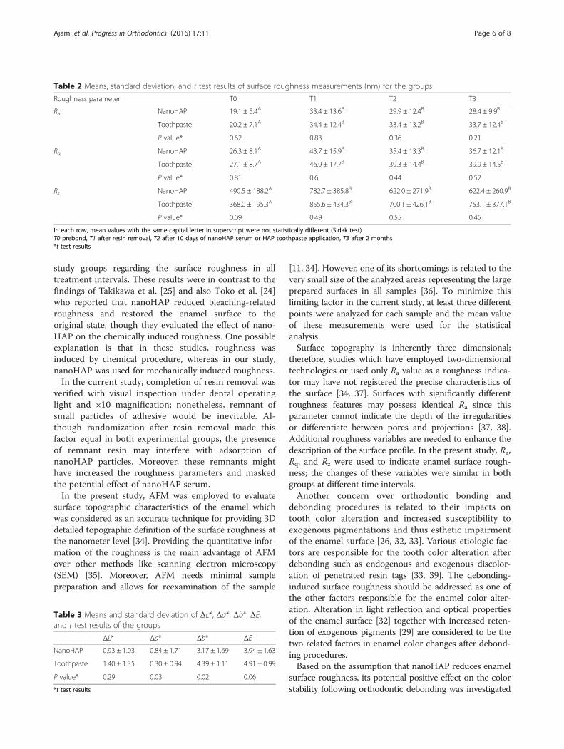
study groups regarding the surface roughness in alltreatment intervals. These results were in contrast to thefindings of Takikawa et al. [25] and also Toko et al. [24]who reported that nanoHAP reduced bleaching-relatedroughness and restored the enamel surface to theoriginal state, though they evaluated the effect of nano-HAP on the chemically induced roughness. One possibleexplanation is that in these studies, roughness wasinduced by chemical procedure, whereas in our study,nanoHAP was used for mechanically induced roughness.In the current study, completion of resin removal was
verified with visual inspection under dental operatinglight and ×10 magnification; nonetheless, remnant ofsmall particles of adhesive would be inevitable. Al-though randomization after resin removal made thisfactor equal in both experimental groups, the presenceof remnant resin may interfere with adsorption ofnanoHAP particles. Moreover, these remnants mighthave increased the roughness parameters and maskedthe potential effect of nanoHAP serum.In the present study, AFM was employed to evaluate
surface topographic characteristics of the enamel whichwas considered as an accurate technique for providing 3Ddetailed topographic definition of the surface roughness atthe nanometer level [34]. Providing the quantitative infor-mation of the roughness is the main advantage of AFMover other methods like scanning electron microscopy(SEM) [35]. Moreover, AFM needs minimal samplepreparation and allows for reexamination of the sample
[11, 34]. However, one of its shortcomings is related to thevery small size of the analyzed areas representing the largeprepared surfaces in all samples [36]. To minimize thislimiting factor in the current study, at least three differentpoints were analyzed for each sample and the mean valueof these measurements were used for the statisticalanalysis.Surface topography is inherently three dimensional;
therefore, studies which have employed two-dimensionaltechnologies or used only Ra value as a roughness indica-tor may have not registered the precise characteristics ofthe surface [34, 37]. Surfaces with significantly differentroughness features may possess identical Ra since thisparameter cannot indicate the depth of the irregularitiesor differentiate between pores and projections [37, 38].Additional roughness variables are needed to enhance thedescription of the surface profile. In the present study, Ra,Rq, and Rz were used to indicate enamel surface rough-ness; the changes of these variables were similar in bothgroups at different time intervals.Another concern over orthodontic bonding and
debonding procedures is related to their impacts ontooth color alteration and increased susceptibility toexogenous pigmentations and thus esthetic impairmentof the enamel surface [26, 32, 33]. Various etiologic fac-tors are responsible for the tooth color alteration afterdebonding such as endogenous and exogenous discolor-ation of penetrated resin tags [33, 39]. The debonding-induced surface roughness should be addressed as one ofthe other factors responsible for the enamel color alter-ation. Alteration in light reflection and optical propertiesof the enamel surface [32] together with increased reten-tion of exogenous pigments [29] are considered to be thetwo related factors in enamel color changes after debond-ing procedures.Based on the assumption that nanoHAP reduces enamel
surface roughness, its potential positive effect on the colorstability following orthodontic debonding was investigated
Table 2 Means, standard deviation, and t test results of surface roughness measurements (nm) for the groups
Roughness parameter T0 T1 T2 T3
Ra NanoHAP 19.1 ± 5.4A 33.4 ± 13.6B 29.9 ± 12.4B 28.4 ± 9.9B
Toothpaste 20.2 ± 7.1A 34.4 ± 12.4B 33.4 ± 13.2B 33.7 ± 12.4B
P value* 0.62 0.83 0.36 0.21
Rq NanoHAP 26.3 ± 8.1A 43.7 ± 15.9B 35.4 ± 13.3B 36.7 ± 12.1B
Toothpaste 27.1 ± 8.7A 46.9 ± 17.7B 39.3 ± 14.4B 39.9 ± 14.5B
P value* 0.81 0.6 0.44 0.52
Rz NanoHAP 490.5 ± 188.2A 782.7 ± 385.8B 622.0 ± 271.9B 622.4 ± 260.9B
Toothpaste 368.0 ± 195.3A 855.6 ± 434.3B 700.1 ± 426.1B 753.1 ± 377.1B
P value* 0.09 0.49 0.55 0.45
In each row, mean values with the same capital letter in superscript were not statistically different (Sidak test)T0 prebond, T1 after resin removal, T2 after 10 days of nanoHAP serum or HAP toothpaste application, T3 after 2 months*t test results
Table 3 Means and standard deviation of ΔL*, Δa*, Δb*, ΔE,and t test results of the groups
ΔL* Δa* Δb* ΔE
NanoHAP 0.93 ± 1.03 0.84 ± 1.71 3.17 ± 1.69 3.94 ± 1.63
Toothpaste 1.40 ± 1.35 0.30 ± 0.94 4.39 ± 1.11 4.91 ± 0.99
P value* 0.29 0.03 0.02 0.06
*t test results
Ajami et al. Progress in Orthodontics (2016) 17:11 Page 6 of 8

in the present study. Our results showed that 10-day appli-cation of nanoHAP serum did not significantly reduce theamount of enamel color discolorations compared to theHAP toothpaste group. This finding was in accordancewith the outcome surface roughness analysis. In addition,the absorption of coffee pigments by remnant resin tagsmay outweigh any probable differences in enamel surfacecharacteristics. Both groups in this investigation indicatedsignificant change in L*, a*, and b* following 1-weekimmersion in coffee solution. Generally, the mean L* valuedecreased while the mean b* increased in both groupsrepresenting darker and more yellowish color for the teeth.The mean values of total color change (ΔE) were greaterthan 3.7 units, a standard value of clinical detection [27],in both groups. However, differences between the groupswith respect to the mean ΔE value and also the number ofteeth which exceeded this threshold value (ΔE > 3.7)tended to be significantly lower in the nanoHAP group.With a larger sample size, the color parameters may reachthe significant level.In the present study, we found that nanoHAP serum
had no significant effect on debonding-induced enamelsurface roughness and tooth color stability. It could behypothesized that for the mechanically induced rough-ness, nanoHAP with higher concentrations or enduredapplication would be more effective. This opinion can beexamined in a future study. It may also be interesting toinvestigate the effect of nanoHAP on the mechanicallyinduced roughness without the adhesive resin as aninterfering factor.
ConclusionsIt could be concluded that orthodontic bonding anddebonding procedures increased enamel surface rough-ness. The nanoHAP serum and HAP toothpaste testedin the present study could not reduce enamel surfaceroughness parameters to restore enamel to its originalroughness condition.Although there were fewer teeth with clinically visible
color change in the nanoHAP group, 10-day applicationof nanoHAP could not improve enamel color stabilityafter orthodontic debonding.
Competing interestsThe authors declare that they have no competing interests.
Authors’ contributionsAll authors contributed in most aspects of the study. The detailedcontributions are as follows: SHA proposed the concept of the research. SHA,HRP, NB carried out the definition of intellectual content, research design,experimental conduct, and manuscript draft preparation. NB conducted thedata acquisition and analysis. All authors read and approved the finalmanuscript.
Received: 11 January 2016 Accepted: 1 March 2016
References1. Eliades T, Gioka C, Eliades G, Makou M. Enamel surface roughness following
debonding using two resin grinding methods. Eur J Orthod. 2004;26:333–8.doi:10.1093/ejo/26.3.333.
2. Özer T, Başaran G, Kama JD. Surface roughness of the restored enamel afterorthodontic treatment. Am J Orthod Dentofac Orthop. 2010;137:368–74.doi:10.1016/j.ajodo.2008.02.025.
3. Arhun N, Arman A. Effects of orthodontic mechanics on tooth enamel: areview. Semin Orthod. 2007;13:281–91. doi:10.1053/j.sodo.2007.08.009.
4. Quirynen M, Bollen CM. The influence of surface roughness and surface-freeenergy on supra- and subgingival plaque formation in man. A review of theliterature. J Clin Periodontol. 1995;22:1–14. doi:10.1111/j.1600-051X.1995.tb01765.x.
5. Krell KV, Courey JM, Bishara SE. Orthodontic bracket removal usingconventional and ultrasonic debonding techniques, enamel loss, and timerequirements. Am J Orthod Dentofac Orthop. 1993;103:258–66.
6. Kim S-S, Park W-K, Son W-S, Ahn H-S, Ro J-H, Kim Y-D. Enamel surfaceevaluation after removal of orthodontic composite remnants by intraoralsandblasting: a 3-dimensional surface profilometry study. Am J OrthodDentofac Orthop. 2007;132:71–6.
7. Pithon MM, Santos Fonseca Figueiredo D, Oliveira DD, Coqueiro Rda S.What is the best method for debonding metallic brackets from the patient’sperspective? Prog Orthod. 2015;16:1–6. doi:10.1186/s40510-015-0088-7.
8. Rouleau BD, Marshall GW, Cooley RO. Enamel surface evaluations afterclinical treatment and removal of orthodontic brackets. Am J Orthod. 1982;81:423–6. doi:10.1016/0002-9416(82)90081-1.
9. Campbell PM. Enamel surfaces after orthodontic bracket debonding.Angle Orthod. 1995;65:103–10. doi:10.1043/0003-3219(1995)065<0103:ESAOBD>2.0.CO;2.
10. Zachrisson BU, Arthun J. Enamel surface appearance after variousdebonding techniques. Am J Orthod. 1979;75:121–7. doi:10.1016/0002-9416(79)90181-7.
11. Karan S, Kircelli BH, Tasdelen B. Enamel surface roughness after debonding.Angle Orthod. 2010;80:1081–8. doi:10.2319/012610-55.1.
12. Hayakawa K. Nd: YAG laser for debonding ceramic orthodontic brackets.Am J Orthod Dentofacial Orthop. 2005;128:638–47. doi:10.1016/j.ajodo.2005.03.018.
13. Dorozhkin SV, Epple M. Biological and medical significance of calciumphosphates. Angew Chemie Int Ed. 2002;41:3130–46. doi:10.1002/1521-3773(20020902)41. 17<3130::AID-ANIE3130>3.0.CO;2-1.
14. Busch S. Regeneration of human tooth enamel. Angew Chemie Int Ed.2004;43:1428–31. doi:10.1002/anie.200352183.
15. Li L, Pan H, Tao J, Xu X, Mao C, Gu X, et al. Repair of enamel by usinghydroxyapatite nanoparticles as the building blocks. J Mater Chem. 2008;18:4079. doi:10.1039/b806090h.
16. Lin K, Wu C, Chang J. Advances in synthesis of calcium phosphate crystalswith controlled size and shape. Acta Biomater. 2014;10:4071–102. doi:10.1016/j.actbio.2014.06.017.
17. Tang R, Wang L, Orme CA, Bonstein T, Bush PJ, Nancollas GH. Dissolution atthe nanoscale: self-preservation of biominerals. Angew Chemie. 2004;116:2751–5. doi:10.1002/ange.200353652.
18. Robinson C, Connell S, Kirkham J, Shore R, Smith A. Dental enamel-abiological ceramic: regular substructures in enamel hydroxyapatite crystalsrevealed by atomic force microscopy. J Mater Chem. 2004;14:2242–8.doi:10.1039/B401154F.
19. Tschoppe P, Zandim DL, Martus P, Kielbassa AM. Enamel and dentineremineralization by nano-hydroxyapatite toothpastes. J Dent. 2011;39:430–7.doi:10.1016/j.jdent.2011.03.008.
20. Comar LP, Souza BM, Gracindo LF, Buzalaf M a R, Magalhães AC. Impact ofexperimental nano-HAP pastes on bovine enamel and dentin submitted toa pH cycling model. Braz Dent J. 2013;24:273–8. doi:10.1590/0103-6440201302175.
21. Huang S, Gao S, Cheng L, Yu H. Combined effects of nano-hydroxyapatiteand Galla chinensis on remineralisation of initial enamel lesion in vitro.J Dent. 2010;38:811–9. doi:10.1016/j.jdent.2010.06.013.
22. Mielczarek A, Michalik J. The effect of nano-hydroxyapatite toothpaste onenamel surface remineralization. An in vitro study. Am J Dent. 2014;27:287–90.
23. Browning WD, Cho SD, Deschepper EJ. Effect of a nano-hydroxyapatitepaste on bleaching-related tooth sensitivity. J Esthet Restor Dent. 2012;24:268–76. doi:10.1111/j.1708-8240.2011.00437.x.
Ajami et al. Progress in Orthodontics (2016) 17:11 Page 7 of 8

24. Toko T, Tamaoka K, Imaizumi A, HISAMITSU H, NISHIO M, ARAKAWA T. Afterbleach treatments for reducing roughness by SPM. J Dent Res. 2004;85:Abstract# 1686.
25. Takikawa R, Fujita K, Ishizaki T, Hayman RE. Restoration of post-bleachenamel gloss using a non-abrasive, nano-hydroxyapatite conditioner. J DentRes. 2006;85:Abstract# 1670
26. Ye C, Zhao Z, Zhao Q, Du X, Ye J, Wei X. Comparison of enameldiscoloration associated with bonding with three different orthodonticadhesives and cleaning-up with four different procedures. J Dent. 2013;41:1–6. doi:10.1016/j.jdent.2013.07.012.
27. Johnston WM, Kao EC. Assessment of appearance match by visualobservation and clinical colorimetry. J Dent Res. 1989;68:819–22.
28. Øgaard B. Oral microbiological changes, long-term enamel alterations dueto decalcification, and caries prophylactic aspects, in: Orthod. Mater. Sci.Clin. Asp., Stuttgart, Germany: Thieme; 2001: pp. 123–142.
29. Berger SB, PALIALOL ARM, Cavalli V, Giannini M. Surface roughness andstaining susceptibility of composite resins after finishing and polishing.J Esthet Restor Dent. 2011;23:34–43.
30. Aykent F, Yondem I, Ozyesil AG, Gunal SK, Avunduk MC, Ozkan S. Effect ofdifferent finishing techniques for restorative materials on surface roughnessand bacterial adhesion. J Prosthet Dent. 2010;103:221–7.
31. Pont HB, Özcan M, Bagis B, Ren Y. Loss of surface enamel after bracketdebonding: an in-vivo and ex-vivo evaluation. Am J Orthod DentofacOrthop. 2010;138:387–9. doi:10.1016/j.ajodo.2010.05.012.
32. Karamouzos A, Athanasiou AE, Papadopoulos MA, Kolokithas G. Tooth-colorassessment after orthodontic treatment: a prospective clinical trial. Am JOrthod Dentofac Orthop. 2010;537:e1–8. doi:10.1016/j.ajodo.2010.03.026.discussion 537–9.
33. Eliades T, Kakaboura a, Eliades G, Bradley TG. Comparison of enamel colourchanges associated with orthodontic bonding using two differentadhesives. Eur J Orthod. 2001;23:85–90. doi:10.1093/ejo/23.1.85.
34. Kakaboura A, Fragouli M, Rahiotis C, Silikas N. Evaluation of surfacecharacteristics of dental composites using profilometry, scanning electron,atomic force microscopy and gloss-meter. J Mater Sci Mater Med. 2007;18:155–63. doi:10.1007/s10856-006-0675-8.
35. Hashimoto Y, Hashimoto Y, Nishiura A, Matsumoto N. Atomic forcemicroscopy observation of enamel surfaces treated with self-etching primer.Dent Mater J. 2013;32:181–8. doi:10.4012/dmj.2012-227.
36. Ferreira FG, Nouer DF, Silva NP, Garbui IU, Correr-Sobrinho L, Nouer PR a.Qualitative and quantitative evaluation of human dental enamel afterbracket debonding: a noncontact three-dimensional optical profilometryanalysis. Clin Oral Investig. 2013 1–12. doi:10.1007/s00784-013-1159-0.
37. Whitehead SA, Shearer AC, Watts DC, Wilson NHF. Comparison of two stylusmethods for measuring surface texture. Dent Mater. 1999;15:79–86.
38. Baumgartner S, Iliadi A, Eliades T, Eliades G. An in vitro study on the effectof an oscillating stripping method on enamel roughness. Prog Orthod.2015;16:1–6. doi:10.1186/s40510-014-0071-8.
39. Eliades T, Gioka C, Heim M, Eliades G, Makou M. Color stability oforthodontic adhesive resins. Angle Orthod. 2004;74:391–3.
Submit your manuscript to a journal and benefi t from:
7 Convenient online submission
7 Rigorous peer review
7 Immediate publication on acceptance
7 Open access: articles freely available online
7 High visibility within the fi eld
7 Retaining the copyright to your article
Submit your next manuscript at 7 springeropen.com
Ajami et al. Progress in Orthodontics (2016) 17:11 Page 8 of 8