Impact of Goal Directed Therapy in Head and Neck ...
13
cancers Article Impact of Goal Directed Therapy in Head and Neck Oncological Surgery with Microsurgical Reconstruction: Free Flap Viability and Complications Blanca Tapia 1, *, Elena Garrido 2 , Jose Luis Cebrian 3 , Jose Luis Del Castillo 3 , Javier Gonzalez 3 , Itsaso Losantos 4 and Fernando Gilsanz 1 Citation: Tapia, B.; Garrido, E.; Cebrian, J.L.; Del Castillo, J.L.; Gonzalez, J.; Losantos, I.; Gilsanz, F. Impact of Goal Directed Therapy in Head and Neck Oncological Surgery with Microsurgical Reconstruction: Free Flap Viability and Complications. Cancers 2021, 13, 1545. https:// doi.org/10.3390/cancers13071545 Academic Editor: David Wong Received: 21 February 2021 Accepted: 25 March 2021 Published: 27 March 2021 Publisher’s Note: MDPI stays neutral with regard to jurisdictional claims in published maps and institutional affil- iations. Copyright: © 2021 by the authors. Licensee MDPI, Basel, Switzerland. This article is an open access article distributed under the terms and conditions of the Creative Commons Attribution (CC BY) license (https:// creativecommons.org/licenses/by/ 4.0/). 1 Anesthesia and Intensive Care Department, University Hospital La Paz, Universidad Autónoma de Madrid, 28046 Madrid, Spain; [email protected] 2 Anesthesia and Intensive Care Department, Wexner Medical Center, 410 W 10th Ave, Columbus, OH 43210, USA; [email protected] 3 Oral and Maxillofacial Surgery Department, University Hospital La Paz, Universidad Autónoma de Madrid, 28046 Madrid, Spain; [email protected] (J.L.C.); [email protected] (J.L.D.C.); [email protected] (J.G.) 4 Statistics Department, Hospital La Paz, Paseo de la Castellana, 261, 28046 Madrid, Spain; [email protected] * Correspondence: [email protected] or [email protected]; Tel.: +34-678-787-670 Simple Summary: Based on the proven benefits of goal directed therapy (GDT) in the perioperative management of different surgical procedures and in high-risk patients, we hypothesised that this approach would also be beneficial in microvascular free flap reconstruction in head and neck cancer. In this study, we investigated whether GDT would directly benefit flap viability in addition to improving morbidity and mortality. As this reconstructive technique is gradually being introduced in more specialist fields, particularly radical oncological surgery, the benefits of GDT in this context could be extended to numerous procedures. Abstract: (1) Background: Surgical outcomes in free flap reconstruction of head and neck defects in cancer patients have improved steadily in recent years; however, correct anaesthesia management is also important. The aim of this study has been to show whether goal directed therapy can improve flap viability and morbidity and mortality in surgical patients. (2) Methods: we performed an observational case control study to analyse the impact of introducing a semi invasive device (Flo Trac ® ) during anaesthesia management to optimize fluid management. Patients were divided into two groups: one received goal directed therapy (GDT group) and the other conventional fluid management (CFM group). Our objective was to compare surgical outcomes, complications, fluid management, and length of stay between groups. (3) Results: We recruited 140 patients. There were no differences between groups in terms of demographic data. Statistically significant differences were observed in colloid infusion (GDT 53.1% vs. CFM 74.1%, p = 0.023) and also in intraoperative and postoperative infusion of crystalloids (CFM 5.72 (4.2, 6.98) vs. GDT 3.04 (2.29, 4.11), p < 0.001), which reached statistical significance. Vasopressor infusion in the operating room (CFM 25.5% vs. GDT 74.5%, p < 0.001) and during the first postoperative 24h (CFM 40.6% vs. GDT 75%, p > 0.001) also differed. Differences were also found in length of stay in the intensive care unit (hours: CFM 58.5 (40, 110) vs. GDT 40.5 (36, 64.5), p = 0.005) and in the hospital (days: CFM 15.5 (12, 26) vs. GDT 12 (10, 19), p = 0.009). We found differences in free flap necrosis rate (CMF 37.1% vs. GDT 13.6%, p = 0.003). One-year survival did not differ between groups (CFM 95.6% vs. GDT 86.8%, p = 0.08). (4) Conclusions: Goal directed therapy in oncological head and neck surgery improves outcomes in free flap reconstruction and also reduces length of stay in the hospital and intensive care unit, with their corresponding costs. It also appears to reduce morbidity, although these differences were not significant. Our results have shown that optimizing intraoperative fluid therapy improves postoperative morbidity and mortality. Cancers 2021, 13, 1545. https://doi.org/10.3390/cancers13071545 https://www.mdpi.com/journal/cancers
Transcript of Impact of Goal Directed Therapy in Head and Neck ...

cancers
Article
Impact of Goal Directed Therapy in Head and NeckOncological Surgery with Microsurgical Reconstruction: FreeFlap Viability and Complications
Blanca Tapia 1,*, Elena Garrido 2, Jose Luis Cebrian 3, Jose Luis Del Castillo 3, Javier Gonzalez 3, Itsaso Losantos 4
and Fernando Gilsanz 1
�����������������
Citation: Tapia, B.; Garrido, E.;
Cebrian, J.L.; Del Castillo, J.L.;
Gonzalez, J.; Losantos, I.; Gilsanz, F.
Impact of Goal Directed Therapy in
Head and Neck Oncological Surgery
with Microsurgical Reconstruction:
Free Flap Viability and Complications.
Cancers 2021, 13, 1545. https://
doi.org/10.3390/cancers13071545
Academic Editor: David Wong
Received: 21 February 2021
Accepted: 25 March 2021
Published: 27 March 2021
Publisher’s Note: MDPI stays neutral
with regard to jurisdictional claims in
published maps and institutional affil-
iations.
Copyright: © 2021 by the authors.
Licensee MDPI, Basel, Switzerland.
This article is an open access article
distributed under the terms and
conditions of the Creative Commons
Attribution (CC BY) license (https://
creativecommons.org/licenses/by/
4.0/).
1 Anesthesia and Intensive Care Department, University Hospital La Paz, Universidad Autónoma de Madrid,28046 Madrid, Spain; [email protected]
2 Anesthesia and Intensive Care Department, Wexner Medical Center, 410 W 10th Ave,Columbus, OH 43210, USA; [email protected]
3 Oral and Maxillofacial Surgery Department, University Hospital La Paz, Universidad Autónoma de Madrid,28046 Madrid, Spain; [email protected] (J.L.C.); [email protected] (J.L.D.C.);[email protected] (J.G.)
4 Statistics Department, Hospital La Paz, Paseo de la Castellana, 261, 28046 Madrid, Spain;[email protected]
* Correspondence: [email protected] or [email protected]; Tel.: +34-678-787-670
Simple Summary: Based on the proven benefits of goal directed therapy (GDT) in the perioperativemanagement of different surgical procedures and in high-risk patients, we hypothesised that thisapproach would also be beneficial in microvascular free flap reconstruction in head and neck cancer.In this study, we investigated whether GDT would directly benefit flap viability in addition toimproving morbidity and mortality. As this reconstructive technique is gradually being introducedin more specialist fields, particularly radical oncological surgery, the benefits of GDT in this contextcould be extended to numerous procedures.
Abstract: (1) Background: Surgical outcomes in free flap reconstruction of head and neck defects incancer patients have improved steadily in recent years; however, correct anaesthesia management isalso important. The aim of this study has been to show whether goal directed therapy can improveflap viability and morbidity and mortality in surgical patients. (2) Methods: we performed anobservational case control study to analyse the impact of introducing a semi invasive device (FloTrac®) during anaesthesia management to optimize fluid management. Patients were divided intotwo groups: one received goal directed therapy (GDT group) and the other conventional fluidmanagement (CFM group). Our objective was to compare surgical outcomes, complications, fluidmanagement, and length of stay between groups. (3) Results: We recruited 140 patients. There wereno differences between groups in terms of demographic data. Statistically significant differenceswere observed in colloid infusion (GDT 53.1% vs. CFM 74.1%, p = 0.023) and also in intraoperativeand postoperative infusion of crystalloids (CFM 5.72 (4.2, 6.98) vs. GDT 3.04 (2.29, 4.11), p < 0.001),which reached statistical significance. Vasopressor infusion in the operating room (CFM 25.5% vs.GDT 74.5%, p < 0.001) and during the first postoperative 24h (CFM 40.6% vs. GDT 75%, p > 0.001)also differed. Differences were also found in length of stay in the intensive care unit (hours: CFM58.5 (40, 110) vs. GDT 40.5 (36, 64.5), p = 0.005) and in the hospital (days: CFM 15.5 (12, 26) vs.GDT 12 (10, 19), p = 0.009). We found differences in free flap necrosis rate (CMF 37.1% vs. GDT13.6%, p = 0.003). One-year survival did not differ between groups (CFM 95.6% vs. GDT 86.8%,p = 0.08). (4) Conclusions: Goal directed therapy in oncological head and neck surgery improvesoutcomes in free flap reconstruction and also reduces length of stay in the hospital and intensive careunit, with their corresponding costs. It also appears to reduce morbidity, although these differenceswere not significant. Our results have shown that optimizing intraoperative fluid therapy improvespostoperative morbidity and mortality.
Cancers 2021, 13, 1545. https://doi.org/10.3390/cancers13071545 https://www.mdpi.com/journal/cancers

Cancers 2021, 13, 1545 2 of 13
Keywords: goal directed therapy; free flap surgery; fluid therapy; cardiac output monitors
1. Introduction
Microvascular free flaps are now the preferred method of reconstruction for mostmajor head and neck cancer defects, giving better functional and cosmetic outcomes andgenerally higher success rates compared to local and regional flaps [1].
With the introduction of more advanced microvascular instruments and the refinementof microvascular techniques, free flap surgery has become a reliable and efficient methodfor reconstructing complex defects [2–4]. Despite reported free flap survival rates of 95% to99%, the possibility of flap failure is still a major concern [4]. The free flap is transferredwith its accompanying artery and vein and reattached to vessels at the donor site usingmicrovascular techniques. Good intraoperative anaesthetic management is consideredcritical in achieving good flap outcome [5,6].
There is a growing body of evidence to recommend the use of cardiac output monitorsin high-risk patients or patients undergoing major surgery [7,8]. Haemodynamic moni-toring together with blood pressure-based perfusion management strategies can reducemorbidity, mortality, and length of stay in both the intensive care unit (ICU) and the ward,resulting in savings to the healthcare system [9]. It is also useful to bear in mind thathaemodynamic monitoring is particularly important in patients with generalised hypop-erfusion undergoing microvascular free flap reconstruction [10]. Goal directed therapy(GDT) involves administering fluids and vasoactive drugs according to haemodynamicobjectives [10]. It was originally used in surgical patients in whom normal or supranormalcardiac output and oxygenation values were required to meet increased perioperativeoxygen demands and prevent organ failure. Individualizing fluid management meanstailoring fluid administration to individual needs by ensuring that the patient’s heart isoperating close to the inflection point on the Frank Starling curve, far from the dangeroushypovolemic and fluid overload zones [11].
This study was designed to evaluate the benefit of using a goal directed algorithmbased on fluid management recommendations for this group of cancer patients.
The primary outcome measure was improved flap survival. Secondary outcomemeasures were reduced morbidity and mortality associated with the procedure and shorterhospital and intensive care unit stay.
Monitoring was performed with Flo Trac ®, a system that uses an algorithm based onthe principle that aortic pulse pressure is proportional to stroke volume (SV) and inverselyrelated to aortic compliance. In addition, compliance inversely affects pulse pressure (PP),as the algorithm compensates for the effects of compliance on PP based on age, gender,and body surface area.
Our findings show that goal directed therapy in free flap reconstruction in head andneck cancer improves free flap survival and reduces the morbidity and mortality associatedwith the procedure. This approach can, therefore, be recommended in this context.
Similarly, the administration of hydroxyethyl starch and vasopressors during theperioperative period does not increase the risk of 30-day complications or the risk offlap necrosis.
2. Results2.1. Demographic Data
There were no differences between groups in terms of demographic data, diseasehistory, or background therapy, as shown in Tables 1–3.

Cancers 2021, 13, 1545 3 of 13
Table 1. Demographic Data.
Conventional FluidManagement (CFM)
Goal DirectedTherapy (GDT)
Gender (male) % 49.1 57.9Age, years (median, interquartile range) 58 (18/87) 58.50 (18/81)
Weight, kilograms (median, interquartile range) 65 (45/95) 65 (46/127)
Table 2. Disease history.
CFM GDT
Smoker (%) 51.9 48.1Hypertension (%) 48.9 51.1
Chronic pulmonary disease (%) 51.5 48.5Alcoholism (%) 55.3 44.7
Dyslipidaemia (%) 52 48Ischaemic heart disease (%) 1.4 8.5
Arrhythmia (%) 1.4 5.2Hypoalbuminemia (%) 4.3 3.4Diabetes mellitus (%) 1.4 6.8
Anaemia (%) 17.9 22.8
Table 3. Background therapy.
CFM GDT
Antidepressants (%) 11.6 8.5Benzodiazepines (%) 23.2 18.6
Calcium channels (CC) blockers (%) 4.3 10.2Angiotensin-converting enzyme (ACE) inhibitors)/Angiotensin II
receptor blockers (ARBs) (%) 19.1 33.9
Beta-blockers (%) 8.7 6.9
Groups were evenly matched in terms of America Society of Anesthesiology(ASA) classification:
ASA I (14.4% vs. 20.3%) ASA II (62.3% vs. 54.2%) ASA III (21.7 vs. 22%), ASA IV (1.4%vs. 3.4%) in groups conventional fluid management (CFM) vs. GDT, respectively.
Anticoagulant or antiplatelet therapy did not differ between groups, and transfusionrates were also similar (p = 0.24).
2.2. Surgical Data
The following results were obtained in different types of flap, with no significantdifferences between groups (Table 4.)
Table 4. Free flap types (percentage).
CFM GDT
Anterolateral thigh 25% 21.7%Radial/cubital/forearm 29.2% 41.7%
Fibula 36.1% 21.7%Iliac crest 4.2% 3.3%
Scapula bone 1.4% 0%Others 4.1% 11.6%
The percentage of bone grafts with respect to soft tissue grafts is higher in the CFMgroup, p = 0.039.

Cancers 2021, 13, 1545 4 of 13
More patients in the CFM group underwent tracheotomy (76.8%) compared to theGDT group (57.6%) (p = 0.024).
The number of patients undergoing neck dissection (unilateral or bilateral) did notdiffer statistically between groups, p = 0.341.
Surgical time was also similar in both groups. CFM: median 600 (320,900) vs. GDT582 (400,1200). p = 0.638 (minutes).
2.3. Fluid Management and Vasopressor Data
Despite differences in the percentage of patient receiving colloids in each group (CFM53.1% vs. GDT 74.1%, p = 0.023), there were no statistically significant differences in thevolume of colloids administered (Figure 1). However, intraoperative and postoperativeinfusion of crystalloids (CFM 5.72 (4.2, 6.98) vs. GDT 3.04 (2.29, 4.11), p < 0.001) differedsignificantly between groups (Figures 2 and 3).
Cancers 2021, 13, x 4 of 14
The percentage of bone grafts with respect to soft tissue grafts is higher in the CFM
group, p = 0.039.
More patients in the CFM group underwent tracheotomy (76.8%) compared to the
GDT group (57.6%) (p = 0.024).
The number of patients undergoing neck dissection (unilateral or bilateral) did not
differ statistically between groups, p = 0.341.
Surgical time was also similar in both groups. CFM: median 600 (320,900) vs. GDT
582 (400,1200). p = 0.638 (minutes).
2.3. Fluid Management and Vasopressor Data
Despite differences in the percentage of patient receiving colloids in each group
(CFM 53.1% vs. GDT 74.1%, p = 0.023), there were no statistically significant differences in
the volume of colloids administered (Figure 1). However, intraoperative and postopera-
tive infusion of crystalloids (CFM 5.72 (4.2, 6.98) vs. GDT 3.04 (2.29, 4.11), p < 0.001) dif-
fered significantly between groups (Figures 2 and 3).
Figure 1. Colloids (Day 1). Classical fluid management (CMF)/Goal directed therapy (GDT). Figure 1. Colloids (Day 1). Classical fluid management (CMF)/Goal directed therapy (GDT).Cancers 2021, 13, x 5 of 14
Figure 2. Crystalloids (mL/kg) (Day 1). Classical fluid management (CMF)/Goal directed therapy (GDT).
Figure 3. Crystalloids (mL/kg/h) (Day 1). Classical fluid management (CMF)/Goal directed therapy (GDT).
Vasopressor infusion in the operating room (CFM 25.5% vs. GDT 74.5%, p < 0.001)
and during the first 24 h (CFM 40.6% vs. GDT 75%, p > 0.001) also differed. Our results
show that the use of vasopressors and perioperative administration of colloids does not
affect flap prognosis or the presence of 30-day complications (particularly renal failure in
Figure 2. Crystalloids (mL/kg) (Day 1). Classical fluid management (CMF)/Goal directed therapy (GDT).

Cancers 2021, 13, 1545 5 of 13
Cancers 2021, 13, x 5 of 14
Figure 2. Crystalloids (mL/kg) (Day 1). Classical fluid management (CMF)/Goal directed therapy (GDT).
Figure 3. Crystalloids (mL/kg/h) (Day 1). Classical fluid management (CMF)/Goal directed therapy (GDT).
Vasopressor infusion in the operating room (CFM 25.5% vs. GDT 74.5%, p < 0.001)
and during the first 24 h (CFM 40.6% vs. GDT 75%, p > 0.001) also differed. Our results
show that the use of vasopressors and perioperative administration of colloids does not
affect flap prognosis or the presence of 30-day complications (particularly renal failure in
Figure 3. Crystalloids (mL/kg/h) (Day 1). Classical fluid management (CMF)/Goal directedtherapy (GDT).
Vasopressor infusion in the operating room (CFM 25.5% vs. GDT 74.5%, p < 0.001) andduring the first 24 h (CFM 40.6% vs. GDT 75%, p > 0.001) also differed. Our results showthat the use of vasopressors and perioperative administration of colloids does not affectflap prognosis or the presence of 30-day complications (particularly renal failure in thefirst 24 h and at 30 days) (Table 5). The table shows the effect of colloids and vasopressorson different events, in other words, the percentage of patients presenting bleeding orflap thrombosis that received colloids and vasopressors and the percentage that did not(Table 6).
Table 5. Colloids and complications.
Yes No p Value
Free flap bleeding (%) 12.5 52.4 *Free flap thrombosis (%) 75.8 90 *
Medical/surgical complications (%) 25.6 20.3 0.633Renal failure (first 48h/30 days) 14.7/13 14.1/3.2 */*
* We were unable to obtain a p-value due to the low number of cases in both groups.
Table 6. Vasopressors and complications.
Yes No p Value
Free flap bleeding (%) 42.9 46.7 1Free flap
78,6 100 *thrombosis (%)Medical/surgical complications (%) 25.4 20.5 0.641
Renal failure (%)15.9/6.5 14/9.8 1/*(24 h/30 days)
* We were unable to obtain a p-value due to the low number of cases in both groups.
2.4. Length of Stay
Differences were also found in length of stay in the intensive care unit (hours: CFM58.5 (40, 110) vs. GDT 40.5 (36, 64.5), p = 0.005) (Figure 4) and in the hospital (days: CFM15.5 (12, 26) vs. GDT 12 (10, 19), p = 0.009) (Figure 5).

Cancers 2021, 13, 1545 6 of 13
Cancers 2021, 13, x 6 of 14
the first 24 h and at 30 days) (Table 5). The table shows the effect of colloids and vasopres-
sors on different events, in other words, the percentage of patients presenting bleeding or
flap thrombosis that received colloids and vasopressors and the percentage that did not
(Table 6).
Table 5. Colloids and complications.
Yes No p Value
Free flap bleeding (%) 12.5 52.4 *
Free flap thrombosis (%) 75.8 90 *
Medical/surgical complications (%) 25.6 20.3 0.633
Renal failure (first 48h/30 days) 14.7/13 14.1/3.2 */*
* We were unable to obtain a p-value due to the low number of cases in both groups.
Table 6. Vasopressors and complications.
Yes No p Value
Free flap bleeding (%) 42.9 46.7 1
Free flap 78,6 100 *
thrombosis (%)
Medical/surgical complications (%) 25.4 20.5 0.641
Renal failure (%) 15.9/6.5 14/9.8 1/*
(24 h/30 days)
* We were unable to obtain a p-value due to the low number of cases in both groups.
2.4. Length of Stay
Differences were also found in length of stay in the intensive care unit (hours: CFM
58.5 (40, 110) vs. GDT 40.5 (36, 64.5), p = 0.005) (Figure 4) and in the hospital (days: CFM
15.5 (12, 26) vs. GDT 12 (10, 19), p = 0.009) (Figure 5).
Figure 4. Intensive care length of stay (hours). Classical fluid management (CMF)/Goal directed therapy (GDT). Figure 4. Intensive care length of stay (hours). Classical fluid management (CMF)/Goal directedtherapy (GDT).
Cancers 2021, 13, x 7 of 14
Figure 5. Hospital length of stay (days). Classical fluid management (CMF)/Goal directed therapy
(GDT)
2.5. Postoperative Results
Nearly twice as many CFM patients presented one or several complications com-
pared to GDT patients (CFM 28.6% vs. GDT 13.7%, p = 0.07), although these differences
were not statistically significant. Bleeding complications (CFM 21% vs. GDT 18.9%, p =
0.82), and kidney disease during hospital stay (CFM 11.3% vs. GDT 22.2%, p = 0.1) or at 30
days (CFM 4.7% vs. GDT 12.5%, p = 0.18) did not differ between groups.
The free flap necrosis rate differed between groups (CFM 37.1% vs. GDT 13.6%, p =
0.003) (Figure 6). Partial necrosis is defined as flaps that became viable after the surgical
review. An analysis of the viability of these flaps (after surgical review) showed no signif-
icant differences (CFM 88.2% vs. GDT 94.5%).
Figure 6. Free flap necrosis rate (%).
Figure 5. Hospital length of stay (days). Classical fluid management (CMF)/Goal directed ther-apy (GDT).
2.5. Postoperative Results
Nearly twice as many CFM patients presented one or several complications comparedto GDT patients (CFM 28.6% vs. GDT 13.7%, p = 0.07), although these differences werenot statistically significant. Bleeding complications (CFM 21% vs. GDT 18.9%, p = 0.82),and kidney disease during hospital stay (CFM 11.3% vs. GDT 22.2%, p = 0.1) or at 30 days(CFM 4.7% vs. GDT 12.5%, p = 0.18) did not differ between groups.
The free flap necrosis rate differed between groups (CFM 37.1% vs. GDT 13.6%,p = 0.003) (Figure 6). Partial necrosis is defined as flaps that became viable after the

Cancers 2021, 13, 1545 7 of 13
surgical review. An analysis of the viability of these flaps (after surgical review) showed nosignificant differences (CFM 88.2% vs. GDT 94.5%).
Cancers 2021, 13, x 7 of 14
Figure 5. Hospital length of stay (days). Classical fluid management (CMF)/Goal directed therapy
(GDT)
2.5. Postoperative Results
Nearly twice as many CFM patients presented one or several complications com-
pared to GDT patients (CFM 28.6% vs. GDT 13.7%, p = 0.07), although these differences
were not statistically significant. Bleeding complications (CFM 21% vs. GDT 18.9%, p =
0.82), and kidney disease during hospital stay (CFM 11.3% vs. GDT 22.2%, p = 0.1) or at 30
days (CFM 4.7% vs. GDT 12.5%, p = 0.18) did not differ between groups.
The free flap necrosis rate differed between groups (CFM 37.1% vs. GDT 13.6%, p =
0.003) (Figure 6). Partial necrosis is defined as flaps that became viable after the surgical
review. An analysis of the viability of these flaps (after surgical review) showed no signif-
icant differences (CFM 88.2% vs. GDT 94.5%).
Figure 6. Free flap necrosis rate (%). Figure 6. Free flap necrosis rate (%).
One-year survival did not differ between groups (CFM 95.6% vs. GDT 86.8%, p = 0.08).
3. Discussion
The results of our study are similar to those reported in surgery involving microvascu-lar anastomoses and the findings of studies in high surgical risk patients. However, unlikeprevious studies, our analysis opens a new field of study in the use of goal directed therapyin free flap surgery that has not hitherto been explored.
For the anaesthesiologist, the objective is to achieve good tissue oxygenation bymaintaining adequate cardiac output to optimize flap perfusion [12]. Insufficient fluid re-placement in the event of hypovolaemia reduces cardiac output (CO) and decreases oxygendelivery (DO2) to the free flap, resulting in graft failure. However, a positive fluid balancecan also be harmful. Flaps have a high risk of developing oedema due to a lack of lymphaticdrainage, denervation, and poor reabsorption of excess interstitial fluid [13]. Earlier studieshave recommended administering less than 6 mL/kg/h for maintenance, but the latestresearch has focused on the use of advanced haemodynamic monitoring and the benefit ofGDT to improve outcomes in critical patients and those undergoing high-risk surgery [14].The question of when to use colloids or crystalloids, however, remains unclear. Firstly,infusion solutions are obviously drugs and as such have indications, contraindications,and side effects [15]. Therefore, current “safety” discussions are misdirected, because theyignore the fact that any potent drug can only be administered after carefully weighing upthe pros and cons on an individual basis. Secondly, colloids and crystalloids are completelydifferent classes of drugs with different pharmacokinetics and target compartments [16].In our study, we have been able to show that the use of colloids is not harmful in the shortand long term, flap survival is not affected by the use or absence of colloids, and the 30-daycomplication rate was not higher in the group receiving hydroxyethyl starch. Chan et al.reported in 1983 that surgical trauma produces oedema in the injured tissue. These authorsshowed that the creation of small bowel anastomosis in rabbits increased tissue weightby 5%–10% due to fluid accumulation. Supplementary intravenous crystalloid infusionof 5 mL/kg/h doubled the size of the oedema and destabilized the anastomosis [17,18].Noblett et al. randomized 108 patients undergoing colorectal resection to intraoperativeGDT or standard fluid management (3638 mL vs. 3834 mL), and showed that GDT signifi-cantly reduced interleukin 6 levels. Therefore, using GDT to ensure splanchnic circulation
Cancers 2021, 13, 1545 8 of 13
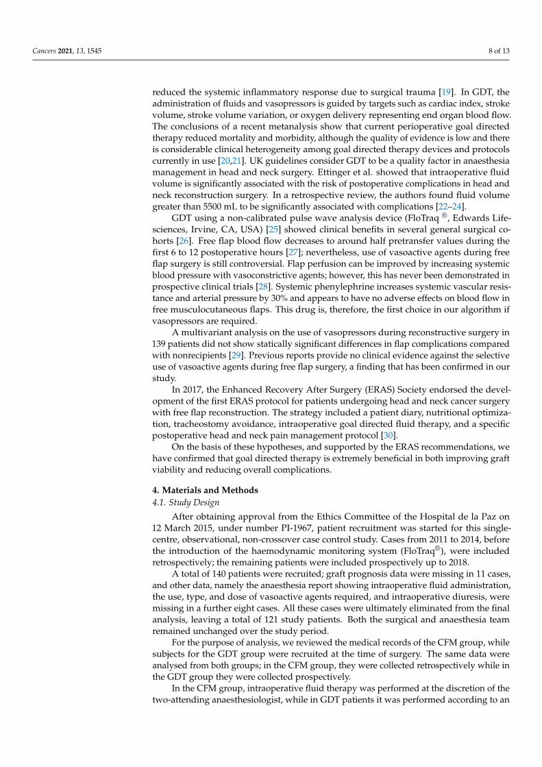
reduced the systemic inflammatory response due to surgical trauma [19]. In GDT, theadministration of fluids and vasopressors is guided by targets such as cardiac index, strokevolume, stroke volume variation, or oxygen delivery representing end organ blood flow.The conclusions of a recent metanalysis show that current perioperative goal directedtherapy reduced mortality and morbidity, although the quality of evidence is low and thereis considerable clinical heterogeneity among goal directed therapy devices and protocolscurrently in use [20,21]. UK guidelines consider GDT to be a quality factor in anaesthesiamanagement in head and neck surgery. Ettinger et al. showed that intraoperative fluidvolume is significantly associated with the risk of postoperative complications in head andneck reconstruction surgery. In a retrospective review, the authors found fluid volumegreater than 5500 mL to be significantly associated with complications [22–24].
GDT using a non-calibrated pulse wave analysis device (FloTraq ®, Edwards Life-sciences, Irvine, CA, USA) [25] showed clinical benefits in several general surgical co-horts [26]. Free flap blood flow decreases to around half pretransfer values during thefirst 6 to 12 postoperative hours [27]; nevertheless, use of vasoactive agents during freeflap surgery is still controversial. Flap perfusion can be improved by increasing systemicblood pressure with vasoconstrictive agents; however, this has never been demonstrated inprospective clinical trials [28]. Systemic phenylephrine increases systemic vascular resis-tance and arterial pressure by 30% and appears to have no adverse effects on blood flow infree musculocutaneous flaps. This drug is, therefore, the first choice in our algorithm ifvasopressors are required.
A multivariant analysis on the use of vasopressors during reconstructive surgery in139 patients did not show statically significant differences in flap complications comparedwith nonrecipients [29]. Previous reports provide no clinical evidence against the selectiveuse of vasoactive agents during free flap surgery, a finding that has been confirmed in ourstudy.
In 2017, the Enhanced Recovery After Surgery (ERAS) Society endorsed the devel-opment of the first ERAS protocol for patients undergoing head and neck cancer surgerywith free flap reconstruction. The strategy included a patient diary, nutritional optimiza-tion, tracheostomy avoidance, intraoperative goal directed fluid therapy, and a specificpostoperative head and neck pain management protocol [30].
On the basis of these hypotheses, and supported by the ERAS recommendations, wehave confirmed that goal directed therapy is extremely beneficial in both improving graftviability and reducing overall complications.
4. Materials and Methods4.1. Study Design
After obtaining approval from the Ethics Committee of the Hospital de la Paz on12 March 2015, under number PI-1967, patient recruitment was started for this single-centre, observational, non-crossover case control study. Cases from 2011 to 2014, beforethe introduction of the haemodynamic monitoring system (FloTraq®), were includedretrospectively; the remaining patients were included prospectively up to 2018.
A total of 140 patients were recruited; graft prognosis data were missing in 11 cases,and other data, namely the anaesthesia report showing intraoperative fluid administration,the use, type, and dose of vasoactive agents required, and intraoperative diuresis, weremissing in a further eight cases. All these cases were ultimately eliminated from the finalanalysis, leaving a total of 121 study patients. Both the surgical and anaesthesia teamremained unchanged over the study period.
For the purpose of analysis, we reviewed the medical records of the CFM group, whilesubjects for the GDT group were recruited at the time of surgery. The same data wereanalysed from both groups; in the CFM group, they were collected retrospectively while inthe GDT group they were collected prospectively.
In the CFM group, intraoperative fluid therapy was performed at the discretion of thetwo-attending anaesthesiologist, while in GDT patients it was performed according to an

Cancers 2021, 13, 1545 9 of 13
algorithm based on recommendations in this type of procedure. The values used in thealgorithm were extracted from the hemodynamic monitoring system.
The goal directed therapy algorithm is described in Figure 7.Cancers 2021, 13, x 10 of 14
Figure 7. GDT algorithm.
Inclusion criteria: All cancer patients scheduled for reconstructive surgery of the
head and neck with a microvascular free flap between 2010 and the first quarter of 2018
were included in the study.
Exclusion criteria: Need for urgent surgery.
All patients were monitored according to the rules of the Spanish Society of Anaes-
thesia (SEDAR): electrocardiogram, pulse oximetry, non-invasive blood pressure (BP)
during induction, after which a radial or femoral arterial line was placed for BP monitor-
ing. In GDT patients, the same line was used to measure haemodynamic parameters.
Depth of anaesthesia was monitored using the BIS (Bispectral Index Sensor).
Active warming systems (convective blanket and fluid warmer) were used in all pa-
tients, and temperature was measured in the GDT group.
Balanced general anaesthesia was performed in all patients. Anaesthesia was in-
duced with 1.5–2 mg/kg propofol, 5–7 mcg/kg fentanyl, and 0.6–0.8 mg/kg rocuronium.
Fibreoptic nasotracheal intubation was performed in patients (the majority) in whom the
airway evaluation predicted the risk of a “cannot intubate, cannot ventilate” scenario. An-
aesthesia was maintained with inhalation agents (sevoflurane) in both groups. After sur-
gery, patients were transferred to the post-anaesthesia care unit (PACU) for a few hours
before extubation until immediate complications (bleeding, suffering of the flap) had been
ruled out.
4.2. Parameters Collected:
1. Demographic data
i. Age
ii. Weight
iii. Sex
2. Preoperative factors
i. Smoking
ii. High blood pressure
iii. Chronic obstructive pulmonary disease
Figure 7. GDT algorithm.
Inclusion criteria: All cancer patients scheduled for reconstructive surgery of the headand neck with a microvascular free flap between 2010 and the first quarter of 2018 wereincluded in the study.
Exclusion criteria: Need for urgent surgery.All patients were monitored according to the rules of the Spanish Society of Anaesthe-
sia (SEDAR): electrocardiogram, pulse oximetry, non-invasive blood pressure (BP) duringinduction, after which a radial or femoral arterial line was placed for BP monitoring. InGDT patients, the same line was used to measure haemodynamic parameters. Depth ofanaesthesia was monitored using the BIS (Bispectral Index Sensor).
Active warming systems (convective blanket and fluid warmer) were used in allpatients, and temperature was measured in the GDT group.
Balanced general anaesthesia was performed in all patients. Anaesthesia was inducedwith 1.5–2 mg/kg propofol, 5–7 mcg/kg fentanyl, and 0.6–0.8 mg/kg rocuronium. Fibre-optic nasotracheal intubation was performed in patients (the majority) in whom the airwayevaluation predicted the risk of a “cannot intubate, cannot ventilate” scenario. Anaesthesiawas maintained with inhalation agents (sevoflurane) in both groups. After surgery, patientswere transferred to the post-anaesthesia care unit (PACU) for a few hours before extubationuntil immediate complications (bleeding, suffering of the flap) had been ruled out.
4.2. Parameters Collected
1. Demographic data
i. Ageii. Weight
iii. Sex
2. Preoperative factors

Cancers 2021, 13, 1545 10 of 13
i. Smokingii. High blood pressure
iii. Chronic obstructive pulmonary diseaseiv. Alcoholismv. Dyslipidaemia
vi. Previous chemotherapyvii. Previous radiation therapy
viii. Acute myocardial infarctionix. History of arrhythmiax. Presence of hypoalbuminemia
xi. Diabetes mellitusxii. ASA classification
xiii. Haemoglobin level, presence of preoperative anaemiaxiv. Background medication: antidepressants, benzodiazepines, antihy-
pertensives (CC blockers, ACE inhibitors/ARBs, beta blockers)xv. Anticoagulants/antiplatelet agents
3. Intraoperative factors
i. Use or non-use of FloTraq®
ii. Type of flapiii. Bone/non-bone flapiv. Surgical timev. Need for transfusion (volume)
vi. Tracheotomyvii. Neck dissection
viii. Fluid therapy
1. Crystalloids2. Colloids
ix. Vasopressors
4. Postoperative factors
i. PACU length of stay (hours)ii. Hospital length of stay (days)
iii. Time on ventilator (hours)iv. Twenty-four hour fluid therapyv. Crystalloids
vi. Colloidsvii. Need for transfusion
viii. Vasopressorsix. Postoperative complications
1. Bleeding2. Thrombosis3. Other complications
a. Pneumoniab. Arrhythmiac. Ischaemic heart diseased. Pulmonary thromboembolisme. Deep vein thrombosisf. Congestive heart failureg. Confusionh. Cerebral vascular accident (CVA)i. Creatinine above 1.2 mg/mLj. Creatinine greater than 24 h per month
4. Diuresis first 24 h

Cancers 2021, 13, 1545 11 of 13
5. Antiplatelet/anticoagulant medication at the time of pedicle section
i. Heparinii. Lysine acelsalicylate
6. Antithrombotic prophylaxis 12 h before the procedure
i. 40 IU low molecular weight heparin (LMWH)/no
7. Graft outcome
i. Necrosis
1. Partial (flaps rescued in a subsequent surgical review)2. Total
ii. Viability
8. Survival
i. Yes/No
4.3. Sample Size Calculation
As the sample size to calculate flap necrosis would be too high, we decided to carryout a pilot study over a period of seven years.
4.4. Statistical Analysis
Qualitative data are presented as absolute frequencies and percentages and quan-titative data as mean ± standard deviation (SD), minimum and maximum if they werenormally distributed, and median and interquartile range if they were not.
The association between qualitative variables was analysed using the chi-square testor Fisher’s exact test.
The nonparametric Mann–Whitney test was used to compare qualitative and quanti-tative data.
Tests were two-tailed and a p value less than 0.05 was considered statistically signifi-cant. Statistical analysis was performed on SAS 9.3 (SAS Institute, Cary, NC, USA).
5. Conclusions
Goal directed therapy in cancer patients undergoing head and neck reconstructionsurgery should be included in all perioperative management protocols, since it improvessurgical outcomes and quality of recovery at discharge, and reduces both postoperativecomplications and length of hospital stay.
Goal directed therapy in head and neck cancer reconstruction surgery improves freeflap outcomes and also reduces hospital and intensive care unit length of stay, with theircorresponding costs. It also appears to reduce morbidity, although these differences werenot significant. Our results have shown that optimizing intraoperative fluid therapyimproves postoperative morbidity and mortality.
We are considering continuing recruitment in order to minimise the possibility thatdifferences between bone/non-bone grafts could be a source of bias in our study.
Author Contributions: B.T.: conceptualization, investigation, methodology, project administration,supervision writing—original draft preparation. E.G.: investigation, methodology, review. J.L.C.:review and editing. J.L.D.C.: methodology. J.G.: resources and review. I.L.: methodology and formalanalysis. F.G.: review discussion and editing. All authors have read and agreed to the publishedversion of the manuscript.
Funding: This research received no external funding.
Institutional Review Board Statement: The study was conducted according to the guidelines of theDeclaration of Helsinki, and approved by Hospital Ethics Committee of the Hospital de la Paz on12 March 2015, under number PI-1967 on 12 March 2015.
Informed Consent Statement: Informed consent was obtained from all subjects involved in the study.

Cancers 2021, 13, 1545 12 of 13
Data Availability Statement: The data presented in this study are available on request from thecorresponding author.
Acknowledgments: Maxillofacial Surgery Team of University Hospital La Paz and Rafa Uña y JoseMaría Muñoz (Anesthesia and Intensive Care Department. University Hospital La Paz).
Conflicts of Interest: The authors declare no conflict of interest.
References1. Urken, M.L.; Weinberg, H.; Buchbinder, D.; Moscoso, J.F.; Lawson, W.; Catalano, P.J.; Biller, H.F. Microvascular free flaps in head
and neck reconstruction. Report of 200 cases and review of complications. Arch. Otolaryngol. Head Neck Surg. 1994, 120, 633–640.[CrossRef]
2. McLean, D.H.; Buncke, H.J. Autotransplant of omentum to a large scalp defect, with microsurgical revascularization. Plast.Reconstr. Surg. 1972, 49, 268–274. [CrossRef] [PubMed]
3. Hagau, N.; Longrois, D. Anesthesia for free vascularized tissue transfer. Microsurgery 2009, 29, 161–167. [CrossRef] [PubMed]4. Khouri, R.K.; Cooley, B.C.; Kunselman, A.R.; Landis, J.R.; Yeramian, P.; Ingram, D.; Natarajan, N.; Benes, C.O.; Wallemark, C.
A Prospective Study of Microvascular Free-Flap Surgery and Outcome. Plast. Reconstr. Surg. 1998, 102, 711–721. [CrossRef][PubMed]
5. Brinkman, J.N.; Derks, L.H.; Klimek, M.; Mureau, M.A.M. Perioperative Fluid Management and Use of Vasoactive and An-tithrombotic Agents in Free Flap Surgery: A Literature Review and Clinical Recommendations. J. Reconstr. Microsurg. 2013, 1,357–366.
6. Quinlan, J. Anaesthesia for Reconstructive Free Flap Surgery. Anaesth. Intensive Care Med. 2003, 4, 87–90.7. Aya, H.D.; Cecconi, M.; Hamilton, M.; Rhodes, A. Goal-directed therapy in cardiac surgery: A systematic review and meta-
analysis. Br. J. Anaesth. 2013, 110, 510–517. [CrossRef]8. Watson, X.; Cecconi, M. Haemodynamic monitoring in the peri-operative period: The past, the present and the future. Anaesthesia
2017, 72, 7–15. [CrossRef]9. Grocott, M.P.W.W.; Dushianthan, A.; Hamilton, M.A.; Mythen, M.G.; Harrison, D.; Rowan, K. Perioperative increase in global
blood flow to explicit defined goals and outcomes after surgery: A Cochrane Systematic Review. Br. J. Anaesth. 2013, 111, 535–548.[CrossRef]
10. Tapia, B.T.; Garrido, E.; Cebrian, J.L.; Del Castillo, J.L.; Alsina, E.; Gilsanz, F. New techniques and recommendations in themanagement of free flap surgery for head and neck defects in cancer patients. Minerva Anestesiol. 2020, 86, 861–871. [CrossRef]
11. Michard, F.; Biais, M.; Lobo, S.M.; Futier, E. Perioperative hemodynamic management 4.0. Best Pract. Res. Clin. Anaesthesiol. 2019,33, 247–255. [CrossRef]
12. MacDonald, D.J.F. Anesthesia for microvascular surgery—A physiological approach. Br. J. Anaesth. 1985, 57, 904–912. [CrossRef]13. Chalmers, A.; Turner, M.W.H.; Anand, R.; Puxeddu, R.; Brennan, P.A. Cardiac output monitoring to guide fluid replacement in
head and neck microvascular free flap surgery-what is current practice in the UK? Br. J. Oral Maxillofac. Surg. 2012, 50, 500–503.[CrossRef]
14. Aditianingsih, D.; Anaesthetist, C. Guiding principles of fluid and volume therapy. Best Pract. Res. Clin. Anaesthesiol. 2014, 28,249–260. [CrossRef]
15. Nolan, J.P.; Mythen, M.G. Hydroxyethyl starch: Here today, gone tomorrow. Br. J. Anaesth. 2013, 111, 321–324. Available online:http://www.ncbi.nlm.nih.gov/pubmed/23946354 (accessed on 10 April 2014). [CrossRef] [PubMed]
16. Chappell, D.; Jacob, M. Role of the glycocalyx in fluid management: Small things matter. Best Pract. Res. Clin. Anaesthesiol. 2014,28, 227–234. [CrossRef]
17. Chan, S.T.F.; Kapadia, C.R.; Johnson, A.W.; Radcliffe, A.G.; Dudley, H.A.F. Extracellular fluid volume expansion and third spacesequestration at the site of small bowel anastomoses. Br. J. Surg. 1983, 70, 36–39. [CrossRef] [PubMed]
18. Voldby, A.W.; Brandstrup, B. Fluid therapy in the perioperative setting—A clinical review. J. Intensive Care 2016, 4, 27. [CrossRef][PubMed]
19. Noblett, S.E.; Snowden, C.P.; Shenton, B.K.; Horgan, A.F. Randomized clinical trial assessing the effect of Doppler-optimizedfluid management on outcome after elective colorectal resection. Br. J. Surg. 2006, 93, 1069–1076. [CrossRef]
20. Chong, M.A.; Wang, Y.; Berbenetz, N.M.; Mcconachie, I. Does goal-directed haemodynamic and fluid therapy improve periopera-tive outcomes? A systematic review and meta-analysis. Eur. J. Anaesthesiol. 2018, 35, 469–483. [CrossRef]
21. Bennett-Guerrero, E. Hemodynamic Goal-Directed Therapy in High-Risk Surgical Patients. JAMA 2014, 311, 2177. Availableonline: http://jama.jamanetwork.com/article.aspx?doi=10.1001/jama.2014.5306 (accessed on 10 April 2014). [CrossRef]
22. Ettinger, K.S.; Arce, K.; Lohse, C.M.; Peck, B.W.; Reiland, M.D.; Bezak, B.J.; Moore, E.J. Higher perioperative fluid administrationis associated with increased rates of complications following head and neck microvascular reconstruction with fibular free flaps.Microsurgery 2017, 37, 128–136. [CrossRef]
23. Kruse, A.L.D.D.; Luebbers, H.T.; Grätz, K.W.; Obwegeser, J.A. Factors influencing survival of free-flap in reconstruction for cancerof the head and neck: A literature review. Microsurgery 2010, 30, 242–248. [CrossRef]
24. Rocca, G.D.; Pompei, L. Goal-directed therapy in anesthesia: Any clinical impact or just a fashion? Minerva Anestesiol. 2011,77, 545–553.

Cancers 2021, 13, 1545 13 of 13
25. Mayer, J.; Boldt, J.; Poland, R.; Peterson, A.; Manecke, G.R. Continuous Arterial Pressure Waveform-Based Cardiac Output Usingthe FloTrac/Vigileo: A Review and Meta-analysis. J. Cardiothorac. Vasc. Anesth. 2009, 23, 401–406. [CrossRef]
26. Bahlmann, H.; Halldestam, I.; Nilsson, L. Goal-directed therapy during transthoracic oesophageal resection does not improveoutcome Randomised controlled trial. Eur. J. Anaesthesiol. 2019, 36, 153–161. [CrossRef] [PubMed]
27. Hallock, G. Critical threshold for tissue viability as determined by laser Doppler flowmetry. Ann. Plast. Surg. 1992, 28, 554–558.[CrossRef] [PubMed]
28. Gupta, D.K. The effects of systemic phenylephrine and epinephrine on pedicle artery and microvascular perfusion in a pig modelof myoadipocutaneous rotational flaps. Plast. Reconstr. Surg. 2007, 120, 1289–1299.
29. Haughey, B.H.; Wilson, E.; Kluwe, L.; Piccirillo, J.; Fredrickson, J.; Sessions, D.; Spector, G. Free flap reconstruction of the headand neck: Analysis of 241 cases. Otolaryngol. Head Neck Surg. 2001, 125, 10–17. [CrossRef]
30. Varadarajan, V.V.; Arshad, H.; Dziegielewski, P.T.T. Head and neck free flap reconstruction: What is the appropriate post-operativelevel of care? Oral Oncol. 2017, 75, 61–66. [CrossRef] [PubMed]