Immunohistochemical and ultrastructural evaluation of the effects of phosphoric acid etching on...
9
Immunohistochemical and ultrastructural evaluation of the effects of phosphoric acid etching on dentin proteoglycans Oyarzu ´n A, Rathkamp H, Dreyer E. Immunohistochemical and ultrastructural evaluation of the eects of phosphoric acid etching on dentin proteoglycans. Eur J Oral Sci 2000; 108: 546–554. # Eur J Oral Sci, 2000 It has been reported that phosphoric acid (PA) produces structural and molecular alterations in dentin collagen fibrils; however, no relevant information exists on the influence of etching with PA on dentin non-collagenous macromolecules. The present study investigated, by immunohistochemistry and ultrastructural histochemistry, the behavior of dentin proteoglycans (PG) after etching human dentin samples with 35% PA gel (thickened with colloidal silica) or with a 35% PA liquid for 15, 30 and 120 s. Immunolabeling with a mouse monoclonal anti-chondroitin sulfate antibody demonstrated that glycosaminoglycans (GAG) were preserved within dentinal tubules opened to the surface after etching with PA gel. In addition, the cationic tracer polyethyleneimine, used for the ultramicroscopic localization of PG anionic sites, revealed that treatment of dentin samples with PA gel preserved the polyanionic peritubular PG in the etched area. On the other hand, etching with the PA liquid produced loss of peritubular GAG and PG anionic sites in the etched dentin surface. The results obtained indicated that similar concentrations of PA in gel or liquid formulations dierently aect the organization of dentin PG. The clinical significance of these in vitro findings and the structural and molecular interactions of dentin PG with adhesive systems are still unknown. Alejandro Oyarzu ´n 1 , Hermann Rathkamp 1 , Erik Dreyer 2 1 Unit of BioStructure, Faculty of Odontology, University of Chile, Santiago, Chile, 2 Department of Prosthetic Dentistry, Faculty of Odontology, University of Chile, Santiago, Chile Alejandro Oyarzu ´ n, Unidad de BioEstructura, Facultad de Odontologı ´a, Universidad de Chile, Casilla 1903, Santiago, Chile Telefax: z56–2–7776062 E-mail: [email protected] Key words: phosphoric acid; acid etching; dentin; proteoglycans Accepted for publication September 2000 Current adhesive operative dentistry involves the use of the total-etch wet-bonding technique, in combination with improved formulations of hydrophilic primers and bonding agents in order to obtain strong and durable adhesion to dentin (1–3). Dentin etching is usually performed by acidic conditioners which remove the smear layer, funnel dentinal tubules, expose collagen fibrils, and modify dentin permeability and wetness (4, 5). In spite of having been marketed as liquids for years, most of the current available etching agents are purposely thickened to improve handling properties. Phosphoric acid (PA) is the most used acid con- ditioner in operative dentistry, in both liquid and gel forms. It has been reported in several studies that dentin collagen fibrils are subjected to PA-induced conformational modifications compatible with col- lagen denaturation (6) by loss of cross-banding and becoming more susceptible to enzymatic degrada- tion (7). However, no relevant information exists yet on the eects of PA or other acidic condi- tioners on non-collagenous macromolecules present in dentin matrix. Proteoglycans (PG) are a group of cell surface and extracellular matrix macromolecules com- posed of a protein core to which one or more glycos- aminoglycan (GAG) chains are attached. PG regulate cell adhesion, cell growth, and connective tissue matrix formation (8). Due to carboxyl and Eur J Oral Sci 2000; 108: 546–554 Printed in UK. All rights reserved
-
Upload
alejandro-oyarzun -
Category
Documents
-
view
213 -
download
0
Transcript of Immunohistochemical and ultrastructural evaluation of the effects of phosphoric acid etching on...

Immunohistochemical andultrastructural evaluation ofthe effects of phosphoricacid etching on dentinproteoglycansOyarzuÂn A, Rathkamp H, Dreyer E. Immunohistochemical and ultrastructuralevaluation of the e�ects of phosphoric acid etching on dentin proteoglycans.Eur J Oral Sci 2000; 108: 546±554. # Eur J Oral Sci, 2000
It has been reported that phosphoric acid (PA) produces structural and molecularalterations in dentin collagen ®brils; however, no relevant information existson the in¯uence of etching with PA on dentin non-collagenous macromolecules.The present study investigated, by immunohistochemistry and ultrastructuralhistochemistry, the behavior of dentin proteoglycans (PG) after etching humandentin samples with 35% PA gel (thickened with colloidal silica) or with a 35%PA liquid for 15, 30 and 120 s. Immunolabeling with a mouse monoclonalanti-chondroitin sulfate antibody demonstrated that glycosaminoglycans (GAG)were preserved within dentinal tubules opened to the surface after etchingwith PA gel. In addition, the cationic tracer polyethyleneimine, used for theultramicroscopic localization of PG anionic sites, revealed that treatment of dentinsamples with PA gel preserved the polyanionic peritubular PG in the etchedarea. On the other hand, etching with the PA liquid produced loss of peritubularGAG and PG anionic sites in the etched dentin surface. The results obtainedindicated that similar concentrations of PA in gel or liquid formulations di�erentlya�ect the organization of dentin PG. The clinical signi®cance of these in vitro®ndings and the structural and molecular interactions of dentin PG with adhesivesystems are still unknown.
Alejandro Oyarzu n1, HermannRathkamp1, Erik Dreyer2
1Unit of BioStructure, Faculty of Odontology,University of Chile, Santiago, Chile,2Department of Prosthetic Dentistry, Faculty ofOdontology, University of Chile, Santiago,Chile
Alejandro OyarzuÂn, Unidad de BioEstructura,Facultad de OdontologõÂa, Universidad de Chile,Casilla 1903, Santiago, Chile
Telefax: z56±2±7776062E-mail: [email protected]
Key words: phosphoric acid; acid etching;dentin; proteoglycans
Accepted for publication September 2000
Current adhesive operative dentistry involves theuse of the total-etch wet-bonding technique, incombination with improved formulations ofhydrophilic primers and bonding agents in orderto obtain strong and durable adhesion to dentin(1±3).
Dentin etching is usually performed by acidicconditioners which remove the smear layer, funneldentinal tubules, expose collagen ®brils, and modifydentin permeability and wetness (4, 5). In spite ofhaving been marketed as liquids for years, mostof the current available etching agents are purposelythickened to improve handling properties.
Phosphoric acid (PA) is the most used acid con-ditioner in operative dentistry, in both liquid and gel
forms. It has been reported in several studies thatdentin collagen ®brils are subjected to PA-inducedconformational modi®cations compatible with col-lagen denaturation (6) by loss of cross-banding andbecoming more susceptible to enzymatic degrada-tion (7). However, no relevant information existsyet on the e�ects of PA or other acidic condi-tioners on non-collagenous macromolecules presentin dentin matrix.
Proteoglycans (PG) are a group of cell surfaceand extracellular matrix macromolecules com-posed of a protein core to which one or more glycos-aminoglycan (GAG) chains are attached. PGregulate cell adhesion, cell growth, and connectivetissue matrix formation (8). Due to carboxyl and
Eur J Oral Sci 2000; 108: 546±554Printed in UK. All rights reserved

sulfate groups present in GAG chains, PG arehighly hydrophilic and polyanionic, which enablethem to attract water and cationic material (9).GAG and PG have been isolated and biochem-
ically identi®ed in dentin and predentin (10±13)and visualized by ultrahistochemical techniques(14±18). More recently, immunohistochemicaltechniques have been introduced in order to local-ize GAG and PG in predentin and dentin. Thus,the majority of stainable GAG (chondroitin-4-sulfate, chondroitin-6-sulfate and keratan sulfate)were distributed in predentin, and only chondroitin-4-sulfate was con®ned mostly to dentinaltubules surrounding odontoblastic processes (19).Moreover, the small leucine-rich proteoglycansdecorin and lumican have been detected in humandentin-predentin complex (20, 21)At present, micromechanical interlocking is cur-
rently accepted as the principal attachment mechan-ism of adhesive resins to exposed dentin collagen®brils (1). However, it is not know if dentin PG/GAG participate directly in the bonding mechan-ism, and to what extent the polyanionic PG aremodi®ed during the dentin-conditioning step.This work was designed to analyze, by combining
immunohistochemistry and ultrastructural histo-chemistry, the e�ects of acid etching on dentin PGand GAG using 35% PA as solution or colloidalsilica-thickened gel.
Material and methods
Tissue preparation
Caries-free human teeth from 20- to 25-yr-oldindividuals were obtained immediately after extrac-tion for orthodontic therapy. The crowns and rootswere cut o� each tooth 2 mm above and belowthe cementum-enamel junction. This was achievedby cutting a deep groove around the tooth witha water-cooled tungsten carbide bur in a high-speedhandpiece. In the tooth discs obtained, the pulpremnants and predentin were carefully removed.Each specimen was further grooved in the mesiodistal plane and subsequently split into buccal andlingual segments.Tooth segments were etched with 35% phos-
phoric acid gel (Scotchbond Etchant N³7523; 3MDental Products, St. Paul, MN, USA) for 15, 30and 120 s (n~15 each) or with a 35% phosphoricacid liquid, obtained by water dilution from stand-ard 85%ortho-phosphoric acid (Merck,Darmstadt,Germany) for 15, 30 and 120 s (n~15 each). Experi-mental samples were washed in distilled waterand then processed for immunohistochemistry orultrastructural histochemistry.
Immunohistochemistry
Tooth segments etched with PA gel (n~30) and PAliquid (n~30) were ®xed in 4% paraformaldehydein 0.1 M phosphate bu�er (pH~7.4) for 48 hat room temperature and then demineralized in5% formic acid (22) for 7 d at room temperature.After extensive washes in running tap water,samples were maintained in phosphate-bu�eredsaline (PBS) for 12 h at 4³C, dehydrated througha graded series of ethanol, cleared in xyleneand embedded in para�n. Sections were cut at5 mm thickness and mounted on silanized glassslides.
After depara�nation, the sections were rinsedwith PBS three times for 5 min. Unspeci®c proteinbinding was blocked with 5% normal goat serumin 3% bovine serum albumin (BSA) in PBS for30 min at room temperature. Then, tissue sectionswere incubated with a mouse monoclonal anti-chondroitin sulfate antibody (Clone CS-56, Sigma,St. Louis, MO, USA) diluted 1:200 in 3% BSA-PBS in a moist chamber for 12 h at 4³C. Thiswell-characterized antibody is speci®c for the GAGchains of native chondroitin sulfate-PG and bindsto both the 4- and 6-sulfated moieties. It doesnot, however, recognize dermatan sulfate (23). Thesections were washed with PBS and exposed toFITC conjugated anti-mouse IgG (Sigma F-3008)1:300 in 3% BSA-PBS for 1 h at 37³C. As negat-ive control, normal serum replaced the primaryantibody.
After rinse with PBS (365 min), the sectionswere mounted in glycerol:PBS 1:1 and examinedin a Nikon Microphot-FXA ¯uorescence micro-scope with 610, 620, 640 and 6100 CF ¯uorobjectives. The light source was a 100 W high-pressure mercury lamp. The FITC ¯uorescencewas studied with an excitation ®lter at 490 nm,a dichroic mirror at 510 nm, and a barrier ®lterat 520 nm. Images were recorded on KodakEktachrome ®lm 400 ASA.
Ultrahistochemistry
Tooth segments etched with PA gel (n~15) andPA liquid (n~15) were ®xed in 4% paraformalde-hyde in 0.1 M sodium cacodylate bu�er, pH 7.4for 48 h at 4³C and then incubated in 0.5%polyethyleneimine (PEI) (MW 1800 Polysciences,Warrington, PA, USA) in 1.1% NaCl, pH 7.4 for12 h at 4³C. Tooth segments were rinsed in 0.1 Msodium cacodylate bu�er, pH 7.4 (3610 min)and next immersed in a mixture of 2% phospho-tungstic acid (PTA) and 0.1% glutaraldehyde incacodylate bu�er for 1 h at 4³C. Subsequently,tissues were washed (3610 min) in cacodylate
E�ects of phosphoric acid on dentin proteoglycans 547

bu�er and post®xed in 1% osmium tetroxide incacodylate bu�er during 1 h at room temperature.Some samples were demineralized in block with10% EDTA, pH 7.4 for 7 d at 4³C after PEIlabeling.
Non-demineralized and demineralized specimenswere dehydrated in graded series of acetone andembedded in EM bed-812 resin (ElectronMicroscopy Sciences, FT Washington, PA, USA).For tissue orientation, semi-thin sections of 1 mmwere cut with a diamond knife and stained withmethylene blue. Ultra-thin sections of 60 nm werestained with uranyl acetate and lead citrate andexamined in a Zeiss AM-109 transmission electronmicroscope operated at 80 kV.
Controls
Tooth segments (n~12) were treated with dis-tilled water instead of PA and then processedfor immunohistochemistry or ultrahistochemistry.Furthermore, some samples etched with 5% formicacid for 2 min were exposed for 3 h to a 2.5%testicular hyaluronidase (Sigma H-3506) solutionin 0.1 M sodium acetate bu�er (pH 5.6) containing0.15 M sodium chloride. Tissue blocks were thenprocessed for PEI staining and embedded in resin.Semi-thin sections were treated with a saturatedsolution of sodium methoxide for 15 min at roomtemperature to remove the resin and then processedfor immunolabeling (24).
Results
Immunohistochemistry
In experimental samples treated with PA gel,a strong ¯uorescence was observed in all dentinaltubules at the etched dentin surface, at all etchingintervals analyzed. The immunolabeling was clearlyobserved in the uppermost part of dentinal tubulesopened to the surface (Fig. 1A, B).
After etching with PA liquid, a loss of immuno-reactivity was evident at the etched dentin surfaceat all times considered and in all samples analyzed.However, not all the tubules in the etched dentinshowed the same pattern of ¯uorescence. The lossof immunolabeling seemed not to be uniformfrom one tubule to another (Fig. 1C, D).
In control specimens, the monoclonal antibodyused showed that chondroitin sulfate groups werestrongly stained along dentinal tubules (Fig. 1E).The immunolabeling, resolved as ¯uorescent spots,was not observed in the intertubular matrix(Fig. 1F). Testicular hyaluronidase pretreatmentresulted in the loss of ¯uorescence in all dentinaltubules in the etched surface (Fig. 1G). When
sections were treated with normal serum insteadof the primary antibody, the ¯uorescence wascompletely abolished (not shown).
Ultrahistochemistry
At the light microscopy level, in nondemineralizedsemi-thin sections, etching with PA gel and PAliquid produced a clean, well-de®ned etching patternat the dentin surface. Dentinal tubules wereenlarged into a funnel shape and the demineral-ization front involved the peritubular matrix andtubule branches in deeper zones of the dentinsegment (Fig. 2A).
In demineralized semi-thin sections incubatedin PEI, a dark brownish stain was observed delin-eating dentin tubules, tubule branches and odonto-blastic processes. The peritubular dentin wasfrequently occupied by granular or ®lamentousmaterial. In deep zones, una�ected by the acidetching, non-homogeneous globular structures wereobservedwithin dentinal tubules.A striking observa-tion was the presence of globular structuresin close apposition to tubule ori®ces (Fig. 2B).In some samples, the globules tended to coalesce,forming a sheet on the dentin surface (not shown).
At the ultrastructural level, in tooth segmentsetched with PA gel, the odontoblast processremnants were strongly stained, and the peritubularspaces were occupied by granular or ®lamentousPEI-positive material in the etched dentin surface,at all etching times analyzed (Fig. 2C, E). Someelectron-dense granular material was observedon the surface of intertubular matrix between onetubule to another (Fig. 2E).
After etching with liquid PA, the dentin tubulesin the demineralized zone were found to be empty.Some scarce PEI-positive material was observedassociated with collagen ®brils of the tubulewall, at all times analyzed (Fig. 2D, F). The ®neelectron-dense granular material on the surfaceof intertubular matrix was not observed (Fig. 2F).
In control specimens, PEI penetrated only a fewmm into the dentin tissue blocks, and scarce darkbrownish globules were seen within dentinal tubules.In some areas, the globular material tended tocoalesce, forming a sheet on the dentin surface(Fig. 3A). Ultrastructural analysis revealed thatPEI-positive granules ®lled the peritubular space,and highly electron-dense winding structuresrelated with odontoblast processes were observed(Fig. 3B). When PEI was omitted in the stainingprocedure, the peritubular ®lamentous or granularmaterial was not observed (Fig. 3C). Testicularhyaluronidase pretreatment resulted in loss ofthe intratubular anionic sites resolvable by PEI(Fig. 3D).
548 OyarzuÂn et al.

E�ects of phosphoric acid on dentin proteoglycans 549

Discussion
This study represents the ®rst investigation ofthe e�ects of acid etching on dentin PG. Ourimmunohistochemical and ultrahistochemicalapproach suggests that 35% PA gel and 35%PA liquid a�ect the organization of dentin PGdi�erently.
Immunohistochemical and ultrastructural fea-tures of the GAG and PG anionic sites contentof dentinal tubules were strikingly di�erentbetween PA gel- and PA liquid-treated samples.In contrast to the PA gel-treated groups, thePA liquid-treated groups showed evidence of lossof chondroitin sulfate (compare Fig. 1A with 1Cand Fig. 1B with 1D) and anionic material(compare Fig. 2C with 2D and Fig. 2E with 2F)from the dentinal tubules in the etched area, at alletching intervals analyzed.
Demineralization is an important step for mor-phological studies of hard tissues. However decal-cifying agents extract organic components fromthe tissues, and the amount of material extractedis dependent on to the decalcifying solutionemployed (25). In order to overcome this meth-odological problem, crucial in proteoglycan histo-chemistry studies, we used 5% formic acid, insteadof EDTA or other acids, since it has been shownto produce minimal or no loss of uronic acidsfrom formaldehyde-®xed and para�n-embeddedcartilage sections, which cannot be appreciatedby histochemical means (22). From this point ofview, the loss of immunoreactivity in the groupstreated with PA liquid cannot be attributed to atechnical artifact during the histological processingof samples.
The acid composition, pH, osmolality, andviscosity produced by thickening agents have beenidenti®ed as the major factors governing thedentin±conditioner interactions (6). PERDIGAO et al.(26) found that PA made into gel with silica hada higher pH than PA thickened with polymer(xanthan gum). Moreover, silica-thickened etchantsresult in reduced extent of peritubular and inter-tubular mineral dissolution compared to condi-tioners with polymer thickeners and unthickenedacid solutions.
Morphologically, silica-thickened PA-treateddentin samples displayed a cu� of super®cialmineralized peritubular dentin at the entrance ofeach tubule, and silica particles contaminate thedentin surface (26). The silica not removed evenafter vigorous rinsing forms a Si-rich layer on thedentin surface (27) and may form bridges withresidual hydroxyapatite crystals that remain in thedecalci®ed dentin area (28). From this point ofview, the electron-dense granular material observedon the surface of intertubular matrix in ourspecimens, could be colloidal silica microparticlesrelated with the exposed collagen network. On theother hand, it is possible that a less aggressivee�ect of PA gel that permits the existence of a cu�of super®cial peritubular dentin and a silica-gellayer after etching, may stabilize the organic matrixof peritubular dentin, thus preventing the loss ofGAG-PG from the etched zone in our experiments.It has not been established whether only the silicathickener is actively engaged in this mechanism.The e�ects of other components of etching gelssuch as surfactants, polymers, or coloring agentson peritubular matrix organization are not yetfully understood.
Application of hypertonic solutions such as 37%phosphoric acid (6070 mOsm/kg) would osmotic-ally draw ¯uid from the dentin toward the surface,thereby inducing convective ¯uid movements (29).It is possible that the loss of GAG in our studymay be a result of the hypertonicity of its etchantrather than acidity per se. Nevertheless, acid-induced conformational changes in protein coresand PG disassembly must be considered possible.
The estimation of the calcium binding capacityof various GAG using equilibrium dialysis indic-ated that chondroitin-4-sulfate bound ®ve timesmore calcium than dermatan sulfate. The dataindicate a possible important role for chondroitin-4-sulfate in dentin physiology, since it could act asa binding site for calcium ions (30). This fact couldexplain that while 35% PA liquid is being rinsedo�, the solubilized calcium-chondroitin-4-sulfatecomplex and other reaction products may bewashed away, leaving semi-®lled or empty dentinaltubules in the demineralized dentin layer.
Fig. 1. Immuno¯uorescence staining of experimental groups with CS-56 monoclonal antibody. A) Note that the immunoreactivitywas observed along dentinal tubules. No di�erences were detected between the distribution of labeling in the etched super®cial zoneand the deeper unaltered dentin. PA gel, 120 s. Bar~100 mm. B) Note that the immunolabeling was preserved in the externalzone where tubules run in angle towards the surface. PA gel, 15 s. Bar~50 mm. C) Note the irregular loss of immunoreactivity inthe external zone exposed to acid conditioning. Aqueous 35% PA, 120 s. Bar~200 mm. D) Note the loss of immunoreactivityin the external zone exposed to acid conditioning. Aqueous 35% PA, 15 s. Bar~200 mm. E) Control specimen. Note thatthe immunolabeling was preserved in tubules opened to the surface. Bar~200 mm. F) Control specimen. High magni®cation view ofdentinal tubules in which the immunolabeling was resolved as ¯uorescent spots. Bar~20 mm. G) Testicular hyaluronidase-digestedsample in which ¯uorescence was not observed in the etched surface. Bar~50 mm.
550 OyarzuÂn et al.
E�ects of phosphoric acid on dentin proteoglycans 551

The movement of material into or across dentinis referred to as dentin permeability. If the materialenters the tubules but does not travel acrossthe dentin, the process is referred to as intra-tubular permeability. Acidic conditioners increase
intratubular permeability by removing smear plugsand the mineral content of peritubular dentin toa depth of approximately 2±7 mm (1, 2). Based onour observations, we can propose that PA liquidincreases intratubular permeability more than PA
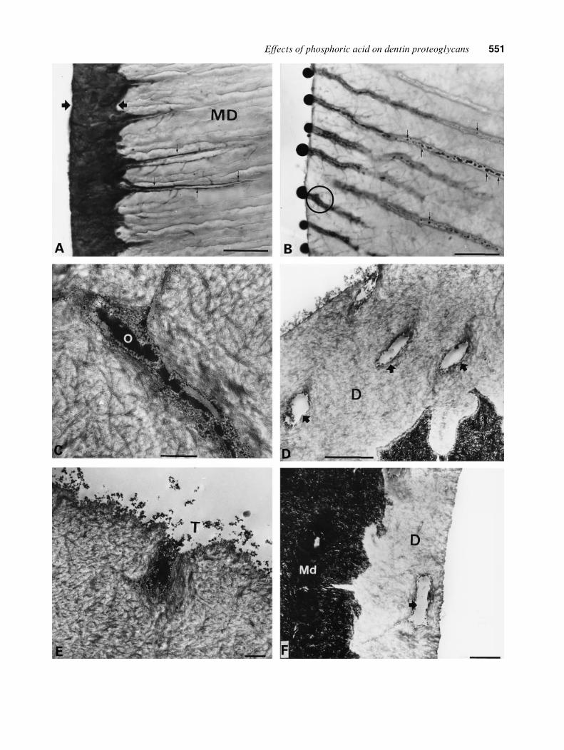
Fig. 2. PEI labeling of experimental groups. A) Light photomicrograph of non-demineralized specimen illustrating the transitionbetween etched intertubular dentin (arrowheads) and mineralized dentin (MD). Note etched areas around dentinal tubules in deepzones (arrows). PA gel, 120 s, bar~20 mm. B) Light photomicrograph showing globular structures in close apposition to tubuleori®ces. Note the presence of non-homogeneous globular material within dentinal tubules in deep zones (arrows). PA gel, 120 s,bar~20 mm. C) TEM photomicrograph of the encircled area in (B). Note the high electron-density of the odontoblast processremnant (O). The peritubular space was ®lled with an electron-dense ®lamentous and granular material. Bar~1 mm. D) TEMphotomicrograph in which empty dentinal tubules (arrows) in the etched dentinal matrix (D) were observed. PA liquid, 120 s,bar~1 mm. E) TEM photomicrograph of a dentinal tubule (T) opened to the surface and ®lled with PEI-positive material. PA gel,15 s, bar~1 mm. F) TEM photomicrograph of an empty dentinal tubule (arrow) in the etched intertubular dentin (D). Md,mineralized dentin. PA liquid, 15 s, bar~0.5 mm.
Fig. 3. Control specimens. A) Light photomicrograph of non-demineralized specimen. Note a few PEI-positive globules (arrows) inthe super®cial zone of mineralized dentin (MD). Bar~20 mm. B) High magni®cation of a dentinal tubule in which winding structures,related with the odontoblast process (O), were observed. Note the peritubular space completely occupied by PEI-positive granules.Bar~500 nm. C) TEM photomicrograph of a dentinal tubule in which PEI was omitted in the staining procedure. Note that the®lament network and peritubular electron-dense granules were not observed (asterisk). Bar~1 mm. D) Testicular hyaluronidase-treated specimen. Note that after PEI staining, anionic sites were not observed in the dentin tubule (asterisk). Bar~500 nm.
552 OyarzuÂn et al.

gel by elimination not only of the mineral contentbut also the organic backbone that supports thespatial organization of hydroxyapatite crystals inperitubular dentin.Several studies have demonstrated that PEI
is a contrast agent for ultrastructural localiza-tion of proteoglycans in many mineralized andnon-mineralized tissues (31±33). The staining prop-erties of PEI are based on the electrostatic attractionbetween the polyanionic GAG and the positivelycharged cationic tracer. This dye is available indi�erent molecular weights (600±60,000 Da) andpenetrates much better into the tissues than othercationic tracers like ruthenium red, alcian blue orthorium dioxide (32). A disadvantage associatedwith the use of conventional chemical ®xation(aqueous aldehydic media), dehydration, andcationic dyes is that GAG chains collapse ontothe protein core, the extent of which depends onthe nature of the dye used (9).In the present investigation, the electron-dense
PEI-positive material that was found to adopta ®lamentous or granular appearance within dentintubules in experimental and control specimensresembles the ultrastructural organization of pro-teoglycans, stained with the same reagent, in boneand cartilage matrix (32). The highly variableappearance of dentin PG after PEI labeling couldbe explained by two or more neighboring PGsubunits coalescing under precipitation to formclumps of variable sizes and shapes. Moreover,the manner in which dentin PG are precipit-ated depends also on the coordinates of thesemolecules with respect to the penetrating dye frontwithin dentinal tubules (9).UEDA et al. (34), using PEI as cationic tracer,
found that it is important to maintain low pHconditions for detecting anionic sites of sulfated PGin cartilage matrix. Since the PEI labeling wasundertaken at a physiological pH in the presentstudy, it is possible that GAG and other anionicmacromolecules were stained in tissue blocks. Thefact that in hyaluronidase-digested samples no PEIpositive material was observed in the etched areasuggests that intratubular anionic sites result fromhyaluronic acid and/or chondroitin sulfate PG.Our observations indicated that the high pre-
cipitative e�ect of PEI produced a severe PGcollapse and gave a good contrast of anionic sitesin dentin tubules at the light microscopic level(Figs. 2B, 3A). This event, not reported in theliterature previously, may be related to the longincubation time used in the present study (12 h at4³C) compared with 1 h at 4³C used in previousreports (31, 32, 34).In several studies, employing some acetone-
based adhesives, resin globules have been identi®ed
in close apposition to tubule ori®ces, withindentinal tubules, and on the bonding dentin surface(35±38). These globules were considered as micellesof resin monomers in the presence of highconcentrations of water (2). Based on our obser-vations, in which single or coalescing PEI-positiveglobules were observed, it may be hypothesizedthat GAG-PG-water complexes in etched dentincould contribute to the formation of the resinglobules previously observed. This concept issupported by the fact that the carbohydratesin the repeating disaccharide chains of GAG carrya negative electric charge. This large polyanionicelectrostatic backbone is highly hydrophilic, whichenables them to attract water and cationic material.In this way the di�usion of hydrophilic primersmight produce, like PEI, a PG collapse andformation of globular structures within dentinaltubules.
Previous ultrastructural studies showed thatstrong dehydrating agents, like ethanol, resultedin the formation of condensed PG in cartilagematrix. Moreover, the use of N-N-dimethylform-amide, as a mild dehydrating agent, improves PGpreservation in an extended ®lamentous network(39). It is possible that the use of acetone- oralcohol-based adhesives in the experiments real-ized by TAY et al. (35±38) produces a chemicaldehydration, a severe PG collapse, and formationof primer globules within dentinal tubules.
The penetration of resin monomers into demin-eralized dentin matrix and the formation of a hybridlayer and resin tags are the principal mechanismsinvolved in adhesive procedures (1±3). Consideringthat PG play an important function in regulatingthe amount of water in many connective tissues,we suggest to analyze the role of dentin PG inthe formation of a hybrid layer and resin tags, in thetotal-etch wet-bonding technique.
In conclusion, the immunohistochemical andultrahistochemical results obtained in the presentstudy showed that PG of the peritubular dentinalmatrix are highly resistant to acid etching withPA gel, thickened with colloidal silica. More-over, treatment with aqueous 35% PA produceda drastic e�ect on the PG in the dentinal tubules,resulting in loss of immunoreactivity and PEI-positive material. Since the immunohistochemicaland ultrastructural information was gathered fromsamples prepared in vitro without pulpal ¯uid andunder physiological pulpal pressure, furtherresearch under clinical conditions is needed.
Acknowledgements ± The authors wish to express their gratitudeto Mr. Walter Poxon and Mr. John Horn for their help andsupport with this study. Special thanks to Mrs. Mo nicaUndurraga (3M Dental Division, Chile).
E�ects of phosphoric acid on dentin proteoglycans 553

References
1. EICK JD, GWINETT AJ, PASHLEY DH, ROBINSON SJ. Currentsconcepts on adhesion to dentin. Crit Rev Oral Biol Med1997; 8: 306±335.
2. PASHLEY DH, CARVALHO RM. Dentin permeability anddentin adhesion. J Dent 1997; 25: 355±372.
3. VAN MEERBEEK B, PERDIGAO J, LAMBRETCHS P, VANHERLE G.The clinical performance of adhesives. J Dent 1998; 26:1±20.
4. BERTOLOTTI RL. Conditioning of the dentin substrate. OperDent Suppl 1992; 17 (Suppl 5): 131±136.
5. ERICKSON RL. Surface interactions of dentin adhesivematerials. Oper Dent Suppl 1992; 17 (Suppl 5): 81-94.
6. ELIADES G, PALAGHIAS G, VOUGIOUKLAKIS G. E�ect ofacidic conditioners on dentin morphology, molecularcomposition and collagen conformation in situ. DentMater 1997; 13: 24±34.
7. OKANAMOTO Y, HEELEY JD, DOGON LL, SHINTANI H. E�ectsof phosphoric acid and tanic acid on dentine collagen.J Oral Rehabil 1991; 18: 507±512.
8. RUOSLATHI E. Structure and biology of proteoglycans.Annu Rev Cell Biol 1988; 4: 229±255.
9. HUNZIKER E, SCHENK R. Structural organization ofproteoglycans in cartilage. In: WIGHT TH, MECHAM RP,eds. Biology of proteoglycans. New York: Academic Press,1987; 155±185.
10. JONES IL, LEAVER AG. Glycosaminoglycans of humandentin. Calcif Tiss Res 1974; 16: 37±44.
11. JONTELL M, LINDE A. Non-collagenous proteins ofpredentine from dentinogenically active bovine teeth.Biochem J 1983; 214: 769±779.
12. HJERPE A, ANTONOPOULOS CA, ENGFELDT B, WIKSTROM B.Analysis of dentin glycosaminoglycans using high-performance liquid chromatography. Calcif Tiss Int 1983;35: 496±501.
13. STEINFORT J, VAN DE STAD R, BEERTSEN W. Identi®cationof new rat dentin proteoglycans utilizing C18 chromato-graphy. J Biol Chem 1994; 269: 22397±22404.
14. GOLDBERG M, SEPTIER D. Electron microscopic visualiza-tion of proteoglycans in rat incisor predentine and dentinewith Cuprolinic Blue. Arch Oral Biol 1983; 28: 79±83.
15. GOLDBERG M, SEPTIER D. Visualization of proteoglycansand membrane-associated components in rat incisor pre-dentine and dentine using ruthenium hexammine trichloride.Arch Oral Biol 1986; 31: 205±212.
16. GOLDBERG M, SEPTIER D, ESCAIG-HAYE F. Glycoconjugatesin dentinogenesis and dentine. Progr Histochem Cytochem1987; 17: 1±113
17. GOLDBERG M, SEPTIER D. Di�erential staining of glycos-aminoglycans in the predentine and dentine of ratusing Cuprolinic Blue at various magnesium chlorideconcentrations. Histochem J 1992; 24: 648±654.
18. GOLDBERG M, TAKAGI M. Dentine proteoglycans: composi-tion, ultrastructure and functions. Histochem J 1993; 25:781±806.
19. TAKAGI M, HISHIKAWA H, HOSOKAWA Y, KAGAMI A,RAHEMTULLA F. Immunohistochemical localization ofglycosaminoglycans and proteoglycans in predentin anddentin of rat incisors. J Histochem Cytochem 1990; 38:319±324.
20. YOSHIBA N, YOSHIBA K, IWAKU M, OZAWA H. Immuno-localization of the small proteoglycan decorin in humanteeth. Arch Oral Biol 1996; 41: 351±357.
21. HALL RC, EMBERY G, LLOYD D. Immunochemical local-ization of the small leucine-rich proteoglycan lumicanin human predentine and dentine. Arch Oral Biol 1997;42: 783±786.
22. IPPOLITO E, LA VELLE S, PEDRINI V. The e�ect of variousdecalcifying agents on cartilage proteoglycans. StainTechnol 1981; 56: 367±372.
23. AVNUR Z, GEIGER B. Immunocytochemical localizationof native chondroitin sulfate in tissues and culturedcells using speci®c monoclonal antibody. Cell 1984; 38:811±822.
24. TAKAHASHI I, MIZOGUCHI I, SASANO Y, SAITOH S, ISHIDA M,KAGAYAMA M, MITANI H. Age-related changes in thelocalization of glycosaminoglycans in condylar cartilageof the mandible in rats. Anat Embryol 1996; 194: 489±500.
25. SCHAJOWICZ F, CABRINI RL. The e�ect of acids (decalcifyingsolutions) and enzymes on the histochemical behaviorof bone and cartilage. J Histochem Cytochem 1955; 33:122±129.
26. PERDIGAO J, LAMBRETCHS P, VAN MEERBEEK B, TOME A,VANHERLE G, LOPES A. Morphological ®eld emission-SEMstudy of the e�ect of six phosphoric acid etching agents onhuman dentin. Dent Mater 1996; 12: 262±271.
27. RUSE ND, SMITH DC. Adhesion to bovine dentin-surfacecharacterization. J Dent Res 1991; 70: 1002±1008.
28. VAN MEERBEEK B, DHEM A, GORET-NICAISE M, BRAEM M,LAMBERTCHS P, VANHERLE G. Comparative SEM andTEM examination of the ultrastructure of the resin-interdi�ussion zone. J Dent Res 1993; 72: 495±501.
29. PASHLEY DH, HORNER JA, BREWER PD. Interactionsof conditioners on the dentin surface. Oper Dent 1992;17 (Suppl 5): 137±150.
30. EMBERY G, REES S, HALL R, ROSE K, WADDINGTON R,SHELLIS P. Calcium- and hydroxyapatite-binding propertiesof glucuronic acid-rich and iduronic acid-rich glycosamino-glycans andproteoglycans.EurJOral Sci1998;106: 267±273.
31. SCHURER JW, KALICHARAN D, HOEDEMAEKER PH,MOLENAAR I. The use of polyethyleneimine for demonstra-tion of anionic sites in basement membranes and collagen®brils. J Histochem Cytochem 1978; 26: 688±689.
32. SAUREN YM, MIEREMERT RH, GROOT CG, KOERTEN HK,SCHERFT JP. Polyethyleneimine as a contrast agent forultrastructural localization and characterization of proteo-glycans in the matrix of cartilage and bone. J HistochemCytochem 1991; 39: 331±340.
33. OYARZUN-DROGUETT A. Ultracytochemical localization ofbasal lamina anionic sites in the rat epithelial attachmentapparatus. J Period Res 1992; 27: 256±263.
34. UEDA H, TORIUMI H, LENG CH, OHNO S. A histochemicalstudy of anionic sites in the intermediate layer of ratfemoral cartilage using polyethyleneimine at di�erent pHlevels. Histochem J 1997; 29: 617±624.
35. TAY FR, GWINETT JA, WEI SHY. Micromorphologicalspectrum from overdrying to overwetting acid-conditioneddentin in water free, acetone-based, single-bottle primer/adhesives. Dent Mater 1996; 12: 236±244.
36. TAY FR, GWINETT JA, PANG KM, WEI SHY. Resinpermeation into acid-conditioned, moist, and dry dentin:A paradigm using water-free adhesive primers. J Dent Res1996; 75: 1034±1044.
37. TAYFR, GWINETT JA, WEI SHY. The overwet phenomenonin two component acetone-based primers containing arylamine and carboxylic acid monomers. Dent Mater 1997;13: 118±127.
38. TAY FR, GWINETT JA, WEI SHY. Relation between watercontent in acetone/alcohol-based primer and interfacialultrastructure. J Dent 1998; 26: 147±156.
39. KAGAMI A, TAKAGI M, HIRAMA M, SAGAMI Y, SHIMADA T.Enhanced ultrastructural preservation of cartilage proteo-glycans in the extended state. J Histochem Cytochem 1990;38: 901±906.
554 OyarzuÂn et al.