Imaging of Urinary tract TB
98
Presenter : Dr. Bidyut Debnath Moderator: Dr.(Prof. ) B. K. Duara Development of the urinary system and Imaging in Urinary Tract Tuberculosis
-
Upload
drbidyut-debnath -
Category
Health & Medicine
-
view
54 -
download
2
Transcript of Imaging of Urinary tract TB

Presenter : Dr. Bidyut DebnathModerator: Dr.(Prof. ) B. K. Duara
Development of the urinary system and Imaging in Urinary
Tract Tuberculosis

Development of the kidney
The kidneys develop from a mesodermal ridge known as the intermediate plate mesoderm, which
develops lateral to the paraxial mesoderm,
along the posterior wall of the abdominal
cavity.

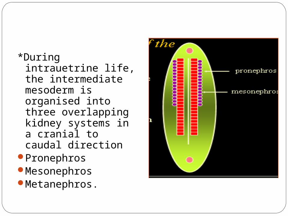
*During intrauetrine life, the intermediate mesoderm is organised into three overlapping kidney systems in a cranial to caudal direction
PronephrosMesonephrosMetanephros.
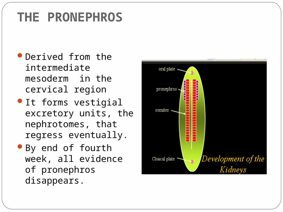
THE PRONEPHROS
Derived from the intermediate mesoderm in the cervical region
It forms vestigial excretory units, the nephrotomes, that regress eventually.
By end of fourth week, all evidence of pronephros disappears.

THE MESONEPHROS
The Mesonephros and Mesonephric ducts are derived from the intermediate mesoderm in the upper thoracic to lumbar L3 region.
• In the 4th week, during regression of the pronephros system, the first excretory tubules of the mesonephros appears.
• The mesonephric ducts become functional between 6 to 10 weeks.
• By the end of the second month, majority of the tubules have disappeared.
• Mesonephric duct remains in males for the formation of the genital system.
• In female they regress.

URETERIC BUD
A ureteric bud arises off the mesonephric duct by 28 days.
This ureteric bud will connect with the metanephric blastema by 32 days.


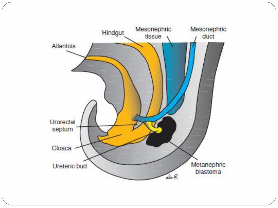

THE METANEPHROS : THE DEFINITIVE KIDNEY
The permanent kidney develops from two sources :
The collecting system from the ureteric bud
The excretory system from the metanephric blastema.
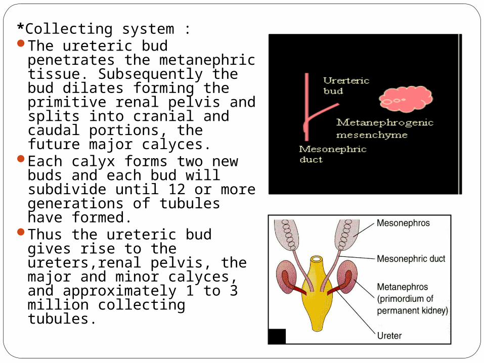
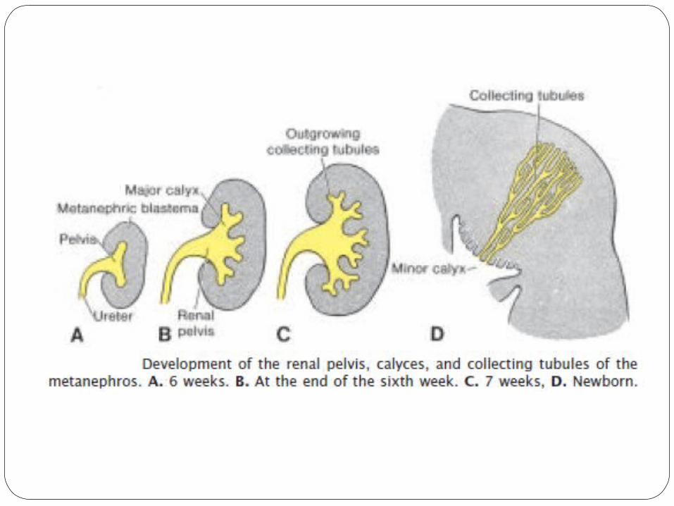
*Collecting system :The ureteric bud penetrates
the metanephric tissue. Subsequently the bud dilates forming the primitive renal pelvis and splits into cranial and caudal portions, the future major calyces.
Each calyx forms two new buds and each bud will subdivide until 12 or more generations of tubules have formed.
Thus the ureteric bud gives rise to the ureters,renal pelvis, the major and minor calyces, and approximately 1 to 3 million collecting tubules.

The excretory system : Develop from the metanephric
mesoderm derived from the posterior intermediate mesoderm.
The ureteric bud induces mesenchyme to form an epithelial aggregate, a metanephric tissue cap. Under the influence of the tubule, cells of the tissue cap form small vesciles, the renal vesciles, which in turn give rise to small S-shaped tubules.
Capillaries grow at one end of the tubules differentiating into glomeruli which will indent into the proximal end of these tubules forming the Bowman’s capsule. The distal end will come in connection with the collecting tubules. Rest of the tubule will form the proximal convoluted tubule, loop of Henle and distal convoluted tubule.


Phenomenon of Reciprocal Induction
Ureteric bud induces the formation of the tubules in the metanephric blastema while the blastema induces the development of the ureteric bud.

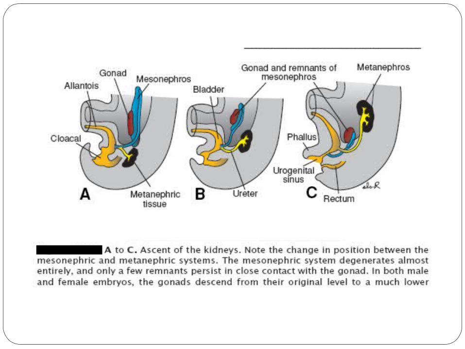
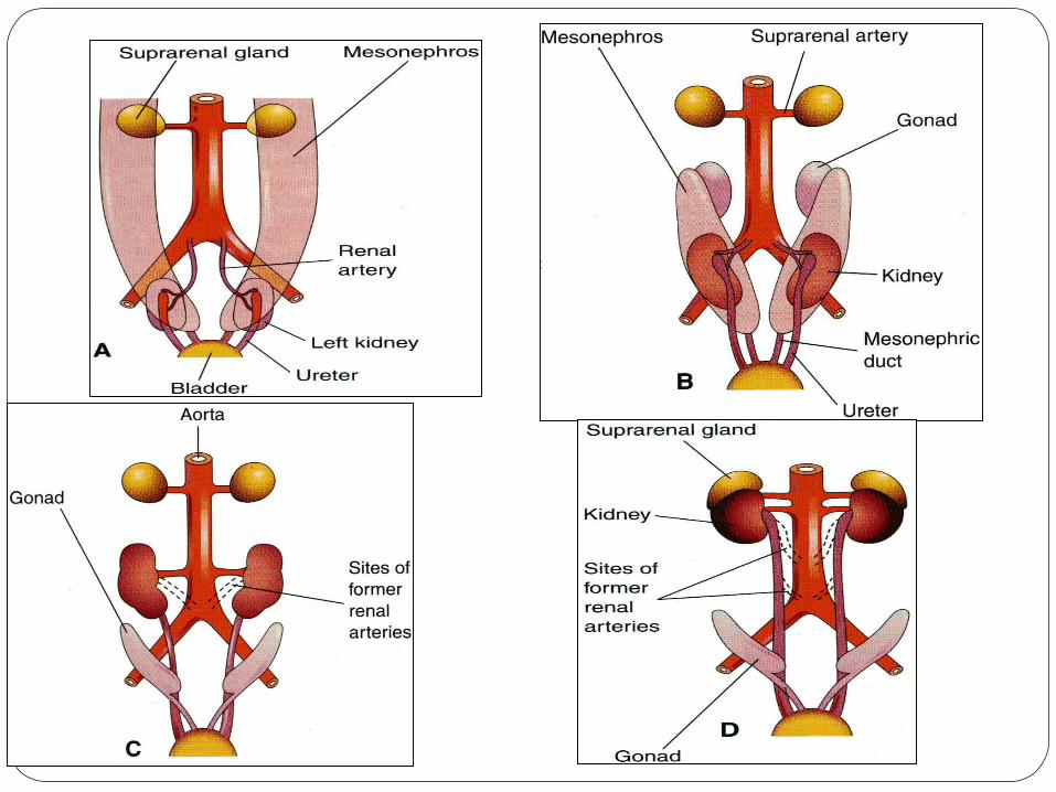
ASCENT OF THE KIDNEY
The kidney is initially in the pelvic region, later migrates to a more cranial position in the abdomen during the 4th to 8th week of gestation.
Ascent is actually caused by diminution of the body curvature and by differential growth of the body in the lumbo-sacral region.
At the same time, the kidneys rotate 90 degrees medially such that the renal pelvis comes to lie anteromedially.
In the pelvis, blood supply is from lateral sacral branches of the aorta. As it ascends it is supplied by continuously higher levels of branches from the aorta upto definitive renal arteries at L1 –L2.

Developmental Abnormalities of kidney
1.Renal dysplasias and agenesis: A spectrum of severe malformations that represent the primary diseases requiring dialysis and transplantation in the first years of life.
o Multicystic dysplastic kidney- Numerous ducts are surrounded by undifferentiated cells. Nephrons fail to develop and the ureteric bud fails to branch, so that the collecting ducts never form.
o In some cases these defects cause involution of the kidneys and renal agenesis. Renal agenesis may also occur if the ureteric bud fails to contact and/or induce the metanephric mesoderm.

2.Congenital polycystic kidney: Numerous cysts form.
ºAutosomal recessive polycystic kidney disease,which occurs in 1/5,000 births, is a progressive disorder in which cystsform from collecting ducts. The kidneys become very large, and renal failureoccurs in infancy or childhood.
ºAutosomal dominant polycystic kidney disease, cysts form from all segments of the nephron and usually do not cause renal failure until adulthood. The autosomal dominant disease is more common (1/500 to 1/1,000 births) but less progressive than the autosomalrecessive disease.
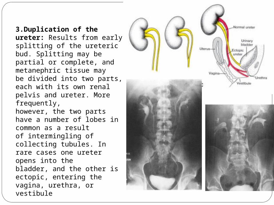
3.Duplication of the ureter: Results from early splitting of the ureteric bud. Splitting may be partial or complete, and metanephric tissue maybe divided into two parts, each with its own renal pelvis and ureter. More frequently,however, the two parts have a number of lobes in common as a resultof intermingling of collecting tubules. In rare cases one ureter opens into thebladder, and the other is ectopic, entering the vagina, urethra, or vestibule
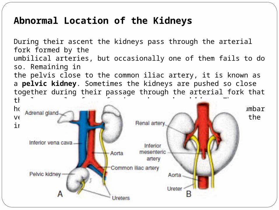
Abnormal Location of the Kidneys
During their ascent the kidneys pass through the arterial fork formed by theumbilical arteries, but occasionally one of them fails to do so. Remaining inthe pelvis close to the common iliac artery, it is known as a pelvic kidney. Sometimes the kidneys are pushed so close together during their passage through the arterial fork that the lower poles fuse, forming a horseshoe kidney. The horseshoe kidney is usually at the level of the lower lumbar vertebrae, since its ascent is prevented by the root of the inferior mesenteric artery.
DEVELOPMENT OF THE URINARY BLADDER AND URETHRA
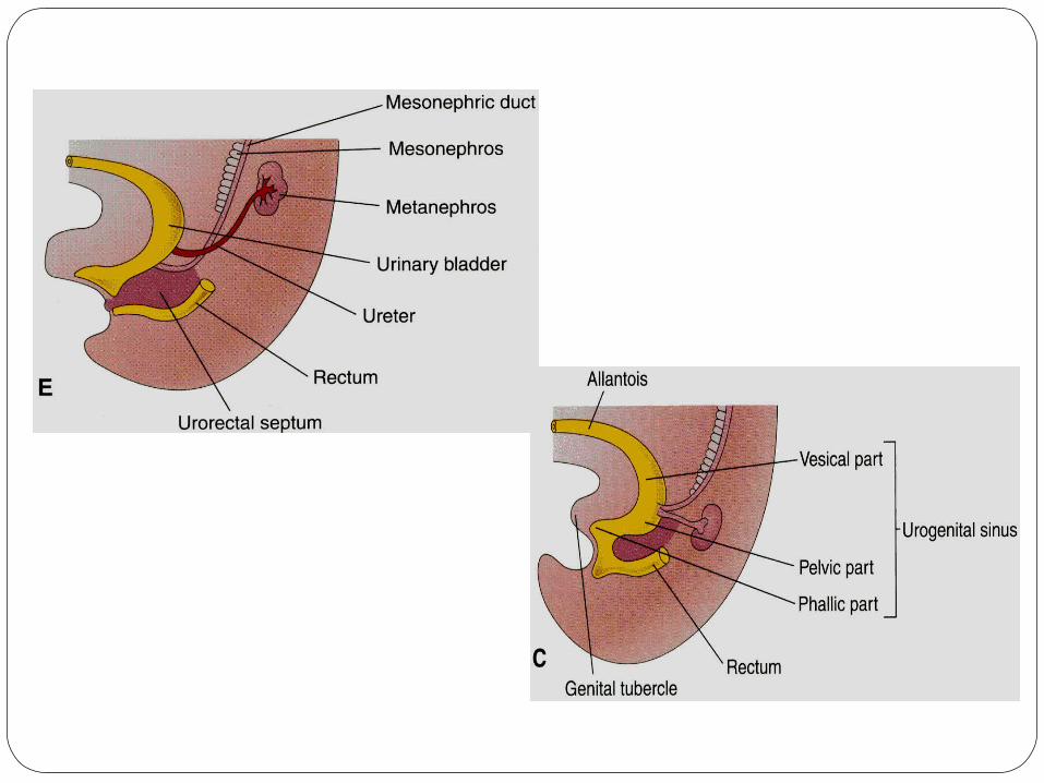
The caudal end of the hindgut is lined with endoderm and is known as the cloaca. During the fourth to seventh weeks of development, the cloaca is divided by a wedge of mesenchyme, the uro-rectal septum, into the uro-genital sinus anteriorly and anal canal posteriorly.
the uro-genital sinus may be divided into three component parts.
The first of these is the cranial portion which is continuous with the allantois and forms the bladder proper.
The pelvic part of the sinus forms the prostatic urethra and membranous urethra in the male and the membranous urethra and part of the vagina in females.
Thirdly, the caudal portion, or definitive uro-genital sinus, forms the penile urethra in males and the vestibule in females.

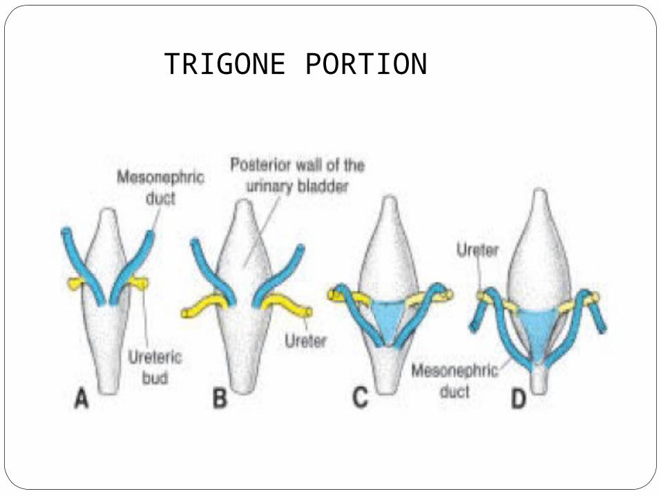
TRIGONE PORTION

Bladder Defects
º When the lumen of the intraembryonic portion of the allantois persists, aurachal fistula may cause urine to drain from the umbilicus.
º If only a local area of the allantois persists, secretory activity of its lining resultsin a cystic dilation, a urachal cyst.
º When the lumen in the upper part persists, it forms a urachal sinus.
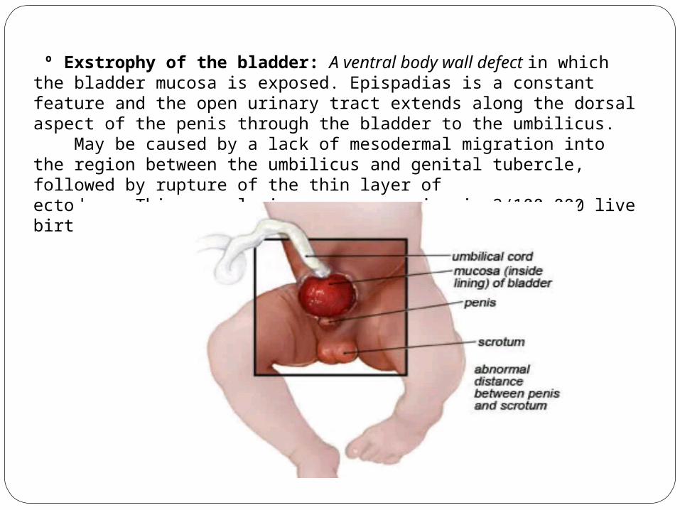
º Exstrophy of the bladder: A ventral body wall defect in which the bladder mucosa is exposed. Epispadias is a constant feature and the open urinary tract extends along the dorsal aspect of the penis through the bladder to the umbilicus. May be caused by a lack of mesodermal migration into the region between the umbilicus and genital tubercle, followed by rupture of the thin layer ofectoderm. This anomaly is rare, occurring in 2/100,000 live births.

Imaging in Urinary Tract Tuberculosis

oTuberculosis (TB), is the commonest worldwide cause of mortality from infectious diseases with nine million new cases and two million fatalities per year.
o Approximately 95% of cases occur in developing countries.
oIn India, more than 1000 lives are lost every day due to TB despitethe availability of modern diagnostic aids and treatment.
oThe genitourinary tract is a primary target of hematogenous infections and is the most common site of extra‑pulmonary TB, comprising 14‑41% of the same.
oGenitourinary tuberculosis (GUTB), a term coined by Wildbolz in 1937.
oWorldwide, 15% of TB patients are co‑infected with HIV, and in HIV‑endemic areas, as many as 75% of patients with GUTB are co‑infected with HIV.
Introduction

Patient Population
o GUTB usually affects adults between the second and fourth decades of life.
o Rare in children and in the fifth and sixth decades.
o Mean age 40.7 years (range: 5‑90 years)
o Long latent period (5‑40 years) between the original pulmonary infection and the appearance of clinical renal Disease*.
oThe youngest reported case of UTB was 2 years old.
oIn India, it is not uncommon to see children with UTB. TB autonephrectomy has been seen in a six year old girl.

Clinical Details
oInsidious onset oNo specific symptoms oAtypical presentations
} which lead to difficulty and delay in diagnosis
oLocal symptoms :Most common
frequent voidingDysuriaPyuriaback, flank, or abdominal painmicroscopic or macroscopic hematuria
oSystemic symptoms :Less commonFeverweight lossanorexia
Hematuria and culture‑negative pyuria may be seen at urine analysis.

Pathogenesis
Causative organismsMycobacterium tuberculosis, an obligate pathogen, is the usual causeMycobacterium bovisMycobacterium avium intracellulare (MAIC)
Spread of tuberculosis to the urinary tract Hematogenous dissemination: from a primary TB focus within the lungs, bone, or other organs Bacille Calmette‑Guerin (BCG): a live vaccine strain can cause renal lesions via reflux, in 0.1% of patients undergoing intravesical instillation of BCG forthe treatment of bladder cancer

Spread of infection at the time of primary tuberculosis
primary infection(Lung)
alveolar macrophages phagocytose one or more mycobacteria lodged within an alveolus
mycobacteria multiply within the macrophages
lymphatic andhematogenous disseminationAnd seeding of tubercularbacilli throughout the body
The kidneys, and possibly the prostate and seminal vesicles, are often the primary sites of GUTB.
All other uro-genital organs, including the epididymis and bladder, become involved by ascent or descent of MTB from a source elsewhere in the genitourinary tract

In most patients, acquired cellular immunity develops and there is inhibition of bacterial multiplication and containment of the disease by the formation of microscopic granulomas.
In immune‑competent patients, these granulomas heal or remain stable for many years.
If there is a breakdown in host immunity, re‑activation or re‑infection occurs. It has been reported that a reduction in serum 25‑OH‑vitamin D levels leads to fall in cell‑mediated immune defenses, which can result in activation of latenttuberculosis.

Renal Parenchymal changes
o Reactivation occurs at the corticomedullary junction.The medullary portion of the renal parenchyma is usually spared initially.
oCortical granulomas enlarge and coalesce, with the bacilli spilling down the nephrons and getting trapped in the narrow segment of the loop of Henle, establishing new foci of infection within the renal pyramid.
o These papillary lesions caseate and cavitate, frequently forming ulcero‑cavernous lesions as they erode into the pelvicalyceal system (PCS).
o Extensive papillary necrosis may develop with the formation of frank cavities and destruction of the adjacent renal parenchyma.
o These may also extend into the collecting system via rupture, or cause parts of the papillae to become necrotic and slough.
o A mass lesion may result from massive destruction and coalescence of granulomas, if they do not rupture into the adjoining calyx.Alternatively, these granulomas may coalesce and form cavities after liquefaction.
o Hypercalcemia may occur, usually secondary to abnormal cortisol production bygranulomatous tissue.
Pelvicalyceal system and Ureteric changes
o When bacilli are shed into the urine, the disease spreads antegradely to involve the urothelium of the renal pelvis, ureter, bladder and, at times, the adjacent genital tract.
o Infection in the walls of the calyces, pelvis, and ureter produces significant inflammatory mucosal thickening. Microscopic granulomas may form here too. Ulceration soon follows.
o In advanced disease, in addition to loss of parenchyma by caseation, intra‑renal scars and strictures lead to obstruction and dilatation of segments of the PCS.Strictures are more common at sites of normal narrowing, such as the calyceal neck, the pelvi‑ureteric junction, and the ureterovesical junction.
o Obstruction may lead to massive hydronephrosis or hydrocalicosis which may be the final stage.

TB of the kidney thus reflects competing processes:(a)The destructive effects of the bacilli, leading to ulceration, cavitation, and fistulization (b) the host’s secondary defense and healing mechanism leading to the formation of granulomas along with fibrosis, calcium deposition, and strictures, which may worsen the obstruction causing progressive renal dysfunction.
However, both processes occur concurrently and may lead to a non‑functioning, calcified kidney of any size; this process is called autonephrectomy. Nephrectomy has been advised to remove the trapped dormant bacilli in such autonephrectomized kidneys.

Tuberculous interstitial nephritis (TIN)
Occasionally, TB can affect the kidney more insidiously, causing TIN, which, if untreated, progresses to renal failure. Rupture of the bacilli into the interstitium can lead to isolated interstitial disease, without persistentpyuria, hematuria, or identifiable AFB in the urine, leading to diagnostic dilemmas.

Changes In Urinary Bladder
o It produces irregular mural thickening which subsequently proceeds to fibrosis.
oThis results in reduction in the bladder capacity and may obstruct the ureters.
oAlternatively, traction on the ureteric orifices may lead to vesicouretcric reflux.
oCalcification (10% )
Urethral TB is very rare despite the constant exposure of the urethra to the infected urine. Most often it occur in association with upper tract involvement. Isolated urethral involvement is extremely uncommon. Tuberculosis of the urethra is usually due to the spread from another focus in the genitourinary tract, the prostate being the common source.


MODALITIES FOR IMAGING
PLAIN RADIOGRAPH
IVU STUDY
RGU MCU
USG AND DOPPLER STUDY
CT SCAN
M R I
RENAL SCINTIGRAPHY
The intravenous urogram (IVU) remains the gold standard inimaging early renal TB.

The plain radiograph
Identification Urinary tract of calcificationSigns of active or inactive extrarenal tuberculosis (such as osseous or paraspinal changes of tuberculosis, as well as old, healed, calcified splenic, hepatic, lymph node, and adrenal granulomas) may be apparent
Patterns varies from few minute areas of calcification to a complete cast of the kidney
o Initially :faint and punctate, may be amorphous, granular, or curvilinear.
o Later : Focal globular calcification involving a renal lobe is frequently associated with a granulomatous mass. Triangular ring‑like calcifications that are characteristic of papillaryNecrosis. ‘Putty kidney’ - Homogeneous and moderately dense, with ground glass appearance (Due to Calcified caseous tissue)

Plain radiograph of the abdomen in a woman with renal tuberculosis shows calcification of varying patterns (curvilinear, amorphous, speckled).

Classic lobar pattern of calcification, which is pathognomonic of end-stage renal tuberculosis, with Ureteral calcification , which is fainter in upper parts.
The occurrence of any upper ureteral calcification (however, faint) along with any other renal calcification is a good pointer of renal TB.
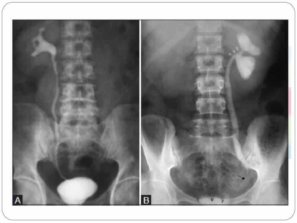
Intravenous urography
Early changes:o Minimal calyceal dilatation(Earliest) and mild loss of calyceal sharpness due to mucosal edema.o As the disease progresses, the calyceal outline becomes more irregular, fuzzy, and ragged and, later, feathery and moth‑eaten in appearance.o Papillary necrosis, o Initial cavitation
Late Changes:oExtensive cavitationoFibrotic stricturesoCortical scarsoMass lesionsoCalcificationoAutonephrectomyoPerinephric abscessoFistula formation.
The finding of hydrocalyces with no pelvis dilatation or an atrophic pelvis is highly suspicious for tuberculosis

A.lower infundibular and renal pelvic scarring,papillarynecrosis
B.papillary necrosis in the upper group of calyces, Healing forniceal papillary necrosis in a lower calyx
C.multiple parenchymal cavities with areas of papillary necrosis

o The cephalic retraction of the inferior medial margin of the renal pelvis at the ureteropelvic junction (UPJ), or the "hiked up renal pelvis, is another suggestive urographic change. .


Autonephrectomy
The late phase of progression of granulomatous destruction of the kidney, with subsequent obstructive uropathy, can lead to an autonephrectomy. This is considered typical of end‑stage renal TB.
There are two types: (1) the caseo‑cavernous autonephrectomized kidney, i.e., an enlarged kidney converted into a caseous filled sac, with or without calcification.
(2) the shrunken, fibrotic, and often calcified kidney. In both instances, there is usually obstruction of the ureter at some point, but this is not essential in type (1). However, both types will be non‑functional on the IVU


False positives/negatives
Renal calcification may occur in nephrocalcinosis, which has several causes. Renal hydatid cysts, renal abscesses, and renal artery aneurysms may calcify and mimic renal tuberculosis.
Calcification may occur within bladder tumors, or calcium may be encrusted on the surface of bladder tumors, which need to be differentiated from bladder tuberculosis. A shrunken bladder may be neurogenic, but the clinical presentation is not that of urinary tuberculosis.
Ureteric and Bladder calcification more commonly occurs in patients with schistosomiasis.
o In schistosomiasis, calcification is first observed in the bladder and then extends upwars to the ureter. In contrast to tuberculosis, bladder capacity and contractilityare surprisingly well preserved.Appeared as eggshell or linear areas of calcification in the submucosa of the bladder and the distalureters
o In tuberculosis, calcification is more amorphous and patchy, and it extends down the ureter


Sonographic features :
o Focal renal lesion–the parenchymal Granuloma / masseso Cavities o Urothelial thickening o Focal caliectasis and Hydronephrosiso Calcificationso Renal Abcess and Perinephric Abcesso Small and fibrotic thick walled bladdero Echogenic foci or calification (Granulomas) in bladder wall near ureteric orifice
USG and Doppler Study
Also Used for USG Guided FNAC
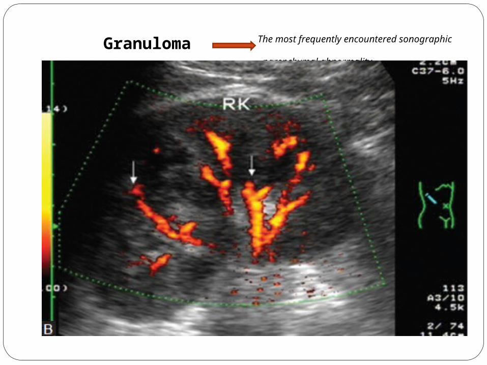
Granuloma The most frequently encountered sonographic parenchymal abnormality
Small (5‑15 mm) º Echogenic º have an echogenic border with a central area of low echogenicity
Granulomas can be better appreciated on color flow imaging, as the ‘cut off’ of the vasculature.
Larger(>15 mm) º Have mixed echogenicity and poorly defined borders

Caseation occurring within the masses parenchymal cavities
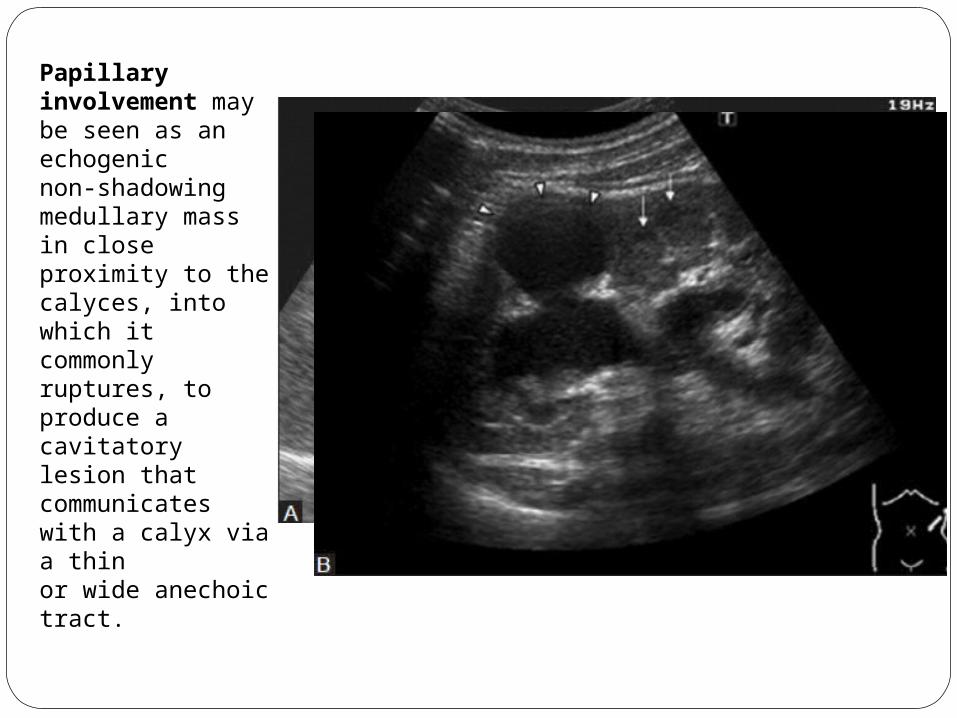
Papillary involvement may be seen as an echogenic non‑shadowing medullary mass in close proximity to thecalyces, into which it commonly ruptures, to produce a cavitatory lesion that communicates with a calyx via a thinor wide anechoic tract.

Infundibular stenosis
Focal caliectasis
Urothelial thickening withvarying degrees of fibrosis
Obstruction affecting different sites
Uneven caliectasis
When the renal pelvis and ureter are involved by TB, the hydronephrosisbecomes severe.
This pattern of diffuse uneven caliectasis (without renal pelvic dilatation) accompanied by urothelial thickening, isa good pointer of renal TB, especiallyin the absence of a renal pelvic calculus.

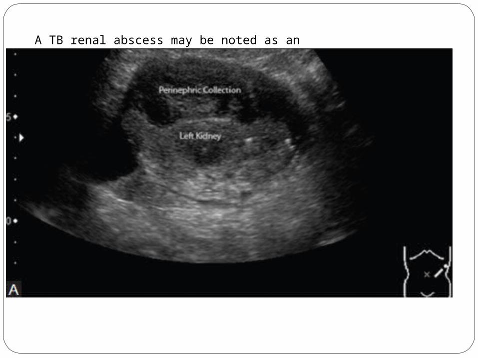
A TB renal abscess may be noted as an irregular sonolucent cavity, with a semi‑solid echogenicity and a thick ill‑defined wall. Necrotic debris and scattered echogenic foci may be noted. The renal abscess can extend outward andrupture, leading to a perinephric abscess and later to a cutaneous fistula.
A thorough analysis of the retroperitoneal compartments and, in particular, the psoas muscle sheath, is required as these are possible sites of a migrating abscess.

Small and fibrotic thick walled bladder
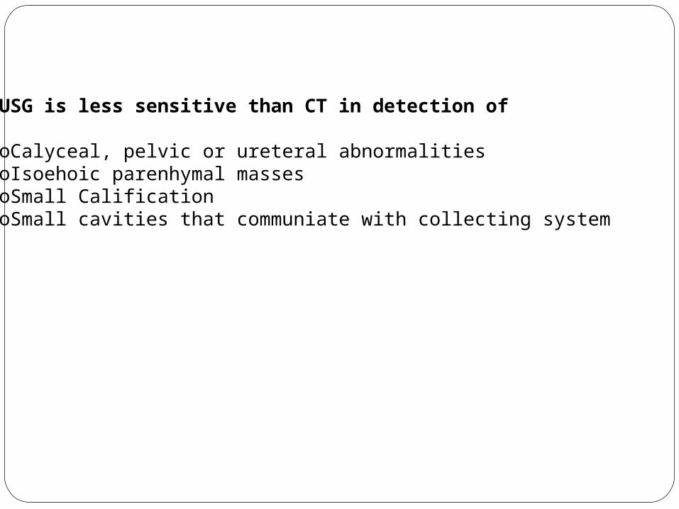
USG is less sensitive than CT in detection of
oCalyceal, pelvic or ureteral abnormalitiesoIsoehoic parenhymal massesoSmall CalificationoSmall cavities that communiate with collecting system

CT Scan
Advantage:
o CT does not require bowel preparation. o It directly visualizes the renal parenchyma, irrespective of renal function.o Assesses extrarenal spread of the disease. o CT is also useful in identifying renal scars, mass lesions, and urothelial thickening.o CT can also substitute for RGU in cases of a tight stricture. o CT can detect calcification with greater accuracy, precision, and sensitivity and is the most sensitive modality for identifying renal calcifications.
MDCT with contrast administration also allows dynamicassessment of the kidney in different phases of excretion andcan define the extent of the disease and identify significantobstruction as well as other complications

Early changes
Papillary necrosis
These can be appreciated on the current high‑end MDCT scanners
Solid mass with little or minimal enhancement after contrast administration. The lesions can occasionally grow to a very large Size and may form masses of mixed density due to the presence of areas of calcification. Coalesced cortical granulomas containing caseous or calcified material can be easily identified on CT.
Granulomas (≤3 mm)
Renal parenchymal changes
In rare cases, there may be single or multiple parenchymal nodules, without collecting system involvement.The nodules are variable‑sized, well‑defined parenchymal lesions on cross‑sectional images and may mimic renal neoplasms, which may lead to unnecessary surgery; these are therefore labeled as the ‘Pseudo‑tumoral’ Type.


oTubercular renal abscesses are seen as hypodense areas of 10‑40 HU with mild peripheral enhancement.

o Cavitation within the renal parenchyma may be seen as irregular pools of contrast material if a calyceal communication exists. Inflammatory granulomatous and caseating masses show enhancement.
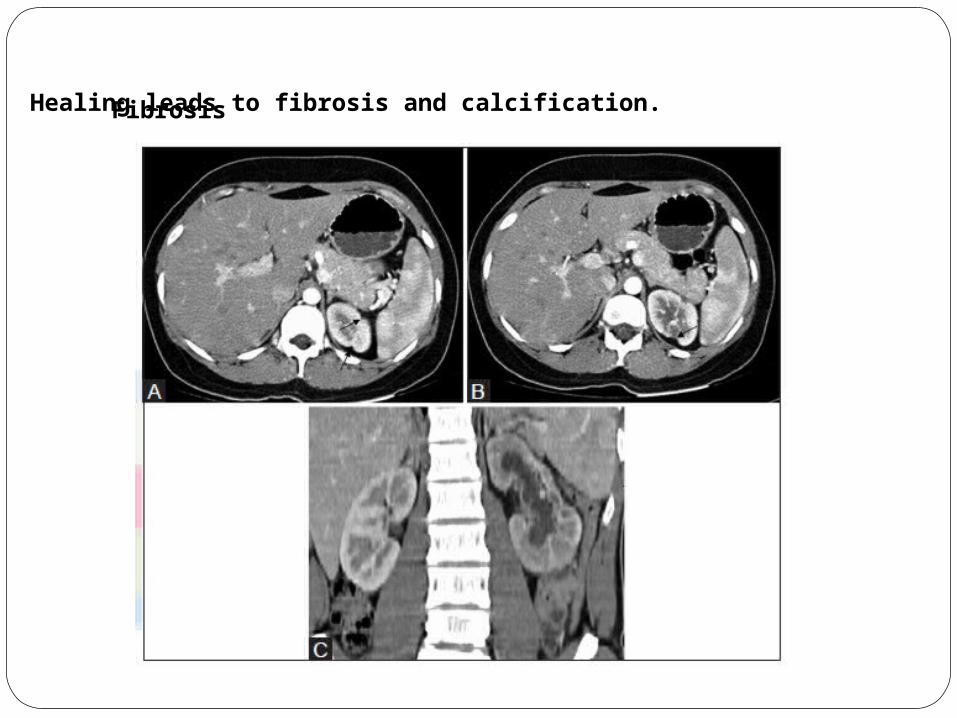
o Healing leads to fibrosis and calcification.Fibrosis

Fine calcifications are best seen on CT and may be good clues to the presence of TB. Varies in appearance depending upon the stage and severity of the disease from punctate, to amorphous, to thin rims surrounding low‑attenuation areas of focal cortical inflammation ; to diffuse uniformly radio‑dense areas, replacing part or all of the renal parenchyma, in late‑stage disease. The lobar pattern of calcification, which is pathognomonic for TB
Calcification
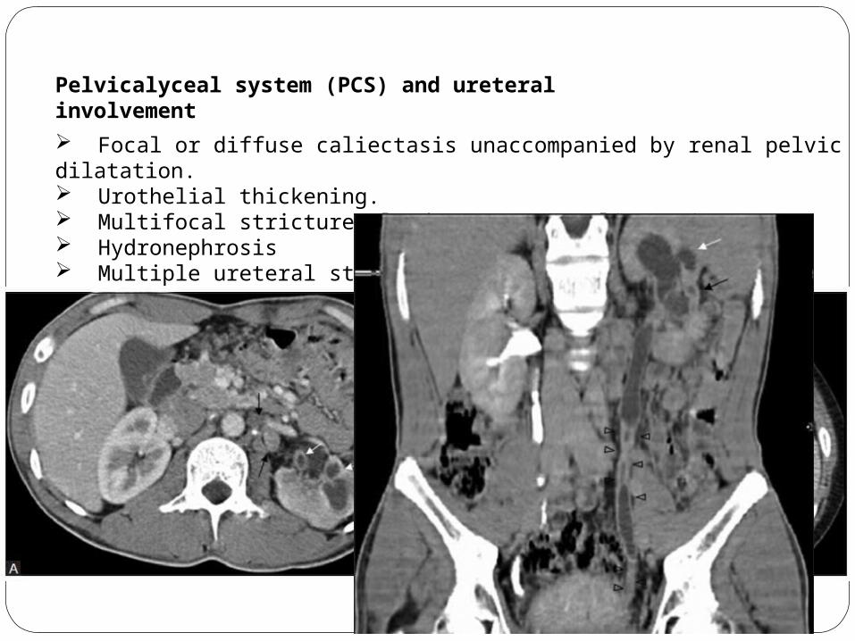
Pelvicalyceal system (PCS) and ureteral involvement
Focal or diffuse caliectasis unaccompanied by renal pelvic dilatation. Urothelial thickening. Multifocal strictures lead to uneven caliectasis. Hydronephrosis Multiple ureteral strictures

With long‑standing renal TB, progressive parenchymal atrophy and hydronephrosis lead to a loss of normal morphology, and the appearance mimics multiple thin‑walled cysts or, occasionally, a multiloculated cyst
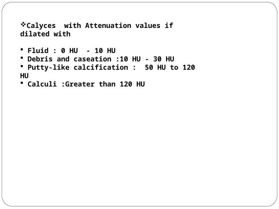
Calyces with Attenuation values if dilated with
Fluid : 0 HU - 10 HU Debris and caseation :10 HU - 30 HU Putty‑like calcification : 50 HU to 120 HU Calculi :Greater than 120 HU

As destruction progresses, the dilated calyces are assimilated into thecaseous renal parenchyma and a unique ‘lobar caseation’ appearance is recognizable. This can be easily differentiated from hydrocalycosis, as each individual lobe does not connect to another as would be expected if they were calyces

Kenny stated that although pelvi‑infundibular strictures, papillary necrosis, cortical low‑attenuating masses, scarring, and calcification may be seen in other conditions, the combination of three or more of these findings is highly suggestive of TB, even in the absence of documented pulmonary disease.
Three imaging patterns were described in CT of renal TB by Wang et al. whenever multiple findings were noted: (a) multiple stricture sites; (b) a single stricture with one other imaging finding; and (c) autonephrectomy along with any other imaging finding barring stricture.
The Lober pattern of calcification is pathognomonic of renal TB and‘lobar caseation’ comes pretty close to the same.

MRI
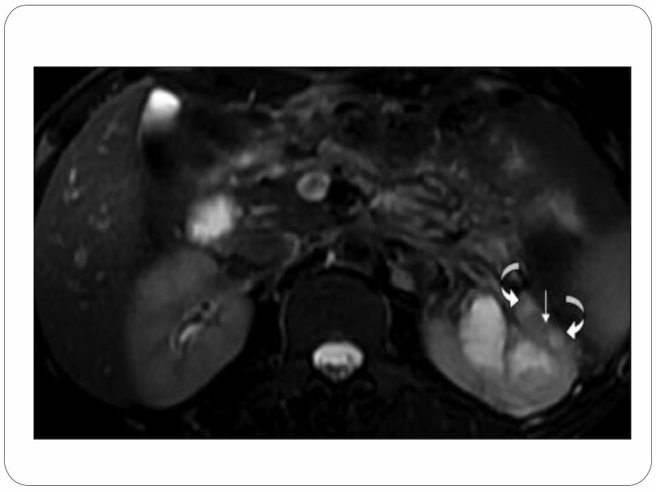
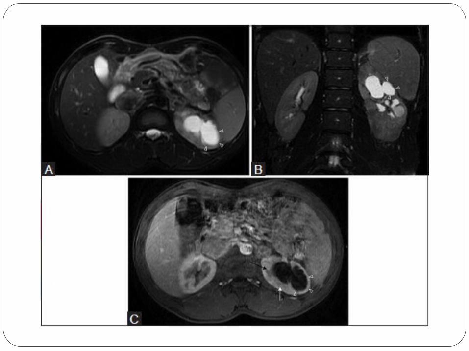
MRI provides good morphological details of the kidneys as well as excellent delineation of the ureters.
MRI is good at depicting tuberculous cavities, sinuses tracts, fistulous communications, and extrarenal spread.
MRI is also useful in the evaluation of peritonitis and adnexal masses.
Renal parenchymal changes not dissimilar to acute pyelonephritis occur in renal involvement with tuberculosis. Active inflammation may cause focal tissue edema and vasoconstriction resulting in focal hypoperfusion well depicted on MRI scans.
Non‑contrast MRI is especially useful in patients with renal failure.

MR urography (MRU) comprises an evolving group of techniques with thepotential for optimal non‑invasive evaluation of urinary tract abnormalities. Both static‑fluid (non‑contrast, heavily T2W sequences) and excretory MRU (performed during the excretory phase of enhancement after intravenousgadolinium) can be combined with conventional MRI for comprehensive evaluation of the urinary tract.
Cine MRU demonstrates the ureters in their entirety and is useful for confirming the presence of stenosis.
Time‑resolved dynamic contrast‑enhanced MRU has been used in the evaluation of ureteral peristalsis in GUTB.

Clues indicating that the focal pyelonephritis has a tuberculous origin:o Loss of interface between the infection and the adjacent renal parenchymao Surrounding tissue edema,o Asymmetric perinephric fat strandingo Thickening of Gerota’s fascia may be.
A TB granuloma is seen as a solid mass of variable size. o The smaller nodular lesions usually appear hypointense on both T1W and T2W images o Larger nodules may reveal central hyperintensity on T2W images.The central T2 hyperintense signal is due to high numbers of macrophages, fibrosis, and gliosis, as well as the increased lipid content, in these lesions.
The necrotic debris along with the caseation and calcification results in a heterogeneous hypo‑ to iso‑intense signal on T2W images. Caseation may have a slightly hyperintense appearance on T2W images.


RENAL SCINTIGRAPHY
The role of radionuclides in imaging patients with renal tuberculosis is confined to assessment of relative renal function by renography when surgery or nephrectomy is contemplated.
The agents used are technetium-99m (99mTc) diethylenetriamine penta-acetic acid (DTPA), 99mTc mercaptotriglycylglycine (MAG-3), and iodine-123 (123I) orthoiodohippurate (OIH).
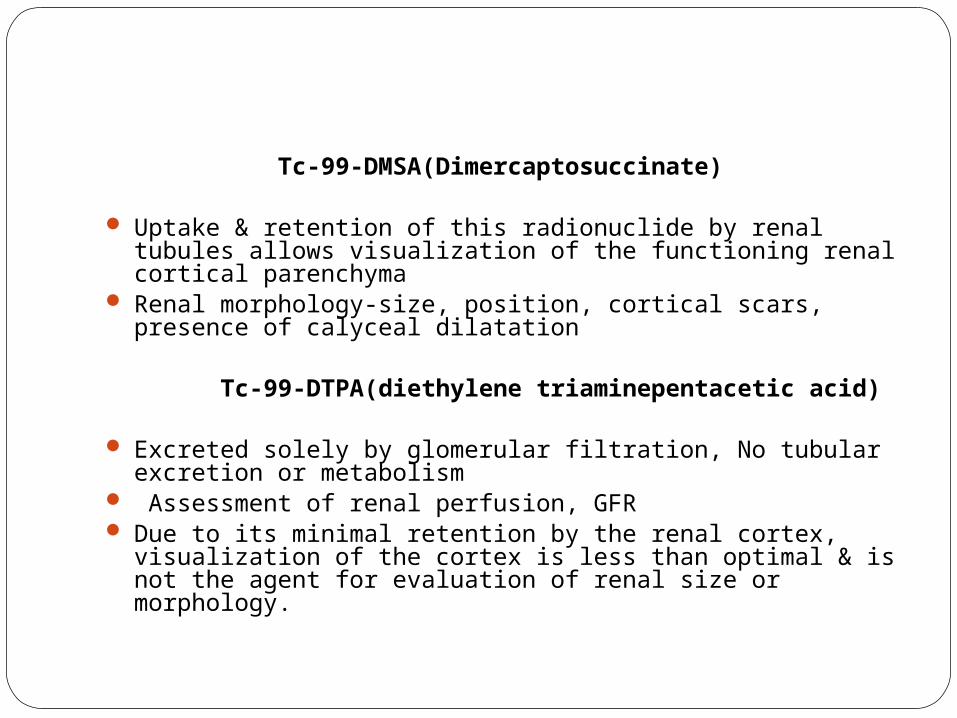
Tc-99-DMSA(Dimercaptosuccinate)
Uptake & retention of this radionuclide by renal tubules allows visualization of the functioning renal cortical parenchyma
Renal morphology-size, position, cortical scars, presence of calyceal dilatation
Tc-99-DTPA(diethylene triaminepentacetic acid)
Excreted solely by glomerular filtration, No tubular excretion or metabolism
Assessment of renal perfusion, GFR Due to its minimal retention by the renal cortex, visualization of the
cortex is less than optimal & is not the agent for evaluation of renal size or morphology.

Tc-99-glucoheptone
Combination agent- both some degree of renal cortical activity & excretion by glomerular filteration.
Major portion excreted by glomerular filteration while rest enters extracellular space. Renal tubular uptake of protein bound fraction & is retained in renal cortex for several hours.
I-131-Ortho iodo hippurate(Hippuran)Tc-99-MAG3 (Mercaptoacetylglycylglycylglycine)


![7 Catheter-associated Urinary Tract Infection (CAUTI) · UTI Urinary Tract Infection (Catheter-Associated Urinary Tract Infection [CAUTI] and Non-Catheter-Associated Urinary Tract](https://static.fdocuments.net/doc/165x107/5c40b88393f3c338af353b7f/7-catheter-associated-urinary-tract-infection-cauti-uti-urinary-tract-infection.jpg)