ilaatu - dm5migu4zj3pb.cloudfront.net · studies onthevelocity of blood flow iv. the velocity of...
25
STUDIES ON THE VELOCITY OF BLOOD FLOW IV. THE VELOCITY OF BLOOD FLOW AND ITS RELATION TO OTHER ASPECTS OF THE CIRCULATION IN PATIENTS WITH ARTERIO- SCLEROSIS AND IN PATIENTS WITH ARTERIAL HYPERTENSION' By HERRMANN L. BLUMGART AND SOMA WEISS (From the Department of Medicine, Harvard Medical School, and the Thorndike Memorial Laboratory, Boston City Hospital) (Received for publication January 25, 1927) This study undertakes the correlation of the clinical manifestations of arteriosclerosis and of hypertension with possible disturbances in the velocity of blood flow, venous and arterial pressures, and vital capacity of the lungs. In studying patients with arteriosclerosis and with evidences of myocardial degeneration it seemed desirable to divide them into two groups. The first group includes patients with regular ventricular rhythm; the second group presents observations on patients who showed total ventricular arrhythmia. I. PATIENTS WITH ARTERIOSCLEROSIS AND WITH EVIDENCES OF MYOCARDIAL DEGENERATION A. Patients with regular rhythm (table 1) Rheumatic infection tends to strike, not only the valves, but also the myocardium. The consequent ill effects on the circulation are a result of these two lesions, the relative importance of which varies from patient to patient. In the group of patients presented here, the lesion is almost entirely myocardial and the rhythm is regular. Con- sequently, the study of such patients provides an opportunity to observe the practically uncomplicated effect on the circulation of but one factor, the myocardium. * All the patients in this group exhibited normal sinus rhythm, with LThis investigation was aided by a grant from the Proctor Fund of the Harvard Medical School for the Study of Chronic Diseases. 173
Transcript of ilaatu - dm5migu4zj3pb.cloudfront.net · studies onthevelocity of blood flow iv. the velocity of...

STUDIES ONTHE VELOCITY OF BLOODFLOW
IV. THE VELOCITY OF BLOODFLOWAND ITS RELATION TO OTHERASPECTS OF THE CIRCULATION IN PATIENTS WITH ARTERIO-
SCLEROSISANDIN PATIENTS WITHARTERIAL HYPERTENSION'
By HERRMANNL. BLUMGARTAND SOMAWEISS
(From the Department of Medicine, Harvard Medical School, and the Thorndike MemorialLaboratory, Boston City Hospital)
(Received for publication January 25, 1927)
This study undertakes the correlation of the clinical manifestationsof arteriosclerosis and of hypertension with possible disturbances inthe velocity of blood flow, venous and arterial pressures, and vitalcapacity of the lungs. In studying patients with arteriosclerosis andwith evidences of myocardial degeneration it seemed desirable todivide them into two groups. The first group includes patients withregular ventricular rhythm; the second group presents observationson patients who showed total ventricular arrhythmia.
I. PATIENTS WITH ARTERIOSCLEROSIS AND WITH EVIDENCES OFMYOCARDIALDEGENERATION
A. Patients with regular rhythm (table 1)
Rheumatic infection tends to strike, not only the valves, but alsothe myocardium. The consequent ill effects on the circulation are aresult of these two lesions, the relative importance of which variesfrom patient to patient. In the group of patients presented here, thelesion is almost entirely myocardial and the rhythm is regular. Con-sequently, the study of such patients provides an opportunity toobserve the practically uncomplicated effect on the circulation of butone factor, the myocardium. *
All the patients in this group exhibited normal sinus rhythm, with
LThis investigation was aided by a grant from the Proctor Fund of theHarvard Medical School for the Study of Chronic Diseases.
173

STUDIES ON THE VELOCITY OF BLOODFLOW
ilaatu avenbs s
lad awui UeTlWln4jt3 X1 - D4 .4in om m ) \so 0
~~tUV~~UO!~~flZ)Ji3 Cl in) in) 0DI tf) to \0 r- 00 0% Cs 0 "~auiii uotjo[n:)!:) N,,N,> N CN C1 . . . C N~~~~~~~~~~~~~~~~~~~CS 0Jx;d Xl)dt13)1WILA ~ % 00 0% 00 00 t'- "I 0% %0- - (~~~~~~-4 - - ~~~~~ -4- ---
kjizrediiz 1IlA veif) 00 CN) 0 ) e1
.Xi 0 C4 000 - 00 r 00 O -
m4)-1 ~4 C14 if) '- '1 -4\ % f)(pal39t~~~~~U') |n 00 U) in c) e§0*om C1VOoo U Um4 Nabnssa.d snouOA C)) uf O
4) | ~ if) 0 00'14s!Ito in 0\-t O +-g O101CW[) 00 if)t- t- if)b0 00 00 if) U) \0
uo}ns> > ci c4l 000 0 00O d'i N
!0 O0if 0f 0 '
° O Off)C %0O q 0000I C0t'
e. %00 4C\4 %04%0%0 C1O
omsvauilnl It o 0oo - 00 00 00 N o 00 "t 'to__- t- \0 U0\0% . O
uoit,nda,dlq4 en~ 0c C) Ch 00 If)O%01
C~~~~~~~~~~~~~l C)EN C1 'I\E2 02 , r3,U2
00 - -- -0 06 01---_--
awe| U 2 2; W U U U $ W m°244
in\0C1 C t-) 4-424424) 0)~
vao0 s__i- ooroa~qwnu lsaI , + 4- 00 I)4-m00 t-
QB
:
W44
C4) CV
-I
O >. *t-*- )
*co z* cow0:
b E Ebb
174
.4)
444
4-)
4.,
fz
0t
0b

HERRMANNL. BLUMGARTAND SOMAWEISS 175
o 0N eq t- 000% eo- eq eq ce . . . U)
U) o 00%Z~0 0Nto - U)
U) U) UCD '-4'-CDZ"000 4~~-eq0 00t~~ -0 00
U) N r- 00 toe~0eq Ce C%4E eqU)q eq0 to so 0 00 %0 i
0o 00 eqoo U VeoooV U) 0
0 U) 00 ea0 u0U )0 U)o) V) oo N 0o U):U)o U)
Uino 0000oIq %0 0 in ino wo inoo + + U)oo00 U)V0PoV
0 4_
) V- U)
_1 0 | 00 V eq eqV008O00 V t- 00 U)in 0 0r-
VO 00 0%00 0 00eq C - eq -C_ C1
- OV\ 0 in, \ eN 00 \0 0 \ o0U)V0 t\. 0% 0%0% 0%0%0% 0% 0%
0 e 00 Z-e000 VZ0
0 U) U) V Vt-_c c .t V
Cd~~~ ~ ~~~~~~~~~~~~~~~~~.0 .d_ ,
-Z
O 0 °O o0 W C00Ch 0
to Uo U) 0 ' U
*~~~_ ._..
o. co Cdc.
6 oc,4~~~~~~~~~~~~~~~~ ~ ~ ~ ~ ~ ~ ~ ~ ~ ~ ~ ~
C4~ee eq eqC
_CS _ N l es4 C.* .eq . * e*q
* . .% . .. 0.*%
* * . . . . .. * .
* * .5 .. .4 .5 5. *0.* . V. * )) eq**
-^4eq . * . *4) .t c4) C4
X ZcZ 0 0

STUDIES ON THE VELOCITY OF BLOODFLOW
clinical and electrocardiographic evidence of myocardial degenerationand of arteriosclerosis. The majority did not show the usual symp-toms and signs of cardiac failure, although a history of weakness andrestricted activity was obtainable in almost every case. A fewcomplained of dyspnea or precordial pain at rest or on exertion, butwere without other signs or symptoms of cardiac weakness. Onlyfive patients (nos. 58, 36, 71, 168 and 384) showed, at the time of thetest of the velocity of blood flow, evidences of congestive failure,namely, moist rales at the bases of one or both lungs. The patients
TABLE 2
A. Patients with signs of arteriosclerosis and of myocardial degeneration without history ofcardiac failure and without signs of congestive failure
Circulation Circulation cqapait
Test number time time per square Vital capacity mer bod Venous pressuremeter surface
seconds seconds cc. cc. cm. H20
17 29 15 5.819 28 17 3,100 1,850 4 533 34 20 4,400 2,570 0.8;48 25 14 5,200 2,950 2.087 32 19 5,700 1,630 2 198 31 17 2,850 1,530 1 2
193 30 19 3,100 1,930 -3.5194 37 23 2,800 1,730. 8.0215 22 11 3,200 1,640 3 5242 39 22 3,750 2,118 -7.0290 25 3,000
Average.... 30 17.7 3,410 1,994 1 7
may be conveniently subdivided according to the signs or symptoms ofcongestive failure.
Under the first heading (table 2) are grouped the patients withouthistory of cardiac failure. In all of them physical activity was re-stricted on account of general weakness.' They had not sufferedfrom congestive failure. In'the second'subdivision (table 3) are'thosepatients who, besides weakness, complained of dyspnea on exertion,but did not show signs of congestive failure. In the third subdivision(table 4) are those who exhibited signs of early congestive failure atthe time of the test. That congestive failure was not- marked is borne
176

HERRMANNL. BLUMGARTAND SOMAWEISS
out by the venous pressure measurements which were within thelimits of normal.
In general, the patients in all three groups showed normal venouspressures, lowered vital capacities, and circulation times that wereslightly, moderately, or greatly prolonged. Caution must be ob-served in the interpretation of the lowered vital capacities because
TABLE 3
B. Patients complaining of dyspnea on exertion but without contgestive failure
r Circulation iCirculation Vital capacityTestumber time time per square Vital capacity per Venous pressuremeter square meter
seconds secoxds CC. cc. cm. HO057 29 18 2,800 1,700 4.863 25 15 2,800 1,800 2.570 30 18 3,100 1,920 2.5
199 37 23 2,350 1,350 3.0223 33 20 2,650 1,500 6.5241 39 27295 45 29 3,200 2,038 1.5
Average.... 34 22 2,817 1,718 3.5
TABLE 4
C. Patients who showed at time of determination signs of congestive failure
Circulation Circulation Vital capacityTest number time time per square Vital capacity per Venous pressure
meter square meter
seconds seconds CC. CC. cm. H20
36 48 29 2,950 1,800 3.058 29 19 2,200 1,140 -1.271 25 14 2,100 1,150 -0.5
168 30 21 2,150 1,120 3.2284 51 32 2,000 1,273 3.5
Average.... 36.6 23 2,280 1,296 1.6
practically all of the patients were of advanced years. All were overfifty except L. C. and D. M. (nos. 19 and 48), who were forty-fourand thirty-eight years of age respectively, the average being sixty.Wintrich (2) in 1854, in the course of his study of the vital capacityof thirty-five hundred individuals, found that it tended to be slightlydiminished between the ages of forty and fifty; while between the
177

STUDIES ON THE VELOCITY OF BLOODFLOW
ages of fifty and sixty years he observed great variation. The reduc-tion in some individuals of advanced years is undoubtedly an expres-sion of underlying emphysema, and is not necessarily due to circula-tory causes. Advanced age in itself does not predispose to reducedvelocity of blood flow, for we have found (3) that persons of approxi-mately the same age as the patients here studied may have bloodflow velocity within the limits of the normal observed in youngerindividuals.
Inspection of table 1 which includes all patients of the three groupsshows that while the circulation times were in general prolonged, thisprolongation was not conspicuous. Of the eighteen patients studied,only four showed arm to arm velocities of blood flow greater thanthirty-five seconds. In seven patients, however, the circulation timeswere between twenty-five and thirty seconds. The vital capacitieswere all definitely reduced except in D. M. (no. 48), in whom it wasnormal, but whose circulation time was just outside of the normallimits. This patient did not give a history of cardiac failure butshowed evidence of unusually advanced arteriosclerosis. In GroupA (table 2) which includes patients without history or signs of con-gestive failure, there is considerable variation of the circulation timealthough, in general, its average prolongation runs parallel to thedecrease in vital capacity. The venous pressures, on the other hand,are, in general, all within the limits of normal, as is to be expected inpatients without conspicuous congestive failure. The patients ofGroup B (table 3) who complained of dyspnea on exertion withoutsigns of congestive failure showed greater retardation of the velocityof blood flow and a more marked reduction in the vital capacity thanpatients of Group A.
Five patients (table 4) entered the hospital because of congestivefailure. On rest in bed, and on administration of digitalis, they im-proved strikingly so that by the time the velocity of blood flow wasmeasured, edema and dyspnea had disappeared and they showed onlymoist rales at the bases of the lungs. In patients who are regainingcardiac compensation, the increased venous pressure tends to disappearearly, followed at first by a return of the velocity of blood flow tonormal, and only later by a rise in the vital capacity. The timerelationship between these three phenomena in patients showing
178

HERRMANNL. BLUMGARTAND SOMAWEISS
circulatory improvement is the reverse of that observed in patientswith increasing failure. The measurement of -the velocity of bloodflow therefore affords information of prognostic value.
While there is a general relationship between the venous pressure,velocity of blood flow, and vital capacity, the results do not permitthe formulation of a definite quantitative relationship. Whethersuch a relationship is possible is very questionable. The velocity ofblood flow from arm to arm reflects the situation existing in the arm,and to a larger extent, in the lungs. But the clinical signs of con-gestive failure may be due to passive congestion of the liver, of thelegs, or of other parts of the body. That there is a precise quantita-tive relationship between the velocity of blood flow through the lungsand in each and every other portion of the circulation is improbable.Vital capacity measurements, furthermore, do not lend themselves toprecise interpretations in persons with generalized arteriosclerosis,because of the tendency to pulmonary emphysema.
In studying patients with evidences of arteriosderosis and myo-cardial degeneration, we have been- impressed by the relatively lateappearance of dyspnea. This may be due in part to the fact thatthese persons frequently experience weakness as their earliest symp-tom, whereas patients with rheumatic or syphilitic heart diseasewhose blood velocity is similarly prolonged do not restrict theiractivities until compelled to do so by dyspnea.. The spontaneousreduction of muscular activity, in patients with arteriosclerosis goesmore or less parallel -with impairment of heart muscle function so thatthese patients live within the limits of the functional capacity of theirhearts and thus do not show symptoms of cardiac insufficiency. Thelate appearance of dyspnea may also be due in part to the presenceof emphysema, for Scott (4) has shown that patients with emphysemaare remarkably insensitive to concentrations of carbon dioxide whichwould be sufficient to cause overstimulation of the respiratory centersof normal persons.
B. Patients with fibrillation of the auricles
The patients presented here (table 5) showed fibrillation of theaurides without antecedent rheumatic or syphilitic infection, butwith signs of advanced arteriosclerosis.
THE JOURNALOF CLINICAL INVESTIGATION, VOL. IV, NO. 2
179

180 . STUDIES ON THE VELOCITY OF BLOODFLOW
The average circulation time of all patients with auricular fibrilla-tion is approximately 100 per cent above the extreme upper limit ofnormal. The conspicuous slowing of the velocity of blood flow is in
TABLE 5
Circulation times and related measurements in patients with fibriUation of auricles and withhistory of dyspnea but without signs of congestive failure at the time
of the determination
Circulation Circulation Vital capacityTest number time time per square Vital capacity per Venous pressuremeter square meter
seconds seconds cc. cc. cm. H20
30 57 34 3,050 1,800 0.331 47 28 3,150 1,870 1.269 42 25 2,500 1,500 1.274 28 16 2,800 1,560 1.276 44 24 3,600 1,960 4.1
226 23 14 3,000 1,820 4.7247 42 26 1,100 688 -3.0
Average.... 40.4 23.9 2,743 1,600 1.4
TABLE 6
Circulation times and related measurements in patients with fibriUation of auricles, withhistory of congestive failure and with signs of congestive
failure at the time of the determinations
Circulation Circulation Vital capacityTest number Clrcuatime time per square Vital capacity per Venous pressure
meter square meter
seconds seconds cc. cc. cm. HsC23 55 29 3,100 1,650 7.090 46 38 2,650 1,490 2;4
100 39 18 2,250 1,030 5.2113 34 14 2,400 1,130 -0.5222 68 45 2,350 1,540 12.5227 36 23 2,250 1,480 13.0229 73 43 1,550 860 16.0246 55 33 2,100 1,272 3.0
Average.... 50.1 29.9 2,581 1,306 7.3
accord with the minute volume output studies of Lundsgaard (5).The degree of prolongation bears a definite relation to the clinicalcondition. These patients were suffering from more severe cardiac

aui uot vInznD 0g oo°~ °° %O eq C4 t- to In ) t- ¢ 9a~~~~~~1n4|4>° mm in InI,-.g8>°u)
4dA | * O g O u >0~~- C> o% in 8O 00 No t- 8 o* _oo oo "id§o,§e + +o
za a!mvatulv! - -< -s -_ -< -O sVx
tm C1 I4 _4 (4 cl _ I 4_
v~~~~~~~~~0tnC-n-
31os;8c (D C4O Sz 0v\O+O O e>1 C04 a00int 0
:)H.1S S to \0tm\e N o X
°° \0 0 . 0o 0o o c eq c14 oSlasi I 1 r- 00 1- t- 00 01 \0 U')
uotlvs!dsoa | 0 m \0 V" in 1*u "+
° co 0E t--o
1 00 00 t-i No t oo osalwaasfitn~~S C,4 47 _0 0 _1 zoo\0 X o Ul0 00 Loo Co4i -
.o.o- .o.o.o .°° ° s.eEt e5
* * 0- j .i *- E
to ~ In --3 3 v ^ B*taqumxtlsa*L|° e + g os cl >° t3 s c> ¢ u) 3vo0od c2s _ _ tcs c
*~~~~~~~~~~~~~~~~~~~~~~~~d0c
*g~~~~~~~~~~~
*t~~~~~~~~~n w7 ..T 4II
.~~~~~~~~~~~dc dC C dC d; 0 dC dC
**~~~~~~~~~~~~~~~~~~ 8wa~~~~~~~~~~~~O*sj 0
*E~~~~~~~~~~~~~~~~~~~~> Cd>
***so~~~~~~~~~~~~~p
* qmn*sg 4C4( 1
b.E~~~~~~~~~~~~~~~~~~~~~~~~~~C4ClC
181HIERRMANNL. BLTUMGARTAND SOMAWEISS
,,1* §
lbX
0
,zolam asnubs I ilsaod aou uopvj=q3n I "m "4 -W so0X- m winm
00 42% 0. m ldfC4 " C4 m mq!.
0*1 . c@|o*' 34
.NO°O - * O tf N S S OR,
W~~~~~~~ Eno bb
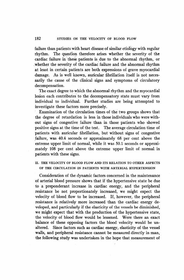
182 STUDIES ON THE VELOCITY OF BLOODFLOW
failure than patients with heart disease of similar etiology with regularrhythm. The questlon therefore arises whether the severity of thecardiac failure in'these patients is due to the abnormal rhythm, or
.whether the severity of the cardiac failure and the abnormal rhythmat least in certain patients are both expressions of grave myocardialdamage. As is well kiiown, auricular fibrillation itself is not neces-sarily the cause of the clinical signs and symptoms of circulatorydecompensation.
The exact degree to which the abnormal rhythm and the myocardiallesion each contributes to the decompensatory state must vary fromindividual to individual. Further studies are being attempted toi-nvestigate these factors more precisely.
Examination of the circulation times of the two groups shows thatthe degree of retardation is less in those individuals who were with-out signs of congestive failure than in those patients who showedpositive signs at the time of the test. The average circulation time ofpatients with auricular fibrillation, but without signs of congestivefailure, was 40.4 seconds or approximately 68 per cent above theextreme upper limit of normal, while it was 50.1 seconds or approxi-mately 108 per cent above the extreme upper limit of normal inpatients with these signs.
II. THEVELOCITY OF BLOODFLOWANDITS RELATION TO OTHERASPECTS
OF THE CIRCULATION IN PATIENTS WITH ARTERIAL HYPERTENSION
Consideration of the dynamnic factors concerned in the maintenanceof arterial blood pressure shows that if the hypertensive state be dueto 'a preponderant increase in cardiac energy, and the peripheralresistance be not proportionately increased, we might expect thevelocity of blood flow to be increased. If, however, the peripheralresistance is relatively more increased than the cardiac energy de-veloped, and particularly if the elasticity of the vessels be diminished,we might expect that with the production of the hypertensive state,the velocity of blood flow would be lessened. Were there an exactbalance of these opposing factors the blood velocity would be un-altered.' Since factors such as cardiac energy, elasticity of the vesselwalls, and peripheral resistance cannot be measured directly in man,the following study was undertaken in the hope that measurement of

HERRMANNL. BLUMGARTAND SOMAWEISS 18
the velocity of blood flow, which is a resultant of many complicateddynamic factors, might aid in our understanding of the mechanismof hypertension.
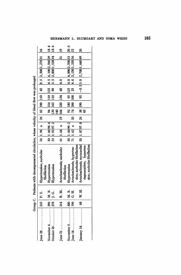
The data obtained in studying patients with hypertension aredivided into three groups (table 8). Group A consists of patientswithout any evidence of circulatory failure at rest at the time of thetests, and in whomthe velocity of blood flow was normal. Group Balso includes patients who did not exhibit symptoms- or signs ofcirculatory failure at rest or on exertion, but in whom there was defi-nite slowing of blood flow. Group C presents patients with decreasedvelocity of blood flow but with symptoms or signs of circulatoryfailure.
The blood pressures of the patients of Group A, with one exception,at the time of test, varied from 160 mm. Hg to 220 mm. Hg systolicand from 76 mm. Hg to 114 mm. Hg diastolic. L. S. (no. 96), whoseblood pressure was normal, had suffered from dizziness and headachesfor three years. His physician had told him that his blood pressurewas elevated and at the time of entry to the hospital the systolic bloodpressure was 195 mm., the diastolic 50 mm. The finding of signs ofcardiac enlargement, in the absence of any signs of cardiac failure,suggested that the blood pressure of this patient had been elevated forsome time. The velocity of blood flow and the vital capacity werenormal. Were his blood pressure to fall without any diminution inthe peripheral resistance the velocity of blood flow might be expected tobecome slowed. That it did not become slowed suggests that hiscapillary resistance was due to functional causes rather than to per-sistent structural alteration such as widespread capillary occlusion dueto arteriosclerosis. E. M. (no. 272) complained of breathlessness onlyon exertion, had never suffered from congestive failure, and his circu-lation was compensated at the time of test.
The presence of a normal velocity of blood flow in the patients ofGroup A is of considerable interest. In no patients with hypertensiondid we find an increased velocity of flow. This indicates that increasedblood pressure, which in itself would tend to increase the speed offlow, is opposed by such factors as increased peripheral resistance.
Group B consists of patients in whom there was a slowing of theblood flow, but who were able to continue their daily duties without
183

STUDIES ON THE VELOCITY OF BLOODFLOW
zoaaui ajenbsaid aUil. Uo°iUlnZ2fl
aUil uoielnanDj
iaalu aenbsjad 4!pvdv Ivl!A
z6cIWedi13!A
ppaauI
aznss2id snou3A
.cd I. :)T OISeLaT
asInd
uoiW.2dso)I
ajnl4Jadutiaj
* 2cn0
co:
awu2v
jaqwunu Isa.I
-4
-Al
Cd
"0
to
00
.0
0
4C)
0
C-)
cna.
0
*S
Cl
-i;
Cd011)
0
C-)-4
0C)
04CU
If)
0000%-~ v-
('i 1If) 0 00 V
f) Q -40e 0ie 1~0eZC T 1400v4
e-i 1-4 V-400 0\ 00t0
00a (00 (Nq If00 0% f) Vo 00 t-
v- v- (N 4 00
00 0000 00 00VDI0% 0 01% 0K 0A
r.V 00 %O
If) (. (f ~4 1.4 ILO
0%tLf - If)
0
Z Zros
C.)bo0
0
0
C-)
0
cd
1:$
B
-)
u
._
CL)
(12
En
0
00
.2Cd
*O
ce
u
rn0GI
- - o--
00
m %O
in to Co
0% 000\0 \0
_ _ _ _-
00 C4o o-
I) oo) oo) 0of
W-1-o v- vo-
0 C% 0% 0 (N(N - - (N_ _
00 \0 \V t- V
t o o o
If %O t dO 0%
V)
oo%0%0 0%
tN - (N -oo) I V If If)
Cd
0 0 00 0~C) C) C) C
+.14i+ 5+
C) C)C)) )'
Cd S mS
mU 00SY00 N-0-s oo
0 0
$-o X - s00000e
184
0
.0
I)
00
C.
.0o
*E
.1
voi-c a3,ejjnS
02V
.!i . cj C'i . C5P4 0. 4 1.4 4i P4
v41cop
HERRMANN,L. BLUMGARTAND SOMAWEISS 18
V 0000 0% OC V
_ _- - N) 0.%
V- C-4 V- CV4-
_) in_in 0 _V 0
00~~ ~ ~
.4 - _ I_
V4
oo oo e e- 0 ~000S00
Y V 0" ) - 0 0oQ if) U)f V- t+ oU)t- f'4~~~~~C
C-)
'4 '4 OM
o 0mf
. 4 4 _-4V4 1
X~~~~co o bun\0 0 N + O ;0;o3o aoos fi v s
0 .. . ..u
___ ________ _ ___
8 -0u°> 0<
'4s
f00*a.a
0 u 0 0mow
eq C4 C4 C4 g B.
I-~~~~~~~~~~~~~~~-
0O>N> >e _
0.. . . .z o .z.

STUDIES ON THE VELOCITY OF BLOODFLOW
experiencing any symptoms of circulatory insufficiency. We layemphasis on these observations because they constitute the onlyinstances in which we have encountered such prolongation in anygroup of patients without signs or symptoms of circulatory failure.It is possible, that the abnornal increase in blood pressure constitutesa compensatory mechanism enabling the normal gaseous exchangesto take place in spite of the subnormal velocity of blood flow.
Group C consists of patients in whomthe slowing of the blood flowwas associated with the symptoms or signs of circulatory failure.This finding is in accord with our experience in patients with normalblood pressure suffering from circulatory failure. The degree ofslowing was approximately that observed in patients with cardio-vascular failure due to other causes. In seven of the patients (nos.198, 296, 304, 305, 307, 308 and 309) the venous pressure was definitelyabove the extreme upper limit of normal, a phenomenon which hasbeen observed by others (6) (7) (8).
The existence of two groups of patients with hypertension similarto our Groups A and B which cannot be differentiated clinically wasalso observed by Boas and Frant (9). They found that in one groupthe capillary pressures were normal, whereas in the other the capillarypressures were elevated. Since we have not measured the capillarypressures of our patients we cannot state whether the two groupsdifferentiated by Boas and Frant correspond to the two groups ob-served by us. The fact that in none of the patients with hypertensiondid we observe increased velocity of blood flow suggests, perhaps, thatthe primary, change in hypertension occurs in the peripheral bloodvessels and that rise in the arterial tension is a secondary reaction onthe part of the body aimed to maintain adequate blood supply to thetissues. For, were the primary change the elevation of the bloodpressure, one would expect to find a period when patients with hyper-tension show an increased velocity of blood flow. This, however, hasnot been observed. In some patients, on the contrary, the velocity ofblood flow is retarded without clinical evidence of decompensation,and we suspect that in these patients the adjustment on the part ofthe heart to the opposed peripheral resistance was incomplete.
186

HERRMANNL. BLTJMGARTAND SOMAWEISS
SUMMARY
1. In this and the preceding (3) communication, eighty-sevenmeasurements of the arm to arm circulation time by the radium Cmethod, on male patients with cardiovascular diseases are presented,and an attempt is made to establish the relationship between thevelocity of blood flow and other fundamental aspects of the circulationsuch as the vital capacity of the lungs, the venous and arterial pres-sures, and the cardiac rate and rhythm.
2. The method as described in a preceding communication hasbeen found adequate for the study of the various aspects of cardio-vascular disease.
The velocity of blood flow and its relation to other aspects of the circulationwere studied in patients: I. With arteriosclerosis and with
evidences of myocardial degeneration
1. Twenty-three measurements of the. arm to arm velocity of bloodflow and related aspects of the circulation were carried out on twentypatients who showed a regular cardiac rhythm.
2. All these patients showed normal.venous pressures, lowered vitalcapacities and circulation times that were slightly, moderately, -orgreatly prolonged, according to the degree of circulatory insufficiency.
3. Sixteen measurements of the arm to arm velocity of blood flowand related aspects of the circulation in thirteen persons with fibrilla-tion of the auricles showed that while the retardation of blood flowcorresponded to the clinical evidences of cardiac decompensation,the prolongation of the circulation time was greater in proportion tothe degree of circulatory decompensation than might be expected onthe basis of our tests on similarly decompensated patients who showeda regular rhythm.
II. With Hypertension
1. Eighteen measurements of the arm to arm velocity of blood flowand related aspects of the circulation are presented on seventeenpatients suffering from arterial hypertension.
2. Patients with hypertension who exhibit no evidence of circula_tory disability may be divided into two groups: in one, the velocity
187.

STUDIES ON THE VELOCITY OF BLOODFLOW
of blood flow is within the limits of normal, whereas in the other, thevelocity of blood flow is retarded.
3. In no patients with hypertension was an abnormally rapidvelocity of blood flow observed.
4. In seven patients without evidences of congestive failure, thevenous pressures were found to be elevated.
5. Patients with hypertension who show congestive failure have aretardation in the velocity of blood flow similar to that of patientswith a corresponding degree of circulatory failure but with a normalblood pressure.
CONCLUSIONS
1. Whereas the arm to arm circulation time in normal, resting, maleindivkduals ranged from eleven to twenty-four seconds, it variedbetween eleven and seventy-three in male patients with compensatedand uncompensated cardiovascular disease.
2. The average arm to arm circulation time in fifty-three normalmale individuals was eighteen seconds, whereas the average in eighty-six determinations in patients with cardiovascular disease was thirty-tvee seconds.
3. The average arm to arm circulation time in those patients whoshQwed no symptoms or signs of circulatory decompensation at thetime of test averaged twenty-four seconds, whereas patients exhibitingsymptoms or signs or cardiac failure showed an average arm to armcirculation time of thirty-eight seconds.
4. The fact that the average circulation time in normal personswas eighteen seconds, and in patients with compensated cardio-vascular disease, was twenty-four seconds, indicates that a retardationin the velocity of blood flow occurs in general before symptoms or signsbecome manifest.
5. In general, the degree of cardiac decompensation at the time ofthe test was closely related to the degree of retardation of the velocityof blood flow.
6. Prolonged circulation times always occurred in the presence ofa failing circulation, except in one group of patients with arterialhypertension in whom a prolongation of the velocity of blood flowwas observed, and who had never shown evidence of circulatory em-barrassment.
188

HERRMANNL. BLUMGARTAND SOMAWEISS
7. Patients with auricular 'fibrillation showed a disproportionateprolongation of the blood flow compared with patients with a similardegree of circulatory decompensation but with a regular rhythm.
8. At the onset of circulatory failure, the retardation in the arm toarm velocity of blood flow appeared earlier than the increase in thevenous pressure, and somewhat later than the reduction in the vitalcapacity. In patients with improving circulatory function the venouspressure first returned to normal. This was followed by a return ofthe velocity of blood flow to within the limits of normal, and some-what later the vital capacity became normal.
9. When the velocity of blood flow was measured several times inthe same patient, it was found that the retardation of the velocityof flow preceded clinical evidence of increasing cardiac failure; andconversely, an increase in the velocity occurred before clinical evidenceof improvement appeared.
Wewish to express our appreciation to Dr. Francis W. Peabody forhis constant advice and encouragement.
BIBLIOGRAPHY
1. Blumgart, Herrmann L., and Weiss, Soma, J. Clin. Invest., 1927, iv, 149.Studies on the Velocity of Blood Flow. III. The Velocity of Blood Flowand its Relation to Other Aspects of the Circulation in Patients with Rheu-matic and Syphilitic Heart Disease.
2. Wintrich, M. A., Krankheiten der Respirationsorgane. InVirchows Handbuchder speciellen Pathologie und Therapie, Erlangen, 1854. Quoted by J. A.Myers, The Vital Capacity of the Lungs. 1925.
3a. Blumgart, Herrmann L., and Yens, Otto C., J. Clin. Invest., 1927, iv 1.Studies in the Velocity of Blood Flow. I. The Method Utilized.
3b. Blumgart, Herrmann L., and Weiss, Soma, J. Clin. Invest., 1927, iv, 15.Studies on the Velocity of Blood Flow. II. The Velocity of Blood Flowin Normal Resting Individuals and a Critique of the Method Used.
4. Scott, R. W., Arch. Int. Med., 1920, xxvi, 544. Observations on the Patho-logic Physiology of Chronic Pulmonary Emphysema.
5. Lundsgaard, C., J. Exper. Med., 1918, xxvii, 219. Studies of Oxygen in theVenous Blood. IV. Determinations on Five Patients with IncompensatedCirculatory Disturbances.
6. Sewall, H., J. A. M. A., 1906, xlvii, 1279. Experiments on Venous BloodPressure and its Relation to Arterial Blood Pressure in Man.
7. Villaret, M., St. Girons, and Greility-Bosviel, Le Jour. Med. Francais, 1921, x,
189

STUDIES ON THE VELOCITY OF BLOODFLOW
359. Contributions a l'etude de la tension veineuse peripherique a l'etatnormal et pathologique.
8. Rotky, H., and Klein, 0., Med. Klin., 1923, xix, 1542. Studien ueber Venen-druck und Kreislaufsuffizienzprufung.
9. Boas, E. P., and Frant, S., Arch.,Int. Med., 1922, xxx, 40. The Capillary BloodPressure in Arterial Hypertension.
APPENDIX
IA. ABSTRACTSOF HISTORIES AND PHYSICAL EXAMINATIONS OF PATIENTS WITHARTERIOSCLEROSISAND EVIDENCES OF MYOCARDIALDEGENERATIONWITH
REGULARRHYTim
215. D. C. entered the hospital complaining of dizziness. Six days before entry-,he suffered what was evidently a cerebral hemorrhage with fainting, vomiting andvertigo. Following this he had been somewhat disoriented until he entered theho'spital. During his stay in the hospital his neurological signs of cerebral hemor-;rhage improved. Occasionally, on physical examination, auricular fibrillationwas found. Heart was not enlarged to percussion, sounds were faint but of fairquality. Discharge diagnosis-cerebral hemorrhage; arteriosclerosis; paroxysmala5u riciar ,fibrillation; chronic myocarditis.
71. T. H. complained of dyspnea, orthopnea and congestive failure for 8 weeks.He was cyanotic at time of admission. P.E. (on admission)-heart was pushedto right and the rhythm was totally irregular. There was fluid in both chests andthoracentesis was performed 3 times. Patient received novarsurol and quinidinand became regular. P.E. (time of test)-heart was regular and the circulationcompensated. No evidence of fluid observed. Few moist rales at left base
"licited. Diagnosis-myocardial degeneration; arteriosclerosis.48. D. M.: No cardiac history obtained. Thickened radial and brachial
arteries palpated. Diagnosis-slight arteriosclerosis.17. E. L. had had no definite history of decompensation. Sclerosed radial and
brachial arteries were prominent. The heart sounds were distant. Diagnosis-moderate arteriosclerosis.
63. J. W. complained of dyspnea. P.E. (date of test)-distant heart soundsheard with rough systolic murmur over precordium, loudest over aortic area.Moderate sclerosis of peripheral vessels was present. Diagnosis-arteriosclerosis;myocardial degeneration.
19, 20, 21. L. C. had had no history of decompensation. The heart soundswere. distant. Moderately tortuous radial and brachial arteries palpated. Diag-nosis-moderate arteriosclerosis.
57. J. W. had had increasing dyspnea for 3 years. He suffered from diabetesfor 4 years. The heart was normal except for systolic murmur over precordium.No history of congestive failure. Beaded peripheral arteries felt. Diagnosis-conspicuous a:rteriosclerosis; myocardial degenerationn.
*190

HERRMANNL. BLUMGARTAND SOMAWEISS 191
58. J. W. had dyspnea and gastric and precordial distress for 3 years, congestivefailure 2 months previously with nocturnal dyspnea. Precordial pain was radiat-ing to the left shoulder. P.E. (at time of test) showed moderate cardiac enlarge-ment. The heart sounds were distant with rough systolic murmur over pre-cordium which was loudest at 2nd right costal space. Moist rales at both baseswere heard. Conspicuous sclerosis of brachials, radials and temporals were noted.Diagnosis-myocardial degeneration; generalized arteriosclerosis.
193. H. B. had had no cardiac history. He entered the hospital because ofbleeding gums and perifollicular hemorrhages which were diagnosed as scurvy.At time of test patient had improved and signs of scurvy had disappeared. P.E.showed distant heart sounds. Size of heart was normal, sounds, regular. Therewas conspicuous sclerosis of the femoral, radial and temporal arteries. Diag-nosis-general arteriosclerosis.
87. T. 0. gave no history of cardiac decompensation. P.E.-size of the heartwas normal; the sounds were inaudible. Rough systolic murmur over the apex washeard. All the palpable vessels were markedly sclerosed. The thorax was fixedand the breath sounds were distant. Diagnosis-arterioscleiosis; senile em-physema.
199. A. C. complained of weakness, loss of weight, shortness of breath on exer-tion. There was no dyspnea or orthopnea, and no history of congestive failure.P.E.-size of the heart was normal; sounds were not heard. Pulse was regular.Conspicuous thickening and tortuosity of peripheral vessels was present. Thechest was barrel-shaped and fixed. The breath sounds were distant. Diagnosis-generalized arteriosclerosis; myocardial degeneration; senile emphysema.
223. H. B. suffered for several years from dyspnea on exertion, and fromoccasional precordial pain. One year ago he felt dizzy, had sharp precordialpain radiating to the left chest, palpitation, sounds regular but distant. The whiteblood cell count was 22,000 per cubic mm. and therefore coronary thrombosis wassuspected. He was fairly comfortable until 4 weeks previous to present admission,when after a short walk, he developed a rather sudden, marked sensation of suffo-cation. He was relieved by nitroglycerine and was well except for short dyspnea.On the day of admission, after a walk, the sense of suffocation rather suddenlyreturned. He vomited once. He had a tight sensation over the upper chest,soreness but not pain, and felt as if he were in extremis. P.E.-Appeated to be"in extremis", with marked dyspnea, orthopnea and slight cyanosis. Heart wasslightly enlarged. Sounds were distant, many crepitant rales at both bases. Noedema noted. White blood cell count was 28,800. He improved gradually.At time of test his circulation was compensated at rest, no signs of congestivefailure were noted, though he was very weak and dyspneic on slight exertion.Electrocardiogram showed signs of coronary occlusion. Diagnosis-coronarythrombosis; arteriosclerosis.
33. J. 0. had had history of painful.swollen right knee 37 years before admis-sion. At time of admission he suffered from painful joints. P.E.-apex impulse

STUDIES ON THE VELOCITY OF BLOODFLOW
was not felt. The left border of cardiac dullness was 9 cm. Sounds were regularand of good quality. Temporal arteries were tortuous, and the brachial andradial arteries were moderately sclerosed. Diagnosis-infectious arthritis; arte-riosclerosis.
168. F. S.: Following rest in bed for 8 weeks he became short of breath. P.E.(at the time of test)-the heart size and sounds were normal. There was flatnesswith suppressed breath sounds on left. RAles at both bases were heart. Pittingedema of legs was present. Diagnosis-myocardial failure.
242. J. B. suffered from periodic attacks of constriction of the chest with epi-gastric pain and vomiting. P.E.-was negative except for marked arteriosclerosis.He was observed in one attack during which the electrocardiogram showed com-plete ventricular asystole of about 11 seconds' duration. After discharge fromthe hospital, patient showed almost daily attacks. He was unconscious duringattack. He had no signs of congestive failure. Diagnosis-Stokes-Adamssyndrome; myocardial degeneration for 9 months.
194. J. W. had had no history of cardiac decompensation. P.E.-apex beat wasnot visible and not felt. The left cardiac border dullness was in the 5th space,9 cm. The sounds were of good quality. Rough systolic murmur with palpablethriU over aortic region was heard. Brachial and radial arteries were hard,tortuous and beaded. There was no edema of the ankles. Diagnosis-general-ized arteriosclerosis.
29. R. B. gave no cardiac history. P.E. (date of test)-Heart sounds weremuffled. Premature ventricular beats from different foci were shown by theelectrocardiogram. Cardiac impulse was not seen or felt. Conspicuous sclerosisof the radial and brachial arteries was observed. Diagnosis-general arterio-sclerosis; myocardial degeneration.
295. M. C. complained of weakness of 6 months' duration. Frequently he wastroubled by painful joints for 15 years. Occasional palpitation with precordialpain was felt for several years which was associated with dyspnea on exertion.At time of test he was unable to walk more than 600 yards without conspicuousdyspnea. There was no sign of congestive failure. P.E.-showed marked emacia-tion. Apex in 5th space was 9 cm. from midsternal line. The sounds were distantand regular. There was slight tortuosity of peripheral arteries. Diagnosis-myocardial degeneration; syphilis.
241. D. M. felt tiredness and shortness of breath on walking, for 2 years. Hegave no history of congestive failure. P.E.-the heart was normal in size. Thesounds were regular and distant. Conspicuous thickening of the peripheral vesselswas noted. Diagnosis-generalized artenrosclerosis, marked.
36. J. G. complained of weakness, shortness of breath, and chronic cough ofseveral months' duration. P.E.-there was slight orthopnea. The heart wasapparently normal. The arteries were sclerosed. Persistent moist rMles at baseswere heard. Diagnosis-myocardial degeneration; emphysema.
284. J. G. entered the hospital because of dyspnea, anorexia, and weaknessbeginning 4 weeks previously, when he developed severe attacks of nocturnal
192

HERRMANN L. BLUMGARTAND SOMAWEISS
dyspnea associated with a sense of pressure over the epigastrium. P.E.-Therewas orthopnea. The sounds were faint. A soft systolic murmur over the aorticarea was heard. Brachial and radial arteries were sclerosed. Moist riles overboth bases were heard. The liver edge was palpable and tender. Slight pittingedema over both ankles was present. Diagnosis-arteriosclerosis; cardiac asthma.
290. N. L. gave no history of congestive failure. P.E.-the heart was normal insize. The sounds were of good quality and regular in rhythm. Arteries weretortuous and thickened. Diagnosis-arteriosclerosis.
IB. ABSTRACTSOF HISTORIES AND PHYSIcAL EXAMINATIONS OF PATIENTS WITHARTERIOSCLEROSISAND EVDENCESOF MYOCARDiALDEGENERATION
WITH FIBRILLATION OF THE AuRiCLES
226. 0. G. gave no cardiac history and had never experienced shortness ofbreath. P.E.-showed the heart moderately enlarged, the sounds rapid anddistant and totally irregular in rhythm. The lungs were hyperresonant andexpansion was diminished. Mark-ed thickening of all palpable arteries waspresent. Both legs were amputated from the thigh. Diagnosis-arteriosclerosis;auricular fibrillation; myocardial degeneration.
100, 113. W. H. gave a history of cardiac failure of one year with markedorthopnea and dyspnea. At time of test he showed orthopnea and dyspnea.P.E.-Left border of cardiac dullness was 13 cm. from the midsternal line. Theheart sounds were distant and totally irregular. There was no pulse deficit.Both bases were flat. Moist riles were heard over the lungs. Pitting edema ofwrist, arms and legs was present. Diagnosis-myocardial degeneration; auricularfibrillation.
74. R. F. had had occasional shortness of breath with attacks of pain over pre-cordium radiating to the left shoulder, arm, and hand. The pain was never sharp,but rather dull and numb. Slight dyspnea on exertion had been present duringthe previous few weeks. P.E.-showed the left border of cardiac dullness 12.5cm. from the midsternal line, sounds of good quality, and no murmurs. Bloodpressure (on entry) was 190 systolic and 100 diastolic. No evidence was presentof congestive failure. Diagnosis-auricular fibrillation.
90, 246. R. F. five years ago, following an operation, had shortness of breath,slight orthopnea, and swelling of legs and abdomen. Diagnosis at that time wasauricular fibrillation, chronic myocarditis, coronary sclerosis, and ascites. Heimproved under digitalis and was able to work. Two days before admission hebecame dyspneic and orthopneic. Legs and abdomen were not swoUen. P.E.The apex impulse was not felt. The left border of cardiac dullness in 5th spacewas 13 cm. from the midsternal line. Systolic murmur was heard over precordium;totally irregular rhythm was present with the apex rate, 80, radial rate, 72. Bub-bling riles were heard over both bases at time of Test 90. Nails were slightlycyanotic. His circulation was compensated; a rough systolic murmur was heardover the precordium. Patient was discharged from the hospital and was well
193

194 STUJDIES ON THE VELOCITY OF BLOODFLOW
until a month before second admission and Test 246, when he experienced coughand dyspnea. One week before this 2nd admission he noted swelling of the ankles.At time of Test 246 he had been completely digitalized and showed evidence ofmild toxic effects such as vomiting. P.E. was essentially the same as at previoustest, except that he showed slight pitting edema over the ankles and of the sub-cutaneous tissues. He was short of breath and unable to walk. Liver edge waspalpable and tender. Diagnosis-myocardial degeneration; auricular fibrillation.
222, 227. J. U. had had rapidly increasing marked dyspnea, soreness overepigastrium, cough, swelling of abdomen and legs of two months' duration. P.E.showed orthopnea, dyspnea and cyanosis. Veins of neck were distended. Theleft border of cardiac dullness was 12 cm. from the midsternal line. Sounds weredistant, rapid and totally irregular. Arteries were soft. The abdomen was largeand the liver edge firm, 5 fingers below the costal margin. There was pittingedema of the lower extremities and over the buttocks. At the time of test thepatient was still markedly decompensated with orthopnea, cyanosis and signs ofcongestive failure. Hgb. 100 per cent. Diagnosis-myocardial degeneration;auricular fibrillation.
76. T. M. had had fatigue for 1 year and dyspnea and paroxysmal palpitationfor 3 months, orthopnea and nocturnal dyspnea for 2 months. P.E. showedorthopnea with rapid breathing, the left border of cardiac dullness being 12.5 cm.from the midsternal line, the heart rate 140, with a pulse deficit of 35. Soundswere weak and totally irregular, and a short blowing systolic murmur was present.Both bases were dull. Liver edge was felt 3 cm. below right costal margin.There was pitting edema of both ankles. The circulation was compensated at timeof test. There was no edema. The heart rate was 72 with no pulse deficit.Slight pitting edema was present over buttocks. Diagnosis-myocardial de-generation; auricular fibrillation.
69. D. M.: No history of congestive failure. At time of test patient was inmoderate distress. P.E. showed the left border of cardiac dullness 12 cm. fromthe midsternal line. Systolic and early diastolic murmurs were heard over apex,with a loud first sound. Rhythm was totally irregular. The apex rate was 120,with a pulse deficit of 15. Diagnosis-mitral stenosis; auricular fibrillation.
247. F. B. had dyspnea on moderate exertion and nocturnal, paroxysmal attacksof precordial distress associated with shortness of breath. There was no historyof congestive failure, P.E. showed heart apex impulse in the 5th space, 11.5 cm.from the midsternal line. Sounds were distant. There was marked sclerosis ofthe peripheral vessels. Diagnosis-myocardial degeneration; auricular fibrilla-tion; arteriosclerosis; cardiac asthma.
,30, 31. J. W. had had shortness of breath and weakness, 8 months prior toadmission, and precordial pain, orthopnea and nocturnal dyspnea. There was nohistory of. congestive failure. Repeated electrocardiographic tracings showedspontaneous changes to normal rhythm, flutter and auticular fibrillation. P.E.at time of test showed the heart slightly enlarged, sounds of fair quality. Radial,

HERRMANNL. BLUMGARTAND SOMAWEISS
brachial and temporal arteries were tortuous and thickened. The circulation wascompensated. Diagnosis-myocardial degeneration; auricular fibrillation.
23. J. S. had had increasing dyspnea for 3 years, orthopnea for 2 years. Onadmission he showed marked cyanosis and general anasarca, and slight jaundice.The vital capacity was 1350 cc. At time of test there were no signs of congestivefailure except rales at both bases. The left border of cardiac dullness was 17cm. from the midsternal line in the sixth interspace. ALsolute irregularity ofventricular rate was noted. Systolic murmur was heard over apex. The brachialand radial arteries were rigid. Ronchi and riles were heard at both bases.Diagnosis-general arteriosclerosis; chronic myocarditis; auricular fibrillation.
II. ABSTRACTSOF HISTORIES AND PHYSICAL EXAmiNATIONS OF PATIENTS WITHHYPERTENSION
307. B. M. had had for 6 years dizziness and headaches but no symptoms ofcardiac decompensation. He had had arterial hypertension for at least 5 months.P.E. showed puffiness about both eyes. The heart was enlarged to the left anda soft blowing systolic murmur was heard over apex. Lungs were clear. Liverwas not felt. Blood pressure at first determination, 5 months previously, was188 systolic, 90 diastolic. Urine was negative. Diagnosis-hypertension.
305. H. M. had had shortness of breath of 2 weeks' duration, and a chokingsensation the night before admission. He had had several similar attacks duringthe previous 2 months but no symptoms of congestive failure. There had beennocturia 2-3 of one month's duration. P.E. showed edema of conjunctivae andeyelids, and the heart was moderately enlarged. The sounds were regular and offair quality. No murmurs were heard. There was no evidence of sclerosis.The chest was fixed and flat. Non-tender liver edge was palpable two fingersbelow costal margin. There was no orthopnea. Urine showed no fixation ofgravity, a slight trace of albumin. There was no nitrogen retention, no signs ofarteriosclerosis or congestive failure. Diagnosis-hypertension.
80. D. C. had had no history or signs of cardiac decompensation. He enteredbecause of accidental fall. The systolic blood pressure of 240 was discoveredaccidentally 4 years ago. The left border of cardiac dullness was 12 cm. from themidsternal line. Sounds were normal. Diagnosis-hypertension.
96. L. S. had had no signs or symptoms of decompensation and occasionalheadaches and dizziness for 3 years. The blood pressure at entry was 195 systolicand 50 diastolic. P.E. showed the heart slightly enlarged, with the left border ofcardiac dullness 12 cm. from the midsternal line. A2 was accentuated. Periph-eral arterial vessels were tortuous. Diagnosis-arteriosclerosis; hypertension.
272. E. M. had had attacks of dizziness, forcing him to lie down, associated withpain over the lower anterior chest and palpitation. Patient was neurotic. Therewas no swelling of ankles or puffiness of face. P.E. showed tortuous retinal vessels,the left border of cardiac dullness 9.5 cm. in the nipple line in the 5th space. Therewas a slight systolic murmur over the tricuspid area in standing position. Thepulses were equal, regular and synchronous, and the radial arteries, neither
195

STUDIES ON THE VELOCITY OF BLOODFLOW
thickened nor scierosed. Lungs were normal. Blood pressure during stay inhospital varied from 170 to 200 systolic and from 110 to 140 diastolic. Urineshowed a specific gravity of 1004, no fixation; slight trace of albumin; no sugar;numerous red cells. 'Phthalein test of kidney function showed 57 per cent thefirst hour and 21 per cent the second hour. Wassermann test was negative.Diagnosis-hypertension; vascular nephritis.
56. B. G., for 2 years had been easily excitable, had occasional palpitation andshortness of breath for a few months. He noticed occasional edema of the leftankle. P.E. showed the left border of cardiac dullness 11.5 cm. in the 5th space,heart sounds regular. A2 accentuated, and a soft systolic murmur, over the pre-cordium. At the time of test the circulation was compensated. Diagnosis-hypertension.
309. J. M. had had dizziness of 7 months' duration but no dyspnea, orthopnea,or evidences of congestive failure. Nocturia 3 had been present for 7 months.P.E. showed the apex impulse in 5th space, 12 cm. from the midsternal line.The heart rate and rhythm were normal and no murmurs were heard. Urineshowed no fixation of specific gravity, very slight trace of albumin. Diagnosis-hypertension.
233. P. S. had had repeated attacks of shortness of breath with congestivefailure during preceding 2 years, and breathlessness and swelling of ankles and legsof 3 weeks' duration. P.E. on admission showed chest increased in anteriorposterior diameter with numerous moist and musical raes heard anteriorly andposteriorly. At time of test there was slight dyspnea and fluid in right chest.Heart showed the left border of dullness well outside nipple line, no enlargementto right, action totally irregular, pulse deficit of 5. There was pitting edema ofankles and buttocks. Diagnosis-myocardial decompensation (mild); generalarteriosclerosis; auricular fibrillation; chronic myocarditis; pulmonary emphysema.
308. M. C. had had precordial pain of several years' duration, with occasionalpalpitation. Patient never stopped his work. There was no dyspnea or or-thopnea and no evidence of congestive failure. P.E. showed heart apex in 5thspace, 12 cm. from the midsternal line, no murmurs, no thrills. The lungsshowed the signs of emphysema. Liver was not felt. Radial and brachial arterieswere sclerosed and somewhat tortuous. There were no signs of congestive failure.Urine was entirely normal with no fixation of gravity. Diagnosis-arterio-sclerosis; hypertension.
304. M. S. had no cardiac history but was troubled by dizziness and headaches.Hypertension was discovered accidentally. P.E. was entirely normal. Urinewas clear with no fixation of specific gravity. There was nitrogen retention.Diagnosis-hypertension.
198. J. M. had been in good health and gave no history of weakness, dyspneaor congestive failure. He came into hospital because of fainting for the first time.P.E. showed the heart apex in 5th space and the left border of dullness 11 cm.from the midsternal line. Sounds were regular and normal. There was slightthickening of brachial arteries. Diagnosis-hypertension.
196

HERRMANNL. BLUMGARTAND SOMAWEISS 197
296, 300. M. B., beginning 5 years before entry, had had attacks of pain inchest radiating to left arm, associated with dyspnea. Three weeks before entry,paroxysms of pain and dyspnea became more frequent and more severe. Par-oxysms lasted about 3 minutes and were agonizing. P.E. showed peripheralvessels sclerosed and tortuous, heart not enlarged. No signs of congestive failure.Urine showed slight trace of albumin and hyaline casts with a slight tendencytoward fixation of specific gravity. Diagnosis-hypertension; chronic nephritis.
278. J. G. had had attacks of abdominal pain, and frequent attacks of severenocturnal dyspnea, lasting 10 to 15 minutes. Heart was normal in size, soundsregular and of good quality. Faint systolic murmur was heard over the mitralarea. Pulses were equal and of increased tension. Liver edge was felt 3 fingersbelow the costal margin, moderately tender. There was no edema over theextremities. Urine showed a tendency toward fixation of specific gravity, slighttrace of albumin, occasional hyaline and cellular cast. There was no nitrogenretention. 'Phthalein output was 45 per cent in 2 hours. Diagnosis-hyper-tension; vascular nephritis.
214. R. M., for 6 weeks, had had marked dyspnea and swelling of extremities.P.E showed the heart markedly enlarged. Sounds were loud, A2 was accentuated.Rhythm was totally irregular. There was fluid in both chests and in the abdomen,and pitting edema over lower extremities. Patient improved markedly underrest and digitalis and rhythm became regular spontaneously. At time of testthere were no signs of congestive failure. His circulation was compensated atrest. Hgb. 70 per cent. Diagnosis-hypertension; auricular fibrillation.
61. H. B. had no history of congestive failure but had pain over left chest.P.E. at time of test showed diffuse pulsation over precordium, with the leftborder of cardiac dullness 11.5 cm. in the 5th space. Vessels were slightlysclerosed. Diagnosis-hypertension; auricular fibrillation; myocardial de-generation.
196. G. H. One week previous to admission ankles and knees began to swelland there was slight shortness of breath on exertion. Patient was weak andunable to walk. P.E.-heart was moderately enlarged, the second aortic soundwas loud and ringing, and a loud, blowing systolic murmur replaced the first soundover the apex. Conspicuous thickening of radial, brachial and femoral arteries,and slight pitting edema of ankles was present. Diagnosis-myocardial de-generation; arteriosclerosis; hypertension.
68. W. H. had had dyspnea and attacks of precordial pain for 6 years, andorthopnea and bloody sputum for 2 months. At time of test patient was indistress, orthopneic, and dyspneic, but had no edema or congestion of lungs.Patient suffered from paroxysmal nocturnal dyspnea. P.E. at time of test showedrapid breathing, heaving cardiac impulse in the 6th space, 14 cm. from the mid-sternal line, a systolic murmur with very loud booming first sound, and the aorticsecond sound ringing. The rhythm was totally irregular, rate 70, no pulse deficit.Urine was negative. No nitrogen retention was present. Diagnosis-hyper-tension; auricular fibrillation.