Hydrogel facilitated bioelectronic integration
15
Biomaterials Science REVIEW Cite this: Biomater. Sci., 2021, 9, 23 Received 15th August 2020, Accepted 28th September 2020 DOI: 10.1039/d0bm01373k rsc.li/biomaterials-science Hydrogel facilitated bioelectronic integration Richard Vo, † Huan-Hsuan Hsu*† and Xiaocheng Jiang * The recent advances in bio-integratable electronics are creating new opportunities for investigating and directing biologically significant processes, yet their performance to date is still limited by the inherent physiochemical and signaling mismatches at the heterogeneous interfaces. Hydrogels represent a unique category of materials to bridge the gap between biological and electronic systems because of their struc- tural/functional similarity to biological tissues and design versatility to accommodate cross-system com- munication. In this review, we discuss the latest progress in the engineering of hydrogel interfaces for bioelectronics development that promotes (1) structural compatibility, where the mechanical and chemi- cal properties of hydrogels can be modulated to achieve coherent, chronically stable biotic-abiotic junc- tions; and (2) interfacial signal transduction, where the charge and mass transport within the hydrogel mediators can be rationally programmed to condition/amplify the bioderived signals and enhance the electrical/electrochemical coupling. We will further discuss the application of functional hydrogels in complex physiological environments for bioelectronic integration across different scales/biological levels. These ongoing research efforts have the potential to blur the distinction between living systems and artifi- cial electronics, and ultimately decode and regulate biological functioning for both fundamental inquiries and biomedical applications. 1. Introduction The relentless evolution of modern electronics is enabling un- precedented capability for information processing and storage. When integrated with biosystems, it allows quantitative interpretation of complex bio-derived signals and dynamic modulation of critical biological functions, empowering influ- ential innovations in glucose monitoring, electrocardiogram and electroencephalogram, cardiac pacemakers, neurostimula- tors and more. 1–5 Central to bioelectronic development is the effective and reliable signal transduction across the biotic/ abiotic interface – a fundamental requisite that continues to challenge current bioelectronic design and operation, as a result of the intrinsic structural and signaling mismatch between the two distinct systems. Structurally, traditional electronics are composed of solid- state materials (e.g. metals and semiconductors) that are chemically inert and orders of magnitude stiffer as compared with soft, bioactive components. 6 This mismatch can adversely affect cell behavior and development, and also lead to insuffi- cient electrode interaction, large contact impedance and poor signal coupling. 7,8 Particularly, for in vivo applications, these stiff materials can cause vascular and tissue damage during implantation, and induce foreign body responses and fibrous encapsulation, thus further impeding the quality of cross- system communication. 9,10 Recent progress in nano- and flex- ible electronics has shown promising improvement for bio- Xiaocheng Jiang Xiaocheng Jiang is the John A. and Dorothy M. Adams Faculty Development Assistant Professor at Tufts University. He received his Ph.D. in physical chemistry from Harvard University in 2011. Prior to joining Tufts, he was an American Cancer Society postdoctoral fellow at Harvard Medical School and Massachusetts General Hospital. He is the recipient of NSF CAREER award (2017) and AFOSR young investigator award (2018). His lab is interested in exploring the unique biomaterial interface between living and electronic systems, with top priorities on (1) developing bio-integratable platforms for probing, interro- gating, and directing biologically significant processes; and (2) pursuing bio-derived materials and bio-inspired approaches for various engineering applications. † These authors contributed equally to this work. Department of Biomedical Engineering, Tufts University, Medford, MA 02155, USA. E-mail: [email protected], [email protected] This journal is © The Royal Society of Chemistry 2021 Biomater. Sci. , 2021, 9, 23–37 | 23 Published on 29 September 2020. Downloaded on 12/25/2021 9:11:57 PM. View Article Online View Journal | View Issue
Transcript of Hydrogel facilitated bioelectronic integration

BiomaterialsScience
REVIEW
Cite this: Biomater. Sci., 2021, 9, 23
Received 15th August 2020,Accepted 28th September 2020
DOI: 10.1039/d0bm01373k
rsc.li/biomaterials-science
Hydrogel facilitated bioelectronic integration
Richard Vo, † Huan-Hsuan Hsu*† and Xiaocheng Jiang *
The recent advances in bio-integratable electronics are creating new opportunities for investigating and
directing biologically significant processes, yet their performance to date is still limited by the inherent
physiochemical and signaling mismatches at the heterogeneous interfaces. Hydrogels represent a unique
category of materials to bridge the gap between biological and electronic systems because of their struc-
tural/functional similarity to biological tissues and design versatility to accommodate cross-system com-
munication. In this review, we discuss the latest progress in the engineering of hydrogel interfaces for
bioelectronics development that promotes (1) structural compatibility, where the mechanical and chemi-
cal properties of hydrogels can be modulated to achieve coherent, chronically stable biotic-abiotic junc-
tions; and (2) interfacial signal transduction, where the charge and mass transport within the hydrogel
mediators can be rationally programmed to condition/amplify the bioderived signals and enhance the
electrical/electrochemical coupling. We will further discuss the application of functional hydrogels in
complex physiological environments for bioelectronic integration across different scales/biological levels.
These ongoing research efforts have the potential to blur the distinction between living systems and artifi-
cial electronics, and ultimately decode and regulate biological functioning for both fundamental inquiries
and biomedical applications.
1. Introduction
The relentless evolution of modern electronics is enabling un-precedented capability for information processing and storage.When integrated with biosystems, it allows quantitativeinterpretation of complex bio-derived signals and dynamicmodulation of critical biological functions, empowering influ-ential innovations in glucose monitoring, electrocardiogramand electroencephalogram, cardiac pacemakers, neurostimula-tors and more.1–5 Central to bioelectronic development is theeffective and reliable signal transduction across the biotic/abiotic interface – a fundamental requisite that continues tochallenge current bioelectronic design and operation, as aresult of the intrinsic structural and signaling mismatchbetween the two distinct systems.
Structurally, traditional electronics are composed of solid-state materials (e.g. metals and semiconductors) that arechemically inert and orders of magnitude stiffer as comparedwith soft, bioactive components.6 This mismatch can adverselyaffect cell behavior and development, and also lead to insuffi-cient electrode interaction, large contact impedance and poorsignal coupling.7,8 Particularly, for in vivo applications, thesestiff materials can cause vascular and tissue damage duringimplantation, and induce foreign body responses and fibrousencapsulation, thus further impeding the quality of cross-system communication.9,10 Recent progress in nano- and flex-ible electronics has shown promising improvement for bio-
Xiaocheng Jiang
Xiaocheng Jiang is the John A.and Dorothy M. Adams FacultyDevelopment Assistant Professorat Tufts University. He receivedhis Ph.D. in physical chemistryfrom Harvard University in2011. Prior to joining Tufts, hewas an American Cancer Societypostdoctoral fellow at HarvardMedical School andMassachusetts General Hospital.He is the recipient of NSFCAREER award (2017) andAFOSR young investigator award
(2018). His lab is interested in exploring the unique biomaterialinterface between living and electronic systems, with top prioritieson (1) developing bio-integratable platforms for probing, interro-gating, and directing biologically significant processes; and (2)pursuing bio-derived materials and bio-inspired approaches forvarious engineering applications.
†These authors contributed equally to this work.
Department of Biomedical Engineering, Tufts University, Medford, MA 02155, USA.
E-mail: [email protected], [email protected]
This journal is © The Royal Society of Chemistry 2021 Biomater. Sci., 2021, 9, 23–37 | 23
Publ
ishe
d on
29
Sept
embe
r 20
20. D
ownl
oade
d on
12/
25/2
021
9:11
:57
PM.
View Article OnlineView Journal | View Issue

integration through the reduction of device dimension11 and/or substrate stiffness,12 enabling less-invasive probe designwith intimate and chronically stable bio-contact for implanta-ble/wearable applications.13 These research efforts will con-tinue to benefit from localized biomaterial engineering at theactive recording/stimulation interfaces to achieve ultimatestructural coherence across the boundary.
Functionally, biological and electrical circuits are proces-sing signals in a completely different modality. Biosystems arecapable of transmitting highly complex and dynamic physio-chemical signals via water-compliant carriers (such as ionsand biomolecules), while conventional electronics representdeterministic systems that rely on the controlled transport ofdelocalized electrons/holes. The cross-system signaling, whichcan be achieved either passively (e.g. with conductive electro-des) or actively (e.g. with field-effect transistors, or FETs),remains a limiting factor in device functioning, especiallyunder physiologically relevant conditions. For example, elec-trophysiological recording by microelectrode arrays (MEAs) can
only detect attenuated, spatially averaged and temporarily fil-tered field-potential as a result of poor electrical coupling atthe device interface.14 Similarly, FET biosensors, whichconvert biologically induced potential variation into conduc-tance changes, typically suffer from compromised signal trans-duction in physiological fluids, as a result of charge screening(Debye length <1 nm in high-ionic strength solutions),15 signaldecay (due to diffusion/neutralization), and nonspecificbinding (by overwhelming background molecules).
Overall, the intrinsic mismatch at the bio-/electronic inter-face, both structurally and functionally, continuously chal-lenges the efficiency and stability of existing devices. Toaccommodate the mismatch, hydrogels, three-dimensionalpolymeric networks with great structural similarity to biologi-cal tissues, have been extensively studied as a bridgingmaterial (Fig. 1). In this review we will discuss the unique pro-perties of hydrogel materials that can be rationally designedand programmed to enhance the structural integration andinterfacial signaling between biological and electronic
Fig. 1 Hydrogel facilitated bioelectronic integration. (Left) Structural integration: hydrogel has unique mechanical and chemical properties tobridge soft, wet, and chemically active biological components with rigid, dry, and inert electronics. Young’s moduli of: different biological com-ponents (e.g. central nervous system; 0.1–10 kPa; lungs: 1–5 kPa; muscle and cardiac: 10–20 kPa; vessels: 125 kPa; liver and kidney: 190 kPa),common hydrogels (hydrogel: 0.1–100 kPa; composite hydrogels: 1–100’s of kPa, tough hydrogel: ∼MPa) and electronic materials/devices. (Right)Functional integration: rationally designed hydrogel interfaces enhance the cross-system signal coupling through: (i) facilitating the electron and/orionic transport; (ii) modulation of local dielectric environment and Debye screening; (iii) dynamic enrichment of molecular biosignals via mass trans-port control; (iv) regulation/filtering of biological inputs/outputs via programmable hydrogel properties (e.g. pore size/surface charge/chemicalaffinity); and (v) active signal transduction/amplification via stimuli-responsive hydrogel design.
Review Biomaterials Science
24 | Biomater. Sci., 2021, 9, 23–37 This journal is © The Royal Society of Chemistry 2021
Publ
ishe
d on
29
Sept
embe
r 20
20. D
ownl
oade
d on
12/
25/2
021
9:11
:57
PM.
View Article Online

systems, and highlight the latest progress in hydrogel-mediated bioelectronic development at molecular, cellular,tissue, and body levels.
2. Hydrogel enhanced structuralintegration
Hydrogels are hydrophilic polymer networks that contain up tothousands of times their dry weight in water.16 They have beenwidely recognized for their unique physiochemical propertiesin favor of bio-integration. Mechanically, the stiffness/Young’smodulus of hydrogels is usually in the range of 0.1–100 kPa.17
Tough hydrogels with stiffness up to MPa have also be gener-ated by regulating the composition and crosslinkingmechanism.18,19 This range accommodates various types ofcells and tissues20 to bridge the gap with stiff electronics(Fig. 1). Chemically, intrinsic or modified surface functionalgroups on hydrogels can provide strong adhesion to biologicalcomponents through non-covalent (e.g. hydrogen bonds, π–πstacking, and cation–π interaction) or covalent interactions.21
Leveraging strategies from emerging biomedical research,additional hydrogel features such as porosity/pore size,16
stretchability,22 water content, topology,23 and conductivity24
can also be tailored to further control the interfacial pro-perties. In general, two types of materials have been exploitedto form hydrogels: (1) naturally derived polymers and (2) syn-thetic macromolecules. Due to their improved uniformity,stability and simplified synthesis/purification, synthetic hydro-gels provide rational control over physical and chemical pro-perties, enabling extensive flexibility in designing bioelectronicinterfaces based on specific demands.25,26 For the structuralintegration of bioelectronics, hydrogels have been exploited asthe interfacing material between biological and electroniccomponents27,28 to improve the structural compatibility. Forexample, hydrogel coatings have been extensively applied inepidermal bioelectronics to ensure conformal and stabledevice-epidermis contacts. This hydrogel-mediated intimateinterface also leads to enhancement in both stimulation andrecording performances due to the reduced gap junction,which will be extensively discussed in the next section.Similarly, hydrogels have found extensive applications in manyother bioelectronic designs, such as electroencephalogram,electrocardiogram, transcutaneous electrical nerve stimu-lation, electronic skin, and highly stretchable wearabledevices.29–31
Different from skin, the integration of bioelectronic deviceswith internal biological systems typically requires invasive pro-cedures, where immune responses and scar formation aroundelectronics are common barriers to electrical recording andstimulation. Soft cells/tissues have a Young’s modulus in therange of 0.5 to 100’s of kPa,32,33 whereas that of typical elec-tronic materials (e.g. gold, silicon, etc.) are closer to 100’s ofGPa.34 These differences cause considerable damage to sur-rounding tissue after electronic implantation due to localmechanical strain.35 Furthermore, immediately after contact,
proteins adsorb to the electronic surface due to their hydro-phobicity and lack of bioactive functional groups. The proteinadsorption then activates immune signaling cascades and pro-inflammatory responses, inducing complex cellular responsesto the devices. This foreign body response can increase theimpedance at the tissue/electrode interface that challenges theelectrical signal transduction.36–38 Therefore, harmonizing themechanical mismatch between tissue and electronics is impor-tant for improving device performance. Recently, hydrogelcoatings have been utilized to improve the long-term biocom-patibility of stiff electronic devices by reducing the largemechanical mismatch to minimize the immune response.39,40
Furthermore, the physical properties of the hydrogel may betuned to match the local biological environment in order toelicit normal behavior after integration with electronics. As themechanical forces acting on cells and tissues can greatly affecttheir function and behavior,41,42 by modifying compositionand crosslinking density, hydrogels have been engineered tohave tissue-like mechanical properties for improving bioelec-tronic integration. For example, polyethylene glycol dimeth-acrylate hydrogel with a stiffness similar to brain tissue (1.6kPa to 171.5 kPa) has been coated on implanted electrodes ofbrain tissues.43 These hydrogel coatings significantly reducedthe local strain caused by the large mechanical mismatchbetween brain tissue and metal electrodes, and micromotionof brain tissue relative to the stationary implanted device. Thedecrease in strain resulted in a reduction of the glial scar for-mation surrounding the implantation site compared touncoated devices.
Overall, hydrogels provide a wide selection in compositions,structures, and functions, which offers unique advantages inthe customization of bioelectronic interfaces for modifyingelectronics to accommodate various biological components,hence, advancing the quality and satiability of existing toolsfor the physiological signal recording/simulation of humantissues. Recent developments in hydrogel-coated bioelectronicsfor in vivo applications were systematically reviewed by Yuket al.17
3. Hydrogel mediated bio-signaltransduction
The functions of living systems rely on highly sensitive,dynamic, and error-tolerant transduction of complex bio-signals through: (1) bioelectrical signaling (e.g. in brain, heart,and muscles), which is mediated by ion fluxes and cell mem-brane potential changes; and (2) biochemical signaling, where(bio)molecules transmit and trigger internal reaction cascades(e.g. metabolism, immune response, tissue regeneration).Coupling these two distinct signaling pathways at the bioelec-tronic interface will allow comprehensive modulation/interrog-ation of biofunctions through electrical inputs/outputs.However, challenges remain in establishing an effective yetreliable cross-system signal coupling at bio-electronic inter-faces, which can be summarized into the following three
Biomaterials Science Review
This journal is © The Royal Society of Chemistry 2021 Biomater. Sci., 2021, 9, 23–37 | 25
Publ
ishe
d on
29
Sept
embe
r 20
20. D
ownl
oade
d on
12/
25/2
021
9:11
:57
PM.
View Article Online

aspects: (1) the physiochemical mismatch between bothsystems can prohibit intimate contacts and lead to signalattenuation (ion/molecule diffusions); (2) the physiologicalfluid presents a high-ionic strength environment with a largeamount of background molecules that jeopardizes theefficiency and accuracy in signal transduction; (3) bio-reco-gnition components (such as enzymes, antibodies, bio-recep-tors) that have been used to facilitate biochemical signal trans-duction usually hold limited lifespans owing to bio-incompati-ble immobilization techniques. Toward overcoming these chal-lenges, hydrogels represent unique interfacing materials asthey provide a biologically relevant microenvironment withtunable mass and/or charge transport properties. The state-of-the-art achievements of the implementation of hydrogels inimproving bioelectronic signal coupling are reviewed in thefollowing sections.
3.1 Bioelectrical signaling
In electrically active cells and tissues (e.g. neurons, musclecells, cardiomyocytes, etc.), the selective ion transport acrosscell membranes and the corresponding membrane potentialchanges are central to the generation and transmission ofbioelectrical signals. The continuous recoding and compre-hensive interpretation of these signals can greatly elevate ourunderstanding in important biological processes,44,45 whilestimulation of these tissues finds critical importance for bothphysiological studies and disease treatments.46 Hence, manystate-of-the-art developments in bioelectronics are targeted atimproving the bi-directional communication between thesetissues and external electronics. Generally, the electricalrecording/simulation of excitable tissues is completed by theconversion between ion- and electron-mediated electricalsignals. At the tissue-electronic interface, equilibrant electro-lyte-electrode interactions (ion diffusion, redox reaction, elec-trical double layer, etc.) can establish a semi-stable electricalpotential. During recording, the ion flux varies the electricalpotential and consequently induces the electron flow in elec-tronics to be detected. In contrast, during stimulation, apply-ing an external electric field can trigger ion re-distribution atthe tissue-electronic interface, altering the membrane poten-tial of excitable cells, and activating ion channels.
As both bioelectrical recording and stimulation are associ-ated with the highly localized, transient ion flux, an intimateand chronically stable tissue-electronic contact becomes criti-cally important for effective interfacial signaling. However, asdiscussed earlier, the intrinsic mismatch in mechanical andchemical properties limits the quality of tissue-electronic con-tacts. Hydrogels have been used as coatings or encapsulatingmaterials on the electrode surface to bridge the structural mis-match between electronics and electrically active tissues17
While demonstrating improved biocompatibility, the insulat-ing nature of hydrogels impedes the signal transductionbetween bio- and electronic-systems. Although hydrogels holdcertain degrees of ionic conductivity47 that can be furtherenhanced by introducing high concentration ionic solutionssuch as ionic liquids and buffers into hydrogel matrixes,48,49
the stability of such ionic conductivity can be disturbed bycontinuous ion diffusion. Consequently, the performance ofhydrogel-coated devices is limited, especially for chronic appli-cations. To overcome this limitation, conductive hydrogels thatdisplay both tissue-like mechanical properties and electricalconductivity have been developed by incorporating differentconductive fillers such as graphene, carbon nanotubes, gold/silver nanoparticles, or conductive polymers into the hydrogelnetwork.50–54 In particular, PEDOT:PSS has been widely usedin fabricating conductive hydrogels for bioelectronic appli-cations due to its high electrical conductivity and solution-based processing capabilities.22,55,56 Liu et al. reported softmicropillar electrodes composed of an electrically conductivehydrogel with tissue-like stiffness for electrophysiologicalrecording of HL-1 cardiomyocytes.55 The soft conductivehydrogel electrodes were composed of PEDOT:PSS modifiedwith ionic liquid and exhibited a Young’s modulus of 13.4kPa. The soft nature of the electrodes allowed for accommo-dation of the movements of cardiomyocytes during beating(Fig. 2a). Furthermore, this conductive hydrogel reduced theimpedance at the tissue-electronic interface to improve trans-duction of electrophysiological signals (Fig. 2b). Altogether,this hydrogel electrode demonstrated a greater quality inrecorded signals in terms of both amplitude and larger signal-to-noise ratio compared to metal electrodes with a stiffness of100 GPa (Fig. 2c). Moreover, Yuk et al. developed a method for3D printing PEDOT:PSS polymers that can be used to formconductive hydrogels.57 After printing and annealing, the dry3D-printed polymer exhibits a conductivity over 155 S cm−1.The conductive polymer can be converted into hydrogel byswelling in aqueous solution. In the hydrogel state, theYoung’s modulus was reported to be 1.1 MPa with an electricalconductivity of 28 S cm−1. This approach was utilized to fabri-cate soft probes for in vivo recording of neurons over a 2-weekperiod (Fig. 3a–c). Dalrymple et al. demonstrated the advan-tages of conductive hydrogel coated platinum electrodes versusbare platinum electrodes implanted in rat cochlea.54 PEDOTwas incorporated into a PVA hydrogel as a conductive hydrogelcoating and electrodes were implanted over a 5-week period.The coated electrodes showed significant improvement of elec-trical properties, displaying significantly higher charge storagecapacity, charge injection limit and lower impedance. Theeffective long-term integration of bioelectronic devices in vivois vital for communication with the body. These studiespresent the use of hydrogel to facilitate structural integrationand improve signal coupling at the bioelectronic interface.Thus, engineering of both hydrogel and device properties tomatch the biological environment offers the potential to over-come the challenges of immune response caused devicefailure.
In addition to common conductive hydrogels, compositehydrogels have been developed to provide additional versatilityin bio-integration due to their tunable soft, conductive, andelastic properties. For example, an interpenetrating hydrogelnetwork composed of both poly(3,4-ethylenedioxythiophene)polystyrene sulfonate (PEDOT:PSS) and polyacrylic acid hydro-
Review Biomaterials Science
26 | Biomater. Sci., 2021, 9, 23–37 This journal is © The Royal Society of Chemistry 2021
Publ
ishe
d on
29
Sept
embe
r 20
20. D
ownl
oade
d on
12/
25/2
021
9:11
:57
PM.
View Article Online

gels was electrically conductive and highly elastic, capable ofstretching over 100% strain while maintaining conductivity.56
The stiffness could be tuned between 8 and 374 kPa by chan-ging the polymer concentrations, making it applicable to
match a wide range of biological tissue. Similarly, Liu et al.demonstrated a 64 channel array of hydrogel electrodes forinterfacing with beating hearts for electrophysiological record-ing in vivo (Fig. 3d).58 The electrodes of this array are designedto be <100 μm for potential single cardiomyocyte recordingand possess tissue-like Young’s modulus and elasticity, whichenable a stable interface with beating cardiac tissue in vivo(Fig. 3e). Additionally, the device was glued to the heart usinga bioadhesive for strengthening hydrogel-heart integrations.This strategy can provide stable signal recording during heartbeating and leads to the improvement in signal quality(Fig. 3f). Moving forward, composite hydrogels may be furtherengineered for additional functions, such as eluting bioactivesubstances (i.e. growth factors or drugs). For example, a multi-functional hydrogel coating incorporated with both conduct-ing polymers and anti-inflammatory drugs was used forimproving the interface of neural cuff electrodes.59 The devicedisplayed significantly increased axon density and decreasedscar tissue formation in the surround area compared tocontrol groups, and was capable of recording and stimulatingover 5 weeks. These studies demonstrate the potential ofhydrogel bioelectronics for long-term in vivo use by matchingthe mechanical properties of the device to the in vivo environ-ment and attenuating the immune response. Overall, theextensive tunability of electrically conductive hydrogelsendows them with great potential for use in implantable bioe-lectronics. By utilizing the tissue-like properties of hydrogelswith the electrical properties of conducting polymers, conduc-tive hydrogels enable improved structural integration andsignal coupling.
3.2 Biochemical signaling
Many biological functions including sensation, metabolism,immune response, etc., are mediated by a series of bio-molecular interactions such as enzymes, membrane/nuclear
Fig. 3 Hydrogels for in vivo tissue-electronics interfacing. (a) Image of3D printed soft neural probe. Scale bar, 2 mm. (b) Images of probesimplanted in mouse. (c) Continuous measurement of local field potential(top) and extracellular action potentials (bottom). Reproduced from ref.57 with permission from Springer Nature, Copyright © 2020. (d)Schematic of the stretchable hydrogel electrode array placed on heart.(e) Images of the hydrogel electrode array conforming to a rabbit heart.(f ) Left: Voltage traces from electrocardiogram and hydrogel electrodes.Right: Voltage trace from hydrogel electrodes with (red) and without(black) bioadhesive gel. Reproduced from ref. 58 with permission fromNational Academy of Sciences, Copyright © 2020.
Fig. 2 Comparison between soft hydrogel probes and rigid metal electrodes for interfacing with beating HL-1 cardiomyocytes. (a) Schematic ofsoft conductive micropillars for electrophysiological recording of HL-1 cardiomyocytes during spontaneous beating. (b) Impedance measurementsof metal micropillars (blue) compared to conductive hydrogel micropillars (red). (c) Extracellular recording of cardiomyocyte activity from the con-ventional metal electrode (top) and soft conductive hydrogel micropillar (bottom). Reproduced from ref. 55 with permission from National Academyof Sciences, Copyright © 2018.
Biomaterials Science Review
This journal is © The Royal Society of Chemistry 2021 Biomater. Sci., 2021, 9, 23–37 | 27
Publ
ishe
d on
29
Sept
embe
r 20
20. D
ownl
oade
d on
12/
25/2
021
9:11
:57
PM.
View Article Online
receptors, and antibodies/immunoglobulin receptors. Theprecise interpretation of these complex biochemical signals inthe quantitative electrical language will provide uniqueinsights into the underlying biological function.Electrochemical methods have been widely used for bio-to-electronic signal transduction. In particular, with the incorpor-ation of bio-recognition molecules that either (1) selectivelyconvert the target analyte into electroactive species or (2) selec-tively bind to the target analyte, the electrochemical sensorscan specifically translate corresponding biological events inthe form of current, potential or impedance changes. A com-prehensive review in electrochemical bioelectronics was pre-sented by Ronkainen et al.60 Alternatively, FETs possessunique capability to actively sense and amplify the variation ofelectrical potential at the device surface. When integrated withbio-recognition molecules such as enzymes, antibodies, andsingle-strand DNA, the selective binding of the target mole-cule, or the generation of biologically derived species inducesa change in local charge and the biological event is transducedinto an electrical signal in real time. This capacity makes FETsan excellent candidate for coupling electronic- and living-signals. While both types of detection mechanisms have beenwidely investigated, further improving the signal transductionat bio-electronic interfaces, especially under physiologicallyrelevant conditions, remains challenging.
First, interfacial signal attenuation becomes significant asthe bioderived molecules are quickly diluted and/or neutral-ized before meaningful information can be transmitted to theelectronics, demanding extremely intimate bio-electronicinterfaces.61–63 In particular, for FET sensing, signal attenu-ation is aggravated by the presence of a high-concentration ofelectrolytes, which induce electrostatic screening.64 Thestrength of the electrical field generated by charged analytes isdiminished at a distance of 0.75 nm in physiological environ-ments. Although diluted buffer solutions, desalting, or purifi-cation can increase the Debye screening length, post-proces-sing compromises the real time sensing capabilities of bioelec-tronics.65 Shorter bioreceptors such as truncated antibodies66
and aptamers67,68 have also been exploited to overcome thecharge screening effect, but their application is typicallylimited by their complex design/synthesis.
Second, nonspecific binding of background species such asserum albumin can induce significant false signals or biofoul-ing interfering with the functioning of bioelectronics. Effectivefiltering of competing biochemical signals has the potential toimprove the device performance in both sensitivity and selecti-vity. Existing strategies (e.g. pre-absorption of blocker pro-teins69 or hydrophilic/hydrophobic modifications) couldreduce the non-specific binding of certain biomolecules, butlack the capability to regulate the accessibility of dynamic bio-chemical signals in general.70
Lastly, the chronic performance of bioelectronics is com-promised by the limited lifetime of bio-recognition com-ponents, which lose their activity quickly as a result of fast andprogressive chemical/structural degradation in non-nativeenvironments. This issue is further amplified by the bio-
incompatible functionalization strategies such as physicaladsorption or chemical conjugation.61 Physical adsorptionusually relies on van der Waals or electrostatic interactions.62
However, these weak interactions can lead to desorption ofbiomolecules and loss of sensitivity over time.63 Chemical con-jugation generates a strong and stable biomolecule attachmentthrough covalent bonding,71 but typically compromises thebioactivity due to the disturbance to the native structure.72
Toward overcoming these mismatches, hydrogels have beenutilized to immobilize molecular biomachinery such asenzymes or antibodies for functionalizing electronics.73 The“hydrogel biotransducer” demonstrates abilities in (1) modify-ing the local dielectric environment thus increasing the Debyescreening length;15,74 (2) regulating the “input” and “output”biosignals through mass transport control,75 which reducesnonspecific absorption/interactions of interference species75,76
while enriching/amplifying the bio-transformed signal; and (3)providing a biologically relevant microenvironment for main-taining the functions of immobilized biomachinery, throughmild, biocompatible fabrication processes. Recent develop-ments in hydrogel enabled structural integration and signalcoupling between biomolecules and electronics are summar-ized in the following sections.
Enzymatic transformation has been widely explored inelectrochemical based sensor design, where hydrogels can pre-serve the activity of encapsulated enzymes77 while providingsufficient porosity to facilitate the contact between electrodesand enzymatic products. Furthermore, the 3-D matrix of hydro-gels can also increase the encapsulation efficiency of enzymescompared to planar electrodes, increasing the amplitude ofgenerated biosignals. These features make hydrogels an excel-lent candidate for enzyme-electronic integration. For example,by immobilizing lactate oxidase inside a dimethylferrocene-modified poly(ethylenimine) hydrogel while incorporating abilirubin oxidase-based cathode, Hickey et al. fabricated a self-powered lactate biosensor with a detection range between0–5 mM with a sensitivity of 45 ± 6 μA mM−1 cm−2.78
Additionally, Wang et al. immobilized alcohol oxidase andglucose oxidase onto the electrodes using a chitosan hydrogel.These hydrogel-based biosensors present the ability to detectalcohol and glucose in bodily fluids by measuring electric cur-rents produced by the enzymatic reactions.79,80
To enable multiplexed sensing capability, Yan et al. fabri-cated a biosensor array through a multistep photo-polymerization to immobilize glucose oxidase and lactateoxidase on separated microelectrodes. This device demon-strates simultaneous detection of glucose and lactate with asensitivity of 0.9 μA cm−2 mM−1 and 1.1 μA cm−2 mM−1,respectively.27 Li et al. also demonstrated the multiplex detec-tion of different biomarkers by functionalizing electrodes withhydrogels through multi-step inkjet printing.81 By loading theprinter cartridges with different bio-inks, electrodes were inde-pendently functionalized with different enzymes sensitive toglucose, lactate, and triglycerides. The sensors perform simi-larly in both phosphate buffer and serum solutions, whichindicates that hydrogels can minimize the interference from
Review Biomaterials Science
28 | Biomater. Sci., 2021, 9, 23–37 This journal is © The Royal Society of Chemistry 2021
Publ
ishe
d on
29
Sept
embe
r 20
20. D
ownl
oade
d on
12/
25/2
021
9:11
:57
PM.
View Article Online

background metabolites and molecules. Besides, the fabrica-tion using ink-jet printing represents the possibility for massproduction of biosensors with customized biomarkerfunctionalization.
Similarly, Bay et al. created a multi-functional FET arrayusing projection microlithography with diffraction-limitedspatial resolution. In this design, enzyme functionalized poly-ethylene diacrylate (PEGDA) hydrogels were individually cross-linked on the top of graphene FET by controlling the area oflight exposure with an inverted microscope and a computer-controlled photomask (Fig. 4a). Multiplex detection wasdemonstrated by sequential photopolymerization of hydrogelscontaining enzymes for the specific detection of penicillin oracetylcholine (Fig. 4a). The hydrogel encapsulation was alsoshown to extend the activity of penicillinase up to 7 days com-pared to only several hours in solution. Additionally, thePEGDA hydrogel was found to significantly reduce the nonspe-cific absorption of bovine serum albumin (BSA, MW 6.65 × 10⁴
g mol−1) to the FET surface.76 To further improve the designflexibility, Dai et al. demonstrated the modular version ofhydrogel-gate FETs made of independently fabricated enzymefunctionalized hydrogels and an electronic transducer that canbe reversibly assembled/disassembled.75 In this work, hydro-gels containing urease and penicillinase were fabricated in a
mold and then integrated onto the FET. The enzymatic reac-tion is highly confined within the hydrogel environment,accumulating within and slowing the diffusion to the externalbuffer environment. This local signal amplification allows forsensing without the permanent surface modification of theFET device and enables the ability to reprogram or replenishthe bioreceptors by switching hydrogels without affecting thedevice sensitivity (Fig. 4b).
For active transducers like FET, another critical challenge isassociated with electrostatic screening as the signal transduc-tion is achieved through biologically induced changes in thelocal electrical field. This becomes particularly challenging inthe physiological environment, where the effective detectionrange (or Debye length) is within the nanometer length-scale.82 By modifying the local dielectric environment andmodulating the charge distribution, hydrogels provide a prom-ising solution to reduce electrostatic screening for high-sensi-tivity FET detection in physiological fluids without pre-proces-sing. For example, Lieber and colleagues presented that theDebye screening length of both silicon nanowire- (SiNW) andgraphene-based FET can be significantly increased by PEGhydrogel functionalization.15,74 First, SiNW-FET modified bythe PEG hydrogel successfully detected prostate specificantigen (PSA) in phosphate buffer solutions (PBS) with concen-trations as high as 150 mM, whereas FETs without PEG couldonly detect PSA in PBS concentrations lower than 10 mM(Fig. 5a). Concentration-dependent measurements alsodemonstrate that in 100 mM PBS, PEG modified SiNW-FET isable to hold linear response to PSA in the range of 10 to 1000nM when implemented.15 Similarly, PEG-modified grapheneFETs also exhibited real-time reversible detection of PSA from1 to 1000 nM in 100 mM PBS. In addition, co-modification ofgraphene FET with PEG and PSA aptamers enabled the sensi-tive yet reversible detection of PSA since (1) the conformationalchanges of these highly charged aptamers upon PSA bindingled to a significant change in the electric field of graphenegate and (2) aptamers have reversible binding ability with PSAwithout loss of activity (Fig. 5b).74 Additionally, recentadvancements in bio-stimuli responsive smart hydrogels rep-resent an alternative strategy to overcome the charge screeningeffect by actively transducing and amplifying the biomolecularbinding within the hydrogel matrix. For example, hydrogelsmade of mannose and N,N-dimethylacrylamide that undergovolume change in response to the formation of lectin-mannose molecular complex are applied as gate materials forfabricating FET-based lectin sensors. The change in hydrogelvolume can introduce a shift in the local electrical field at thegate electrode, which can be detected by the FET.83 Manysmart hydrogels have been developed recently, includingantigen-,84 nucleic acid-,85 and enzymatic reaction-responsivehydrogels.86 We believe that functions of molecular-level bio-electronics can be broadened to a new level through furtherexploring possibilities in smart hydrogel-electronicsintegration.
In hydrogel transducers, mass transport inside the hydrogelmatrix determines the accessibility of ions and molecules to
Fig. 4 Designs of multifunctional-hydrogel-based-bioelectronics: (a)Left: schematic of projection lithography setup for hydrogel patterning.(inset of (a)) Image of hydrogels containing red, blue, and green fluor-escence dyes. Scale bar, 20 μm. Right: Multiplex sensing of penicillin(blue), acetylcholine (green) and no-enzyme control (red). Reproducedfrom ref. 76 with permission from American Chemical Society,Copyright © 2019. (b) Left: Schematic of hydrogel-enabled modularizedFET. Right: Performance of a modularized FET biosensor functionalizedby the urease-encoded hydrogel (red) and penicillinase-encoded hydro-gel (blue). Reproduced from ref. 75 with permission from AmericanChemical Society, Copyright © 2019.
Biomaterials Science Review
This journal is © The Royal Society of Chemistry 2021 Biomater. Sci., 2021, 9, 23–37 | 29
Publ
ishe
d on
29
Sept
embe
r 20
20. D
ownl
oade
d on
12/
25/2
021
9:11
:57
PM.
View Article Online

the FET gate, providing additional control over device sensi-tivity and selectivity based on specific demands. In general,the mass transport properties of the hydrogel material can beregulated by tuning the molecular weight of the monomer,87
cross-linking density,88 or through the introduction of specific-sized porogens.89 In the modular FET design presentedearlier,94 for example, the diffusion of methylene blue (MB,MW 320 g mol−1) exhibits a substantially varied rate in hydro-gels crosslinked from PEGDA, gelatin methacrylate (GelMA),and alginate, as a result of the difference in pore size (Fig. 6ainsert).75 Correspondingly, the FET functionalized with GelMAshows a 4 mV signal after the introduction of poly-L-lysine(PLL) solution, while the same PLL solution cannot induce adetectable signal in the PEG functionalized FET (Fig. 6a).75
This difference in mass transport demonstrates a significanteffect in preventing the nonspecific binding from large bio-molecules with a hydrogel-gate design. Similar results have
also been demonstrated in the research of Burrs et al., inwhich, alcohol oxidase was immobilized onto a nanoplati-num–graphene–modified electrode using hydrogel made ofchitosan, poly–N–isopropylacrylamide (PNIPAAM), silk fibroin,and cellulose nanocrystals. The results demonstrated that highporosity of chitosan and PNIPAAM hydrogels can lead to bettersensitivity and faster response time during alcohol sensing.90
Also, Kim et al. demonstrated the PEG hydrogel functionali-zation of interdigitated microelectrodes for the detection ofamyloid beta 42 (Aβ42, 2.2 nm diameter) and prostate-specificantigen (PSA, 4.1 nm diameter) via antibody–antigenbinding.91 The hydrogel porosity was adjusted between twosizes, “loose” and “dense”, by tuning the molecular weight ofPEG monomers. The dense hydrogel enabled the diffusion ofAβ42 selectively, where the diffusion of PSA was inhibited.Detection of PSA was achieved on devices functionalized by aloose hydrogel where the diffusion of PSA results in signalsgreater than twice that of both dense hydrogel- and non-modi-fied devices (Fig. 6b). Besides, the results indicated that thehydrogel functionalization also increased the device sensi-tivity, owing to its three orders of magnitude increase inimmobilized antibodies as compared to electrodes withouthydrogels.
In addition to mass transport, the chemical properties ofhydrogels can be tuned to achieve selective diffusion of mole-cules with a certain charge or chemical affinity.92 This generalstrategy could serve to promote the real-time and label freedetection of analytes in physiological solutions. The additionalselectivity can increase the functionality of the bioelectronicsfor real-time sensing applications, potentially decreasing theneed for pretreating samples to remove background species orsignificantly reducing biofouling and nonspecific adsorptionfor in vivo implantation. Besides, computational modelingcould provide useful insights into the interfacial transportprocesses93,94 which will further assist the hydrogel design forboth signal enrichment and reduced nonspecific binding. Dueto these unique advantages in hydrogel functionalization,various bioreceptors have been incorporated with the hydro-gel-based bioelectronics to transduce biological signals suchas femtomolar levels of disease antibodies, nucleic acids, andsingle viruses.95–97 These approaches have opened many newopportunities in bioelectronics such as biosensing, implanta-ble stimulators, drug screening, disease models, brain-machine interfaces and more.
4. State-of-the-art applications ofhydrogel-based bioelectronics4.1 Tissue-electronic interfaces
Hydrogels have been widely utilized as soft, bioactive coatings,or 3-D constructs to improve the integration of cells with syn-thetic substrates/scaffolds, which can promote cell adhesion,proliferation, and lifetime.98–100 In the context of bioelectro-nics, hydrogel mediators have been found to benefit cell func-tioning and bi-directional signaling for both electroactive- (e.g.
Fig. 5 Hydrogel coating for reduced charge screening. (a) PEGmodified SiNW-FET, which demonstrated reversible detection of PSAantigen in 150 mM PBS solution, while FET without PEG showed nosignal. Reproduced from ref. 15 with permission from AmericanChemical Society, Copyright © 2015. (b) Graphene FET co-modifiedwith PEG and PSA aptamers, which exhibited real-time reversible detec-tion of PSA from 1 to 1000 nM in 100 mM PBS. Reproduced from ref. 74with permission from National Academy of Sciences, Copyright © 2016.
Review Biomaterials Science
30 | Biomater. Sci., 2021, 9, 23–37 This journal is © The Royal Society of Chemistry 2021
Publ
ishe
d on
29
Sept
embe
r 20
20. D
ownl
oade
d on
12/
25/2
021
9:11
:57
PM.
View Article Online

neurons,101,102 cardiomyocytes103,104 etc.) and non-electroac-tive-cells (macrophages,105 HeLa cancer cells,106 etc.). In termsof electroactive cells, hydrogels offer superior biocompatibilityto maintain their morphology and functions such as metab-olism, proliferation and differentiation, while providingsufficient porosity to ensure the transduction of physiologicalsignals. For example, a fibrin-based hydrogel was used as asoft substrate for integrating human induced pluripotent stemcell (iPSC) derived cardiomyocytes with nanomesh probes.103
The soft mechanical and elastic properties of both the hydro-gel and probes allowed cardiomyocytes to perform contractionand relaxation motions comparable to the one without nano-mesh attachment. This device enabled the recording of electro-physiological signals of the cardiomyocytes over 96 hourswithout significant cell damage. Moreover, Kujala et al.applied a micro-molded gelatin hydrogel to integrate cardio-myocytes with microelectrode arrays. On this device, theimmobilized cells were able to develop normally to formlaminar cardiac tissues, which were then exploited to investi-gate the pharmacological effects of the β-adrenergic agonistand terfenadine in human cardiac cells with electrophysiologi-cal recording.104 The latest developments in this directionhave been discussed in the review articles published by Kitsaraet al. and Fattahi et al.107,108
In addition to cell/tissue recording on a planar substrate,there have been substantial on-going effort towards the con-struction of 3D electronic-innervated cells/tissues. Manystudies suggested that the organization, development, andcommunication of cells are significantly different when cul-tured/immobilized on 2-D substrates as compared with theirnormal conditions in a native 3-D matrix.109,110 This differencecan lead to bias/error in the in vitro studies in cellular beha-
viors and functions using planar bioelectronics. In tissueengineering, 3-D cell cultures are a popular approach, whichprovide a biological relevant microenvironment to ensure thenormal behavior of cells.111 In order to enable the electricalaccess to these 3-D cultured cellular networks, many hydrogel-based 3-D electronics have been developed. In 2019, Kalmykovet al. demonstrated the use of self-rolling electrode arrays forinterfacing with 3-D hydrogel cardiac models (Fig. 7a andb).112 The 3-D hydrogel creates a natural microenvironment byproviding a scaffold that allows biologically relevant cell–celland cell–matrix interaction, recapitulating the in vivo environ-ment that cannot be achieved in 2-D cell culture.113,114 Thisallows for the detection of biologically relevant behavior fromin vitro models. Self-rolling the electrode array around thehydrogel spheroid enables electrophysiological recording of3-D signal propagation (Fig. 7c). Similarly, Soscia et al.reported the use of flexible 3-D microelectrode arrays for inter-facing with and recording from 3-D neuron cultures in a col-lagen-based hydrogel (Fig. 7d and e). The hydrogel cell culturecreates an environment that aims to recapitulate real brainfunction by facilitating cell–cell communication and inter-actions. The flexible electrodes could bend vertically 90degrees in order to record in 3-D hydrogels. After vertical align-ment of electrodes, the microelectrode arrays were seeded withhuman iPSC-derived neurons and astrocytes in a collagenhydrogel containing extracellular matrix proteins.Electrophysiological recordings were conducted (Fig. 7f) andneurons were found to be viable for over 30 days, demonstrat-ing the potential for long-term studies in vitro.101,102
Hydrogel electronics have also been exploited to improvethe electrical-to-biological signal transduction. Zhao et al.developed an electronic circuit made of salt/PEG two–phase
Fig. 6 Bio-signal filtering by modulating the mass transport of the hydrogel matrix. (a) Compositional controls: poly-lysine nonspecific bindingtests on FET passivated with PEG (red) and GelMA hydrogel. Results indicated that PEG can effectively prevent external noise from poly-lysine of dueto its small pore size/low mass transport. Insert: Diffusion of methylene blue inside PEG, GelMA, and alginate hydrogel over time. Results present theinfluence of different hydrogel components in mass transport. Reproduced from ref. 75 with permission from American Chemical Society, Copyright©2019. (b) Schematic of electrodes functionalized with the hydrogel. Inset: Impedance changes in planar electrodes and with dense and loosehydrogels by binding of Aβ42. Insert: Impedance changes in planar electrodes and with dense and loose hydrogel by binding of PSA. Reproducedfrom ref. 91 with permission from Elsevier, Copyright © 2020.
Biomaterials Science Review
This journal is © The Royal Society of Chemistry 2021 Biomater. Sci., 2021, 9, 23–37 | 31
Publ
ishe
d on
29
Sept
embe
r 20
20. D
ownl
oade
d on
12/
25/2
021
9:11
:57
PM.
View Article Online

hydrogels that is capable of effective modulation of culturedneuron cells (SH–SY5Y) and skeletal muscle tissue.48 In thisdesign, high ionic conductivity salt–solutions were stablyencapsulated within PEG hydrogel matrices. Patterning of thehydrogel circuit enables control over ionic current for highresolution stimulation both in vitro and in vivo. For in vitroneuron cell stimulation, a hydrogel based electronic circuitcomposed of four pairs of electrodes was applied, which deli-vered 3.6 V cm−1 electrical field to cells for stimulation (Fig. 8aand b). The results showed that the cells at the stimulatedspots exhibited higher intracellular calcium increase comparedto cells located at the resting spots, indicating successfulcross-system signal transduction (Fig. 8c). For in vivo stimu-lation, a hydrogel ionic stimulator made of one pair of electro-des was interfaced with the tibialis anterior muscle at theknees of Sprague–Dawley rats. The stimulation results showedthe force generated from stimulation increased slightly from300 mN at a voltage of 0.9 V to a plateau of 380 mN with vol-tages of either 1.6 or 2.5 V. Additionally, compared with thegold electrode, a lower voltage (2.5 V vs. 4 V) was required togenerate a similar force (1.38 N vs. 1.33 N) when a hydrogelstimulator was used, indicating more efficient electrical signaltransmission/delivery.
Similarly, Liu et al. utilized micropatterned electrically con-ductive hydrogels (MECH) to fabricate microelectrodes forinterfacing the nervous system of mice.22 Owing to their elec-trical and ionic conductivity as well as soft mechanical pro-perties, the MECH-based microelectrodes feature a contactimpedance >90% lower as compared with the conductivehydrogel coated Au electrode and >95% lower than the silane-
Fig. 7 3D electrode interface with 3D in vitro models. (a) 3D schematic of organoid interfacing with self-rolled biosensor array. (b) Confocal imageof cardiac spheroid labeled with the fluorescent calcium indicator. Scalebar, 50 μm. (c) Field potential measurements from recording elementsaround the spheroid. Reproduced from ref. 112 with permission from American Association for the Advancement of Science, Copyright © 2019. (dand e) Image of the device and close-up of bent electrodes. (f ) Recording of neuronal activity in the 3D culture from a single electrode. Reproducedfrom ref. 101 with permission from Royal Society of Chemistry, Copyright © 2020.
Fig. 8 Hydrogel enabled bioelectronic interface for the manipulation ofcellular functions (a) the schematic of the hydrogel ionic electrode arrayfor in vitro neuron cell stimulation. (b) Image of the actual electroniccircuit made of the PEG hydrogel with 20% w/w PEGDMA 8000, 20%w/w PEGDA 700, and 1% w/w irgacure 2959. Scale bar, 1 cm. (c) Left:The intracellular calcium fluorescence change during stimulation (errorbars indicate standard deviation, N = 3). Spot 1 was stimulated, while theother spots were at rest. At 20 and 30 min, the fluorescence at stimu-lated spot (#) was significantly different from that at resting spots (*) (p< 0.05). Right: The corresponding fluorescence images at time 0 and30 min at each spot. A higher fluorescence increase was seen at thestimulated spots. Scale bar is 100 µm. Reproduced from ref. 48 with per-mission from John Wiley & Sons, Inc., Copyright © 2018.
Review Biomaterials Science
32 | Biomater. Sci., 2021, 9, 23–37 This journal is © The Royal Society of Chemistry 2021
Publ
ishe
d on
29
Sept
embe
r 20
20. D
ownl
oade
d on
12/
25/2
021
9:11
:57
PM.
View Article Online
crosslinked PEDOT:PSS coating. This low contact impedanceenables the delivery of an excitation current density as high as10 mA cm−2 at a low voltage of 50 mV, whereas the Pt electroderequires at least 500 mV to achieve observable leg movements.The experimental results demonstrated that MECH can locallystimulate the subgroups of peripheral nerve bundles to syn-chronize individual toe movements with the stimulationfrequency.
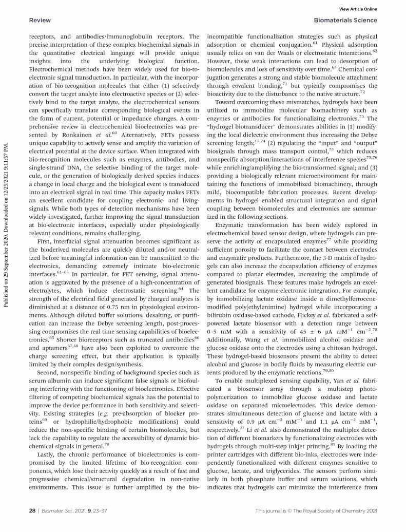
In terms of non-electroactive cells, most of their functionsare regulated by biochemical signals. The specific electrical/electrochemical transduction of these signals relies on theappropriate functioning and effective integration of bio-reco-gnition molecules, where the hydrogel could enable uniquepossibilities to promote interfacial signaling as discussedearlier. For instance, Misun et al. demonstrated the ampero-metric detection of glucose consumption and lactate pro-duction from human colon carcinoma spheroids.115 Thedevice consisted of two modular components: a microfluidicplatform for media perfusion and glass plug-in with electrodecomponents (Fig. 9a). The electrodes were functionalized withthe enzymes: glucose oxidase or lactate oxidase immobilizedin a hydrogel, enabling the real time detection of cell metab-olism. The device measured the real time secretion/consump-tion of analytes from the perfused cell media (Fig. 9b). Lian
et al. reported the amperometric detection of hydrogen per-oxide secreted from HeLa cells utilizing horseradish peroxi-dase (HRP) functionalized hydrogel coating on a glassy carbonelectrode (Fig. 9c).106 HeLa cells were cultured on top of bio-active hydrogels, showing activity for up to two weeks. Cellswere stimulated with Phorbol 12-myristate 13-acetate (PMA) totrigger hydrogen peroxide production. Horseradish peroxidase(HRP) was immobilized in the hydrogel, enabling the real timedetection of hydrogen peroxide (Fig. 9d). The hydrogel alsoserved to inhibit the diffusion of hydrogen peroxide secretedfrom cells, effectively increasing the concentration that directlyinteracts with the HRP enzyme. A similar design has beenapplied by Yan et al. to study the metabolism of macro-phages.105 These studies demonstrate the possibility for realtime interpretation of cellular metabolic signals, which couldbe further expanded through incorporating different bio-markers and/or bioreceptors for real time drug screening,disease monitoring and personalized medicine.
4.2 Wearable bioelectronics
Wearable bioelectronics are capable of real-time, noninvasivemonitoring of physiological signals, and have become increas-ingly common in our everyday lives, e.g. in the form of smartwatches/bands that can continuously measure heart rate or
Fig. 9 Hydrogel functionalization enables real time monitoring of cell metabolism. (a) Schematic of the biosensor device with hanging drop net-works for cell culture and hydrogels functionalized with lactate oxidase and glucose oxidase. (b) Real time monitoring of glucose consumption andlactate production. Reproduced from ref. 115 with permission from Springer Nature, Copyright © 2016. (c) Schematic of hydrogel formation and cellintegration for electrochemical biosensing of H2O2 after chemical stimuli. (d) Current response of the sensor with (red) and without (black) HeLacells after chemical stimulation. Reproduced from ref. 106 with permission from American Chemical Society, Copyright © 2016.
Biomaterials Science Review
This journal is © The Royal Society of Chemistry 2021 Biomater. Sci., 2021, 9, 23–37 | 33
Publ
ishe
d on
29
Sept
embe
r 20
20. D
ownl
oade
d on
12/
25/2
021
9:11
:57
PM.
View Article Online

blood oxygen saturation.116 However, these commercially avail-able wearable devices share some similar challenges withmetal/semiconductor based bioelectronics with unstable bodycontact that is associated with low sensitivity and fluctuationin sensing results.117 To address this issue, flexible andstretchable electronics have been developed that comply withthe curvatures of the human body; maintaining stable contactsto ensure consistent sensing results. Toward this goal, hydro-gels are suggested as ideal body-electronics interfacingmaterials due to their superior mechanical properties andtunable bio-adhesiveness. For example, Pan et al. reportedhydrogel-elastomer composites with low stiffness and highadhesiveness for interfacing with skin.118 Gold nanofilms wereincorporated into the hydrogel structure for electrical conduc-tivity and were demonstrated for on-skin electromyographyand electrocardiography. The reported Young’s modulus of thehydrogel composite was reported to be near 5.3 kPa and couldstretch 25 times its length, enabling conformal contact withthe skin. This work provides a general strategy for on-skinbioelectronics by engineering the hydrogel properties.
In wearable electronics, body motions are one of the mostcommon challenges that can lead to device detachment,abrasion, fracture, and eventually failure of device functions.Recent studies in stretchable-, tough- and healable-hydrogelsprovide potential solutions to this challenge.119,120 Withfurther enhanced ionic conductivity, these novel hydrogelsshow potential to replace state-of-the-art substrates (e.g. metal,semiconductor, dry polymer, etc.) in the development of nextgeneration wearable electronics. For example, Zhao et al. fabri-cated a conductive hydrogel from a supramolecular assemblyof polydopamine decorated silver nanoparticles, polyaniline,and polyvinyl alcohol. The conductive hydrogel displayedtunable stiffness (132 Pa to 40 kPa), stretchiness (0.01–500%),self–adhesiveness and self-healing capacity, and has been suc-cessfully implemented as epidermal motion sensors and dia-betic wound dressing.121 Also, Liu et al. created a microfluidic-based, ultra-stretchable hydrogel network with metallic con-ductivity using liquid metal as conductive fillers.122 Thisdevice showed good stretchability and flexibility, which remainfunctional under many types of deformations (e.g. up to 550%stretch, cyclic stretches, bends, and twists). Due to the metallicconductivity, this hydrogel can be applied to the fabrication ofwireless bioelectronics for monitoring physiological conditionsof the human body using near-field communication techno-logy. Furthermore, a variety of functional hydrogel designs forwearable electronics have been comprehensively reviewed byYang and Suo.123
Additionally, multifunctional wearable hydrogel bioelectro-nics has been developed for simultaneous monitoring of thephysiological environment and delivery of drugs for treatment.For example, contact lenses are hydrogel-based medicaldevices that have long been used to correct vision. By embed-ding sensors within the lens, smart contact lenses have beenused for monitoring diseases such as glaucoma anddiabetes.124,125 Keum et al. demonstrated contact lensescapable of monitoring glucose levels from tears in rabbits and
delivery of the drugs metformin and genistein for the treat-ment of hyperglycemia and diabetic retinopathy.126 Similarly,a smart bandage was developed for monitoring of the woundenvironment and delivery of antibiotics.127 Overall, hydrogelscan create many new possibilities in wearable electronicsowing to their programmable mechanical, electrical, andchemical properties.128,129
5. Conclusions
Engineered hydrogel interfaces have shown great promisetowards the seamless structural and functional integrationbetween biological and electronic systems, which is transform-ing the design and development of next-generation bioelectro-nics across molecular, cellular, tissue and body levels. Themismatch at the heterogeneous interface, both structurallyand functionally, can be blurred by rationally programmingthe physiochemical parameters through controlled hydrogelsynthesis/fabrication. In terms of structures, hydrogels providea mechanically compliant, chemically active, and biologicallyfavourable microenvironment for seamless bio-integration thatis difficult to achieve on a traditional electronic interface. Interms of functions, hydrogels can facilitate the signal trans-duction between bio- (ions & molecules) and electrical-(elec-trons & holes) circuit by precisely regulating interfacial massand transport, enabling localized amplification and/or filteringof bio-derived signals. At the molecular to the cellular level,the spatial organization and hierarchical assembling of func-tionalized hydrogels will create new signal transduction andenergy conversion cascades with electrically controllableinputs and outputs for novel biosensor and biocatalyst devel-opments.130 At the tissue to the body level, recent develop-ments in stretchable-,131 biodegradable-,132 self-healing-,133
and bio-adhesive-hydrogels134 offer opportunities in designingnew bioelectronic interfaces with intimate contact, minimalinvasiveness, and maximized motion-compliance. Throughthese new bioelectronic interfaces, long term, continuousprobing and regulation of human functions will be achieved,which are expected to contribute significantly to disease diag-nosis and personalized medicine. Overall, we believe thathydrogel-mediated bio-integratable electronics can initiate anevolution in the way we communicate with biological systemsby unambiguously decoding critical biological languages andprecisely defining/regulating complex bio-functions.
The future of hydrogel-based bioelectronics is anticipatedto implement more advanced functions beyond the currentscope of bioelectronics. However, before hydrogels can fullyaddress the interfacing challenges, more validation andoptimizations are required. Mainly, their long-term perform-ance and biocompatibility demand further evaluation andoptimization in order to obtain intimately integrated, yetchronically stable bio-interfaces, which are critically importantto in vivo and implanted applications. Other concerns includedegradation and potential cytotoxicity of different synthetichydrogels, as well as additional complexity and variability in
Review Biomaterials Science
34 | Biomater. Sci., 2021, 9, 23–37 This journal is © The Royal Society of Chemistry 2021
Publ
ishe
d on
29
Sept
embe
r 20
20. D
ownl
oade
d on
12/
25/2
021
9:11
:57
PM.
View Article Online

transducing and interpreting bioderived signals. In the longterm, given the ability to tune the physical and chemical pro-perties, biological interactions, and more, we are optimistic forhydrogels with the potential to address many challenges inbioelectronics.
Conflicts of interest
There are no conflicts to declare.
Acknowledgements
The authors gratefully acknowledge support from the NationalScience Foundation (CBET-1803907 and DMR-1652095).
References
1 J. Wang, Chem. Rev., 2008, 108, 814–825.2 H. Berger, Arch. Psychiatr. Nervenkrankh., 1929, 87, 527–
570.3 M. AlGhatrif and J. Lindsay, J. Community Hosp. Intern.
Med. Perspect., 2012, 2, 14383.4 O. Aquilina, Images Paediatr. Cardiol., 2006, 8, 17–81.5 J. Gardner, Soc. Stud. Sci., 2013, 43, 707–728.6 J. P. Seymour, F. Wu, K. D. Wise and E. Yoon, Microsyst.
Nanoeng., 2017, 3, 1–16.7 T. Yeung, P. C. Georges, L. A. Flanagan, B. Marg, M. Ortiz,
M. Funaki, N. Zahir, W. Ming, V. Weaver and P. A. Janmey,Cell Motil., 2005, 60, 24–34.
8 M. HajjHassan, V. Chodavarapu and S. Musallam, Sensors,2008, 8, 6704–6726.
9 V. S. Polikov, P. A. Tresco and W. M. Reichert, J. Neurosci.Methods, 2005, 148, 1–18.
10 N. Wisniewski, F. Moussy and W. M. Reichert,Fresenius. J. Anal. Chem., 2000, 366, 611–621.
11 R. C. Kelly, M. A. Smith, J. M. Samonds, A. Kohn,A. B. Bonds, J. A. Movshon and T. S. Lee, J. Neurosci.,2007, 27, 261–264.
12 A. Zhang and C. M. Lieber, Chem. Rev., 2016, 116, 215–257.
13 T. Zhou, G. Hong, T.-M. Fu, X. Yang, T. G. Schuhmann,R. D. Viveros and C. M. Lieber, Proc. Natl. Acad.Sci. U. S. A., 2017, 114, 5894–5899.
14 M. E. Spira and A. Hai, Nat. Nanotechnol., 2013, 8, 83–94.15 N. Gao, W. Zhou, X. Jiang, G. Hong, T.-M. Fu and
C. M. Lieber, Nano Lett., 2015, 15, 2143–2148.16 A. S. Hoffman, Adv. Drug Delivery Rev., 2012, 64, 18–23.17 H. Yuk, B. Lu and X. Zhao, Chem. Soc. Rev., 2019, 48,
1642–1667.18 S. Lin, C. Cao, Q. Wang, M. Gonzalez, J. E. Dolbow and
X. Zhao, Soft Matter, 2014, 10, 7519–7527.19 B. Xu, P. Zheng, F. Gao, W. Wang, H. Zhang, X. Zhang,
X. Feng and W. Liu, Adv. Funct. Mater., 2017, 27, 1604327.
20 J. L. Drury and D. J. Mooney, Biomaterials, 2003, 24, 4337–4351.
21 Y. Liu, W. He, Z. Zhang and B. P. Lee, Gels, 2018, 4, 46.22 Y. Liu, J. Liu, S. Chen, T. Lei, Y. Kim, S. Niu, H. Wang,
X. Wang, A. M. Foudeh, J. B.-H. Tok and Z. Bao, Nat.Biomed. Eng., 2019, 3, 58–68.
23 J. Yang, R. Bai, B. Chen and Z. Suo, Adv. Funct. Mater.,2020, 30, 1901693.
24 D. Gao, K. Parida and P. S. Lee, Adv. Funct. Mater., 2020,30, 1907184.
25 Y. S. Zhang and A. Khademhosseini, Science, 2017, 356,eaaf3627.
26 X. Li, Q. Sun, Q. Li, N. Kawazoe and G. Chen, Front.Chem., 2018, 6, 499.
27 J. Yan, V. A. Pedrosa, A. L. Simonian and A. Revzin, ACSAppl. Mater. Interfaces, 2010, 2, 748–755.
28 J. Chen, Q. Peng, T. Thundat and H. Zeng, Chem. Mater.,2019, 31, 4553–4563.
29 N. A. Alba, R. J. Sclabassi, M. Sun and X. T. Cui, IEEE.Trans. Neural. Syst. Rehabil. Eng., 2010, 18, 415–423.
30 S. Ji, C. Wan, T. Wang, Q. Li, G. Chen, J. Wang, Z. Liu,H. Yang, X. Liu and X. Chen, Adv. Mater., 2020, 32, 2001496.
31 M. Baumgartner, F. Hartmann, M. Drack, D. Preninger,D. Wirthl, R. Gerstmayr, L. Lehner, G. Mao, R. Pruckner,S. Demchyshyn, L. Reiter, M. Strobel, T. Stockinger,D. Schiller, S. Kimeswenger, F. Greibich, G. Buchberger,E. Bradt, S. Hild, S. Bauer and M. Kaltenbrunner, Nat.Mater., 2020, 1–8.
32 Z. Taylor and K. Miller, J. Biomech., 2004, 37, 1263–1269.33 W. Hiesinger, M. J. Brukman, R. C. McCormick,
J. R. Fitzpatrick, J. R. Frederick, E. C. Yang, J. R. Muenzer,N. A. Marotta, M. F. Berry, P. Atluri and Y. J. Woo,J. Thorac. Cardiovasc. Surg., 2012, 143, 962–966.
34 B. Wang, W. Huang, L. Chi, M. Al-Hashimi, T. J. Marksand A. Facchetti, Chem. Rev., 2018, 118, 5690–5754.
35 S. R. Goldstein and M. Salcman, IEEE Trans. Biomed. Eng.,1973, BME-20, 260–269.
36 J. W. Salatino, K. A. Ludwig, T. D. Y. Kozai andE. K. Purcell, Nat. Biomed. Eng., 2017, 1, 862–877.
37 A. Prasad and J. C. Sanchez, J. Neural Eng., 2012, 9, 026028.38 K. Woeppel, Q. Yang and X. T. Cui, Curr. Opin. Biomed.
Eng., 2017, 4, 21–31.39 J. Goding, A. Gilmour, P. Martens, L. Poole-Warren and
R. Green, Adv. Healthcare Mater., 2017, 6, 1601177.40 L. Ferlauto, A. N. D’Angelo, P. Vagni, M. J. I. Airaghi
Leccardi, F. M. Mor, E. A. Cuttaz, M. O. Heuschkel,L. Stoppini and D. Ghezzi, Front. Neurosci., 2018, 12, 648.
41 R. G. Wells, Hepatology, 2008, 47, 1394–1400.42 A. J. Engler, C. Carag-Krieger, C. P. Johnson, M. Raab,
H.-Y. Tang, D. W. Speicher, J. W. Sanger, J. M. Sanger andD. E. Discher, J. Cell Sci., 2008, 121, 3794–3802.
43 K. C. Spencer, J. C. Sy, K. B. Ramadi, A. M. Graybiel,R. Langer and M. J. Cima, Sci. Rep., 2017, 7, 1–16.
44 J. D. Enderle, in Introduction to Biomedical Engineering, ed.J. D. Enderle and J. D. Bronzino, Academic Press, Boston,3rd edn, 2012, ch. 8, pp. 447–508.
Biomaterials Science Review
This journal is © The Royal Society of Chemistry 2021 Biomater. Sci., 2021, 9, 23–37 | 35
Publ
ishe
d on
29
Sept
embe
r 20
20. D
ownl
oade
d on
12/
25/2
021
9:11
:57
PM.
View Article Online

45 M. Escabí, in Introduction to Biomedical Engineering, ed.J. D. Enderle and J. D. Bronzino, Academic Press, Boston,3rd edn, 2012, ch. 11, pp. 667–746.
46 J. D. Enderle, in Introduction to Biomedical Engineering, ed.J. D. Enderle and J. D. Bronzino, Academic Press, Boston,3rd edn, 2012, ch. 12, pp. 747–815.
47 C.-J. Lee, H. Wu, Y. Hu, M. Young, H. Wang, D. Lynch,F. Xu, H. Cong and G. Cheng, ACS Appl. Mater. Interfaces,2018, 10, 5845–5852.
48 S. Zhao, P. Tseng, J. Grasman, Y. Wang, W. Li, B. Napier,B. Yavuz, Y. Chen, L. Howell, J. Rincon, F. G. Omenettoand D. L. Kaplan, Adv. Mater., 2018, 30, 1800598.
49 I. Noshadi, B. W. Walker, R. Portillo-Lara, E. S. Sani,N. Gomes, M. R. Aziziyan and N. Annabi, Sci. Rep., 2017,7, 4345.
50 H. Jo, M. Sim, S. Kim, S. Yang, Y. Yoo, J.-H. Park,T. H. Yoon, M.-G. Kim and J. Y. Lee, Acta Biomater., 2017,48, 100–109.
51 X. Liu, A. L. Miller, S. Park, B. E. Waletzki, Z. Zhou,A. Terzic and L. Lu, ACS Appl. Mater. Interfaces, 2017, 9,14677–14690.
52 P. Baei, S. Jalili-Firoozinezhad, S. Rajabi-Zeleti,M. Tafazzoli-Shadpour, H. Baharvand and N. Aghdami,Mater. Sci. Eng., C, 2016, 63, 131–141.
53 L. Xu, X. Li, T. Takemura, N. Hanagata, G. Wu andL. L. Chou, J. Nanobiotechnol., 2012, 10, 16.
54 A. N. Dalrymple, U. A. Robles, M. Huynh, B. A. Nayagam,R. A. Green, L. A. Poole-Warren, J. B. Fallon andR. K. Shepherd, J. Neural Eng., 2020, 17, 026018.
55 Y. Liu, A. F. McGuire, H.-Y. Lou, T. L. Li, J. B.-H. Tok,B. Cui and Z. Bao, Proc. Natl. Acad. Sci. U. S. A., 2018, 115,11718–11723.
56 V. R. Feig, H. Tran, M. Lee and Z. Bao, Nat. Commun.,2018, 9, 2740.
57 H. Yuk, B. Lu, S. Lin, K. Qu, J. Xu, J. Luo and X. Zhao, Nat.Commun., 2020, 11, 1–8.
58 J. Liu, X. Zhang, Y. Liu, M. Rodrigo, P. D. Loftus,J. Aparicio-Valenzuela, J. Zheng, T. Pong, K. J. Cyr,M. Babakhanian, J. Hasi, J. Li, Y. Jiang, C. J. Kenney,P. J. Wang, A. M. Lee and Z. Bao, Proc. Natl. Acad.Sci. U. S. A., 2020, 117, 14769.
59 D. N. Heo, S.-J. Song, H.-J. Kim, Y. J. Lee, W.-K. Ko, S. J. Lee,D. Lee, S. J. Park, L. G. Zhang, J. Y. Kang, S. H. Do, S. H. Leeand I. K. Kwon, Acta Biomater., 2016, 39, 25–33.
60 N. J. Ronkainen, H. B. Halsall and W. R. Heineman,Chem. Soc. Rev., 2010, 39, 1747–1763.
61 W. Schramm, S.-H. Paek and G. Voss, ImmunoMethods,1993, 3, 93–103.
62 N. R. Mohamad, N. H. C. Marzuki, N. A. Buang, F. Huyopand R. A. Wahab, Biotechnol. Biotechnol. Equip., 2015, 29,205–220.
63 U. Guzik, K. Hupert-Kocurek and D. Wojcieszyńska,Molecules, 2014, 19, 8995–9018.
64 E. Stern, R. Wagner, F. J. Sigworth, R. Breaker,T. M. Fahmy and M. A. Reed, Nano Lett., 2007, 7, 3405–3409.
65 G. Zheng, F. Patolsky, Y. Cui, W. U. Wang andC. M. Lieber, Nat. Biotechnol., 2005, 23, 1294–1301.
66 R. Elnathan, M. Kwiat, A. Pevzner, Y. Engel, L. Burstein,A. Khatchtourints, A. Lichtenstein, R. Kantaev andF. Patolsky, Nano Lett., 2012, 12, 5245–5254.
67 N. Nakatsuka, K.-A. Yang, J. M. Abendroth, K. M. Cheung,X. Xu, H. Yang, C. Zhao, B. Zhu, Y. S. Rim, Y. Yang,P. S. Weiss, M. N. Stojanović and A. M. Andrews, Science,2018, 362, 319–324.
68 N. Kumar, M. Gray, J. C. Ortiz-Marquez, A. Weber,C. R. Desmond, A. Argun, T. van Opijnen and K. S. Burch,Med. Devices Sens., 2020, e10121.
69 J. E. Contreras-Naranjo and O. Aguilar, Biosensors, 2019, 9,15.
70 P. Roach, D. Farrar and C. C. Perry, J. Am. Chem. Soc.,2005, 127, 8168–8173.
71 Y. Li, T. L. Ogorzalek, S. Wei, X. Zhang, P. Yang,J. Jasensky, C. L. Brooks, E. N. G. Marsh and Z. Chen,Phys. Chem. Chem. Phys., 2018, 20, 1021–1029.
72 S. Datta, L. R. Christena and Y. R. S. Rajaram, 3 Biotech,2013, 3, 1–9.
73 J. Tavakoli and Y. Tang, Polymers, 2017, 9, 364.74 N. Gao, T. Gao, X. Yang, X. Dai, W. Zhou, A. Zhang and
C. M. Lieber, Proc. Natl. Acad. Sci. U. S. A., 2016, 113,14633–14638.
75 X. Dai, R. Vo, H.-H. Hsu, P. Deng, Y. Zhang and X. Jiang,Nano Lett., 2019, 19, 6658–6664.
76 H. H. Bay, R. Vo, X. Dai, H.-H. Hsu, Z. Mo, S. Cao, W. Li,F. G. Omenetto and X. Jiang, Nano Lett., 2019, 19, 2620–2626.
77 J. Kunkel and P. Asuri, PLoS One, 2014, 9, e86785.78 D. P. Hickey, R. C. Reid, R. D. Milton and S. D. Minteer,
Biosens. Bioelectron., 2016, 77, 26–31.79 J. Kim, I. Jeerapan, S. Imani, T. N. Cho, A. Bandodkar,
S. Cinti, P. P. Mercier and J. Wang, ACS Sens., 2016, 1,1011–1019.
80 A. J. Bandodkar, W. Jia, C. Yardımcı, X. Wang, J. Ramirezand J. Wang, Anal. Chem., 2015, 87, 394–398.
81 L. Li, L. Pan, Z. Ma, K. Yan, W. Cheng, Y. Shi and G. Yu,Nano Lett., 2018, 18, 3322–3327.
82 M. J. Schöning and A. Poghossian, Analyst, 2002, 127,1137–1151.
83 Y. Maeda, A. Matsumoto, Y. Miura and Y. Miyahara,Nanoscale Res. Lett., 2012, 7, 108.
84 T. Miyata, N. Asami and T. Uragami, Nature, 1999, 399,766–769.
85 T. Aoki, K. Nakamura, K. Sanui, A. Kikuchi, T. Okano,Y. Sakurai and N. Ogata, Polym. J., 1999, 31, 1185–1188.
86 K. Podual, F. J. Doyle and N. A. Peppas, J. ControlledRelease, 2000, 67, 9–17.
87 P. B. Welzel, S. Prokoph, A. Zieris, M. Grimmer,S. Zschoche, U. Freudenberg and C. Werner, Polymers,2011, 3, 602–620.
88 H. Chavda and C. Patel, Int. J. Pharm. Invest., 2011, 1, 17–21.
Review Biomaterials Science
36 | Biomater. Sci., 2021, 9, 23–37 This journal is © The Royal Society of Chemistry 2021
Publ
ishe
d on
29
Sept
embe
r 20
20. D
ownl
oade
d on
12/
25/2
021
9:11
:57
PM.
View Article Online

89 N. W. Choi, J. Kim, S. C. Chapin, T. Duong, E. Donohue,P. Pandey, W. Broom, W. A. Hill and P. S. Doyle, Anal.Chem., 2012, 84, 9370–9378.
90 S. L. Burrs, D. C. Vanegas, M. Bhargava, N. Mechulan,P. Hendershot, H. Yamaguchi, C. Gomes andE. S. McLamore, Analyst, 2015, 140, 1466–1476.
91 H. J. Kim, W. Choi, J. Kim, J. Choi, N. Choi andK. S. Hwang, Sens. Actuators, B, 2020, 302, 127190.
92 R. J. Russell, A. C. Axel, K. L. Shields and M. V. Pishko,Polymer, 2001, 42, 4893–4901.
93 B. Amsden, Macromolecules, 1998, 31, 8382–8395.94 E. Axpe, D. Chan, G. S. Offeddu, Y. Chang, D. Merida,
H. L. Hernandez and E. A. Appel, Macromolecules, 2019,52, 6889–6897.
95 A. Star, E. Tu, J. Niemann, J.-C. P. Gabriel, C. S. Joiner andC. Valcke, Proc. Natl. Acad. Sci. U. S. A., 2006, 103, 921–926.
96 E. Stern, J. F. Klemic, D. A. Routenberg, P. N. Wyrembak,D. B. Turner-Evans, A. D. Hamilton, D. A. LaVan,T. M. Fahmy and M. A. Reed, Nature, 2007, 445, 519–522.
97 F. Patolsky, G. Zheng, O. Hayden, M. Lakadamyali,X. Zhuang and C. M. Lieber, Proc. Natl. Acad. Sci. U. S. A.,2004, 101, 14017–14022.
98 G. L. Mario Cheong, K. S. Lim, A. Jakubowicz,P. J. Martens, L. A. Poole-Warren and R. A. Green, ActaBiomater., 2014, 10, 1216–1226.
99 J. O. Winter, S. F. Cogan and J. F. Rizzo, J. Biomed. Mater.Res., Part B, 2007, 81, 551–563.
100 M. L. McCain, A. Agarwal, H. W. Nesmith, A. P. Nesmithand K. K. Parker, Biomaterials, 2014, 35, 5462–5471.
101 D. A. Soscia, D. Lam, A. C. Tooker, H. A. Enright,M. Triplett, P. Karande, S. K. G. Peters, A. P. Sales,E. K. Wheeler and N. O. Fischer, Lab Chip, 2020, 20, 901–911.
102 D. Lam, H. A. Enright, S. K. G. Peters, M. L. Moya,D. A. Soscia, J. Cadena, J. A. Alvarado, K. S. Kulp,E. K. Wheeler and N. O. Fischer, J. Neurosci. Methods,2020, 329, 108460.
103 S. Lee, D. Sasaki, D. Kim, M. Mori, T. Yokota, H. Lee,S. Park, K. Fukuda, M. Sekino, K. Matsuura,T. Shimizu and T. Someya, Nat. Nanotechnol., 2019, 14,156–160.
104 V. J. Kujala, F. S. Pasqualini, J. A. Goss, J. C. Nawroth andK. K. Parker, J. Mater. Chem. B, 2016, 4, 3534–3543.
105 J. Yan, Y. Sun, H. Zhu, L. Marcu and A. Revzin, Biosens.Bioelectron., 2009, 24, 2604–2610.
106 M. Lian, X. Chen, Y. Lu and W. Yang, ACS Appl. Mater.Interfaces, 2016, 8, 25036–25042.
107 M. Kitsara, D. Kontziampasis, O. Agbulut and Y. Chen,Microelectron. Eng., 2019, 203–204, 44–62.
108 P. Fattahi, G. Yang, G. Kim and M. R. Abidian, Adv.Mater., 2014, 26, 1846–1885.
109 J. L. Bourke, A. F. Quigley, S. Duchi, C. D. O’Connell,J. M. Crook, G. G. Wallace, M. J. Cook and R. M. I. Kapsa,J. Tissue Eng. Regener. Med., 2018, 12, 490–493.
110 S. P. Paşca, Nature, 2018, 553, 437–445.
111 R. Edmondson, J. J. Broglie, A. F. Adcock and L. Yang,Assay Drug Dev. Technol., 2014, 12, 207–218.
112 A. Kalmykov, C. Huang, J. Bliley, D. Shiwarski,J. Tashman, A. Abdullah, S. K. Rastogi, S. Shukla,E. Mataev, A. W. Feinberg, K. J. Hsia and T. Cohen-Karni,Sci. Adv., 2019, 5, eaax0729.
113 A. P. Napolitano, D. M. Dean, A. J. Man, J. Youssef,D. N. Ho, A. P. Rago, M. P. Lech and J. R. Morgan,BioTechniques, 2007, 43, 494–500.
114 M. W. Tibbitt and K. S. Anseth, Biotechnol. Bioeng., 2009,103, 655–663.
115 P. M. Misun, J. Rothe, Y. R. F. Schmid, A. Hierlemann andO. Frey, Microsyst. Nanoeng., 2016, 2, 1–9.
116 D. Phan, L. Y. Siong, P. N. Pathirana and A. Seneviratne,in 2015 International Symposium on Bioelectronics andBioinformatics (ISBB), 2015, pp. 144–147.
117 Z. Lou, L. Wang and G. Shen, Adv. Mater. Technol., 2018,3, 1800444.
118 S. Pan, F. Zhang, P. Cai, M. Wang, K. He, Y. Luo, Z. Li,G. Chen, S. Ji, Z. Liu, X. J. Loh and X. Chen, Adv. Funct.Mater., 2020, 26, 1909540.
119 J.-Y. Sun, X. Zhao, W. R. K. Illeperuma, O. Chaudhuri,K. H. Oh, D. J. Mooney, J. J. Vlassak and Z. Suo, Nature,2012, 489, 133–136.
120 T. Kakuta, Y. Takashima, M. Nakahata, M. Otsubo,H. Yamaguchi and A. Harada, Adv. Mater., 2013, 25, 2849–2853.
121 Y. Zhao, Z. Li, S. Song, K. Yang, H. Liu, Z. Yang, J. Wang,B. Yang and Q. Lin, Adv. Funct. Mater., 2019, 29, 1901474.
122 Y. Liu, T. Yang, Y. Zhang, G. Qu, S. Wei, Z. Liu andT. Kong, Adv. Mater., 2019, 31, 1902783.
123 C. Yang and Z. Suo, Nat. Rev. Mater., 2018, 3, 125–142.124 G. E. Dunbar, B. Y. Shen and A. A. Aref, Clin. Ophthalmol.,
2017, 11, 875–882.125 M. Senior, Nat. Biotechnol., 2014, 32, 856–856.126 D. H. Keum, S.-K. Kim, J. Koo, G.-H. Lee, C. Jeon,
J. W. Mok, B. H. Mun, K. J. Lee, E. Kamrani, C.-K. Joo,S. Shin, J.-Y. Sim, D. Myung, S. H. Yun, Z. Bao andS. K. Hahn, Sci. Adv., 2020, 6, eaba3252.
127 P. Mostafalu, A. Tamayol, R. Rahimi, M. Ochoa,A. Khalilpour, G. Kiaee, I. K. Yazdi, S. Bagherifard,M. R. Dokmeci, B. Ziaie, S. R. Sonkusale andA. Khademhosseini, Small, 2018, 14, 1703509.
128 Z. Lou, L. Wang, K. Jiang, Z. Wei and G. Shen, Mater. Sci.Eng., R, 2020, 140, 100523.
129 J. C. Yang, J. Mun, S. Y. Kwon, S. Park, Z. Bao and S. Park,Adv. Mater., 2019, 31, 1904765.
130 L. Hsu and X. Jiang, Trends Biotechnol., 2019, 37, 795–796.131 H. Liu, M. Li, C. Ouyang, T. J. Lu, F. Li and F. Xu, Small,
2018, 14, 1801711.132 B. Guo, A. Finne-Wistrand and A.-C. Albertsson, Chem.
Mater., 2011, 23, 1254–1262.133 Z. Deng, H. Wang, P. X. Ma and B. Guo, Nanoscale, 2020,
12, 1224–1246.134 H. Chopra, S. Kumar and I. Singh, in Bioadhesives in Drug
Delivery, John Wiley & Sons, Ltd, 2020, ch. 6, pp. 147–170.
Biomaterials Science Review
This journal is © The Royal Society of Chemistry 2021 Biomater. Sci., 2021, 9, 23–37 | 37
Publ
ishe
d on
29
Sept
embe
r 20
20. D
ownl
oade
d on
12/
25/2
021
9:11
:57
PM.
View Article Online