History Organism Echinococciasis Hydatidosis Epidemiology ...
Upload
trinhtuongCategory
view
216download
0
Pathology and Laboratory Medicine 2017; 1(1): 1-4
http://www.sciencepublishinggroup.com/j/plm
doi: 10.11648/j.plm.20170101.11
Case Report
Hydatid Cyst of the Kidney: About Two Rare Cases
Oukkas Siham1, *
, El Mezouari El mostafa1, Redouane Rhoukhsi
2, Redouane Moutaj
1
1Department of Parasitology Mycology, Avicenna Military Hospital, Medical School, Cadi Ayyad University, Marrakech, Morocco 2Department of Radiology, Avicenna Military Hospital, Medical School, Cadi Ayyad University, Marrakech, Morocco
Email address:
*Corresponding author
To cite this article: Oukkas Siham, El Mezouari El mostafa, Redouane Rhoukhsi, Redouane Moutaj. Hydatid Cyst of the Kidney: About Two Rare Cases.
Pathology and Laboratory Medicine. Vol. 1, No. 1, 2017, pp. 1-4. doi: 10.11648/j.plm.20170101.11
Received: January 26, 2017; Accepted: February 13, 2017; Published: March 2, 2017
Abstract: Hydatidosis is a rare parasitic disease that is endemic in some countries of the Mediterranean basin. Renal
localization is rare and exists only in 5% of the visceral forms. The diagnosis of hydatid cyst of the kidney is suspected in
epidemiological, clinical, radiological and biological arguments. The clinical symptomatology is variable, and depends on the
evolutionary stage of the cyst. Hydration is the only pathognomonic sign, but it exists in only 10 to 30% of cases. Ultrasound
makes it possible to suspect the hydatic nature of the lesion in more than 50% of cases; Computed tomography and magnetic
resonance imaging are useful in cases of diagnostic doubt. The reference treatment of the renal hydatid cyst is Conservative
surgery with resection of the protruding dome. A total nephrectomy is conceived only in front of a kidney completely destroyed.
Keywords: Cyst, Echinococcus Granulosus, Marrakech
1. Introduction
Hydatidosis is an antropozoonosis due to development in
the larval form of the Taenia: Echinococcus granulosus. Like
all tapeworms, the evolutionary cycle of the parasite takes
place between the final host (canidae) and the intermediate
host (several herbivorous or omnivorous mammals including
sheep and accidentally man. The intermediate host is
contaminated by ingestion of embryonated eggs
(embryophores) eliminated in the external environment by
the final host. The hexacanth embryo, released into the
gastrointestinal tract, passes through the intestinal wall and
gains, via the bloodstream, the liver and lungs. Other organs
may be affected. It is stopped in 50-60% of cases by the first
filter (hepatic filter), then in 30% to 40% of cases by the
second filter (pulmonary filter) and is found in the rest of the
body (bone, brain, Thyroid...) in 10% of the cases [1]. The
ingestion of a hydatid cyst by a canine results in the release of
larvae (scolex) into the intestine. At this stage, the scolex
becomes adult worms. Human infestation occurs by
accidental ingestion of E. granulosus, either by absorption of
dirty foods or by contact with a dog. It is thus favored by
promiscuity with dogs and herbivores, which explains its
high prevalence in rural areas, man constituting a parasitic
impasse in that it is no longer usually a prey for canids. Renal
localization is the 3rd place after pulmonary and hepatic
localization. It is rare and represents less than 5% of the
visceral forms. The most frequent visceral sites are the liver
(60%) and the lung (30%). However, it threatens the
functional prognosis of the kidney and even the prognosis
because it exposes the risk of rupture and superinfection of
the cyst. The imagery makes it possible to make the diagnosis,
to appreciate the impact and to look for complications.
2. Observations
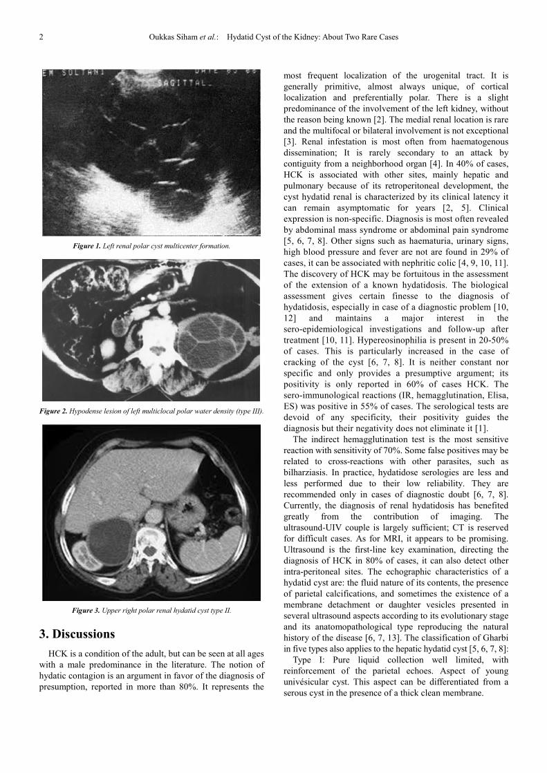
Observation 1: Man of 45 who exhibits receding left lumbar
pain in analgesic treatments and in whom an ultrasound
[Figure 1] completed with a CT scan showing the presence of a
type III hydatid cyst has been performed [Figure 2].
Observation 2: Patient operated on a hydatic cyst of the
liver 6 years ago who is suffering from the pains of the right
hypochondrium posterior irradiation and in whom abdominal
CT carried out has objectified the presence of a type II
hydatid cyst [Figure 3].
2 Oukkas Siham et al.: Hydatid Cyst of the Kidney: About Two Rare Cases
Figure 1. Left renal polar cyst multicenter formation.
Figure 2. Hypodense lesion of left multiclocal polar water density (type III).
Figure 3. Upper right polar renal hydatid cyst type II.
3. Discussions
HCK is a condition of the adult, but can be seen at all ages
with a male predominance in the literature. The notion of
hydatic contagion is an argument in favor of the diagnosis of
presumption, reported in more than 80%. It represents the
most frequent localization of the urogenital tract. It is
generally primitive, almost always unique, of cortical
localization and preferentially polar. There is a slight
predominance of the involvement of the left kidney, without
the reason being known [2]. The medial renal location is rare
and the multifocal or bilateral involvement is not exceptional
[3]. Renal infestation is most often from haematogenous
dissemination; It is rarely secondary to an attack by
contiguity from a neighborhood organ [4]. In 40% of cases,
HCK is associated with other sites, mainly hepatic and
pulmonary because of its retroperitoneal development, the
cyst hydatid renal is characterized by its clinical latency it
can remain asymptomatic for years [2, 5]. Clinical
expression is non-specific. Diagnosis is most often revealed
by abdominal mass syndrome or abdominal pain syndrome
[5, 6, 7, 8]. Other signs such as haematuria, urinary signs,
high blood pressure and fever are not are found in 29% of
cases, it can be associated with nephritic colic [4, 9, 10, 11].
The discovery of HCK may be fortuitous in the assessment
of the extension of a known hydatidosis. The biological
assessment gives certain finesse to the diagnosis of
hydatidosis, especially in case of a diagnostic problem [10,
12] and maintains a major interest in the
sero-epidemiological investigations and follow-up after
treatment [10, 11]. Hypereosinophilia is present in 20-50%
of cases. This is particularly increased in the case of
cracking of the cyst [6, 7, 8]. It is neither constant nor
specific and only provides a presumptive argument; its
positivity is only reported in 60% of cases HCK. The
sero-immunological reactions (IR, hemagglutination, Elisa,
ES) was positive in 55% of cases. The serological tests are
devoid of any specificity, their positivity guides the
diagnosis but their negativity does not eliminate it [1].
The indirect hemagglutination test is the most sensitive
reaction with sensitivity of 70%. Some false positives may be
related to cross-reactions with other parasites, such as
bilharziasis. In practice, hydatidose serologies are less and
less performed due to their low reliability. They are
recommended only in cases of diagnostic doubt [6, 7, 8].
Currently, the diagnosis of renal hydatidosis has benefited
greatly from the contribution of imaging. The
ultrasound-UIV couple is largely sufficient; CT is reserved
for difficult cases. As for MRI, it appears to be promising.
Ultrasound is the first-line key examination, directing the
diagnosis of HCK in 80% of cases, it can also detect other
intra-peritoneal sites. The echographic characteristics of a
hydatid cyst are: the fluid nature of its contents, the presence
of parietal calcifications, and sometimes the existence of a
membrane detachment or daughter vesicles presented in
several ultrasound aspects according to its evolutionary stage
and its anatomopathological type reproducing the natural
history of the disease [6, 7, 13]. The classification of Gharbi
in five types also applies to the hepatic hydatid cyst [5, 6, 7, 8]:
Type I: Pure liquid collection well limited, with
reinforcement of the parietal echoes. Aspect of young
univésicular cyst. This aspect can be differentiated from a
serous cyst in the presence of a thick clean membrane.

Pathology and Laboratory Medicine 2017; 1(1): 1-4 3
Type II: Liquid collection with a detachment of the
membrane which can produce a floating membrane
appearance. This type is pathognomonic of the hydatic cyst.
Type III: Liquid collection partitioned with multivesicular
aspect.
Type IV: Formations of heterogeneous echostructure,
which can be predominantly fluid or solid.
Type V: Reflective dense image with a posterior shadow
cone corresponding to a frequently sterile calcified cyst. This
aspect is suggestive of diagnosis in an endemic region.
However, other lesions may give the same appearance, such
as kidney cancer or renal tuberculosis cave.
The diagnostic value of ultrasound for Renal Hydatid Cysts
is small when these cysts are small with a size less than 2 cm;
Differential diagnosis with a tumor then becomes difficult [6, 7,
8]. Similarly, the heterogeneous, hyperechogenic,
well-circumscribed pseudotumoral aspect of a renal type IV
cyst may evoke a renal tumor or an abscess. Echodoppler may
provide an additional argument for the diagnosis of Renal Type
IV Hydatid Cyst because there is no intra- and perilional
vascular flow [6, 7, 8]. Intravenous urography has no
contribution in the etiological diagnosis. It shows an avascular
mass syndrome, with no sign of invasion of the excretory
pathways, or even a silent kidney [4, 5, 13]. Calcifications in
projection of the renal area are visible in more than 30%
preparation. This rare sign has a strong diagnostic orientation
when the calcifications are organized in peripheral arciform
rim [6, 7, 8]. Besides the avascular tumor mass syndrome, it
may be a distortion of the contours of the kidney, a
compression, a repression or a stretch of the excretory cavities.
The appearance of opacified extracalicial cavity, which is the
seat of gaps (bead bag appearance), is strongly evoked [6, 7, 8].
An obstructive syndrome, secondary to the encompassment of
the excretory pathway in the pericystic gangue or the migration
of daughter vesicles into the ureter is observed in 5% of cases
[5, 6, 10, 11]. The uro-scanner brings a lot of additional
information. It is necessary whenever the diagnosis is uncertain,
especially for cysts type IV and I. It makes it possible to draw
up a precise topographical assessment, to look for possible
extrarenal localizations. It is more sensitive than ultrasound to
detect calcifications; It better evaluates the nature of the cystic
content and defines the relationship of the cyst with the
neighboring organs and also makes it possible to objectify any
communication with the excretory pathways [6, 7, 8]. Each
tomodensitometric form of HC is designated by 2 parameters:
The type which corresponds to the state of the contents and the
index which specifies the state of the wall [5, 10, 11]:
� type I: homogeneous univésicular cyst;
� type II: heterogeneous univesicular cyst;
� type III: multivesicular cyst;
� type IV: cyst with air inside;
� type V: fully calcified cyst.
index a: thin wall;
index b: thick wall;
index c: calcified wall;
index d: membrane separation
Magnetic resonance imaging is not common practice in
hydatid disease. It offers the possibility of obtaining
multidimensional images.. The vesicular fluid is in
hyposignal in T1 and hypersignal in T2 The cyst then appears
hyper-intense on the sequences weighted in T2 with a
peripheral halo representing the hyposignal of the periyste [6].
The treatment of HCK is essentially surgical. It should
preserve as much as possible the functional renal tissue. Open
surgery remains the classically practiced approach. New less
invasive therapeutic techniques are based on video-assisted
surgery and percutaneous surgery. The approach is dictated
by two parameters: cyst measurements and these
relationships with neighboring organs. In the case of an
enlarged cyst with a previous development or in association
with another intraperitoneal localization, the surgical
approach is performed by a classic lombotomy or sometimes
by an anterior incision type baraya or median or a subcostal
incision in case Of diagnostic difficulty with renal cancer [5,
10, 11]. After careful protection of the operating field with
hypertonic saline or hydrogen peroxide and sterilization of
the cyst by intracystic injection of hypertonic saline serum,
the actual treatment of the cyst. Partial pericystectomy. Or the
resection of the protruding dome which is the preferred
surgical method, it gives excellent results and allows a good
reexpansion of the renal parenchyma. It consists in removing
the exteriorized, superficial and avascular part of the periyste
[1]. Of saline serum re-suction under ultrasound or CT scan
used initially as a diagnostic means, currently allows
non-surgical treatment of hydatic cyst [5, 10, 11]. However,
the risk of spin-off or anaphylactic shock remains
unpredictable.
The place of medical treatment is controversial, for most
authors it is insufficient alone, its main indications are: the
disseminated or multiple hydatidosis and the residual KHR
after surgery. Recently, we are interested in benzimedazole
derivatives:
Albendazole, mebendazole, flubendazole and praziquantel;
These products appear to be active on the larval stages of
Echinococcus granulosus, scolex and especially the
protoscolex [5, 10, 11]. But the use of medical treatment
long-term, clinical and biological monitoring to prove its
effectiveness.
The major complication is the rupture of the cyst in the
urinary tract, which can be manifested by a hydration with the
risk of infection and destruction of the kidney, the other
possible complications are Suppuration of the residual cavity:
In some series, the postoperative suppuration rate reaches 8%
and recidivism which is exceptional [5, 6, 7]. Postoperative
progression is frequently favorable in the literature, except
for a recurrence estimated at less than 5% of cases [5, 6, 7, 8].
The prognosis remains very good in the absence of other
localizations and prophylaxis remains the best treatment in
endemic areas.
4. Conclusion
HCK is a very rare condition. Its clinical semiology is rich,
where only the hydatura is pathognomonic The

4 Oukkas Siham et al.: Hydatid Cyst of the Kidney: About Two Rare Cases
epidemiological context, the biological data and the
echographic or scannographic aspect allow a preoperative
diagnosis of presumption in endemic areas. The treatment is
essentially surgical and conservative. The prognosis is
frequently favorable but this should not make us forget that
the true treatment of hydatidosis remains prophylactic for the
eradication of hydatid disease.
References
[1] T. I. Boudhaye, M. M. Taleb Jiddou, T. Mohamed, B. Salem, C. Jdoud. The primary renal hydatid cyst: A first Mauritanian case. African Journal of Urology, Volume 22, Issue 4, December 2016, Pages 325-328.
[2] N. Bentani, D. Basraoui, B. Wakrim, M. R. Hiroual, N. Cherif Idrissi Ganouni, Z. Dahami, M. S. Moudouni, I. Sarf. Renal hydatid cyst: Radiology and therapeutic.(2012) 22, 999—1003.
[3] A. Darbi 1, D. Bassou 1, S. Akjouj 1, M. Ameur 2, S. Chaouir 1, M. Benameur 1, A. El Kharras 1 Imaging hydatid disease of the kidney Feuillets de Radiologie 2008, 48, n° 5, 283-290.
[4] Hammoudi F, Hartani M Imaging of renal hydatid cyst ― Comments about 35 cases. J Radiol 1989; 70: 549–55.
[5] H. Fekak *, S. Bennani, R. Rabii, M. H. Mezzour, A. Debbagh, A. Joual, M. El Mrini. Hydatic cyst of kidney: about 90 cases. Annales d’urologie 37 (2003) 85–89.
[6] H. Ketata, M. Peyromaure * Service d’urologie, hôpital Cochin, 27, rue du Faubourg-Saint-Jacques, 75014 Paris, France Hydatic cyst of kidney Annales d’urologie 38 (2004) 259–265.
[7] Andersen FL. Introduction to cystic Echinococcosis and description of cooperative research project in Morocco. In: Andersen FL, Ouhelli H, Kachani M, editors. Compendium on cystic Echinococcosis in Africa and Middle Eastern countries with special reference to Morocco. Provo: Brigham Young University; 1997. p. 1–17.
[8] Zmerli S, Ayed M, Horchani A, Chami I, El Ouakdi M, Ben Slama MR. Hydatid cyst of the kidney: diagnosis and treatment. World J Surg 2001; 25: 68–74.
[9] Vonsinner WN, Hellstrom M, Kagevi I, Norlen BJ. Hydatid disease of the urinary tract. J Urol 1993; 149: 557–80.
[10] Biava MF, Kures L. Biological diagnosis of echinococciasis. Rev Prat (Paris) 1990; 40: 201–4.
[11] Bensaid M, Ben Rachid MS, Khaled S, Poirot JL, Roux P, GolvanYT. Serologic screening of hydatidosis in Tunisia. Tunis Méd 1986; 64: 321–4.
[12] Benchekroun A, Lakrissa A, Essakalli N, Abakka T, Faik M, Hachimi M, et al. Hydatic cyst in kidney. Study of 30 cases. J Urol 1986; 92: 171–9.
[13] Murat Uc¸ar a, Ahsen Karago¨zlu¨ Akgu¨l a, Fatih C¸elik b, Nizamettin Kılıc¸ Excisional treatment of renal hydatid cyst mimicking renal tumor with diode laser technique: A case report Journal of Pediatric Urology (2016) xx, 1.e1e1.e5.