Prevalence of Sheep Hydatidosis in North West Bank- … · An-Najah National University Faculty of...
66
An-Najah National University Faculty of Graduate Studies Prevalence of Sheep Hydatidosis in North West Bank- Palestine By Jehad Hammed El-Ibrahim Supervisor Dr Ayman Hussein This thesis is submitted in Partial Fulfillment of the Requirements for the Degree of Master in Public Health of Faculty of Graduate Studies, at An-Najah National University, Nablus, Palestine . 2009
-
Upload
vuongnguyet -
Category
Documents
-
view
213 -
download
0
Transcript of Prevalence of Sheep Hydatidosis in North West Bank- … · An-Najah National University Faculty of...

An-Najah National University Faculty of Graduate Studies
Prevalence of Sheep Hydatidosis in North West Bank- Palestine
By Jehad Hammed El-Ibrahim
Supervisor Dr Ayman Hussein
This thesis is submitted in Partial Fulfillment of the Requirements for the Degree of Master in Public Health of Faculty of Graduate Studies, at An-Najah National University, Nablus, Palestine .
2009


iii
Dedication
This work is dedicated to :
Soul of :
Gaza children

iv
Acknowledgement
I would like to acknowledge An-Najah National University for allowing me to get a master degree in public health . Many thanks go to my theses chairperson Dr. Ayman Hussein , who provided valuables comments and guidance through different stages of this study, with deepest appreciation because without his support , this study could not be achieved .
v
قـرار اإل
:أنا الموقع أدناه مقدم الرسالة التي تحمل العنوان
Prevalence of Sheep Hydatidosis in North West Bank- Palestine
دراسة وبائية فلسطين -األكياس المائية في األغنام في شمال الضفة الغربية
باستثناء مـا تمـت اقر بأن ما اشتملت عليه هذه الرسالة إنما هي نتاج جهدي الخاص،
اإلشارة إليه حيثما ورد، وان هذه الرسالة ككل، أو أي جزء منها لم يقدم من قبل لنيل أية درجة
.علمية أو بحث علمي أو بحثي لدى أية مؤسسة تعليمية أو بحثية أخرى
Declaration
The work provided in this thesis, unless otherwise referenced, is the
researcher's own work, and has not been submitted elsewhere for any other
degree or qualification.
:Student's name :اسم الطالب
:Signature :التوقيع
:Date :التاريخ
vi
List of Contents
No. Subject Page
Dedication III Acknowledgement IV Declaration V List of Contents VI List of tables VIII List of Figures IX Abstract XI Chapter one 1
Introduction and literature review 1 1.1 Background and significance 2 1.2 Diagnosis of Echinococcus 5 1.3 Burden of Hydatid cyst disease 6 1.4 Vaccination 7 1.5 Literature review 8 1.6 Aims of the study 15
Chapter Two 2 Materials and Methods 17 2.1 Design of the study 18 2.2 Study area 19 2.3 Sample size 20 2.4 Examination of slaughtered animals 20 2.5 Data analysis 21
Chapter Three 3 Results 23 3.1 General characteristics 24 3.2 Cyst location 27 3.3 Age and location of cyst 28 3.4 Number and size of cyst 30 3.5 Nature of cysts 31 3.6 Age of sheep and number of cysts 33 3.7 Age of sheep and size of cysts 34
Chapter Four Discussion 36 Limitations of the study 42

vii
No. Subject Page
Recommendation 43 References 44 الملخص ب
viii
List of Tables
Table No. Contents Page
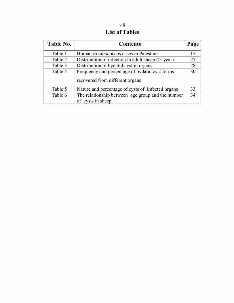
Table 1 Human Echinococcus cases in Palestine 15 Table 2 Distribution of infection in adult sheep (>1year) 25 Table 3 Distribution of hydatid cyst in organs 28 Table 4 Frequency and percentage of hydatid cyst forms
recovered from different organs
30
Table 5 Nature and percentage of cysts of infected organs 33 Table 6 The relationship between age group and the number
of cysts in sheep34
ix
List of Figures
Figures No.
Contents page
Fig. 1 Life cycle of Echinococcus species 3Fig. 2 Hydatid cysts in various organs of sheep 4Fig. 3 Global distribution of Echinococcus granulosus 9Fig 4 The regional setting of West Bank and location of
the cities Nablus, Jenin and Tubas19
Fig. 5 Distribution of total sheep sample 20 Fig. 6 Examination of slaughtered animals 22 Fig. 7 Distribution of sheep according to age 24 Fig. 8 percentage of hydatid cyst in various age-group 27 Fig. 9 Distribution of infected organs in sheep 29 Fig. 10 size of cysts (cm) in infected sheep 31 Fig. 11 Echinococcus granulosus Protoscolices 32 Fig. 12 The relationship between age groups of sheep and
size of hydatid cysts 35
x
Prevalence of sheep hydatidosis in north West Bank Palestine By
Jehad Hammed El-Ibrahim Supervisor
Dr Ayman Hussein Abstract
Hydatidosis is a zoonotic disease that occurs throughout the world and causes considerable economic losses and public health problems in
many countries, it is a major helminthes parasitic infection in West Bank . The current study aims to explore the size of the problem in sheep of
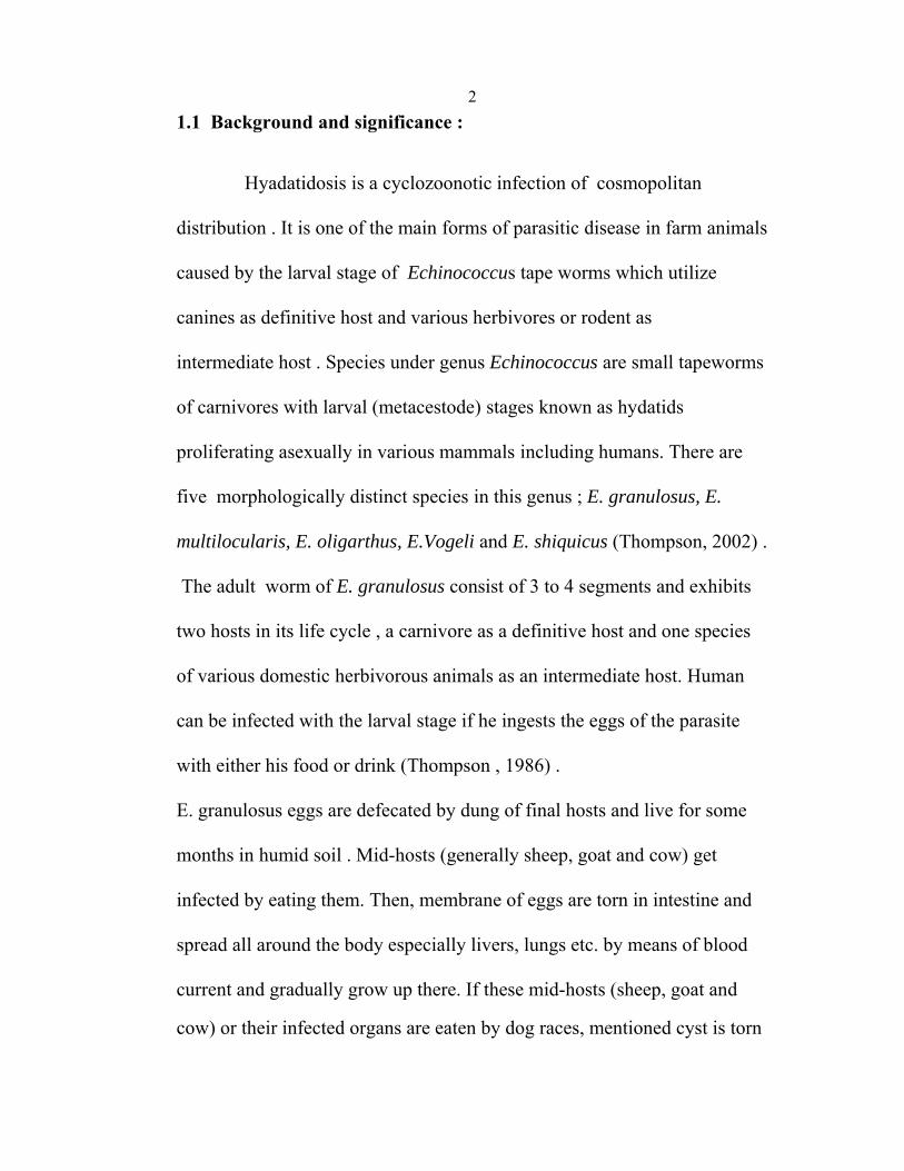
West-Bank and answering the question : what is the prevalence of sheep hydatidosis in Palestine ?. A total of 1000 indigenous sheep carcasses were selected from the municipalities abattoirs and slaughterhouses of Nablus, Jenin and Tubas districts . Each animal carcass was inspected carefully, cysts of each organ ( liver , lung …) were counted, measured and examined microscopically to determine the fertility .The total prevalence of hydatid cyst was 9 %, distributed according to age of sheep as : 0.6% in hoggets ( ≤ 1 year ), 10 % in 1-2 years, 24% in 2-3 years, 27% in >3 years age . Our research showed that liver was the most infected organ, 51% of cysts infected both liver and lung together (mixed infection ), 31% were liver cases alone, while involvement of lung alone was observed in 13 % of the cases .The lower number was in spleen 3% and viscera 1% . Microscopic examination of infected cysts revealed that 17% of organs had fertile cysts . 61% of cysts size were <4 cm and 38 % were >4 cm .

1
Chapter One Introduction and literature review
21.1 Background and significance :
Hyadatidosis is a cyclozoonotic infection of cosmopolitan distribution . It is one of the main forms of parasitic disease in farm animals caused by the larval stage of Echinococcus tape worms which utilize canines as definitive host and various herbivores or rodent as intermediate host . Species under genus Echinococcus are small tapeworms of carnivores with larval (metacestode) stages known as hydatids proliferating asexually in various mammals including humans. There are five morphologically distinct species in this genus ; E. granulosus, E. multilocularis, E. oligarthus, E.Vogeli and E. shiquicus (Thompson, 2002) . The adult worm of E. granulosus consist of 3 to 4 segments and exhibits two hosts in its life cycle , a carnivore as a definitive host and one species of various domestic herbivorous animals as an intermediate host. Human can be infected with the larval stage if he ingests the eggs of the parasite with either his food or drink (Thompson , 1986) . E. granulosus eggs are defecated by dung of final hosts and live for some months in humid soil . Mid-hosts (generally sheep, goat and cow) get infected by eating them. Then, membrane of eggs are torn in intestine and spread all around the body especially livers, lungs etc. by means of blood current and gradually grow up there. If these mid-hosts (sheep, goat and
cow) or their infected organs are eaten by dog races, mentioned cyst is torn
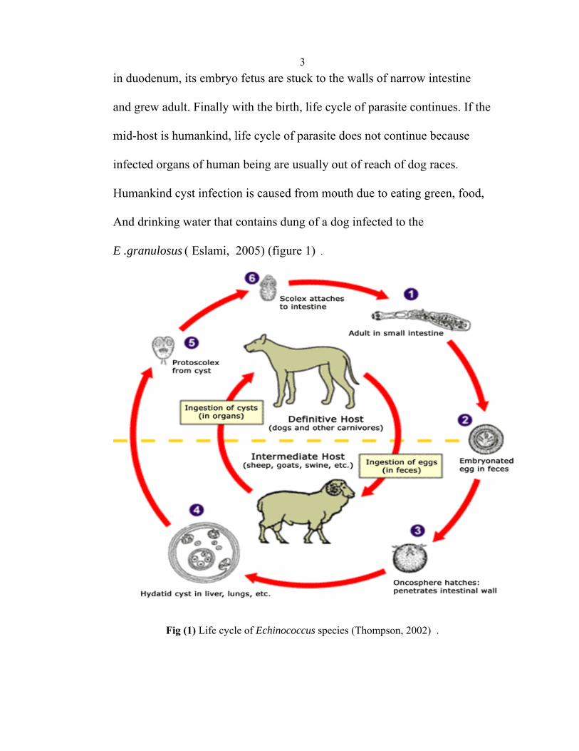
3in duodenum, its embryo fetus are stuck to the walls of narrow intestine and grew adult. Finally with the birth, life cycle of parasite continues. If the mid-host is humankind, life cycle of parasite does not continue because infected organs of human being are usually out of reach of dog races. Humankind cyst infection is caused from mouth due to eating green, food, And drinking water that contains dung of a dog infected to the E .granulosus ( Eslami, 2005) (figure 1) .
Fig (1) Life cycle of Echinococcus species (Thompson, 2002) .
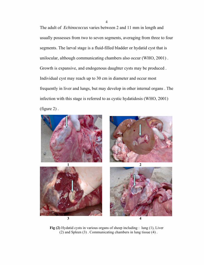
4The adult of Echinococcus varies between 2 and 11 mm in length and usually possesses from two to seven segments, averaging from three to four segments. The larval stage is a fluid-filled bladder or hydatid cyst that is unilocular, although communicating chambers also occur (WHO, 2001) . Growth is expansive, and endogenous daughter cysts may be produced . Individual cyst may reach up to 30 cm in diameter and occur most frequently in liver and lungs, but may develop in other internal organs . The infection with this stage is referred to as cystic hydatidosis (WHO, 2001) (figure 2) .
1 2
3 4
Fig (2) Hydatid cysts in various organs of sheep including : lung (1), Liver
(2) and Spleen (3) . Communicating chambers in lung tissue (4) .
5Hydatid cyst of E. granulosus is unilocular . Its growth is expansive by concentric enlargement . A well developed cyst contains three layers; fibrous capsule of host origin . The middle one is the laminated membrane which is secreted by the thin (germinal) layer and therefore is of parasite origin . The germinal layer gives rise to the broad capsule and daughter cysts ( Thompson, 1986) . The cysts are mainly found in the liver (and every possible organ: spleen, kidney, bone, brain, tongue and skin ) and asymptomatic until their growing size produces symptoms or accidentally discovered . Cysts are full of fluid and its brood capsules containing protosculex on its inner walls. The fertility of the cysts depends on presence of protoscolices in the fluid, it is one of the important factors in the epidemiology of E. granulosus . The fertility of cycts varies depends on the intermediate hosts and geographical situation (Farah, 1987). 1.2 Diagnosis of Echinococcus : The diagnosis of Echinococcosis in dogs or other carnivores requires the demonstration of the adult cestodes and eggs of Echinococcus spp. in their faeces or the small intestine (Nonaka et al. , 2008) . While diagnosis in intermediate hosts occurs by necropsy finding . Whereas surveillance for E. granulosus in domestic animals may take place in licensed slaughter houses, that for Echinococcus sp. in wildlife must be done by field surveys. In livestock intermediate hosts, molecular methods are, however, important

6in identification of isolates or strains of E. granulosus for epidemiological
purposes (Mcmanus & Bryant , 1995 ) . Serological or immunological tests, useful in humans, are less sensitive and specific in livestock and at present cannot replace necropsy . Antibodies directed against oncosphere, cyst fluid and protoscolex antigens can be detected in the serum of infected sheep, but this approach is presently of limited practical use as it does not distinguish between current and previous infections . Copro antigen tests based on a faecal antigen-detection antibody can be detected shortly after infection (10–14 days) (Craig , 1997 ) . DNA recognition methods is currently used mainly for confirmatory testing of coproantigen-positive samples or for identification of taenid eggs recovered from faeces using the different PCR primers from faeces in definitive hosts of genus Echinococcus (Mcmanus & Bryant, 1995) .
1.3 Burden of Hydatid cyst disease :
Based on Food and Agricultural Organization (FAO) report, economic
damages caused by parasite infections in developed and developing
countries are respectively 16% and 30% of their whole livestock
production and it is even more in countries where there is no serious
prevention policy against parasite infections (WHO, 2001) .
As 75% of world population live in developing countries and they possess
65% of total animals, economic damages are more serious (Taghizadeh and
Hoshiar, 2003 ) .
7Echinococcus and its metacestode stage in herbivores and humans have been recognized as the most important helminthes zoonoses, with great economic and public health significances in developing countries (Eckert , 1982) . Cystic Echinococcosis causes a huge health problems in animals and economical disadvantages because of production loss . It posses both a human health risk and economic loss to the country, which can takes the form of a reduction in live weight gain, reduced yield of milk, reductions in the fertility rates and reductions in the value of wool or other products . It may also be the most important one . Totally or partially discard of infected organs causes largest costs . Financial loss could be as high as 10% ( Torgerson et al., 2000 ) . It has a marked social impact because it is also frequently found in the human population (Eckert et al., 2001) . 1.4 Vaccination : Application of an effective vaccine to reduce hydatid infection in livestock would be likely to have a substantial impact on the rate of transmission of the disease to humans (Lightowlers, 2006) . As E. granulosus belongs to the Taeniid family, many aspects of its immunological relationship with its intermediate host are similar to that
occurring in Taenia species . Moreover, it was considered that the vaccine development approach used in Taenia species such as the native host-
8protective antigens of T. ovis would also be successful for E. granulosus (Lightowlers et al., 1996). Using recombinant DNA technology, an oncosphere antigen vaccine EG95 was shown to be capable of inducing a high level of protection against experimental challenge infection with E. granulosus eggs in sheep (Lightowlers et al., 1996).
1.5 Literature review :
The prevalence of hydatid cyst disease either in man or animals has
been studied extensively . In this study we are concentrated on E.
granulosus in sheep . E. granulosus has a global distribution; E.
multilocularis occurs in wide areas of the Northern Hemisphere, E. shiquicus is found in the People’s Republic of China and E. oligarthrus and E. vogeli are confined to Central and South America (WHO, 2001). All five species are infective to humans causing various forms of echinococcosis . Human cystic echinococcosis, caused by E. granulosus and alveolar echinococcosis, caused by E. multilocularis, are the most important public health threats in many parts of the world (WHO, 2001) . There are about 3 million patients who are infected in the world ( Craig et
al., 1996) . Latency can be up to 50 years, and mostly found in South and Central America , the Middle East , China, and the West of the U.S.A. ( Craig et al., 1996). Hydatid cyst disease has world-wide distribution which
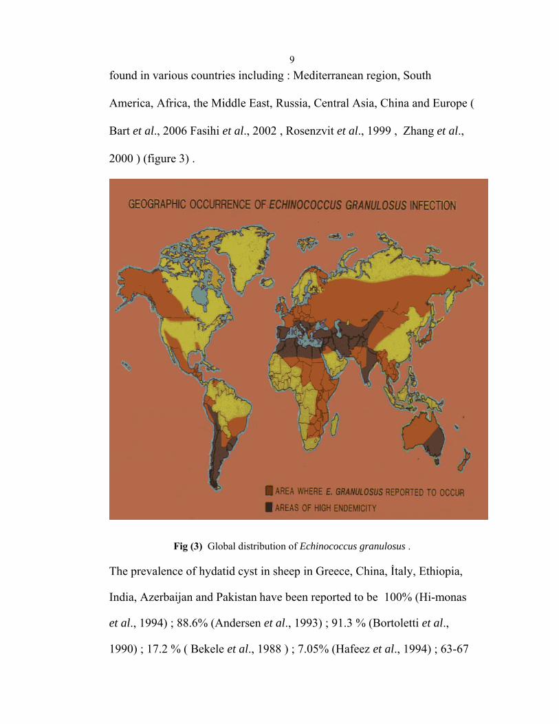
9found in various countries including : Mediterranean region, South America, Africa, the Middle East, Russia, Central Asia, China and Europe ( Bart et al., 2006 Fasihi et al., 2002 , Rosenzvit et al., 1999 , Zhang et al., 2000 ) (figure 3) .
Fig (3) Global distribution of Echinococcus granulosus .
The prevalence of hydatid cyst in sheep in Greece, China, İtaly, Ethiopia,
India, Azerbaijan and Pakistan have been reported to be 100% (Hi-monas
et al., 1994) ; 88.6% (Andersen et al., 1993) ; 91.3 % (Bortoletti et al.,
1990) ; 17.2 % ( Bekele et al., 1988 ) ; 7.05% (Hafeez et al., 1994) ; 63-67
10% (Chobanov et al., 1991) ; 3.04 % ( Anwar et al., 1993) , respectively .
In Iran many studies have been performed ; in Sanandaj area, western Iran
and Kashan area, results indicated an infection rate of ; 51.9% (LAkhlagh
et al ., 2005) ; 11.1% ( Dalimi et al ., 2002) and 2.7% ( Arbabi and
Hooshyar 2005) respectively . Another study on sheep which has been
carried out in Turkey and showed an infection rate of 3.50% (Meltem et
al . , 2007 ) .
Sheep, goats, cattle, camels, buffaloes, pigs and donkeys have
been repeatedly found infected with hydatid cysts in Iraq, Jordan, Lebanon,
Syria, Kuwait and Saudi Arabia ( Molan, 1993 ; Al-yaman, 1985 ; Abdel-
Hafez, 1986 ; Abo-Shehada, 1993 ; Dajani, 1978 ; Hassounah, 1976 ;
Ghandour, 1988) . The prevalence of Hydatid disease in sheep from Saudi
Arabia, Kuwait, Jordan, Morocco, Syria, Sudan, and Iraq have been
reported to be 4.6% (Farah, 1984), 12.8% (Hassonah and Behbehani
, 1976), 4% (Al-Yaman et al. , 1985), 5.3% (Pandey et al., 1988) 4.5%
( Dajani, 1978), 6.9 % (Elmahdi et al. , 2001 ), 5.9 %, 4.5 % (Al-
Abbassy et al. , 1980 and Molan, 1993 ), respectively .
Azlaf and Dakkak studied the prevalence of cystic echinococcosis in
Morocco in 2004, after the post mortem inspection concerned 2948 sheep
in five different regions, the global CE infection prevalence rates obtained
was 10.58% (Azlaf and Dakkak , 2004 ) .
Examination of 471 sheep, slaughtered in abattoirs in North Jordan was
carried out during March–May 1984 and showed an overall infection rate
11of 27.8% (Abdel-Hafez et al .,1986) . Another study in the same country in
1992 using indigenous sheep from five regions of Jordan showed an
infection rate of 12.9% , The higher prevalence was (27.6%) which
observed in sheep from Karak (Kamhawi et al ., 1992) .
Five thousand and Five hundred and ninety-six head of sheep (443 local,
473 Romanian and 4680 Australian) slaughtered in Amman Central
Abattoir during November -December 1999 were examined in routine meat
inspection procedures for hydatid cyst . 20.3% of the local sheep were
infected .While 12.8% of Australian sheep were infected, ( Anwar, 1999 ) .
In Egypt Hydatidosis was investigated among sheep in Egyptian official
abattoirs, from August 2000 to August 2005. The overall five years
hydatidosis prevalence was 0.3% . (Haridy et al ., 2005 ) .
A study in Hadhramout (Yemen) on 218 sheep carcasses were examined in
(2005/2006) . The prevalence was 3.21% (Baswaid , 2007) . In Libya astudy in an Shahat abattoir in Al-Jabal showed an infection rate of (8.7%), of 554 sheep, 48 sheep were infected (Al-Khalidi , 1998 ) .
The relationship between age and infection with E. granulosus was
investigated in many studies . In Thrace (Turkey ), The cysts were found in
2.64% of 720 lambs (<1 year old) and in 31.8% of 22 sheep (between 1-6
years old) (Meltem et al ., 2007) . In North Jordan the rate of hydatid
infection increased with age and reached as high as 63.7 percent in ewes 4
years of age and older (Abdel-Hafez et al .,1986 ) . Abo-Shehada in 1993
12studied the relation between age and infection . He found that infection in
sheep age less than two years was 0.4 %, 2-4 years : 46.3 %, 5-6 years :
78.8 % and 7-8 years : 84.8 % ( Abo-Shehada, 1993) . The same result was
recorded by Baswaid in Yemen : Below 1 year was 0% ; 1-2 years
: 2.3% ; 2-3 years : 12% ; over 3 years : 17% .
Location of cysts has been also investigated . The liver was the predilection site of infection .These findings were reported from studies in Saudi Arabia (Farah, 1984), Yemen (Baswaid, 2007), Jordan (Al-Yaman et al 1985), (Abdel-Hafez et al .,1986), (Kamhawi et al ., 1992) and (Abo-Shehada, 1993) . The liver was also the predominant site of infection in other studies. In Turkey, cysts were encountered in the liver of 96.2%, in the lungs of 26.9%, and in the spleen of 3.85% . Out of infected sheep 23.1% had fertile cysts (Meltem et al ., 2007) . In North Jordan 71.1 % of liver and lung and 7.6 % of spleen had cysts (Abdel-Hafez et al ., 1986 ) . While in Yemen the liver was the predominant site of infection 29% but liver and lung 71% (Baswaid , 2007) .
In Libya Liver infection was 87% while lungs infection 33.4 % and Spleen 4.2% (Al-Khalidi ,1998 ) . But lungs are the most predominant organs in Pandey study in Morocco (Pandey et al , 1988) . Firtility of cysts was reported in many studies to be : in Yemen (46.8%) (Baswaid , 2007), Iraq (39.4%) (Al-Abbassy, 1980), Kuwait (88.2%) (Hassonah and Behbehani ,1976) . In Jordan, the percentages of sheep, with

13 fertile cysts were reported in the range of 7.1– 68.7, (Al-Yaman et al, 1985) , (Abdel-Hafez et al .,1986), (Kamhawi et al ., 1992) , (Abo-Shehada, 1993), (Dajani and Khalaf, 1981) . History of hudatidosis In Palestine : In human : History of the endemic nature of hydatidosis in palestine was reported in 1933 by Witenberg and in 1947 by Torance . These authors noted a high incidence of cysts in slaughtered animals and the common occurrence of human patients in Jerusalem, Jaffa, and other cities in the area (Matossian et. al, 1977) . Rakover indicated that Israel has had a high morbidity rate of about 100 cases per annum of human hydatidosis (5 per 100 000 population ). This, however, has been ascribed, in part, to the immigration of infected patients (Matossian et. al, 1977) . The clinical, surgical, and diagnostic problems of hyddatidosis have been described by Levy and Peller et al. (Levy, 1970 ; Peller et al., 1973) . In 2002 Abu-Hassan et al studied human cystic echinococcosis in West- Bank by investigating the surgical incidence in hospitals between 1990 to 1997. A total of 390 surgically confirmed cases were recorded throughout the 8-year period, with an overall mean annual surgical incidence (MASI) of 3.1 per 100,000. A high MASI of 4.9, 5.0 and 5.1 per 100,000 was found in Hebron, Jericho and Bethlehem Governorates, respectively. Yata town,
14 Hebron governorate, showed the highest MASI, at 16.8 per 100,000 ( Abu- Hasan et al. , 2002 ) . According to Palestinian Ministry of health Reports ; table 1 summarizes the number of human cases of Echinococcus disease from 1999 to 2008 in Palestine (MOH, 2008) . . In animals : In 1977 its prevalence has varied from 0.02% in Tel-Aviv to 12% in Beersheba (Matossian et. al, 1977) . A survey carried out in 1991-1992 in the town of Yirka in Northern Israel on sheep slaughtered in the local abattoir during a one-month period revealed E. chinococcus cysts in 10% of the sheep (Furth et al. ,1992) . In the same contents there is no official records in Palestine regarding the prevalence of hydatid cyst disease in animals either in the Palestinian ministry of agriculture or the veterinary services or municipality abattoirs ( MOA, 2008 ) .
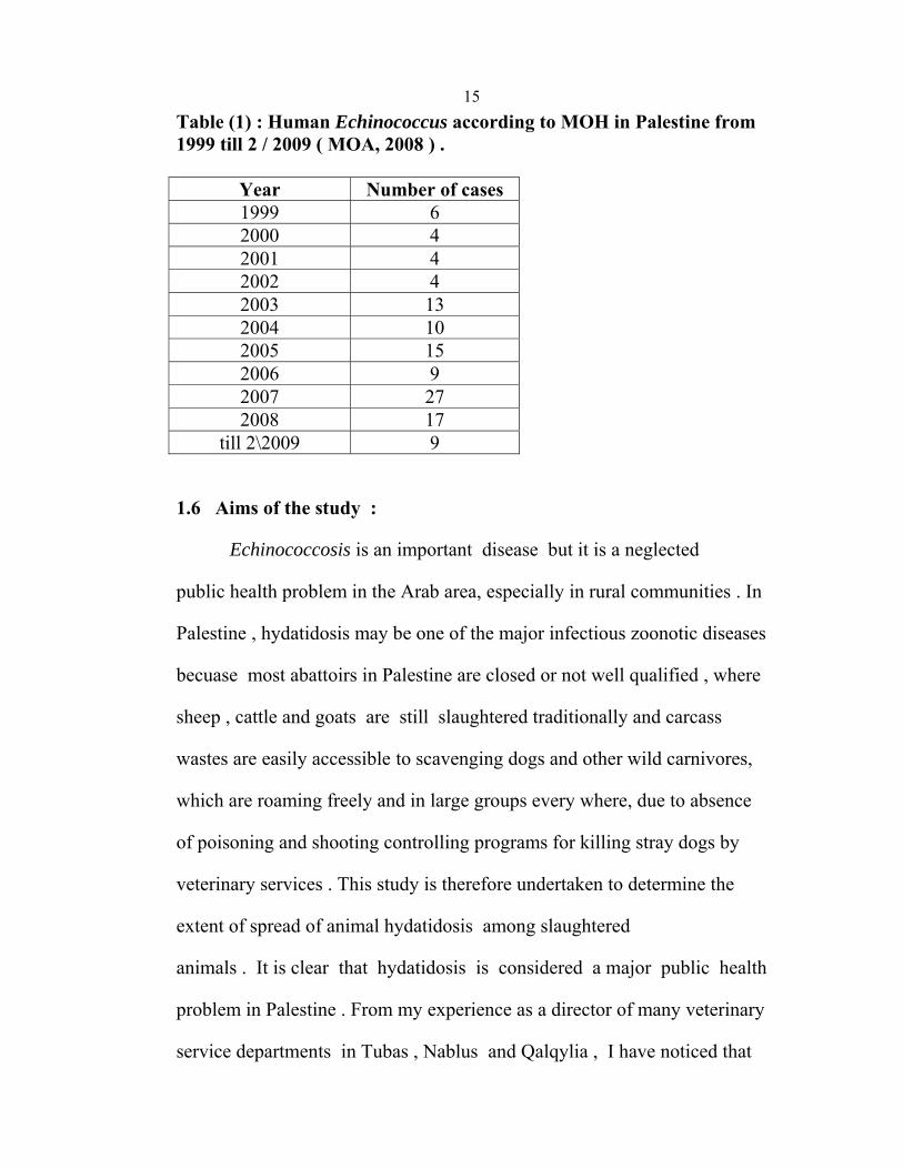
15Table (1) : Human Echinococcus according to MOH in Palestine from 1999 till 2 / 2009 ( MOA, 2008 ) .
Year Number of cases 1999 62000 4 2001 4 2002 4 2003 13 2004 10 2005 15 2006 9 2007 27 2008 17
till 2\2009 9
1.6 Aims of the study :
Echinococcosis is an important disease but it is a neglected
public health problem in the Arab area, especially in rural communities . In
Palestine , hydatidosis may be one of the major infectious zoonotic diseases
becuase most abattoirs in Palestine are closed or not well qualified , where
sheep , cattle and goats are still slaughtered traditionally and carcass
wastes are easily accessible to scavenging dogs and other wild carnivores,
which are roaming freely and in large groups every where, due to absence
of poisoning and shooting controlling programs for killing stray dogs by
veterinary services . This study is therefore undertaken to determine the
extent of spread of animal hydatidosis among slaughtered
animals . It is clear that hydatidosis is considered a major public health
problem in Palestine . From my experience as a director of many veterinary
service departments in Tubas , Nablus and Qalqylia , I have noticed that
16many animals are infected with hydatid cyst disease . Since the animals
share the same life cycle as man , therefore determination of the
prevalence of the disease in West Bank is very important in order to
explore the size of the problem which helps to control the disease . This
work is the first epidemiologically analyses of sheep hydatidosis in
Palestine which aims to alert policy makers to design governmental control
programs against hydatidosis and to minimize prevalence in Palestine
either in human or in animals .

17
Chapter Two Materials and Methods
182.1 Design of the study :
A cross-sectional study was carried out in order to investigate the
prevalence of hydatid cyst disease in sheep in North of the West-Bank .
Indigenous sheep which were slaughtered at local abattoirs were selected
for the current study .
2.2 Study area:
Three districts in North of West-Bank were selected for this study .
These districts are Nablus, Jenin and Tubas (Figure 4 ) . These
governorates are considered one of the most sheep raising areas in
Palestine . The population of the area is approximately 400, 000
inhabitants, most of them (60%) are living in rural areas and are involved
in animal production (PCBS , 2007) . The number of animals in these areas
is nearly about 300,000 heads of sheep; 120,000 in Nablus , 130,000 in
Jenin and 50,000 in Tubas (PCBS , 2007) . Sheep are one of the most
principal slaughtered animals for human consumption on social and
religious occasions . Slaughtering in Nablus district occurs in the main
abattoir . However, Tubas and Jenin still lack abattoirs and the process
takes place in streets , traditional slaughter houses, and markets . The dogs
in these areas which are the final host of the parasite carrying hydatid cyst
disease either live with herds of sheep or look after the houses or farms.
Meanwhile, the stray dogs roam also freely and live on food garbage's
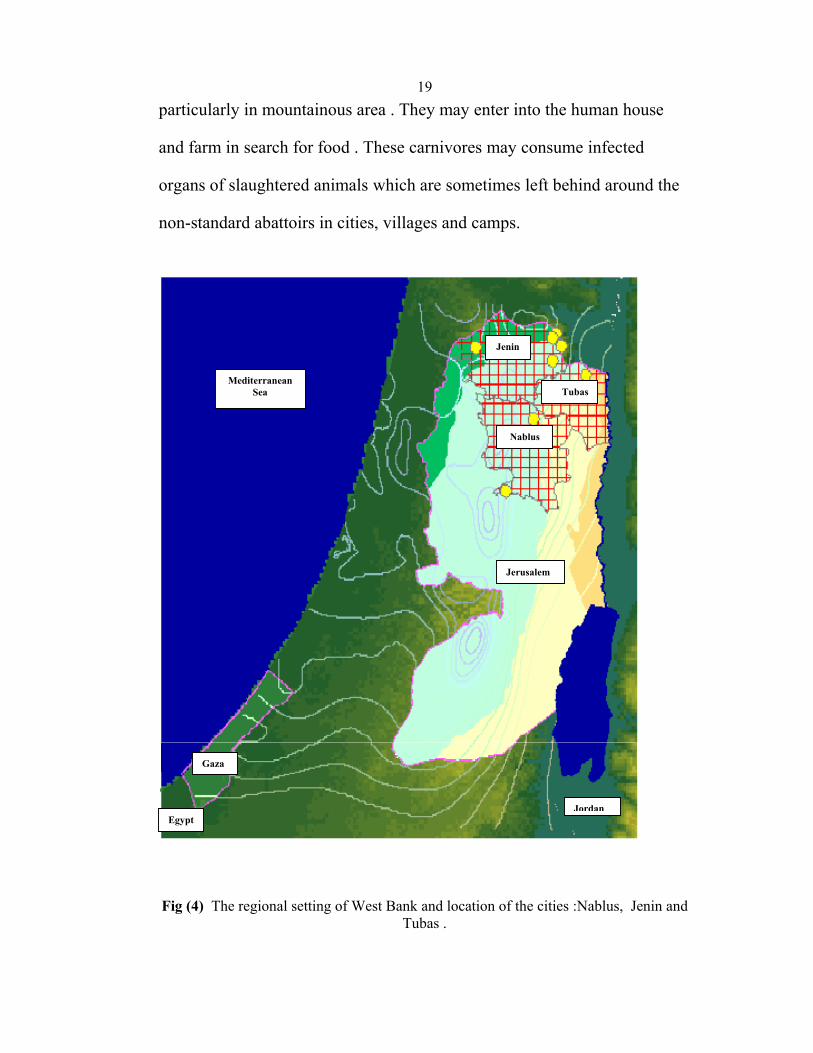
19particularly in mountainous area . They may enter into the human house
and farm in search for food . These carnivores may consume infected
organs of slaughtered animals which are sometimes left behind around the
non-standard abattoirs in cities, villages and camps.
Fig (4) The regional setting of West Bank and location of the cities :Nablus, Jenin and
Tubas .
Jenin
Tubas
Nablus
Jerusalem
Gaza
Mediterranean Sea
JordanEgypt
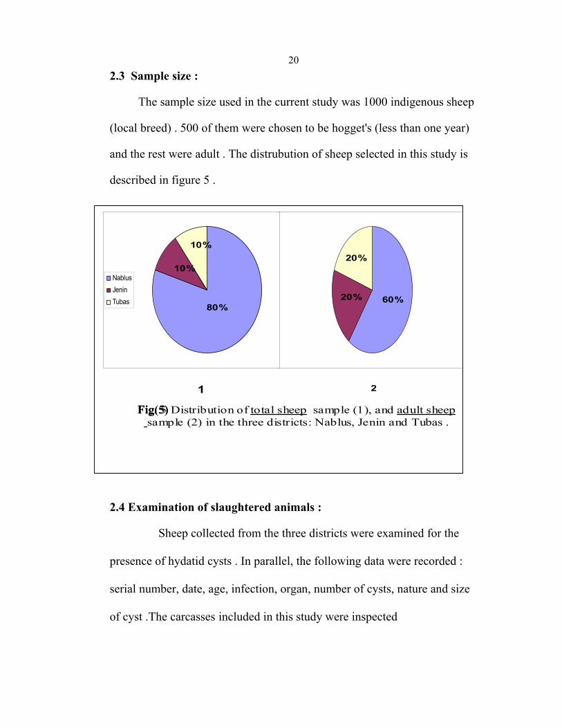
202.3 Sample size :
The sample size used in the current study was 1000 indigenous sheep
(local breed) . 500 of them were chosen to be hogget's (less than one year)
and the rest were adult . The distrubution of sheep selected in this study is
described in figure 5 .
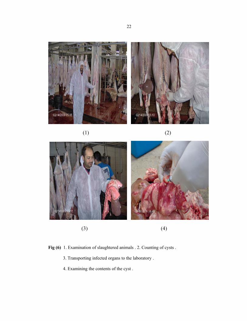
2.4 Examination of slaughtered animals :
Sheep collected from the three districts were examined for the presence of hydatid cysts . In parallel, the following data were recorded : serial number, date, age, infection, organ, number of cysts, nature and size of cyst .The carcasses included in this study were inspected
60%20%
20%
NablusJeninTubas
80%
10%
10%
1 2
Fig. 5 Distribution of total sheep sample (1), and adult sheepsample (2) in the three districts: Nablus, Jenin and Tubas .
Fig(5)
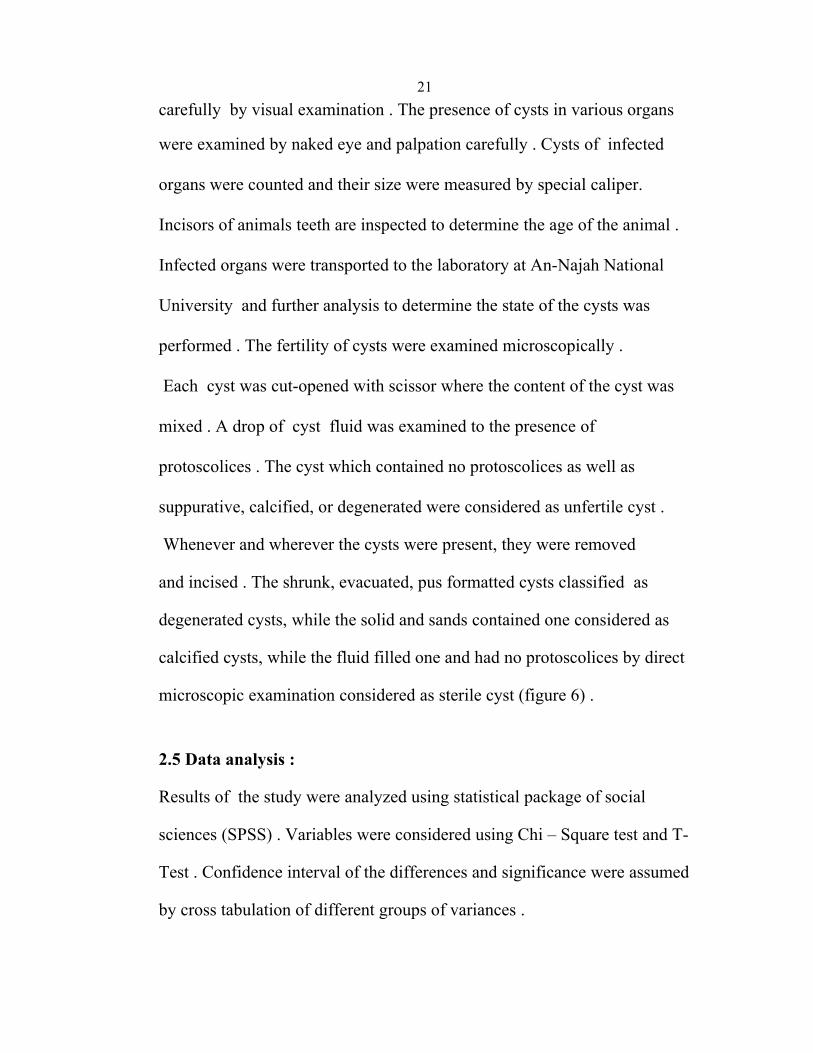
21carefully by visual examination . The presence of cysts in various organs were examined by naked eye and palpation carefully . Cysts of infected organs were counted and their size were measured by special caliper. Incisors of animals teeth are inspected to determine the age of the animal . Infected organs were transported to the laboratory at An-Najah National University and further analysis to determine the state of the cysts was performed . The fertility of cysts were examined microscopically . Each cyst was cut-opened with scissor where the content of the cyst was mixed . A drop of cyst fluid was examined to the presence of protoscolices . The cyst which contained no protoscolices as well as suppurative, calcified, or degenerated were considered as unfertile cyst .
Whenever and wherever the cysts were present, they were removed
and incised . The shrunk, evacuated, pus formatted cysts classified as
degenerated cysts, while the solid and sands contained one considered as
calcified cysts, while the fluid filled one and had no protoscolices by direct
microscopic examination considered as sterile cyst (figure 6) .
2.5 Data analysis :
Results of the study were analyzed using statistical package of social
sciences (SPSS) . Variables were considered using Chi – Square test and T-
Test . Confidence interval of the differences and significance were assumed
by cross tabulation of different groups of variances .
22
(1) (2)
(3) (4)
Fig (6) 1. Examination of slaughtered animals . 2. Counting of cysts .
3. Transporting infected organs to the laboratory .
4. Examining the contents of the cyst .

23
Chapter Three Results
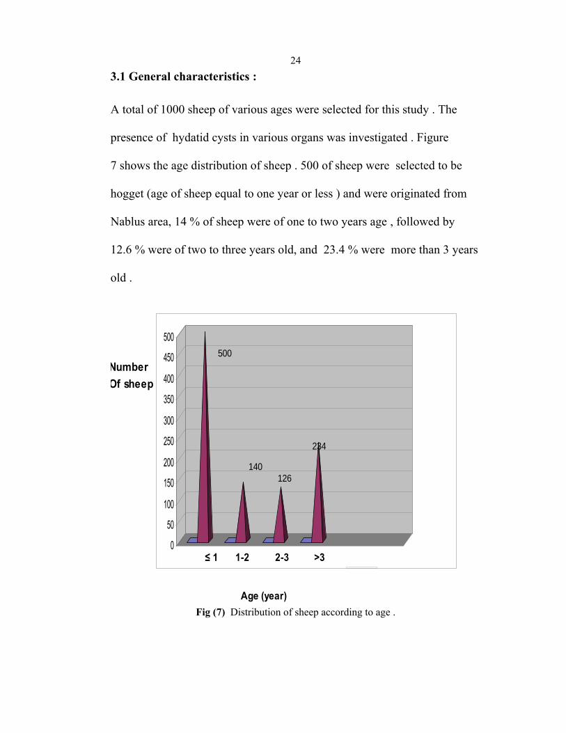
243.1 General characteristics : A total of 1000 sheep of various ages were selected for this study . The presence of hydatid cysts in various organs was investigated . Figure 7 shows the age distribution of sheep . 500 of sheep were selected to be hogget (age of sheep equal to one year or less ) and were originated from Nablus area, 14 % of sheep were of one to two years age , followed by 12.6 % were of two to three years old, and 23.4 % were more than 3 years old .
0
50
100
150
200
250
300
350
400
450
500
Number Of sheep
≤ 1 1-2 2-3 >3
Age (year)
500
234
140126
Fig (7) Distribution of sheep according to age .
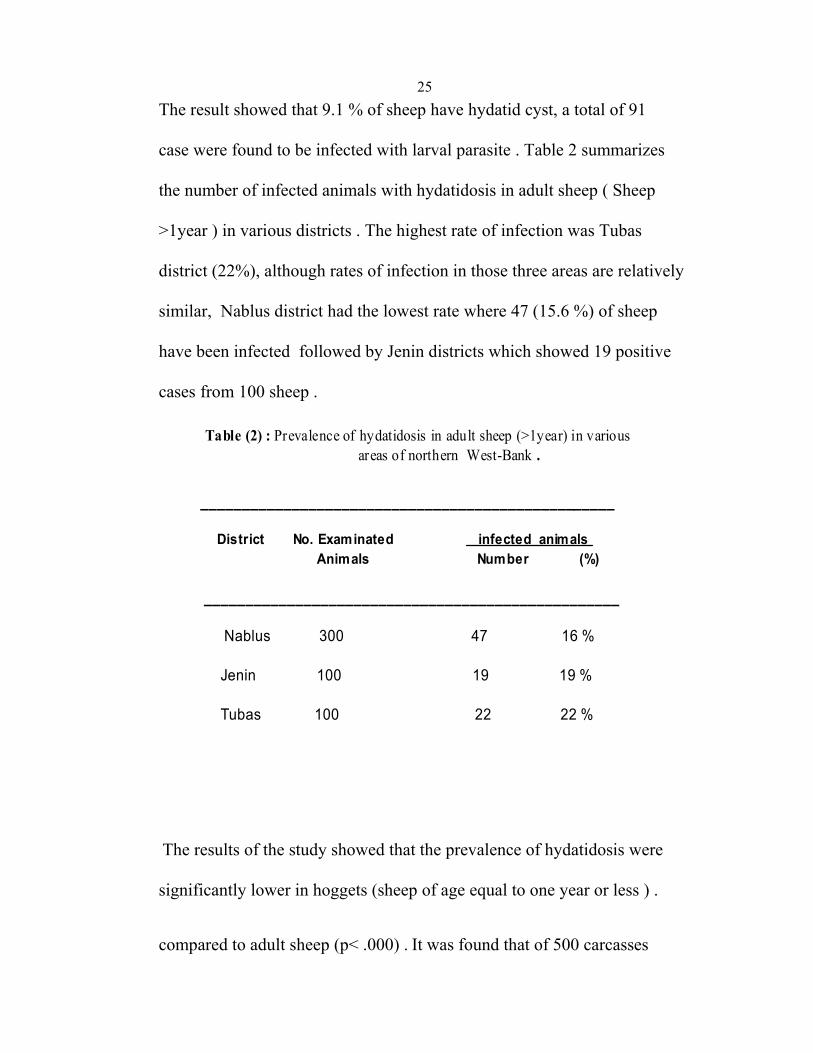
25The result showed that 9.1 % of sheep have hydatid cyst, a total of 91 case were found to be infected with larval parasite . Table 2 summarizes the number of infected animals with hydatidosis in adult sheep ( Sheep >1year ) in various districts . The highest rate of infection was Tubas district (22%), although rates of infection in those three areas are relatively similar, Nablus district had the lowest rate where 47 (15.6 %) of sheep have been infected followed by Jenin districts which showed 19 positive cases from 100 sheep .
__________________________________________________
District No. Examinated infected animals Animals Number (%)
__________________________________________________
Nablus 300 47 16 %
Jenin 100 19 19 %
Tubas 100 22 22 %
Table (2) : Prevalence of hydatidosis in adult sheep (>1year) in various areas of northern West-Bank .
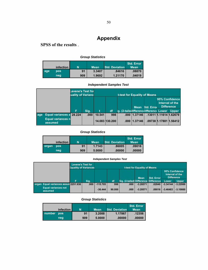
The results of the study showed that the prevalence of hydatidosis were significantly lower in hoggets (sheep of age equal to one year or less ) . compared to adult sheep (p< .000) . It was found that of 500 carcasses
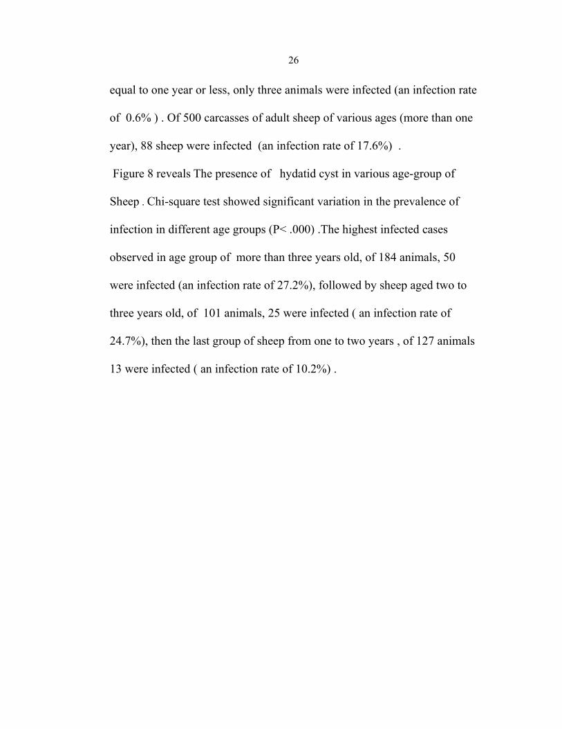
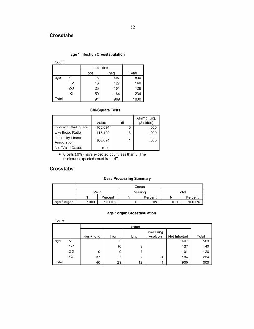
26 equal to one year or less, only three animals were infected (an infection rate of 0.6% ) . Of 500 carcasses of adult sheep of various ages (more than one year), 88 sheep were infected (an infection rate of 17.6%) . Figure 8 reveals The presence of hydatid cyst in various age-group of Sheep . Chi-square test showed significant variation in the prevalence of infection in different age groups (P< .000) .The highest infected cases observed in age group of more than three years old, of 184 animals, 50 were infected (an infection rate of 27.2%), followed by sheep aged two to three years old, of 101 animals, 25 were infected ( an infection rate of 24.7%), then the last group of sheep from one to two years , of 127 animals 13 were infected ( an infection rate of 10.2%) .

27
Percentage Of infection
Age (year)
0
5
10
15
20
25
30
(≤1) (1-2) (2-3) >3
0.6%
27.2%24.7%
10.2%
Fig. 8 : percentage of hydatid cyst in variousage-group of sheep
3.2 Cyst location : The location of cysts over various tissues has been examined . Our research showed that liver was the most infected organ with hydatidosis where 75 cases (83 %) were found infected, lung was the second organ where 58 cases (64%) . Spleen had the lowest number 3 % . Table 3 summarizes the distribution of hydatid cyst in organs of studied sheep, 46 (51%) of cysts from both liver and lung together (mixed infection ), 29 (31.8%) were only liver cases .
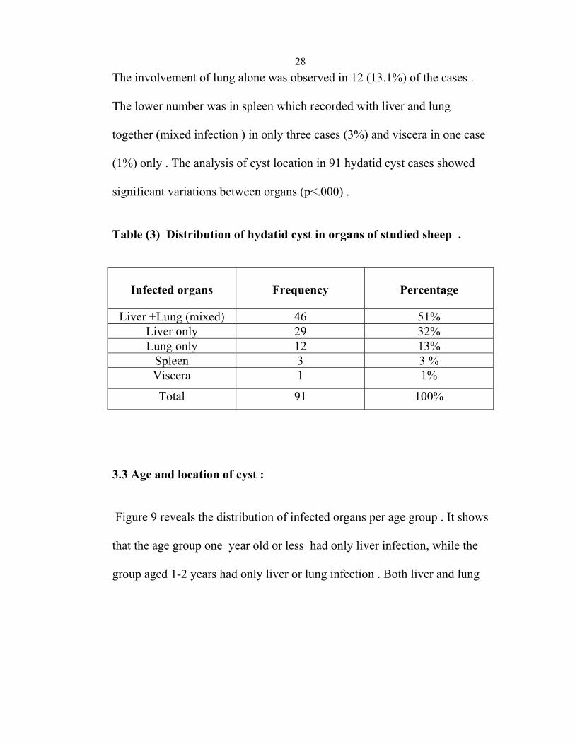
28The involvement of lung alone was observed in 12 (13.1%) of the cases . The lower number was in spleen which recorded with liver and lung together (mixed infection ) in only three cases (3%) and viscera in one case (1%) only . The analysis of cyst location in 91 hydatid cyst cases showed significant variations between organs (p<.000) . Table (3) Distribution of hydatid cyst in organs of studied sheep .
Infected organs
Frequency
Percentage
Liver +Lung (mixed) 46 51%
Liver only 29 32% Lung only 12 13%
Spleen 3 3 % Viscera 1 1%
Total 91 100%
3.3 Age and location of cyst : Figure 9 reveals the distribution of infected organs per age group . It shows that the age group one year old or less had only liver infection, while the group aged 1-2 years had only liver or lung infection . Both liver and lung
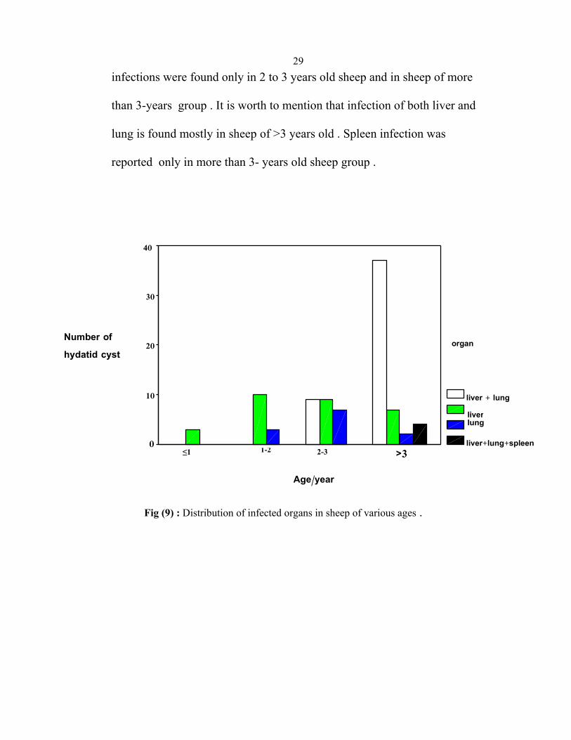
29infections were found only in 2 to 3 years old sheep and in sheep of more than 3-years group . It is worth to mention that infection of both liver and lung is found mostly in sheep of >3 years old . Spleen infection was reported only in more than 3- years old sheep group .
Age/year
>3 2-31-2≤1
40
30
20
10
0
organ
liver + lung
liverlung
liver+lung+spleen
Fig (9) : Distribution of infected organs in sheep of various ages .
Number of
hydatid cyst
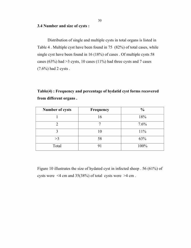
303.4 Number and size of cysts :
Distribution of single and multiple cysts in total organs is listed in
Table 4 . Multiple cyst have been found in 75 (82%) of total cases, while
single cyst have been found in 16 (18%) of cases . Of multiple cysts 58
cases (63%) had >3 cysts, 10 cases (11%) had three cysts and 7 cases
(7.6%) had 2 cysts .
Table(4) : Frequency and percentage of hydatid cyst forms recovered
from different organs .
Number of cysts Frequency %
1 16 18%
2 7 7.6%
3 10 11%
>3 58 63%
Total 91 100%
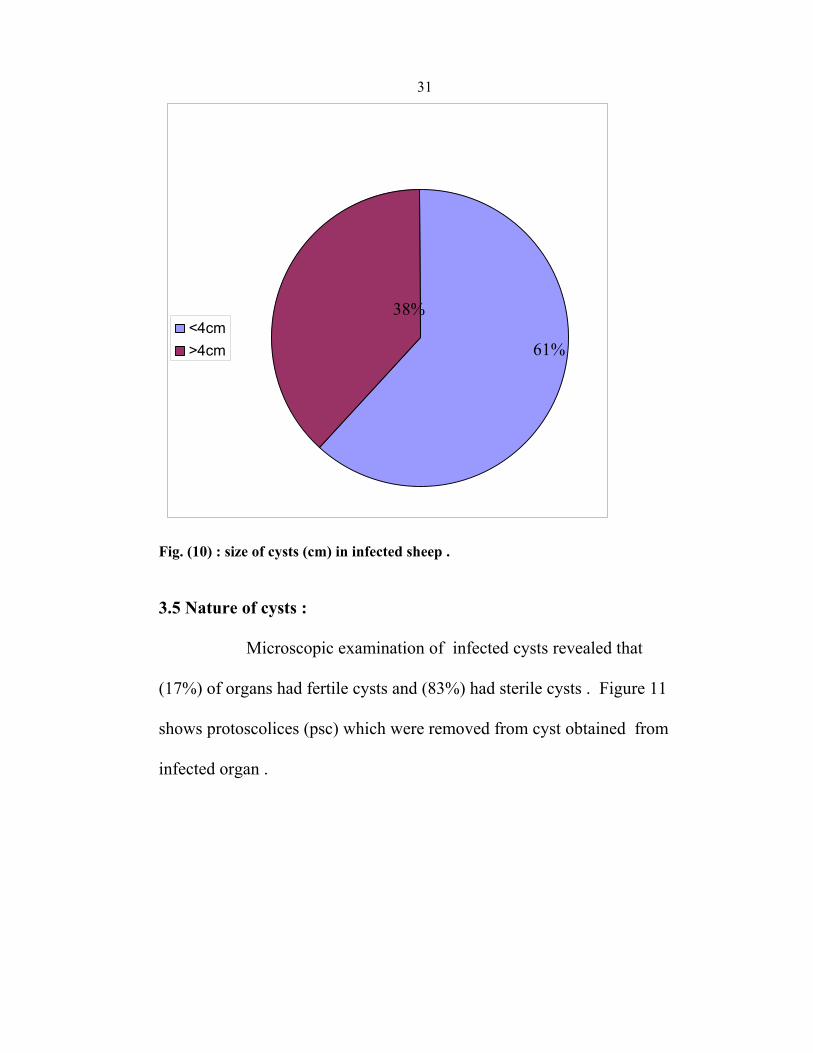
Figure 10 illustrates the size of hydated cyst in infected sheep . 56 (61%) of
cysts were <4 cm and 35(38%) of total cysts were >4 cm .
31
<4cm>4cm
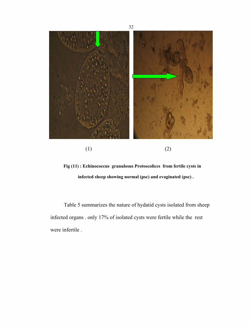
Fig. (10) : size of cysts (cm) in infected sheep . 3.5 Nature of cysts : Microscopic examination of infected cysts revealed that (17%) of organs had fertile cysts and (83%) had sterile cysts . Figure 11 shows protoscolices (psc) which were removed from cyst obtained from infected organ .
38% 61%
32
(1) (2)
Fig (11) : Echinococcus granulosus Protoscolices from fertile cysts in
infected sheep showing normal (psc) and evaginated (psc) .
Table 5 summarizes the nature of hydatid cysts isolated from sheep infected organs . only 17% of isolated cysts were fertile while the rest were infertile .
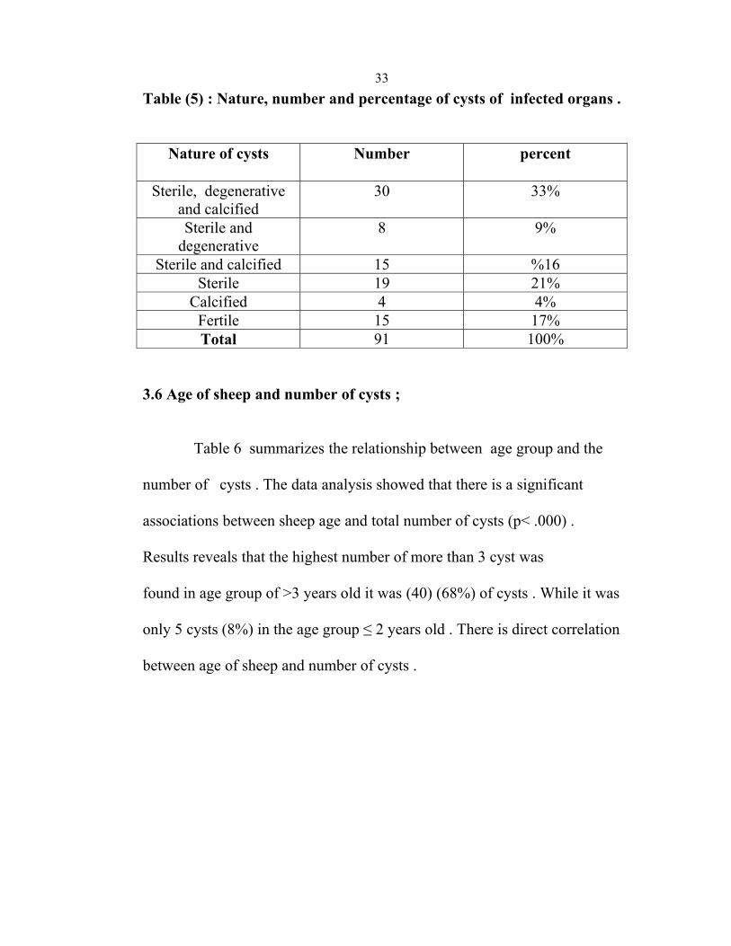
33Table (5) : Nature, number and percentage of cysts of infected organs .
Nature of cysts Number percent
Sterile, degenerative and calcified
30 33%
Sterile and degenerative
8 9%
Sterile and calcified 15 16% Sterile 19 21%
Calcified 4 4% Fertile 15 17% Total 91 100%
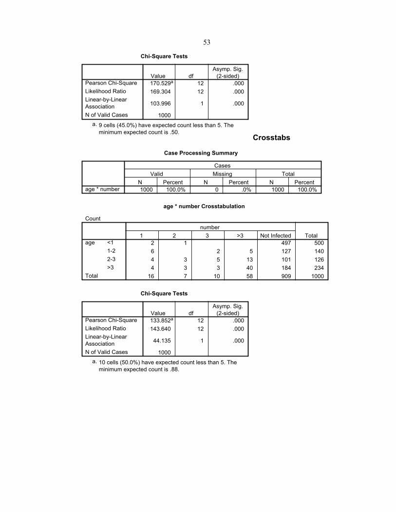
3.6 Age of sheep and number of cysts ; Table 6 summarizes the relationship between age group and the number of cysts . The data analysis showed that there is a significant associations between sheep age and total number of cysts (p< .000) . Results reveals that the highest number of more than 3 cyst was found in age group of >3 years old it was (40) (68%) of cysts . While it was only 5 cysts (8%) in the age group ≤ 2 years old . There is direct correlation between age of sheep and number of cysts .
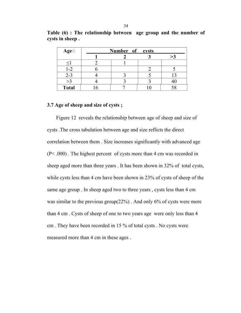
34Table (6) : The relationship between age group and the number of cysts in sheep .
◌Age ____________cysts of _Number_________
1 2 3 >3 ≤1 2 1 1-2 6 2 5 2-3 4 3 5 13 >3 4 3 3 40
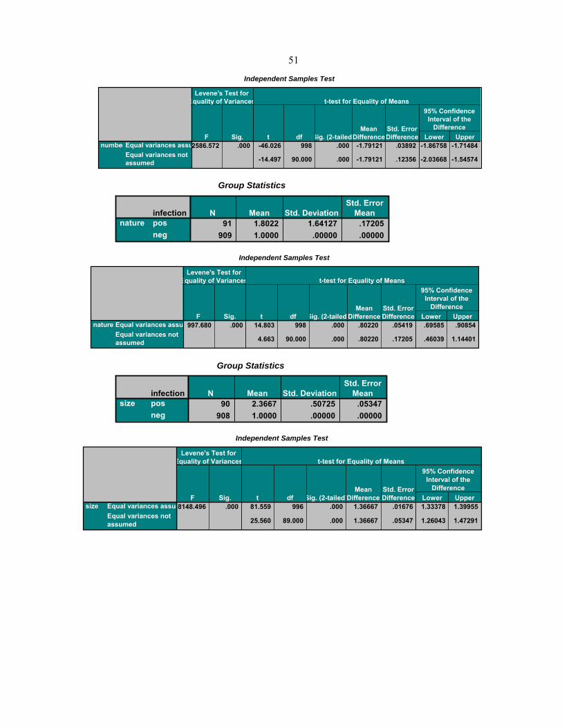
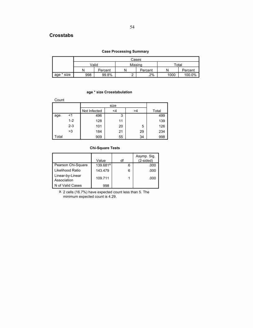
Total 16 7 10 58 3.7 Age of sheep and size of cysts ; Figure 12 reveals the relationship between age of sheep and size of cysts .The cross tabulation between age and size reflicts the direct correlation between them . Size increases significantly with advanced age (P< .000) . The highest percent of cysts more than 4 cm was recorded in sheep aged more than three years . It has been shown in 32% of total cysts, while cysts less than 4 cm have been shown in 23% of cysts of sheep of the same age group . In sheep aged two to three years , cysts less than 4 cm was similar to the previous group(22%) . And only 6% of cysts were more than 4 cm . Cysts of sheep of one to two years age were only less than 4 cm . They have been recorded in 15 % of total cysts . No cysts were measured more than 4 cm in these ages .

35
0
5
10
15
20
25
30
Size of hudatid cyst (cm)
(≤1) (1-2) (2-3) >3
Age of sheep (year)Series1
Series2
Fig (12) : The relationship between age groups of sheep and size of hydatid cysts .
: < 4 Cm cyst . : > 4 Cm cyst .

36
Chapter Four
Discussion
37In North West Bank, where the largest open area of Palestine, particularly Tubas and Jenin destricts, there are large population of stray dogs and other wild carnivorous live near human houses, interaction between domestic cycle and wild cycles may occur . In this regard, animals may become infected by consuming infected organs of slaughtered animals left around the non-standard small abattoirs and slaughtered houses in villages. Among the present study which showed that cystic echinococcosis is spreading among sheep, in addition to its presence in human beings in West Bank 3.1 per 100,000 (Abu-Hasan et al., 2002 ) . Our study has revealed that 9.1 % of sheep have hydatid cyst . The results are similar to other reports in Israil from where the rate of infection in sheep of age less than one year was 0.02% (Matossian et. al, 1977) . , More over, the rate of infection in sheep of older age in other studies in Israel are similar where Lass et al study showed 12% in Beersheba .Our study showed also a similar rate of infection in Northern Israel 10% (Furth et al., 1993) . It worth mentioning that area under study either in our study (North W.B.) or Furth et al study (North Israil) are under same area . In comparison to other regions, it is clear that the prevalence of hydatidosis in our study has higher rate . For example, the prevalence in the current study is about 2-fold of prevalence reported from : Saudi Arabia : 4.6% (Farah, 1984), Yemen : 3.21% (Baswaid , 2007) , Jordan : 4% (Al-
38Yaman et al., 1985), Morocco : 5.3% (Pandey et al., 1988), Iraq : 5.9%, 4.5% (Al-Abbassy et al., 1980 and Molan, 1993), Syria ; 4.5% ( Dajani, 1978), Sudan : 6.9 % (Elmahdi et al., 2001 ), Egypt : 0.3% (Haridy et al., 2005 ), Libya : 8.7% (Al-Khalidi ., 1998 ), Kashan region of Iran : 2.7% ( Arbabi and Hooshyar 2005) and Turkey : 3.50% (Meltem et al., 1998) . It is however, similar to the results of other studies : 12.8% in Kuwait (Hassonah and Behbehani ,1976), 27.8 in North Jordan (Abdel-Hafez et al., 1986), 12.9% in five regions of Jordan (Kamhawi S et al.,1992), 20.3% in Amman Central Abattoir (Anwar , 1999) , 10.58% in Morocco (Azlaf , Dakkak , 2004), 51.9% in Sanandaj area in Iran ((LAkhlagh et al., 2005) and 11.1% in western Iran ( Dalimi et al., 2002) .This difference is attributed, perhaps to the variability of the following : origin of animals, mode of grazing , presence of definitive host (carnivore), degree of contamination with parasite and other environmental factors as periodical destruction of dogs, improved standards of meat inspection, overall improvement in socio economic conditions, hygienic status of sheep herds, variations in the temperature, environmental conditions, the nature of the pasture and the way of raising of these animals . The age of the animals is another factor in these variations . In our survey, the majority of slaughtered animals were bred out doors, and there was a strong practical relationship between animal offal and scavenging dogs .

39The highest rate of infection was in Tubas district . The reason behind it could be of geographic reason, , out door breeding with open grazing areas, more carnivorous Population (sheep dogs ), no central abattoirs and there is no hygienic elimination of sheep's offal with more environmental parasite contamination . In Jenin district the situation is similar to Tubas, but more awareness about anti helmentic drugs . Nablus district had the lowest rate, this may be attributed to elevation nature of the district, central official hygienic slaughtering, more knowledge about anthelmentic dosage, and indoor breeding . The liver in my study was the most infected organ . These findings were similar to the observations reported in Saudi Arabia (Farah, 1984), Yemen (Baswaid , 2007), Jordan (Al-Yaman et al., 1985),(Abdel -Hafez et al., 1986), (Kamhawi et al.,1992) and (Abo-Shehada, 1993), but did not coincide with those of Pandey et al., In Morocco (Pandey et al., 1988), where lungs are the most predominant organs . The liver was the most common site of infection in sheep .The lungs came in the second place in the present study . This is mainly due to the fact that the liver is the first organ the blood flows through after leaving the intestine . Therefore, most of the oncospheres hatched in the intestine are filtered in it . The ones that are not trapped in the liver are passed to the lungs then other organs (Al-Khalidi, 1998)

40In our study the older animals were highly infected, more number of cysts, bigger size of cysts, and more infected organs, while the younger ones had low rate of infection, less number of cysts, smaller size of cysts, and less infected organs . This is attributed to various factors . Firstly, higher age reflects a much longer period of risk of infection . Secondly, the chances of detecting cysts at meat inspection are higher in aged animals due to the bigger size of the cyst . Finally, the older animal cysts have more time to enlarge and transport cysts to other organs . In our survey only three sheep less than one year of age were found positive . Echinococcus egg, in general, requires at least one year before the hydatid cyst stage grows sufficiently to produce protoscolices capable of infecting the carnivore host (Smyth, 1964) . These three positive cases may be due to wrong dentation . The fertility of cyst is an important factor that can effect stability of E. granulosus cycle depending on geographical situation, kind of infected host site and size of cyst . Fertility was 17% in my study, which is substantially lower compared to what has been observed in Yemen (46.8%) (Baswaid , 2007), Iraq (39.4%) (Al-Abbassy, 1980), Kuwait (88.2%) (Hassonah and Behbehani ,1976), Jordan (38.1%) (Kamhawi et al., 1995) but higher compared to that observed in Jordan (8.0%) (Al-Yaman et al., 1985) . In Jordan, the percentages of sheep, with fertile cysts were reported in the range of 7.1– 68.7%, (Al-Yaman et al.,1985), (Abdel-Hafez et al., 1986),
41 (Kamhawi et al., 1992) , (Abo-Shehada, 1993), (Dajani and Khalaf, 1981) The low rate of fertile cysts and small size of cysts in our study is contributed to the high contamination and parasitic infestation in sheep and over use and multi use of antiparasitic and anthelmantic derivatives such as Ivermectine, Albendazole, Febendasole and others for long period before slaughtering to increase sheep weight .
42
Limitations :
1. Lack of official abattoirs in the study area especially in Tubas and Jenin
districts, which makes the researcher to looks for traditional slaughter houses in different villages and locations.
2. Time of slaughtering occurred at dawn, this makes it difficult for the scholar to carry out inspection procedure in proper time.
3. Difficulty in dealing with butchers without reward, because he feels shameful at slaughtering adult sheep, that affects his reputation .
4. Difficulty in transporting infected organs to the laboratory on time for recognizing the nature of cyst. 5. Difficulty in determining the age of animal by dentations . 6. Difficulty in diagnosis of cysts in sheep less than one year due to its small size . 7. Lack of adult sheep (more than one year) specially in Tubas and Jenin, and if there it slaughtered in mysterious way . 8. Lack of scientific periodicals references, books and magazines related to parasitology in palestine .
43
Recommindation :
* Scope of my thesis is to alert policy makers to design governmental
control programs against hydatidosis to minimize prevalence in Palestine,
and ensure effective protection not only for animal population but also
for humans at risk of contracting the infection . * New checks and controls are hoped for at a political level which will increase the financial support for the farmers and encourage importing and testing of vaccines which have already been tested in other areas in the world . * Killing rambling dogs and preparing identity card and collar for them . * Treatment of animals with anti-parasite medicines ( specially sheep and dogs) and prophylactic anthelmentic dosage four times yearly for all animal farms . * Preventing illegal slaughtering and making healthy slaughter houses ( not to allow rambling dogs enter to the field of slaughterhouses ) . * Public health learning through TV, and teaching livestock holders and people who are at risk about periodic epidemiologic investigations .
44
References
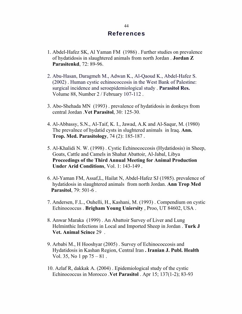
1. Abdel-Hafez SK, Al Yaman FM (1986) . Further studies on prevalence of hydatidosis in slaughtered animals from north Jordan . Jordan Z Parasitenkd, 72: 89-96. 2. Abu-Hasan, Daragmeh M., Adwan K., Al-Qaoud K., Abdel-Hafez S. (2002) . Human cystic echinococcosis in the West Bank of Palestine: surgical incidence and seroepidemiological study . Parasitol Res. Volume 88, Number 2 / February 107-112 . 3. Abo-Shehada MN (1993) . prevalence of hydatidosis in donkeys from central Jordan .Vet Parasitol, 30: 125-30. 4. Al-Abbassy, S.N., Al-Taif, K. I., Jawad, A.K and Al-Saqur, M. (1980) The prevalnce of hydatid cysts in slughtered animals in Iraq. Ann. Trop. Med. Parasitology, 74 (2): 185-187 . 5. Al-Khalidi N. W. (1998) . Cystic Echinococcosis (Hydatidosis) in Sheep, Goats, Cattle and Camels in Shahat Abattoir, Al-Jabal, Libya Proceedings of the Third Annual Meeting for Animal Production Under Arid Conditions, Vol. 1: 143-149 . 6. Al-Yaman FM, Assaf,L, Hailat N, Abdel-Hafez SJ (1985). prevalence of hydatidosis in slaughtered animals from north Jordan. Ann Trop Med Parasitol, 79: 501-6 . 7. Andersen, F.L., Ouhelli, H., Kashani, M. (1993) . Compendium on cystic Echinococcus . Brigham Young Uniersity , Proo, UT 84602, USA . 8. Anwar Maraka (1999) . An Abattoir Survey of Liver and Lung Helminthic Infections in Local and Imported Sheep in Jordan . Turk J Vet. Animal Scince 29 . 9. Arbabi M., H Hooshyar (2005) . Survey of Echinococcosis and Hydatidosis in Kashan Region, Central Iran . Iranian J. Publ. Health Vol. 35, No 1 pp 75 – 81 . 10. Azlaf R, dakkak A. (2004) . Epidemiological study of the cystic Echinococcus in Morocco .Vet Parasitol . Apr 15; 137(1-2); 83-93
4511. Bart J.M., Abdukader M., Zhang Y.L., Lin R.Y., Wang Y.H., Nako M., Ito A., Craig P.S., PiarrouxR.,Vuitton D.A. & Wen H. (2006) . Genotyping of human cystic echinococcosis in Xinjiang, PR China. Parasitology, 133, 571–579. 12. Baswaid S. H. (2007) . Prevalence of Hydatid cyst in slaughtered sheep and goats in Hadramout (Yemen) . Ass. Univ. Bull. Environ. Res. Vol. 10 No. 2 . 13. Bekele T, Mukasa-Mugerwa E, Kasali OB. (1988) . The prevalence of cysticercosis and hydatidosis in Ethiopian sheep. Vet Parasitol, 28(3): 267-270 . 14. Bortoletti G, Gabriele F, Seu V, Palmas C. (1990) . Epidemiology of hydatid disease in Sardinia: a study of fertility of cysts in sheep. J Helminth, 64(3): 212-216 . 15. Chobanov RE, Salekhov AA, Iskenderov VS, Alieva TI,Dzhafarova IA. (1991).Epidemiologyof echinococcosis under conditions of transhumant husbandry in Azerbaijan. Veterinariya Moskva, 12: 33-34. 16. Craig P.S , M.T. Rogan, J.C. Allan (1996) . Detection, screening and community epidemiology of taeniid cestode zoonoses: cystic echinococcosis, alveolar echinococcosis and neurocysticercosis, Adv. Parasitol. 38 169–250.
17. Craig P.S (1997). Immunodiagnosis of Echinococcus granulosus and a comparison of techniques for diagnosis of canine echinococcosis. In: Compendium on Cystic Echinoccosis in Africa and in Middle Eastern Countries with Special Reference to Morocco, Andersen F.L., Ouhelli H. & Kachani M., eds. Brigham Young University Print Services, Provo, Utah, USA. 18. Dajani YF, Khalaf FH . (1981) . Hydatidosis and tenuicollosis in sheep and goats of Jordan: a comparative study. Ann Trop Med Parasitol, 75: 175-79. 19. Dalimi A, Motamedi G, Hossini M, Mohammadian B, Malaki H, Ghamari Z, Ghaffari F.F., 2002 . Ehinococcus in Western Iran . Vet Parasitol, 105(2) : 161-76 .

4620. Eckert J, Gemmel MA, Meslin FX, Pawlowski ZS . (2001) . Manual on Echinococcosis in Humans an Animals: a Public Health Problem of Global Concern. WHO/OIE, Paris. 21. Eckert J. (1982) . Echinococcosis/ hydatidosis survallence, prevention and control . FAO/UNEP/WHO gudlines., FAO . Animal production and health, p 92 . 22. Elmahdi IE, Magzoub MM. Ibraheem AM, Saad MB, Roming T . (2001) . Cystic Echinococcou of life stock and human in central Sudan . Ann Trop Med. Parasitol . Jul : 98(5) ; 473-9 . 23. Farah MO. (1984). Infection rates, cyst fertility and larval viability of hydatid disease in camels, sheep and cattle in Gassim, Saudi Arabia. Vet Res Commun, 11: 493-495 . 24. Fasihi Harandi M., Hobbs R.P., Adams P.J., Mobedi I., Morgan-Ryan U.M. & Tompson R.C.A. (2002) . Molecular and morphological characterization of Echinococcus granulosus of human and animal origin in Iran. Parasitology, 125, 367–373. 25. Furth M, Hoida G, Nahmias J, Greenberg Z, Barzilay A, Goldsmith RS, el-On J.(1993) The development of new foci of echinococcosis in northern Israel: prevalence in domestic animals. J Helminthol, 68(1); 45- 47 . 26. Ghadour AM (1988). Health hazards in humans and animals caused by imported livestock disease in Saudi Arabia. Fauna Saudi Arabia, 9: 468-477 . 27. Hafeez MD, Reddy PR, Hasina S, Prasad KLG, Nirmala DK,Thayeeb MD. (1994). Fertility rate of hydatidosis in cattle, buffaloes, sheep and pigs. Indian J Anim Sci, 64(1): 46-47 . 28. Haridy FM, Morsy TA, El-Sherbini GT, Sultan DM, Awad SE, ElShazly AM, I brahim BB (2005). Hydatidosis granulosus in Egyptian Slaughtered animals in the years 2000- 2005 . J Egypt Soc Parasitol Dec:36(3): 1087-100 .
4729. Hassounah A, Behbehani K (1976) .The epidemiology of Echinococcus infection in Kuwait. J Helminthol, 50: 65-73. 30. Himonas C, Antoniadou SK, Papadopoulos E. (1994). Hydatidosis of food animals in Greece: prevalence of cysts containing viable protoscoleces. J Helminth, 68(4): 311-313 (ref: VETCD 1/89-11/96). 31. Kamhawi S, Hijjawi N, Abu-Gazaleh A, Abbass M, (1992) . Prevalence of hydatid cysts in livestock from five regions of Jordan . Parasitol Int. 2006;55 Suppl:S197-202 . 32. Eslami, A. (2005). Helmintology, Cestodes. University of Tehran pub., Tehran, 3rd ed. 33. LAkhlaghi , JMassoud , A Housaini . (2005) . Observation on Hydatid cyst Infection in Kordestan Province (West of Iran) using Epidemiological and Seroepidemiological Criteria . Iranian J Publ Health, Vol. 34, No. 4, pp.73-75 . 34. Levy, M. (1970) . Israel journal of medical sciences, 6:388-392 . 36. Lightowlers M.W. (2006) . Cestode vaccines: origins, current status and future prospects. Parasitology, 133, S27–42. 37. Lightowlers M.W., Lawerence S.B. Gauci C.G., Young J, Ralston M. Maas D. And Heath D.D. (1996) . Vaccination against hydatidosis using a difined recumbenant antigene . Int . J. Parasitol., 18, 457-462 . 38. Matossian R.M., Rickard M.D. & Smyth J.D. (1977) . Hydatidosis : A global problem of increasing importancre . Bulletin of the world health organization, 55(4) 499-507 . 39. McManus D.P. & Bryant C. (1995) . Biochemistry, Physiology and Molecular Biology of Echinococcus. In: Echinococcus and Hydatid Disease, Thompson R.C.A. & Lymbery A.J., eds. CAB International, Wallingford, UK, 135–182 . 40. McManus DP, Smyth JD, (1986) . Hydatidosis changing conceptsin epidemiology and speciation. Parasitol Today, 2: 163-168.
4841. Meltem U. E., Erkut T. (2007) . Prevalenc of Hydatidosis in Slaughtered animal in Thrace, Turkey . Parazitologi Dergisi, 31, (1) ; 4145 . 42. MOA. Plestenian Menistry of Agriculture website (2008) . Available on : http;// www. moa. Gov.ps . 43. MOH. Plestenian Menistry of health website . (2008) . Available on : http//www.google.de/search/hl=ar&ei=gacnSpShOIPBsAaMw7yDBg&sa =x&oi=spell&resnum=0&ct=1&q=Palestinian+ministry+of+Health+websit e&spell +1 44. Molan AL. (1993) . Epidemiology of hydatidosis and echinococcosis in Theqar province, southern Iraq. Jpn Med Sci Biol, 46;29-35 . 45. Nonaka N., Oka M., Kamiya M. & Oky Y (2008). A latex agglutination test for the detection of Echinococcus multilocularis coproantigen in the definitive hosts. Vet. Parasitol., (in press). 46. Palestinian Central Bureau of Statistics Website (2007). Available on :
http://atlas.pcbs.gov.ps/atlas/ASD/Agriculture/Amaps2006.asp http://atlas.pcbs.gov.ps/Website/ASD/Agriculture/Agr_Sheep_number_06/viewer.htm 47. Pandey, V. S., Ohelli, H. and Moumen, A (1988) : Epidemiology of hydatidosis)-Echinococcosis in Quarzazte, The Pre .Saharian region of Morocco. Ann. Trop .Med. Parasitology, 82 (5) : 461-470 . 48. Peller, N. ET Al. (1973) . Journal of the Israel Medical Association, .84: 73-74 . 49. Rosenzvit M.C., Zhang L.H., Kamenetzky L., Canova S.G., Guarnera E.A. & Mcmanus D.P.,(1999) . Genetic mvariation and epidemiology of Echinococcus granulosus in Argentina. Parasitology, 118, 523–530. 50. Smyth, J., (1964): The biology of the hydatid .organisms. In: Advanced in Parasitology ,B. Dawes (ed.), Academic Press, N. York .2: 169-219
4951. Taghizadeh, S. and H. Hoshiar (2003) . Evaluation of Economic
Damages of Human Hydatidosis in Two of Tehran Hospitals, 4th
Iranian Parasitology and Parasitic Diseases Congress, pp: 83-87.
52. Thompson (1986) . The biology of echinococcus . 53. Thompson R.C.A & Mcmanus D.P. (2002) . Towards a taxonomic revision of the genus Echinococcus. Trends Parasitol., 18, 452–457. 54. Torgerson PR, Carmona C, Bnifacino R . (2000) . Estimating the economic effects of cystic echinococcosis. Uruguay, a developing country with upper-middle income. Ann Trop Med Parasitol, 94: 703- 713 . 56. World Health Organisation (WHO)/ (OIE) (2001). WHO/OIE Manual on Echinococcosis in Humans and Animals: a Public Health Problem of Global Concern, Eckert J., Gemmell, M.A., Meslin F.-X., Pawlowski Z.S., eds. OIE (World Organisation for Animal Health), Paris, France, 1–265. 57. Zhang L.H., Joshi D.D. & Macmanus D.P. (2000) . Three genotypes of Echinococcus granulosus identified in Nepal using mitochondrial DNA markers. Trans. R. Soc. Trop. Med. Hyg., 94, 258–260.
50
Appendix SPSS of the results .
Group Statistics
91 3.3407 .84616 .08870909 1.9692 1.21170 .04019
infectionposneg
ageN Mean Std. Deviation
Std. ErrorMean
Independent Samples Test
28.224 .000 10.541 998 .000 1.37146 .13011 1.11614 1.62679
14.083 130.200 .000 1.37146 .09738 1.17881 1.56412
Equal variances asEqual variances nassumed
ageF Sig.
Levene's Test forquality of Variance
t df ig. (2-tailedMean
DifferenceStd. ErrorDifference Lower Upper
95% ConfidenceInterval of the
Difference
t-test for Equality of Means
Group Statistics
91 1.7143 .86005 .09016909 5.0000 .00000 .00000
infectionposneg
organN Mean Std. Deviation
Std. ErrorMean
Independent Samples Test
2251.938 .000 -115.705 998 .000 -3.28571 .02840 -3.34144 -3.22999
-36.444 90.000 .000 -3.28571 .09016 -3.46483 -3.10660
Equal variances assumEqual variances notassumed
organF Sig.
Levene's Test forEquality of Variances
t df Sig. (2-tailed)Mean
DifferenceStd. ErrorDifference Lower Upper
95% ConfidenceInterval of the
Difference
t-test for Equality of Means
Group Statistics
91 3.2088 1.17867 .12356909 5.0000 .00000 .00000
infectionposneg
numberN Mean Std. Deviation
Std. ErrorMean
51
Independent Samples Test
2586.572 .000 -46.026 998 .000 -1.79121 .03892 -1.86758 -1.71484
-14.497 90.000 .000 -1.79121 .12356 -2.03668 -1.54574
Equal variances assuEqual variances notassumed
numberF Sig.
Levene's Test forEquality of Variances
t df Sig. (2-tailed)Mean
DifferenceStd. ErrorDifference Lower Upper
95% ConfidenceInterval of the
Difference
t-test for Equality of Means
Group Statistics
91 1.8022 1.64127 .17205909 1.0000 .00000 .00000
infectionposneg
natureN Mean Std. Deviation
Std. ErrorMean
Independent Samples Test
997.680 .000 14.803 998 .000 .80220 .05419 .69585 .90854
4.663 90.000 .000 .80220 .17205 .46039 1.14401
Equal variances assuEqual variances notassumed
natureF Sig.
Levene's Test forEquality of Variances
t df Sig. (2-tailed)Mean
DifferenceStd. ErrorDifference Lower Upper
95% ConfidenceInterval of the
Difference
t-test for Equality of Means
Group Statistics
90 2.3667 .50725 .05347908 1.0000 .00000 .00000
infectionposneg
sizeN Mean Std. Deviation
Std. ErrorMean
Independent Samples Test
8148.496 .000 81.559 996 .000 1.36667 .01676 1.33378 1.39955
25.560 89.000 .000 1.36667 .05347 1.26043 1.47291
Equal variances assumEqual variances notassumed
sizeF Sig.
Levene's Test forEquality of Variances
t df Sig. (2-tailed)Mean
DifferenceStd. ErrorDifference Lower Upper
95% ConfidenceInterval of the
Difference
t-test for Equality of Means
52
Crosstabs
age * infection Crosstabulation
Count
3 497 50013 127 14025 101 12650 184 23491 909 1000
<11-22-3>3
age
Total
pos neginfection
Total
Chi-Square Tests
103.824a 3 .000118.129 3 .000
100.074 1 .000
1000
Pearson Chi-SquareLikelihood RatioLinear-by-LinearAssociationN of Valid Cases
Value dfAsymp. Sig.
(2-sided)
0 cells (.0%) have expected count less than 5. Theminimum expected count is 11.47.
a.
Crosstabs
Case Processing Summary
1000 100.0% 0 .0% 1000 100.0%age * organN Percent N Percent N Percent
Valid Missing TotalCases
age * organ Crosstabulation
Count
3 497 50010 3 127 140
9 9 7 101 12637 7 2 4 184 23446 29 12 4 909 1000
<11-22-3>3
age
Total
liver + lung liver lungliver+lung+spleen Not Infected
organ
Total
53
Chi-Square Tests
170.529a 12 .000169.304 12 .000
103.996 1 .000
1000
Pearson Chi-SquareLikelihood RatioLinear-by-LinearAssociationN of Valid Cases
Value dfAsymp. Sig.
(2-sided)
9 cells (45.0%) have expected count less than 5. Theminimum expected count is .50.
a.
Crosstabs
Case Processing Summary
1000 100.0% 0 .0% 1000 100.0%age * numberN Percent N Percent N Percent
Valid Missing TotalCases
age * number Crosstabulation
Count
2 1 497 5006 2 5 127 1404 3 5 13 101 1264 3 3 40 184 234
16 7 10 58 909 1000
<11-22-3>3
age
Total
1 2 3 >3 Not Infectednumber
Total
Chi-Square Tests
133.852a 12 .000143.640 12 .000
44.135 1 .000
1000
Pearson Chi-SquareLikelihood RatioLinear-by-LinearAssociationN of Valid Cases
Value dfAsymp. Sig.
(2-sided)
10 cells (50.0%) have expected count less than 5. Theminimum expected count is .88.
a.
54
Crosstabs
Case Processing Summary
998 99.8% 2 .2% 1000 100.0%age * sizeN Percent N Percent N Percent
Valid Missing TotalCases
age * size Crosstabulation
Count
496 3 499128 11 139101 20 5 126184 21 29 234909 55 34 998
<11-22-3>3
age
Total
Not Infected <4 >4size
Total
Chi-Square Tests
139.681a 6 .000143.479 6 .000
109.711 1 .000
998
Pearson Chi-SquareLikelihood RatioLinear-by-LinearAssociationN of Valid Cases
Value dfAsymp. Sig.
(2-sided)
2 cells (16.7%) have expected count less than 5. Theminimum expected count is 4.29.
a.
جامعة النجاح الوطنية
كلية الدراسات العليا
فلسطين -األكياس المائية في األغنام في شمال الضفة الغربية
دراسة وبائية
إعداد
جهاد حمد اإلبراهيم
إشراف
الدكتور أيمن حسين
ة الدراسات لمتطلبات درجة الماجستير في الصحة العامة بكلي قدمت هذه األطروحة استكماال
.فلسطين جامعة النجاح الوطنية في نابلس، العليا في 2009

ب
دراسة وبائية فلسطين -األكياس المائية في األغنام في شمال الضفة الغربية
إعداد
جهاد حمد اإلبراهيم
إشراف
الدكتور أيمن حسين
الملخص
شر في العالم مسببا خسائراألكياس المائية مرض مشترك بين اإلنسان والحيوان ينت
إقتصادية وخسائر في الصحة العامة في العديد من البلدان ، وهو مرض ديداني طفيلي منتشر
هذه الدراسة تهدف الى استكشاف حجم المشكلة في أغنام الضفة. في الضفة الغربية
من ذبيحةف تم اختيار أل. كم هي وبائية المرض في فلسطين : الغربية واإلجابة على السؤال
كل جثة تم فحصها. األغنام المحلية من مسالخ ومجازر محافظات نابلس، جنين وطوباس
ها وقياسهاإحصائو......) كبد، رئة (بعناية، ثم تم فحص األكياس في االعضاء المصابة
وهي موزعة% 9.1وبائية المرض في فلسطين كانت . واختبارها بالمجهرلتحديد خصوبتها
2-1في األعمار % 10في المواليد أقل أو تساوي سنة، % 0.6: كما يلي حسب العمر
أظهرت الدراسة أن .سنوات 3> في األعمار % 27سنة، 3-2في األعمار % 24سنة،
إصابة( من األكياس أصابت الرئة والكبد في نفس الوقت % 51الكبدأكثر األعضاء إصابة،
من الحاالت،% 13إصابت الرئة وحدها كان التي أصابت الكبد منفردا، بينما% 31، )مختلطة
من% 17أظهر أن يالفحص المجهر% . 1واألحشاء % 3أقل إصابة كانت في الطحال
أكبر % 38سم و 4من األكياس كان حجمها اقل من % 61. الحاالت تحوي أكياسا مخصبة
.سم 4من