Human A & P Bone Structure and Function. I. Introduction to The Skeletal System A. Background...
89
Human A & P Bone Structure and Function
-
Upload
philip-jeremy-higgins -
Category
Documents
-
view
221 -
download
1
Transcript of Human A & P Bone Structure and Function. I. Introduction to The Skeletal System A. Background...

Human A & P
Bone Structure and Function

I. Introduction to The Skeletal System A. Background information about the skeletal
system:1. The skeletal system includes the entire framework of ____________ and
their _____________.
2. Each bone is considered to be an ___________.
BonesCartilage
organ

3. Bone tissue is a __________________ tissue. a. The _______________ is:
i. _________________________ - to
provide hardnessii. ______________ - to provide
some flexibility.
iii. _____________
connectivematrixCrystallized Minerals
Collagen fibers
Water
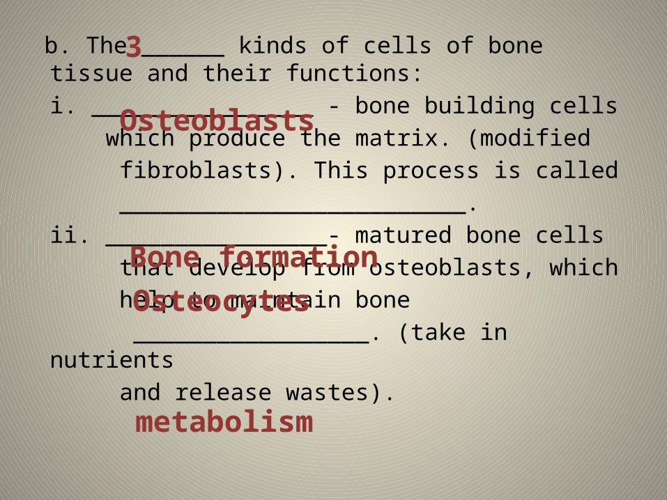
b. The ______ kinds of cells of bone tissue and their functions:
i. ________________ - bone building cells which produce the matrix. (modified fibroblasts). This process is called _________________________.ii. ________________- matured bone cells that develop from osteoblasts, which help to maintain bone _________________. (take in nutrients and release wastes).
3
Osteoblasts
Osteocytes
metabolism
Bone formation

iii. ______________________- (modified macrophage) huge cells made from 50+ WBCs that produce lysosomal enzymes & acids to break down bone matrix. This is a process called __________________.
Osteoclasts
resorption

II. Gross Anatomy of BoneA. Classification of bones based on shape
1. _______________- greater in length than in width. Ex –
2. _______________- nearly equal in length and width Ex -
Long Bone
Femur, tibia, ulna, humerus, phalanges
Short Bone
Wrist, & ankle bones

3. ________________ - thin and flat Ex –
4. _________________- complex shapes that do not fit other categories.Ex -
Flat BoneRibs, cranium, sternum, shoulder blades
Irregular Bone
Vertebra, pelvis, some facial bones


B. Macroscopic Structure of Bone1. gross view of outside of bone

_____________________- end of boneepiphysis
_____________________- end of boneepiphysis

_____________________- main middle portion of the bone.
Diaphysis

_______________ - region in mature bone where diaphysis meets epiphysismetaphysis
Metaphysis

_____________________- thin layer of cartilage over the epiphysis where the bone connects with another bone. It has two features:
1. ________________ & protects the ends of bone.2. _________________ability to repair itself. Why????
Articular Cartilage
cushions
limited
Articular Cartilage

2. gross view of the inside of the bone

_____________________- a layer of cartilage in growing bone where the diaphysis can grow in __________.
- when the bone stops growing in length, bone will replace the cartilage and become the _____________________________.
Epiphyseal Plate
LENGTH
Epiphyseal Line

Site of _______________________ in babies and adults which is where blood cell production occurs.
Red bone marrow

_____________________- looks like a network of bone with marrow in between.
Spongy Bone

_____________________- single layer of bone-forming cells membrane that lines the inside of the medullary cavity.
Endosteum

_____________________- dense bone that serves to protect and support.
Compact Bone

_____________________- dense irregular connective tissue that surrounds bone where articular cartilage is absent. Serves the following functions:
1.____________ the bone & assists in fracture repair. 2. ____________ point for ligaments & tendons.3. _____________ & thickens the bone. BUT DOES NOT LENGTHEN!4. _______________ bone tissue.
ACRONYM HELP – P.A.W.N.
Periosteum
Protects
Attachment
Widens
Nourishes

_____________________- contains __________________ in babies, but as we age, this marrow becomes __________________ as adults which acts as fat storage.
Medullary CavityRed bone marrow
Yellow bone marrow

_____________________- transports nutrients and waste into & out of bone. (This is how breaking one’s “femur” could be a life-threatening, blood loss situation).
Nutrient Artery


III. Microanatomy of Compact and Spongy Bone

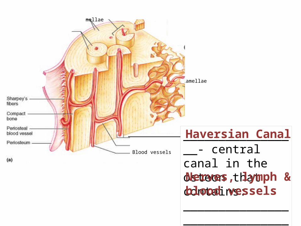
A. Anatomy of both types of bones.1. Compact bone ___________________- units
that compact bone are arranged in. (also called ____________.)
Haversian System
osteons
Circumferential Lamellae
Blood vessels

___________________- rings of hard, calcified matrix around the Haversian canal.
Concentric LamellaeCircumferential Lamellae
Blood vessels
Circumferential Lamellae
Blood vessels
_________________- central canal in the osteon that contains: ___________________________________.
Haversian Canal
Nerves, lymph & blood vessels
Circumferential Lamellae

Circumferential Lamellae
Blood vessels
_________________- Leads to the periosteum
Perforating Canal
Circumferential Lamellae

Concentric Lamellae
Compact Bone (continued)

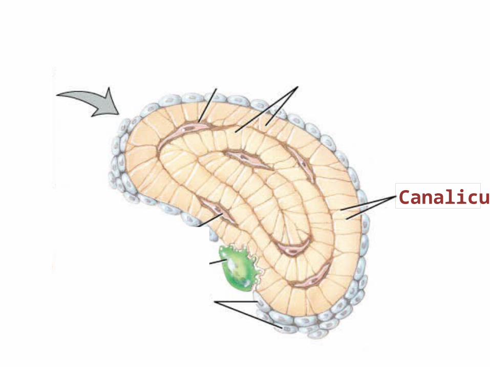
______________- the bone cell.Osteocyte

______________- (“small lake”) – small space that holds the osteocyte.
Lacunae

__________________- small channels filled with extracellular fluid which connects lacunae w/ each other and Haversian canal
Canaliculi

Haversian Canal

Red Space for ___________ bone marrow.
2. Spongy Bone

_______________- a network of thin columns of bone.
Trabeculae

Lacuna

Concentric Lamellae
Canaliculi

____________- fiber makersOsteoblasts

_______________- microbe killersOsteoclasts

____________- bone “maintainers”
Osteocyte

B. Differences between compact and spongy bone:
1. anatomical differences of each type:a. __________________ with Haversian canals are unique to compact bone.
b. ___________________ are unique
to spongy bone.
Osteons
Trabeculae

2. Location of each type in the body:
a. COMPACT bone is found in the ________________ of long bones.
b. SPONGY bone is found in:i. the _______________ and near the _____________________ of long bone.ii. Makes up most of: ___________ __________________________.
Diaphysis
epiphysisMedullary cavity
flat, short & irregular


3. Density differences of each type:a. Spongy bone is _______________
with empty spaces in between for red bone marrow to fill.
b. Compact bone is ____________ packed
with few spaces in between cells & ___________.
lighter
tightly
matrix

III. Physiological Features of Bone TissueA. Main Functions of the Bones & Skeletal System:
(Quick Glance) 1. ___________________or _______________ -
occurs in ___________________ only. 2. __________ - provides a framework for muscles to
attach to. 3. __________________ (detailed later) 4. _____________________ - works with muscles 5. __________ heart & other internal organs 6. _____________________________ in yellow
bone marrow. Acronym help: BS MA PhD
Support
ProtectsAssists In Movement
Mineral Homeostasis
Blood Cell Production
Deposits & stores adipose tissue
hemopoiesisRed bone marrow

B. Bone formation & ossification1. Definition of ossification:
_____________
_________________________________
2. When does ossification occur?a. begins about the ________ week
of embryonic life and continues into ___________ (ages18-25).
the process of bone formation
6th
adulthood

3. Two methods of ossification:a. _________________________- bone forms directly on or in loose fibrous
connective tissue.i. Where does this occur?
1. _____________________2. _____________________
Intramembraneous
Flat bones of skullMandible (lower jaw)

b. ____________________- bone forms within the cartilage. ____________________ in the body form this way.
EndochondralMOST BONES

4. Process of endochondral ossification:STEP #1:________________________________Development of the Cartilage Model
a). fetal _______________ cells crowd together in the shape of a future bone. (mesenchymal cells are embryonic tissue cells from which ALL connective tissue arises.)
mesenchymal

b). mesenchymal cells turn into _________________.chondroblasts

c). chondroblasts produce _________________ cartilage and the _________________ - membrane around the cartilage.
hyaline
perichondrium

STEP #2. _________________________________Growth of Cartilage Model
a). Chondroblasts become ________________ and some start to burst, triggering _________________.b). Remaining chondrocytes die b/c they cannot get enough _____________ in the calcifying matrix.c). When they die, _____________ form and merge into small cavities.
chondrocytescalcification
nutrients
lacunae

STEP #3. _________________________________Development of Primary Ossification Center
a). Primary ossification proceeds ____________________________.inward

b). A nutrient _________________ penetrates the middle of the cartilage.c) This stimulates _______________ cells to become ___________________ which lay the matrix to form the __________________ of spongy bone
artery
Osteogenic osteoblasts
trabeculae

d). ____________________ dissolves some of the newly formed trabeculae to create the ____________________ which fills with _____________________________.
Osteoclasts
Medullary cavity
Red bone marrow

STEP #4. _________________________________Development of Secondary Ossification Center
a). Like primary ossification except the bone remains as _______________.Spongy bone

STEP #5. _________________________________Formation of articular cartilage and epiphyseal plate
a). ________________ cartilage is replaced with ___________________ cartilage.
hyaline
articular

b). ______________ remains.
Spongy bone

c). ____________________ is the only remaining hyaline cartilage that allows the bone to ______________ until it calcifies. (usually between 18-25).
Epiphyseal plate
lengthens

IV. Homeostasis in Bone Tissue & Complications from an Imbalance
A. _____________________- the study of bone structure and the treatment of disorders.
Osteology

B. Bone Tissue “regeneration”:
1. old bone is constantly being ___________ by osteoclasts & new
bone tissue is being formed by _______________.(EVEN IN ADULTHOOD.)
resorbed
osteoblasts
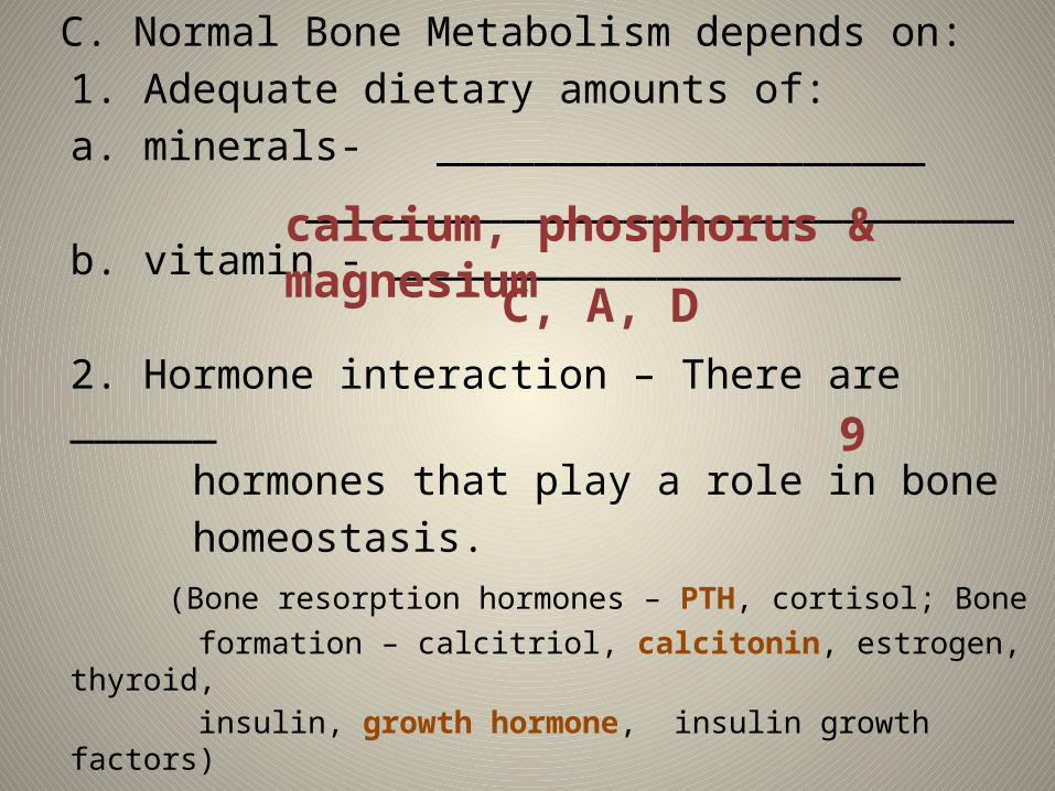
C. Normal Bone Metabolism depends on:1. Adequate dietary amounts of:
a. minerals- ____________________ _____________________________
b. vitamin - _____________________
2. Hormone interaction – There are ______ hormones that play a role in bone homeostasis.
(Bone resorption hormones – PTH, cortisol; Bone formation – calcitriol, calcitonin, estrogen, thyroid,
insulin, growth hormone, insulin growth factors)
calcium, phosphorus & magnesium
C, A, D
9

D. How bone aids in the homeostasis of Calcium in the blood:
1. if blood-calcium levels are too __________, PTH
(___________________), from the parathyroid gland is released which activates the osteoclasts to _______________ calcium into the blood via resoprtion.
low Parathyroid hormone
release

2. If blood-calcium levels are too _________, then the thyroid gland releases _________________________ which activates the osteoblasts to take up calcium via bone formation.
3. Effects of calcium imbalance:a. Too low ______________________b. Too high ______________________
high
calcitonin
Breathing stopsHeart stops

Calcium Homeostasis Song (Tune: Jingle Bells, start with “Dashing…..”)
• If calcium gets low • Parathyroid goes to work• Releases PTH • Bones give up calcium• Kidneys take in less• So it stays in the blood• Vitamin D makes intestines take it from
grub.

Song continued…• If calcium gets too high • Stimulates thyroid• Thyroid makes calcitonin• To bring the level down• Calcitonin stimulates deposition in the
bones• Kidney leave more calcium in urine • Now we’re done!

E. Impact on Human Growth Hormone (hGH) on Bone Homeostasis
1. hGH stimulates ___________________ to make more _________________ which will cause bone formation.
chondrocytescartilage

2. effects of hGH imbalance:a. Undersecretion:
i. ______________________ - when too little hGH is secreted and the
epiphyseal plate closes too soon.
Pituitary dwarfism


b. Oversecretion:i. during ________________ could lead
to ___________________ which is caused by an abnormal lengthening of bones.
childhoodgigantism

• Gigantism slide here

ii. During _________________ could lead to ___________________ which causes a thickening of the hand, feet, cheek and jaw bones. (bones won’t lengthen b/c epiphyseal plate is already calcified).
adulthoodacromegaly


• Pic of NFL issue and growth hormone here

F. Maintaining Bone Mass1. How can bone mass be maintained or strengthened?
a. ________________________i. Why?
1. mechanical stress (like the pull of gravity or the pull
of skeletal muscles) ____________________ bones.
Weight bearing Exercise
Strengthens and thickens

2. How can bone mass be lost?a. when bone ________________ occurs faster than bone formation. In healthy individuals, this happens when:
i. ___________________________ii. _______________ - break in bone
resorption
A limb is unusedA bone fractures

1. four descriptions of fractures:a. _____________________ - an incomplete break across the bone,
such as a crack.
Partial fracture


b. _______________________- a complete break across the bone so that the bone is in two or more pieces.
Complete Fracture


c. _______________________ - the fractured bone does not break through the skin. (It could be ________________ or ________________)
Closed fracture
partial complete
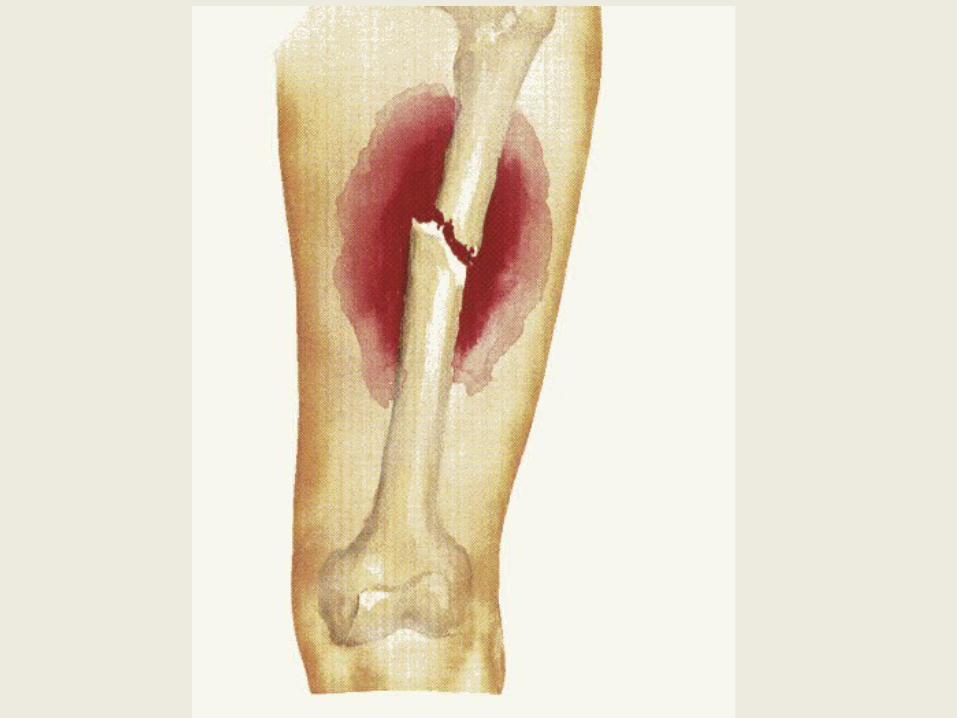

d. ________________________- the broken end of the bone protrudes through the skin. (Can only be a ________________ fracture.)
open fracture
compound


G. ____________________- a condition of porous bones caused by a depletion of ____________ in the body.
1. What can it cause?a. ______________________b. _________________(especially
hip fractures)c. ______________________d. ______________________
Osteoporosis
calcium
Bone mass lossfractures
Shortened height (hunch back)Bone pain

2. Who is most likely to get it & why?a. ____________________- estrogen (an osteoblast stimulator) declines dramatically during menopause.
3. What can be done to prevent getting it later in life?
a. ________________________b. ________________________
Middle aged women
Adequate calcium in dietWeight bearing exercise in early years