Hssasr volume5
69
High School Students for Agricultural Science Research Volume 5 I I I S A P “Agriculture meets Biomedicine” Proceedings of the V Congress PIIISA-CSIC May 2016 2016
-
Upload
german-tortosa -
Category
Science
-
view
543 -
download
0
Transcript of Hssasr volume5
High School Students for Agricultural Science Research
Volume 5
I I
I S A
P
“Agriculture meets Biomedicine”
Proceedings of the V Congress PIIISA-CSIC
May 2016
2016

High School Students for Agricultural Science Research
Volume 5 May 2016
EDITORIAL BOARD Juan de Dios Alché
Manuel Espinosa-Urgel
Francisco Martínez-Abarca
José Manuel Palma
ISSN: 2340-9746
Published in Granada by Estación Experimental del Zaidín. CSIC
High School Students for Agricultural Science Research. Vol. 5 by Estación Experimental del Zaidín is licensed under a Creative Commons Reconocimiento-NoComercial-SinObraDerivada 4.0 Internacional License.


Index Foreword/prefacio M. Rodríguez, A. Castellano ...................................................................................................................... i-ii Editorial: Agriculture meets Biomedicine M. Espinosa-Urgel ..................................................................................................................................... iii “What pepper beneath”: Biochemical and molecular characterization of catalase in pepper fruit. L. Sáez Martín, M. Jiménez Carretero, A. García Pérez, A. Padial Raya, D. Adamuz Puerto, C. Ruiz, M. Rodríguez Ruiz .......................................................................... 1-9 Brainstorming in Agricultural Sciences: Seeking the antioxidative enzymes in pomegranate (Punica granatum L.) A. del Águila Gómez, D.J. Carricondo Pérez, M.R. Píñar Espejo, C. Sánchez Rodríguez, A. Yasser Mossad, F. del Águila Gómez, M.J. Campos, J. Iglesias, J.M. Palma .................................................................................................................................................... 10-17 Study of the ultra-violet light effect on the mutation frequency in a soil bacterium A.M. Bolivar, M.L. Candela, P. Collado, M.L. Freire, C. Molina, L. Torres, C.M. Vargas, F. Martínez-Abarca ....................................................................................................... 18-26 Genomic variability in humans: our genetic fingerprint I.M. Bautista-Plaza, L. Jiménez-Molina, A. Manzano-Martín, B. Ortiz-Rodríguez, M. Pedregosa-Ruiz, F. Matesanz, A. Barroso-del Jesus ........................................................... 27-35 Resistance to repeated radiotherapy in glioblastoma cells is partially prevented by the use of a DNA repair inhibitor E. Gómez, L. Muros, D. Oliver, M., J.L. Ruiz, N. Ruiz, L.M. López, J. Oliver ............................. 36-41 Biofertilisers with olive-oil taste: isolation of plant growth-promoting rhizobacteria (PGPR) from “alperujo” compost A. Díaz Arco, M. Molina Muñoz, E. Navarro García, C. Ortega Fernández, L. Palma Pérez, M.C. Sarmiento Vega, A. Castellano Hinojosa, G. Tortosa Muñoz, E.J. Bedmar .............. 42-50 A microscopical garden at the Estación Experimental del Zaidín E. Lima, M.E. Ramos, I. Cabello-Cano, E.L. Carretero-Quero, A.M. García-Gil, J. Moreno-García, A.M. Romero-López, M. Ruiz-López, J.D. Alché ............................................ 51-58

i
Foreword: the PIIISA experience in the eyes of young researchers
Although a number of researchers with previous experience in PIIISA have participated in
most of the projects, this is the first project that I supervise, so for me personally it has extra value. It has been a gratifying experience that has allowed me to see different aspects of research activity. But it has also been a challenge that has made me improve at the scientific and personal levels, since it has tought me how to design a project, to organize ideas and to be more determined.
Being able to show and disseminate our work to people who do not have daily contact with the scientific world is motivating: you discover that students are actually able to make the project progress through their eagerness and curiosity. During the course of the sessions one can feel how the group quickly takes in all the information that one tries to transmit. This shows their great capacity to adapt and integrate into a world that for them is rather unknown and sometimes even idealized. It is remarkable how fast they handle the techniques and lab instruments, prepare reagents and interpret results.
All the above can be taken as a clear indication that PIIISA is a tool that opens the world of science to young people in an easy and close way. This is a clear necessity, since scientific work is often seen by society as something very distant from their lives. With this kind of activities we help to reduce that distance. I believe the final result will be a great example of team work with excellent research projects carried out by youngsters that will not only reflect scientific results, but also their views and experiences along the way. I hope initiatives such as this one are not put aside, but rather keep growing and expanding.
Marta Rodríguez Ruiz Estación Experimental del Zaidín. CSIC
From the researcher’s perspective, it is a very interesting and rewarding experience to be able to feel the enthusiasm that young students display during their visit to our laboratories. It is a challenge for us to be able to explain concepts and ensure that we are understood by the students. When we speak to students and realise that they have understood the essence of the concept so well that they can explain it in their own words, we most definitely feel like real scientists.
I think that disclosure is essential to evoke interest and pass on knowledge to the future generations. The PIIISA project is a good way to approach society and incorporate it within research. I’m sure that these talented young individuals will go far and it will be pleasing to know that this experience has been important in helping them realise their scientific vocation.
Antonio Castellano Hinojosa Estación Experimental del Zaidín. CSIC

ii
Prefacio: La experiencia PIIISA a los ojos de jóvenes investigadores
Aunque en la mayoría de los proyectos han tomado parte un número variado de investigadores que han participado en ediciones anteriores, a nivel personal este es el primer proyecto que dirijo por lo que tiene un valor añadido para mí. Ha sido una experiencia gratificante que me ha permitido ver otros aspectos del ámbito de la investigación. Ha sido un reto que me ha permitido mejorar a nivel científico y personal ya que me ha enseñado a esbozar un proyecto, organizar ideas y a ser más resolutiva.Poder divulgar y mostrar el trabajo que hacemos a personas que no tienen contacto día a día con el mundo de la ciencia es motivador: Compruebas cómo los estudiantes son los que hacen crecer el proyecto a través de su ilusión y de sus inquietudes.
A lo largo de las sesiones se aprecia cómo el grupo va absorbiendo rápidamente todo el flujo de información que se le intenta transmitir, demostrando su gran capacidad de adaptación e integración a un mundo para ellos no tan conocido y en ocasiones hasta mitificado. Es remarcable cómo rápidamente manejan las técnicas, los instrumentos del laboratorio, preparan los reactivos e interpretan los datos obtenidos.
Todo lo expuesto puede tomarse como un referente claro de que el proyecto PIIISA es una herramienta que abre el mundo de la ciencia a los jóvenes de forma fácil y cercana, una cuestión más que necesaria ya que el mundo de la ciencia es bastante distante en muchas ocasiones para la sociedad. Con este tipo de actividad contribuimos a que esa lejanía merme. Creo que el resultado final será una gran muestra de trabajo en equipo donde se verán grandes proyectos de investigación realizados por jóvenes donde queda plasmado no solo un resultado científico sino también sus opiniones y experiencias a lo largo de todo el trayecto. Espero que iniciativas como esta no queden en el olvido y sigan desarrollándose y ampliándose.
Marta Rodríguez Ruiz Estación Experimental del Zaidín. CSIC
Desde el punto de vista del investigador esta experiencia resulta de gran interés ya que resulta muy gratificante sentir el entusiasmo que ponen estos jóvenes estudiantes durante su estancia en nuestros laboratorios. Se trata de un reto para nosotros puesto que tenemos que ser capaces de explicar y hacernos entender. Sin duda, cuando hablamos con ellos y sentimos que han captado la idea y que saben explicarla adecuadamente nos sentimos como verdaderos científicos.
Pienso que la divulgación es esencial para despertar el interés y transmitir el conocimiento a las nuevas generaciones. El proyecto PIIISA es una buena forma de acercarnos a la sociedad y hacerla participe de la importante labor de la investigación. Estoy seguro que estos jóvenes talentos llegarán tan lejos como se propongan y será grato saber que esta experiencia fue importante para ellos y despertó su vocación científica.
Antonio Castellano Hinojosa Estación Experimental del Zaidín. CSIC
iii
Editorial
Agriculture meets Biomedicine
Manuel Espinosa-Urgel
Estación Experimental del Zaidín. CSIC [email protected]
High School Students for Agricultural Science Research reaches its volume number 5. Five consecutive years of a periodical publication in which science breaks the barrier that separates researchers from young students that are still trying to discover their own professional vocations.
This new issue of High School Students for Agricultural Science Research represents a milestone in the brief but intense life of this publication. Up until now, the published articles mainly reflected the work carried out at the Estación Experimental del Zaidín (EEZ) as part of the yearly PIIISA program. Being a research institute devoted to agricultural sciences, it is not surprising that soil, plants and plant-associated microorganisms played the leading role. This year, however, we have crossed the boundaries with two articles coming from the Institute of Parasitology and Biomedicine “López-Neyra”. One of them deals with human genetic variability and the other with anti-cancer research. They accompany five other communications ranging from plant biology (with pepper, pomegranate and pollen as the subjects of research) to soil microbiology (finding new plant-beneficial bacteria) or microbial genetics (the influence of UV light on mutation rates). It is a clear indication that science knows and cares very little about the boxes or labels that we artificially use to parcel it. Perhaps the forces that join in this issue will lead to an even more expanded scope in the future, so that the seed for a forthcoming “High School Students for Scientific Research” may have been sown with this volume. Or maybe not; after all, evolution finds its own –and often unexpected– ways.
Breaking boundaries has been the aim of PIIISA from the start: a program to make high school students participate and immerse into the meaning of research. The idea is to learn about science not as an abstract concept, or something that is already known and complete and can be found in text books. Rather, to discover first hand that scientific progress is an ongoing process that requires asking the right questions, finding the best techniques to answer them, and then asking again what we have learnt and what we should ask next. And also, that research is challenging but in this challenge there are great doses of fun. Check the separate “My own ideas” section after each article, where students recollect their participation in PIIISA. There you will find the true nature of this experience.
As a scientific publication, our pretensions are modest. We do not intend to become impact factor-maniacs, and we do not have a fancy layout on glossy paper. What we aim at is completely different: to be the vehicle that disseminates the work in which young students have participated, to teach them how science is transmitted, and to show the next generation of high school students that they, too, can become scientists. For researchers reding this volume, we hope they are at least intrigued by the things that can be done in this context. And that they are encouraged to pursue that little side project scribbled on a notepad that has been sitting in a drawer waiting for a chance.

iv
1
“What pepper beneath”. Biochemical and molecular characterization of catalase in pepper
fruit
Luis Sáez Martín1, Mónica Jiménez Carretero 1, Ana García Pérez 2, Alejandra Padial Raya 3, David Adamuz Puerto 4, Carmelo Ruiz5, Marta Rodríguez Ruiz5*
1IES Padre Manjón. Gonzalo Gallas s/n, 18003 Granada, Spain 2IES José de Mora.C/ Blas Infante, 9, 18800, Baza,Granada, Spain.
3CDP Juan XXIII-Zaidín. Camino Santa juliana s/n, 18006 Granada,Spain 4 IES Hiponova. C/Federico García Lorca, 1, 18270, Montefrío, Granada, Spain
5Departamento de Bioquímica Biología Celular y Molecular de Plantas,Estación Experimental del Zaidín, CSIC, Profesor Albareda 1, 18008 Granada, Spain
*Corresponding author: e-mail: [email protected]
SUMMARY
Biochemical and molecular properties of catalase were studied in green and red pepper (Capsicum annuum L.) fruits. Catalase activity was analyzed, obtaining higher values in green peppers. Analysis by SDS-PAGE and immunoblot displayed similar pattern to catalase activity and showed that the pepper catalase could be composed of subunits of about 21.5 kDa. The molecular weight of the native protein was also determined for green and red peppers and results rendered a weight around 125-129 kDa.
INTRODUCTION (AND OBJECTIVES)
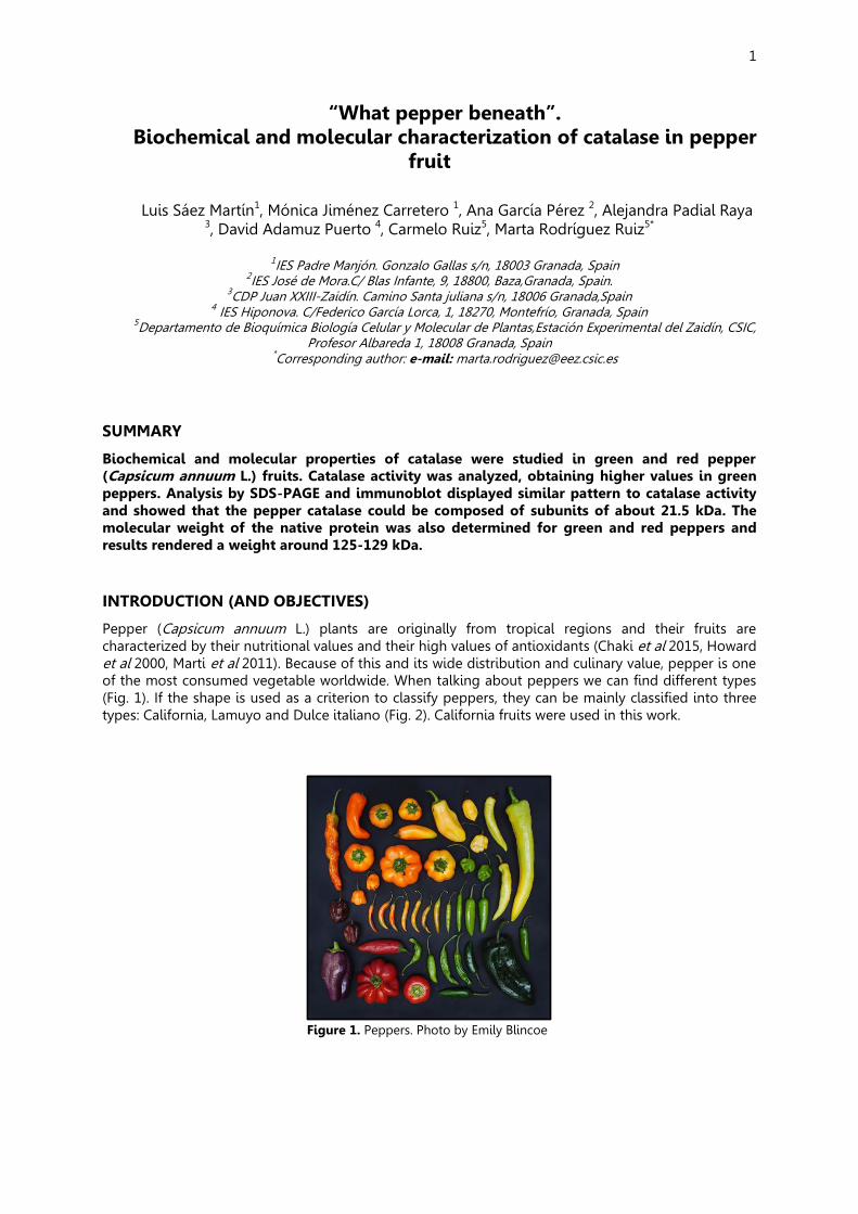
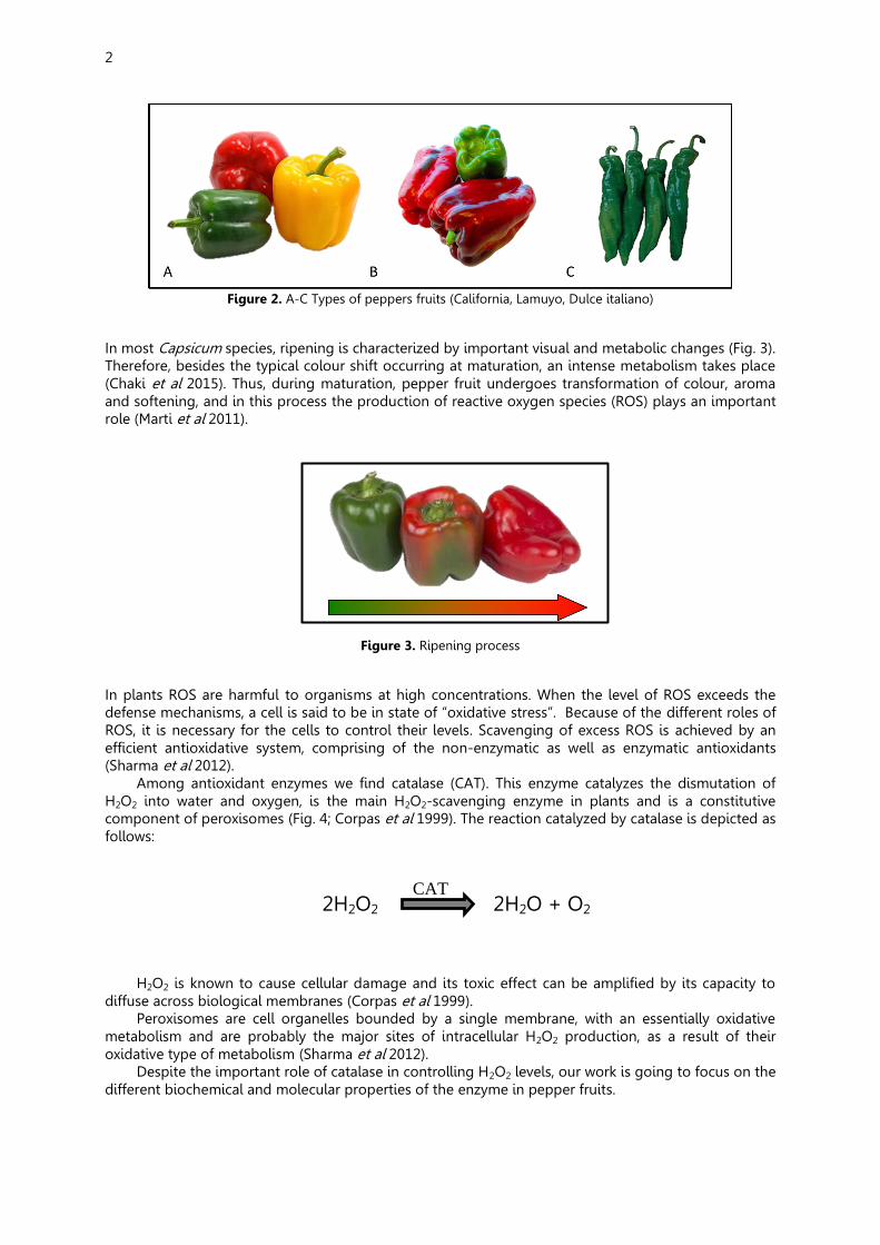
Pepper (Capsicum annuum L.) plants are originally from tropical regions and their fruits are characterized by their nutritional values and their high values of antioxidants (Chaki et al 2015, Howard et al 2000, Marti et al 2011). Because of this and its wide distribution and culinary value, pepper is one of the most consumed vegetable worldwide. When talking about peppers we can find different types (Fig. 1). If the shape is used as a criterion to classify peppers, they can be mainly classified into three types: California, Lamuyo and Dulce italiano (Fig. 2). California fruits were used in this work.
Figure 1. Peppers. Photo by Emily Blincoe
2
Figure 2. A-C Types of peppers fruits (California, Lamuyo, Dulce italiano)
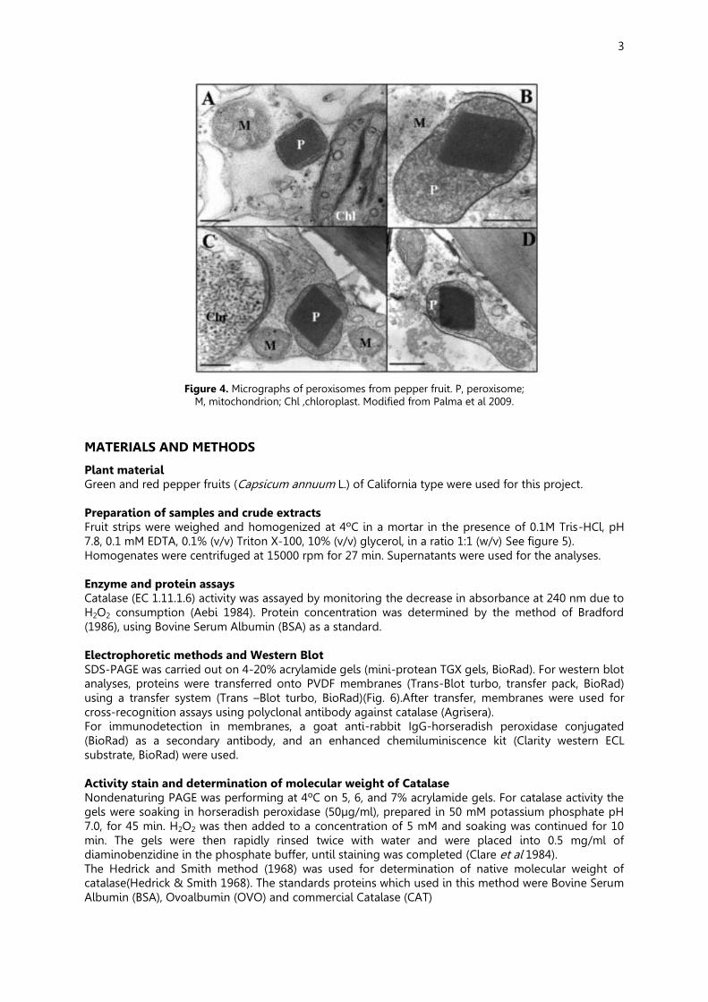
In most Capsicum species, ripening is characterized by important visual and metabolic changes (Fig. 3). Therefore, besides the typical colour shift occurring at maturation, an intense metabolism takes place (Chaki et al 2015). Thus, during maturation, pepper fruit undergoes transformation of colour, aroma and softening, and in this process the production of reactive oxygen species (ROS) plays an important role (Marti et al 2011).
Figure 3. Ripening process
In plants ROS are harmful to organisms at high concentrations. When the level of ROS exceeds the defense mechanisms, a cell is said to be in state of “oxidative stress”. Because of the different roles of ROS, it is necessary for the cells to control their levels. Scavenging of excess ROS is achieved by an efficient antioxidative system, comprising of the non-enzymatic as well as enzymatic antioxidants (Sharma et al 2012).
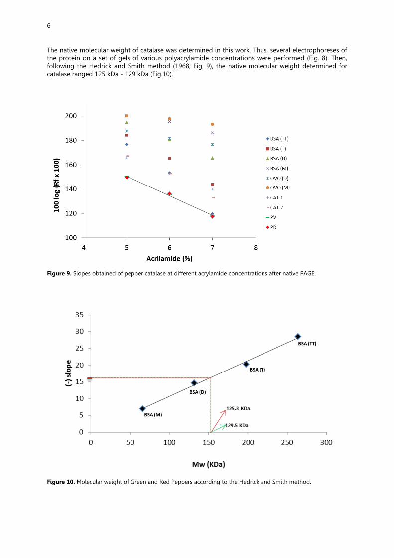
Among antioxidant enzymes we find catalase (CAT). This enzyme catalyzes the dismutation of H2O2 into water and oxygen, is the main H2O2-scavenging enzyme in plants and is a constitutive component of peroxisomes (Fig. 4; Corpas et al 1999). The reaction catalyzed by catalase is depicted as follows:
2H2O2 2H2O + O2
H2O2 is known to cause cellular damage and its toxic effect can be amplified by its capacity to diffuse across biological membranes (Corpas et al 1999).
Peroxisomes are cell organelles bounded by a single membrane, with an essentially oxidative metabolism and are probably the major sites of intracellular H2O2 production, as a result of their oxidative type of metabolism (Sharma et al 2012).
Despite the important role of catalase in controlling H2O2 levels, our work is going to focus on the different biochemical and molecular properties of the enzyme in pepper fruits.
CAT
3
Figure 4. Micrographs of peroxisomes from pepper fruit. P, peroxisome;
M, mitochondrion; Chl ,chloroplast. Modified from Palma et al 2009. MATERIALS AND METHODS
Plant material Green and red pepper fruits (Capsicum annuum L.) of California type were used for this project. Preparation of samples and crude extracts Fruit strips were weighed and homogenized at 4ºC in a mortar in the presence of 0.1M Tris-HCl, pH 7.8, 0.1 mM EDTA, 0.1% (v/v) Triton X-100, 10% (v/v) glycerol, in a ratio 1:1 (w/v) See figure 5). Homogenates were centrifuged at 15000 rpm for 27 min. Supernatants were used for the analyses. Enzyme and protein assays Catalase (EC 1.11.1.6) activity was assayed by monitoring the decrease in absorbance at 240 nm due to H2O2 consumption (Aebi 1984). Protein concentration was determined by the method of Bradford (1986), using Bovine Serum Albumin (BSA) as a standard. Electrophoretic methods and Western Blot SDS-PAGE was carried out on 4-20% acrylamide gels (mini-protean TGX gels, BioRad). For western blot analyses, proteins were transferred onto PVDF membranes (Trans-Blot turbo, transfer pack, BioRad) using a transfer system (Trans –Blot turbo, BioRad)(Fig. 6).After transfer, membranes were used for cross-recognition assays using polyclonal antibody against catalase (Agrisera). For immunodetection in membranes, a goat anti-rabbit IgG-horseradish peroxidase conjugated (BioRad) as a secondary antibody, and an enhanced chemiluminiscence kit (Clarity western ECL substrate, BioRad) were used. Activity stain and determination of molecular weight of Catalase Nondenaturing PAGE was performing at 4ºC on 5, 6, and 7% acrylamide gels. For catalase activity the gels were soaking in horseradish peroxidase (50µg/ml), prepared in 50 mM potassium phosphate pH 7.0, for 45 min. H2O2 was then added to a concentration of 5 mM and soaking was continued for 10 min. The gels were then rapidly rinsed twice with water and were placed into 0.5 mg/ml of diaminobenzidine in the phosphate buffer, until staining was completed (Clare et al 1984). The Hedrick and Smith method (1968) was used for determination of native molecular weight of catalase(Hedrick & Smith 1968). The standards proteins which used in this method were Bovine Serum Albumin (BSA), Ovoalbumin (OVO) and commercial Catalase (CAT)

4
Figure 5. Preparation of crude extracts
Figure 6. Preparation of electrophoresis and western blotting
RESULTS
The analysis of catalase activity in green and red fruit was performed and revealed that this activity was higher in green fruit than in red fruit, but this different was not significant (Fig.7) and pattern was similar to immunoblot analysis carried out with an antibody against catalase, where the expression of this protein was higher in green fruit than red fruit (Fig. 7). Western blot analysis showed up a recognition band around 21.5 kDa for this type of fruits (Fig.7).

5
Figure 7. Catalase activity and determination of the subunit size by western blotting. Immunoblot of pepper fruit (30 g protein) was probed with an anti-Cat (dilution 1/5000). GF, green fruit; RF, red fruit
Figure 8. Electrophoresis of pepper catalase at different acrylamide concentrations. Coomassie Stain of standards proteins (left) and catalase activity (right) are displayed; BSA, bovine serum albumin; OV, ovoalbumin; CAT, commercial catalase; GF, green fruit; RF, red fruit..
6
The native molecular weight of catalase was determined in this work. Thus, several electrophoreses of the protein on a set of gels of various polyacrylamide concentrations were performed (Fig. 8). Then, following the Hedrick and Smith method (1968; Fig. 9), the native molecular weight determined for catalase ranged 125 kDa - 129 kDa (Fig.10).
Figure 9. Slopes obtained of pepper catalase at different acrylamide concentrations after native PAGE.
Figure 10. Molecular weight of Green and Red Peppers according to the Hedrick and Smith method.

7
CONCLUSIONS
1. Catalase activity in green pepper is higher, but not significant, than red pepper. 2. The molecular weight of green and red peppers ranges 125-129 kDa. 3. The amount of catalase protein in green pepper is higher than in red pepper and the subunit
size for this variety of peppers was calculated to be about 22 kDa. 4. The atypical value obtained in this work for the catalase size of catalase is different from
others enzymes that have been reported before. Therefore, it is necessary and important to have more information about this enzyme in pepper fruits.
REFERENCES Aebi H. 1984. Catalase in vitro. Methods in enzymology 105: 121-6 Bradford MM (1986) A rapid and sensitive method for the quantitation of microgram quantities of
protein utilizing the principle of protein-dye binding. Analytical Biochemistry 72: 248-254. Clare DA, Minh Ngoc D, Darr D, Archibald F, Fridovich I. 1984. Effects of molecular oxygen on detection
of superoxide radical with nitroblue tetrazolium and on activity stains for catalase. Analytical Biochemistry 140: 532-37
Corpas FJ, Palma JM, Sandalio LM, Lopez-Huertas E, Romero-Puertas MC, et al. 1999. Purification of catalase from pea leaf peroxisomes: identification of five different isoforms. Free radical research 31 Suppl: S235-41
Chaki M, Alvarez de Morales P, Ruiz C, Begara-Morales JC, Barroso JB, et al. 2015. Ripening of pepper (Capsicum annuum) fruit is characterized by an enhancement of protein tyrosine nitration. Annals of botany 116: 637-47
Hedrick JL, Smith AJ. 1968. Size and charge isomer separation and estimation of molecular weights of proteins by disc gel electrophoresis. Archives of biochemistry and biophysics 126: 155-64
Howard LR, Talcott ST, Brenes CH, Villalon B. 2000. Changes in Phytochemical and Antioxidant Activity of Selected Pepper Cultivars ( Capsicum Species) As influenced by Maturity. Agric.Food.Chem. 48: 1713-20
Marti MC, Camejo D, Vallejo F, Romojaro F, Bacarizo S, et al. 2011. Influence of fruit ripening stage and harvest period on the antioxidant content of sweet pepper cultivars. Plant Foods Hum Nutr 66: 416-23
Palma JM, Corpas FJ, del Rio LA. 2009. Proteome of plant peroxisomes: new perspectives on the role of these organelles in cell biology. Proteomics 9: 2301-12
Sharma P, Jha A.B, Dubey R.S, M. P. 2012. Reactive Oxygen Species,Oxidative Damage, and Antioxidative Defense Mechanism in Plants under Stressful conditions. Journal of Botany 2012: 26

8
MY OWN IDEAS Mónica Jiménez Carretero, IES Padre Manjón-Zaidín
In my opinion, this project has been very interesting, because I have had the opportunity to work in a laboratory with scientists who have helped me with the project. The most important thing I have learned is how to set out a research and work in a laboratory. Moreover, I have acquired new and specific vocabulary about the subject. In this project, we have worked with peppers. Although most of us don't like them, I have realized that they are important fruits. Peppers have a lot of vitamin C (especially the red ones). In fact, they contain much more than fruits like oranges or strawberries. Also, they are important sources of antioxidants. Now that I know all this about them, I try to eat more peppers than before. During the research, we have been using many complex instruments that I hadn't seen before. The most difficult part was when we had to load the gels for the electrophoresis. But I have enjoyed working there, and I hope I will work in a place like this in the future. I could go to all the sessions, so I have learned every detail about the research. Even though some concepts and ideas were new to me, I have been able to understand all the proceedings we have done. Now that everything is finished I think that we have achieved our objective, because our results are really good. We have been able to characterize the Catalase in red and green peppers. From my point of view, people would learn more if they had the chance to work in a project like this. Students should have more practical lessons at school so they could use the concepts they learn, because otherwise they will just memorize the concepts but they will not understand them. To conclude, I have to say that this experience has taught me very important things about the world of science that I would not know otherwise. Also, I have to thank the researchers who have told me everything about this project. Luis Sáez Martín, IES Padre Manjón-Zaidín
I still remember when my teacher of physics and chemistry offered me to participate in this project, it was still early and I was not sure yet what decision to make. now I can say that this has been one of the best decisions I have made along this year, to work on this project has been a different experience, a unique opportunity to reach a privileged few as my colleagues and me that decided to get out of that normal academic activity of the high school to observe science and research through the eyes of a scientist. In the months that I have been working within the walls of the experimental station of Zaidin, I have discovered an unknown world for me, so far I have learned to use many machines which I had never heard, I have acquired a good scientific culture and I have learned a lot of new vocabulary, I also learned many scientific techniques. When my teacher told me to work with peppers, it was not something that fascinated me but once the work is completed I am so glad. I have learned many things about this fascinating fruit and the protein that we have studied, “the catalase”. It has served to bring into practice the lessons that I had learned in biology and biochemistry. Finally I would like to thank in writing Marta and Carmelo, they were lovely, without them this project would not have been the same, they have been receptive to help at any time it a has been a pleasure for me to work with them. I can only say that participating in PIIISA has been an experience that I will never forget I’ve enjoyed and I’ve learned a lot and in addition I have met good people, I will always be grateful for this opportunity and I encourage all students to participate in it.
9
Ana García Pérez, IES José de Mora (Baza)
I think that this type of projects are able to draw the attention of young people to introduce them into investigation and I can say completely sure that they work. My experience in this project was awesome, I worked with machines that I had never seen and I've used methods that I had never known so now I'm more interested in the investigation scientific research. Alejandra Padial Raya, CDP Juan XXIII_Zaidín
En mi opinión es una grata experiencia sé que no lo voy a olvidar, y voy a tener el recuerdo conmigo siempre que pueda y lo mejor es que me ha hecho comprender la ciencia , acercarme a ella, darle vida, darle forma, darle interés, porque, he tenido muy buenas emociones con toda esta experiencia. Y he conocido a muchas personas impresionantes, tanto mis compañeros como mis investigadores Marta y Carmelo. He podido vivir en primera persona como se trabaja en un laboratorio, la forma de organizarse y como utilizar los aparatos y utensilios con los que hemos llevado a cabo algunos experimentos con los que nos han servido de gran motivación para acercarnos a este mundo tan complejo como fascinante que es la ciencia. Agradezco mucho esta oportunidad de haber participado en este proyecto que me ha ayudado a entender el mundo de la investigación y a conceder la importancia que conlleva el trabajo previo en un laboratorio para poder aplicar posteriormente todos esos conocimientos y resultados obtenidos en proyectos. David Adamuz Puerto, IES Hiponova Montefrio
At first, I went to this project because i wanted to lose class time but on the first day, I changed my mind. I wanted work with liquid nitrogen and with the pepper. I would have wanted go at extra meetings but I couldn’t. The firsts days were funny because we worked in the laboratory but the last days, were bored because we were doing the poster and proceeding. This project has helped me to know the pepper better and to know how to work in the laboratory and of course to make friends.
10
Brainstorming in Agricultural Sciences: Seeking the antioxidative enzymes in pomegranate
(Punica granatum L.) (http://brainstorming-eez.blogspot.com.es/)
Alba del Águila Gómez1, Diego Jesús Carricondo Pérez2, María Rosa Píñar Espejo3,
Cristina Sánchez Rodríguez4, Ahmed Yasser Mossad5, Fátima del Águila Gómez1, María Jesús Campos6, Jessica Iglesias6, José M. Palma6,*
1 IES Avenmoriel. Parque San León, 18817 Benamaurel, Granada, Spain. 2IES Cartuja. Julio Moreno Dávila, 18, 18011 Granada, Spain.
3 IES Jiménez de Quesada. Camino Sta. Teresa, 1, 18320 Santa Fe, Granada, Spain. 4IES Aricel. Calle del Aricel, 18220 Albolote, Granada, Spain.
5Centro Juan XXIII-Zaidín. Camino Santa Juliana, s/n, 18016 Granada, Spain 6Grupo de Antioxidantes, Radicales Libres y Óxido Nítrico en
Biotecnología y Agroalimentación, Estación Experimental del Zaidín, CSIC, Profesor Albareda 1, 18008 Granada, Spain
*Corresponding author: e-mail: [email protected]
HIGHLIGHTS
• The PIIISA-Granada students have successfully achieved a whole research project, from design to conclusions, for the first time.
• This pioneer initiative has allowed them proposing which plant species should be investigated, which strategy to be followed, how to perform the experiments, which conclusions could be withdrawn and, finally, how the research can be addressed in the future.
• The superoxide dismutase isoenzymatic pattern of pomegranate seeds has been reported for the first time. In fact, this is the first report of an antioxidative enzyme system in this plant species.
SUMMARY
The main goal of this work was to issue a project from the very beginning, emerging from a brainstorming, as it should be when achieving Science. But, in our case, the storm started at the student minds. Accordingly, a new topic was introduced in our research group objectives: the analysis of the antioxidative enzymes in a still little studied plant crop, the pomegranate. We set the procedure to extracts protein (if any) from aril juice, skin and seeds. No proteins were practically detected in both arils and skin, whereas, in seeds, at least eight major protein bands were found in SDS-polyacrylamide gels stained with silver. The analysis of superoxide dismutase (SOD) activity by native PAGE in all tissues rendered 4-6 isoenzymes, only in seeds, and this was depending on the variety/location analyzed. This is the first report of SOD activity in pomegranate and points towards a latent metabolism of reactive oxygen species (ROS) in seeds.
INTRODUCTION (AND OBJECTIVES)
The pomegranate (Punica granatum L.) is a fruit-bearing deciduous shrub or small tree from the family Lythraceae that grows up to 5 -10 m. The pomegranate has multiple spiny branches, and is extremely long-lived, with some specimens surviving for 200 years [1-3]. Pomegranate leaves are opposite or subopposite, glossy, narrow oblong, entire, 3–7 cm long and 2 cm broad. The flowers are bright red with 3 cm in diameter and three to seven petals. P. granatum has more than 500 named cultivars, but evidently there is considerable synonymy in which the same genotype is named differently across regions of the world [1,3].
11
Pomegranate is originally native of the Northern India and Iran, and it has been cultivated and naturalized since ancient times over the entire Mediterranean region and Middle East, and later in the United States, South America, and Central America [4].
The fruit produced by this small tree is a rounded shape, large, deep red berry (5-12 cm in diameter), with leathery and thick skin, and crowned by a pointed calyx. It contains numerous arils, each surrounded by a translucent juice-containing sac which engulf the own juice and one seed [4-6]. The number of seeds in a pomegranate can vary from 200 to about 1400. Juice represents about 30% of the fruit weight, whereas seeds are around 3%.
Several papers have reported the chemical composition of the different parts of the pomegranate, also conducted to find the relationship with its biological activity [1–3]. This fruit has high antioxidant content, and its pharmaceutical properties, such as anti-inflammatory and anticarcinogenic effects, the reduction of cardiovascular risks and the control of kidney troubles have partially been attributed to substances such as ellagic acid, ellagitannins (including punicalagins), punic acid, anthocyanins, flavonols, flavan-3-ols, and flavones [4,5]. Juice also contains beneficial compounds such as catequines, vitamin C, and phytosterols, among others [1,3-5].
Only few studies have been devoted to investigate pomegranate proteins, which represent ca. 120 g kg−1 of the seeds. Furthermore, these studies have been limited only to specific proteins [6-10]. Yang et al. [8] isolated a new class III chitinase from pomegranate seeds. More recently, the proteome of pomegranate has been reported where a list of 1,488 proteins was obtained, although only six of which belonged to pomegranate species [6]. Most pomegranate proteins identified from seeds are storage proteins, whose major components are globulins and albumins, followed by glutelin and prolamin [6].
Regarding to antioxidative proteins from pomegranate, no data are known thus far, and investigation in this issue is of great interest, not only from of a scientific point of view, but also from its repercussion in human diet. In this work, the identification of antioxidative enzymes from pomegranate fruits was initiated.
MATERIALS AND METHODS
Plant material Pomegranate (Punica granatum L.) fruits from two locations of the province of Granada were analyzed: Benamaurel (37°36′30″N 2°41′50″O, altitude, 723 m) y El Fargue (37°12′N 3°33′O; altitude, 1111 m). Fruits were separated into different parts according to their structure. Thus, skin, juice from arils and seeds were pooled separately for further analysis. Preparation of crude extracts To obtain juice from fruits, arils were squeezed within two-layers nylon cloth bags, and then diluted with buffer Tris-HCl 0,1 M, pH 7,5, glycerol 20% (v/v), EDTA 2 mM, DTT 5 mM in a ratio 1:1 (juice:buffer). Crude extracts from skin and seeds (once removed the external layer of the aril) were prepared in Tris-HCl buffer 50 mM, pH 7,5, glycerol 10% (v/v), EDTA 1 mM, DTT 5 mM, in a ratio 1:4 (plant material:buffer). Protein determination For protein concentration the method of Bradford [11] was followed, using bovine serum albumin as standard. SDS-PAGE (proteins) SDS-PAGE of pomegranate samples was carried out on 12% polyacrylamide gels as described by Laemmli [12]. Prior to electrophoresis, samples were heated at 95ºC for 5 min in the presence of 0.1% (w/v) SDS and 5 mM DTT. Gels were stained with Coomassie Brilliant Blue R-250 and also with silver nitrate as reported earlier [13,14] for proteins detection. Native PAGE (SOD activity) Native polyacrylamide gel electrophoresis was performed using acrylamide gels as described by Davis [15]. SOD (EC 1.15.1.1) isozymes were separated by nondenaturing polyacrylamide gel electrophoresis (PAGE) on 10% acrylamide gels and visualized by a photochemical NBT (nitroblue tetrazolium) reduction method [16].
12
RESULTS
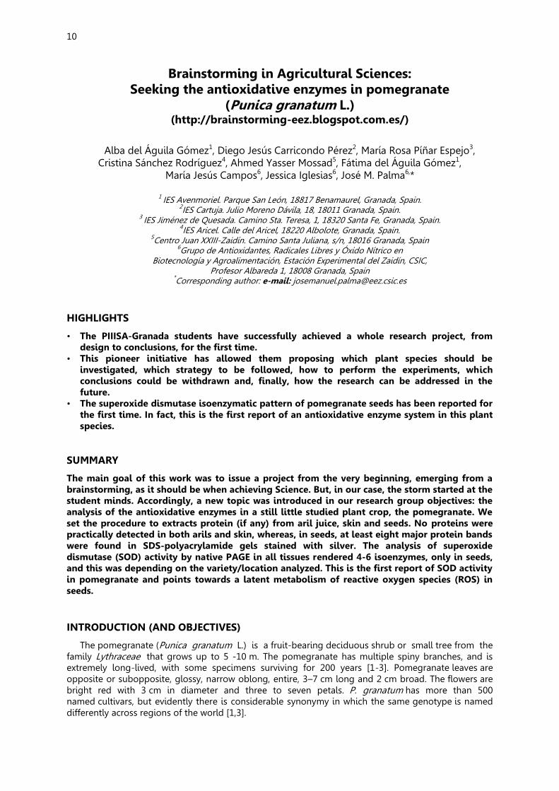
In Fig. 1 the design of the protocol to obtain the proteins from the different parts of pomegranate fruits is depicted.
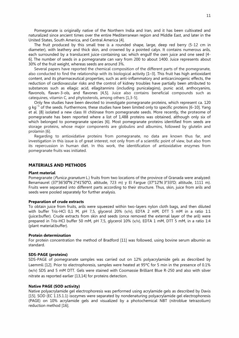
Figure 1. Protocol set for the preparation of crude extracts from different parts of the pomegranate fruits. For the analysis of the protein pattern of juice, skin and seeds from fruits, SDS-PAGE in 12% polyacrylamide electrophoresis was performed. Gels were initially stained with the Coomassie blue method, but no bands were visible in gels (results not reported). Then, silver staining was achieved, and the results obtained are shown in Figure 2.
Figure 2. SDS-PAGE and protein staining of samples from pomegranate fruits harvested at two locations of the province of Granada. Fruits were collected from Benamaurel and El Fargue. A, arils; Sk, skin; Sd, seeds. Molecular weight markers are indicated on the left.
13
As it can be seen in the Fig. 2, only one protein band was detected in juice from Benamaurel fruits, whereas in skin no proteins were visible in fruits from the two locations. Regarding to the analysis of seeds, up to 8 protein bands with similar mobility were observed in both locations.
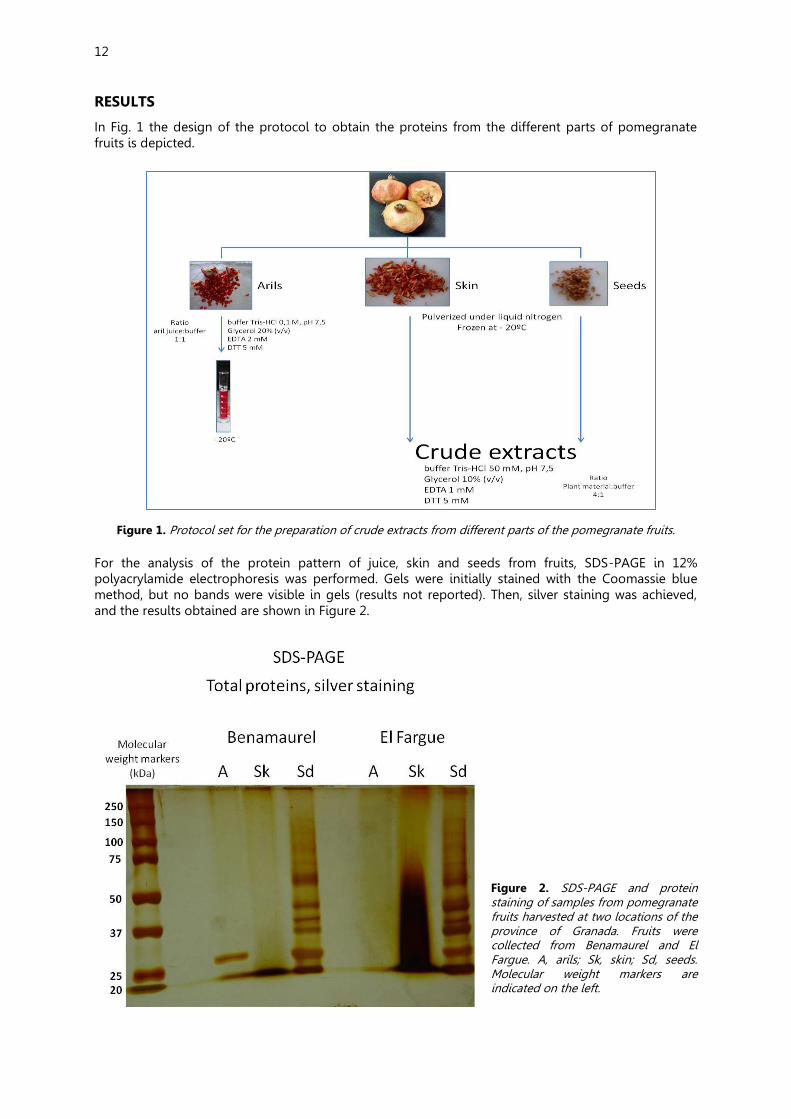
The isoenzimatic SOD pattern in samples was also determined by native PAGE. Although no clear
activity bands were observed in juice from both locations, the presence of this enzymatic system cannot be discarded in view of the faint band detected at the bottom of the gel (Figure 3). The samples which showed a well defined activity profile were seeds, where up to six isoenzymes could be visible (marked with asterisks). This is the first report of the presence of this enzymatic system in this plant species. The activity smeared lanes observed in skin from both locations and in leaves from Benamaurel pomegranate are due to unspecific staining, but not to SOD isoenzymes (Figure 3). More research is necessary to identify the nature of the diverse isoenzymes.
As negative results, proper of an initial research, it was found the impossibility to determine both
protein concentration by the method followed in this work and catalase activity, which was not detected in any of the samples analyzed.
Figure 3. Native PAGE and SOD activity staining of samples from pomegranate fruits harvested at two locations of the province of Granada. Fruits were collected from Benamaurel and El Fargue. A, arils; Sk, skin; Sd, seeds; L, leaves. The different isozymes detected in seeds are marked with asterisks.
CONCLUSIONS
1. Proteins were only detected in pomegranate seeds. In spite of the few and little abundant proteins found in this fruit, the identification of antioxidative enzymes was achieved.
2. This is the first report of superoxide dismutase (SOD) activity in pomegranate fruits. The presence of this enzymatic system with so many isozymes indicates a wide distribution of the metabolism of reactive oxygen species in seeds, specially that involving superoxide free radicals.
3. The SOD isoenzymatic patterns reported here in fruits from two locations suggest that SOD might be used as indicator of both varieties and acclimation to distinct environmental conditions. Further research is necessary to address this point.
14
ACKNOWLEDGEMENTS
This work was supported by Project AGL2015-65104-P from the MINECO, Spain.
REFERENCES
[1] Morton JF (1987). Pomegranate, Punica granatum L. Fruits of Warm Climates. Purdue New Crops Profile. pp. 352–355.
[2] López-González G (2001) Los Árboles y Arbustos de la Península Ibérica e Islas Baleares. Tomo I. Ediciones Mundi Prensa, Madrid. ISBN 84-7114-994-X.
[3] López-Mejía OA, López-Malo A, Palou E (2010) Granada (Punica granatum L.): una fuente de antioxidantes de interés actual. Tem Select Ing Alim 4: 64-73.
[4] Faria A, Calhau C (2011) The bioactivity of pomegranate: impact on health and disease. Crit Rev Food Sci Nutr 51: 626–634.
[5] Lansky E, Newman R (2007) Punica granatum (pomegranate) and its potential for prevention and treatment of inflammation and cancer. J Ethnopharmacol 109: 177–206.
[6] Capriotti AL, Caruso G, Cavaliere C, Foglia P, Piovesana S, Samperi R, Laganà A (2013) Proteome investigation of the non-model plant pomegranate (Punica granatum L. ). Anal Bioanal Chem 405: 9301–9309.
[7] Yang H, Zhang T, Masuda T, Lv C, Sun L, Qu G, Zhao G (2011) Chitinase III in pomegranate seeds (Punica granatum Linn.): a high-capacity calcium-binding protein in amyloplasts. Plant J 68: 765–776.
[8] Yang H, Li M, Qi X, Lv C, Deng J, Zhao G (2012) Identification of seven water-soluble non-storage proteins from pomegranate (Punica granatum Linn.) seeds. Food Sci Technol Int 18: 329–338.
[9] Elfalleh W, Nasri N, Sarraï N, Guasmi F, Triki T, Marzougui N, Ferchichi A (2010) Storage protein contents and morphological characters of some Tunisian pomegranate (Punica granatum L.) cultivars. Acta Bot Gallica 157: 401-409.
[10] Elfalleh W, Hannachi H, Guetat A, Tlili N, Guasmi F, Ferchichi A, Ying M (2012) Storage protein and amino acid contents of Tunisian and Chinese pomegranate (Punica granatum L.) cultivars. Genet Resour Crop Evol 59: 999-1014.
[11] Bradford MM (1986) A rapid and sensitive method for the quantitation of microgram quantities of protein utilizing the principle of protein-dye binding. Anal Biochem 72: 248-254.
[12] Laemmli UK (1970) Cleavage of structural proteins during the assembly of the head of bacteriophage T4. Nature 227: 680-685.
[13] Distefano S, Palma, JM, McCarthy I, del Río LA. 1999. Proteolytic cleavage of plant proteins by peroxisomal endoproteases from senescent pea leaves. Planta 209: 308-313.
[14] Corpas Corpas FJ, Barroso JB. Sandalio LM, Distefano S, Palma JM, Lupiáñez JA, del Río, LA (1998) A dehydrogenase-mediated recycling system of NADPH in plant peroxisomes. Biochem J 330. 777–784.
[15] Davis BJ (1964) Disc gel electrophoresis. II. Method and application to human serum proteins. Ann NY Acad Sci 121: 404–427.
[16] Beauchamp C, Fridovich I (1971) Superoxide dismutase: improved assays and an assay applicable to acrylamide gels. Anal Biochem 44: 276–287.
15
MY OWN IDEAS Alba del Águila Gómez, Benamaurel
(First feelings) Hola, voy a hablar sobre mi primer día en PIIISA. Al llegar al recinto, me impactó bastante, ya que era un sitio inmensamente grande para mí, puesto que no hay nada parecido en mi pueblo. Cuando entramos, estaba un tanto nerviosa porque llegué sin saber mucho de biología, ya que mi instituto, profesores y alumnos son un poco peculiares. Empezó la charla, era interesante hasta que empezaron a llamarnos. Me inquieté más de lo que estaba, ya que no me gusta estar en público, pero fue soportable. Nos dispusimos a hacer la foto y fuimos a una pequeña sala, donde nuestros profesores nos empezaron a explicar el mundo del pimiento. A decir verdad, yo he venido a aprender, y en tan solo un día que llevo creo que he aprendido más que en mi trayectoria de 3º ESO. Terminada la charla, nos llevaron a la cafetería. Tras el desayuno, nuestro monitor Pepe nos hizo una guía por las instalaciones. Nos enseñó los invernaderos, muchas salas, aparatos que podríamos utilizar para nuestro futuro proyecto, nuestra próxima sala de trabajo... Yo estaba hipnotizada con tantas máquinas a nuestra disposición, con tanta libertad de creación, y a la misma vez, con tanta confianza por parte de los investigadores (incluyendo a nuestro profesor) hacia nosotros. Él también nos enseñó el funcionamiento de los artilugios, como íbamos a funcionar en las clases, como trabajaríamos... y terminada la ruta, nos pusimos manos a la obra con lo más importante: nuestro proyecto. Nos puso unas diapositivas y estuvimos dialogando sobre cómo podría ser nuestro trabajo, de qué podíamos hacerlo... pero al final, nos dejó solos para pensar con libertad. Transcurrido el tiempo, lo avisamos y le dijimos una serie de ideas, la primera fue que si era posible trabajar con sangre animal, pero no pudo ser. La segunda propuesta era sobre el maíz, pero nos respondió con que estaba muy explotado, y la tercera fue la granada, y como bien dice el dicho, a la tercera va la vencida. Le gustó la idea. Así que nuestro proyecto va a tratar sobre la granada. Pero nosotros vamos a trabajar con el fruto en sí, no con los efectos del fruto en ratas u otros seres vivos, nuestro trabajo no está investigado...aún.
Mi segundo día y mi primera tarde en PIIISA fue bastante entretenida, divertida e interesante. Yo me encargaba de traer el material, que eran un par de granadas y algunas hojas del árbol de este. Nuestro investigador también aportó otro par de granadas para hacer después la comparación de los resultados de los diferentes frutos. Trajimos a nuestros padres porque nuestro profesor quería hablarles de nuestro proyecto, en qué nos habíamos metido, y qué suponía este proyecto para nosotros, para nuestros padres y para el CSIC. Terminada la charla, Pepe les hizo un pequeño recorrido por las instalaciones. No pudo entretenerse demasiado porque teníamos poco tiempo y debíamos empezar con la tarea. Al llegar al laboratorio, padres y madres se quedaron con nosotros para vernos trabajar. Nos reunimos con nuestro monitor y con otra investigadora también participante del proyecto PIIISA llamada Marta, para que nos ayudase y explicase cómo debíamos trabajar. Finalmente, nos explicó nuestro instructor la faena que íbamos a desempeñar. La tarea consistía en una serie de pasos: 1º Separar la piel del grano, sin dañar a este para que no se desperdiciase el jugo. 2º Limpiar bien la piel para que no se contaminasen las muestras. 3º Colocar los granos en unos trapos muy finos para separar sin dañar la semilla del jugo. Este proceso se hacía exprimiendo los granos para dejar el jugo en una probeta y apartar las semillas. 4º Tenía preparado Pepe un tampón de extractos de granada compuesto por: Tris-HCI 0,1M, pH 7,5, Glicerol 20%, Ácido etilendiaminotetraacetico (EDTA) 2mM, Ditiotreitol (DTT) 5mM. Lo mezclamos con el zumo de la granada para impedir el cambio de pH, para que no se oxidara nuestra muestra y lo pusimos en frío. 5º Mientras unos cortaban la piel en tiras, otros limpiaban bien las semillas para que no hubiera otras sustancias distintas. 6º Terminadas las limpiezas, debíamos meter las tiras en nitrógeno líquido, y también el molinillo en este para que se endureciese. 7º Los recipientes de plástico que íbamos a utilizar para poner las muestras (previamente marcados para diferenciarlos), los debíamos de meter en nitrógeno líquido, para que las sustancias mantuvieran su temperatura del nitrógeno.

16
8º Picábamos los trozos hasta convertirlos en polvo (con cuidado de no sobre calentar el aparato) y rápidamente los vertíamos en los recipientes (anteriormente nombrados) y los volvíamos a colocar en el nitrógeno. 9º Repetimos este proceso con las semillas. 10º Finalmente, pusimos las muestras en una bandeja y le añadimos nitrógeno líquido, terminado este proceso, nuestro instructor los metió en una sala a -80º para que las muestras estuviesen bien conservadas. Diego Jesús Carricondo Pérez, Granada
Cuando fui por primera vez al proyecto PIIISA, creía que sería un proyecto interesante como los demás, pero tras la primera reunión vi que este proyecto era diferente. Durante todos los días de reunión obligatoria, como las reuniones voluntarias, hemos visto de primera mano la investigación y el descubrimiento de la granada, que era nuestro tema, como la utilización de los materiales de innovación. Sinceramente creo que colaborar y ver de primera mano el trabajo intenso de un laboratorio hace ver con mejor expectativa la importancia de la investigación. Creo que lo más complicado del proyecto fue al extraer la información y resultados finales del proyecto. Para mi es una experiencia que recomiendo a todos los alumnos que tengan la posibilidad.
María Rosa Píñar Espejo, Santa Fe
(First feelings) Mi experiencia en los proyectos PIIISA ha sido muy satisfactoria ya que pensaba que no me iban a escoger debido a que había muchos centros que se presentaban y muchos alumnos; cuando mi profesor nos consultó este tipo de trabajo no tenía pensado apuntarme ya que creía que sería difícil compaginarlo con los estudios de 1º de bachillerato, lo consulté con mi gente más cercana y me dijeron que no lo dudará ya que es un experiencia increíble y que no me iba a ser difícil llevar ambas tareas porque creen que soy muy organizada. Yo espero llevarlo lo mejor posible y espero estar a la altura en este proyecto ya que mi instituto me ha dejado a cargo esta gran oportunidad y no pienso desaprovecharla, creo que aprenderé mucho en todo tipo de aspectos y ámbitos tanto científico como personal y también social, debido a que soy una persona muy tímida y que me cuesta hablar en público aunque lo haré. Espero tener más seguridad en mi misma y confiar más porque es una parte fundamental en el futuro. Creo que puedo vencer mis miedos y que mis familiares y amigos se sientan orgullosos de mí, de que puedo conseguir lo que me proponga aunque cueste.
Gracias por elegirme y espero que sea, y estoy segura que será, una experiencia única, inmemorable e inmejorable. (Late feelings) Mi experiencia en este proyecto ha sido muy gratificante, no creía que me iba a llegar tanto pero he aprendido muchísimas cosas y he mejorado como persona, he conocido a gente maravillosa e inolvidable y me llevo unos amigos fantásticos. Este trabajo me ha acercado mucho más al campo de la investigación y me ha ayudado a saber elegir qué es lo que quiero en mi futuro. Ha sido una experiencia única que no voy a olvidar nunca y que me da realmente tristeza que se haya acabado tan pronto ya que me ha parecido poquísimo y espero poder repetirlo cuanto antes, pero en el mismo sitio. Termino dando enormemente las gracias por todo a nuestro investigador por creer en nosotros. Cristina Sánchez Rodríguez, Albolote
(First feelings) Mi primera impresión de PIIISA ha sido muy buena. Al principio me ha dado un poco de miedo cuando el director del proyecto nos ha comunicado a todos como íbamos a exponer el proyecto, ya que lo tendremos que exponer uno a uno y en inglés, pero después he pensado que puede venir bien para un futuro.
Conforme el investigador nos iba diciendo ideas del proyecto me iba gustando más la idea de pertenecer a este grupo porque el hecho de trabajar en un laboratorio tan grande con tanta maquinaria tan avanzada interesa bastante. Sólo espero que todo el trabajo que realicemos durante el curso nos salga muy bien y nos llevemos una grata impresión sobre la ciencia aunque ya nos la ha dado desde el primer día.
17
Ahmed Yasser Mossad, Granada
Ha sido una pena que haya terminado la parte práctica ya que me ha gustado mucho y he aprendido una gran cantidad de cosas que desconocía; pero ahora toca exponer nuestro trabajo en el cual hemos puesto mucho empeño y esfuerzo. Espero que el año que viene pueda volver a participar en PIIISA. Fátima del Águila Gómez, Benamaurel
Hola, en esta redacción voy a hablarle sobre mi primer día en el PIIISA. Mi idea era asistir a una clase únicamente como observadora (ya que yo había solicitado este proyecto junto con otros más del CSIC en mi centro, pero que como usted sabe, no me concedieron) y aprender el funcionamiento del personal en un laboratorio, el método de investigación etc; pero cuál fue mi sorpresa cuando me ofrecieron la posibilidad de participar en el proyecto durante una sesión. Por tanto, supongo que mi primer día en este proyecto PIIISA fue diferente al de mis compañeros.
Mi primera impresión (todas las que me llevo han sido positivas) comenzó con el impactante palacete en el que se encontraba el centro del CSIC, que si no recuerdo mal, Pepe nos explicó que fue construido por un ingeniero belga durante la época de la caña de azúcar para su amada. Después, nos enseñó varias zonas de las instalaciones (entre ellas el lugar donde se realizaría la exposición), y después subimos a una sala, donde nos mostró en una presentación de diapositivas en qué consistía este proyecto y los estudios que por consiguiente realizarían los participantes. Su tema principal se desarrollaría entorno a una investigación sobre la granada y la misma sería expuesta en una serie de congresos en inglés. Después de esta explicación, Pepe me invitó a ayudar en el proyecto, y nos condujo a todos los presentes al laboratorio donde tendrían lugar las prácticas del proyecto. Allí nos dieron unas batas blancas y unos guantes, y comenzamos a recoger una serie de muestras de semillas, piel y zumo de este fruto para la futura elaboración de la investigación, que la realizaríamos con la comparación de datos entre las granadas de "Benamaurel" y de "El Fargue". Mi trabajo comenzó numerando y acotando los distintos recipientes en los que almacenaríamos las muestras (posteriormente conservadas en el congelador), mientras mis compañeros realizaban la extracción de las muestras. Después, con unas pipetas, coloqué el zumo mezclado con un tampón de glicerol (si mal no recuerdo) en los frascos que anteriormente estuve catalogando.
Cuando finalicé mi tarea, observé fascinada cómo mis compañeros manipulaban el nitrógeno líquido, y pensé en lo afortunados que éramos por poder ser partícipes de aquella investigación, y por poder realizar una investigación científica a tan temprana edad, disponiendo del apoyo de los centros y la confianza que transmitían las personas con las que trabajé, tanto el profesor como los compañeros.
Aquí termina mi redacción, no sé si habré cumplido con la extensión que esperaba, pero espero haberlo hecho. P.D.: No sé si usted sabrá que no estudio biología este año, y mi memoria no da para más, así que si he cometido algún fallo, ruego me disculpe. UN SALUDO. ¡HASTA LA PRÓXIMA!
18
Study of the ultra-violet light effect on the mutation frequency in a soil bacterium
Bolivar, A.M.1, Candela, M.L.. 2, Collado, P3, Freire, M.L.. 4, Molina, C. 5, Torres, L.6, Vargas,
C.M.7 & Martínez-Abarca F.8
1 IES La Vega de Atarfe. Atarfe 2 IES Avenmoriel. Benamaurel
3CDP Dulce Nombre de Maria (Escolapios). Granada. 4IES Miguel de Cervantes. Granada.
5IES Alba Longa. Armilla 6IES Zaidín Vergeles. Granada 7IES Severo Ochoa. Granada
8Departmento de Microbiología y Sistemas Simbióticos Estación Experimental del Zaidín – CSIC – Granada, Spain
*Corresponding author: fmabarca @eez.csic.es HIGHLIGHTS
Students have determined how a soil bacteria – Sinorhizobium meliloti- can be used as a test for evaluation the DNA damage caused by ultraviolet light exposure. SUMMARY
Reverse mutation assays designed in bacterial cultures have been extensively used in the screening of chemicals and physical stresses for mutagenicity tests. One of the most commonly used is known as the ‘Ames test’. It is based on Salmonella strains; a strain will not grow on agar plates unless there is a mutation on the histidine operon. In this study we wonder if another type of bacteria can be used for this aim. The chosen microorganism is a soil Gram- bacterium named Sinorhizobium meliloti that belongs to a group collectively referred to as rhizobia that together with leguminous plants contributes the largest input of combined nitrogen into terrestrial ecosystems. It is a genetically tractable model species for investigating rhizobial biology.
In a previous study, it was determined the mutation frequency in this soil bacteria based on the effect of the activity of the gene sacB. Similarly to the Ames test, only 10-5 to 10-6 bacteria containing a mutation in this gene were able to grow on plates containing 5% of sucrose. In this study we analyze the effect of the irradiation with ultra-violet (uv) light of S. meliloti. Experiments were conducted to observe the effect of the uv light treatments on the bacterial cultures and if there is a direct effect on the mutation frequency depending of the time exposure. Keywords: Sinorhizobium meliloti; Saccharose resistance phenotype; uv-effect

19
INTRODUCTION
Mutations have many possible causes. Some mutations seem to happen spontaneously without any outside influence. However, others are caused by environmental factors. Anything in the environment that can cause a mutation is known as a mutagen [1]. Examples of mutagens are: (i) Physical stresses as Uv radiation or X Rays; (ii) Chemicals: substances derived from smoking, compounds used on processive food, and (iii) infection agents, like viruses or bacteria [1]. A method to determine the mutagenicity of any stress or chemical compound is based on bacterial culture assays. One of the most common used is the standard ‘Ames assay’, which is based on a histidine dependent Salmonella strain assay. A strain will not grow on agar plates unless there is a mutation on the histidine operon [2]. It is known that not all the bacteria present identical sensitivity to a particular mutagen it could be interesting to test alternative methods.
Our bacteria the study is a soil Gram- bacterium named Sinorhizobium meliloti that belongs to a group collectively referred to as rhizobia that together with leguminous plants contributes the largest input of combined nitrogen into terrestrial ecosystems (3). It is a genetically tractable model species for investigating rhizobial biology.
The isolation of the structural gene sacB from Bacillus subtilis have made it possible to design a means to positively select mutations in Gram negative bacteria [4]. The sacB gene encodes levansucrase (sucrose:2,6-P-D-fructan6-3-Dfructosyl-transferase; EC2.4.1.10). The production of levansucrase in E. coli and other Gram negative bacteria is lethal in the presence of 5% sucrose in agar medium, causing lysis within 1 h or inhibition of growth [4]. This property has been used to develop suicide vectors which inserted in bacterial genomes generate strains with saccharose sensitive phenotype (Figure 1) [5].
Figure 1. Scheme of the Mob-Sac system designed to generate bacterial strains with saccharose sensitive phenotype [5]. The Mob-Sac cassette is integrated into the chromosome via homologous recombination. Only cells disrupted in the 1.4 kb SacB gene are able to grow on TY agar plates containing 5% sucrose and Kanamycin 200 ug/ml.
In a previous PIIISA project It was determined the mutation frequency of this bacteria based on
this saccharose assay test [6]. In this study we analyze the effect of the irradiation with ultra-violet (uv) light of S. meliloti. Experiments were conducted to observe the effect of the uv light treatments on the bacterial cultures and if there is a direct effect on the mutation frequency depending of the time exposure. MATERIALS AND METHODS
Bacterial strains The bacterial strains used in this work, their source and relevant characteristics are listed on table 1.

20
Table 1. Bacterial strains used in this study.
Relevant characteristics* Reference or source
GR4 S. meliloti uv resistant [7] GR4 ΔRecA S. meliloti uv sensitive [7] RMO17(3I) SacS/KmR; MobSacB vector (11,764) [8]
*In parenthesis is indicated the insertion site in the chromosome of the 11.3 Kb of the MobSacB cassette. Media and growth conditions Triptone yeast (TY) solid media were used for maintenance and grow at 28ºC S. meliloti strains. Eventually parallel plates were cultured at home in a warm and dry folder. Liquid media cultures were grown in agitation chamber at 28ºC. Optical Density measurements Different bacterial cultures were grown in agitation chamber for 3-4 days and generally the Optical density at 600 nm of the culture or a dilution of 1/5 of the culture was measured on a spectrophotometer (Pharmacia Biotech Company). All the cultures used for the experiments were diluted to an OD of 0.5 or less. Ultraviolet treatments A time course on a wt and RecA mutant (table 1) was performed in order to determine the final uv treatment in our experiments (Figure 2). Identical spread of every strain was subjected to a time course (0 to 30 seconds) of a transilluminator with UV light (302 nm). After incubation for 3-4 days, a clear effect over the uv sensistive strain (RecA) could be observed. This effect was less pronounced at the wild type strain. Final design of the experiments was performed as follows: for every culture, 3.0 ml of S. meliloti cell suspensions (OD 0.5) were spread onto plastic plates and subsequently irradiated for 0, 15 and 30 seconds with UV light (302 nm; Figure 3).
Figure 2. Time course of uv treatment. The doses might be sufficient to have an effect on the culture but not so high to kill all the cells. 0 to 30 seconds of ultra violet light doses were performed over a spreading of bacteria on agar plates. A higher effect can be observed over the sensitive strain: RecA than over the wild type, indicating that the treatments were affecting to the bacterial culture. See details in main text.
Bacterial Plate dilutions and spread. In order to establish a general scheme to count total and mutant bacteria present in the different cultures and treatments a general protocol was determined (Figure 3). Serial dilutions of TY media containing the bacterial cultures were performed in a series of 100 ul in a 1 ml (vortexing carefully) in such way that: dilution (0) is equivalent to a bacterial culture reaching to OD600nm of 0.5. 100 ul of dilutions (0) and (-1) were spread on plates and used to obtain an accounting number of colonies of SacR mutants (Approx 10-1000 per plate); On the other hand, 100 ul of dilutions (-5) and (-6) were used for accounting the total number of cells in the culture.
21
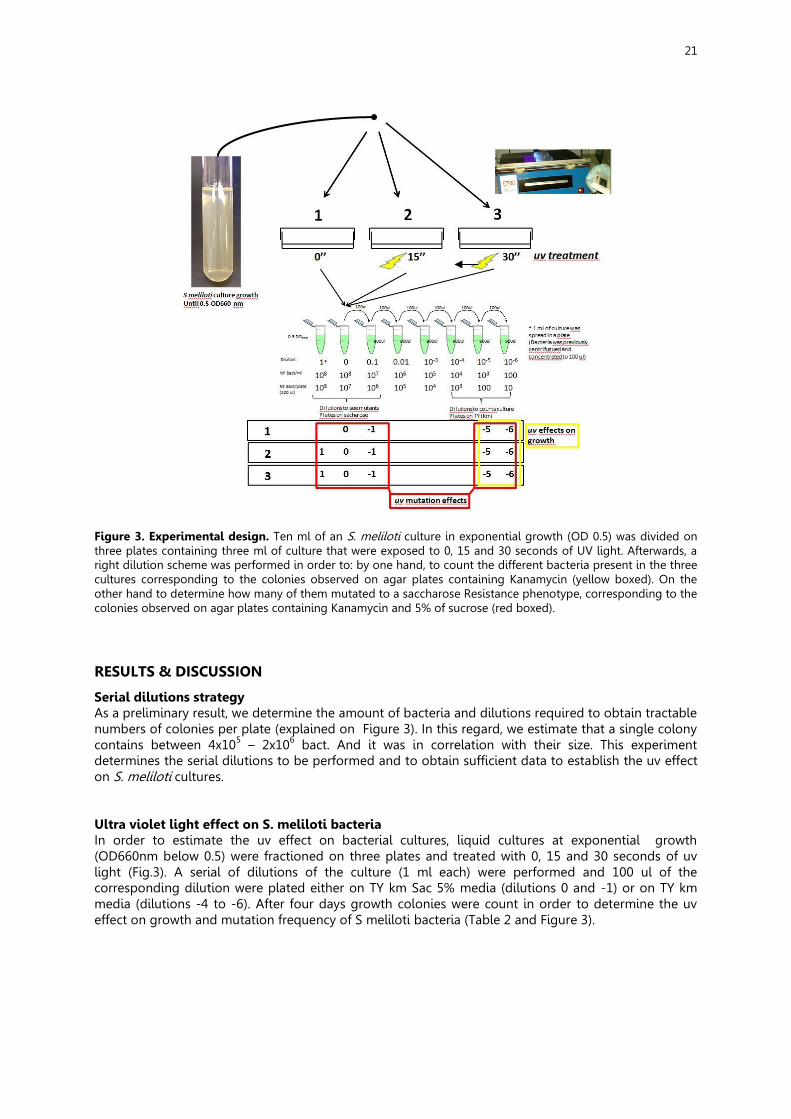
Figure 3. Experimental design. Ten ml of an S. meliloti culture in exponential growth (OD 0.5) was divided on three plates containing three ml of culture that were exposed to 0, 15 and 30 seconds of UV light. Afterwards, a right dilution scheme was performed in order to: by one hand, to count the different bacteria present in the three cultures corresponding to the colonies observed on agar plates containing Kanamycin (yellow boxed). On the other hand to determine how many of them mutated to a saccharose Resistance phenotype, corresponding to the colonies observed on agar plates containing Kanamycin and 5% of sucrose (red boxed). RESULTS & DISCUSSION
Serial dilutions strategy As a preliminary result, we determine the amount of bacteria and dilutions required to obtain tractable numbers of colonies per plate (explained on Figure 3). In this regard, we estimate that a single colony contains between 4x10⁵ – 2x10⁶ bact. And it was in correlation with their size. This experiment determines the serial dilutions to be performed and to obtain sufficient data to establish the uv effect on S. meliloti cultures. Ultra violet light effect on S. meliloti bacteria In order to estimate the uv effect on bacterial cultures, liquid cultures at exponential growth (OD660nm below 0.5) were fractioned on three plates and treated with 0, 15 and 30 seconds of uv light (Fig.3). A serial of dilutions of the culture (1 ml each) were performed and 100 ul of the corresponding dilution were plated either on TY km Sac 5% media (dilutions 0 and -1) or on TY km media (dilutions -4 to -6). After four days growth colonies were count in order to determine the uv effect on growth and mutation frequency of S meliloti bacteria (Table 2 and Figure 3).
22
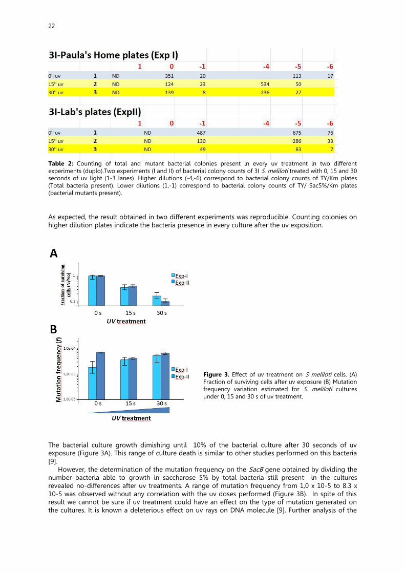
Table 2: Counting of total and mutant bacterial colonies present in every uv treatment in two different experiments (duplo).Two experiments (I and II) of bacterial colony counts of 3I S. meliloti treated with 0, 15 and 30 seconds of uv light (1-3 lanes). Higher dilutions (-4,-6) correspond to bacterial colony counts of TY/Km plates (Total bacteria present). Lower dilutions (1,-1) correspond to bacterial colony counts of TY/ Sac5%/Km plates (bacterial mutants present). As expected, the result obtained in two different experiments was reproducible. Counting colonies on higher dilution plates indicate the bacteria presence in every culture after the uv exposition.
Figure 3. Effect of uv treatment on S meliloti cells. (A) Fraction of surviving cells after uv exposure (B) Mutation frequency variation estimated for S. meliloti cultures under 0, 15 and 30 s of uv treatment.
The bacterial culture growth dimishing until 10% of the bacterial culture after 30 seconds of uv exposure (Figure 3A). This range of culture death is similar to other studies performed on this bacteria [9].
However, the determination of the mutation frequency on the SacB gene obtained by dividing the number bacteria able to growth in saccharose 5% by total bacteria still present in the cultures revealed no-differences after uv treatments. A range of mutation frequency from 1,0 x 10⁻⁵ to 8.3 x 10⁻⁵ was observed without any correlation with the uv doses performed (Figure 3B). In spite of this result we cannot be sure if uv treatment could have an effect on the type of mutation generated on the cultures. It is known a deleterious effect on uv rays on DNA molecule [9]. Further analysis of the
23
sequence of the SacB gene in uv-treated bacterial colonies might show a difference in the type of mutation generated. CONCLUSIONS
1) We demonstrate Sinorhizobium meliloti growth is sensitive to uv treatments 2) uv treatments do not increase the mutation frequency in S. meliloti 3) We cannot discard a putative effect on the type of mutation generated.
ACKNOWLEDGEMENTS
This work was performed in the Microbiology and Symbiotic Systems department in the Estación Experimental del Zaidín – Consejo Superior de Investigaciones Científicas. It was supported by research projects MICINN Consolider-Ingenio 2010. CSD2009-00006; BIO-2011-24401 and BIO2014-51953-P. Secondary Schools Institutes and Centers: Zaidín Vergeles, Severo Ochoa, CDP Dulce Nombre de Maria (Escolapios), Miguel de Cervantes from Granada Capital and La Vega de Atarfe. Atarfe; Avenmoriel. Benamaurel and Alba Longa. Armilla. Also we appreciate the coordination of All Secondary Schools teachers involved and particularly to Javier Cáceres as promoter of PIIISA project and Antonio Quesada Ramos for his help in developing our blog: http://mutandogenes.blogspot.com.es/. REFERENCES
[1] Bertram, John S. (2000). "The molecular biology of cancer". Molecular Aspects of Medicine (Amsterdam, the Netherlands: Elsevier) 21 (6): 167–223.
[2] Maron & Ames (1983) Revised methods for the Salmonella mutagenicity test. Mutat. Res. 113: 173–215.
[3] Newton WE (2000) in Nitrogen Fixation: from Molecular to Crop Productivity, eds Pedrosa FO, Hungria M, Yates MG, Newton WE (Kluwer, Dordrecht, The Netherlands), pp3-8.
[4] Gay, P., D. LeCoq, M. Steinmetz, E. Ferrari, and J. A. Hoch. (1983) Cloning structural gene sacB, which codes for exoenzyme levansucrase of Bacillus subtilis: expression of the gene in Escherichia coli. J. Bacteriol 153: 1424-1431.
[5] Selbitschka W, Niemann S, Puhler A (1993) Construction of gene replacement vectors for Gram- bacteria using a genetically modified sacRB gene as a positive selection marker. Appl Microbiol Biotechnol 38:615-618.
[6] Benavente C, Trespando VF, Iglesias M, Leal E, Padilla AJ, Pardo L, & Martínez-Abarca F. (2015) Variations in the spontaneous mutation rate of a soil bacterium. Effect of gene location. In: Alche et al. (eds) High School Students for Agricultural Science Research.IV. Estación Experimental del Zaidín, Granada, pp 23-40. ISSN 2340-9746.
[7] Martínez-Abarca F, Toro N (2000) RecA-independent ectopic transposition in vivo of a bacterial group II intron. Nucleic Acids Res. 28:4397-4402.
[8] García-Rodríguez FM, Hernández-Gutiérrez T, Díaz-Prado V, Toro N. (2014) Use of the computer-retargeted group II intron RmInt1 of Sinorhizobium meliloti for gene targeting. RNA Biol 11: 391-401.
[9] Selbitschka W, Arnold W, Priefer UB, Rottschäfer T, Schmidt M, Simon R, Pühler A. (1991) Characterization of recA genes and recAmutants of Rhizobium meliloti and Rhizobium leguminosarum biovar viciae. Mol Gen Genet. 229(1):86-95.
24
MY OWN IDEAS Matilde López Freire
At the beginning of the project Piiisa I was a few scared, I didn’t know where I had got into. The first that we did was have a touchdown with the laboratory and with the objects that would use, and more or less learn to use as for example the pipette with its different types, depending on the volume you want to take 10µl, 100µl… to take dilutions would serve a great help in the tedious task of counting bacteria. The first experiment that we did was from a culture in which we pike on colonies with different diameters on the order of >2mm, 2-0,5mm y <0,5mm to see how many bacteria had. The nest experiment that we did already with the cultures 3G and 3I of our beloved soil bacterium Sinorhizobium meliloti to see where growing better to decided where would realize the next experiments. Other machines we used were the spectrometer to measure the optical density and a centrifuge. In subsequent experiments we were using one or both cultures 3G or 3I and half of the plates were with Ty Kanamycin and the other half of the plates were also with 5% sucrose. With 5% sucrose the bacteria cannot grow with which it has suffered a mutation to can grow. And the goal of this project was see if submitting the bacteria to 0, 15 and 30 seconds of ultra-violet light, it could see an acceleration on the frequency mutation. Along our way not everything was perfect, accidents occurred either time when we did the dilutions or problems with moisture what did he had to repeat the experiments and the last case discard one. But looking back, I don’t regret having targeted in this wonderful project, in which I reaffirmed my ideas when I want to study and which I want to do in the not too distant future. The end of the project give me a bittersweet feeling because I’m happy, exciting and proud to see it finished and think that I participated in that as a real research but I’m also sad because the finish won’t step on a lab to take a few years and is really fascinating to be there with some project in hands. Al principio del proyecto Piiisa estaba un poco asustada, no sabía donde me había metido. Lo primero que hicimos fue tener una toma de contacto con el laboratorio y los objetos que utilizaríamos y más o menos aprender a usarlos, como por ejemplo la pipeta con sus diferentes tipos, dependiendo del volumen que se quería coger 10µl, 100µl… para poder hacer las diluciones que nos servirían de gran ayuda en la tediosa tarea de contar bacterias. El primer experimento que hicimos fue a partir de un cultivo en el que picamos sobre colonias de diferentes diámetros del orden de >2mm, 2-0,5mm y <0,5 para ver cuantas bacterias habían. El siguiente que hicimos fue ya con los cultivos 3G y 3I de nuestra querida bacteria del suelo Sinorhizobium Meliloti para ver en que casa se realizarían los posteriores experimentos. Otras máquinas utilizamos fueron el espectrómetro para medir la densidad óptica y una centrifugadora. En los posteriores experimentos usábamos uno o los dos cultivos 3G y 3I, y la mitad de las placas estaban en un medio con Ty Kanamicina y la otra mitad estaba en un medio que tenía además 5% de sacarosa. En un medio con sacarosa la bacteria no puede crecer con lo cuál ha sufrido una mutación para poder hacerlo. Y la meta de este proyecto era ver si sometiendo los cultivos a 0,15 y 30 segundos de rayos ultra violeta se pudiera ver una aceleración en la frecuencia de mutación. A lo largo de nuestro camino no todo fue perfecto, ocurrieron accidentes bien a la hora de las diluciones o problemas de humedad que hicieron que tuviéramos que repetir los experimentos y en el último caso descartar uno. Pero echando la vista atrás no me arrepiento de haberme apuntado a este maravilloso proyecto, con el cuál he reafirmado mis ideas a la hora de que quiero estudiar y dedicarme en un futuro no muy lejano. El final del proyecto me provoca una sensación agridulce ya que estoy contenta, emocionada y orgullosa por verlo acabado y pensar que he participado en eso como una auténtica investigadora pero también siento tristeza debido a que al acabarse ya no volveré a pisar un laboratorio hasta que pasen unos años y la verdad es fascinante estar allí con algún proyecto entre manos. Carmen Molina Muñoz
I have participated in a microbiology investigating project which consists of detecting if the ultra-violet light treatment has an effect on the mutation frequency of a soil bacterium called Sinorhizobium meliloti. At the beginning I did not know how the project was going to turn out. I was lost in the laboratory, but Paco was very patient with me and my teammates and he taught us about dilutions, colonies count

25
and mathematical operations. We worked with two different bacteria: 3G and 3I. We did the experiments with both of them, but unfortunately, we committed some mistakes and we finally only obtained results from 3I. Now, we are able to affirm that ultra- violet light treatment does not affect on the mutation frequency of our bacterium. As days passed, I was learning more and more about our project, about how to interpret the data that we have obtained and about how to do a scientific research. What I have liked the most was doing dilutions and what I have liked less was doing mathematical operations because it wasn’t easy at the beginning; but at the same time, I think it is one of the most important parts of our project. I have met a small part of microbiology and how Scientifics work in order to find the answer of a certain question. At the same time, that solution will propose new questions that will be solved in future researches. And that is the magic of the science, there is an infinite world to discover. I feel very proud of being part of this investigation because not everyone has the same opportunity. I have learnt so much and I have enjoyed. I am sure that this project will help me to choose the career I would like to do in the future. So thanks to Paco, my teacher and all the people that has made this possible. Cristina Martín Vargas
Since the very first time I met my team I was very enthusiastic about the project. The girls and our researcher seemed kind and likeable. The first day, I remembered it like the most important day in our project, well, there were lots of important days but it was a remarkable date. We met each other and our researcher, Paco, explained us about what we were going to do. I didn't understand almost nothing and I was impressed with the lab. Then the next session was with our parents and some teacher, aiming to know more about this. We counted the colonies that we took to our houses. When we pickep up the bacterias and incubated then in each house was astonishing. That was my first contact with a lab and I think it was for everyone, too. After that we started with the real experiment, that was to find out if our bacteria, s.meliloti suffer a mutation when it's exposed to uv radiation. I was really amazed about everything we were doing, the lab tools, the way we worked and also counting bacterias. But it wasn't perfect at all, we messed a bit our experiment. There were humidity in the plates and a foreign organism in one of them, so we had to prepare the plates again. The next time we couldn't take the data of 3G due to it was imposible to count the colonies, although that doesn't stop us to finish our project, we just used the other one that was all right. In spite of I expected to have different conclusions, I know it doesn't matter, because it was my first time I thought we could discover something bigger. But the uv radiation doesn't change the mutation frequency in s.meliloti. To be honest, I think is a good conclussion, even though anything changes. To sum up I have to say it was an unique experience that I will never forget about, because for me. it was something big, I had never been in a lab and I met new people. Thanks to our project I think I discover what I want to do in the future, study biochemistry or something related to it. Alba Mª Bolívar Moreno
Our Piiisa project has just finished. This experience was amazing. I have learned a lot of things about science and I have learned how the investigators work in the laboratory of microbiology, what instruments they use and how they make their researches. These months have been very intense with a lot of work and stress but it’s worth the effort. I grew as a person, I met some incredible clever girls and our Investigator was so funny. He teached us a lot of things. This amazing experience is unforgettable for me. Thanks to all the people who made this possible, to my teacher Elisa Valero , to my teammate, to the organization Piisa and of course to our incredible investigator Fco Martínez Abarca. Paula Collado Cordón
This project has totally changed the way I thought science would be. I did not expect that it had so many hours of thinking and making hypothesis as it really has. I have to say that this is not my favorite part of the project, but it was interesting. The laboratory was amazing. I wanted to do everything that I could, and using ultra-violet light made it even better.
26
At the beginning, I did not choose this PIIISA project as my first option, and now that it is about to end, I have to admit my mistake, because I could not have chosen anyone better. I’m so glad to be part of this incredible experience and also relieved to have some ideas about what I’d like to be in the future a little bit clearer. As a conclusion, I’d like to congratulate our investigator. I think all of my mates would agree if I say that his patience, his hard work and his enthusiasm has made this project an unique project, so I just can say: thank you! Mónica López Candela
These 6 months, has been very special for my, because I' ve enjoyed this experience in the lab, with my "classmates" and my investigator. I love science, and in the future, I would like to study biochemistry. However, I like music and I,m in the Conservatory, so I,m very confused because I like the two options. But with this esperiment, I've learnt much things and I' ve known my classmates. They are very friendly and nice. I would like to go to de next exposition in the EEZ, but that days I'll be in a cruise, so I' ll support my classmates from Italy. Also, say that I'm very happy with the results of the experiment, the way of work, because I think is the most important to do the experimet, and my investigator, because, with he I' ve learnt about biochemistry, one of my favourites carreers. Finally, I' m very excited with the project and the next year, I'll try to go. Lucía Torres Gurrea
I signed up for PIIISA-project because I wanted to live this experience. I wanted to be able to know how it was work in a laboratory, working with a researcher. The first day was somewhat shocking because we had our first contact with Paco – our researcher -, He told us the topic of the project, and none of us were able to understand. We were a little lost, new things, new concepts. A new world to discover. Then, the first contact with the laboratory came, more striking but also the most complicated part. In general, this first day was strong in emotions. As the days pass, the project was getting a little easier because we were going to know something else. We understood both as objectives and methods in every session increasing our interest. But, we had to repeat several experiments because we are not properly and therefore we did not get the expected results. Finally, even that part of the experiment we ruled out, we succesfully got end the project: ready to write the report and publish. .But, then we arrived the moment that makes me more nervous; the time to read our results to experienced public that knows what I'm talking about; the fear that ask me and not knowing what to say; to go blank. The fear of ridicule after all. But I guess like everyone says fears are only obstacles on the road they are overcome. In summary, This has been one of the best experiences of my life. I take friends, new learning and a great model for improvement. And also a great example of wisdom as our researcher shown us that everything can be in life and what is sought is achieved. Yo me apunte a PIIISA porque quería vivir esta experiencia, quería tener la posibilidad de saber cómo era el trabajo en un laboratorio, el trabajo con un investigador. El primer día fue algo impactante porque tuvimos nuestra primera toma de contacto con Paco (nuestro investigador), nos contó de que iba el proyecto y ninguna comprendíamos mucho digamos que estábamos un poco perdidas, cosas nuevas, conceptos nuevos, todo un mundo nuevo por descubrir. Después vino la primera toma de contacto con el laboratorio la parte mas vistosa aunque también la mas complicada. En general ese primer día fue de emociones fuertes. Conforme los días fueron pasando cada vez se fue haciendo un poco más sencillo ya que íbamos sabiendo algo más de qué iba nuestro proyecto. Fuimos comprendiendo tanto procedimientos como objetivos y sesión tras sesión fue siendo cada vez mas interesante. Tuvimos que repetir varios experimentos porque no los hacíamos de forma correcta y por lo tanto no obteníamos los resultados esperados. Pero, finalmente aunque una parte del experimento se nos cayó, lo conseguimos. El experimento estaba terminado, listo para redactar el informe y publicar. Y entonces llegó el momento que más miedo me daba y me sigue dando y es el momento de leer en publico los resultados; de enfrentarte a un publico experimentado y que sabe de lo que estoy hablando; al miedo de que me pregunten y no saber que responder; a quedarme en blanco. Miedo al ridículo al fin y al cabo. Pero, supongo que como todos dicen los miedos solo son obstáculos en el camino que se superan. En fin esta ha sido una de las mejores experiencias de mi vida de la que me llevo amigas, nuevos aprendizajes y un gran modelo de superación y ejemplo de sabiduría como es Paco que nos a demostrado que todo se puede y que en la vida lo que se persigue se consigue.
27
Genomic variability in humans: our genetic fingerprint
Isabel M. Bautista-Plaza1, Lorena Jiménez-Molina2, Alejandro Manzano-Martín3, Blanca Ortiz-Rodríguez4, María Pedregosa-Ruiz5, Fuencisla Matesanz6, Alicia Barroso-del Jesus7*
1IES Cartuja. Julio Moreno Dávila 18, 18011 Granada, Spain. 2Colegio Salesianos San Juan Bosco. Almuñecar 9, 18006 Granada, Spain.
3Colegio “El Carmelo”. Palau y Quer 7, 18008 Granada, Spain. 4IES Padre Poveda. Avda de Mariana Pineda, 18, 18500 Guadix, Granada, Spain
5IES Jiménez de Quesada. Camino Sta. Teresa 1 18328 Santa Fe, Granada, Spain. 6Instituto de Parasitología y Biomedicina “López-Neyra”, CSIC. Avda del Conocimiento s/n, 18016 Armilla,
Granada, Spain. 7Unidad de Genómica. Instituto de Parasitología y Biomedicina “López-Neyra”, CSIC. Avda del Conocimiento
s/n, 18016 Armilla, Granada, Spain.
*Corresponding author: e-mail: [email protected]
SUMMARY
In terms of DNA sequence, we share a 92% of our genome with mouse just because we both are mammals, 98% with chimpanzees and 99.9% with other humans. Although the DNA of any two people on Earth is, in fact, 99.9% identical, we accumulate differences (1 base every 1200 or 1500 bases) that turn out in clear distinct phenotypes and can have a big effect if any of these differences is located in a critical gene. In this work we have used a STR Analysis to demonstrate the proof of principle that every human has a unique genetic fingerprint. We have investigated 9 variable loci across the human genome in 15 independent DNA samples and established their genetic profiles. The results have been also applied to prove the parental relationship among members of three families included in a multiple sclerosis disease genetic association project. INTRODUCTION
On average, in terms of DNA sequence all humans are 99.9% similar to any other humans [1]. However, no two humans are genetically identical. Even monozygotic twins, who develop from one zygote, have infrequent genetic differences due to mutations occurring during development and gene copy-number variation [2].
Inherited differences in DNA sequence contribute to phenotypic variation, influencing an individual's anthropometric characteristics, risk of disease and response to the environment. A central goal of genetics is to pinpoint the DNA variants that contribute most significantly to population variation in each trait.
Human genetic variation is the genetic differences both within and among populations. There may be multiple variants of any given gene in the human population, leading to polymorphism. Many genes are not polymorphic, meaning that only a single allele is present in the population: the gene is then said to be fixed. Alleles occur at different frequencies in different human populations, with populations that are more geographically and ancestrally remote tending to differ more.
Causes of differences between individuals include the exchange of genes during meiosis and various mutational events. There are, at least, two reasons why genetic variation exists between populations. Natural selection may confer an adaptive advantage to individuals in a specific environment if an allele provides a competitive advantage. Alleles under selection are likely to occur only in those geographic regions where they confer an advantage. The second main cause of genetic variation is due to the high degree of neutrality of most mutations. Most mutations do not appear to have any selective effect one way or the other on the organism. The main cause is genetic drift; this is the effect of random changes in the gene pool. In humans, founder effect and past small population
28
size (increasing the likelihood of genetic drift) may have had an important influence in neutral differences between populations.
The study of human genetic variation has both evolutionary significance and medical applications. It can help scientists understand ancient human population migrations as well as how different human groups are biologically related to one another. For medicine, study of human genetic variation may be important because some disease-causing alleles occur more often in people from specific geographic regions. New findings show that each human has on average 60 new mutations compared to their parents [3]. Apart from mutations, many genes that may have aided humans in ancient times plague humans today. For example, it is suspected that genes that allow humans to more efficiently process food are those that make people susceptible to obesity and diabetes today.
Differences between individuals, even closely related individuals, are the key to techniques such as genetic fingerprinting. The system of DNA profiling used today is based on PCR and uses simple sequences [4] or short tandem repeats (STR). This method uses highly polymorphic regions that have short repeated sequences of DNA (the most common is 4 bases repeated, but there are other lengths in use, including 3 and 5 bases). Because unrelated people almost certainly have different numbers of repeat units, STRs can be used to discriminate between unrelated individuals. These STR loci (locations on a chromosome) are targeted with sequence-specific primers and amplified using PCR. The DNA fragments that result are then separated and detected using electrophoresis. There are two common methods of separation and detection, capillary electrophoresis (CE) and gel electrophoresis.
Each STR is polymorphic, but the number of alleles is very small. Typically each STR allele will be shared by around 5 - 20% of individuals. The power of STR analysis comes from looking at multiple STR loci simultaneously. The pattern of alleles can identify an individual quite accurately. Thus STR analysis provides an excellent identification tool. The more STR regions that are tested in an individual the more discriminating the test becomes.
MATERIALS AND METHODS
Samples From the 15 samples analyzed, 6 samples corresponded to the co-authors of this study and 9 corresponded to subjects of a multiple sclerosis disease genetic association project. These samples were collected at the Virgen Macarena Hospital in Sevilla. Informed consent was obtained for each patient.
DNA extraction DNA samples from multiple sclerosis project were kindly provided by Dr. Fuencisla Matesanz´s laboratory. DNA from PIIISA tutor and students was extracted from saliva samples using the QiaAMP kit (Qiagen) with the Qiacube® robot. Briefly, 1 ml of saliva was collected from each subject and diluted with 4 ml of PBS buffer. Cells were recovered by centrifugation at 1800 xg for 5 min and resuspended in 180 µl of PBS buffer and 20 µl of RNase A solution at 20 mg/ml. Then samples were further processed in the Qiacube® robot using the protocol recommended by the manufacturer. DNAs were eluted in 10 mM Tris-ClH pH 8.5 in a final volume of 100 µl.
DNA quantification Genomic DNAs were quantified by measuring absorbance at 260 nm in a Nanodrop® spectrophotometer. STRs amplification STRs loci used in this study were: Amelogenin (sex determination locus), vWA, THO1, TPOX, CSF, D5, D7, D13 and D16. Genomic location and primers used for amplification are listed in table 1. Forward primers were 5´end labelled with 6-FAM. PCR reactions were multiplexed as follows: PCR1 included primers for vWA, TPOX, THO1, CSF1 and Amelogenin loci; PCR2 included primers for D5, D7, D13 and D16 loci. Concentration of primer pairs was previously optimized in the lab and adjusted to obtain 10x stock solutions. PCRs were performed using 1.5 mM MgCl2, 0.2 mM dNTPs, 1.5 µl Primer Mix (1 or 2) 10x, 2 ng genomic DNA and 0.75 units of Taq DNA polymerase (Biotools) in a final volume of 15 µl.
Capillary electrophoresis PCR reactions were tenfold diluted in H2O. One µl of this dilution was mixed with 0.4 µl of GS500 Rox-labeled size marker (ThermoFisher) and 12 µl of desionized formamide. Samples were denatured by
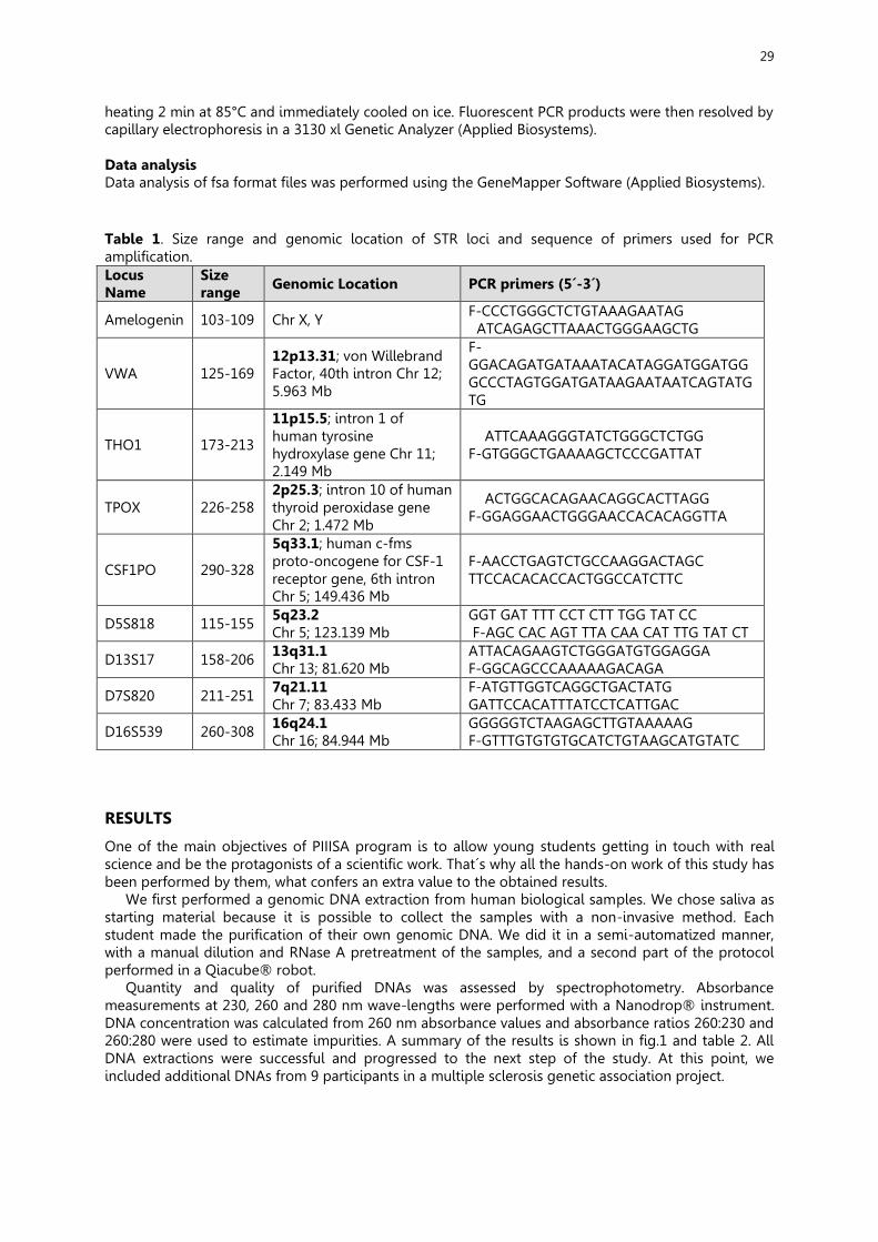
29
heating 2 min at 85°C and immediately cooled on ice. Fluorescent PCR products were then resolved by capillary electrophoresis in a 3130 xl Genetic Analyzer (Applied Biosystems). Data analysis Data analysis of fsa format files was performed using the GeneMapper Software (Applied Biosystems). Table 1. Size range and genomic location of STR loci and sequence of primers used for PCR amplification. Locus Name
Size range Genomic Location PCR primers (5´-3´)
Amelogenin 103-109 Chr X, Y F-CCCTGGGCTCTGTAAAGAATAG ATCAGAGCTTAAACTGGGAAGCTG
VWA 125-169 12p13.31; von Willebrand Factor, 40th intron Chr 12; 5.963 Mb
F-GGACAGATGATAAATACATAGGATGGATGG GCCCTAGTGGATGATAAGAATAATCAGTATGTG
THO1 173-213
11p15.5; intron 1 of human tyrosine hydroxylase gene Chr 11; 2.149 Mb
ATTCAAAGGGTATCTGGGCTCTGG F-GTGGGCTGAAAAGCTCCCGATTAT
TPOX 226-258 2p25.3; intron 10 of human thyroid peroxidase gene Chr 2; 1.472 Mb
ACTGGCACAGAACAGGCACTTAGG F-GGAGGAACTGGGAACCACACAGGTTA
CSF1PO 290-328
5q33.1; human c-fms proto-oncogene for CSF-1 receptor gene, 6th intron Chr 5; 149.436 Mb
F-AACCTGAGTCTGCCAAGGACTAGC TTCCACACACCACTGGCCATCTTC
D5S818 115-155 5q23.2 Chr 5; 123.139 Mb
GGT GAT TTT CCT CTT TGG TAT CC F-AGC CAC AGT TTA CAA CAT TTG TAT CT
D13S17 158-206 13q31.1 Chr 13; 81.620 Mb
ATTACAGAAGTCTGGGATGTGGAGGA F-GGCAGCCCAAAAAGACAGA
D7S820 211-251 7q21.11 Chr 7; 83.433 Mb
F-ATGTTGGTCAGGCTGACTATG GATTCCACATTTATCCTCATTGAC
D16S539 260-308 16q24.1 Chr 16; 84.944 Mb
GGGGGTCTAAGAGCTTGTAAAAAG F-GTTTGTGTGTGCATCTGTAAGCATGTATC
RESULTS
One of the main objectives of PIIISA program is to allow young students getting in touch with real science and be the protagonists of a scientific work. That´s why all the hands-on work of this study has been performed by them, what confers an extra value to the obtained results.
We first performed a genomic DNA extraction from human biological samples. We chose saliva as starting material because it is possible to collect the samples with a non-invasive method. Each student made the purification of their own genomic DNA. We did it in a semi-automatized manner, with a manual dilution and RNase A pretreatment of the samples, and a second part of the protocol performed in a Qiacube® robot.
Quantity and quality of purified DNAs was assessed by spectrophotometry. Absorbance measurements at 230, 260 and 280 nm wave-lengths were performed with a Nanodrop® instrument. DNA concentration was calculated from 260 nm absorbance values and absorbance ratios 260:230 and 260:280 were used to estimate impurities. A summary of the results is shown in fig.1 and table 2. All DNA extractions were successful and progressed to the next step of the study. At this point, we included additional DNAs from 9 participants in a multiple sclerosis genetic association project.
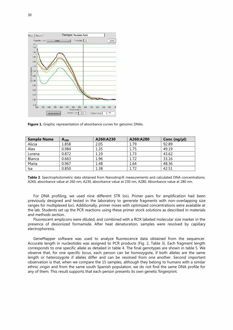
30
Figure 1. Graphic representation of absorbance curves for genomic DNAs. Sample Name A260 A260:A230 A260:A280 Conc (ng/µl) Alicia 1.858 2.05 1.79 92.89 Alex 0.984 1.35 1.75 49.19 Lorena 0.872 1.19 1.73 43.62 Blanca 0.663 1.96 1.72 33.16 María 0.967 1.48 1.64 48.36 Isa 0.850 1.38 1.72 42.51 Table 2. Spectrophotometric data obtained from Nanodrop® measurements and calculated DNA concentrations. A260, absorbance value at 260 nm; A230, absorbance value at 230 nm, A280, Absorbance value at 280 nm.
For DNA profiling, we used nine different STR loci. Primer pairs for amplification had been previously designed and tested in the laboratory to generate fragments with non-overlapping size ranges for multiplexed loci. Additionally, primer mixes with optimized concentrations were available at the lab. Students set up the PCR reactions using these primer stock solutions as described in materials and methods section.
Fluorescent amplicons were diluted, and combined with a ROX labeled molecular size marker in the presence of desionized formamide. After heat denaturation, samples were resolved by capillary electrophoresis.
GeneMapper software was used to analyze fluorescence data obtained from the sequencer. Accurate length in nucleotides was assigned to PCR products (Fig. 2, Table 3). Each fragment length corresponds to one specific allele as detailed in table 4. The final genotypes are shown in table 5. We observe that, for one specific locus, each person can be homozygote, if both alleles are the same length or heterozygote if alleles differ and can be resolved from one another. Second important observation is that, when we compare the 15 samples, although they belong to humans with a similar ethnic origin and from the same south Spanish population, we do not find the same DNA profile for any of them. This result supports that each person presents its own genetic fingerprint.
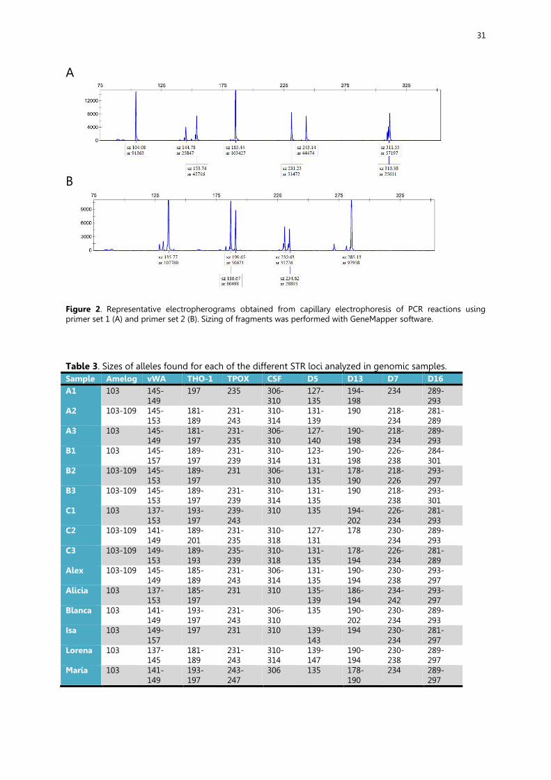
31
A
B
Figure 2. Representative electropherograms obtained from capillary electrophoresis of PCR reactions using primer set 1 (A) and primer set 2 (B). Sizing of fragments was performed with GeneMapper software. Table 3. Sizes of alleles found for each of the different STR loci analyzed in genomic samples. Sample Amelog vWA THO-1 TPOX CSF D5 D13 D7 D16 A1 103 145-
149 197 235 306-
310 127-135
194-198
234 289-293
A2 103-109 145-153
181-189
231-243
310-314
131-139
190 218-234
281-289
A3 103 145-149
181-197
231-235
306-310
127-140
190-198
218-234
289-293
B1 103 145-157
189-197
231-239
310-314
123-131
190-198
226-238
284-301
B2 103-109 145-153
189-197
231 306-310
131-135
178-190
218-226
293-297
B3 103-109 145-153
189-197
231-239
310-314
131-135
190 218-238
293-301
C1 103 137-153
193-197
239-243
310 135 194-202
226-234
281-293
C2 103-109 141-149
189-201
231-235
310-318
127-131
178 230-234
289-293
C3 103-109 149-153
189-193
235-239
310-318
131-135
178-194
226-234
281-289
Alex 103-109 145-149
185-189
231-243
306-314
131-135
190-194
230-238
293-297
Alicia 103 137-153
185-197
231 310 135-139
186-194
234-242
293-297
Blanca 103 141-149
193-197
231-243
306-310
135 190-202
230-234
289-293
Isa 103 149-157
197 231 310 139-143
194 230-234
281-297
Lorena 103 137-145
181-189
231-243
310-314
139-147
190-194
230-238
289-297
María 103 141-149
193-197
243-247
306 135 178-190
234 289-297
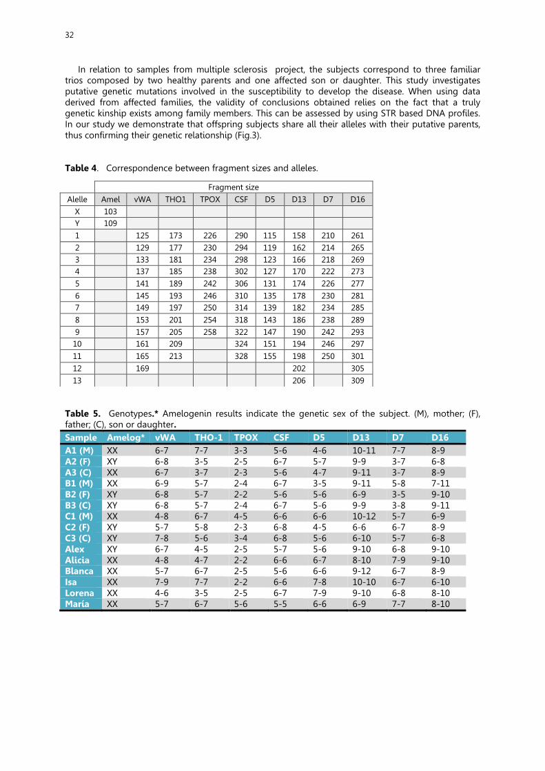
32
In relation to samples from multiple sclerosis project, the subjects correspond to three familiar trios composed by two healthy parents and one affected son or daughter. This study investigates putative genetic mutations involved in the susceptibility to develop the disease. When using data derived from affected families, the validity of conclusions obtained relies on the fact that a truly genetic kinship exists among family members. This can be assessed by using STR based DNA profiles. In our study we demonstrate that offspring subjects share all their alleles with their putative parents, thus confirming their genetic relationship (Fig.3).
Table 4. Correspondence between fragment sizes and alleles.
Table 5. Genotypes.* Amelogenin results indicate the genetic sex of the subject. (M), mother; (F), father; (C), son or daughter. Sample Amelog* vWA THO-1 TPOX CSF D5 D13 D7 D16 A1 (M) XX 6-7 7-7 3-3 5-6 4-6 10-11 7-7 8-9 A2 (F) XY 6-8 3-5 2-5 6-7 5-7 9-9 3-7 6-8 A3 (C) XX 6-7 3-7 2-3 5-6 4-7 9-11 3-7 8-9 B1 (M) XX 6-9 5-7 2-4 6-7 3-5 9-11 5-8 7-11 B2 (F) XY 6-8 5-7 2-2 5-6 5-6 6-9 3-5 9-10 B3 (C) XY 6-8 5-7 2-4 6-7 5-6 9-9 3-8 9-11 C1 (M) XX 4-8 6-7 4-5 6-6 6-6 10-12 5-7 6-9 C2 (F) XY 5-7 5-8 2-3 6-8 4-5 6-6 6-7 8-9 C3 (C) XY 7-8 5-6 3-4 6-8 5-6 6-10 5-7 6-8 Alex XY 6-7 4-5 2-5 5-7 5-6 9-10 6-8 9-10 Alicia XX 4-8 4-7 2-2 6-6 6-7 8-10 7-9 9-10 Blanca XX 5-7 6-7 2-5 5-6 6-6 9-12 6-7 8-9 Isa XX 7-9 7-7 2-2 6-6 7-8 10-10 6-7 6-10 Lorena XX 4-6 3-5 2-5 6-7 7-9 9-10 6-8 8-10 María XX 5-7 6-7 5-6 5-5 6-6 6-9 7-7 8-10
Fragment size Alelle Amel vWA THO1 TPOX CSF D5 D13 D7 D16
X 103 Y 109 1 125 173 226 290 115 158 210 261 2 129 177 230 294 119 162 214 265 3 133 181 234 298 123 166 218 269 4 137 185 238 302 127 170 222 273 5 141 189 242 306 131 174 226 277 6 145 193 246 310 135 178 230 281 7 149 197 250 314 139 182 234 285 8 153 201 254 318 143 186 238 289 9 157 205 258 322 147 190 242 293
10 161 209 324 151 194 246 297 11 165 213 328 155 198 250 301 12 169 202 305 13 206 309
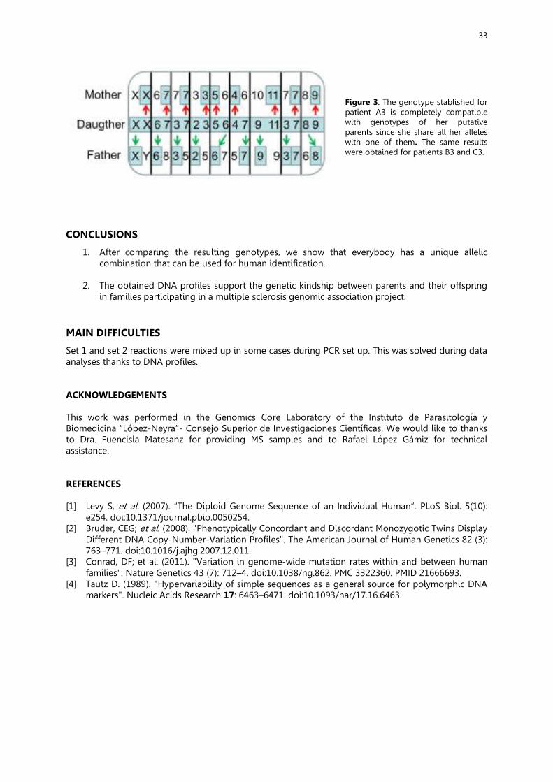
33
Figure 3. The genotype stablished for patient A3 is completely compatible with genotypes of her putative parents since she share all her alleles with one of them. The same results were obtained for patients B3 and C3.
CONCLUSIONS
1. After comparing the resulting genotypes, we show that everybody has a unique allelic combination that can be used for human identification.
2. The obtained DNA profiles support the genetic kindship between parents and their offspring
in families participating in a multiple sclerosis genomic association project. MAIN DIFFICULTIES
Set 1 and set 2 reactions were mixed up in some cases during PCR set up. This was solved during data analyses thanks to DNA profiles. ACKNOWLEDGEMENTS This work was performed in the Genomics Core Laboratory of the Instituto de Parasitología y Biomedicina “López-Neyra”- Consejo Superior de Investigaciones Científicas. We would like to thanks to Dra. Fuencisla Matesanz for providing MS samples and to Rafael López Gámiz for technical assistance. REFERENCES [1] Levy S, et al. (2007). “The Diploid Genome Sequence of an Individual Human”. PLoS Biol. 5(10):
e254. doi:10.1371/journal.pbio.0050254. [2] Bruder, CEG; et al. (2008). "Phenotypically Concordant and Discordant Monozygotic Twins Display
Different DNA Copy-Number-Variation Profiles". The American Journal of Human Genetics 82 (3): 763–771. doi:10.1016/j.ajhg.2007.12.011.
[3] Conrad, DF; et al. (2011). "Variation in genome-wide mutation rates within and between human families". Nature Genetics 43 (7): 712–4. doi:10.1038/ng.862. PMC 3322360. PMID 21666693.
[4] Tautz D. (1989). "Hypervariability of simple sequences as a general source for polymorphic DNA markers". Nucleic Acids Research 17: 6463–6471. doi:10.1093/nar/17.16.6463.
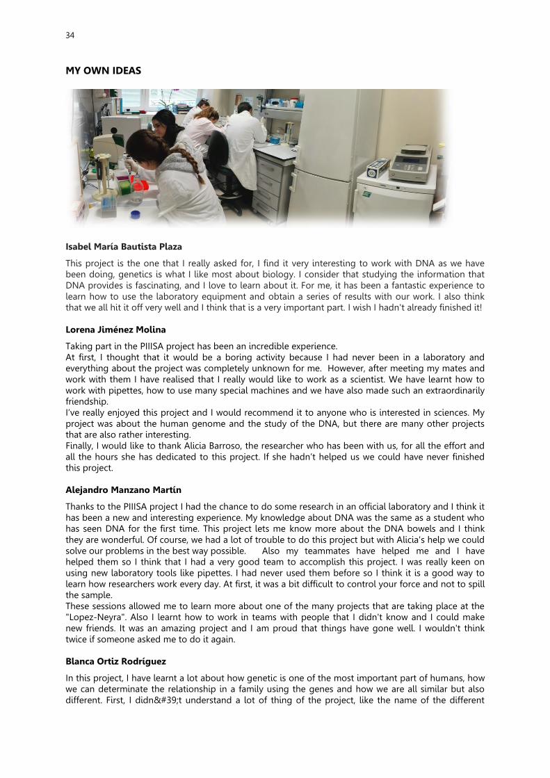
34
MY OWN IDEAS
Isabel María Bautista Plaza
This project is the one that I really asked for, I find it very interesting to work with DNA as we have been doing, genetics is what I like most about biology. I consider that studying the information that DNA provides is fascinating, and I love to learn about it. For me, it has been a fantastic experience to learn how to use the laboratory equipment and obtain a series of results with our work. I also think that we all hit it off very well and I think that is a very important part. I wish I hadn't already finished it! Lorena Jiménez Molina
Taking part in the PIIISA project has been an incredible experience. At first, I thought that it would be a boring activity because I had never been in a laboratory and everything about the project was completely unknown for me. However, after meeting my mates and work with them I have realised that I really would like to work as a scientist. We have learnt how to work with pipettes, how to use many special machines and we have also made such an extraordinarily friendship. I’ve really enjoyed this project and I would recommend it to anyone who is interested in sciences. My project was about the human genome and the study of the DNA, but there are many other projects that are also rather interesting. Finally, I would like to thank Alicia Barroso, the researcher who has been with us, for all the effort and all the hours she has dedicated to this project. If she hadn’t helped us we could have never finished this project. Alejandro Manzano Martín
Thanks to the PIIISA project I had the chance to do some research in an official laboratory and I think it has been a new and interesting experience. My knowledge about DNA was the same as a student who has seen DNA for the first time. This project lets me know more about the DNA bowels and I think they are wonderful. Of course, we had a lot of trouble to do this project but with Alicia's help we could solve our problems in the best way possible. Also my teammates have helped me and I have helped them so I think that I had a very good team to accomplish this project. I was really keen on using new laboratory tools like pipettes. I had never used them before so I think it is a good way to learn how researchers work every day. At first, it was a bit difficult to control your force and not to spill the sample. These sessions allowed me to learn more about one of the many projects that are taking place at the "Lopez-Neyra". Also I learnt how to work in teams with people that I didn't know and I could make new friends. It was an amazing project and I am proud that things have gone well. I wouldn't think twice if someone asked me to do it again. Blanca Ortiz Rodríguez
In this project, I have learnt a lot about how genetic is one of the most important part of humans, how we can determinate the relationship in a family using the genes and how we are all similar but also different. First, I didn't understand a lot of thing of the project, like the name of the different

35
instruments that we use or the name of some of the process we follow to obtain the results. But then, in the third or the fourth session I began to understand all and things became easier. I really enjoyed the project and the people in it. Now more than ever, I am sure that's the job I want to do went I grow up. It has been a great experience that I recommend to every one because is a big opportunity to us. María Pedregosa Ruiz
For me the PIIISA project has been one of the best experiences of my life, I would repeat this experience 1,000 times if I could. I was delighted to be working in a laboratory although I admit it's not easy and you have to be very attentive to everything because any failure insignificant can spoil all the work that has been done. I must also say that I found it easier and incredible thanks to all the people I've ever met.
36
Resistance to repeated radiotherapy in glioblastoma cells is partially prevented by the use of a DNA repair inhibitor
Eliana Gómez Jiménez1, Lucía Muros García2, Daniel Oliver de la Rubia3, Marta Rodríguez Camacho4, José Luis Ruiz Benito5, Natalia Ruiz Montosa6, Laura M. López Jiménez7, Javier
Oliver7*
1IES Vega de Atarfe, Atarfe, Granada 2IES Francisco de Ayala, Granada 3CDP Sagrado Corazón, Granada
4IES Luis Bueno Crespo, Armilla, Granada 5IES Ángel Ganivet, Granada
6IES La Madraza, Granada 7Instituto de Parasitología y Biomedicina López Neyra, CSIC, Granada
*corresponding author. email: [email protected]
All authors contributed equally to this work
HIGHLIGHTS
In this study we have evaluated the increased radioresistance of glioblastoma cells, one of the most deadly tumors in human, after two rounds of gamma-irradiation by determining induction of DNA damage and ability to repair DNA. We also found that disabling DNA repair might be helpful in radisensitizing these cells. SUMMARY
Glioblastoma multiforme (GBM) is the most common primary brain tumour in adults and one of the most aggressive cancers.The present study aimed to investigate the alteration of the DNA damage signaling pathway profile in radiation-treated glioblastoma and also aimed to explore potential targets for overcoming glioblastoma radioresistance. To this end we have used a human glioblastoma cell line LN229, and we have analysed the ability to damage DNA and repair it upon treatment with gamma irradiation. We have tested the hypothesis that repeated treatment could select a radioresistant population in the tumor cells therefore making more difficult the antitumor effect of radiation. In terms of DNA damage, GBM cells did not respond at the second treatment while co-treatment with an agent that blocks DNA repair, olaparib, increased DNA damage in these cells. In conclusion, our results suggest that combination with small molecules that inhibit DNA repair might improve the effect of gamma-irradiation in GBM cells after repeated treatment. INTRODUCTION
Glioblastoma multiforme (GBM) are lethal brain tumours, highly resistant to therapy. An important improvement in therapeutic response came from the use of the alkylating agent temozolomide (TMZ) in combination with ionizing radiation (IR).With the administration of the standard treatment of maximal safe surgical resection followed by radiotherapy combined with concomitant and adjuvant temozolomide (TMZ) chemotherapy, newly diagnosed patients with GBM demonstrate an average survival time of only 12-14 months (1). Almost one half of patients with GBM do not survive the first year subsequent to diagnosis. In cancer cells DNA damage may cause cell death by apoptosis which is the main purpose of radio and chemotherapy. In the course of treatment GBM become resistant to radio and chemotherapy then boosting tumor re-growth with the consequent aggravation of the

37
clinical status of the patient. Clarifying molecular resistance mechanisms could prompt the development of new drugs and facilitate more effective treatment combinations. In a previous a publication from our group we have shown that the use of a DNA repair inhibitor, more precisely an inhibitor of poly (ADP-ribose-ribose) polymerase (PARP), lead to cell death of GBM bearing DNA repair defects by homologous recombination, inducing mitotic catastrophe (2). In the current project we aimed to analyze if repeated radiotherapy treatment increased radioresistance in tumor cells based of the use of the comet assay as a readout of the cell’s ability to cope with DNA damage. In conclusion, our results suggest that repeated treatment with radiotherapy may affect the resistance of GBM cells to radiotherapy and the combination with small molecules that inhibit DNA repair might improve the effect of gamma-irradiation in GBM cells after repeated treatment. MATERIAL AND METHODS
Cell culture, reagents and treatments The LN-229 cell line was established in 1979 from cells taken from a patient with right frontal parieto-occipital glioblastoma (Female, 60 years, White). The culture medium consisted in Dulbecco's Modified Eagle's Medium (DMEM), Inactive fetal bovine serum (FBSi) to a final concentration of 10%, and Antiobiotic: penicillin/streptomycin. Some genetic alterations have been identified for this cells: p53 + (mutated, CCT (Pro) --> CTT (Leu) mutation at codon 98), PTEN + (wild type), p16 - (deleted), p14ARF - (deleted). γ-irradiation. The γ-irradiation on LN229 cells was performed using a 137Caesium source (dose rate 3 Gy/min). The PARP inhibitor olaparib (Astrazeneka) (3) was used at a dose of 10M 90 minutes before the second irradiation. Cells were irradiated at 3Gy and the effects were evaluated during 2 weeks. The schedule of the treatment was as follows: Day 1: Culture of cells Day 2: Irradiation (IR1) Day 9 (1 week): 10µM Olaparib + Irradiation (IR2) Comet Assay It is based upon the ability of denatured, cleaved DNA fragments to migrate out of the nucleoid under the influence of an electric field, whereas undamaged DNA migrates slower and remains within the confines of the nucleoid when a current is applied. DNA damage was analyzed using COMET assay (Assay kit , R&D Systems, Trevigen, MD, USA) with some modifications. 1 × 10⁵ cells/ml were mixed with molten LM agarose at 37 °C at a ratio of 1:10 (vol/vol) and pipetted onto a COMET slide. The slides were placed for 10 min in the dark at 4 °C and were immersed in pre-chilled lysis solution. The slides were then removed from lysis buffer, washed in TBE buffer and transferred to a horizontal electrophoresis chamber. Voltage (1 V/cm) was applied for 20 min. After washed in distilled water, the slides were immersed in 70% ethanol for 5 min and allowed to air dry. Slides were stained with SYBR Green and then analyzed by fluorescence microscopy. 70-90 cells were evaluated in each sample using the COMET Assay Software Open Comet. DNA damage was quantified by measuring the Tail Moment calculated as percentage of DNA in the tail × tail length (figure 1).
Figure 1. To analyze this assay we use Open Comet software that allows us to quantify the lenght of the tail and the head’s size. Tail Moment= [(Tail Mean-Head mean) / 100] x Tail% DNA
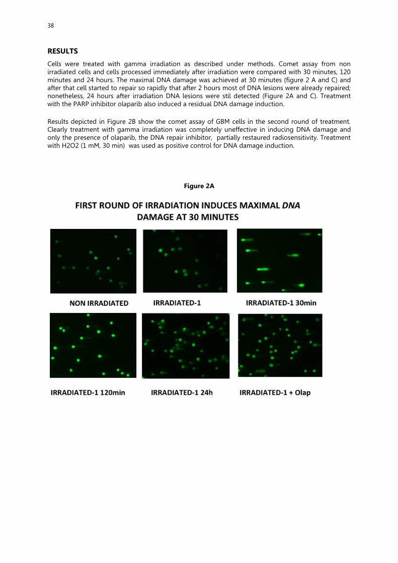
38
RESULTS
Cells were treated with gamma irradiation as described under methods. Comet assay from non irradiated cells and cells processed immediately after irradiation were compared with 30 minutes, 120 minutes and 24 hours. The maximal DNA damage was achieved at 30 minutes (figure 2 A and C) and after that cell started to repair so rapidly that after 2 hours most of DNA lesions were already repaired; nonetheless, 24 hours after irradiation DNA lesions were stil detected (Figure 2A and C). Treatment with the PARP inhibitor olaparib also induced a residual DNA damage induction. Results depicted in Figure 2B show the comet assay of GBM cells in the second round of treatment. Clearly treatment with gamma irradiation was completely uneffective in inducing DNA damage and only the presence of olaparib, the DNA repair inhibitor, partially restaured radiosensitivity. Treatment with H2O2 (1 mM, 30 min) was used as positive control for DNA damage induction.
Figure 2A
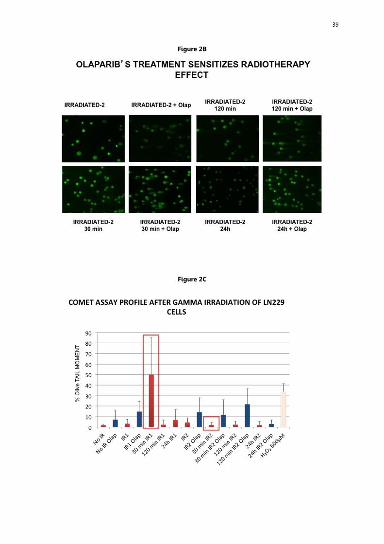
39
Figure 2B
Figure 2C
40
DISCUSSION
The results presented in this study have to be taken with caution as they preliminary results and not enough experiments have been performed as to have statistical validity. Nonetheless, the tendencies are different enough as to suggest that the first round of RT operates, through unknown mechanisms, as to render the cells much more resistant to therapy. This change might be occurring by selecting the cells able to cope more efficiently with DNA damage induced by gamma irradiation. There is a model that postulate that tumors arise from a small population of “Tumor Initiating cells” that behave as stem cells including the ability to undergo asymetrical cell division (4). These cells are particularly resistant to radiotherapy then after a first round of RT most cells are eliminated but this population is enriched. In any case more studies are needed to characterize in detail this effect including cell viability assays and characterization of the cell population remaining after different rounds of RT. The radiopotentiation effect of olaparib needs to be further characterized as a way to overcome GBM resistance to RT following repeated treatment. CONCLUSIONS
1. After the first round of radiotherapy (RT) there is a large induction of DNA damage which is maximal after 30 minutes. DNA repair is completed after 2 hours. 2. The second round of RT is completely uneffective in the induction of DNA damage. 3. Co-treatment with a PARP inhibitor (Olaparib) seems to increase the effect of RT on DNA repair after the second round. Final conclusion: The initial RT induces changes in glioma cells that select a radio-resistant cell population. The use of PARP inhibitor may overcome, at least partially, this radioresistant phenotype. ACKNOWLEDGEMENTS
We thanks to Profesora Francisca Ríos for her collaboration in this Project.This work was supported by Junta de Andalucía, project of Excellence P10-CTS-0662, Spanish Ministery of Economy and Competitiveness SAF2009-13281-C02-01, SAF2012-40011-C02-01 and RTICC RD12/0036/0026. REFERENCES
[1] Stupp R, Mason WP, van den Bent MJ, Weller M, Fisher B, Taphoorn MJ, et al. Radiotherapy plus concomitant and adjuvant temozolomide for glioblastoma. N Engl J Med. 2005;352(10):987-96.
[2] Majuelos-Melguizo J, Rodriguez MI, Lopez-Jimenez L, Rodriguez-Vargas JM, Marti Martin-Consuegra JM, Serrano-Saenz S, et al. PARP targeting counteracts gliomagenesis through induction of mitotic catastrophe and aggravation of deficiency in homologous recombination in PTEN-mutant glioma. Oncotarget. 2015;6(7):4790-803.
[3] Fong PC, Boss DS, Yap TA, Tutt A, Wu P, Mergui-Roelvink M, et al. Inhibition of poly(ADP-ribose) polymerase in tumors from BRCA mutation carriers. N Engl J Med. 2009;361(2):123-34.
[4] Anido J, Saez-Borderias A, Gonzalez-Junca A, Rodon L, Folch G, Carmona MA, et al. TGF-beta Receptor Inhibitors Target the CD44(high)/Id1(high) Glioma-Initiating Cell Population in Human Glioblastoma. Cancer Cell. 2010;18(6):655-68.

41
MY OWN IDEAS Eliana Gómez Jiménez
This experience has been amazing and unique, I have learned a lot of things like how it works in a investigation or how to use a pipette. From my point of view, I think that the problem of cells originating of the glioblastoma, which are more resistant, they could be use to treat our own body. Because, as happens in transplants, if cells are not compatible with our inmune system which attacks, so it can happen with the cells of the tumour, because its are not equal to the rest of our organism. If it doesn’t work. We can create an artificial inmune system with nanotecnology, for nanorobots attack this cells, for example desactivating this sequence of DNA. Lucía Muros García
I think this proyect is a great way to know more about cancer and its treatment. We have worked in the laboratory using cells of glioblastoma and we have learned scientific methods to follow to find the result we want get, for example, the electrophoresis, to look through the fluorescence microscope and the comet assay. We have felt like true scientists forming part of a major project. This experience will be decisive in the decisions we make in the future, such as choosing the career or work. In my opinion, the results we have obtained can be used for treatment after radiotherapy. We know the inhibitor repair DNA damage gets radiation resistant cells of glioblastoma don't increase much. We would continue investigating about the action of the inhibitor repair DNA damage. Daniel Oliver de la Rubia
I really enjoyed the experience of working in the Project PIIISA. It has been a great opportunity to know from inside how a research Project is developed and the work behind the resulst of the research. The topic was also very interesting. I think that the use of DNA repair inhibitors can help in the treatment of cáncer. What I like the most was the interpretation of the results from the image of the comet assay and the use of the software to generate the data. The research project could continue by analyzing how good is this treatment in eliminating cancer cells (not only comet assay) José Luis Ruiz Benito
As far as I am concerned, this experience was really instructive and gratifying. I was able to discover the work in a laboratory and some sophisticated techniques that scientists often use in their researches. In that way, I have took my first professional experience as a researcher, which will help me for deciding about my academic future. I really enjoyed the project and I haven’t nothing to discuss. In my opinion, all the methods and procedures that we used were correct, because the success of our research. In addition, I think that researchers of CSIC do a great task for the society, improving our life and doing it easier. Finally, I would like to express thanks for this unique opportunity. If I could, I would undoubtedly repeat. Natalia Ruiz Montosa
My experience on this PIIISA project has been really interesting and gratifying for me; because it has allowed me to learn new concepts about biology and work on a laboratory with professional scientists. It is also curious to me the theme of this project; we have made a research on a type of cerebral cancer, concretely from glioblastoma cells. To study how is the resistance of these cells, we have introduced a damage in their DNA that allows us to make our study. After this research, we have verified that these cells are resistant to the radiotherapy treatment; so it is a bit impossible to recover if you have this type of cancer. In addition, I think this type of project is worth because we can continue the research against a horrible disease such as cancer. In my opinion, it should be more investigations that attempt to find a cure to this disease because it could save a lot of people’s life. Unfortunately nowadays, there is a high amount of people who suffer cancer. In conclusion, projects who are made to try to find the cure for a disease must go on because the most important aspect in life is health.

42
Biofertilisers with olive-oil taste: isolation of plant growth-promoting rhizobacteria (PGPR) from “alperujo” compost
Alba Díaz Arco1, María Molina Muñoz2, Elena Navarro García3, Carlos Ortega Fernández4,
Laura Palma Pérez5, María Carmen Sarmiento Vega2, Antonio Castellano Hinojosa*6, Germán Tortosa Muñoz**6, Eulogio J. Bedmar6
1IES Hiponova, Avda. Miguel Hernández (antes Antonio Luis Soler) s/n 18270 Montefrío, Granada, Spain.
2IES Alba Longa, C/ San Miguel, s/n, 18100, Armilla, Granada, Spain. 3CDP Dulce Nombre de María, Paseo de los Basilios 2, 18008, Granada, Spain. 4IES Pedro Jiménez Montoya, Calle Isaac Peral, 2, 18800 Baza, Granada, Spain.
5IES Aricel, Calle del Aricel, S/N, 18220 Albolote, Granada, Spain. 6Department of Soil Microbiology and Symbiotic Systems, Estación Experimental del Zaidín, CSIC, Profesor
Albareda 1, 18008 Granada, Spain. Corresponding authors: *[email protected], ** [email protected]
HIGHLIGHTS
- “Alperujo” composts are an important source of N2-fixing bacteria. - There have been isolated and identified four PGPR bacteria in an “alperujo compost”: Burkholderia phymatum, Bacillus aryabhattai, Gluconacetobacter diazotrophicus and Pseudomonas stutzeri. - “Alperujo” composts are potentially biological fertilisers. SUMMARY
Composting is a microbial technology which can be effectively used for transforming organic wastes into valuable organic amendments and fertilisers. Also, composts may be considered as biological fertilisers due to containing beneficial microorganisms capable of promoting plant growth and development. The aim of this research was the isolation of some plant growth-promoting rhizobacteria (PGPR) present in composts made of “alperujo”, the main organic waste of the Spanish olive oil industry. We have focused in the nitrogen-fixing bacteria that were able or not to form symbiosis with legumes like soybean, common bean and chickpea. Also, free-living nitrogen-fixing bacteria and their ability to produce siderophores and solubilise insoluble soil phosphate were studied. We have found some bacteria that are able to nodulate common bean roots and we have identified four PGPR bacteria: Burkholderia phymatum, Bacillus aryabhattai, Gluconacetobacter diazotrophicus and Pseudomonas stutzeri.
INTRODUCTION (AND OBJECTIVE)
The world's population is rapidly growing and demands an increase of global food production [1]. Moreover, over the last 50 years, synthetic industrial fertilizers application (nitrates, ammonia, urea, etc.) has generated negative effects on the environment. Nutrient leakage has contaminated freshwater courses (surface and groundwater) and has provoked loss of soil biodiversity. Moreover, an increase in the emission of greenhouse gases from agriculture has been registered, which is directly related to global warming and climate change.
European Union has adopted a legislative initiative to promote sustainable development called "the Circular Economy" [2]. Its objectives are focused in reducing raw materials use, minimize waste production and encourage reuse and recycling. In Spain, olive oil industry generates a large amount of a solid by-product called “alperujo”, a highly polluting organic waste that needs to be treated for its revalorisation [3]. In order to achieve that, composting can be performed due to it being a simple, inexpensive and effective method for transforming organic waste as "alperujo" (AL) into organic amendments and fertilisers [4]. The compost produced could also be used as a biological fertiliser

43
(biofertiliser). It may contain several microorganisms capable of promoting the development and growth of plants, although this issue has not been yet well documented.
Nowadays, it is well know that many bacteria can form beneficial associations with plants in their natural environments. It has been scientifically proven that plant growth-promoting rhizobacteria (PGPR) can improve development and growth of many plants. This is performed though several mechanisms such as biological control of pathogens, induced plant resistance, phytostimulation or increase of soil nutrients bioavailability.
In the latter, biological nitrogen fixation is an important mechanism that bacteria can use to transform atmospheric nitrogen gas (N2) into ammonia using the nitrogenase enzyme. This process can be carried out through two ways, under symbiotic relationship with a certain group of plants such as legumes or under free-living conditions [5]. Other mechanisms are related to the production of iron chelators or siderophores and solubilisation of insoluble phosphate, both involved in increasing the availability of these soil nutrients for plants [6].
The aim of this research project was the isolation and identification of some bacteria with PGPR
properties which are presented in AL composts. Namely, we have focused into their ability for fixing nitrogen, producing siderophores and solubilising insoluble soil phosphate.
MATERIALS AND METHODS
We have studied the presence of PGPR bacteria in a AL compost using two approaches: bacteria-legume symbiosis and free-living conditions. More information about agrochemical characteristics of the AL compost and the experimental PGPR procedures used in this study can be found in [4] and [6]. 1. Bacteria-legume symbiosis In order to elucidate if the AL compost used contained bacteria that were able to form symbiosis with legumes, two set of experiments were carried out. The legume plants used in this study were soybean, common bean and chickpea. They were selected due to their agronomic relevance. Seeds were surface-sterilized and germinated in petri dishes (10–12 seeds each) with 1 % agar (w/v) in darkness during 3-4 days at 30 °C. Selected seedlings were planted in hydroponic pots filled with vermiculite using the Leonard jar method (Figure 1), containing a mineral solution without any nitrogen source.
Figure 1. Leonard Jar system used in this study.
In the first experiment, solid AL compost (5 g per pot) was used and in the second one, liquid compost was added (1mL per seed). Liquid compost was obtained using a 1:20 solid to liquid ratio during 2 h of mechanical extraction. Both experiments consisted in two treatments, with and without compost, and 3 replicates per treatment were performed.
44
The experiments were carried out under sterilized conditions during 3-4 weeks at the facilities of Greenhouse and Growth Chamber Service of Estación Experimental del Zaidín (EEZ). 2. Free-living conditions 2.1. Liquid compost and isolation of N2-fixing bacteria Liquid compost was obtained by mechanical extraction using 1:20 solid to liquid ratio for 2 h with sterile saline solution (NaCl 0.9℅). After that, serial dilutions were prepared up to 10-6 . Petri dishes containing Burk medium (specific medium for isolation of N2-fixing bacteria without any nitrogen source) were inoculated. 30 μL of each dilution were added and extended using a Drigalsky spatula. Cultures were incubated for 4 weeks in darkness at 30 ºC. This process was duplicated. 2.2. Selection of bacteria Bacteria who showed different morphology (color and shape) were selected using a magnifying glass and a light microscope. They were cultured separately in a new petri dish containing Burk medium and incubated at 30 °C for several weeks. Trace N in the media was prevented by using high purity products. Once grown, the isolates which showed a better growth were re-grown in Burk liquid medium at 30 °C for 1 week. For each isolate, two tubes were prepared. One of them was for the isolation of DNA and the remaining for PGPR tests. 2.3. Plant growth-promoting properties (PGPR): siderophore production and solubilization of inorganic phosphates. In order to check the PGPR properties of the N2-fixing strains isolated, 3 mL of each bacterial culture was used to inoculate Petri dishes containing two specific media for qualitative PGPR analysis: Phosphate solubilisation: the culture medium contained insoluble tricalcium phosphate.
Solubilisation of phosphate was considered positive if a clear halo was formed around the colony.
Production of siderophores: the culture medium contained a blue chromogenic compound. Siderophore production was considered positive if an orange halo was formed around the colony.
The cultures were incubated for two weeks in darkness at 30 °C. Next, halo diameters and colony sizes were recorded. The phosphate and siderophore efficiency (E) of each strain was determined by the formula: [E = diameter of PGPR activity / diameter growth x 100]. E data were then refereed to A. brasilense C16 as a PGPR standard (100%). 2.4. Identification of isolates 2.4.1 Isolation of DNA and amplification of the 16S rRNA gene For DNA extraction and PCR amplification, genomic DNA was isolated from bacterial cells using the Real Pure Genomic DNA Extraction kit (Durviz, Spain), according to the manufacturer’s instructions. Quantity of DNA was determined using a Nanodrop spectrophotometer (NanoDrop ND1000, Thermo Fisher Scientific, USA). To identify the selected microorganisms, PCR amplifications of 16S rRNA gene fragments were performed using the two universal opposing primers, fD1 and rD1, which amplify conserved sequences [7]. 2.4.2 Electrophoresis To check the results from PCR amplification, electrophoresis technique was used. This technique allowed to separate molecules according to their mobility in an electric field. 5 μL from the PCR reaction were run in a 0.7% agarose gel in TBE buffer at 90V. The gel was stained GelRedTM (Biotium) and visualized under UV light. 2.4.3 Sequence analysis PCR products were purified and sequenced in the Sequencing Service available in the Experimental Station of Zaidín (EEZ-CSIC). The sequences obtained were compared with the metagenomic bacteria database EzTaxon available in http://www.ezbiocloud.net/eztaxon.

45
RESULTS AND DISSCUSSION
Biological N2-fixation by bacteria-legume symbiosis. Although N2 constitutes around 80% of the atmosphere, fixed nitrogen is a limiting nutrient in most environments. N2 cannot be directly assimilated by plants, but it becomes available through biological fixation, a process carried out exclusively by prokaryotes.
In the first plant experiment, no nodules were recorded in soybean, common bean and chickpea roots when solid AL compost was added. On the other hand, in the second experiment, nodules were found only in common bean roots when liquid compost extract was used (Figure 2).
These results did not seen to be representative of the symbiotic nitrogen fixation for no differences between plants growth with and without AL compost were found (Figure 3). More experiments need to be carried out to elucidate if N2-fixing bacteria, present in AL compost, are able to nodulate common bean or not.
Figure 2. Nodulated roots of common bean. Figure 3. Soybean, common bean and chickpea
plants treated with and without AL compost. In vitro plant growth-promoting traits Biological N2-fixation presents considerable interest among PGPRs properties and, accordingly, it was set as a priority in the search for PGPRs isolated from the AL compost. The culture dependent approach was used in this study to isolate N2-fixing bacteria from the AL compost. A total of 38 strains with different morphology (color and size) (Figure 4 and 5) were selected but only 9 were derived to PGPR tests.
Figure 4. Cultivable N2-fixing bacteria isolated from AL compost.
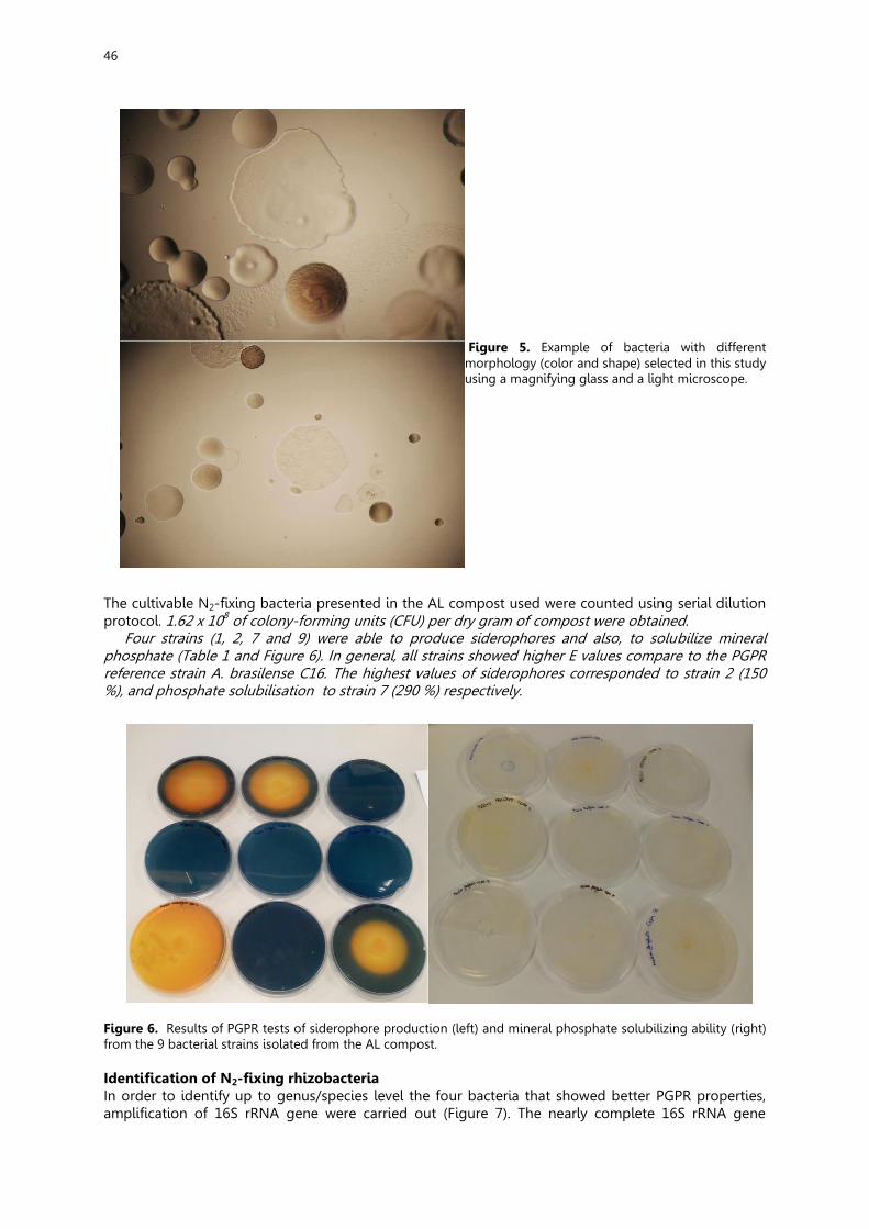
46
Figure 5. Example of bacteria with different morphology (color and shape) selected in this study using a magnifying glass and a light microscope.
The cultivable N2-fixing bacteria presented in the AL compost used were counted using serial dilution protocol. 1.62 x 10⁸ of colony-forming units (CFU) per dry gram of compost were obtained.
Four strains (1, 2, 7 and 9) were able to produce siderophores and also, to solubilize mineral phosphate (Table 1 and Figure 6). In general, all strains showed higher E values compare to the PGPR reference strain A. brasilense C16. The highest values of siderophores corresponded to strain 2 (150 %), and phosphate solubilisation to strain 7 (290 %) respectively.
Figure 6. Results of PGPR tests of siderophore production (left) and mineral phosphate solubilizing ability (right) from the 9 bacterial strains isolated from the AL compost. Identification of N2-fixing rhizobacteria In order to identify up to genus/species level the four bacteria that showed better PGPR properties, amplification of 16S rRNA gene were carried out (Figure 7). The nearly complete 16S rRNA gene
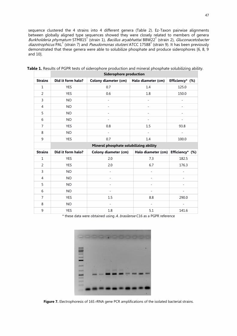
47
sequence clustered the 4 strains into 4 different genera (Table 2). Ez-Taxon pairwise alignments between globally aligned type sequences showed they were closely related to members of genera Burkholderia phymatum STM815T (strain 1), Bacillus aryabhattai B8W22T (strain 2), Gluconacetobacter diazotrophicus PALT (strain 7) and Pseudomonas stutzeri ATCC 17588T (strain 9). It has been previously demonstrated that these genera were able to solubilize phosphate and produce siderophores [6, 8, 9 and 10].
Table 1. Results of PGPR tests of siderophore production and mineral phosphate solubilizing ability. Siderophore production
Strains Did it form halo? Colony diameter (cm) Halo diameter (cm) Efficiency* (%)
1 YES 0.7 1.4 125.0
2 YES 0.6 1.8 150.0
3 NO - - -
4 NO - - -
5 NO - - -
6 NO - - -
7 YES 0.8 1.5 93.8
8 NO - - -
9 YES 0.7 1.4 100.0
Mineral phosphate solubilizing ability
Strains Did it form halo? Colony diameter (cm) Halo diameter (cm) Efficiency* (%)
1 YES 2.0 7.3 182.5
2 YES 2.0 6.7 176.3
3 NO - - -
4 NO - - -
5 NO - - -
6 NO - - -
7 YES 1.5 8.8 290.0
8 NO - - -
9 YES 1.8 5.1 141.6
* these data were obtained using A. brasilense C16 as a PGPR reference
Figure 7. Electrophoresis of 16S rRNA gene PCR amplifications of the isolated bacterial strains.
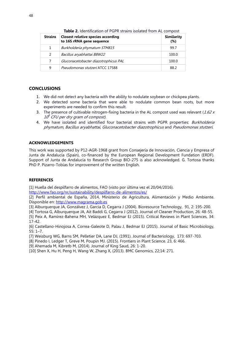
48
Table 2. Identification of PGPR strains isolated from AL compost Strains Closest relative species according
to 16S rRNA gene sequence Similarity
(%)
1 Burkholderia phymatum STM815 99.7
2 Bacillus aryabhattai B8W22 100.0
7 Gluconacetobacter diazotrophicus PAL 100.0
9 Pseudomonas stutzeri ATCC 17588 88.2
CONCLUSIONS
1. We did not detect any bacteria with the ability to nodulate soybean or chickpea plants. 2. We detected some bacteria that were able to nodulate common bean roots, but more
experiments are needed to confirm this result. 3. The presence of cultivable nitrogen-fixing bacteria in the AL compost used was relevant (1.62 x
108 CFU per dry gram of compost). 4. We have isolated and identified four bacterial strains with PGPR properties: Burkholderia
phymatum, Bacillus aryabhattai, Gluconacetobacter diazotrophicus and Pseudomonas stutzeri.
ACKNOWLEDGEMENTS
This work was supported by P12-AGR-1968 grant from Consejería de Innovación, Ciencia y Empresa of Junta de Andalucía (Spain), co-financed by the European Regional Development Fundation (ERDF). Support of Junta de Andalucía to Research Group BIO-275 is also acknowledged. G. Tortosa thanks PhD P. Pizarro-Tobías for improvement of the written English.
REFERENCES
[1] Huella del despilfarro de alimentos, FAO (visto por última vez el 20/04/2016). http://www.fao.org/nr/sustainability/despilfarro-de-alimentos/es/ [2] Perfil ambiental de España, 2014, Ministerio de Agricultura, Alimentación y Medio Ambiente. Disponible en: http://www.magrama.gob.es [3] Alburquerque JA, Gonzálvez J, Garcia D, Cegarra J (2004). Bioresource Technology, 91, 2: 195-200. [4] Tortosa G, Alburquerque JA, Ait Baddi G, Cegarra J (2012). Journal of Cleaner Production, 26: 48-55. [5] Peix A, Ramírez-Bahena MH, Velázquez E, Bedmar EJ (2015). Critical Reviews in Plant Sciences, 34: 17-42. [6] Castellano-Hinojosa A, Correa-Galeote D, Palau J, Bedmar EJ (2015). Journal of Basic Microbiology, 55: 1–7. [7] Weisburg WG, Barns SM, Pelletier DA, Lane DJ, (1991). Journal of Bacteriology, 173: 697-703. [8] Pinedo I, Ledger T, Greve M, Poupin MJ. (2015). Frontiers in Plant Science. 23, 6: 466. [9] Ahemada M, Kibretb M, (2014). Journal of King Saud, 26: 1-20. [10] Shen X, Hu H, Peng H, Wang W, Zhang X, (2013). BMC Genomics, 22;14: 271.
49
MY OWN IDEAS Elena Navarro García
Al comenzar este proyecto no sabía muy bien cómo se iba a desarrollar y no estaba segura de si no me arrepentiría de participar en él, pero con el paso del tiempo y de las sesiones me ha ido gustando cada vez más y pienso que es una experiencia que debería vivir todo aquel que se sienta atraído por la ciencia.
La razón principal por la que pienso de esta manera es debido a que soy una firme defensora del aprendizaje práctico, es decir, en mi opinión, los conceptos se afianzan mucho mejor si ves o haces una prueba de ello. Gracias a este proyecto he podido poner en práctica muchos de los conceptos aprendidos anteriormente en mi instituto ya que, debido a la falta de tiempo, no solemos hacerlo y aquí nos ha dado la oportunidad de ello. María Molina Muñoz.
Cursábamos cuarto de la ESO cuando nuestra profesora Soledad de física y química entró en la clase con una noticia que cultivó intriga e interés en todo el aula. La oportunidad de poder participar en un proyecto de investigación como tal, con científicos, medios y las instalaciones adecuadas era posible. El CSIC, la Universidad de Granada y los proyectos PIIISA nos brindaban esta oportunidad: mutación de bacterias, cúmulos estelares, el genoma humano, biología tumoral, jardín microscópico, agujeros negros… Infinidad de temas y proyectos estaban a solo unos pequeños pasos. Desafortunadamente, tanto para el resto de mis compañeros como para mí, las plazas se agotaron antes de que nuestra profesora pudiera apuntar ningún nombre, pese a su esfuerzo, las oportunidades cesaron a tiempo récord. Llegado el 2016, nuevo año, nueva vida y nuevas ocasiones de participación. Fue entonces cuando por fin se me ofreció el disfrute de este proyecto. Sinceramente, una de mis mejores experiencias como estudiante. Todavía recuerdo como el ayer, el primer día que llegamos al CSIC, nuestra profesora nos invitó a desayunar y nos deseó suerte para el resto del día. Quedamos todos los alumnos reunidos en un gran salón de actos, recuerdo también girar mi cabeza varias veces para contemplar con intriga la sala, cuando vi a dos investigadores sentados en las sillas del fondo. Quién me iba a mí a decir, que estos dos jóvenes se convertirían en mis investigadores, en los propulsores de tanta ilusión y gusto: Germán Tortosa y Antonio Castellano.
Bajo el proyecto: ‘’Biofertilizantes con sabor a aceite de oliva” y las puertas del laboratorio 202 un pequeño equipo de seis alumnos de diferentes centros estaría trabajando al unísono durante varios meses como verdaderos científicos: y es que nuestros coordinadores nos vistieron con una bata blanca, guantes y nos dejaron, como bien dice Germán ‘’cacharrear’’ con todo el material científico, ‘’eso es lo que a ellos les gusta, cacharrear’’. Pudimos usar lupas, microscopios ópticos, programas de identificación de bacterias, cabinas de flujo laminar, pipetas. ¡Cultivamos en casa nuestras propias bacterias y dos macetitas de soja o garbanzo fueron regadas por nosotros mismos! Alucinante.
El trato de ellos hacia nosotros, fue grandioso. Todas las sesiones nos invitaban a desayunar, nos presentaban a sus compañeros de laboratorio, resolvían dudas e incluso varias fotos y vídeos nos eran sacadas.
Realmente esta experiencia ha alcanzado parámetros de ilusión que no pensé que fueran posible. He vivido cada sesión con tanta ilusión que dudo mucho que se me vaya a olvidar fácilmente, he disfrutado tanto viendo crecer a nuestras cepas, aislando su ADN y poniéndoles nombres que el deleite ha sido máximo. En definitiva me sentía grande dentro de mi pequeño cuerpo, visualizaba mi futura profesión a cada paso del procedimiento. Me sentía realmente parte de esta gran comunidad científica y aunque mis inseguridades eran notables; pues nunca antes había usado una pipeta ni inoculado placas Petri, la confianza y paciencia de nuestros investigadores fue de gran ayuda.
Así que no me queda más que agradecerles a ellos y a los coordinadores de esta actividad, por recibirnos con los brazos tan abiertos, por su energía y su capacidad transmisora de amor a la ciencia, por su apoyo altruista e iniciativa y por convertir en verídico este proyecto haciendo felices a tantos pequeños científicos por unos pocos días. Laura Palma Pérez.
Cuando me dijeron que me había tocado este proyecto pensé que iba a estar bien pero sinceramente antes de haberlo hecho hubiera preferido entrar en el de genética, pero desde el primer día que pisamos el laboratorio supe que este proyecto era el mejor de todos porque en comparación con
50
otros compañeros de otros proyectos, nosotros hemos estado prácticamente todo el rato en el laboratorio. Por otra parte la relación con los compañeros de proyecto ha sido muy buena y desde el primer día nos hemos llevado muy bien y no hemos tenido ningún problema en ayudarnos entre nosotros. Gracias a este proyecto he podido descubrir cómo se trabaja de verdad en un laboratorio, y la verdad que me ha dado muchas ideas a la hora de elegir una carrera para el futuro. Mª Carmen Sarmiento Vega.
En conclusión, esta experiencia me ha encantado. Es impresionante ver el lugar en donde grandes investigadores trabajan a diario para ayudar al mundo. Lo que más me ha gustado ha sido poder trabajar con los aparatos específicos de investigación, pero sobretodo el microscopio. Este proyecto me ha servido muchísimo para saber que es trabajar en equipo y el esfuerzo que requiere descubrir cualquier cosa por pequeña que sea. Carlos Ortega Fernández.
La impresión que he tenido sobre este proyecto ha sido muy gratificante debido a que he podido experimentar como es el trabajo en un laboratorio utilizando todos los instrumentos que eran necesarios para realizar nuestro proyecto relacionado con el compost. La parte que más me ha gustado del proyecto fue cuando miramos las bacterias a través del microscopio electrónico y luego hicimos una placa master con las bacteria que fuimos seleccionando según su morfología para luego crecerlas y extraer su ADN. Esto puede que no me haya servido de nada por ahora, pero estoy seguro de que en un futuro me será muy útil ya sea para decidir la carrera que quiero estudiar o para cualquier otra cosa. Alba Díaz Arco.
Este proyecto me ha encantado ya que está relacionado con la ciencia y sobre el medio ambiente, de gran importancia para nuestro futuro. Al haber tratado con diversas prácticas y haber aprendido cosas sobre este tema por primera vez he aprendido como se trabaja en un laboratorio ya que en mi instituto lo utilizamos pocas veces. También me ha ayudado a saber si realmente me gustaría trabajar en estos ya que si no pruebas algo no sabes realmente como es, como suele pasar cuando tienes expectativas. Es muy importante este tipo de proyectos por esto mismo ya que muchas personas cambian de carrera porque eso no es lo que pensaban o tenían otra idea de ello, de esta manera podemos evitarlo.
51
A microscopical garden at the “Estación Experimental del Zaidín”
Elena Lima1, María Eugenia Ramos2, Irene Cabello-Cano3, Eugenia Laura
Carretero-Quero4, Alba María García-Gil5, Javier Moreno-García6, Alba María Romero-López7, Mar Ruiz-López5, Juan de Dios Alché1*
1Department of Biochemistry, Cell and Molecular Biology of Plants, Estación Experimental del Zaidín (CSIC),
Profesor Albareda 1, 18008 Granada, Spain 2Grupo de Pastos y Sistemas Silvopastorales Mediterráneos, Estación Experimental del Zaidín (CSIC), Profesor
Albareda 1, 18008 Granada, Spain 3CDP dulce nombre de María, Paseo de los Basilios, 2, 18008 Granada
4CDP Sagrado Corazón, Paseo de los Basilios, 3, 18008 Granada 5IES Zaidín Vergeles, Primavera 24, 26, 18080 Granada, Spain. 6IES Padre Suarez, Calle Gran Vía de Colón, 61, 18001 Granada
7IES Padre Poveda, Av de Mariana Pineda, 18, 18500 Guadix, Granada, *Corresponding author: e-mail: [email protected]
SUMMARY
Pollen grains represent the male gametophyte in higher plants. They offer a large variability in sizes, shapes and textures, and are used as taxonomic characters for the identification of plant species. The aim of the present study was to set the procedures to identify and characterize the pollen from those species present as ornamentals in the gardens of the EEZ-CSIC institute in Granada, which have been previously recorded in botanic guides. The procedure has been implemented in five species flowering at wintertime, such as Lagerstroemia indica, Chimonanthus praecox, Yucca elephantipes, Teucrium fruticans L. and Spiraea x vanhouttei. Moreover, the pollen from other plant species widely used in the Institute for research purposes, like Olea europaea L. and Chenopodium album (both used for studies on allergy) were also used in the present study. Microscopic analyses were carried out by light microscopy with differential interferential contrast (DIC) and by confocal laser scanning microscopy, using the autofluorescence of the pollen exine for pollen imaging. The morphology of the pollens studied here largely coincided with the existing literature, which is mainly based on SEM (scanning electron microscopy) analyses. However we discovered that the use of confocal microscopy represents an excellent alternative to SEM, by offering an adequate, although yet lower level of resolution, but without the need of expensive and time consuming sample preparation procedures. INTRODUCTION (AND OBJECTIVES)
Pollen grains represent whole microscopic organisms with a reduced genetic package as regards to the sporophyte, because they are formed through meiosis. They are able to keep themselves alive in the environment, and to resist unfavourable ecological conditions, mainly thanks to physiological adaptations like the accumulation of reserves and the presence of a highly evolved pollen wall, which represents a unique taxonomic feature.
In order to complete their function (addressing the sperm nuclei to the embryo sac), pollen grains must be able to maintain a high metabolic rate, and to emit a pollen tube, sometimes unbelievable large. Some pollen grains contain proteins, which make them allergenic and terribly uncomfortable for humans.
Several botanic itineraries have been described in the EEZ-CSIC institute, including numerous species, both Spanish and from exotic countries. Most species present in these routes have been catalogued and botanic information is already available. However, information regarding palynological characteristics of these taxa is lacking.
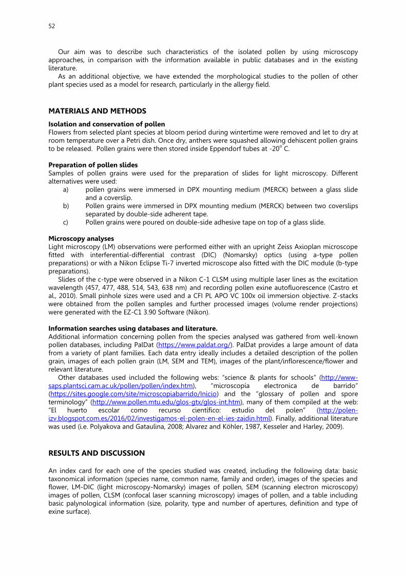
52
Our aim was to describe such characteristics of the isolated pollen by using microscopy approaches, in comparison with the information available in public databases and in the existing literature.
As an additional objective, we have extended the morphological studies to the pollen of other plant species used as a model for research, particularly in the allergy field.
MATERIALS AND METHODS
Isolation and conservation of pollen Flowers from selected plant species at bloom period during wintertime were removed and let to dry at room temperature over a Petri dish. Once dry, anthers were squashed allowing dehiscent pollen grains to be released. Pollen grains were then stored inside Eppendorf tubes at -20o C.
Preparation of pollen slides Samples of pollen grains were used for the preparation of slides for light microscopy. Different alternatives were used:
a) pollen grains were immersed in DPX mounting medium (MERCK) between a glass slide and a coverslip.
b) Pollen grains were immersed in DPX mounting medium (MERCK) between two coverslips separated by double-side adherent tape.
c) Pollen grains were poured on double-side adhesive tape on top of a glass slide.
Microscopy analyses Light microscopy (LM) observations were performed either with an upright Zeiss Axioplan microscope fitted with interferential-differential contrast (DIC) (Nomarsky) optics (using a-type pollen preparations) or with a Nikon Eclipse Ti-7 inverted microscope also fitted with the DIC module (b-type preparations).
Slides of the c-type were observed in a Nikon C-1 CLSM using multiple laser lines as the excitation wavelength (457, 477, 488, 514, 543, 638 nm) and recording pollen exine autofluorescence (Castro et al., 2010). Small pinhole sizes were used and a CFI PL APO VC 100x oil immersion objective. Z-stacks were obtained from the pollen samples and further processed images (volume render projections) were generated with the EZ-C1 3.90 Software (Nikon).
Information searches using databases and literature. Additional information concerning pollen from the species analysed was gathered from well-known pollen databases, including PalDat (https://www.paldat.org/). PalDat provides a large amount of data from a variety of plant families. Each data entry ideally includes a detailed description of the pollen grain, images of each pollen grain (LM, SEM and TEM), images of the plant/inflorescence/flower and relevant literature.
Other databases used included the following webs: “science & plants for schools” (http://www-saps.plantsci.cam.ac.uk/pollen/pollen/index.htm), “microscopía electronica de barrido” (https://sites.google.com/site/microscopiabarrido/Inicio) and the “glossary of pollen and spore terminology” (http://www.pollen.mtu.edu/glos-gtx/glos-int.htm), many of them compiled at the web: “El huerto escolar como recurso científico: estudio del polen” (http://polen-izv.blogspot.com.es/2016/02/investigamos-el-polen-en-el-ies-zaidin.html). Finally, additional literature was used (i.e. Polyakova and Gataulina, 2008; Alvarez and Köhler, 1987, Kesseler and Harley, 2009). RESULTS AND DISCUSSION An index card for each one of the species studied was created, including the following data: basic taxonomical information (species name, common name, family and order), images of the species and flower, LM-DIC (light microscopy-Nomarsky) images of pollen, SEM (scanning electron microscopy) images of pollen, CLSM (confocal laser scanning microscopy) images of pollen, and a table including basic palynological information (size, polarity, type and number of apertures, definition and type of exine surface).
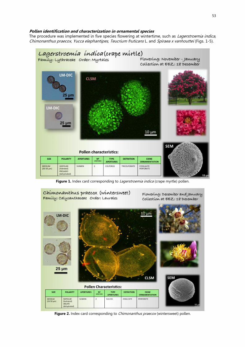
53
Pollen identification and characterization in ornamental species The procedure was implemented in five species flowering at wintertime, such as Lagerstroemia indica, Chimonanthus praecox, Yucca elephantipes, Teucrium fruticans L. and Spiraea x vanhouttei (Figs. 1-5).
Figure 1. Index card corresponding to Lagerstroemia indica (crape myrtle) pollen.
Figure 2. Index card corresponding to Chimonanthus praecox (wintersweet) pollen.

54
Figure 3. Index card corresponding to Yucca elephantipes (spineless yucca) pollen.
Figure 4. Index card corresponding to Teucrium fruticans L. (tree germander) pollen.
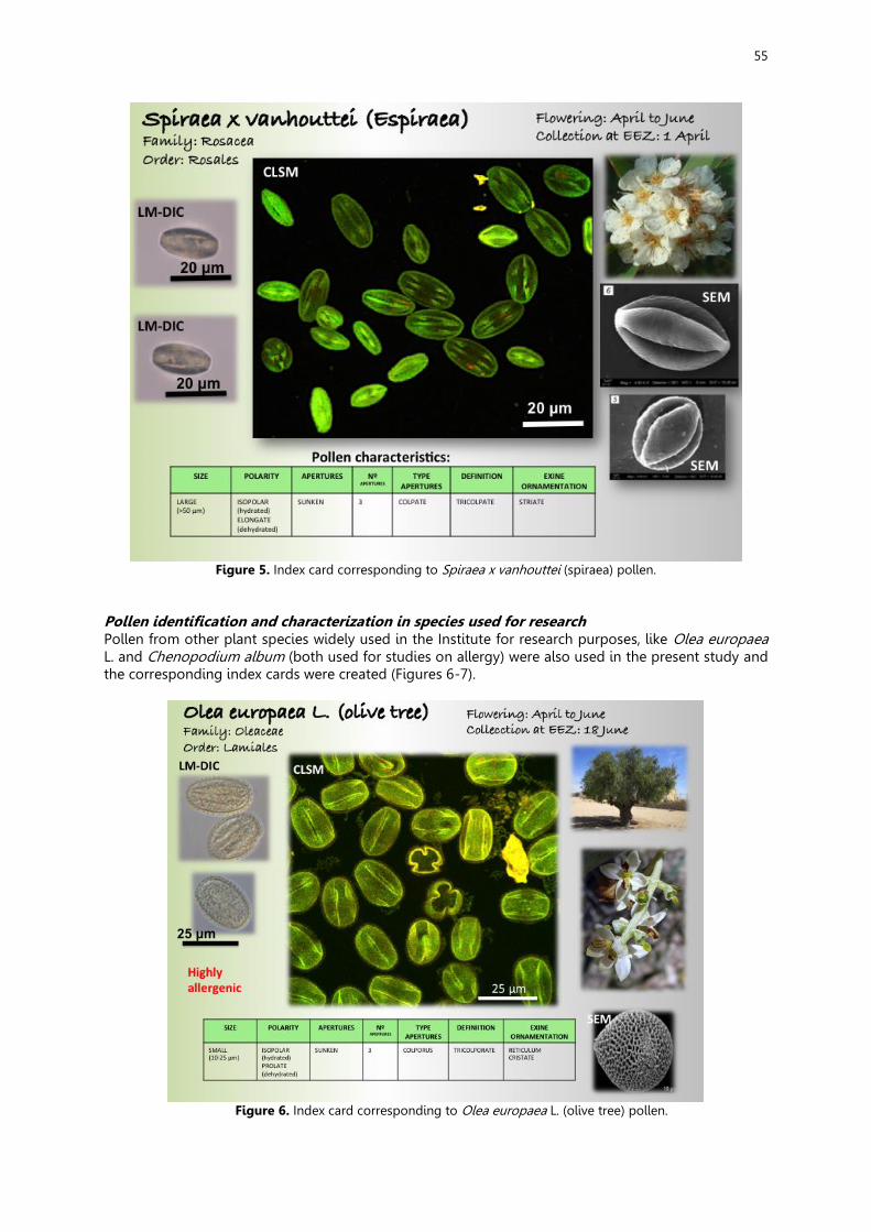
55
Figure 5. Index card corresponding to Spiraea x vanhouttei (spiraea) pollen.
Pollen identification and characterization in species used for research Pollen from other plant species widely used in the Institute for research purposes, like Olea europaea L. and Chenopodium album (both used for studies on allergy) were also used in the present study and the corresponding index cards were created (Figures 6-7).
Figure 6. Index card corresponding to Olea europaea L. (olive tree) pollen.
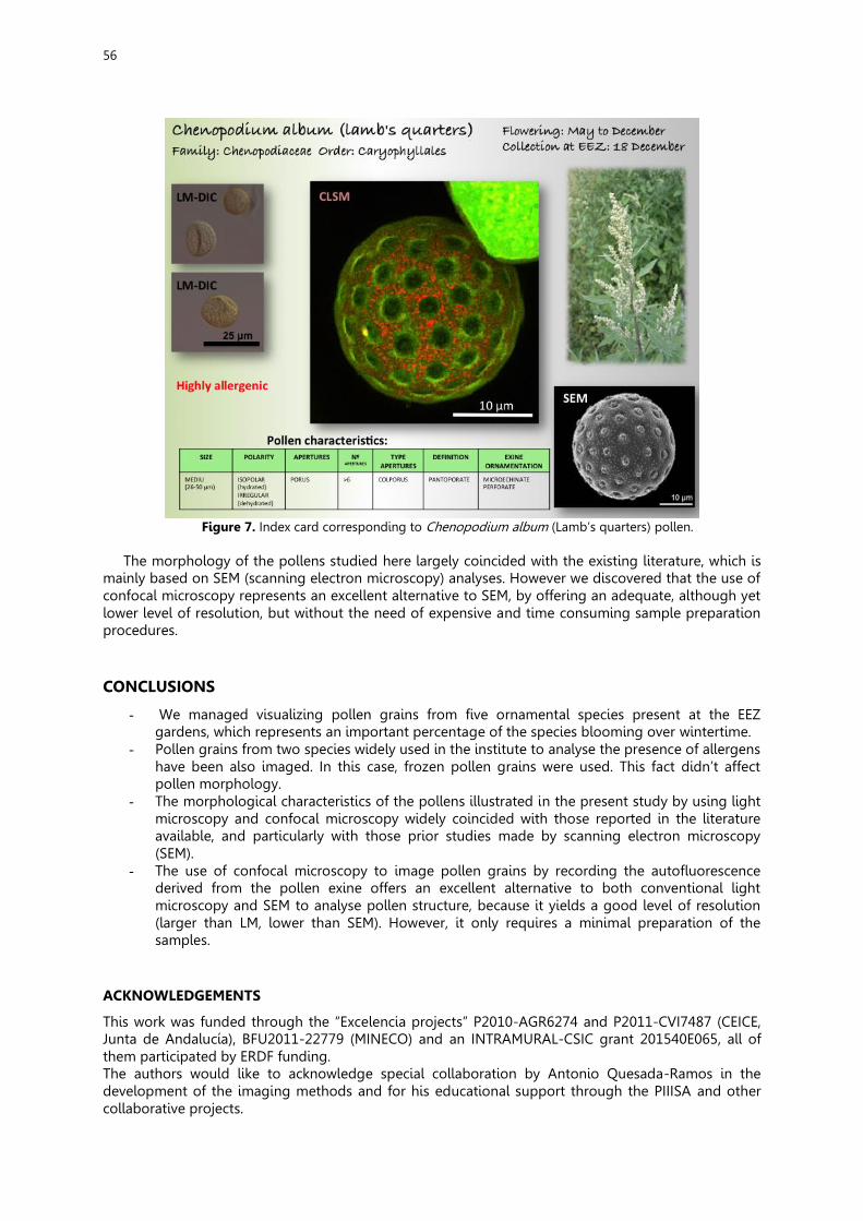
56
Figure 7. Index card corresponding to Chenopodium album (Lamb’s quarters) pollen.
The morphology of the pollens studied here largely coincided with the existing literature, which is
mainly based on SEM (scanning electron microscopy) analyses. However we discovered that the use of confocal microscopy represents an excellent alternative to SEM, by offering an adequate, although yet lower level of resolution, but without the need of expensive and time consuming sample preparation procedures. CONCLUSIONS
- We managed visualizing pollen grains from five ornamental species present at the EEZ gardens, which represents an important percentage of the species blooming over wintertime.
- Pollen grains from two species widely used in the institute to analyse the presence of allergens have been also imaged. In this case, frozen pollen grains were used. This fact didn’t affect pollen morphology.
- The morphological characteristics of the pollens illustrated in the present study by using light microscopy and confocal microscopy widely coincided with those reported in the literature available, and particularly with those prior studies made by scanning electron microscopy (SEM).
- The use of confocal microscopy to image pollen grains by recording the autofluorescence derived from the pollen exine offers an excellent alternative to both conventional light microscopy and SEM to analyse pollen structure, because it yields a good level of resolution (larger than LM, lower than SEM). However, it only requires a minimal preparation of the samples.
ACKNOWLEDGEMENTS
This work was funded through the “Excelencia projects” P2010-AGR6274 and P2011-CVI7487 (CEICE, Junta de Andalucía), BFU2011-22779 (MINECO) and an INTRAMURAL-CSIC grant 201540E065, all of them participated by ERDF funding. The authors would like to acknowledge special collaboration by Antonio Quesada-Ramos in the development of the imaging methods and for his educational support through the PIIISA and other collaborative projects.
57
REFERENCES
Alvarez A and Köhler E. 1987. Morfología del polen de las Agavaceae y algunos géneros afines, Grana, 26:1, 25-46, DOI: 10.1080/00173138709428902 Castro AJ, Rejón JD, Fendri M, Jiménez-Quesada MJ, Zafra A, Jiménez-López JC, Rodríguez-García MI and Alché JD. 2010. Taxonomical discrimination of pollen grains by using confocal laser scanning microscopy (CLSM) imaging of autofluorescence. In: “Microscopy: Science, Technology, Applications and Education” Vol. 1. Antonio Méndez-Vilas and Jesús Díaz Álvarez (Eds.). Formatex Research Center pp:607-613 ISBN-13: 978-84-614-6189-9. Kesseler R and Harley M. 2009. Pollen. The hidden sexuality of flowers. Third Ed. Rob Kesseler, Madeline Harley and Papadakis Publisher. ISBN: 978 1 906506 01 8. Polyakova TA and Gataulina GN. 2008. Morphology and Variability of Pollen of the Genus Spiraea L. (Rosaceae) in Siberia and the Far East. In: Contemporary Problems of Ecology, 2008, Vol. 1, No. 4, pp. 420–424. Pleiades Publishing, Ltd. ISSN 1995-4255. Original Russian Text: T.A. Polyakova, G.N. Gataulina, 2008, published in Sibirskii Ekologicheskii Zhurnal, 2008, Vol. 15, No. 4, pp. 545–551.

58
MY OWN IDEAS Mar Ruiz López
If somebody asked me now why I chose this project, I would probably answer him/her that it was because I truly love nature and green landscapes. Yes, just because of that I chose this project at the EEZ. My first impression of the place was breathtaking. It's preserved in excellent conditions. I really couldn't expect a place like that in the middle of a town like Granada. Regarding the research, I have to admit that it was very interesting, though the mystery of pollen is not one of my favourites scientific fields; I have learned a lot of things that I'm sure will be useful to me in my future, like being extremely organised with the information we get, the importance of the good presentation at the moment of elaborating the slides with the conclusions... And last, but not least, I've had the chance of sharing this experience with one of my best classmates and meeting new incredible and well-formed people that have made these months funny and didactic. Alba García Gil
At this project we have been employed with many trees and plants. First we gathered the pollen and learned to do several preparations to the microscope. Then, together with our investigator we discover many types of pollen and also differentiating them and to be able to classify them. Finally we realize the presentation of our work collaborating all the companions and each one investigated a plant/ tree and his pollen respectively. It has been a very interesting project in which beside learning, me entered the world of the botany and I have known persons and splendid investigators. Javier Moreno García If I had to sum up my experience during this project with an only word, I am sure that I would use ‘Fantastic’. I have enjoyed every minute of it (well, apart from when we were doing the presentation… That was tough!). Not only I found the recollection of pollen fascinating, but the work environment is what I have enjoyed the most. I loved watching researchers doing their work while we were there, and I am sure that in a few years time I will be working like them, with my little microscope and my notepad. This experience would not have been as great as it has, without my friends from the project and the two best and nicest researchers of the world. Thank you for everything! Eugenia Laura Carretero Quero
I love science, so it has been one of the best experiences of my life, because it has given me the chance to feel like a real scientist, to work in a profesional laboratory, to use wonderful objects, like big microscopes, and the luck of being able to learn new things. After this project I can discover other things about something I really love (new skills for microscopes, new tools...). Also I have met new great people in my team, the best. Alba María Romero López
First I want thank all the people who have made possible this project. In these laboratories there are diverse fantastic tools of work, but especially what impressed me the most were the microscopes, which we have had the possibility to observe many details of the gathered pollen with. Finally, I think that this has been a great experience, where I have got to know fantastic people, have learned many new things ... so I recommend to those who have this opportunity to take part in this type of projects.

II
I SA
P