HPV mRNA test in triage of women with minor cervical lesions - Munin
23
HPV mRNA test in triage of women with minor cervical lesions; experiences from the University Hospital of North Norway S.W. Sorbye a* , S. Fismen a , T.J. Gutteberg b,c , E.S. Mortensen a,c a Department of Clinical Pathology, The University Hospital of North Norway, 9038 Tromso, Norway b Department of Microbiology and Infection Control, The University Hospital of North Norway, 9038 Tromso, Norway c Department of Medical Biology, Faculty of Health Sciences, University of Tromso, Norway *Corresponding author at: Department of Clinical Pathology, University Hospital of North Norway, 9038 Tromso, Norway. Tel.: +47 77 62 72 23; fax: +47 77 62 72 04 E-mail address: [email protected] (S.W. Sorbye). *Manuscript Click here to view linked References
Transcript of HPV mRNA test in triage of women with minor cervical lesions - Munin
HPV mRNA test in triage of women with minor cervical lesions; experiences
from the University Hospital of North Norway
S.W. Sorbyea*
, S. Fismena, T.J. Gutteberg
b,c, E.S. Mortensen
a,c
aDepartment of Clinical Pathology, The University Hospital of North Norway, 9038 Tromso,
Norway
bDepartment of Microbiology and Infection Control, The University Hospital of North Norway,
9038 Tromso, Norway
cDepartment of Medical Biology, Faculty of Health Sciences, University of Tromso, Norway
*Corresponding author at: Department of Clinical Pathology, University Hospital of North
Norway, 9038 Tromso, Norway. Tel.: +47 77 62 72 23; fax: +47 77 62 72 04
E-mail address: [email protected] (S.W. Sorbye).
*ManuscriptClick here to view linked References
Abstract
In the Norwegian Cervical Cancer Screening Programme tests for detection of human
papillomavirus (HPV) are used to triage women with minor cytological cervical lesions. The
material in this study comprises samples from 1 798 women in the period 2006-08. The HPV test
was performed according to the guidelines of the Norwegian Cancer Registry. The HPV mRNA
test (PreTect HPV-Proofer) detects and types 5 high-risk genotypes (16, 18, 31, 33 and 45). The
HPV mRNA results were compared to cytology and then biopsy up to December 2009. Women
with minor cytological cervical lesions and negative HPV test were followed with a new PAP
smear after 12 months. A total of 327 women (18 %) were HPV mRNA positive. Of the 1 798
women with minor cytological lesions, 232 women (13 %) had moderate dysplasia, severe
dysplasia or cancer and 144 women (8 %) had severe dysplasia or cancer in biopsy. 57 % of the
women with a positive HPV mRNA test had moderate dysplasia, severe dysplasia or cancer. 37 %
had severe dysplasia or cancer. The sensitivity of the HPV mRNA test to detect moderate dysplasia,
severe dysplasia or cancer was 81 %. The specificity for moderate dysplasia, severe dysplasia or
cancer was 91 %. The negative predictive value (NPV) of the HPV mRNA test for moderate
dysplasia, severe dysplasia or cancer was 97 %. Of 11 women with cervical cancer, 10 were
positive for HPV type 16 or 18.
Compared to existing literature the HPV mRNA test seems more suitable than HPV DNA
tests to triage women with minor cytological cervical lesions due to its higher specificity.
Key words: HPV, mRNA, dysplasia, cytology, cervical cancer, screening.

Cervical cancer is the second most common cancer affecting women worldwide. About 300 women
in Norway get cervical cancer each year and 80 of these die from the disease. In 1995 a Norwegian
National Cervical Cancer screening programme was established. As a part of the programme,
women aged 25 – 69 years are invited to cytological screening every three years, totalling 400 000
smears a year. Every year 25 000 women are retested due to minor cytological lesions.
Approximately 10 000 women are referred to colposcopy, and 3 000 treated by conisation because
of high grade changes confirmed by biopsy (Cancer Registry of Norway;
http://www.kreftregisteret.no).
It has been estimated that without the screening programme, one might expect 600 cases of
cervical cancer in Norway each year (Cancer Registry of Norway; http://www.kreftregisteret.no).
The Norwegian guidelines recommend treatment with conisation of the cervix after histologically
confirmed high grade dysplasia. 3 000 women are conisated every year to avoid 300 cases of
cancer, which suggests considerable overtreatment. Currently no methods exist for distinguishing
high grade dysplasia with high risk of progression to invasive cancer from high grade dysplasia that
is going to regress spontaneously without treatment. The assay PreTect HPV-Proofer (NorChip AS,
Norway) detects the E6/E7 oncogene expression which is necessary for malignant transformation
and may therefore be a possible candidate to identify underlying cervical cancer precursors with
high probability of progression to cancer among women with minor cytological abnormalities.
Women with minor cytological cervical lesions have an increased risk of having, or
developing, high grade dysplasia compared to women with normal cytology. However, most minor
cytological lesions regress spontaneously and therefore careful triage is crucial. In this short
communication the routine diagnostic practice at the University Hospital of North Norway is
evaluated including the use of the HPV E6/E7 mRNA test PreTect HPV-Proofer in triage of
women with minor cytological cervical lesions for the detection of cervical dysplasia and cancer.
About 23 000 cervical smears are analysed annually at the Department of Clinical
Pathology at the University Hospital of North Norway. Between 2006 and 2008, smears from
48 781 women aged 25-69 years were analysed. A total of 2 314 women (4.7 %) were diagnosed
with minor cervical lesions. For these women, repeat cytology and HPV mRNA test after 6 months
is recommended. The compliance was high; as liquid based control cytology was received 1 798
women were tested with the HPV mRNA test (78 % of the 2 314 women). Cells were extracted
from the ThinPrep (Cytyc Corporation, Marlborough, MA, USA) for cytological examination.
The diagnoses of the cervical smears and the biopsies were taken from the diagnostic
database for cytological and histological samples (SymPathy) at the Department of Clinical
Pathology, University Hospital of North Norway. The HPV mRNA testing was performed
according to the national guidelines for HPV testing (Figure 1). In the department of clinical
pathology all biopsies with high-grade dysplasia and cancer are evaluated by experienced
pathologists. Biopsies with uncertain cellular changes are immunostained with p16.
The sensitivity of the HPV mRNA test is defined as the proportion of high grade dysplasia
detected by HPV mRNA or repeat cytology that is positive for HPV mRNA. In the calculations of
specificity and negative predictive values (NPV) it is assumed that cytonegative and HPV mRNA
negative samples without detected dysplasia during the follow up period do not contain disease.
Of the 1 798 women who had a HPV-test done, 327 (18 %) were HPV mRNA positive. A
total of 232 women (13 %) had moderate dysplasia or worse and 144 women (8 %) had severe
dysplasia or cancer in biopsy. The absolute number of true-positives (TP) for moderate dysplasia or
worse was 188. The TP was 121 for severe dysplasia or cancer. 57 % (188/327) of the women with
a positive HPV mRNA test had moderate dysplasia or worse. 37 % (121/327) had severe dysplasia
or cancer. The sensitivity of the HPV mRNA test to detect moderate dysplasia or worse was 81 %
(188/232). The sensitivity for severe dysplasia or cancer was 84 % (121/144). The specificity for
moderate dysplasia or worse was 91 % (1427/1566). The specificity for severe dysplasia or cancer

was 88 % (1448/1654). The negative predictive value (NPV) of the HPV mRNA test was 97 %
(1427/1471) for moderate dysplasia or worse. NPV for severe dysplasia or cancer was 98 %
(1448/1471).
For women with equivocal cervical lesions (atypical squamous cells of undetermined
significance) the positive predictive value (PPV) for moderate dysplasia or worse was 57 %
(83/145). The PPV for severe dysplasia or cancer was 41 % (60/145). The specificity was 95 %
(1088/1150) and 93 % (1092/1177), respectively. For women with low-grade cytological lesions
(low grade squamous intraepitelial lesion) the PPV for moderate dysplasia or worse was 58 %
(105/182). The PPV for severe dysplasia or cancer was 34 % (61/182). The specificity of low grade
cytological lesions was 82 % (339/416) and 75 % (356/477), respectively (Tables 1 and 2).
Of the 327 women with a positive HPV mRNA test, 204 women (62 %) were infected with
HPV type 16 or 18. Of the 187 HPV positive women with moderate dysplasia or worse, 129 (69 %)
had HPV 16 or 18. Of the 121 HPV positive women with severe dysplasia or cancer, 94 (78 %) had
HPV type 16 or 18. Despite low-grade changes in PAP-smear six months earlier, 11 women
actually had cancer in biopsy. 10 of these (91 %) had HPV type 16 or 18 (figure 2). One woman
with a lymphoepithelial carcinoma of the cervix was HPV mRNA negative.
In general, the use of HPV DNA tests generates more positive results than the HPV mRNA
test. This is because DNA tests detect the mere presence of the virus and will therefore also detect
harmless transient infections which are handled by the immune system (Cattani et al., 2009b). The
HPV DNA test Hybrid Capture 2 detects 13 HPV types and the HPV mRNA test PreTect HPV
Proofer detects five HPV types. In Europe most of the HPV positive cervical cancer cases are types
16, 18, 31, 33 and 45 (Smith et al., 2007), and these HPV types are included in the HPV mRNA
test. The specificity and the positive predictive value (PPV) of HPV DNA tests for moderate
dysplasia or worse are low, especially when young, sexually active women are tested. The real
cause of cervical cancer is not the HPV-virus infection per se, but continuous over-expression of
the viral oncogenes E6 and E7 from oncogenic HPV-types (zur Hausen, 2002). There may be
several reasons why E6 and E7 are over-expressed, but one of the main reasons is integration of the
virus into the human genome (loss of E2 gene results in loss of regulation of transcripts). The loss
of the E2 gene only occurs in a small fraction of the high number of women with HPV infection
(Collins et al., 2009). This implies that a test that detects the over-expression of E6 and E7 mRNA
is more specific than a test that detects the general presence of viral DNA.
In triage of minor cytological cervical lesions, in comparison with follow-up cytology, HPV
DNA testing was more sensitive and equally specific for triage of equivocal lesions and for
predicting recurrence of cervical intraepitelial neoplasia in women treated for high-grade dysplasia
(Arbyn et al., 2005). Due to the high rate of HPV positivity, this is not the case for patients with
cytological low grade lesions, as the DNA test Hybrid Capture 2 showed a substantially and
significantly lower specificity than the repeat Pap smear, as demonstrated in a meta-analysis by
Arbyn et al (Arbyn et al., 2004;Arbyn et al., 2005). When triaging women with low grade lesions, a
reflex Hybrid Capture 2 test did show a significantly higher sensitivity, but at the same time a
significantly lower specificity compared to repeat cytology (Arbyn et al., 2006).
In studies where cervical samples have been tested with both the HPV mRNA test and HPV
DNA tests, the test positivity rate of the HPV mRNA test is one third of the HPV DNA tests. The
sensitivity for moderate dysplasia or worse is lower, but the specificity is higher (Cattani et al.,
2009a;Keegan et al., 2009;Lie et al., 2005;Szarewski et al., 2008). It is well-known that many
cervical lesions with moderate or severe dysplasia will regress spontaneously. Only 31% of
colposcopically visible lesions with severe dysplasia will progress to invasive cancer within 30
years (McCredie et al., 2008). Although the HPV mRNA test has a lower sensitivity in detecting
moderate or severe dysplasia, it is probable that it still identifies the lesions that are destined to
progress to cancer. Furthermore, several studies have shown that the sensitivity of the HPV mRNA
test for cervical cancer is the same as for the HPV DNA tests (Basu et al., 2009;Hovland et al.,
2010;Kraus et al., 2006;Lie et al., 2005).
The HPV test is used to triage women with minor cytological cervical lesions. In triage it is
important to have a test with high specificity in order to identify who should be referred to
colposcopy and biopsy from those that only need control cytology 12 months later. The sensitivity
is of minor importance because a positive diagnosis has already been made using cytology. A
positive HPV test will result in biopsy when the control cytology has minor lesions. In cases where
a HPV test with a low specificity is used, a high number of women will be referred to colposcopy
and biopsy unnecessarily. Many unnecessary conisations are performed because many lesions with
moderate or severe dysplasia will regress spontaneously without treatment. Moreover, unnecessary
conisations should be avoided given the risk of obstetrical complications associated with such
treatments (Arbyn et al., 2008).
One of the most used HPV tests in Norway is the Digene Hybrid Capture® HPV DNA Test
(Qiagen, Gaithersburg, MD, USA). This test detects whether or not any of the 13 high risk HPV
viruses are present but a positive test does not give any information concerning which type of virus
is causing the positive result. The HPV DNA test is shown to have a high clinical sensitivity and a
high negative predictive value (NPV), but the specificity for high grade dysplasia is low (Solomon
et al., 2001). For instance, using high grade lesions as outcome, the average specificity of the HPV
DNA test Hybrid Capture in triage of equivocal cervical lesions is 62.5% (95% CI: 57.8-67.3%)
and only 28.6% (95% CI: 22.2-35.0) in triage of low grade cervical lesions (Arbyn et al., 2006).
While HPV DNA tests correlate poorly with the grade of dysplasia (Trope et al., 2009), the
experiences from the University Hospital of North Norway show that the HPV mRNA test
correlates well with histologically confirmed grade of dysplasia. This indicates that HPV E6/E7
mRNA is a good biomarker for severe dysplasia and cancer. As moderate dysplasia regresses
spontaneously more often than severe dysplasia, it is probable that use of a HPV mRNA test in

triage reduces overtreatment of young women compared to the use of a HPV DNA test. However,
if moderate or severe dysplasia is confirmed by biopsy, ethical considerations make it difficult to
delay treatment to evaluate the rate of regression even though the women have a negative HPV
mRNA test.
In one study the HPV mRNA test was as sensitive for high grade dysplasia but more
specific that HPV DNA testing with PCR (Molden et al., 2005). In a Norwegian study by
Vintermyr et al. using samples from 435 women with minor cervical lesions the test positivity rate
of the HPV DNA test Hybrid Capture 2 was 53 % (232/435); the sensitivity for high grade lesions
was 94 % (47/50); the specificity 52 % (200/385); and the PPV was 20 % (47/232) (Vintermyr et
al., 2008). In the material from the University Hospital of North Norway 18 % had a positive HPV
mRNA test; the sensitivity for high grade lesions was 81 %; specificity 91 %; and the PPV was 57
%.
The experience from the University Hospital of North Norway is that the HPV mRNA test
has a high specificity and a high positive predictive value. This makes it useful in triage of women
with equivocal and low grade cytological lesions. Together with the cervical cytology, this HPV
mRNA test detects more high-grade dysplasia than cytology alone without increasing the number
of biopsies. Compared to existing literature the HPV mRNA test seems more suitable than HPV
DNA tests to triage women with minor cytological cervical lesions due to its higher specificity.
Conflicts of interest
None
Table 1
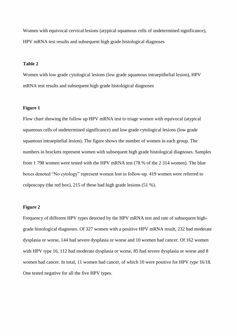
Women with equivocal cervical lesions (atypical squamous cells of undetermined significance),
HPV mRNA test results and subsequent high grade histological diagnoses
Table 2
Women with low grade cytological lesions (low grade squamous intraepithelial lesion), HPV
mRNA test results and subsequent high grade histological diagnoses
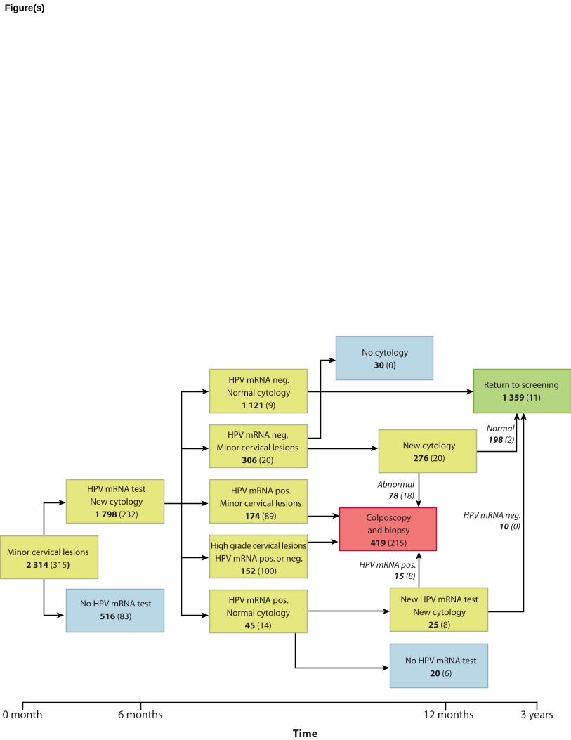
Figure 1
Flow chart showing the follow up HPV mRNA test to triage women with equivocal (atypical
squamous cells of undetermined significance) and low grade cytological lesions (low grade
squamous intraepitelial lesion). The figure shows the number of women in each group. The
numbers in brackets represent women with subsequent high grade histological diagnoses. Samples
from 1 798 women were tested with the HPV mRNA test (78 % of the 2 314 women). The blue
boxes denoted “No cytology” represent women lost in follow-up. 419 women were referred to
colposcopy (the red box), 215 of these had high grade lesions (51 %).
Figure 2
Frequency of different HPV types detected by the HPV mRNA test and rate of subsequent high-
grade histological diagnoses. Of 327 women with a positive HPV mRNA result, 232 had moderate
dysplasia or worse, 144 had severe dysplasia or worse and 10 women had cancer. Of 162 women
with HPV type 16, 112 had moderate dysplasia or worse, 85 had severe dysplasia or worse and 8
women had cancer. In total, 11 women had cancer, of which 10 were positive for HPV type 16/18.
One tested negative for all the five HPV types.

References
Arbyn, M., Buntinx, F., Van Ranst, M., Paraskevaidis, E., Martin-Hirsch, P., Dillner, J., 2004.
Virologic versus cytologic triage of women with equivocal Pap smears: a meta-analysis of
the accuracy to detect high-grade intraepithelial neoplasia. J. Natl. Cancer Inst. 96, 280-293.
Arbyn, M., Kyrgiou, M., Simoens, C., Raifu, A.O., Koliopoulos, G., Martin-Hirsch, P., Prendiville,
W., Paraskevaidis, E., 2008. Perinatal mortality and other severe adverse pregnancy
outcomes associated with treatment of cervical intraepithelial neoplasia: meta-analysis. BMJ
337, a1284.
Arbyn, M., Paraskevaidis, E., Martin-Hirsch, P., Prendiville, W., Dillner, J., 2005. Clinical utility
of HPV-DNA detection: triage of minor cervical lesions, follow-up of women treated for
high-grade CIN: an update of pooled evidence. Gynecol. Oncol. 99, S7-11.
Arbyn, M., Sasieni, P., Meijer, C.J., Clavel, C., Koliopoulos, G., Dillner J., 2006. Chapter 9:
Clinical applications of HPV testing: a summary of meta-analyses. Vaccine 24 Suppl 3, S3-
78-S3/89.
Basu, P., Roychowdhury, S., Bafna, U.D., Chaudhury, S., Kothari, S., Sekhon, R., Saranath, D.,
Biswas, S., Gronn, P., Silva, I., Siddiqi, M., Ratnam, S., 2009. Human papillomavirus
genotype distribution in cervical cancer in India: results from a multi-center study. Asian
Pac. J. Cancer Prev. 10, 27-34.
Cattani, P., Siddu, A., D'Onghia, S., Marchetti, S., Santangelo, R., Vellone, V.G., Zannoni, G.F.,
Fadda, G., 2009a. RNA (E6 and E7) assays versus DNA (E6 and E7) assays for risk
evaluation for women infected with human papillomavirus. J. Clin. Microbiol. 47, 2136-
2141.
Cattani, P., Zannoni, G.F., Ricci, C., D'Onghia, S., Trivellizzi, I.N., Di Franco, A., Vellone, V.G.,
Durante, M., Fadda, G., Scambia, G., Capelli, G., De Vincenzo, R., 2009b. Clinical
performance of human papillomavirus E6 and E7 mRNA testing for high-grade lesions of
the cervix. J. Clin. Microbiol. 47, 3895-3901.
Collins, S.I., Constandinou-Williams, C., Wen, K., Young, L.S., Roberts, S., Murray, P.G.,
Woodman, C.B., 2009. Disruption of the E2 gene is a common and early event in the
natural history of cervical human papillomavirus infection: a longitudinal cohort study.
Cancer Res. 69, 3828-3832.
Hovland, S., Muller, S., Skomedal, H., Mints, M., Bergstrom, J., Wallin, K.L., Karlsen, F.,
Johansson, B., Andersson, S., 2010. E6/E7 mRNA expression analysis: a test for the
objective assessment of cervical adenocarcinoma in clinical prognostic procedure. Int. J.
Oncol. 36, 1533-1539.
Keegan, H., Mc Inerney, J., Pilkington, L., Gronn, P., Silva, I., Karlsen, F., Bolger, N., Logan, C.,
Furuberg, L., O'Leary, J., Martin, C., 2009. Comparison of HPV detection technologies:
Hybrid capture 2, PreTect HPV-Proofer and analysis of HPV DNA viral load in HPV16,
HPV18 and HPV33 E6/E7 mRNA positive specimens. J. Virol. Methods 155, 61-66.

Kraus, I., Molden, T., Holm, R., Lie, A.K., Karlsen, F., Kristensen, G.B., Skomedal, H., 2006.
Presence of E6 and E7 mRNA from human papillomavirus types 16, 18, 31, 33, and 45 in
the majority of cervical carcinomas. J. Clin. Microbiol. 44, 1310-1317.
Lie, A.K., Risberg, B., Borge, B., Sandstad, B., Delabie, J., Rimala, R., Onsrud, M., Thoresen, S.,
2005. DNA- versus RNA-based methods for human papillomavirus detection in cervical
neoplasia. Gynecol. Oncol. 97, 908-915.
McCredie, M.R., Sharples, K.J., Paul, C., Baranyai, J., Medley, G., Jones, R.W., Skegg, D.C.,
2008. Natural history of cervical neoplasia and risk of invasive cancer in women with
cervical intraepithelial neoplasia 3: a retrospective cohort study. Lancet Oncol. 9, 425-434.
Molden, T., Nygard, J.F., Kraus, I., Karlsen, F., Nygard, M., Skare, G.B., Skomedal, H., Thoresen,
S.O., Hagmar, B., 2005. Predicting CIN2+ when detecting HPV mRNA and DNA by
PreTect HPV-proofer and consensus PCR: A 2-year follow-up of women with ASCUS or
LSIL Pap smear. Int. J. Cancer 114, 973-976.
Smith, J.S., Lindsay, L., Hoots, B., Keys, J., Franceschi, S., Winer, R., Clifford, G.M., 2007.
Human papillomavirus type distribution in invasive cervical cancer and high-grade cervical
lesions: a meta-analysis update. Int. J. Cancer 121, 621-632.
Solomon, D., Schiffman, M., Tarone, R., 2001. Comparison of three management strategies for
patients with atypical squamous cells of undetermined significance: baseline results from a
randomized trial. J. Natl. Cancer Inst. 93, 293-299.
Szarewski, A., Ambroisine, L., Cadman, L., Austin, J., Ho, L., Terry, G., Liddle, S., Dina, R.,
McCarthy, J., Buckley, H., Bergeron, C., Soutter, P., Lyons, D., Cuzick, J., 2008.
Comparison of predictors for high-grade cervical intraepithelial neoplasia in women with
abnormal smears. Cancer Epidemiol. Biomarkers Prev. 17, 3033-3042.
Trope, A., Sjoborg, K., Eskild, A., Cuschieri, K., Eriksen, T., Thoresen, S., Steinbakk, M., Laurak,
V., Jonassen, C.M., Westerhagen, U., Jacobsen, M.B., Lie, A.K., 2009. Performance of
human papillomavirus DNA and mRNA testing strategies for women with and without
cervical neoplasia. J. Clin. Microbiol. 47, 2458-2464.
Vintermyr, O.K., Skar, R., Iversen, O.E., Haugland, H.K., 2008. Usefulness of HPV test on cell
sample from the cervix. Tidsskr. Nor Laegeforen. 128, 171-173.
zur Hausen, H, 2002. Papillomaviruses and cancer: from basic studies to clinical application. Nat.
Rev. Cancer 2, 342-350.

Mark Arbyn
Unit of Cancer Epidemiology, Scientific Institute of Public Health, Brussels, Belgium
Kate Cuschieri
Scottish HPV Reference Laboratory, Specialist Virology Centre, Royal Infirmary of Edinburgh,
Edinburgh EH16 4SA, UK
Paola Cattani
Institute of Microbiology, Università Cattolica del Sacro Cuore, Largo A Gemelli 8, Rome 00168,
Italy
*Potential Reviewers
Response to comments from the reviewers Reviewer #1:
General comments:
Cervical cancer screening is still based on cytology, with recent introduction of detection of HPV
DNA along with cytology. The change now being proposed is in the detection of the mRNA of
HPV oncogenic proteins (E6 and E7) as a marker of clinically significant cervical disease
because detecting DNA alone yields too many negative biopsies (low specificity).
Specific comments:
The authors reported comprehensive evaluation of the utility of HPV E6 and E7 mRNA testing.
Their histologic-virologic investigation, testing a large number of women (1798) for HPV
mRNA, in the triage of ASCUS and LSIL, convincingly demonstrate that the detection of E6 and
E7 viral transcripts is a specific tool for prediction of high grade lesions, correlating significantly
with histological confirmed grade of dysplasia.
These findings have important clinical implications. HPV mRNA testing may be more efficient
and effective for triage and follow-up of females with abnormal cytology, reducing the number of
patients referred to colposcopy and biopsy.
A minor suggested change is that the authors may consider reducing the number of tables (table1
and 2 could be incorporated comparing results for CIN2+ and CIN3+ in the same table)
Our response: CIN2+ and CIN3+ are now in the same table. However, reviewer #2
recommended dividing the material into women with cytological ASC-US versus women with
LSIL, respectively. For this reason there are still two tables.
Reviewer #2:
GENERAL COMMENTS
Women with ASCUS/LSIL have an increased risk for having or developing high grade CIN
compared to women with normal cytology. However, most minor lesions regress spontaneously
and therefore careful triage is crucial. This study demonstrates good specificity of the HPV
Proofer containing probes for the 5 main HPV types in triage of women with minor cytological
lesions (ASCUS/LSIL). Triage with HPV DNA testing using the HC2 assay has significantly
higher sensitivity and similar specificity compared to repeat cytology. However in LSIL triage,
HC2 is highly sensitive but has very low specificity. For this reason it would be appropriate that
the authors provide separate data split by triage group: ASCUS and LSIL.
Our response: The data is now split by triage group ASC-US (table 1) and LSIL (table 2), as
recommended by reviewer #2.
It is recommended also to present the performance of triage by repeat cytology.
*Response to Reviewers
Our response: Unfortunately, we cannot compare triage by repeat cytology with triage by HPV-
testing in our material. According to the guidelines from the Norwegian Cervical Cancer
Screening Programme, colposcopy and biopsy are not recommended after repeat cytology (ASC-
US / LSIL x 2) without a positive HPV-test. However, colposcopy and biopsy are recommended if
the woman has minor cytological abnormalities (ASC-US / LSIL x 3) over a period of 18 months
even though the HPV-test is negative.
Certain improvements in describing accuracy parameters are needed (see below).
Our response: Accuracy parameters have been improved according to Reviewer 2`s
recommendations (see below)
References to studies addressing 1ary screening are irrelevant. The authors should refer to
studies where HPV tests in a triage setting are used.
Our response: We agree and have included the following paragraph (page 6, ln 5-14)
“In triage of minor cytological cervical lesions, in comparison with follow-up cytology, HPV
DNA testing was more sensitive and equally specific for triage of equivocal (ASC-US) lesions
and for predicting recurrence of cervical intraepitelial neoplasia (CIN) in women treated for
high-grade dysplasia (CIN2+). Due to the high rate of HPV positivity, this is not the case for
patients with cytological low grade dysplasia (LSIL), as the DNA test Hybrid Capture 2 showed a
substantially and significantly lower specificity than the repeat Pap smear, as demonstrated in a
meta-analysis by Arbyn et al. When triaging women with low-grade squamous intraepithelial
lesions (LSIL), a reflex Hybrid Capture 2 test did show a significantly higher sensitivity, but at
the same time a significantly lower specificity compared to a repeat Pap smear.”
Pages could be numbered to facilitate reviewing.
Our response: The pages have been numbered.
INTRODUCTION
Page 5,3rd
§: the accuracy estimates of cytology in primary screening (Nanda, Koliopoulous,
Gage) are irrelevant for this study, where triage of ASCUS/LSIL is addressed. The authors
should refer to previously published meta-analyses on triage of these lesions: (see Arbyn JNCI
2004; Gynecol Oncol 2005 and Vaccine 2006).
Our response: We agreee and have included the following paragraph (page 6, ln 5-14)
“In triage of minor cytological cervical lesions, in comparison with follow-up cytology, HPV
DNA testing was more sensitive and equally specific for triage of equivocal (ASC-US) lesions
and for predicting recurrence of cervical intraepitelial neoplasia (CIN) in women treated for
high-grade dysplasia (CIN2+). Due to the high rate of HPV positivity, this is not the case for
patients with cytological low grade dysplasia (LSIL), as the DNA test Hybrid Capture 2 showed a
substantially and significantly lower specificity than the repeat Pap smear, as demonstrated in a
meta-analysis by Arbyn et al. When triaging women with low-grade squamous intraepithelial
lesions (LSIL), a reflex Hybrid Capture 2 test did show a significantly higher sensitivity, but at
the same time a significantly lower specificity compared to a repeat Pap smear.”
Page 5, last line: “.candidate”; complete: . candidate “to identify underlying cervical cancer
precursors with high probability of progression among women with minor cytological
abnormalities”.
Our response: We agree and have inserted this sentence according to the reviewers
recommendations: (page 3, ln 17-19) ”candidate to identify underlying cervical cancer
precursors with high probability of progression among women with minor cytological
abnormalities.”
Page 6, first §. This § essentially concerns primary screening. The authors should focus on the
topic of their manuscript which is triage of ASC-US/LSIL. Only the Solomon reference is
relevant. This paragraph should be dropped.
Our response: We agree and this paragraph has been omitted.
MATERIAL AND METHOD
Page 5, 3rd
§. “Cells are extracted with the ThinPrep®. Change “with” into “from”
Our response: We agree and have changed the sentence accordingly (page 4, ln 9).
As mentioned before, the authors should provide separate data for ASC-US and LSIL.
Our response: We agree. The data are now presented separately (table 1 and 2).
The authors should put the assumption forward that cytonegative/RNA negative samples do not
contain disease. Otherwise the specificity and NPV cannot be derived from these data since this
would require verification of all mRNA neg samples.

Our response: We agree, and have added this sentence accordingly (page 4, ln 17-20) “In the
calculations of specificity and negative predictive values (NPV) it is assumed that cytonegative
and HPV mRNA negative samples without detected dysplasia during the follow up period do not
contain disease.”
RESULTS
Page 7: Give the absolute number of true-positives (TP) for CIN2+ and CIN3+.
Our response: We agree and have included these data (page 4, ln 23-24) “The absolute number
of true-positives (TP) for CIN2+ and CIN3+ is 188 and 121 respectively.”
It is recommended to provide systematically numerator and denominator for all computed
accuracy measures in the paper.
Our response: We agree and we have included these numbers in all the presented data.
The PPV values are mentioned twice (0.57 and 57%; 0.37 and 37%). Be consistent in describing
accuracy parameters: always as a percentage for instance (57%) but not in fraction (0.57).
Our response: We agree. All the PPV and NPV are now presented as percentages.
The authors should define sensitivity of mRNA as the proportion of CIN2/3+ detected by mRNA
or repeat cytology that is positive for mRNA.
Our response: We agree. This sentence has been included accordingly (page 4, ln 16-17) “The
sensitivity of the HPV mRNA test is defined as the proportion of high grade dysplasia (CIN2+)
detected by HPV mRNA or repeat cytology that is positive for HPV mRNA.”
The title of fig 1 can be shortened, since the fig is self-explanatory.
Our response: We agree. It is now shortened.
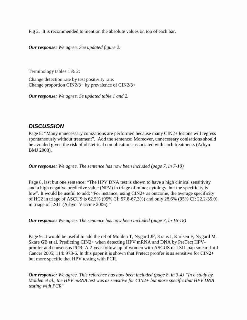
Fig 2. It is recommended to mention the absolute values on top of each bar.
Our response: We agree. See updated figure 2.
Terminology tables 1 & 2:
Change detection rate by test positivity rate.
Change proportion CIN2/3+ by prevalence of CIN2/3+
Our response: We agree. Se updated table 1 and 2.
DISCUSSION
Page 8: “Many unnecessary conizations are performed because many CIN2+ lesions will regress
spontaneously without treatment”. Add the sentence: Moreover, unnecessary conisations should
be avoided given the risk of obstetrical complications associated with such treatments (Arbyn
BMJ 2008).
Our response: We agree. The sentence has now been included (page 7, ln 7-10)
Page 8, last but one sentence: “The HPV DNA test is shown to have a high clinical sensitivity
and a high negative predictive value (NPV) in triage of minor cytology, but the specificity is
low”. It would be useful to add: “For instance, using CIN2+ as outcome, the average specificity
of HC2 in triage of ASCUS is 62.5% (95% CI: 57.8-67.3%) and only 28.6% (95% CI: 22.2-35.0)
in triage of LSIL (Arbyn Vaccine 2006).”
Our response: We agree. The sentence has now been included (page 7, ln 16-18)
Page 9: It would be useful to add the ref of Molden T, Nygard JF, Kraus I, Karlsen F, Nygard M,
Skare GB et al. Predicting CIN2+ when detecting HPV mRNA and DNA by PreTect HPV-
proofer and consensus PCR: A 2-year follow-up of women with ASCUS or LSIL pap smear. Int J
Cancer 2005; 114: 973-6. In this paper it is shown that Pretect proofer is as sensitive for CIN2+
but more specific that HPV testing with PCR.
Our response: We agree. This reference has now been included (page 8, ln 3-4) “In a study by
Molden et al., the HPV mRNA test was as sensitive for CIN2+ but more specific that HPV DNA
testing with PCR”

Editor’s Comments:
Do not use the first-person when rewriting (ie. “we” and “our”).
Our response: the use of first-person has been deleted in the manuscript
There are grammatical errors which must be corrected.
Our response: Grammatical errors have been corrected and the manuscript has been edited by a
scientific English editing service
- The paper must be rewritten in grammatical English with the help of a native English speaking-
scientist or scientific English editing service:
http://www.elsevier.com/wps/find/authors.authors/languagepolishing
http://authorservices.wiley.com/bauthor/english_language.asp
An example of the corrections necessary throughout the text will be e-mailed to you
separately / is attached.
Do not use the acronyms ASCUS, LSIL, CIN+, UNN. Do not use acronyms in the title of the
manuscript.
Our response: OK
Reference List:
Please check the style of the Journal
Do not use the word “and” in the list of authors
Our response: this has been corrected according to the style of the journal.
The title page must be retyped according to the style of the Journal.
Our response: this has been corrected according to the style of the journal.
0 month
Minor cervical lesions
2 314 (315)
High grade cervical lesions
HPV mRNA pos. or neg.
152 (100)
Colposcopy
and biopsy
419 (215)
HPV mRNA pos.
Normal cytology
45 (14)
No HPV mRNA test
516 (83)
No HPV mRNA test
20 (6)
New HPV mRNA test
New cytology
25 (8)
HPV mRNA test
New cytology
1 798 (232)
HPV mRNA neg.
Normal cytology
1 121 (9)
HPV mRNA neg.
Minor cervical lesions
306 (20)
HPV mRNA pos.
Minor cervical lesions
174 (89)
No cytology
30 (0)
New cytology
276 (20)
HPV mRNA neg.
10 (0)
Abnormal
78 (18)
HPV mRNA pos.
15 (8)
Return to screening
1 359 (11)
Normal
198 (2)
6 months
Time
12 months 3 years
Figure(s)

180
160
140
120
100
80
60
40
20
0
Number of women
HPV type
HPV 16
HPV mRNA positive
Moderate dysplasia or worse
Severe dysplasia or worse
Cancer
162
112
85
8
42
17
9
2
21
94
60
33
17
42
17
6
HPV 18 HPV 31 HPV 33 HPV 45
Figure(s)
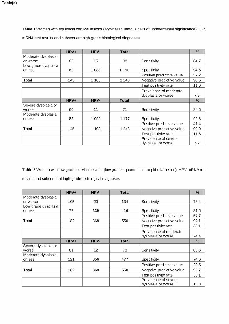
Table 1 Women with equivocal cervical lesions (atypical squamous cells of undetermined significance), HPV
mRNA test results and subsequent high grade histological diagnoses
HPV+ HPV- Total %
Moderate dysplasia or worse 83 15 98 Sensitivity 84.7
Low grade dysplasia or less 62 1 088 1 150 Specificity 94.6
Positive predictive value 57.2
Total 145 1 103 1 248 Negative predictive value 98.6
Test positivity rate 11.6
Prevalence of moderate dysplasia or worse 7.9
HPV+ HPV- Total %
Severe dysplasia or worse 60 11 71 Sensitivity 84.5
Moderate dysplasia or less 85 1 092 1 177 Specificity 92.8
Positive predictive value 41.4
Total 145 1 103 1 248 Negative predictive value 99.0
Test positivity rate 11.6
Prevalence of severe dysplasia or worse 5.7
Table 2 Women with low grade cervical lesions (low grade squamous intraepithelial lesion), HPV mRNA test
results and subsequent high grade histological diagnoses
HPV+ HPV- Total %
Moderate dysplasia or worse 105 29 134 Sensitivity 78.4
Low grade dysplasia or less 77 339 416 Specificity 81.5
Positive predictive value 57.7
Total 182 368 550 Negative predictive value 92.1
Test positivity rate 33.1
Prevalence of moderate dysplasia or worse 24.4
HPV+ HPV- Total %
Severe dysplasia or worse 61 12 73 Sensitivity 83.6
Moderate dysplasia or less 121 356 477 Specificity 74.6
Positive predictive value 33.5
Total 182 368 550 Negative predictive value 96.7
Test positivity rate 33.1
Prevalence of severe dysplasia or worse 13.3
Table(s)