HowADAM-9andADAM-11DifferentiallyFromEstrogenReceptor ... · members of the MMP-family, MMP-2 and...
12
How ADAM-9 and ADAM-11 Differentially From Estrogen Receptor Predict Response to Tamoxifen Treatment in Patients with Recurrent Breast Cancer: a Retrospective Study Anieta M. Sieuwerts, Marion E. Meijer-van Gelder, Mieke Timmermans, Anita M.A.C. Trapman, Roberto Rodriguez Garcia, Miranda Arnold, Anneke J.W. Goedheer, Henk Portengen, Jan G.M. Klijn, and John A. Foekens Abstract Purpose: To evaluate the predictive value of the disintegrin and metalloproteinases, ADAM-9, ADAM-10, ADAM-11, and ADAM-12, and of the matrix metalloproteinases, MMP-2 and MMP-9, in patients with recurrent breast cancer treated with tamoxifen. Experimental Design: A retrospective study was done on 259 frozen specimens of estrogen receptor ^ positive primary breast carcinomas from patients who developed recurrent disease and were treated with tamoxifen as the first line of therapy. The expression levels of the biological factors were assessed by real-time quantitative reverse transcriptase PCR. Results: Using log-transformed continuous variables, increasing levels of ADAM-9 [odds ratio (OR) = 1.41; P = 0.015] and decreasing levels of MMP-9 (OR, 0.81; P = 0.035) predicted favorable disease control independent from the traditional predictive factors. Furthermore, when tumors were dichotomized at the median level of 70% tumor cell nuclei, our univariate analysis showed particularly strong results for the group of 153 patients with primary tumors containing 30% or more stromal cells. Although estrogen receptor levels lost their predictive power for this group of patients, high levels of ADAM-9 (OR, 1.59; P = 0.007) and ADAM-11 (OR, 1.65; P = 0.001) were significantly associated with a higher efficacy of tamoxifen therapy. Conclusions: Our results show that especially for primary tumors containing stromal elements, the assessment of mRNA expression levels of ADAM-9 and ADAM-11could be useful to identify patients with recurrent breast cancer who are likely to benefit or fail from tamoxifen therapy. The ADAMs, which stands for a disintegrin and metalloprotei- nase, also known as MDCs, are a newly discovered family of membrane proteins. All ADAMs possess some or all of the following domains: a signal peptide, a propeptide, a metal- loprotease, a disintegrin, a cysteine-rich domain, an epidermal growth factor–like domain, a transmembrane sequence, and a cytoplasmic tail. The propeptide might be involved in latency with activation upon loss, the metalloprotease domain in proteolysis, the disintegrin domain in adhesion, the cysteine- rich domain in fusion and adhesion, the epidermal growth factor – like domain in growth factor activity, and the cytoplasmic tail in cell signaling. The possession of these multiple domains with their potential functions makes them likely candidates to play a role in cancer cell invasion and metastasis. Indeed, some of these ADAMs have already been linked to various diseases including cancer (1–5). Despite the above findings, a definite role for any ADAM in either cancer formation, progression, or response to therapy, remains to be shown. In recurrent breast cancer, steroid hormone receptor status is one of the variables often used to determine the choice of endocrine therapy. Thus far, tamoxifen is the most extensively used hormonal treatment, although only 50% to 60% of the treated patients will benefit (6 – 8). Because proteases such as the urokinase-type plasminogen activator have been shown to be associated with failure of tamoxifen therapy in patients with recurrent breast cancer (9, 10), we hypothesized that specific ADAMs might also be associated with therapeutic failure. Thus far, >30 different ADAMs have been described, of which 19 appear in humans. For our study, we selected four ADAMs for which no pseudogenes have been described and which have already been shown to be expressed to some extent in human breast cancer. Of these four, only ADAM-11, also named MDC, does not possess an active matrix metal- loproteinase (MMP) – like domain. However, based on its location within a loss of heterozygosity region of chromosome 17q21 (11, 12), ADAM-11 has been proposed to be a candi- date tumor suppressor gene for human breast cancer (13, 14) and was therefore included in our study. The other three members included in this study, ADAM-9 (MDC9, meltrin-g), ADAM-10 (Kuz, SUP-17, MADM), and ADAM-12 (meltrin-a) all possess an active MMP-like domain, and in addition have all been reported to be increased in malignant compared with Imaging, Diagnosis, Prognosis Authors’ Affiliation: Department of Medical Oncology, Erasmus Medical Center, Rotterdam, the Netherlands Received 3/11/05; revised 7/9/05; accepted 7/20/05. The costs of publication of this article were defrayed in part by the payment of page charges. This article must therefore be hereby marked advertisement in accordance with 18 U.S.C. Section 1734 solely to indicate this fact. Requests for reprints: Anieta M. Sieuwerts, Erasmus Medical Center, Josephine Nefkens Institute, Room Be 400, P.O. Box 1738, 3000 DR Rotterdam, the Netherlands. Phone: 31-10-408-8372; Fax: 31-10-408-8377; E-mail: a.sieuwerts@ erasmusmc.nl. F 2005 American Association for Cancer Research. doi:10.1158/1078-0432.CCR-05-0560 www.aacrjournals.org Clin Cancer Res 2005;11(20) October 15, 2005 7311 Research. on May 31, 2017. © 2005 American Association for Cancer clincancerres.aacrjournals.org Downloaded from
Transcript of HowADAM-9andADAM-11DifferentiallyFromEstrogenReceptor ... · members of the MMP-family, MMP-2 and...

How ADAM-9 and ADAM-11Differentially From Estrogen ReceptorPredict Response to Tamoxifen Treatment in PatientswithRecurrent Breast Cancer: a Retrospective StudyAnietaM. Sieuwerts, Marion E.Meijer-van Gelder, Mieke Timmermans, AnitaM.A.C.Trapman,Roberto Rodriguez Garcia, Miranda Arnold, Anneke J.W. Goedheer, Henk Portengen,Jan G.M. Klijn, andJohn A. Foekens
Abstract Purpose: To evaluate the predictive value of the disintegrin and metalloproteinases, ADAM-9,ADAM-10, ADAM-11, and ADAM-12, and of the matrix metalloproteinases, MMP-2 and MMP-9,in patients with recurrent breast cancer treatedwith tamoxifen.Experimental Design: A retrospective study was done on 259 frozen specimens of estrogenreceptor ^ positive primary breast carcinomas from patients who developed recurrent diseaseandwere treated with tamoxifen as the first line of therapy.The expression levels of the biologicalfactors were assessed by real-time quantitative reverse transcriptase PCR.Results: Using log-transformed continuous variables, increasing levels of ADAM-9 [odds ratio(OR)=1.41;P =0.015] anddecreasing levels ofMMP-9 (OR,0.81;P =0.035) predicted favorabledisease control independent from the traditional predictive factors. Furthermore, when tumorswere dichotomized at the median level of 70% tumor cell nuclei, our univariate analysis showedparticularly strong results for the group of 153 patients with primary tumors containing 30% ormore stromal cells. Although estrogen receptor levels lost their predictive power for this group ofpatients, high levels of ADAM-9 (OR,1.59; P = 0.007) and ADAM-11 (OR,1.65; P = 0.001) weresignificantly associated with a higher efficacy of tamoxifen therapy.Conclusions: Our results show that especially for primary tumors containing stromal elements,the assessment of mRNA expression levels of ADAM-9 and ADAM-11could be useful to identifypatients with recurrent breast cancer who are likely to benefit or fail from tamoxifen therapy.
The ADAMs, which stands for a disintegrin and metalloprotei-nase, also known as MDCs, are a newly discovered family ofmembrane proteins. All ADAMs possess some or all of thefollowing domains: a signal peptide, a propeptide, a metal-loprotease, a disintegrin, a cysteine-rich domain, an epidermalgrowth factor–like domain, a transmembrane sequence, and acytoplasmic tail. The propeptide might be involved in latencywith activation upon loss, the metalloprotease domain inproteolysis, the disintegrin domain in adhesion, the cysteine-rich domain in fusion and adhesion, the epidermal growthfactor–like domain in growth factor activity, and the cytoplasmictail in cell signaling. The possession of these multiple domainswith their potential functions makes them likely candidates toplay a role in cancer cell invasion and metastasis. Indeed, someof these ADAMs have already been linked to various diseases
including cancer (1–5). Despite the above findings, a definiterole for any ADAM in either cancer formation, progression, orresponse to therapy, remains to be shown.
In recurrent breast cancer, steroid hormone receptor status isone of the variables often used to determine the choice ofendocrine therapy. Thus far, tamoxifen is the most extensivelyused hormonal treatment, although only 50% to 60% of thetreated patients will benefit (6–8). Because proteases such asthe urokinase-type plasminogen activator have been shown tobe associated with failure of tamoxifen therapy in patients withrecurrent breast cancer (9, 10), we hypothesized that specificADAMs might also be associated with therapeutic failure.
Thus far, >30 different ADAMs have been described, ofwhich 19 appear in humans. For our study, we selected fourADAMs for which no pseudogenes have been described andwhich have already been shown to be expressed to someextent in human breast cancer. Of these four, only ADAM-11,also named MDC, does not possess an active matrix metal-loproteinase (MMP)–like domain. However, based on itslocation within a loss of heterozygosity region of chromosome17q21 (11, 12), ADAM-11 has been proposed to be a candi-date tumor suppressor gene for human breast cancer (13, 14)and was therefore included in our study. The other threemembers included in this study, ADAM-9 (MDC9, meltrin-g),ADAM-10 (Kuz, SUP-17, MADM), and ADAM-12 (meltrin-a)all possess an active MMP-like domain, and in addition have allbeen reported to be increased in malignant compared with
Imaging, Diagnosis, Prognosis
Authors’Affiliation: Department of Medical Oncology, Erasmus Medical Center,Rotterdam, the NetherlandsReceived 3/11/05; revised 7/9/05; accepted 7/20/05.The costs of publication of this article were defrayed in part by the payment of pagecharges.This article must therefore be hereby marked advertisement in accordancewith18 U.S.C. Section1734 solely to indicate this fact.Requests for reprints: Anieta M. Sieuwerts, Erasmus Medical Center, JosephineNefkens Institute, Room Be 400, P.O. Box 1738, 3000 DR Rotterdam, theNetherlands. Phone: 31-10-408-8372; Fax: 31-10-408-8377; E-mail: [email protected].
F2005 American Association for Cancer Research.doi:10.1158/1078-0432.CCR-05-0560
www.aacrjournals.org Clin Cancer Res 2005;11(20) October15, 20057311
Research. on May 31, 2017. © 2005 American Association for Cancerclincancerres.aacrjournals.org Downloaded from

normal breast tissue (3, 15). We also included two well-knownmembers of the MMP-family, MMP-2 and MMP-9, which havealso been reported to be increased in malignant compared withnormal breast tissue (16 – 20). Because most MMPs arelocalized to the tumor stroma (17, 21), we suspected that thismight also be the case for the ADAMs. We therefore comparedmRNA levels measured from human breast tissue sectionscontaining predominantly (>70%) tumor cells with thosemeasured in sections containing at least 30% stromal cells.
In this report of our retrospective study which includes 259patients with estrogen receptor (ER)–positive primary breasttumors treated with tamoxifen for recurrent breast cancer, weshow that, especially in stroma-enriched primary tumors,ADAM-9 and ADAM-11 are able to predict the efficacy offirst-line tamoxifen therapy.
Patients andMethods
Patients. The Medical Ethical Committee of the Erasmus MedicalCenter Rotterdam, the Netherlands, approved our study design (MEC02.953). This retrospective study included 259 female breast cancerpatients for which the following inclusion criteria were used: allpatients should have measurable disease that was treated withtamoxifen as first-line treatment for metastatic disease; all patientsunderwent primary surgery for breast cancer; diagnosis took placebetween 1979 and 1996; the primary tumor should be ER-positive and>100 mg tissue should be available. Exclusion criteria were: neo-adjuvant therapy or adjuvant hormonal treatment; if the follow-upperiod during tamoxifen treatment was only 6 months or less andpatient was still alive but showed no response or therapy was stoppedfor other reasons than progression (e.g., subjective or objective toxicity)during these 6 months; if previous other cancers were experienced(except basal cell skin cancer or early-stage cervical cancer stage Ia/Ib).Following the above criteria, 340 tumors were available for analysis. Ofthe tissues, 15% were excluded from this study because the sectionscontained <30% tumor cell nuclei (see below). Another 9% wereexcluded because of poor RNA quality (see below). The remaining 259eligible patients were treated either with breast-conserving surgery(36%) or with modified mastectomy (64%). An axillary dissection wasdone in 94% of the patients (n = 244). Twenty-five patients receivedcyclophosphamide, methotrexate, 5-fluorouracil, whereas 17 patientsreceived anthracyclin-containing adjuvant chemotherapy. Relevantclinicopathologic characteristics of the patients and their primarytumor are given in Table 1. Follow-up scheduling of physical andinstrumental exams, which, depending on the type of metastasis,included computerized tomography scan, bone scan, magnetic reso-nance imaging, X-rays as well as plasma tumor marker levels, were doneas described recently in detail (22). The date of diagnosis of metastasiswas defined as that at confirmation of metastasis after symptomsreported by the patient, detection of clinical signs, or at regular follow-up. Twenty-four patients presented with distant metastasis at diagnosisor developed distant metastasis (including supraclavicular lymph nodemetastasis) within 1 month after primary surgery (M1 patients). These24 patients and the 235 patients who developed a recurrence duringfollow-up (24 patients with local-regional relapse, 211 patients withdistant metastasis) were treated with first-line tamoxifen (40 mg daily).Of the 235 M0 patients, the median time between primary surgery andstart of therapy was 27 months (range, 4-164 months). At the time ofsurgical removal of the primary tumor, the median age of the patientswas 58 years (range, 26-89 years), and at the start of tamoxifen therapyfor recurrent disease, the median age of the patients was 61 years(range, 29-90 years). Response to tamoxifen therapy was defined bystandard Unio Internationale Contra Cancrum criteria (23). Objectiveresponse was observed in 53 patients (12 complete remission and41 partial remission), and 87 patients had an increase in tumor size
of 25% or more, or showed new tumor lesions within 3 months(progressive disease). The 119 patients with no evident tumor reductionof 50% or more (complete remission and partial remission) or a tumor-progression (progressive disease), were considered as patients with nochange. These patients with no change were divided into 103 patientswho had no change at >6 months (defined as stable disease) and 16patients with no change at V6 months. The median progression-freesurvival ratios were: complete remission, 37 months; partial remission,16 months; stable disease, 14 months; no change at V6 months,5 months; and for progressive disease, 3 months. Because the patientswith stable disease had a progression-free survival similar to patientswith partial remission, we classified these patients as responders totamoxifen as advised by the European Organization for Research andTreatment of Cancer (24). Therefore, as has been done before (25–27),disease control was defined in our study as complete remission + partialremission + stable disease. For 156 patients (60%), disease wascontrolled by tamoxifen therapy. The median follow-up of patientsalive after surgery was 90 months (range, 10-190 months) and37 months (range, 4-131 months) after start of tamoxifen therapy. Atthe end of the follow-up period, 238 (92%) patients had developedtumor progression and 202 (78%) patients had died.
Tissue processing. After primary surgery, a representative part of the
tumor was selected by the pathologist, frozen in liquid nitrogen, and sent
to our laboratory for routine determination of ER and progesterone
receptor (PgR) by ligand binding assay or enzyme immunoassay (28).
Tumor cytosols were prepared and processed as recommended by the
European Organization for Research and Treatment of Cancer (29). The
cut-point used to classify tumors as ER- or PgR-positive was 10 fmol/mg
cytosolic protein. The remainder of the tumor tissue was stored in our
liquid nitrogen tumor bank at the Erasmus MC. For RNA isolation,
20 to 60 cryostat sections of 30 Am, corresponding to 30 to 100 mg, were
cut from these tissues. Before, during, and after cutting the sections for
RNA isolation, 5-Am sections were cut for H&E staining to assess the
amount of tumor cells relative to the amount of surrounding stromal
cells. The amount of nuclei evidently of epithelial tumor cell origin
relative to the amount of surrounding stromal cells was estimated with a
100-fold magnification in 10 different areas covering the area of each of
the three H&E sections. The fraction of tumor cells over stromal cells
throughout the sections did not change greatly between the first and last
section (mean coefficient of variation, 6%). Only specimens with at least
30% of the nuclei evidently of epithelial tumor cell origin and distributed
uniformly over at least 70% of the section area were included.RNA isolation and cDNA synthesis. Total RNA was extracted with
RNABee (Campro, Veenendaal, the Netherlands) according to themanufacturer and stored aliquoted in RNase/DNase-free water at�80jC. Five micrograms of total RNA sample aliquots were reverse-transcribed with oligo(dT)12-18 and random hexamer primers in a finalvolume of 40 AL using the Superscript II RNase H- kit from Invitrogen(Breda, the Netherlands) and used according to the manufacturer’sinstructions. Prior to PCR, the resulting cDNA samples were treated for30 minutes at 37jC with four units of RNase H- (Ambion, Huntingdon,United Kingdom). The quantity and quality of the isolated RNA wasestablished by UV spectroscopy, by examination of rRNA bands afteragarose gel electrophoresis, and by the ability of the sample to belinearly amplified in a serial dilution with our housekeeping gene set(see next section for further details). Samples of total RNA not showingboth the 18S and 28S bands (6%) or at 15 ng reverse-transcribed totalRNA not amplifiable within 26 cycles at our fixed threshold value of0.02 (see below) with our housekeeping set, which was the case for3% of our samples, were excluded from this study.
Quantification of specific mRNA species. Real-time quantitative PCRwas done in an ABI Prism 7700 Sequence Detection System (AppliedBiosystems, Nieuwerkerk a/d IJssel, the Netherlands) using both theAssay-on-Demand kits from Applied Biosystems and the intron-spanning forward and reverse primer combinations shown in Table 2.PCR reactions were done in a final volume of 25 AL containing cDNAsynthesized from 5 to 15 ng of total RNA, 330 nmol/L forward and
Imaging, Diagnosis, Prognosis
www.aacrjournals.orgClin Cancer Res 2005;11(20) October15, 2005 7312
Research. on May 31, 2017. © 2005 American Association for Cancerclincancerres.aacrjournals.org Downloaded from

reverse primer and 12.5 AL SYBR-green PCR master mixture (AppliedBiosystems) or Brilliant SYBR Green Master Mix (Stratagene, Amster-dam, the Netherlands). For the Assay-on-Demand kits, the protocolwith 40 rounds of amplification recommended by the manufacturerwas used. For the SYBR-based assays, the following protocol was used.After 10 minutes of denaturation and activation of the Taq-DNApolymerase, PCR products were amplified in 35 cycles with 15 secondsof denaturing at 95jC, 30 seconds of annealing at 62jC, 10 seconds oframping to 72jC, 20 seconds of extension at 72jC, 10 secondsof ramping to 79jC, and 20 seconds at 79jC. To avoid possibledetection of primer-dimers, which usually melt at lower temperatures,SYBR green fluorescent signals of the products were acquired after each
cycle at 79jC for PCR products with melting temperatures >80jC andonly at 72jC for those with melting temperatures <80jC. A referencedye, ROX, was included in all assays to normalize data for non–PCR-related signal variation. Initial PCRs followed by product-melting curveanalyses and gel electrophoresis experiments were done to ensure thatwith the PCR conditions and the different primer sets used, only oneproduct of the expected size was amplified, and that for each gene, anadditional cycle resulted in a doubling of PCR product, i.e., that allgenes were amplified with an efficiency of at least 95%. In addition, thePCR efficiency of each gene-specific real-time PCR session was validatedwith a standard curve constructed from a simultaneously run seriallydiluted cDNA pool of human breast fibroblasts and cell-lines. Negative
Table1. Associations of biological factors with clinicopathologic factors
Clinicopathologicfactors
Median (and interquartile range) of biological factors after normalization to the housekeeper set*
No. ofpatientsc
ER-A(�100)
PgR(�100)
ADAM-9(�100)
ADAM-10(�10�1)
ADAM-11(�10�3)
ADAM-12(�10�4)
MMP-2(�100)
MMP-9(�10�1)
Menopausal statusb
Premenopausal 68 3.30(4.83) 1.04 (1.79) 4.32 (7.53) 3.34 (2.38) 3.85 (7.94) 4.76 (7.03) 5.91 (9.53) 4.34 (10.57)Postmenopausal 191 8.39 (12.56) 0.67 (3.03) 4.17 (7.13) 3.12 (2.09) 3.15 (7.17) 3.99 (6.77) 4.70 (7.25) 3.86 (8.64)
P < 0.01x P = 0.84x P = 0.85x P = 0.43x P = 0.71x P = 0.41x P = 0.18x P = 0.99x
Tumor size (cm)V2 71 8.30 (11.30) 0.53 (2.20) 4.97 (9.33) 3.29 (1.92) 3.41 (9.28) 4.76 (7.37) 5.59 (8.71) 4.79 (10.36)>2 to V5 147 6.79 (10.59) 1.06 (3.42) 4.32 (6.41) 3.15 (2.28) 3.17 (6.46) 4.22 (6.47) 4.90 (8.13) 4.19 (9.32)>5 + pT4 41 4.96 (8.14) 0.67 (2.23) 3.60 (5.34) 2.82 (1.66) 2.48 (4.42) 3.00 (7.17) 3.29 (6.41) 1.88 (3.61)
P = 0.16k P = 0.19k P = 0.57k P = 0.11k P = 0.20k P = 0.06k P = 0.04k P < 0.01kTumor gradeGood/moderate 32 10.31 (12.90) 0.73 (2.47) 3.22 (8.01) 3.20 (2.16) 4.25 (12.22) 3.99 (7.32) 5.12 (6.64) 3.61 (9.83)Poor 137 4.93 (8.01) 0.67 (2.55) 4.11 (6.89) 3.12 (2.25) 2.39 (5.50) 4.15 (6.84) 4.63 (7.26) 4.19 (8.02)
P < 0.01x P = 0.49x P = 0.57x P = 0.16x P = 0.11x P = 0.71x P = 0.63x P = 0.84x
Histologic typeInfiltrating ductalcarcinoma
155 6.47 (10.93) 0.53 (2.24) 3.91 (7.19) 2.93 (1.87) 3.20 (6.98) 4.49 (6.41) 4.97 (6.91) 4.18 (10.03)
Infiltrating lobularcarcinoma
31 7.24 (10.66) 1.16 (3.65) 4.01 (9.29) 3.56 (2.51) 4.30 (8.82) 3.40 (4.90) 4.39 (9.66) 2.86 (9.74)
Ductal carcinoma 17 4.48 (4.85) 1.01 (0.90) 3.55 (12.93) 3.22 (3.04) 2.13 (3.57) 5.61 (10.77) 7.84 (12.27) 4.80 (5.76)in situ + infiltratingductal carcinoma
P = 0.17k P = 0.12k P = 0.97k P = 0.02k P = 0.21k P = 0.58k P = 0.72k P = 0.66k
Nodal statusN0 118 7.63 (12.94) 0.40 (1.99) 5.22 (7.74) 3.20 (2.46) 4.32 (7.41) 4.76 (6.82) 4.85 (9.04) 3.84 (8.65)N1-3 54 6.03 (9.47) 1.12 (3.14) 3.67 (7.82) 3.00 (2.55) 2.81 (7.97) 4.03 (8.66) 5.20 (7.81) 4.35 (7.92)N>3 72 4.78 (8.21) 1.15 (3.14) 2.87 (4.39) 3.07 (1.74) 2.25 (3.94) 3.32 (4.62) 3.62 (6.37) 3.51 (10.66)
P = 0.02k P < 0.01k P = 0.01k P = 0.27k P = 0.01k P = 0.01k P = 0.15k P = 0.89k
Dominant site of relapseSoft 30 5.45 (12.34) 0.72 (2.26) 4.21 (7.26) 3.15 (2.48) 4.59 (9.46) 2.30 (6.09) 4.69 (6.03) 3.75 (11.21)Bone 133 6.78 (8.39) 0.67 (2.25) 4.38 (7.97) 3.20 (2.25) 3.27 (7.27) 4.13 (7.70) 4.63 (8.92) 3.83 (6.49)Viscera 96 7.44 (11.81) 0.92 (3.25) 3.98 (5.53) 3.08 (1.88) 3.04 (5.57) 4.53 (6.52) 4.91 (6.80) 4.27 (9.45)
P = 0.28k P = 0.90k P = 0.79k P = 0.82k P = 0.48k P = 0.27k P = 0.59k P = 0.92k
Disease-free interval (y)V1 64 6.68 (8.89) 0.59 (1.70) 4.05 (6.08) 3.01 (2.09) 2.23 (6.03) 4.05 (6.51) 4.65 (6.90) 4.27 (10.01)1-3 119 7.11 (10.65) 0.85 (2.44) 4.52 (8.15) 3.14 (2.23) 3.55 (5.65) 4.43 (7.71) 4.67 (9.08) 4.13 (8.10)>3 76 6.07 (14.33) 1.00 (3.34) 4.08 (6.80) 3.30 (2.18) 3.18 (9.42) 3.65 (6.48) 4.90 (6.37) 3.83 (9.18)
P = 0.92k P = 0.12k P = 0.75k P = 0.52k P = 0.22k P = 0.34k P = 0.74k P = 0.93k
*Due to different assay conditions and amplicon lengths, absolute values of the biological factors can only be compared within a gene assay.cBecause of others and unknowns, numbers do not always add up to 259.bAt start of first-line therapy for recurrent disease.xP for Mann-Whitney U test.kP for Kruskal-Wallis test, including a Wilcoxon-type test for trend when appropriate.
ADAMs and Response toTamoxifen
www.aacrjournals.org Clin Cancer Res 2005;11(20) October15, 20057313
Research. on May 31, 2017. © 2005 American Association for Cancerclincancerres.aacrjournals.org Downloaded from

controls included samples without reverse transcriptase and sampleswhere total RNA and cDNA was replaced with genomic DNA. Quan-titative values were obtained from the threshold cycle (Ct) at which theincrease in SYBR green or TaqMan probe fluorescent signal associatedwith an exponential increase of PCR products reached the fixedthreshold value of 0.02, which was in all cases, at least 10-fold thestandard deviation of the background signal. To enable comparison ofthe levels of specific mRNAs in different samples, they were evaluatedrelative to the average expression levels of three housekeeping genes: thelow abundance housekeeping gene porphobilinogen deaminase(PBGD), the medium abundance housekeeping gene hypoxanthine-guanine phosphoribosyltransferase (HPRT), and the high abundancehousekeeping gene h-2-microglobulin (b2M). With this set ofhousekeeping genes, the potential influence of sample-specific fluctua-tions in one of the housekeeping genes will be minimized. Levels of thetarget genes expressed relative to this housekeeping set were quantifiedas follows: mRNA target = 2(mean Ct housekeeping genes � mean Ct target).
Immunohistochemistry. To assess the source of the relevant mRNAspecies for this study, formalin-fixed, paraffin-embedded breast tumortissues were analyzed by immunohistochemistry. Formalin-fixed,paraffin-embedded tumors were sectioned at 5 Am, mounted onStarFrost slides, dried, deparaffinized in xylene and rehydrated ingraded solutions of ethanol and distilled water. Prior to immunostain-ing, specimens were pretreated with 1 mmol/L EDTA (pH 8.0) for 10minutes at 121jC in an autoclave, cooled to room temperature, rinsedin PBS followed by a 15-minute peroxidase (0.3%) and a 30-minutebovine serum albumin (5%) block. The following primary antibodieswere used: anti-ADAM-9 goat polyclonal antibody (Santa CruzBiotechnology, Santa Cruz, CA; clone C-15; dilution 1:200); anti-ADAM-11 goat polyclonal antibody (Santa Cruz Biotechnology, cloneH-19; dilution 1:200); anti-PR mouse monoclonal antibody (DakoDiagnostica GmbH, Hamburg, Germany; clone 1A6; dilution 1:320);anti-ER-a mouse monoclonal antibody (Dako Diagnostica; clone 1D5;
dilution 1:320). After the primary antibody, ADAM-9 and ADAM-11immunoreactions were visualized by a standard streptavidin-biotin-peroxidase complex (Strept ABC) method (DAKO, Diagnostica GmbH,Hamburg, Germany) followed by 3,3V-diaminobenzidine enzymaticdevelopment. ER-a and PgR were visualized using the DAKOEnVision+System-HRP mouse kit (DAKO). Sections were counter-stained with hematoxylin. The specificity of immunostaining wascontrolled using normal goat and mouse IgG and by omitting theprimary antibodies.
To assess the correlation between ER-a and PgR mRNA and proteinlevels, ER-a and PgR immunoreactivity was also assessed in 108randomly selected frozen sections matching the frozen sections used forRNA isolation. These sections were cut, fixed in 4% paraformaldehyde,and analyzed for ER-a and PgR immunoreactivity as described abovefor the paraffin-embedded samples, except for the deparaffinization andpretreatment steps and with antibodies diluted 1:320. The percentageof tumor cells with positive nuclei was estimated with a 100-foldmagnification in 10 different areas covering the section and scored infive categories as follows: 0% (1), <10% (2), 10% to 25% (3), 25% to50% (4), >50% (5).
Statistics. Differences in levels were assessed with the Mann-Whitney U test or Kruskal-Wallis test, including a Wilcoxon-type test fortrend, when appropriate. In these tests, patient and tumor character-istics were used as grouping variables. The strengths of the associationsbetween continuous variables were tested with the Spearman rankcorrelation (r s). For the analysis of treatment benefit, transformationsof the variables were explored with fractional polynomials. The gain inm2 values was not substantial when using transformations other thanlog-transformations. Fractional polynomials did not result in statisti-cally significantly better fit. The relation with disease control-to-therapywas examined with logistic regression analysis. Odds ratios (OR) werecalculated and are presented with their 95% confidence interval (CI).The likelihood ratio test in logistic regression models was used to test
Table 2. Intron-skipping primers used for real-time PCR
Gene Assay-on-Demand kit Exon boundary spanned according to product insert
ADAM-9* Hs00177638_m1 15-16ADAM-10* Hs00153853_m1 11-12ADAM-11* Hs00253742_m1 26-27ADAM-12* Hs00222216_m1 18-19
Gene Forward primer, sequence 5V!3V Reverse primer, sequence 5V!3V Product size (bp)
ADAM-9c exon16, CCAGCTAGGATCAGATGTTC exon18, CACTTCCTCCGTATCCTTTAG 230ADAM-11c exon 3, CCAGCCTTCAACTCAAACTTC exon 5, GAGCTTCCCCTGGTAGTAG 147MMP-2b exon 7, CGCAGTGACGGAAAGATGTG exon 8, TGGGACAGACGGAAGTTCTTG 203MMP-9c exon 7, TGCCCGGACCAAGGATACAG exon 8, GGCACTGAGGAATGATCTAAG 83ER-ab exon 4, ATCCTACCAGACCCTTCAGTG exon 5, GCCAGACGAGACCAATCATC 186PgRb exon 6, CAAGTTAGCCAAGAAGAGTTC exon 7, ACTTCGTAGCCCTTCCAAAG 78HPRTb exon 3 TATTGTAATGACCAGTCAACAG exon 7 GGTCCTTTTCACCAGCAAG 192PBGDb exon1, CATGTCTGGTAACGGCAATG exon 4, GTACGAGGCTTTCAATGTTG 139b2Mb exon 2, CTTTGTCACAGCCCAAGATAG exon 4, CAATCCAAATGCGGCATCTTC 83
NOTE:Twenty-seven percent of the samples analyzed forADAM-11with the SYBR-based assay and 2% of the samples analyzed forADAM-12 with the probe-basedassay did not show detectable levels after, respectively, 35 and 40 cycles of amplification. To validate our personally designed SYBR-based ADAM-9 and ADAM-11assays, we also analyzed samples with the commercially available probe-based Assay-on-Demand kits forADAM-9 and ADAM-11. These assays correlated well withour personally-designed SYBR-based assays (Spearman rs = 0.75; n = 245, P < 0.001forADAM-9 and rs = 0.45; n = 243, P < 0.001forADAM-11).We chose to use ourpersonally designed SYBR-based quantitative PCR assays for all factors, except forADAM-10 and ADAM-12, for whichwe used theAssay-on-Demand kit.Abbreviations: HPRT, hypoxanthine-guanine phosphor-ribosyl-transferase; PBGD, porphobilinogen deaminase; b2M, h-2-microglobulin.*Assay done withTaqMan probes in Universal PCRmaster mixture (Applied Biosystems).cAssay done in Brilliant SYBR green PCRmaster mixture (Stratagene).bAssay done in SYBR green PCRmaster mixture (Applied Biosystems).
Imaging, Diagnosis, Prognosis
www.aacrjournals.orgClin Cancer Res 2005;11(20) October15, 2005 7314
Research. on May 31, 2017. © 2005 American Association for Cancerclincancerres.aacrjournals.org Downloaded from

for differences. The Cox proportional hazard model was used tocalculate the hazard ratio and 95% CI in the analysis of progression-freesurvival. Progression-free survival was the time that the patients weretreated with tamoxifen as first-line systemic treatment for recurrentdisease. The start of tamoxifen therapy was set at zero and the end pointat the stop-date of tamoxifen therapy or last date of follow-up. Theproportionality assumption was investigated using a test based on theSchoenfeld residuals (30). Three equal thirds were used to categorizethe variable to low, intermediate, and high. Survival curves weregenerated using the method of Kaplan and Meier (1958) and the log-rank test was used to test for differences. All P values are two-sided andP < 0.05 was considered statistically significant. Computations weredone with the use of STATA statistical package, release 8.2 (STATACorp., College Station, TX).
Results
Correlations between biological factors. To verify that thesections used for RNA isolation were representative of thewhole tumor with respect to ER and PgR levels, all sampleswere analyzed for ER and PgR mRNA expression. In agreementwith the selection of ER protein–positive samples, none of theRNA samples tested negative for ER mRNA. In addition, ER andPgR mRNA levels correlated significantly with the amount ofER or PgR protein as measured in the cytosols (Spearman rankcorrelation, rs = 0.62; P < 0.001 for ER, n = 259; and rs = 0.63;P < 0.001 for PgR; n = 255) and by immunohistochemistry(Kruskal-Wallis test: m2 = 31.09; df = 4; P < 0.001 for ER, n =108; and m2 = 55.95; df = 4; P < 0.001 for PgR, n = 108).
Spearman rank correlation further revealed meaningful (de-fined as P < 0.001 for n = 250 to 259) correlations betweenADAM-9 and ADAM-10 (rs = 0.28), ADAM-12 (rs = 0.28),MMP-2 (rs = 0.36), and MMP-9 (rs = 0.27). In addition, ADAM-10 correlated with ADAM-12 (rs = 0.41), MMP-2 (rs = 0.34),and MMP-9 (rs = 0.24), ADAM-12 with MMP-2 (rs = 0.69) andMMP-9 (rs = 0.34), and MMP-2 with MMP-9 (rs = 0.34).ER-a mRNA only correlated with PgR mRNA (rs = 0.25), andADAM-11 showed no correlation (P < 0.001) with any of thebiological factors studied.
Associations of the expression levels with clinicopathologicfactors. The associations of clinicopathologic factors with thebiological factors at the median mRNA level are depicted inTable 1. None of the mRNA levels correlated with the dominantsite of relapse or disease-free interval. ER-a mRNA levels wereinversely related with grade and were higher in tumors frompostmenopausal patients compared with premenopausalpatients. MMP-2 and MMP-9 mRNA expression levels wereinversely related with tumor size, and ADAM-10 expressionlevels varied between histologic subtypes. The association withnodal status is less straightforward. Although PgR mRNA levelsin these ER-positive tumors were significantly lower in node-negative patients, ER-a, ADAM-9, ADAM-11, and ADAM-12mRNA levels were negatively related with the number ofpositive lymph nodes.
Univariate and multivariate analysis for disease control. Inour analysis of the predictive value of the ADAMs and the
Table 3. Cox univariate andmultivariate regression analysis for disease control with first-line tamoxifen therapy
Factor No. of patients* Disease control (%) Univariate analysis Multivariate analysisc
OR (95% CI) P OR (95% CI) P
259 60Menopausal statusb
Premenopausal 68 51 1 1Postmenopausal 191 63 1.63 (0.93-2.85) 0.087 1.36 (0.72-2.59) 0.342
Dominant site of relapseLocal-regional relapse 30 63 1 1Bone 133 58 0.80 (0.35-1.80) 0.91 (0.37-2.24)Viscera 96 63 0.96 (0.41-2.26) 0.730 0.90 (0.36-2.26) 0.840
Disease-free interval (y)V1 64 34 1 11-3 119 66 3.77 (1.99-7.16) 3.94 (2.03-7.68)>3 76 72 5.00(2.43-10.28) <0.001 4.95 (2.32-10.56) <0.001
ER-ac 259 1.66 (1.21-2.28) 0.002 1.55 (1.08-2.22) 0.018PgRc 259 1.16 (1.02-1.31) 0.024 1.10 (0.96-1.27) 0.172
Additions to the basemodel
ADAM-9c 259 1.39 (1.07-1.79) 0.012 1.41 (1.06-1.85) 0.015ADAM-10c 250 1.61 (0.89-2.91) 0.114 1.35 (0.71-2.58) 0.363ADAM-11c 259 1.30 (1.05-1.61) 0.016 1.20 (0.95-1.51) 0.126ADAM-12c 250 0.82 (0.61-1.10) 0.189 0.78 (0.56-1.08) 0.137MMP-2c 259 0.85 (0.64-1.12) 0.245 0.80 (0.59-1.08) 0.138MMP-9c 259 0.82 (0.68-0.98) 0.034 0.81 (0.66-0.98) 0.035
*Because of missing values, numbers do not always add up to 259.cBiological factors were separately introduced as log-transformed continuous variable to the base multivariate model that included the factors menopausal status,dominant site of relapse, disease-free interval, and ER-a and PgRmRNA levels as log-transformed continuous variables.bAt start of first-line therapy for recurrent disease.
ADAMs and Response toTamoxifen
www.aacrjournals.org Clin Cancer Res 2005;11(20) October15, 20057315
Research. on May 31, 2017. © 2005 American Association for Cancerclincancerres.aacrjournals.org Downloaded from
MMPs, the main clinical end point was the measurable effect oftamoxifen therapy on tumor size (disease control) from thestart of therapy. In univariate analysis using log-transformedcontinuous variables, increasing levels of ER-a, PgR, ADAM-9,and ADAM-11, and decreasing levels of MMP-9 predicted afavorable disease control (Table 3). In contrast, no significantassociations with treatment benefit were observed for ADAM-10, ADAM-12, and MMP-2 (Table 3). The predictive value ofthe factors for disease control was studied with multivariatelogistic regression analysis (Table 3). For this multivariateanalysis, we used the same base multivariate model includingthe traditional predictive factors as described previously for alarger group of 691 patients treated with first-line tamoxifen forrecurrent disease (10). This base multivariate model includesthe traditional predictive factors menopausal status, dominantsite of relapse, disease-free interval, and ER and PgR tumorlevels. The contributions of the biological factors that wereshown to be significantly related with benefit of tamoxifentreatment in the univariate analysis were separately included aslog-transformed continuous variables (Table 3). The analysesshowed that only ADAM-9 (OR, 1.41; P = 0.015) and MMP-9(OR, 0.81; P = 0.035) provided additional predictive informa-tion over the traditional predictive factors of the base model.
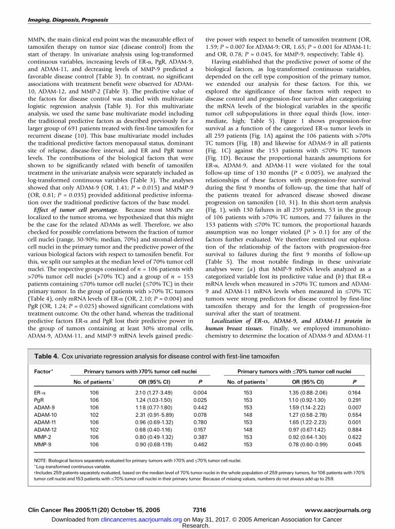
Effect of tumor cell percentage. Because most MMPs arelocalized to the tumor stroma, we hypothesized that this mightbe the case for the related ADAMs as well. Therefore, we alsochecked for possible correlations between the fraction of tumorcell nuclei (range, 30-90%; median, 70%) and stromal-derivedcell nuclei in the primary tumor and the predictive power of thevarious biological factors with respect to tamoxifen benefit. Forthis, we split our samples at the median level of 70% tumor cellnuclei. The respective groups consisted of n = 106 patients with>70% tumor cell nuclei (>70% TC) and a group of n = 153patients containing V70% tumor cell nuclei (V70% TC) in theirprimary tumor. In the group of patients with >70% TC tumors(Table 4), only mRNA levels of ER-a (OR, 2.10; P = 0.004) andPgR (OR, 1.24; P = 0.025) showed significant correlations withtreatment outcome. On the other hand, whereas the traditionalpredictive factors ER-a and PgR lost their predictive power inthe group of tumors containing at least 30% stromal cells,ADAM-9, ADAM-11, and MMP-9 mRNA levels gained predic-
tive power with respect to benefit of tamoxifen treatment (OR,1.59; P = 0.007 for ADAM-9; OR, 1.65; P = 0.001 for ADAM-11;and OR, 0.78; P = 0.045, for MMP-9, respectively; Table 4).
Having established that the predictive power of some of thebiological factors, as log-transformed continuous variables,depended on the cell type composition of the primary tumor,we extended our analysis for these factors. For this, weexplored the significance of these factors with respect todisease control and progression-free survival after categorizingthe mRNA levels of the biological variables in the specifictumor cell subpopulations in three equal thirds (low, inter-mediate, high; Table 5). Figure 1 shows progression-freesurvival as a function of the categorized ER-a tumor levels inall 259 patients (Fig. 1A) against the 106 patients with >70%TC tumors (Fig. 1B) and likewise for ADAM-9 in all patients(Fig. 1C) against the 153 patients with V70% TC tumors(Fig. 1D). Because the proportional hazards assumptions forER-a, ADAM-9, and ADAM-11 were violated for the totalfollow-up time of 130 months (P < 0.005), we analyzed therelationships of these factors with progression-free survivalduring the first 9 months of follow-up, the time that half ofthe patients treated for advanced disease showed diseaseprogression on tamoxifen (10, 31). In this short-term analysis(Fig. 1), with 130 failures in all 259 patients, 53 in the groupof 106 patients with >70% TC tumors, and 77 failures in the153 patients with V70% TC tumors, the proportional hazardsassumption was no longer violated (P > 0.1) for any of thefactors further evaluated. We therefore restricted our explora-tion of the relationship of the factors with progression-freesurvival to failures during the first 9 months of follow-up(Table 5). The most notable findings in these univariateanalyses were: (a) that MMP-9 mRNA levels analyzed as acategorized variable lost its predictive value and (b) that ER-amRNA levels when measured in >70% TC tumors and ADAM-9 and ADAM-11 mRNA levels when measured in V70% TCtumors were strong predictors for disease control by first-linetamoxifen therapy and for the length of progression-freesurvival after the start of treatment.
Localization of ER-a, ADAM-9, and ADAM-11 protein inhuman breast tissues. Finally, we employed immunohisto-chemistry to determine the location of ADAM-9 and ADAM-11
Table 4. Cox univariate regression analysis for disease control with first-line tamoxifen
Factor* Primary tumorswith >70% tumor cell nuclei Primary tumorswith V70% tumor cell nuclei
No. of patientsc OR (95% CI) P No. of patientsc OR (95% CI) P
ER-a 106 2.10 (1.27-3.49) 0.004 153 1.35 (0.88-2.06) 0.164PgR 106 1.24 (1.03-1.50) 0.025 153 1.10 (0.92-1.30) 0.291ADAM-9 106 1.18 (0.77-1.80) 0.442 153 1.59 (1.14-2.22) 0.007ADAM-10 102 2.31 (0.91-5.89) 0.078 148 1.27 (0.58-2.78) 0.554ADAM-11 106 0.96 (0.69-1.32) 0.780 153 1.65 (1.22-2.23) 0.001ADAM-12 102 0.68 (0.40-1.16) 0.157 148 0.97 (0.67-1.42) 0.884MMP-2 106 0.80 (0.49-1.32) 0.387 153 0.92 (0.64-1.30) 0.622MMP-9 106 0.90 (0.68-1.19) 0.462 153 0.78 (0.60-0.99) 0.045
NOTE: Biological factors separately evaluated for primary tumors with >70% and V70% tumor cell nuclei.*Log-transformed continuous variable.cIncludes 259 patients separately evaluated, based on the median level of 70% tumor nuclei in the whole population of 259 primary tumors, for106 patients with >70%tumor cell nuclei and153 patients with V70% tumor cell nuclei in their primary tumor. Because of missing values, numbers do not always add up to 259.
Imaging, Diagnosis, Prognosis
www.aacrjournals.orgClin Cancer Res 2005;11(20) October15, 2005 7316
Research. on May 31, 2017. © 2005 American Association for Cancerclincancerres.aacrjournals.org Downloaded from

protein in our human breast tumor tissues and compared thiswith the location of ER-a protein. Representative results areshown in Fig. 2 for staining of preexistent mammary glandtissue (Fig. 2A-D), carcinoma in situ components (Fig. 2E-H),and lobular breast carcinomas (Fig. 2I-L). Whereas ER-astaining is mainly localized to the nuclei of tumor cells,ADAM-9 and ADAM-11 are most commonly found in thecytoplasm and less commonly at the cell membrane. Immu-nohistochemical staining of ADAM-9 and ADAM-11 protein inhuman breast carcinomas yielded heterogeneous results withboth proteins found in tumor cells (Fig. 2J and K), adipocytes,smooth muscle cells of vessel walls, and the myoepithelial andluminal layers of nonneoplastic epithelium of the mammarygland (Fig. 2B and C).
Discussion
Endocrine therapy is the most common treatment in breastcancer patients with tumors that express ER-a and/or PgR.Even though the ER-a is the prime target for endocrine
therapy, the failure or success of this therapy is poorlyunderstood. Systemic endocrine therapy in patients withrecurrent disease at distant sites is merely palliative andaccomplishes a disease control in about 50% to 60% of thepatients. However, progression is inevitable in these patientsbecause of the occurrence of acquired therapy resistance. Froma biological point of view, first-line single-agent endocrinetherapy in patients with recurrent breast cancer is an excellentsetting to study response to therapy because it is less subject toprognostic influences unavoidably present when a similarstudy would be done in the adjuvant setting. In the presentstudy, the effect of endocrine therapy on size of the metastaticor the occurrence of new lesions were used as the mainclinical end point. We defined the type of response strictlybeforehand, and when there was any doubt, patients were notincluded in this study. The size of the metastases or theoccurrence of new lesions is an objective measure of treatmenteffect. However, because of the retrospective nature of ourstudy, the differentiation between partial remission and nochange was difficult to assess, especially in patients with bone
Table 5. Cox univariate regression analysis of biological factors in primary tumors with >70% or V70% tumor cellnuclei for disease control with first-line tamoxifen and progression-free survival (restricted to the first 9 months) afterstart of tamoxifen therapy
Factor andlevels*
Tumor cells (%)c No. ofpatientsb
Disease control (%)b Disease control Progression-free survival
OR (95% CI) P Hazard ratio (95% CI) P
ER-a(�10�0) >70<5.0 36 50 1 15.0-13.6 35 57 1.33 (0.52-3.40) 1.19 (0.66-2.14)>13.6 35 86 6.00 (1.90-18.96) 0.003 0.28 (0.12-0.64) <0.001
PgR(�10�0) >70<0.2 36 53 1 10.2-1.8 35 63 1.51 (0.59-3.91) 0.72 (0.38-1.36)>1.8 35 77 3.02 (1.08-8.42) 0.094 0.58 (0.30-1.12) 0.253
ADAM-9(�10�0) V70<2.9 51 37 1 12.9-7.6 51 69 3.68 (1.62-8.36) 0.36 (0.21-0.64)>7.6 51 67 3.37 (1.49-7.60) 0.002 0.47 (0.28-0.80) <0.001
ADAM-11(�10�3) V70<1.7 51 45 1 11.7-5.0 51 49 1.17 (0.54-2.55) 0.84 (0.50-1.40)>5.0 51 78 4.43 (1.86-10.52) <0.001 0.48 (0.27-0.85) 0.029
MMP-9(�10�0) V70<0.3 51 67 1 10.3-1.0 51 51 0.52 (0.23-1.16) 1.45 (0.83-2.51)>1.0 51 55 0.61 (0.27-1.36) 0.245 1.33 (0.76-2.33) 0.388
NOTE: Due to different assay conditions and amplicon lengths, absolute values can only be compared within a gene assay.*Three equal thirds were used to categorize the variable in the specific tumor cell subpopulation in low, intermediate, and high.cBasedon themedian level of 70% tumor cell nuclei in thewhole populationof 259 primary tumors, separately evaluated for106 patientswith >70% tumor cell nuclei and153 patients with V70% tumor cell nuclei in their primary tumor.bNumber of patients entered into the study and corresponding disease control data are given for the low, intermediate, and high mRNA expression levels in the specifictumor cell subpopulation.
ADAMs and Response toTamoxifen
www.aacrjournals.org Clin Cancer Res 2005;11(20) October15, 20057317
Research. on May 31, 2017. © 2005 American Association for Cancerclincancerres.aacrjournals.org Downloaded from

metastasis (60%). In our study, the progression-free survivalof patients with stable disease (no change >6 months) wascomparable with the progression-free survival of patients withpartial remission and could therefore be considered asresponders. This is in agreement with a previously publishedprospective study which also reported that objective benefitwas not always easy to assess and in which prolonged stabledisease was categorized as response (6).
In this study, ER and PgR were determined in cytosols bybiochemical methods and the cutoff used to classify tumors asER- or PgR-positive was 10 fmol/mg cytosolic protein. Thesedata correlated significantly with ER and PgR mRNA expressionlevels. However, although these quantitative procedures are themost accurate methods, it is not currently the most widely usedmethod to evaluate hormonal receptor status in breast cancer.In fact, immunohistochemistry is nowadays more commonlyused for routine ER and PgR measurements. Because this studyshows a possible application for current clinical practice, wecompared the biochemical and immunohistochemical meth-ods in a randomly selected subgroup of patients. In agreementwith a previously published study in which ligand binding
assay and immunohistochemistry were compared in predictingresponse to tamoxifen in 205 patients with ER-positivemetastatic breast cancer (32), ER and PgR levels also showedcomparable differences in response rates in our study, whetherdefined by mRNA, by biochemical methods, or by immuno-histochemistry.
The main findings of our study are that ADAM-9 and ADAM-11 differentially from ER predict the type of response totamoxifen treatment in patients with recurrent breast cancerand that the fraction of tumor cells and stromal elements areimportant in this respect. The actual ER level in the ER-positivetumors (>10 fmol/mg cytosol protein) containing >30%stromal elements did not further contribute to the rate ofresponse. This finding supports the results of a previous reportthat showed an association between ER level and the volumefraction of actual cancer cells present in the tumors (33).Therefore, it was advised that, when quantitative ER levels areused to predict the response of tumors to hormonal therapy,the cellularity of tumors should be taken into consideration.We followed this approach in our study by discriminatingbetween tumors with >70% tumor cells and tumors with 30%
Fig. 1. Kaplan-Meier curves of progression-free survival with log-rank testing restricted to the first 9 months of follow-up for patients with advanced disease treated withfirst-line tamoxifen.The mRNA levels divided in three equal thirds given inTable 5 for ER-a (A + B) and ADAM-9 (C + D) were assessed in tumors before (A + C) and after(B + C) dichotomization on the basis of the median level of 70% tumor cell nuclei in the total group of 259 patients. Numbers below the x-axis show the patients at risk at theindicated time points.
Imaging, Diagnosis, Prognosis
www.aacrjournals.orgClin Cancer Res 2005;11(20) October15, 2005 7318
Research. on May 31, 2017. © 2005 American Association for Cancerclincancerres.aacrjournals.org Downloaded from

to 70% tumor cells. Our results show that for tumors with arelatively low percentage of epithelial tumor cells, a marker setincluding ADAM-9 and ADAM-11 may have potential to assessthe efficacy of tamoxifen therapy.
Of the ADAMs and MMPs studied, all, except ADAM-11, werereadily detected by real-time PCR in all samples. The absence ofdetectable ADAM-11 mRNA levels in 29% of our primary breasttumors is most likely a reflection of the loss of heterozygosityon chromosome 17q21, where ADAM-11 is located (13), asdescribed to be the case for 30% of the tumors (11). Patterns ofcopy number gains and losses define breast tumors withdistinct clinicopathologic features and patient prognosis (34).Several studies have already shown that ERBB2 amplification isassociated with a shorter disease-free and overall survival in thesubgroup of patients receiving adjuvant tamoxifen therapywhen compared with the untreated group (35–37). However,whereas ERBB2 is located on cytoband 17q12, a region of copy
gain, ADAM-11 is located on cytoband 17q21, a region of copyloss. Our finding that low tumor ADAM-11 mRNA levels areassociated with poor efficacy of tamoxifen treatment supportsthe hypothesis that ADAM-11 is a candidate tumor suppressorgene for human breast cancer (13, 14), and extends its role as acandidate tumor suppressor gene to a candidate tamoxifensusceptibility gene.
Our study shows that ADAM-9 and ADAM-11 mRNA levelsare especially informative with respect to tamoxifen treatmentoutcome in tumors containing a relatively large proportion ofstromal cells. In agreement with a previously published studydescribing the expression of ADAM-9, ADAM-10, ADAM-12,ADAM-15, and ADAM-17 in breast cancer specimens (38),immunohistochemical staining of ADAM-9 and ADAM-11protein in human breast carcinomas yielded heterogeneousresults with both proteins found in tumor cells, adipocytes,nonneoplastic epithelium of the mammary gland, and smooth
Fig. 2. Immunohistochemical localizationof ER-a, ADAM-9, and ADAM-11in breastcancer tissue. A-D,�20 magnification.Preexistent mammary gland tissueexpressing occasional positive nuclearstaining for ER-a (A), abundant staining ofthe myoepithelial layer and weak staining ofthe luminal layer forADAM-9 (B), and weakstaining of both layers forADAM-11 (C).E-H,�40 magnification. Carcinoma in situcomponent expressing positive nuclearstaining for ER-a (E), intermediatecytoplasmic staining forADAM-9 (F) andabundant cytoplasmic andmembranestaining forADAM-11 (G). I-L ,�40magnification. Lobular carcinomaexpressingpositivenuclear staining for ER-a(I), weak cytoplasmic staining forADAM-9(J), and medium cytoplasmic staining forADAM-11 (K).The specificity ofimmunostaining was controlled usingnormal goat andmouse IgGandby omittingthe primary antibodies (negative controls,D, H, and L).
ADAMs and Response toTamoxifen
www.aacrjournals.org Clin Cancer Res 2005;11(20) October15, 20057319
Research. on May 31, 2017. © 2005 American Association for Cancerclincancerres.aacrjournals.org Downloaded from

muscle cells of vessel walls. The question of how ADAM-11and ADAM-9, either stromal or tumor cell–derived, mightprevent the development of tamoxifen resistance remains tobe solved. Because proteases such as urokinase-type plasmin-ogen activator (9, 10) and MMP-2 (39) have been shown tobe related to tamoxifen resistance, we hypothesized thatspecific ADAMs might also be related to tamoxifen resistance.We found that high levels of ADAM-9 and ADAM-11 mRNAwere related to a better response rate. This is in contrast withthe findings for urokinase-type plasminogen activator (10),showing high levels to be associated with poor benefit oftamoxifen treatment in recurrent breast cancer, and for MMP-2 (39), showing that high levels predicted failure to adjuvantantiestrogen therapy. This suggests that ADAM-9 and ADAM-11 function differently from urokinase-type plasminogenactivator and MMP-2, and that it is therefore perhaps notthe protease function of the ADAMs that is important in theprevention or delay of tamoxifen resistance. Increasingevidence indicates that abnormalities occurring in growthfactor signaling pathways, as currently well-documented forepidermal growth factor receptor (ERBB1) and ERBB2 (HER2/neu), could dramatically influence steroid hormone actionand may be critical for anti–hormonal-resistant breast cancercell growth (7, 8, 36, 40–43). From this point of view, onemight expect factors that target growth factor signalingpathways are potentially able to prevent the development oftamoxifen resistance. Many intercellular signaling moleculesare membrane-anchored proteins, which are proteolyticallyprocessed after becoming membrane-bound, to liberate theirextracellular domains (ectodomain shedding). Genetic andbiochemical studies have shown that some ADAMs participatein these events (3). Therefore, it is perhaps the ectodomainshedding function of the ADAMs that plays a role in theprevention of tamoxifen resistance. Furthermore, the disinte-grin domain of ADAM-9 can function as an adhesion
molecule by interacting with an a(v)h(5) integrin (44), thuslimiting the metastatic potential of the cell.
In summary, our study shows that patients with primarytumors exhibiting a high percentage of tumor cell nuclei overstromal cells combined with high levels of ER-a have a goodchance to benefit from tamoxifen therapy. For patients withtumors displaying z30% stromal components intermingledwith epithelial tumor cells, the additional assessment of tumormRNA levels of ADAM-9 and ADAM-11 could be helpful torefine treatment strategies for these patients. However, takinginto account that only patients with ER-positive primarytumors entered this study, this may only apply to patientswith ER-positive primary tumors. Further studies are requiredto verify whether the results of our study can be adapted to fitall patients, irrespective of the ER status of the primary tumor.Based on recent advances in breast cancer management,endocrine therapy with aromatase inhibitors may become thetreatment of choice for postmenopausal women (45). Becauseboth aromatase inhibitors and tamoxifen aim to deprive the ERfrom estrogens, it would be interesting to learn whether ADAM-9 and ADAM-11 could also be linked to disease control ofaromatase inhibitors. In addition, as the majority of patientsreceive adjuvant treatment today, it will be important to learnwhether ADAM-9 and ADAM-11 could also be informative fordetermining the outcome of breast cancer patients treated withadjuvant endocrine therapy.
Acknowledgments
We thankMaxime Look for her expert support with clinical data analysis and Irisvan Staveren andMaaike Kiel forhelpingwith theRNA isolation.We especially thankthe surgeons, pathologists, and internists of the St. Clara Hospital, Ikazia Hospital,St. Fransiscus Gasthuis, Erasmus MC at Rotterdam, and Ruwaard van Putten Hos-pital at Spijkenisse for their assistance in collecting the tumor tissues and patient’sclinical follow-up data.
References1.Wolfsberg TG, Primakoff P, Myles DG,White JM.ADAM, a novel family of membrane proteins containingA disintegrin and metalloprotease domain: multipoten-tial functions in cell-cell and cell-matrix interactions.JCell Biol1995;131:275^8.
2. Schlondorff J, Blobel CP. Metalloprotease-disinte-grins: modular proteins capable of promoting cell-cellinteractions and triggering signals by protein-ectodo-main shedding. JCell Sci1999;112:3603^17.
3. Duffy MJ, Lynn DJ, Lloyd AT, O’Shea CM. TheADAMs family of proteins: from basic studies to po-tential clinical applications. Thromb Haemost 2003;89:622^31.
4.White JM. ADAMs: modulators of cell-cell and cell-matrix interactions. Curr Opin Cell Biol 2003;15:598^606.
5. Seals DF, Courtneidge SA. The ADAMs family ofmetalloproteases: multidomain proteins with multiplefunctions. Genes Dev 2003;17:7^30.
6. Ravdin PM, Green S, Dorr TM, et al. Prognostic sig-nificance of progesterone receptor levels in estrogenreceptor-positive patients with metastatic breast can-cer treated with tamoxifen: results of a prospectiveSouthwest Oncology Group study. JClin Oncol1992;10:1284^91.
7. Nicholson RI, GeeJM, KnowldenJ, et al.The biologyof antihormone failure in breast cancer. Breast CancerResTreat 2003;80 Suppl1:29^34; discussion S5.
8. Hayes DF.Tamoxifen: Dr. Jekyll andMr. Hyde? JNatlCancer Inst 2004;96:895^7.
9. Foekens JA, Look MP, Peters HA, et al. Urokinase-
type plasminogen activator and its inhibitor PAI-1:predictors of poor response to tamoxifen therapy inrecurrent breast cancer. J Natl Cancer Inst 1995;87:751^6.
10.Meijer-van Gelder ME, Look MP, Peters HA, etal. Urokinase-type plasminogen activator systemin breast cancer: association with tamoxifen ther-apy in recurrent disease. Cancer Res 2004;64:4563^8.
11. DeMarchis L, Cropp C, Sheng ZM, Bargo S,Callahan R. Candidate target genes for loss of hetero-zygosity on human chromosome 17q21. Br J Cancer2004;90:2384^9.
12. Orsetti B, Nugoli M, Cervera N, et al. Genomic andexpression profiling of chromosome 17 in breast can-cer reveals complex patterns of alterations and novelcandidate genes. Cancer Res 2004;64:6453^60.
13. Emi M, Katagiri T, Harada Y, et al. A novel metallo-protease/disintegrin-like gene at17q21.3 is somaticallyrearranged in two primary breast cancers. Nat Genet1993;5:151^7.
14. Katagiri T, Harada Y, Emi M, Nakamura Y. Humanmetalloprotease/disintegrin-like (MDC) gene: exon-intron organization and alternative splicing. CytogenetCell Genet1995;68:39^44.
15. O’Shea C, McKie N, Buggy Y, et al. Expression ofADAM-9 mRNA and protein in human breast cancer.IntJCancer 2003;105:754^61.
16. Azzam HS, Arand G, Lippman ME,Thompson EW.Association of MMP-2 activation potential with meta-static progression in human breast cancer cell lines in-
dependent of MMP-2 production. J Natl Cancer Inst1993;85:1758^64.
17.Heppner KJ,Matrisian LM, JensenRA, Rodgers WH.Expression of most matrix metalloproteinase familymembers in breast cancer represents a tumor-inducedhost response. AmJPathol1996;149:273^82.
18. Duffy MJ, Maguire TM, Hill A, McDermott E,O’Higgins N. Metalloproteinases: role in breast carci-nogenesis, invasion and metastasis. Breast CancerRes 2000;2:252^7.
19. Ranuncolo SM, Armanasco E, Cresta C, Bal De KierJoffe E, Puricelli L. Plasma MMP-9 (92 kDa-MMP)activity is useful in the follow-up and in the assess-ment of prognosis in breast cancer patients. Int JCancer 2003;106:745^51.
20. Tester AM,Waltham M, Oh SJ, et al. Pro-matrixmetalloproteinase-2 transfection increases orthotopicprimary growth and experimental metastasis of MDA-MB-231 human breast cancer cells in nude mice.Cancer Res 2004;64:652^8.
21. JonesJL, Glynn P,Walker RA. ExpressionofMMP-2and MMP-9, their inhibitors, and the activator MT1-MMP in primary breast carcinomas. J Pathol 1999;189:161^8.
22.Martens JW, Nimmrich I, Koenig T, et al. Associationof DNAmethylationof phosphoserine aminotransferasewith response to endocrine therapy in patients withrecurrent breast cancer. Cancer Res 2005;65:4101^17.
23. Hayward JL, Carbone PP, Heuson JC, et al. Assess-ment of response to therapy in advancedbreast cancer:a project of the Programme on Clinical Oncology of
Imaging, Diagnosis, Prognosis
www.aacrjournals.orgClin Cancer Res 2005;11(20) October15, 2005 7320
Research. on May 31, 2017. © 2005 American Association for Cancerclincancerres.aacrjournals.org Downloaded from

ADAMs and Response toTamoxifen
www.aacrjournals.org Clin Cancer Res 2005;11(20) October15, 20057321
the International Union Against Cancer, Geneva, Swit-zerland. Cancer1977;39:1289^94.
24. European Organization for Research and Treatmentof Cancer, Breast Cancer Cooperative Group. Manualfor clinical research and treatment in breast cancer.Excerpta Medica. Almere, the Netherlands; 2000.p.116^7.
25. Ravdin PM, Burris HA III, Cook G, et al. Phase II trialof docetaxel in advanced anthracycline-resistant oranthracenedione-resistant breast cancer. JClin Oncol1995;13:2879^85.
26. FoekensJA, Portengen H, Look MP, et al. Relation-ship of PS2 with response to tamoxifen therapy inpatients with recurrent breast cancer. Br J Cancer1994;70:1217^23.
27. RobertsonJF,Willsher PC, Cheung KL, Blamey RW.The clinical relevance of static disease (no change)category for 6 months on endocrine therapy inpatients with breast cancer. Eur J Cancer 1997;33:1774^9.
28. Foekens JA, Portengen H, van PuttenWL, et al.Prognostic value of estrogen and progesterone recep-tors measured by enzyme immunoassays in humanbreast tumor cytosols. Cancer Res1989;49:5823^8.
29. European Organization for Research and Treatmentof Cancer Breast Cancer Cooperative group. Revisionof the standards for the assessment of hormonereceptors in human breast cancer; report of the sec-ond E.O.R.T.C. Workshop, held on 16 ^ 17 March,1979, in theNetherlandsCancer Institute. EurJCancer1980;16:1513^5.
30. Grambsch P, Louis TA, Bostick RM, et al. Statisticalanalysis of proliferative index data in clinical trials. StatMed1994;13:1619^34.
31. Bonneterre J, Thurlimann B, Robertson JF, et al.
Anastrozole versus tamoxifen as first-line therapy foradvanced breast cancer in 668 postmenopausalwomen: results of theTamoxifen orArimidexRandom-ized Group Efficacy andTolerability study. JClin Oncol2000;18:3748^57.
32. Elledge RM, Green S, Pugh R, et al. Estrogen recep-tor (ER) and progesterone receptor (PgR), by ligand-binding assay compared with ER, PgR and pS2, byimmuno-histochemistry in predicting response totamoxifen in metastatic breast cancer: a SouthwestOncology Group Study. IntJCancer 2000;89:111^7.
33. HelleM, Helin H, IsolaJ, Krohn K. Oestrogen recep-tor content and cancer cell/stroma ratio in mammarycarcinoma. APMIS1988;96:1140^2.
34. Rennstam K, Ahlstedt-Soini M, Baldetorp B, et al.Patterns of chromosomal imbalances defines sub-groups of breast cancer with distinct clinical featuresand prognosis. A study of 305 tumors by compara-tive genomic hybridization. Cancer Res 2003;63:8861^8.
35. Borg A, Baldetorp B, FernoM, et al. ERBB2 amplifi-cation is associated with tamoxifen resistance in ste-roid-receptor positive breast cancer. Cancer Lett 1994;81:137^44.
36. De Placido S, Carlomagno C, De Laurentiis M,BiancoAR. c-erbB2 expression predicts tamoxifen ef-ficacy in breast cancer patients. Breast Cancer ResTreat1998;52:55^64.
37. Lipton A, Ali SM, Leitzel K, et al. Serum HER-2/neuand response to the aromatase inhibitor letrozole ver-sus tamoxifen. JClin Oncol 2003;21:1967^72.
38. Lendeckel U, Kohl J, Arndt M., Carl-McGrath S,Donat H, Rocken C. Increased expression of ADAMfamily members inhumanbreast cancer andbreast can-cer cell lines. JCancer Res Clin Oncol 2005;131:41^8.
39. Talvensaari-Mattila A, Paakko P, Blanco-SequeirosG,Turpeenniemi-HujanenT. Matrix metalloproteinase-2 (MMP-2) is associated with the risk for a relapse inpostmenopausal patients with node-positive breastcarcinoma treated with antiestrogen adjuvant therapy.Breast Cancer ResTreat 2001;65:55^61.
40. Newby JC, Johnston SR, Smith IE, Dowsett M.Expression of epidermal growth factor receptor andc-erbB2 during the development of tamoxifen resis-tance in human breast cancer. Clin Cancer Res 1997;3:1643^51.
41. KurokawaH, Arteaga CL. ErbB (HER) receptors canabrogate antiestrogen action in human breast cancerby multiple signaling mechanisms. Clin Cancer Res2003;9:511^5S.
42. Schiff R, Massarweh SA, Shou J, et al. Cross-talk between estrogen receptor and growth factorpathways as a molecular target for overcoming en-docrine resistance. Clin Cancer Res 2004;10:331^6S.
43. Shou J, Massarweh S, Osborne CK, et al.Mechanisms of tamoxifen resistance: increased es-trogen receptor-HER2/neu cross-talk in ER/HER2-positive breast cancer. J Natl Cancer Inst 2004;96:926^35.
44. ZhouM, Graham R, Russell G, Croucher PI. MDC-9(ADAM-9/Meltrin g) functions as an adhesion mole-cule by binding the a(v)h(5) integrin. Biochem Bio-phys Res Commun 2001;280:574^80.
45.Winer EP, Hudis C, Burstein HJ, et al. AmericanSociety of Clinical OncologyTechnology, Assessmenton the use of aromatase inhibitors as adjuvant therapyfor postmenopausal women with hormone receptor-positive breast cancer: status report 2004. J ClinOncol 2005;23:619^29.
Research. on May 31, 2017. © 2005 American Association for Cancerclincancerres.aacrjournals.org Downloaded from

2005;11:7311-7321. Clin Cancer Res Anieta M. Sieuwerts, Marion E. Meijer-van Gelder, Mieke Timmermans, et al. StudyPatients with Recurrent Breast Cancer: a RetrospectiveReceptor Predict Response to Tamoxifen Treatment in How ADAM-9 and ADAM-11 Differentially From Estrogen
Updated version
http://clincancerres.aacrjournals.org/content/11/20/7311
Access the most recent version of this article at:
Cited articles
http://clincancerres.aacrjournals.org/content/11/20/7311.full.html#ref-list-1
This article cites 40 articles, 19 of which you can access for free at:
Citing articles
/content/11/20/7311.full.html#related-urls
This article has been cited by 15 HighWire-hosted articles. Access the articles at:
E-mail alerts related to this article or journal.Sign up to receive free email-alerts
Subscriptions
Reprints and
To order reprints of this article or to subscribe to the journal, contact the AACR Publications
Permissions
To request permission to re-use all or part of this article, contact the AACR Publications
Research. on May 31, 2017. © 2005 American Association for Cancerclincancerres.aacrjournals.org Downloaded from