Thermal and Mechanical Hazards of Nitrocellulose and its ...
Houghton
1
Supporting Online Material Materials and Methods
Bone marrow transplantation.
Bone marrow was isolated from the femurs and tibias of 6-8 week old
C57BL/6JGtrosa26 (ROSA26) or C57BL/6-TgN [ACTbEGFP] (GFP) mice. Total bone
marrow was washed, triturated using a 20 gauge needle and passed through a 40 µm
nylon mesh cell strainer (Becton Dickson, Franklin Lakes, NJ) to produce a single cell
suspension in PBS. Recipient C57BL/6J mice were irradiated with 900 rads from a
cesium 137-gamma cell irradiator, reconstituted with 3 x 10⁶ donor marrow cells via a
single tail vein injection, and used for experiments after 4 weeks of recovery. The overall
level of engraftment was 60-80% as assessed by analysis of beta-galactosidase or GFP in
peripheral leukocytes. Non-transplanted C57BL/6 mice and C57BL/6 transplanted with
C57BL/6 marrow from liter mates served as controls for all injury models. C57BL/6
mice transplanted with marrow from ROSA26 or GFP transgenic mice without further
intervention served as controls for baseline engraftment. All mice received a single 1
mg/kg BrdU intraperitoneal injection prior to euthanasia. All animal work was
performed at the University of Massachusetts Medical School under approval of the
University of Massachusetts Institutional Animal Care and Use Committee.
Induction of acute gastric ulceration.
Under anesthesia, a left lateral incision was made and the stomach exteriorized. Acute
gastric ulceration was induced by application of liquid nitrogen to the serosal surface for
Houghton
2
15 seconds or intramucosal injection of 20 µl 20% acetic acid to the oxyntic/antral
border. The stomach was gently replaced and the wound closed with surgical clips. Mice
were euthanized and tissues examined at 4, 10 or 20 days.
Induction of non-inflammatory, reversible oxyntic atrophy.
DMP777 ([S-(R*,S*)]-N-{1,3-benzodioxol-5-yl)butyl]-3,3diethyl-2-[4-[(4-methyl-1-
piperazinly)carbonyl] phenoxy] -4-oxo—1-azetidinecarbocamide) was formulated as a
suspension in 0.5% methylcellulose and administered 300 mg/kg/day orally by gavage
for 7 days and mice euthanized at 7, 9 or 210 days.
Helicobacter infection.
Helicobacter felis (strain 49179) was obtained from the American Type Cell Culture
(Rockville, MD), grown as recommended and bacteria enumerated as previously
described (1). Mice were infected by oral gavage with 1 x 10⁷ colony forming units
every other day for 3 days, and euthanized at intervals from 3 weeks to 16 months.
Tissue processing.
Determination of enzyme (X-gal) activity.
After intracardiac perfusion (4% paraformaldehyde, 2% glutaraldehyde in 0.1 M
Sorensen’s phosphate buffer (pH 7.4) with 2 mM MgCl2 and 5 mM EGTA), stomachs
were removed, opened along the greater curvature, washed, and linear longitudinal
sections from the squamocolumnar junction through the pylorus collected. Tissue was
embedded in OCT compound (SAKURA Torrance USA), snap frozen, and sectioned on

Houghton
3
a cryostat for enzyme histochemistry. Frozen sections (10µm) were washed in
Sorensen’s buffer containing 0.01 % sodium deoxycholate and 0.02 % Nonidet P-40 and
incubated for 4 hours at 37°C in a 0.1 % X-gal solution (4 % 4-chloro-5-bromo-3-
indolyl-D-galactopyranoside (X-gal) dissolved in dimethylformamide, 5 mM K3Fe
(CN)6, 5 mM K4Fe (CN)6.6H2O in 0.1 M Sorensen’s phosphate buffer) and
counterstained with nuclear fast red. Additional sections had nuclear fast red omitted,
and cells were instead labeled using antibodies directed against TFF2 (a kind gift from N.
A. Wright),
Preparation of tissue for immunohistochemistry.
Mice were euthanized by CO2 inhalation, gastric tissue was removed, washed, sectioned
as described above and fixed in 10% neutral-buffered formalin, 100% ethanol, or Prefer
(Anatech, Ltd., Battle Creek, MI), followed by standard histologic processing. Sections
were stained using routine H&E. For immunohistochemistry, sections labeled using
antibodies directed against BrdU (Zymed, San Francisco, CA), or bacterial beta-
galactosidase (Promega, Madison, WI) or GFP (Abcam, Cambridge, UK). For mouse-
on-mouse immunostaining the ARK kit (DAKO, Carpinetera, CA) was employed. For
dual fluorescence immunohistochemistry, cell phenotype markers were indirectly labeled
with streptavidin-fluorescein using antibodies directed towards pan-cytokeratin
(AE1/AE3; DAKO) or CD45 (leukocyte common antigen, Ly-5, BD Biosciences, San
Diego, CA) followed by a biotin block and serial application of anti-beta-galactosidase
antibody labeled indirectly with streptavidin-Cy3. Sections were mounted with anti-fade
Vectashield with DAPI (Vector Laboratories, Burlingame, CA), fields viewed and

Houghton
4
captured with the Zeiss Axiopath system (Thornwood, NY), and serial 3-color images
were overlaid using Photoshop 6.0 (Adobe Systems, San Jose, CA).
Co-localization of Y-chromosome and cytokeratin in tissue sections.
Y chromosome was detected using only the components for Y-chromosome of the Dual
Color Detection kit (ID BioLabs- Y-chromosome FITC-label), followed by mouse anti-
cytokeratin complex antibody (AE1/AE2; DAKO Corporation, Carpinteria, CA) at 1/50
dilution at 37°C for 20 minutes, followed by goat anti-mouse / Texas Red conjugate
(Molecular Probes, Eugene, OR) at 1/100 dilution and incubated at 37°C for 20 minutes.
Slides were mounted with antifade, cover slipped and analyzed.
Laser capture microdissection of beta-galactosidase positive glands
Frozen sections were prepared and stained with X-gal as outlined above. Individual
gastric cells within positive glands, individual lymphocytes within an area of submucosal
infiltrate or negative glands (control) were captured using the Laser Capture Microscope
PixCell II, (Arcturus Engineering Inc., Mountain View, CA, U.S.A). Twenty to 30 cells
were captured for each area of interest. DNA was extracted using the PicoPure DNA
extraction kit (Arcturus Engineering Inc., Mountain View, CA, USA ) and GAPDH (210
bp) (2) or 140 bp of the LacZ/NEO fusion gene sequence amplified as follows:
Annealing temperature 56°, forward primer 5′-CGGGCTGCAGCCAATATGGGATCG-
3′, reverse primer 5′-GCCGGAACACGGCGGCATCAGAGC-3′ Using HotStarTaq
Master Mix (Qiagen, Valencia, CA, USA). Primer sequence for LacZ/NEO was a kind
gift from D.K. Kotton.
Houghton
5
Single cell preparation for FACS analysis and FISH
Stomachs were removed, opened and extensively washed. A small longitudinal section
from the squamocolumnar junction through the antrum was processed as outlined above
with standard H&E, anti-beta galactosidase or cytokeratin IHC. The fundic mucosa was
gently scraped free from the serosa, minced and digested for 2 hours in 1mM EDTA, 2%
BSA and 0.1% pronase in PBS at 37° and filtered through a 40 µm nylon mesh strainer.
For determination of DNA content, cells were fixed in 70% ethanol for 48 hours, stained
with PI, and DNA content determined by FACS. For X and Y chromosome
determination, single cell preparations from stomachs of sex-mismatched, GFP marrow
transplanted mice were prepared as detailed above. Cells were washed twice in PBS on
ice, resuspended in 0.5 ml of staining buffer (2% FBS, 0.01% sodium azide in PBS) and
stained with anti-CD45-PE Ab (BD Pharmingen) according to manufacturer protocol.
FACS sorted cells which were GFP+ and CD45- , GFP+ and CD45+, GFP- and CD45+
and GFP- and CD45- were collected. An aliquot of each was prepared and analyzed for
DNA content as described above and analyzed using WinMD2.8 software. For
cytokeratin immunocytochemistry, an aliquot of cells were spun onto slides (Thermo
Shandon) at a speed of 500 rpm for 5 minutes at a density of 5,000 cells per slide, fixed
in 4% paraformaldehyde pH of 7.4 for 1 hour and incubated with the primary Ab for
AE1/AE2 keratin complex (DAKO Corporation, Carpinteria, CA) at a dilution of 1/50
using the Animal Research kit (DAKO corporation, Carpinteria, CA) followed by
incubation with diaminobenzidine/hydrogen peroxidase as chromogen-substrate and
counterstained with hematoxylin. X- and Y-chromosomes were detected using the Dual

Houghton
6
Color Detection kit (ID BioLabs) according to the manufacturer’s protocol (DAPI for
nuclear staining, FITC for Y chromosomes and Texas Red for X chromosomes) and
immediately viewed with a fluorescent microscope (Olympus, Japan).
Western Blot analysis.
Gastric tissues from long term infected or age matched non-transplanted control mice
were homogenized in 0.3-0.5 ml ice-cold lysis buffer containing 8mmol/L Na2HPO4,
3mmol/L NaH2PO4 and EDTA-free protease inhibitor (Roche Applied Science,
Indianapolis, IN), and sonicated. Protein concentration was determined (Bio-Rad
Laboratories, Hercules, CA) and 250 µg of protein resuspended in loading buffer (0.1M
Tris-HCl, pH6.8, 0.2M DTT, 4% SDS, 0.2% bromophenol blue and 20% glycine),
denatured by boiling for 5 minutes, and loaded onto 12% SDS-polyacrylamide separating
gel. After electrophoresis, proteins were transferred to nitrocellulose membrane, blocked
with 5% non-fat dry milk, washed, incubated with primary antibody against SCF-1 (Santa
Cruz Biotechnology, Santa Cruz, CA ) or SDF-1 (R&D Systems, Minneapolis, MN),
washed and incubated with secondary horseradish peroxidase-conjugated antibody
(Amersham Biosciences, Piscataway, NJ). The immune complexes were detected by an
enhanced chemiluminescence system (Amersham Piscataway NJ.).
Gastric mucosal cell culture
The entire stomach was removed from C57BL/6 mice, opened along the greater curvature
and washed extensively in sterile PBS, 20% FCS, supplemented with
penicillin/streptomycin and amphoteracin. The fundic mucosa was scraped free from the
Houghton
7
serosa using a sterile scalpel, minced, manually disaggregated using a pipette, and the cell
suspension equally divided into the bottom wells of the Transwell™ culture plate.
Medium was changed after 24 hours to remove dead cells.
Bone marrow culture
Total marrow was collected as described above into PBS containing
penicillin/streptomycin 1% and 5% fetal calf serum. RBCs were lysed. Bone marrow
was depleted of differentiated cells using an antibody cocktail of TER, YW25.12.7,
MAC-1, GR-1, LYT-2, L3T4, B220 and separated using Dynal™ magnetic bead. Depleted
bone marrow was stained with rhodamine and Hoechst dye and a lineage depleted,
rhodamine dull, Hoechst dull ( lin-RhodullHodull) population isolated as the HSC
population (3) using the Cytomation MoFlo (Dakocytomation, Carpenteria CA).
Mesenchymal stem cells were isolated by culturing whole marrow in complete medium
(DMEM, FBS 10%, L-glutamine 1%, penicillin/streptomycin 1%, sodium pyruvate 1%,
and nonessential amino acids 1%) for 3 days, discarding non-adherent cells, and
retaining adherent cells as MSC. HSC or MSC (2000 cells) were plated into the upper
well of a 0.4µm pore 6.5 mm Transwell culture dish with polycarbonate membrane
(Corning Costar Corporation, Cambridge MA). The bottom dish contained either
complete medium or complete medium containing gastric mucosa (see above). Cells
were cultured at 37°, 5% CO2 for 24 or 48 hours. RNA was isolated using RNeasy
Protect Mini kit, and reverse transcribed using equivalent amounts of RNA using
Omniscript Reverse Transcriptase and PCR with HotStarTaq Master Mix kit as per
manufacturers instructions (Qiagen, Valencia, CA, USA). Primer sequences were as
Houghton
8
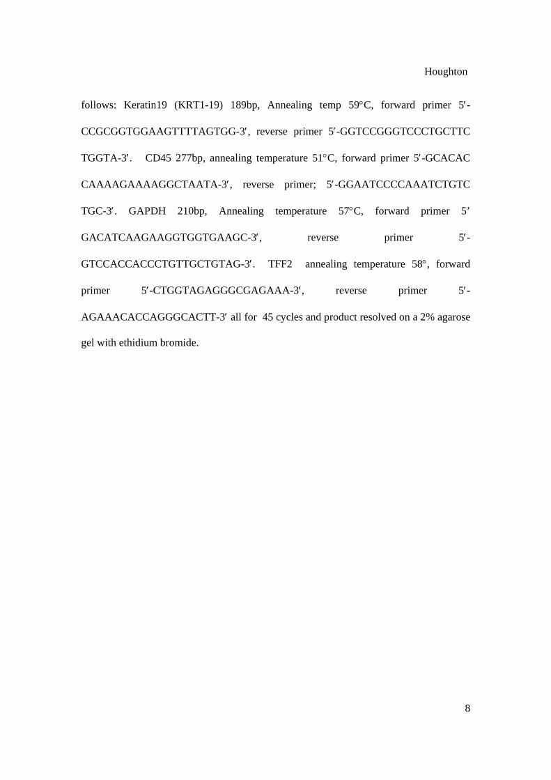
follows: Keratin19 (KRT1-19) 189bp, Annealing temp 59°C, forward primer 5′-
CCGCGGTGGAAGTTTTAGTGG-3′, reverse primer 5′-GGTCCGGGTCCCTGCTTC
TGGTA-3′. CD45 277bp, annealing temperature 51°C, forward primer 5′-GCACAC
CAAAAGAAAAGGCTAATA-3′, reverse primer; 5′-GGAATCCCCAAATCTGTC
TGC-3′. GAPDH 210bp, Annealing temperature 57°C, forward primer 5’
GACATCAAGAAGGTGGTGAAGC-3′, reverse primer 5′-
GTCCACCACCCTGTTGCTGTAG-3′. TFF2 annealing temperature 58°, forward
primer 5′-CTGGTAGAGGGCGAGAAA-3′, reverse primer 5′-
AGAAACACCAGGGCACTT-3′ all for 45 cycles and product resolved on a 2% agarose
gel with ethidium bromide.
Houghton
9
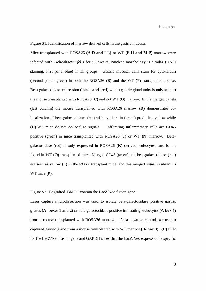
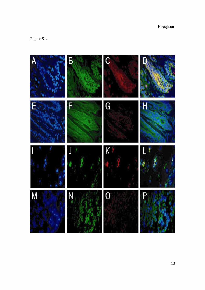
Figure S1. Identification of marrow derived cells in the gastric mucosa.
Mice transplanted with ROSA26 (A-D and I-L) or WT (E-H and M-P) marrow were
infected with Helicobacter felis for 52 weeks. Nuclear morphology is similar (DAPI
staining, first panel-blue) in all groups. Gastric mucosal cells stain for cytokeratin
(second panel- green) in both the ROSA26 (B) and the WT (F) transplanted mouse.
Beta-galactosidase expression (third panel- red) within gastric gland units is only seen in
the mouse transplanted with ROSA26 (C) and not WT (G) marrow. In the merged panels
(last column) the mouse transplanted with ROSA26 marrow (D) demonstrates co-
localization of beta-galactosidase (red) with cytokeratin (green) producing yellow while
(H).WT mice do not co-localize signals. Infiltrating inflammatory cells are CD45
positive (green) in mice transplanted with ROSA26 (J) or WT (N) marrow. Beta-
galactosidase (red) is only expressed in ROSA26 (K) derived leukocytes, and is not
found in WT (O) transplanted mice. Merged CD45 (green) and beta-galactosidase (red)
are seen as yellow (L) in the ROSA transplant mice, and this merged signal is absent in
WT mice (P).
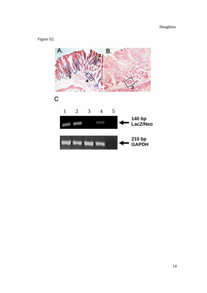
Figure S2. Engrafted BMDC contain the LacZ/Neo fusion gene.
Laser capture microdissection was used to isolate beta-galactosidase positive gastric
glands (A- boxes 1 and 2) or beta-galactosidase positive infiltrating leukocytes (A-box 4)
from a mouse transplanted with ROSA26 marrow. As a negative control, we used a
captured gastric gland from a mouse transplanted with WT marrow (B- box 3). (C) PCR
for the LacZ/Neo fusion gene and GAPDH show that the LacZ/Neo expression is specific

Houghton
10
to engrafted cells. The numbers correspond to the boxes in panels (A) and (B). Lane 5 is
a water control.
Figure S3. Engraftment of BMDC into non-inflamed, non-injured or acutely injured
gastric mucosa is a rare event. C57BL/6 mice were transplanted with ROSA26 marrow,
mock infected and gastric tissue examined at 30 weeks. (A.) A single beta-galactosidase
positive (blue) gland in the oxyntic mucosa (square box- enlarged view B) and sparse
mononuclear leukocytes (rectangular box- large view C) demonstrate that engraftment
in the absence of H. felis infection is a rare event. (D). Acute 4-day cryoinjury in a
ROSA26 transplanted mouse. The ulcer is clearly visible with accompanying
submucosal edema and lymphedema, and beta-galactosidase positive staining cells at the
ulcer edge. Higher power (E) reveals these cells to be mononuclear leukocytes. (F)
Reparative phase 10 days post-cryoinjury with glandular reconstitution from endogenous
gastric stem cell (beta-gal negative) precursors. (G) BMDC-derived mesenchymal cells
(arrows), including plump spindloid fibroblast-like cells, participate in injury repair. (H)
Acute depletion of parietal cells with DMP777 (I) Repopulated parietal cells (arrows)
210 days post-DMP777 administration are derived from gastric precursors (beta-
galactosidase negative) and not BMDC.
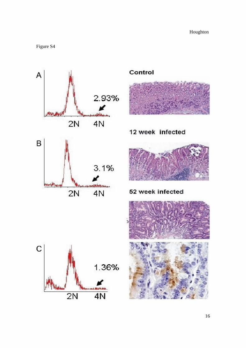
Figure S4. Further evidence that stable-fusion is not the mechanism by which BMDC
differentiate to gastric mucosal cells, metaplasia, dysplasia or early cancer.
(A) DNA content and gastric mucosal histology from a one year old male WT mouse
without Helicobacter infection (B) WT transplanted with ROSA26 marrow and infected

Houghton
11
for 12 weeks or (C) 52 weeks. Single cell preparations were prepared from the gastric
mucosa and analyzed by FACS for DNA content. The number of >2N cells was not
different between groups. Histology confirms the presence of a single nucleus in each
mucosal cell. In long term infected mice (C) GIN is seen in the H&E section, with the
majority of mucosa at the squamocolumnar junction along the lesser curvature replaced
by BMDC (brown staining- IHC for beta-galactosidase, bottom panel). Only single nuclei
are seen in these BMD-gastric epithelial cells.
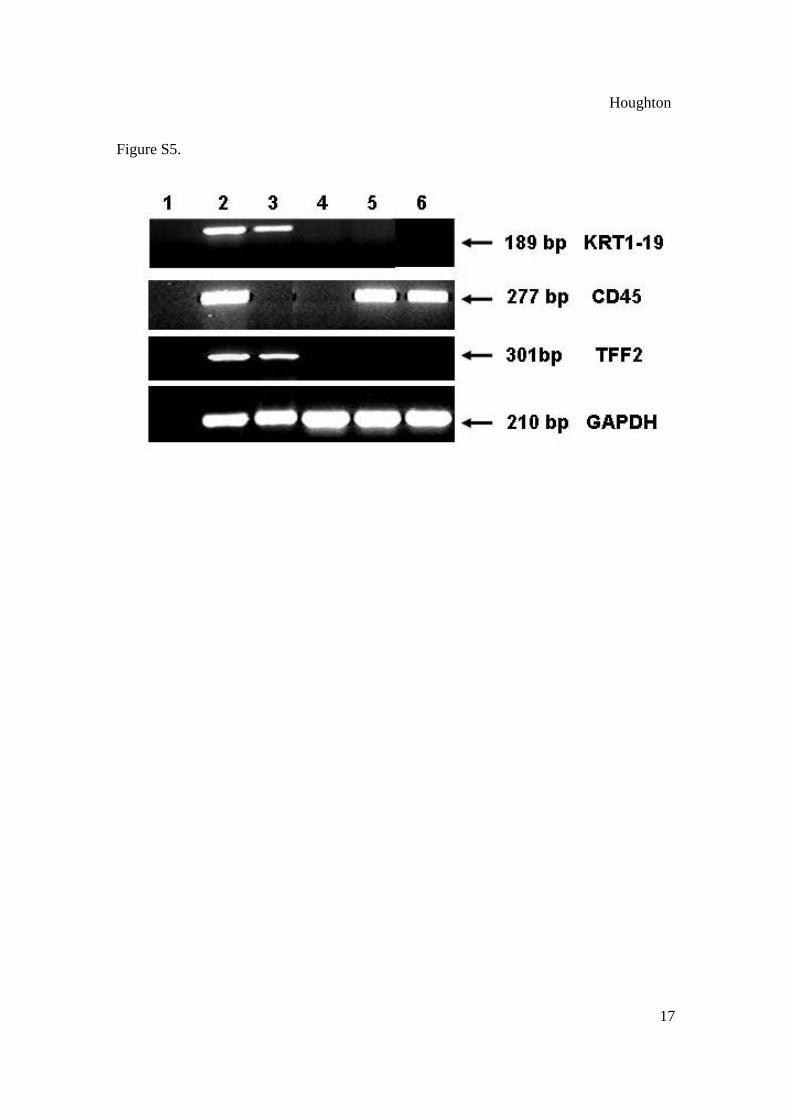
Figure S5. The gastric mucosa promotes differentiation of mesenchymal stem cells
toward an epithelial cell phenotype.
HSC or MSC were cultured in control medium or with the soluble components of a
primary gastric mucosal cell culture followed by RT-PCR after 48-hours. Lane 1:
Negative water control. Lane 2: Positive control- RNA isolated from the gastric mucosa
from an infected male mouse KRT1-19 (epithelial cell cytokeratin), CD45 (from
infiltrating leukocytes), and TFF2 (metaplastic cell lineage marker) are all expressed.
Lane 3: Mesenchymal stem cells cultured in the presence of gastric mucosa do not
express CD45 but do express both KRT1-19 and TFF2. MSC do not express these
epithelial cell markers under control conditions (lane 4). Lane 5- Lineage depleted
rhodull, Hodull (HSC) cells express CD45, but do not express KRT1-19 or TFF2 when
exposed to gastric mucosal environment or with control medium (lane 6). Loading
quantity was standardized with GAPDH.
Figure S6. The gastric mucosa is a permissive environment for stem cell recruitment.
Western blot analysis of the gastric mucosa from (1) control, (2) 12 month (3) 16 month

Houghton
12
Helicobacter felis infected male WT mice show SCF-1 and SDF-1 are upregulated with
infection.
Table S1. Gastric glands along the lesser curvature at the squamocolumnar junction
derived from bone marrow cells.
Houghton
13
Figure S1.
Houghton
14
Figure S2.
140 bp LacZ/Neo
210 bp GAPDH
C
A. B.
1 2 3 4 5

Houghton
15
Figure S3
Houghton
16
Figure S4
Houghton
17
Figure S5.

Houghton
18
Figure S6.

Houghton
19
_______________________________________________________________________ Table S1 Gastric glands along the lesser curvature at the squamocolumnar junction derived from bone marrow cells. _______________________________________________________________________ Tissue number of sections approximate clusters of percent of examined number of glands < 4 cells beta-gal(+)
examined glands _______________________________________________________________________ Non-infected* WT 138 2,070 0 0 ROSA 138 1,780 2 0 DMP777 treated** WT 54 800 0 0 ROSA 54 942 0 0 Acute gastric ulceration *** WT 72 1,050 0 0 ROSA 72 1,070 0 0 Helicobacter infection WT**** 138 2,000 0 0 ROSA 3wk 18 220 0 0 20wk 30 400 0 <5% 30wk 30 450 0 20% 40wk 30 400 0 50% 52wks 30 425 0 90% _______________________________________________________________________ WT- C57BL/6 mouse transplanted with C57BL/6 marrow ROSA- C57BL/6 mouse transplanted with C57BL/6JGtrosa26 marrow * 3 week through 52 week time points combined ** 7, 9 and 140 day time points combined *** liquid nitrogen and acetic acid ulcers at 4, 10 and 20 days combined **** wild type infected mice combined from 3 weeks through 52-week time points. Glands were considered (+) for beta-gal when >50% of cells were showed positive staining.

Houghton
20
References 1. J. Houghton et al. Infection and Immunity 68, 1189 (2000). 2. C. Stoicov et al. J. Immunology 173, 3329 (2004). 3. P.J. Quesenberry, G.A. Colvin, J. Lambert. Blood 100, 4266 (2002).